Article

Circulating Serum Cell-Free Mitochondrial DNA in Amyotrophic Lateral Sclerosis

1
Department of Life Sciences, University of Modena and Reggio Emilia, 41125 Modena, Italy
2
Department of Neurosciences, Ospedale Civile di Baggiovara, Azienda Ospedaliero-Universitaria di Modena, 41126 Modena, Italy
3
Neuroscience PhD Program, Department of Biomedical, Metabolic, and Neural Sciences, University of Modena and Reggio Emilia, 41125 Modena, Italy
4
Department of Biomedical, Metabolic and Neural Sciences, University of Modena and Reggio Emilia, 41125 Modena, Italy
*
Authors to whom correspondence should be addressed.
Cells 2025, 14(18), 1433; https://doi.org/10.3390/cells14181433
Submission received: 18 July 2025 / Revised: 21 August 2025 / Accepted: 10 September 2025 / Published: 12 September 2025

Abstract

Mitochondrial dysfunction is a key pathological hallmark in amyotrophic lateral sclerosis (ALS), yet the role of circulating cell-free mitochondrial DNA (Cf-mtDNA) as a biomarker remains unclear. This study aimed to investigate serum Cf-mtDNA levels in ALS patients compared to healthy controls and explore its associations with disease biomarkers, clinical progression, and survival. We conducted a case–control study measuring Cf-mtDNA levels in serum samples from 54 ALS patients and 36 age- and sex-matched healthy controls using quantitative droplet digital PCR. Correlations between Cf-mtDNA levels and clinical features, neurofilament concentrations, inflammatory indices, and survival were assessed. The average Cf-mtDNA level in ALS patients was 2,426,315 copies/mL of serum (IQR: 865000–2475000), compared to 1,885,667 copies/mL of serum (IQR: 394250–2492500) in controls (p = 0.308). ROC analysis yielded an AUC of 0.595 (95% CI: 0.468–0.721), indicating very limited discriminant ability. Cf-mtDNA levels were inversely correlated with serum creatinine concentrations (r = –0.335, p = 0.018), but showed no significant associations with ALS phenotype, disease staging, neurofilaments, inflammatory indices, or survival. These findings suggest that, in a predominantly sporadic ALS cohort, serum Cf-mtDNA may not serve as a standalone diagnostic or prognostic biomarker, in contrast to previous reports. Methodological differences, cohort composition, and genetic heterogeneity may account for these discrepancies. Our results underscore the importance of further large-scale, longitudinal studies incorporating genetic stratification and multi-biomarker approaches to better elucidate the role of Cf-mtDNA in ALS pathophysiology.

1. Introduction

Amyotrophic lateral sclerosis (ALS) is a neurodegenerative disease that irreversibly causes death of the lower motor neurons in the brainstem and spinal cord and of the upper motor neurons in the motor cortex. Mitochondrial dysfunction represents one of the critical factors in the pathogenesis of ALS [1,2]. Unlike nuclear DNA, mitochondrial DNA (mtDNA) lacks protective histones and robust repair systems, making it especially prone to damage from oxidative stress [3]. Such instability is particularly harmful to energy-demanding, post-mitotic cells like neurons and myocytes, which are sensitive to disruptions in the respiratory chain and ROS-induced damage [4]. As a consequence of this vulnerability, fragments of mtDNA may be released into the bloodstream, a phenomenon referred to as circulating cell-free mitochondrial DNA (Cf-mtDNA) [5], via apoptosis, necrosis, or active secretion, and may activate the immune system, acting as damage-associated molecular patterns (DAMPs) [6,7]. Resembling bacterial DNA, these fragments can trigger innate immune responses and inflammation across various diseases [5,8,9,10,11]. Interestingly, studies in ALS mouse models have demonstrated that the accumulation of misfolded proteins, such as SOD1 and TDP-43, can interfere with mitochondrial function, leading to the release of mtDNA into the cytoplasm and initiating immune responses [12,13]. Recently, Cf-mtDNA has emerged as a promising non-invasive diagnostic indicator of ongoing cellular damage and mortality, providing insight into the pathological mechanisms of neurodegenerative disorders, including Parkinson’s disease (PD) and Alzheimer’s disease (AD) [14]. Despite these findings, similar investigations in ALS patients are scarce [15]. Although neurofilaments are well-established and sensitive biomarkers for ALS [16,17,18], their limited specificity across neurodegenerative conditions [19] and their inability to reflect the full biological complexity of the disease highlight the need for additional, complementary biomarkers [20,21]. In this context, we investigated Cf-mtDNA levels in the serum of ALS patients compared to healthy controls and examined their association with other biomarkers and clinical measures of disease progression.

2. Materials and Methods

2.1. Study Population

ALS patients were recruited from among residents of the Emilia-Romagna region, Northern Italy, who presented to the ALS Center of the Modena University Neurological Department between 1 January 2018 and 31 December 2023. Eligible participants were considered all patients who received a diagnosis of definite or probable ALS according to El Escorial-revised criteria [22] and who still had at least 0.5 mL of serum available for analysis. ALS diagnosis was confirmed by subsequent application of the Gold Coast Criteria [23], after reviewing the medical histories of each individual patient. Serum samples were collected and biobanked as part of routine diagnostic and research procedures. Healthy control (HC) participants were recruited from the same clinical center. They consisted of either first-degree relatives of ALS patients or healthy volunteers, including blood donors, and were age- and sex-matched to the ALS group to ensure demographic comparability and reduce potential environmental bias.

2.2. Standard Protocol Approvals, Registrations, and Patient Consents

This retrospective exploratory study was approved by the Ethical Committee of Area Vasta Emilia Nord (file number 309/2024/TESS/AOUMO). All participants signed written informed consent for venipuncture and gave permission for the use of their biological specimens, which were stocked in the Neurobiobank of Modena (NBBM), for future research studies.

2.3. Clinical Measures

We collected the following demographic and anthropometric variables for each patient: sex, age at symptom onset, and weight loss at sampling. The onset of disease was determined based on the patient-reported occurrence of a distinct motor impairment in a single district [24]. Subsequently, we considered the site of onset (bulbar, spinal) and clinical phenotype (flail, upper motor neuron predominant (UMNp), bulbar, respiratory, classic) [25]. All patients started riluzole treatment after ALS diagnosis. Genetic analysis was performed for 49 patients, following previously described protocols [26]. Clinical variables used as proxies for disease progression included respiratory function, as assessed by FVC at sampling; King’s ALS staging and ALS Milano–Torino Staging (ALS-MiToS) at sampling and at last observation; disease progression rate (DPR), calculated as the monthly decline in the ALSFRS-R score from onset to diagnosis, assuming a total score of 48 at onset [27], as well as at the time of the sampling and at the last observation [28]. Slow, intermediate, and fast progressors were categorized according to DPR at sampling below 0.50, between 0.5 and 1, and above 1, respectively. We also considered time to non-invasive ventilation (NIV), invasive ventilation (IV), percutaneous endoscopic gastrostomy (PEG). Tracheostomy-free survival was defined as the timespan between disease onset and date of death or tracheostomy, whichever came first.

2.4. Sample Collection and Laboratory Assays

Serum samples were obtained via venipuncture and processed according to standard procedures. Following centrifugation for 10 min at 1300× g, the supernatant was aliquoted in polypropylene tubes and stored at −80 °C until analysis. Quantification of Neurofilament Light Chain (NfL), Neurofilament Heavy Chain (NfH), chitinase-3-like-1 (CHI3L1), SerpinA1, and Triggering Receptor Expressed on Myeloid cells 2 (TREM2) was performed using an automated next generation ELISA, based on Ella Simple Plex assay technology (BioTechne, ProteinSimple, San Jose, CA 95134, USA) following the manufacturers’ instructions [18,29]. In this immunoassay samples run through a channel each composed of three glass nano reactors pre-coated with a capture antibody, allowing for automated triplicate measurements of each sample. Samples were loaded into the cartridges with the following dilutions: 1:200,000 for the serum SerpinA1 cartridge; 1:2 for the NfL cartridge; 1:2 for the serum pNfH cartridge; 1:10 for the serum CHI3L1 cartridge; 1:10 for the serum TREM2. Intra-assay and inter-assay variability were evaluated by the manufacturer. Blood samples were analyzed at the Central Laboratory of the Azienda Ospedaliero-Universitaria di Modena using a DxH 900 analyzer (Beckman Coulter, Brea, CA, USA), a fully automated hematology system for white blood cell (WBC) counts and five-part leucocyte differential counting, and red blood cell (RBC) counts. The analyzer employs volume, conductivity, and scatter parameters for controlled flow cytometric analysis of WBC differential. The analytical software is self-gating and separates WBC populations by automatic logic pathways. All analyses were performed in accordance with the manufacturer’s instructions. The neutrophil-to-lymphocyte ratio (NLR), monocyte-to-lymphocyte ratio (MLR), systemic inflammation response index (SIRI), aggregate systemic inflammation index (AISI) and systemic–immune–inflammation index (SII) were derived from the complete blood count.

2.5. DNA Extraction and mtDNA Quantification Through Digital Droplet PCR

Total DNA was extracted from serum using the DNeasy Blood & Tissue Kit (QIAgen, Hilden, Germany) according to the manufacturer’s instructions, and the DNA quantity was assessed using the NanoDrop ND-1000 (Thermo Fisher Scientific, Waltham, MA, USA). mtDNA quantification was performed using droplet digital PCR (ddPCR). Each ddPCR reaction mixture (final volume of 20 μL) consisted of 10 ng of DNA samples, 2X ddPCR Supermix for Probes (Bio-Rad), and 1.1 μL of each mtDNA custom assay (Bio-Rad Laboratories, Hercules, CA, USA). The thermal protocol conditions were set as follows: initial denaturation at 95 °C for 10 min, followed by 40 cycles of denaturation at 94 °C for 30 s and annealing/extension at 55 °C for 1 min, and a final extension at 98 °C for 10 min. The droplets were then read using the QX200 ddPCR droplet reader (Bio-Rad), and analysis was performed using QuantaSoft Analysis software (version 1.7.4.0917, Bio-Rad). The wet-validated primers used were as follows: ND2 (Bio-Rad, #10031252) and Actb (Bio-Rad, #10042961).

2.6. Statistical Methods

Continuous variables were reported as means ± standard deviations (SDs), medians, and interquartile ranges (IQRs), whereas categorical variables were reported as absolute numbers and percentages. The use of non-parametric methods, when possible, was preferred due to the low sample sizes and because visual inspection revealed non-adherence of most of the biomarkers, including Cf-mtDNA, to the Gaussian distribution. We compared Cf-mtDNA between ALS patients and the HC group using the two-tailed Wilcoxon test. A p-value < 0.05 was considered statistically significant. Correlations between biomarker numerical variables were assessed using the Spearman rank correlation coefficient. Receiver operating characteristic (ROC) analysis was used to evaluate the overall performance to discriminate between ALS patients versus HC, by measuring the area under the curve (AUC). The relationship between Cf-mtDNA and clinical variables was assessed with linear (for continuous variables) or Poisson (for discrete variables) regression models, and the results were expressed as the mean difference (MD) or mean ratio (MR) related to a Cf-mtDNA increase of a half million copies per mL of serum. The relationship between Cf-mtDNA and time-to-event variables was assessed with the unadjusted Cox proportional hazards regression model. Cox analysis results were expressed as the hazard ratio (HR) related to a Cf-mtDNA increase of a half million copies per mL of serum. Uncertainty in association measures was reported with the 95% confidence interval (CI). Data analysis was performed using STATA 17 (StataCorp.2017.College Station, TX, USA) and R 3.4.3 (The R Foundation for Statistical Computing, Wien, Austria) statistical software.

3. Results

Based on the inclusion criteria, the serum from 54 ALS patients (27 males, 27 females, mean age at sampling: 52.88 ± 13.28 years) and 36 HC (19 male, 17 females, mean age at sampling: 51.23 ± 10.49 years) were available for analysis. ALS patients’ features are summarized in Table 1. The average value of serum NfL was 116.88 ± 68.22 pg/mL, and the average creatinine was 0.71 ± 0.20 mg/mL.

3.1. Biomarkers’ Distribution in ALS Patients and Healthy Controls

The average serum Cf-mtDNA was not statistically different between ALS patients and HC (copies per mL of serum: 2,426,315 ± 2,793,092, IQR: 865,000–2,475,000 vs. 1,885,667 ± 2,194,552, IQR: 394,250–2,492,500, p = 0.308) (Figure 1). When analyzing Cf-mtDNA discriminant ability, ROC analysis showed an AUC of 0.595 (95% CI: 0.468–0.721). Mean values for each biomarker in the ALS cohort are visible in Table S2.

3.2. Cf-mtDNA Association with Clinical Indicators of Progression in ALS

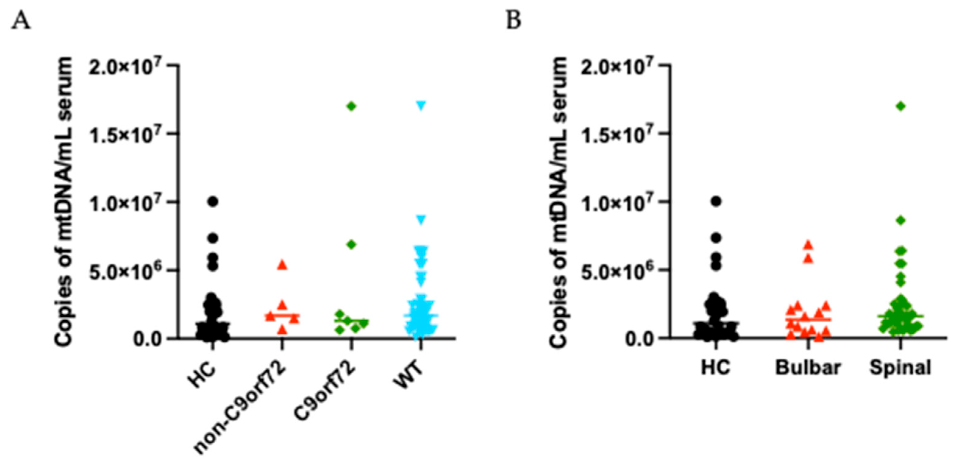
We evaluated potential associations between Cf-mtDNA serum levels and ALS clinical features, as detailed in Table 2. No significant associations were found between Cf-mtDNA concentrations and any of the clinical variables analyzed. No statistically significant differences were observed in relation to FVC at the time of sampling or to clinical staging scores, either at sampling or at the last follow-up. Similar non-significant trends were also noted for ALSFRS-R scores and disease progression rates at both time points. When stratifying by genotype, comparisons between C9orf72 expansion carriers and either non-mutated (WT) or other mutation groups (non-C9orf72) (Figure 2A), as well as multi-group analyses across different genetic subtypes, revealed no significant differences in Cf-mtDNA serum levels. We also stratified the data based on disease progression rate (slow, intermediate, or fast) or site of onset (bulbar or spinal) without finding significant differences between these groups and HC (Figure 2B). Finally, no significant associations were identified between Cf-mtDNA levels and comorbidities or demographic variables among ALS patients, as reported in Table S1.

3.3. Correlation of Cf-mtDNA with ALS Biomarkers

A complete list of biomarker distributions among ALS patients is reported in Table S2. In the present analysis, Cf-mtDNA levels did not show significant correlations with well-established ALS biomarkers, as detailed in Table 3. Notably, Cf-mtDNA showed an inverse correlation with serum creatinine levels (r = –0.280, p = 0.0442). Finally, no significant correlations were observed between Cf-mtDNA and inflammatory indices, such as NLR, MLR, SIRI, AISI, and SII and lipid metabolism-related markers.

3.4. Cf-mtDNA and Survival

Cox analysis was used to assess the prognostic role of each clinical variable and biomarker in our cohort (Table 4). In the survival analysis, Cf-mtDNA levels demonstrated no significant association with key clinical outcomes in patients with ALS, including death, tracheostomy-free survival, and the use of supportive interventions, such as NIV, PEG, and IV.

4. Discussion

In this exploratory study, we explored serum Cf-mtDNA levels in ALS patients, assessing their diagnostic and prognostic potential in comparison with age- and sex-matched healthy controls. Cf-mtDNA concentrations did not differ significantly between ALS patients and the controls and showed no diagnostic discriminative power, which is in contrast to findings from a previous study [30]. Several potential factors may account for the discordance between these studies. First, while both studies analyzed Cf-mtDNA in serum, differences in sample preparation protocols, Cf-mtDNA quantification techniques, and cohort composition may have contributed to the observed discrepancies. It is important to note that Cf-mtDNA quantification can be highly sensitive to pre-analytical variables, particularly the DNA extraction method. In our study, we used a standardized protocol across all samples, employing the kit and protocol, which has been previously reported to offer the most consistent performance in Cf-mtDNA recovery [31]. In particular, Li et al. used a different quantitative assay, which may have affected their sensitivity and specificity in detecting Cf-mtDNA variations. Moreover, differences in sample processing and storage duration may introduce pre-analytical variability, which is increasingly recognized as a major challenge in the field of CfDNA-based biomarker discovery. Also, in Li’s study, a genetically distinct cohort enriched with SOD1 mutation carriers was included, in whom they observed the highest Cf-mtDNA levels and the strongest associations with disease progression. ALS exhibits high clinical and genetic heterogeneity; thus, subgroup-specific patterns may be masked in aggregated analyses, particularly in smaller and mainly sporadic cohorts like ours. Interestingly, while Li et al. identified an inverse correlation between Cf-mtDNA and ALS progression rate, particularly in SOD1 mutation carriers, we found no association between Cf-mtDNA and clinical measures of disease severity or survival. SOD1-associated ALS may be characterized by pronounced mitochondrial involvement [32], potentially leading to increased systemic Cf-mtDNA release.
Moreover, Cf-mtDNA was not significantly associated with core biomarkers of neurodegeneration and inflammation. Interestingly, a significant inverse correlation was identified between Cf-mtDNA levels and serum creatinine, a finding that warrants further investigation. While creatinine is commonly used as a proxy for muscle mass and renal function, its association with Cf-mtDNA could suggest shared underlying mechanisms related to mitochondrial metabolism or systemic catabolism. However, this relationship must be interpreted with caution given the complexity of metabolic alterations in ALS and the potential influence of comorbidities. In this context, elevated Cf-mtDNA has been linked to oxidative stress and systemic inflammation, while reduced creatinine levels—when reflecting malnutrition or sarcopenia—may serve as indicators of advanced disease stage. Both markers, therefore, could represent distinct yet converging facets of the same pathological trajectory. Furthermore, the reduction in serum creatinine commonly observed in ALS is consistent with progressive muscle atrophy. At the same time, ALS may cause progressive muscle atrophy, in this way inducing ongoing muscle damage and increased mitochondrial turnover that promotes the release of Cf-mtDNA into the circulation, potentially contributing to the inverse association observed in our cohort. Next, it is worth noting that a previous study reported a correlation between urinary mtDNA levels and creatinine in patients with hypertension-related kidney dysfunction [33]. While this evidence pertains to a different biological matrix and clinical context, it may support the plausibility of a link between mitochondrial nucleic acids and creatinine metabolism.
In addition to ALS, Cf-mtDNA alterations have been reported in other neurodegenerative disorders. In the case of multiple sclerosis (MS) [34], elevated levels were associated with disease activity and neuroinflammation, while no differences were observed in cases of PD and multiple system atrophy [35]. The picture is more complex as far as AD is concerned, as mtDNA levels have been shown to be lower than controls, but deeply influenced by environmental factors [36,37]. A recent review by Aydın et al. [38], emphasized the diagnostic utility of Cf-mtDNA in conditions such as AD and PD, where elevated Cf-mtDNA levels, when observed, could be interpreted as a surrogate marker of ongoing neuroinflammation and mitochondrial dysfunction. In ALS, however, the absence of a significant increase in Cf-mtDNA, along with the lack of correlation with established biomarkers of neurodegeneration and inflammation, suggests a disease-specific behavior of this biomarker. One possible explanation lies in the distinct pathophysiological mechanisms of ALS. Although mitochondrial dysfunction is well recognized in ALS, it may lead to a more compartmentalized or cell-type-specific release of Cf-mtDNA, limiting its detectability in peripheral circulation. Supporting this hypothesis, Cf-mtDNA levels in our cohort did not correlate with neurofilaments, which are considered robust markers of neuronal damage in ALS and other neurodegenerative conditions. This lack of association may imply that Cf-mtDNA reflects a separate pathophysiological axis, potentially more related to metabolic or systemic stress than to direct neuroaxonal injury. Another key consideration is the biological matrix. While Aydın et al. discuss Cf-mtDNA derived from both plasma and CSF [38], our study is limited to peripheral blood serum. It is conceivable that Cf-mtDNA dynamics in ALS are more prominent within the central nervous system or in neuron-derived vesicles, and are thus underrepresented in peripheral blood.
Future studies using CSF samples or cell-specific profiling approaches—such as analysis of neuronal exosomes—will be essential to clarify the source and relevance of Cf-mtDNA in ALS pathophysiology.
The present study has some strengths and limitations. All ALS patients were deeply clinically characterized and followed up, and a broad panel of inflammatory and neurodegeneration biomarkers allowed for a multifaceted examination of Cf-mtDNA relevance within ALS pathophysiology. Survival data and a wide list of clinical indicators constitute a significant added value to our work. Moreover, healthy controls were age- and sex-matched with our ALS population, therefore reducing the risk of confounding bias. However, the sample size, although comparable to similar exploratory studies, may have been underpowered to detect small-to-moderate associations. Another critical limitation affecting the clinical translatability of our findings is the absence of a replication cohort and cerebrospinal fluid (CSF) data, which restricts the assessment of compartment-specific dynamics and limits generalizability. Finally, the design of the study precludes conclusions about the longitudinal dynamics of Cf-mtDNA throughout disease progression. Despite these limitations, our study offers insights by showing that Cf-mtDNA, at least in serum, may not reflect disease severity, inflammation, or survival in ALS. The observed inverse correlation with creatinine is intriguing and may point toward a link with muscle mass and systemic catabolism, both of which are altered in ALS progression. However, this finding requires further exploration.

5. Conclusions

In conclusion, our findings suggest that Cf-mtDNA levels in serum are not significantly altered in ALS nor associated with disease progression or survival. We could not find correlations with other biomarkers of neurodegeneration or neuroinflammation. Although a potential link with serum creatinine levels was observed, the overall data suggest that Cf-mtDNA, as measured in peripheral blood, may not capture the specific neurodegenerative processes characteristic of ALS. These results contrast with previous findings in ALS and other neurodegenerative disorders, highlighting the need for further investigation into the biological matrices, temporal dynamics, and disease-specific factors that modulate Cf-mtDNA levels. Future studies should consider larger cohorts, longitudinal sampling, and the exploration of Cf-mtDNA in different biofluids to better understand its compartment-specific relevance and mechanistic roles in ALS. Our work emphasizes the importance of harmonizing analytical methods and considering disease compartmentalization in future studies.

Supplementary Materials

The following supporting information can be downloaded at https://www.mdpi.com/article/10.3390/cells14181433/s1: Table S1. Association between Cf-mtDNA, comorbidities and clinical characteristics in ALS patients of the study. Table S2. Biological indicators’ distribution in ALS patients of the study.

Author Contributions

Conceptualization: J.M. and M.P.; methodology, M.P., G.Z. and G.S.; validation, M.P., G.Z. and G.S.; formal analysis, E.Z. and F.B.; investigation, I.M., E.Z., G.G., C.S. and A.G.; resources, J.M. and M.P.; data curation, G.Z., I.M. and E.Z.; writing—original draft preparation, I.M., E.Z. and G.Z.; writing—review and editing, I.M., E.Z., J.M. and M.P.; supervision, J.M. and M.P.; project administration, M.P.; funding acquisition, J.M. and M.P. All authors have read and agreed to the published version of the manuscript.

Funding

This research was funded by the European Union—Next Generation EU, Mission 6, Component 2, CUP F93C24000310005, project “INSPIRER” (to E.Z., J.M., G.G., C.S.) and by Fondazione Cassa di Risparmio di Modena (Neurobiobanca di Modena).

Institutional Review Board Statement

This study was conducted in accordance with the Declaration of Helsinki and approved by the Ethics Committee of Area Vasta Emilia Nord (protocol code 309/2024/TESS/AOUMO on 1 August 2024).

Informed Consent Statement

Informed consent was obtained from all subjects involved in the study.

Data Availability Statement

Data are available from the authors upon reasonable request and after providing the approval of the ethical committee.

Conflicts of Interest

The authors declare no conflicts of interest.

Abbreviations

The following abbreviations are used in this manuscript:
ADAlzheimer’s disease
AISIAggregate systemic inflammation index
ALSAmyotrophic lateral sclerosis
ALS-FRSrAmyotrophic Lateral Sclerosis Functional Rating Scale—revised
AUCArea under the curve
C9ORF72Chromosome 9 open reading frame 72
CHI3L1Chitinase-3-like protein 1
CIConfidence interval
COPDChronic obstructive pulmonary disease
DPRDisease progression rate
FTDFronto-temporal dementia
FVCForced vital capacity
HCHealthy controls
HRHazard ratio
IVInvasive ventilation
MLRMonocyte-to-lymphocyte ratio
NFNeurofilament
NfLNeurofilament light chain
NIVNon-invasive ventilation
NLRNeutrophil-to-lymphocyte ratio
PDParkinson’s disease
PEGPercutaneous endoscopic gastrostomy
pNfHNeurofilament heavy chain
ROCReceiver operating characteristic
SDStandard deviation
SIISystemic–immune–inflammation index
SIRISystemic inflammation response index
WBCWhite blood cell

References

  1. Genin, E.C.; Abou-Ali, M.; Paquis-Flucklinger, V. Mitochondria, a Key Target in Amyotrophic Lateral Sclerosis Pathogenesis. Genes 2023, 14, 1981. [Google Scholar] [CrossRef]
  2. Smith, E.F.; Shaw, P.J.; De Vos, K.J. The Role of Mitochondria in Amyotrophic Lateral Sclerosis. Neurosci. Lett. 2019, 710, 132933. [Google Scholar] [CrossRef]
  3. Newman, L.E.; Shadel, G.S. Mitochondrial DNA Release in Innate Immune Signaling. Annu. Rev. Biochem. 2023, 92, 299–332. [Google Scholar] [CrossRef]
  4. Xu, X.; Pang, Y.; Fan, X. Mitochondria in Oxidative Stress, Inflammation and Aging: From Mechanisms to Therapeutic Advances. Signal Transduct. Target. Ther. 2025, 10, 190. [Google Scholar] [CrossRef]
  5. Gambardella, S.; Limanaqi, F.; Ferese, R.; Biagioni, F.; Campopiano, R.; Centonze, D.; Fornai, F. cCf-mtDNA as a Potential Link Between the Brain and Immune System in Neuro-Immunological Disorders. Front. Immunol. 2019, 10, 1064. [Google Scholar] [CrossRef] [PubMed]
  6. Pinti, M.; Cevenini, E.; Nasi, M.; De Biasi, S.; Salvioli, S.; Monti, D.; Benatti, S.; Gibellini, L.; Cotichini, R.; Stazi, M.A.; et al. Circulating Mitochondrial DNA Increases with Age and Is a Familiar Trait: Implications for “Inflamm-Aging”. Eur. J. Immunol. 2014, 44, 1552–1562. [Google Scholar] [CrossRef]
  7. Zanini, G.; Selleri, V.; Lopez Domenech, S.; Malerba, M.; Nasi, M.; Mattioli, A.V.; Pinti, M. Mitochondrial DNA as Inflammatory DAMP: A Warning of an Aging Immune System? Biochem. Soc. Trans. 2023, 51, 735–745. [Google Scholar] [CrossRef]
  8. De Gaetano, A.; Solodka, K.; Zanini, G.; Selleri, V.; Mattioli, A.V.; Nasi, M.; Pinti, M. Molecular Mechanisms of mtDNA-Mediated Inflammation. Cells 2021, 10, 2898. [Google Scholar] [CrossRef] [PubMed]
  9. Zhou, M.; Zhang, H.; Xu, X.; Chen, H.; Qi, B. Association between Circulating Cell-Free Mitochondrial DNA and Inflammation Factors in Noninfectious Diseases: A Systematic Review. PLoS ONE 2024, 19, e0289338. [Google Scholar] [CrossRef] [PubMed]
  10. Kunze, R.; Fischer, S.; Marti, H.H.; Preissner, K.T. Brain Alarm by Self-Extracellular Nucleic Acids: From Neuroinflammation to Neurodegeneration. J. Biomed. Sci. 2023, 30, 64. [Google Scholar] [CrossRef]
  11. Giordano, L.; Ware, S.A.; Lagranha, C.J.; Kaufman, B.A. Mitochondrial DNA Signals Driving Immune Responses: Why, How, Where? Cell Commun. Signal. 2025, 23, 192. [Google Scholar] [CrossRef] [PubMed]
  12. Tan, H.Y.; Yong, Y.K.; Xue, Y.C.; Liu, H.; Furihata, T.; Shankar, E.M.; Ng, C.S. cGAS and DDX41-STING Mediated Intrinsic Immunity Spreads Intercellularly to Promote Neuroinflammation in SOD1 ALS Model. iScience 2022, 25, 104404. [Google Scholar] [CrossRef]
  13. Yu, C.-H.; Davidson, S.; Harapas, C.R.; Hilton, J.B.; Mlodzianoski, M.J.; Laohamonthonkul, P.; Louis, C.; Low, R.R.J.; Moecking, J.; De Nardo, D.; et al. TDP-43 Triggers Mitochondrial DNA Release via mPTP to Activate cGAS/STING in ALS. Cell 2020, 183, 636–649.e18. [Google Scholar] [CrossRef] [PubMed]
  14. Park, S.S.; Jeong, H.; Andreazza, A.C. Circulating Cell-Free Mitochondrial DNA in Brain Health and Disease: A Systematic Review and Meta-Analysis. World J. Biol. Psychiatry 2022, 23, 87–102. [Google Scholar] [CrossRef] [PubMed]
  15. Risi, B.; Imarisio, A.; Cuconato, G.; Padovani, A.; Valente, E.M.; Filosto, M. Mitochondrial DNA (mtDNA) as Fluid Biomarker in Neurodegenerative Disorders: A Systematic Review. Eur. J. Neurol. 2025, 32, e70014. [Google Scholar] [CrossRef]
  16. Verde, F.; Otto, M.; Silani, V. Neurofilament Light Chain as Biomarker for Amyotrophic Lateral Sclerosis and Frontotemporal Dementia. Front. Neurosci. 2021, 15, 679199. [Google Scholar] [CrossRef]
  17. Poesen, K.; De Schaepdryver, M.; Stubendorff, B.; Gille, B.; Muckova, P.; Wendler, S.; Prell, T.; Ringer, T.M.; Rhode, H.; Stevens, O.; et al. Neurofilament Markers for ALS Correlate with Extent of Upper and Lower Motor Neuron Disease. Neurology 2017, 88, 2302–2309. [Google Scholar] [CrossRef]
  18. Simonini, C.; Zucchi, E.; Bedin, R.; Martinelli, I.; Gianferrari, G.; Fini, N.; Sorarù, G.; Liguori, R.; Vacchiano, V.; Mandrioli, J. CSF Heavy Neurofilament May Discriminate and Predict Motor Neuron Diseases with Upper Motor Neuron Involvement. Biomedicines 2021, 9, 1623. [Google Scholar] [CrossRef]
  19. Zucchi, E.; Bonetto, V.; Sorarù, G.; Martinelli, I.; Parchi, P.; Liguori, R.; Mandrioli, J. Neurofilaments in Motor Neuron Disorders: Towards Promising Diagnostic and Prognostic Biomarkers. Mol. Neurodegener. 2020, 15, 58. [Google Scholar] [CrossRef]
  20. Sturmey, E.; Malaspina, A. Blood Biomarkers in ALS: Challenges, Applications and Novel Frontiers. Acta Neurol. Scand. 2022, 146, 375–388. [Google Scholar] [CrossRef]
  21. Simonini, C.; Zucchi, E.; Martinelli, I.; Gianferrari, G.; Lunetta, C.; Sorarù, G.; Trojsi, F.; Pepe, R.; Piras, R.; Giacchino, M.; et al. Neurodegenerative and Neuroinflammatory Changes in SOD1-ALS Patients Receiving Tofersen. Sci. Rep. 2025, 15, 11034. [Google Scholar] [CrossRef]
  22. Brooks, B.R.; Miller, R.G.; Swash, M.; Munsat, T.L.; World Federation of Neurology Research Group on Motor Neuron Diseases. El Escorial Revisited: Revised Criteria for the Diagnosis of Amyotrophic Lateral Sclerosis. Amyotroph. Lateral Scler. Other Motor Neuron Disord. 2000, 1, 293–299. [Google Scholar] [CrossRef]
  23. Shefner, J.M.; Al-Chalabi, A.; Baker, M.R.; Cui, L.-Y.; de Carvalho, M.; Eisen, A.; Grosskreutz, J.; Hardiman, O.; Henderson, R.; Matamala, J.M.; et al. A Proposal for New Diagnostic Criteria for ALS. Clin. Neurophysiol. 2020, 131, 1975–1978. [Google Scholar] [CrossRef]
  24. Benatar, M.; Granit, V.; Andersen, P.M.; Grignon, A.-L.; McHutchison, C.; Cosentino, S.; Malaspina, A.; Wuu, J. Mild Motor Impairment as Prodromal State in Amyotrophic Lateral Sclerosis: A New Diagnostic Entity. Brain J. Neurol. 2022, 145, 3500–3508. [Google Scholar] [CrossRef] [PubMed]
  25. Chio, A.; Calvo, A.; Moglia, C.; Mazzini, L.; Mora, G.; PARALS Study Group. Phenotypic Heterogeneity of Amyotrophic Lateral Sclerosis: A Population Based Study. J. Neurol. Neurosurg. Psychiatry 2011, 82, 740–746. [Google Scholar] [CrossRef]
  26. Gianferrari, G.; Martinelli, I.; Zucchi, E.; Simonini, C.; Fini, N.; Vinceti, M.; Ferro, S.; Gessani, A.; Canali, E.; Valzania, F.; et al. Epidemiological, Clinical and Genetic Features of ALS in the Last Decade: A Prospective Population-Based Study in the Emilia Romagna Region of Italy. Biomedicines 2022, 10, 819. [Google Scholar] [CrossRef]
  27. Kimura, F.; Fujimura, C.; Ishida, S.; Nakajima, H.; Furutama, D.; Uehara, H.; Shinoda, K.; Sugino, M.; Hanafusa, T. Progression Rate of ALSFRS-R at Time of Diagnosis Predicts Survival Time in ALS. Neurology 2006, 66, 265–267. [Google Scholar] [CrossRef] [PubMed]
  28. Mandrioli, J.; Biguzzi, S.; Guidi, C.; Sette, E.; Terlizzi, E.; Ravasio, A.; Casmiro, M.; Salvi, F.; Liguori, R.; Rizzi, R.; et al. Heterogeneity in ALSFRS-R Decline and Survival: A Population-Based Study in Italy. Neurol. Sci. 2015, 36, 2243–2252. [Google Scholar] [CrossRef]
  29. Martinelli, I.; Zucchi, E.; Simonini, C.; Gianferrari, G.; Bedin, R.; Biral, C.; Ghezzi, A.; Fini, N.; Carra, S.; Mandrioli, J. SerpinA1 Levels in Amyotrophic Lateral Sclerosis Patients: An Exploratory Study. Eur. J. Neurol. 2024, 31, e16054. [Google Scholar] [CrossRef]
  30. Li, J.; Gao, C.; Wang, Q.; Liu, J.; Xie, Z.; Zhao, Y.; Yu, M.; Zheng, Y.; Lv, H.; Zhang, W.; et al. Elevated Serum Circulating Cell-free Mitochondrial DNA in Amyotrophic Lateral Sclerosis. Eur. J. Neurol. 2024, 31, e16493. [Google Scholar] [CrossRef] [PubMed]
  31. Diefenbach, R.J.; Lee, J.H.; Kefford, R.F.; Rizos, H. Evaluation of Commercial Kits for Purification of Circulating Free DNA. Cancer Genet. 2018, 228–229, 21–27. [Google Scholar] [CrossRef] [PubMed]
  32. Higgins, C.M.J.; Jung, C.; Ding, H.; Xu, Z. Mutant Cu, Zn Superoxide Dismutase That Causes Motoneuron Degeneration Is Present in Mitochondria in the CNS. J. Neurosci. 2002, 22, RC215. [Google Scholar] [CrossRef]
  33. Eirin, A.; Saad, A.; Tang, H.; Herrmann, S.M.; Woollard, J.R.; Lerman, A.; Textor, S.C.; Lerman, L.O. Urinary Mitochondrial DNA Copy Number Identifies Chronic Renal Injury in Hypertensive Patients. Hypertension 2016, 68, 401–410. [Google Scholar] [CrossRef] [PubMed]
  34. Nasi, M.; Bianchini, E.; De Biasi, S.; Gibellini, L.; Neroni, A.; Mattioli, M.; Pinti, M.; Iannone, A.; Mattioli, A.V.; Simone, A.M.; et al. Increased Plasma Levels of Mitochondrial DNA and Pro-Inflammatory Cytokines in Patients with Progressive Multiple Sclerosis. J. Neuroimmunol. 2020, 338, 577107. [Google Scholar] [CrossRef] [PubMed]
  35. Ying, C.; Li, Y.; Zhang, H.; Pang, S.; Hao, S.; Hu, S.; Zhao, L. Probing the Diagnostic Values of Plasma Cf-nDNA and Cf-mtDNA for Parkinson’s Disease and Multiple System Atrophy. Front. Neurosci. 2024, 18, 1488820. [Google Scholar] [CrossRef]
  36. Gorham, I.K.; Reid, D.M.; Sun, J.; Zhou, Z.; Barber, R.C.; Phillips, N.R. Blood-Based mtDNA Quantification Indicates Population-Specific Differences Associated with Alzheimer’s Disease-Related Risk. J. Alzheimers Dis. 2024, 97, 1407–1419. [Google Scholar] [CrossRef]
  37. Huang, J.; Song, Z.; Wei, B.; Li, Q.; Lin, P.; Li, H.; Dong, K. Immunological Evaluation of Patients with Alzheimer’s Disease Based on Mitogen-Stimulated Cytokine Productions and Mitochondrial DNA Indicators. BMC Psychiatry 2023, 23, 145. [Google Scholar] [CrossRef]
  38. Aydın, Ş.; Özdemir, S.; Adıgüzel, A. The Potential of CfDNA as Biomarker: Opportunities and Challenges for Neurodegenerative Diseases. J. Mol. Neurosci. 2025, 75, 34. [Google Scholar] [CrossRef]
Figure 1. Cf-mtDNA distribution in serum from ALS patients (red) and HC (black). Scatter dot plots of Cf-mtDNA concentrations in serum of ALS patients and HC.
Cells 14 01433 g001
Figure 2. Cf-mtDNA distribution in serum from ALS patients stratified by genotype and site of onset. (A) Scatter dot plots of Cf-mtDNA concentrations in serum of C9orf72 mutated ALS patients (green), non-C9orf72 ALS patients (red), WT ALS patients (light blue), and HC (black). (B) Scatter dot plots of Cf-mtDNA concentrations in serum of bulbar ALS patients (red), spinal ALS patients (green), and HC (black).
Cells 14 01433 g002
Table 1. Demographic and clinical characteristics of ALS patients in the study.
VariablePatients (n = 54),
n (%), Mean [SD] Median [IQR]
Age at onset, years52.03 [13.15] 50.47 [40.74–62.70]
Weight loss at sampling, Kg2.70 [6.09] 0.50 [0.00–3.75]
Mutational status †
C9ORF72/other/WT7 (14.3%)/5 (10.2%)/40 (74.1%)
Site of onset
Bulbar, spinal14 (25.9%)/40 (74.1%)
Phenotype
Flail, UMNp, bulbar, classic8 (14.8%)/3 (5.6%)/9 (16.7%)/34 (63.0%)
ALSFRS-r total score at sampling, points40.87 [4.39] 42.00 [38.25–44.00]
DPR at sampling, points/month1.25 [2.16] 0.65 [0.36–1.21]
MiToS score at sampling0.15 [0.36] 0.00 [0.00–0.00]
King’s staging at sampling1.72 [0.79] 2.00 [1.00–2.00]
FVC at sampling, %94.24 [20.02] 91.50 [79.75–106.00]
NIV34 (63.0%)
PEG29 (53.7%)
IV 22 (40.7%)
ALSFRS-r total score at last observation, points14.85 [10.58] 14.00 [6.00-20-00]
DPR at last observation, points/month1.12 [0.90] 0.84 [0.70–1.26]
MiToS score at sampling2.60 [1.25] 2.00 [2.00–4.00]
King’s staging at sampling3.49 [0.72] 4.00 [3.00–4.00]
Comorbidities
Depression/psychosis15 (27.8%)/2 (3.7%)
COPD/other respiratory disease4 (7.4%)/5 (9.3%)
Diabetes4 (7.4%)
Hypertension20 (37.0%)
Cardiopathies6 (11.1%)
Dyslipidemia16 (29.6%)
Autoimmune diseases4 (7.4%)
Oncological history5 (9.3%)
Notes: Means with standard deviations [SDs], medians, and interquartile ranges (IQRs) are reported for numerical variables and absolute numbers with percentages (%) for categorical variables. † Genetic analysis available for 49 patients. Legend: COPD: Chronic Obstructive Pulmonary Disease. SD: standard deviation. UMNp: Upper Motor Neuron predominant. WT: wild-type. ALSFRS-r: Amyotrophic Lateral Sclerosis Functional Rating Scale—revised. DPR: disease progression. FVC: forced vital capacity. NIV: non-invasive ventilation. PEG: percutaneous endoscopic gastrotomy. IV: invasive ventilation.
Table 2. Associations between Cf-mtDNA and clinical variables of disease progression in ALS patients in the study.
VariableAssociation Measure
(95% CI)
p-Value
FVC at samplingMD = 0.074 (−0.900, 1.048)0.8822
ALSFRS-R at samplingMD = −0.040 (−0.253, 0.174)0.7166
DPR at samplingMD = −0.045 (−0.150, 0.059)0.3993
MiTos at samplingMR = 0.989 (0.862, 1.135)0.8750
King’s staging at samplingMR = 1.007 (0.973, 1.042)0.7020
ALSFRS-R at last observationMD = −0.200 (−0.718, 0.318)0.4529
DPR at last observationMD = −0.002 (−0.047, 0.042)0.9144
MiTos at last observationMR = 1.010 (0.982, 1.038)0.5006
King’s staging at last observationMR = 1.004 (0.979, 1.029)0.7830
Legend: ALSFRS-r: Amyotrophic Lateral Sclerosis Functional Rating Scale—revised. DPR: disease progression. FVC: forced vital capacity. MD = mean difference. MR = mean ratio. CI = confidence interval. Association measures are related to a Cf-mtDNA increase of a half million copies per mL of serum.
Table 3. Correlations between Cf-mtDNA concentrations and other biomarkers.
VariableSpearman Correlationp-Value
NfLserum0.0220.8774
pNfHserum−0.0540.7022
SerpinA1serum−0.1100.4438
TREM2serum0.1850.2078
CHIT3L1serum−0.0530.7109
NLR−0.0430.7625
MLR0.0740.5967
SIRI0.0380.7879
AISI0.0430.7603
SII0.0010.9961
Total Cholesterol−0.0460.7493
HDL Cholesterol−0.1660.2592
LDL Cholesterol−0.0800.5884
Triglycerides−0.0150.9164
Creatinine−0.2800.0442
Legend: NLR: neutrophil-to-lymphocyte ratio, MLR: monocyte-to-lymphocyte ratio, SIRI: systemic inflammation response index, AISI: aggregate systemic inflammation index, SII: systemic–immune–inflammation index.
Table 4. Association of Cf-mtDNA concentrations with survival outcomes.
OutcomeHR (95% CI)p-Value
NIV1.005 (0.935–1.079)0.8991
PEG1.020 (0.970–1.074)0.4332
IV1.021 (0.967–1.078)0.4485
Death1.016 (0.967–1.067)0.5279
Tracheostomy-free survival1.016 (0.974–1.059)0.4665
Notes: Hazard ratios are presented with 95% confidence interval and p-value. CI: confidence interval. HR: hazard ratio. NIV: non-invasive ventilation. PEG: percutaneous endoscopic gastrotomy. IV: invasive ventilation. Hazard ratios are related to a Cf-mtDNA increase of a half million copies per mL of serum.
Disclaimer/Publisher’s Note: The statements, opinions and data contained in all publications are solely those of the individual author(s) and contributor(s) and not of MDPI and/or the editor(s). MDPI and/or the editor(s) disclaim responsibility for any injury to people or property resulting from any ideas, methods, instructions or products referred to in the content.