Review

Barriers in the Nervous System: Challenges and Opportunities for Novel Biomarkers in Amyotrophic Lateral Sclerosis

by
Lorena Pisoni
1,*,†,
Luisa Donini
1,†,
Paola Gagni
2,
Maria Pennuto
3,4,
Antonia Ratti
5,6,
Federico Verde
6,7,
Nicola Ticozzi
6,7,
Jessica Mandrioli
8,9,
Andrea Calvo
10,11 and
Manuela Basso
1,*
1
Department of Cellular, Computational and Integrative Biology-CIBIO, University of Trento, 38123 Trento, Italy
2
Consiglio Nazionale delle Ricerche, Istituto di Scienze e Tecnologie Chimiche “Giulio Natta” (SCITEC-CNR), 20131 Milano, Italy
3
Department of Biomedical Sciences, University of Padova, 35131 Padova, Italy
4
Veneto Institute of Molecular Medicine (VIMM), 35129 Padova, Italy
5
Department of Medical Biotechnology and Translational Medicine, Università degli Studi di Milano, 20133 Milan, Italy
6
Department of Neurology and Laboratory of Neuroscience, IRCCS Istituto Auxologico Italiano, 20145 Milan, Italy
7
Department of Pathophysiology and Transplantation, “Dino Ferrari” Center, Università degli Studi di Milano, 20122 Milan, Italy
8
Department of Neurosciences, Ospedale Civile Baggiovara, Azienda Ospedaliero Universitaria di Modena, 41126 Modena, Italy
9
Department of Biomedical, Metabolic and Neural Sciences, University of Modena and Reggio Emilia, 41125 Modena, Italy
10
ALS Centre, “Rita Levi Montalcini” Department of Neuroscience, University of Torino, 10120 Torino, Italy
11
Azienda Ospedaliero-Universitaria Città della Salute e della Scienza di Torino, SC Neurologia 1U, 10120 Torino, Italy
*
Authors to whom correspondence should be addressed.
These authors contribute equally to this work.
Cells 2025, 14(11), 848; https://doi.org/10.3390/cells14110848
Submission received: 11 May 2025 / Revised: 2 June 2025 / Accepted: 3 June 2025 / Published: 5 June 2025

Abstract

Amyotrophic Lateral Sclerosis (ALS) is a complex neurodegenerative disorder characterized by wide phenotypic heterogeneity. Despite efforts to carefully define and stratify ALS patients according to their clinical and genetic features, prognosis prediction still remains unreliable. Biomarkers that reflect changes in the central nervous system would be useful, but the physical impossibility of direct sampling and analysis of the nervous system makes them challenging to validate. A highly explored option is the identification of neuronal-specific markers that could be analyzed in peripheral biofluids. This review focuses on the description of the physical and biological barriers to the central nervous system and of the composition of biofluids in which ALS disease biomarkers are actively searched. Finally, we comment on already validated biomarkers, such as the neurofilament light chain, and show the potential of extracellular vesicles (EVs) and cell-free DNA as additional biomarkers for disease prediction.

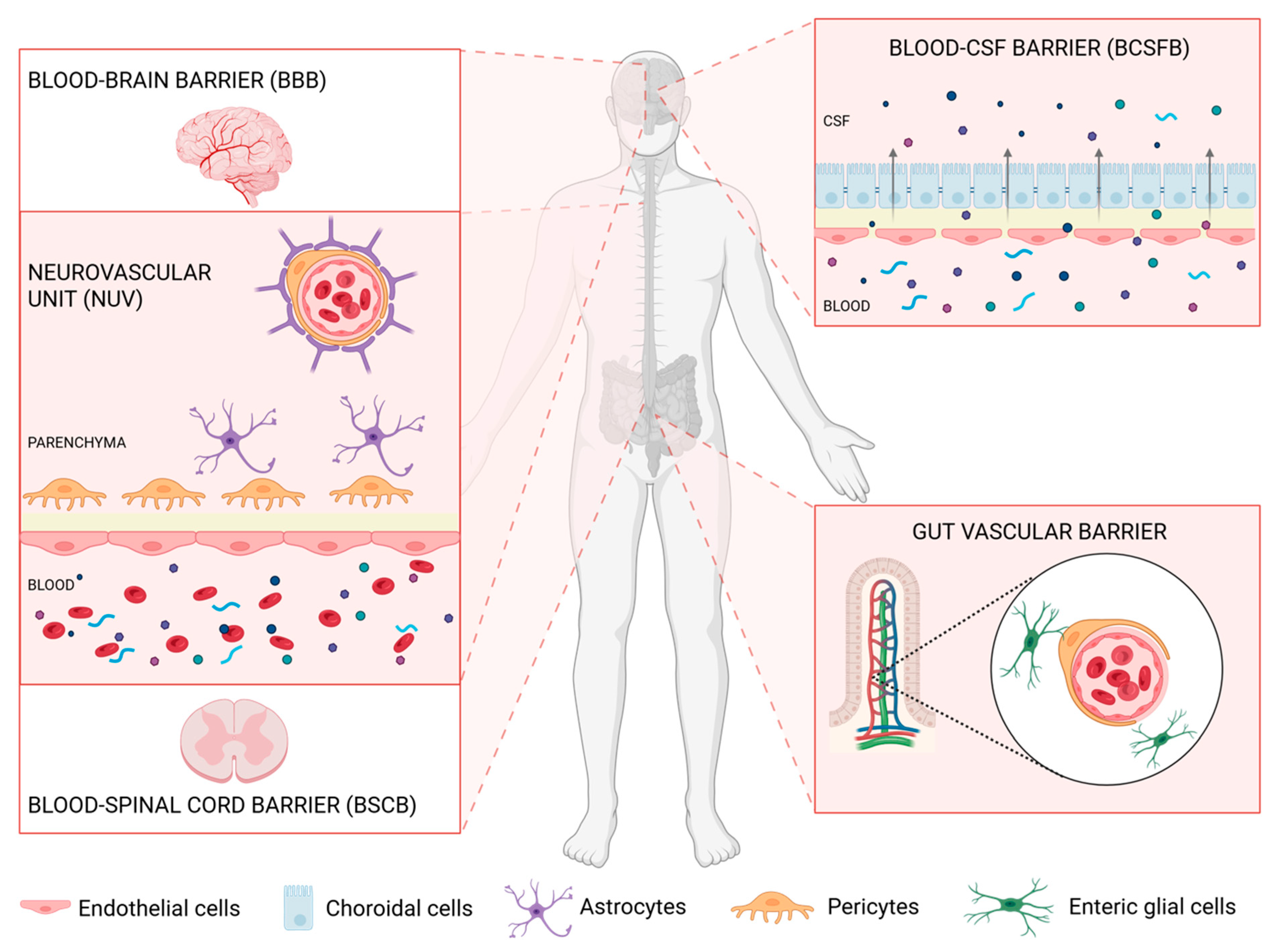
1. Barriers Delimiting and Protecting the Nervous System

In the central nervous system (CNS), we can identify three main barriers regulating the influx of solvents and preventing the intravenous influx of neurotoxic and vasculogenic molecules that may harm CNS cells [1]. These barriers are represented by the blood–cerebrospinal fluid barrier (BCSFB), the blood–brain barrier (BBB), and the blood–spinal cord barrier (BSCB). There is also an extra-neuronal barrier, called the gut–vascular barrier (GVB), whose activity also affects the CNS [2] (Figure 1).

1.1. The Blood–CSF Barrier (BCSFB)

The BCSFB is represented by the choroid plexus located in each of the four cerebral ventricles. Remarkably, the choroid plexus is made of meningeal granular protrusions in the lumen of the cerebral ventricles. These are characterized by a central fenestrated capillary, derived from anterior and posterior choroidal arteries, and an epithelial layer of choroidal cells, which are continuous with the ependymal cells covering the cerebral ventricles’ surface [3]. The choroidal cells are polarized with a basolateral side in contact with the blood and an apical side characterized by villi releasing the cerebrospinal fluid (CSF) into the cerebral ventricles [4]. These cells are connected with tight and adherens junctions that allow the selective passage of ions and other micronutrients, such as vitamins B1, B12, and C, preventing the paracellular movement of molecules [3,5].

1.2. The Blood–Brain Barrier (BBB)

Brain vascularization derives from large and interconnected arteries forming the circle of Willis at the base of the brain, an anastomotic arterial network with a polygonal shape that provides blood nutrient supply to the CNS [6,7].
At the cellular level, endothelial cells, pericytes, vascular smooth muscle cells, astrocytes, oligodendrocytes, and neurons constitute the neurovascular unit (NUV), essential to the nervous system’s functionality. Particularly, endothelial cells, pericytes, and astrocytes are involved in forming the BBB at the level of the vast capillary network which vascularizes the brain. The BBB has the important role of controlling the exchange of molecules between the blood and the CNS, preventing the passage of potentially toxic molecules [8,9,10].
Endothelial cells are enriched with mitochondria and express proteins involved in forming tight and adherens junctions (claudins, occludins, junctional adhesion molecules, and Zonula Occludens (ZO)) [11]. The cell junctions are essential for providing a physical barrier, allowing the free passage of only oxygen, carbon dioxide, and small lipophilic molecules from the blood to the interstitial fluid to prevent the influx of potentially toxic molecules that could damage the CNS. These cells also provide a biochemical barrier through the expression of many transporters of efflux, which pump out unwanted molecules, as well as carriers that regulate the trans-endothelial movement of carbohydrates, hormones, ions, and other molecules circulating into the bloodstream [10]. Concerning the biochemical barrier properties of the BBB, several ATP-binding cassettes, the breast cancer resistance protein (BCRP), and multidrug-resistance-associated proteins (MRPs) are expressed, with the most important ones represented by P-glycoprotein and ATP-binding cassette subfamily G member 2 (ABCG2) [12]. Moreover, the endothelial cells of the BBB express some enzymes of the cytochrome P450 family, with cytochrome P450 family 1 subfamily B (CYP1B1) being the predominant one [12].
The pericytes form direct contact with the endothelium through N-cadherin and connexins, encapsulating the capillary wall [8]. These cells regulate the integrity of the BBB and the permeability of the blood vessels. In various transgenic mouse models expressing mutant platelet-derived growth factor (PDGF)-β/PDGF receptor β signaling, which is involved in the recruitment of pericytes during angiogenesis, proliferation, and migration, several tracers of different molecular weights accumulated in the brain parenchyma, unlike in wild-type mice, showing the loss of integrity of the BBB [13]. Pericytes also have contractile features similar to smooth muscle cells, thus allowing them to regulate the diameter of the brain vessels and the flux of blood into the brain and retinal capillaries in response to vasoactive molecules such as catecholamines, endothelin-1, vasopressin, angiotensin II, ATP, and glutamate [14,15]. In support of this, a reduction in the pericyte population is correlated with an increased vessel diameter and vessel density.
Finally, the astrocytes, with their endfoot processes, define the perivascular space, regulate the ions’ homeostasis, and control the flux of water into the parenchyma through the expression of several transporters and channels. These cells are highly polarized and express protein carriers at the interface with the blood vessels [16,17]. An astrocyte-specific carrier is represented by aquaporin 4 (AQP4), whose expression is restricted to astrocytes and to a subpopulation of ependymal cells localized in the subfornical organ and the supraoptic nucleus. AQP4 is implicated in the osmoregulation between brain parenchyma, CSF, blood, and the clearance of CNS [18,19,20].

1.3. The Blood–Spinal Cord Barrier (BSCB)

Similarly to the BBB, the blood vessels that provide nutrients to the spinal cord are highly specialized to regulate the exchange of solutes between blood and the spinal cord parenchyma. The BSCB is morphologically similar to the BBB and hence characterized by non-fenestrated capillaries with endothelial cells strictly associated with tight and adherens junctions, which prevent the transcellular diffusion of molecules, and expressing influx and efflux transporters that finely control the transcellular transport. Endothelial cells are closely associated with pericytes and astrocytes, and their cytoplasmic endfeet envelop the blood vessels [21,22].
This barrier is more permeable than the BBB; in fact, the expression of tight junction proteins, such as ZO-1, occludin, vascular endothelial cadherin, and β-catenin, is reduced in BSCB [23]. Consistent with this observation, Winkler et al. demonstrated that, upon injection of fluorescent cadaverine, this biogenic amine accumulates in the spinal cord, but not in the brain parenchyma [24]. Furthermore, the immunostaining of CD13 and PDGFRB revealed a lower pericyte coverage in the anterior horn capillaries of the cervical, thoracic, and lumbar spinal cord compared to the brain capillaries. This condition leads to an increased paracellular flow of molecules, favoring the accumulation of plasma proteins in the spinal cord [24].

1.4. Defects in the CNS Barriers in Amyotrophic Lateral Sclerosis

Functional alterations of cell types within the BBB, such as endothelial or mural cells, cause vascular defects and neurological deficits [25]. Changes in the structure of the CNS barriers have been reported in neurodegenerative diseases, such as Alzheimer’s disease (AD), Parkinson’s disease (PD), Huntington’s disease (HD), and Amyotrophic Lateral Sclerosis (ALS), as well as in acute neurological disorders [26].
ALS is the most common motor neuron disease, characterized by the selective degeneration of upper and lower motor neurons [27]. ALS is extremely heterogeneous clinically and genetically [28]. A total of 90% of individuals with ALS present a sporadic form, while in 10% of cases, the disease is familial, with more than 30 genes considered pathogenetic. Accumulation of an RNA-binding protein known as TAR DNA-binding protein 43 (TDP-43), encoded by the gene TARDPB, has been reported in 97% of ALS cases [27]. Structural and functional abnormalities of the barriers that protect the CNS have been demonstrated in animal models mimicking ALS pathology and in human postmortem brain tissues, in studies with a prominent focus on the BSCB; only a few studies have also assessed the BBB in the cortex in ALS [29].
Several mouse and rat experimental models, comprising mice carrying SOD1 and TARDBP mutations, along with C9ORF72 repeat expansion, have been used to study alterations of the CNS barriers from a presymptomatic to a late stage of the disease. In particular, leakage of microvessels, possibly caused by endothelial and basement membrane dysfunction, showed diffusion of Evans Blue, a dye that binds to serum albumin, in the parenchyma of the cervical and lumbar spinal cord from transgenic mutant SOD1 rats and mice at the early symptomatic stage [30,31], along with deposits of hemosiderin, a breakdown product of hemoglobin, in CNS parenchyma [32]. The tight junction proteins ZO-1, occludin, and claudin-5 were found to have reduced expression in spinal cord capillaries from presymptomatic mutant SOD1 mouse models in comparison with control littermates; this is an event happening before motor neuron degeneration which is then exacerbated at the end stage of the disease [33,34]. ZO-1 reduction was confirmed in cortical endothelial cells of the brain microvasculature in transgenic C9orf72 ALS mice at the early stage of the disease [35]. Alterations in the basement membrane in transgenic SOD1G93A mice have also been reported; in particular, collagen IV is reduced in the vascular structure, predominantly in the anterior horn [36], and laminin-1 is decreased, suggesting basement membrane disruption [30]. Moreover, in ALS rats, a decreased expression of agrin, a proteoglycan component of the basement membrane, is observed in association with the disorganization of the extracellular matrix [31]. AQP4, mainly localized in the astrocytic endfeet, is also increased in transgenic SOD1G93A mice [37,38].
Another aspect concerns the induction of perivascular fibroblasts at the presymptomatic stage, possibly involved in remodeling blood vessels in the CNS. In transgenic SOD1G93A ALS mice, there was an increased expression of collagen type VI alpha 1 chain (COL6A1) and secreted phosphoprotein 1 (SPP1) [39]. This was paired with a reduction in capillary density and blood flow in the anterior horn of the lumbar cord occurring at the presymptomatic stage [32,36]. Furthermore, in an in vitro model of BBB from patient donors carrying C9orf72 expansion, compromised barrier integrity and increased activity of the P-glycoprotein transporter have been recently reported [40]. No brain vascular leakage or altered BBB passive diffusion was observed in C9orf72 mice [35], and no ultrastructural changes of BSCB in the brainstem, cervical, and lumbar spinal cords were revealed in capillary inter-endothelial tight junctions in SOD1G93A mice [41], suggesting a discrepancy between protein alteration, functional, and structural measurements.
Most of the observations in murine ALS models have been confirmed in human postmortem ALS specimens. Several independent neuropathological studies revealed CNS barrier perturbation and changes in the neurovascular unit composition in postmortem tissues collected from sporadic ALS patients. At the level of the medulla, spinal cord, and choroid plexus, the protein levels of ZO-1, occludin, claudin-1-3-5, junctional adhesion molecule 1 (JAM-1), and vascular endothelial (VE)-cadherin are reduced [42,43,44]. In BSCB and BCSFB, the staining of capillary endothelium with PECAM-1 and CD105 markers emphasizes a discontinuous endothelial lining [42] along with a reduction in the number of PDGFRβ-positive cells and erythrocyte extravasation in cervical spinal cord anterior horn gray matter [45]. Similarly, Yamadera et al. found a significant decrease in pericyte coverage in the ventral horn of ALS patients compared to controls; this abnormality was also linked to a significantly increased microvascular density [46]. The pericyte reduction was also confirmed at the level of the BCSFB in the choroid plexus [44]. Like in animal models, disorganized collagen IV accumulates in the surrounding leaked vessels. Microvascular barrier abnormalities have been observed in diseased tissues, with microvascular remodeling in the gray and white matter of patients’ medulla, cervical, and lumbar spinal cords. Microvessels are deformed in the spinal cord of ALS patients, with an increased density in patients who underwent artificial respiratory support [46].
Nevertheless, it is still unclear whether the alterations of CNS barriers are an early event contributing to disease onset or just a downstream event exacerbating motor neuron degeneration during disease progression [45]. The analysis of biofluids collected from ALS patients can certainly provide some insights to address this issue. In fact, blood-derived proteins have been identified in the CSF [47,48], and several CNS-specific proteins, like neurofilaments, glial fibrillary acidic protein (GFAP), and tau, are currently measured in the blood of ALS patients, thus offering new means to monitor neuronal health.

1.5. Extra-Neuronal Barriers Affecting the CNS: The Gut–Vascular Barrier

Recent evidence demonstrates a direct link between the CNS and the gut. The gut has a physical and immunological barrier that prevents microorganisms and toxic substances from entering the body. This physical semipermeable barrier is composed of a monolayer of epithelial cells that are strictly associated with each other, thanks to tight and adherens junctions, preventing paracellular flow. Interspersed between epithelial cells, goblet cells secrete mucus, protecting the epithelium from toxic materials and pathogens. At the level of the gut’s blood vessels, a physical barrier that shares several features with the BBB is called the gut–vascular barrier (GVB). Starting from the vessel’s lumen, endothelial cells, pericytes, and enteric glial cells prevent the paracellular flow of molecules bigger than 4kDa [49]. Enteric glial cells (EGCs) play a key role in maintaining the barrier’s integrity [50]. Accordingly, mice with induced ablation of EGC show higher barrier permeability, the release of substances and bacteria into the bloodstream, and a severe inflammatory response [51].
The gut microbiota includes several species of the realms of bacteria, fungi, and viruses, most of which are commensal or mutualistic microorganisms. The composition varies from person to person, depending on several factors such as demography, ethnicity, sex, age, diet, exposure to antimicrobial agents, and healthy or pathological state [52,53,54].
The microbiota has been demonstrated to be involved in several functions of the organism, including the regulation of CNS development and homeostasis [55]. Among these functions, the gut residents regulate the turnover of neurotransmitters, such as noradrenalin, dopamine, and serotonin, and the expression of several proteins associated with synaptogenesis, including postsynaptic density protein 95 (PSD95) and synaptophysin in the striatum. Furthermore, the composition of the microbiota also impacts behavior and anxiety [54,56,57].
The composition of gut microbiota species is essential to benefit the host, including the BBB integrity. In support of this, mice born from germ-free mothers show higher BBB permeability without presenting differences in vascularization and pericyte coverage. Decreased expression of tight junction proteins, like claudin 5, ZO-1, and occludin, in the striatum, cortex, and hippocampus is re-established by colonizing the germ-free mice with the gut microbiota of control mice [58].
Alterations in the composition of the microbiota have been reported in mouse models and patients of several chronic diseases, such as metabolic disorders, atherosclerosis, asthma, autism spectrum disorder, and neurodegenerative disorders [59]. In neurodegenerative disorders, gut microbiota alterations are associated with a dysfunction of the BBB [59]. In an in vivo model of multiple sclerosis (MS), claudin 5 is decreased, and the BBB is more permeable [60]. In animal models of MS, fecal microbiota transplantation (FMT) is associated with a decrease in axonal damage markers (e.g., myelin binding protein and neurofilament light polypeptide (NFL)), intact myelin sheaths in the thoracic spinal cord, and a reduction in the number of activated microglia [61]. Also, germ-free mice show a decrease in the activation of microglia, motor impairment, and inclusion of alpha-synuclein after both supplementation of short-chain fatty acids (SCFAs) and FMT from healthy human donors; inversely, when an FMT is performed from PD patients, the symptoms are exacerbated [62]. The absence of gut microbiota influences the integrity of both BBB and BCSFB by decreasing tight junction expression. This phenotype is rescued in germ-free mice and mice treated with antibiotics after recolonization and in broad-spectrum antibiotic-treated mice by supplementary SCFAs such as propionate and butyrate [63].
This practice is now being explored in humans, and it has been proven to be well tolerated and safe, although efficacy data are still lacking [64,65]. Two ALS patients who underwent FMT showed improvement on the clinical scale ALSFRS-R [59]. A recent Chinese trial was unable to demonstrate an effect of FMT on ALSFRS-R, but recruitment was terminated early before reaching the prespecified sample size due to funding constraints [66]. The results of a recently terminated clinical trial of FMT in 42 ALS patients in Italy are awaited [67]. Notably, individuals with ALS and spinal onset presented gastrointestinal dysbiosis, while patients with a bulbar form of ALS showed dysbiosis in the saliva microbiota [68]. Deciphering how the general microbiome and the GVB influence the integrity of the BBB, BSCB, and BCSFB in ALS and identifying biomarkers of these events could be an added value to monitoring the disease progression.

2. Does the Damage to the Barriers Offer Opportunities to Search for Biomarkers in Circulation?

The physical rupture or damage of cellular barriers should favor the exchange of tissue-specific molecules between different body districts and the search for altered circulating biomarkers in the different biofluids. Indeed, detecting differentially expressed molecules in biofluids in a disease state compared to the physiological condition is promising and receiving great attention in ALS research. However, it is not trivial to detect CNS-specific proteins in peripheral biofluids.
Here, we will give an overview of the main features of the different human biofluids, focusing on well-established CNS-protein biomarkers for ALS disease and illustrating novel avenues of investigation (Figure 2).

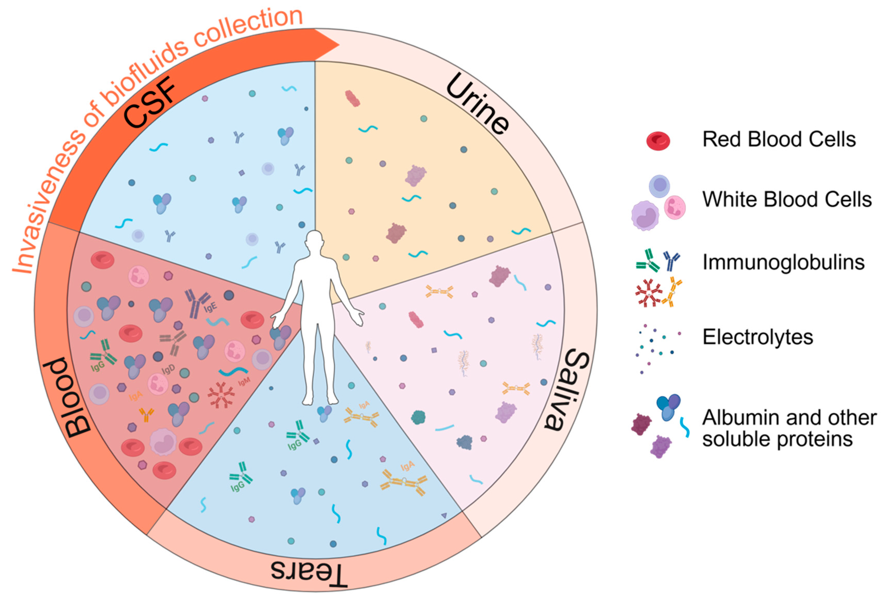
Biofluids for Biomarker Detection

  • The cerebrospinal fluid and the interstitial fluid
The cerebrospinal fluid (CSF) is a chemically stable biofluid that circulates in the CNS, particularly in the subarachnoid space and cerebral ventricles [69]. It has several functions, including regulating the homeostasis of the interstitial fluid of the brain parenchyma, reducing the weight of the CNS by 30 times through its buoyancy property, nourishing and protecting the CNS from mechanical injury, and removing metabolic waste. A human adult contains almost 150 milliliters of CSF, which is completely replaced four to five times every 24 h. This replacement rate is reduced in the elderly [70]. CSF is not a simple blood filtrate; it exhibits a higher concentration of sodium, chloride, folate, amino acids, and magnesium and a lower concentration of potassium, phosphate, albumin, glucose, and calcium than blood [71]. The protein concentration is low, with only 0.025 g of protein per 100 mL, the majority of which is albumin, and the number of cells is less than 5 per mL of CSF. Ions and micronutrients like glucose, vitamins, folate, vasopressin, nitric oxide, and arginine pass through the epithelial cells into the cerebral ventricles at the level of the choroid plexus. This process leads to an increase in the osmotic gradient and, consequently, the passage of water into the CSF through aquaporin 1 [70].
More specifically, the CSF is produced at the level of the choroid plexus, which is in each cerebral ventricle, and at the level of the capillaries of the BBB. The production of CSF is regulated by different factors, such as intraventricular pressure, the autonomic nervous system, Atrial Natriuretic Peptide (ANP), vasopressin, and others. The CSF enters the subarachnoid space through the foramen of Magendie, located in the fourth ventricle, which surrounds the brain and the spinal cord. Next, the CSF can be reabsorbed at the level of the arachnoid granulations or flow into the cranial and spinal subarachnoid space and, thus, into the Virchow–Robin perivascular space. In particular, the subarachnoid space is continuous with the perivascular space surrounding the cortical and penetrating arteries [72]. Iliff et al., in 2012, demonstrated that the CSF flows between the media tunica and the astrocytic endfeet of the arteries and veins driven by arterial pulsation [16]. Based on their size, the solutes present in the CSF can pass through the endfeet of the astrocytes and enter the parenchyma. Here, the bulk flow, which depends upon the movement of water across the astrocytes, determines the flow of the interstitial fluid into the perivenous space and so the clearance of the parenchyma from the metabolic waste, including, for instance, amyloid β 1–40 [16]. This process is associated with the so-called “glymphatic system”, named for its lymphatic-like function and because it depends on glial water flux mediated by AQP4 expressed in the endfeet of astrocytes (https:\doi.org\10.1016\j.nbd.2023.106035). The interstitial fluid enriched with the cells’ metabolic waste must be drained into the bloodstream; however, how the drainage of waste and liquid occurs in the CNS is not as well characterized as in the periphery. One important CSF efflux route is represented by the basal and dorsal meningeal lymphatic vessels. They are lymphatic vessels located in the dura mater meningeal sheath, deprived of the smooth muscle layer and with limited lymphatic valves [73,74,75]. These vessels are mainly localized around the venous sinuses, and with a higher density in the meningeal than in the periosteal layer of the dura mater [76]. The involvement of the meningeal lymphatic vessels in liquor reabsorption was discovered in 2015 in mouse models. Both Louveau and Aspelund demonstrated with fluorescent tracers that the interstitial fluid and the CSF are drained into the subarachnoid space, then into the meningeal lymphatic, and subsequently move into the deep cervical lymph nodes, and then into the superficial cervical lymph nodes [73,74]. In support of this, a reduced clearance and a subsequent accumulation of ovalbumin in deep cranial lymph nodes are reported in transgenic mice defective for lymphatic vessels. Recently, amyloid β 1–40, phosphorylated tau 181 (pTau181), GFAP, and NFL were successfully measured in the human cervical lymph node, providing an additional compartment to analyze and monitor neurodegenerative processes [77].
Due to its proximity to the neuronal and glial cells constituting the nervous system, the CSF is considered the best source for exploring biomarkers for neurological conditions. The most reproducible biomarkers for ALS that have been identified in the CSF are neuronal structural proteins such as NFL, neurofilament heavy (NFH and phosphorylated NFH), tau, and its phosphorylated forms (phosphorylated Tau 181 and 217) [78]. In addition, there are also chitinases, enzymes secreted by macrophage and activated microglia in the inflammatory state, namely the myeloid protein chitotriosidase-1 (CHIT1), the glial protein YKL-40, also known as chitinase-3-like protein 1 (CHI3L1), and YKL-39, also known as chitinase-3-like protein 2 (CHI3L2) (reviewed in [79]). Specifically, each of these markers could have different applications in clinical practice. Neurofilaments have been identified as the most promising biomarkers for ALS, having diagnostic, prognostic, and disease monitoring roles [80]. Chitinases demonstrate diagnostic value, with CHIT1 and CHIT3L1 being associated with disease severity and progression [81]. Instead, tau and its phosphorylated forms correlate with disease severity [82]. Of interest, a novel diagnostic protein biomarker is derived from the cryptic exon-containing hepatoma-derived growth factor-like protein 2 (HDGFL2), whose content in the CSF is strictly dependent on TDP-43 loss of splicing activity in ALS and FTD and is significantly increased in sporadic ALS patients as well as in presymptomatic and symptomatic C9ORF72 mutation carriers [83]. Unfortunately, the CSF collection is an invasive intervention with a lumbar puncture [84]. Therefore, the applicability of these biomarkers is limited and not for routine monitoring. The most studied CSF biomarkers are reported in Table 1.
  • The blood
A human adult contains approximately 4–5 L of blood [85]. This liquid tissue is composed of a cellular fraction (45%) and a liquid phase called plasma (55%). The blood has several functions, including the transport of oxygen and carbon dioxide from the lungs to the tissues and vice versa, and the transport of metabolites, ions, and nutrients [86]. It is involved in the hemostasis process and in protecting the organism from infections. It also carries substances from the site of production or storage to target organs, such as hormones and vitamins [87]. More specifically, the corpuscular part of the blood comprises red blood cells, white blood cells, and platelets. These cells are produced starting from hemopoietic stem cells (HSCs) in the bone marrow, specifically at the level of the HSC niches [88].
Plasma mainly comprises water (90–92%) and more than a thousand proteins, electrolytes, nutrients, metabolites, and dissolved gases (O2, CO2, and N2). The most abundant proteins are albumin, globulins, and fibrinogen [89]. The majority of plasma proteins, such as albumin, fibrinogen, and the other coagulation factors, are produced at the level of the liver, whereas immunoglobulins are produced by B cells [85].
Blood is highly investigated due to its ease of accessibility and low invasiveness. However, it is an extremely complex matrix to analyze, due to the high level of soluble proteins and lipoproteins that could influence the identification of biomarkers [79]. For this reason, very few biomarkers have been validated across laboratories, reflecting alterations in the nervous system in ALS. The most reproducible biomarker is the NFL, which can be used for diagnosis, prognosis, and disease monitoring in ALS (reviewed in [90]). Recent data show promising results for the diagnostic role of phosphorylated tau [78] and the prognostic role of GFAP [91,92], two structural proteins derived from neurons and astrocytes, respectively. Another promising biomarker is the cardiac troponin T, a protein of muscle origin, which was found to be increased in ALS patients [93] and reviewed in [83], demonstrating a role in disease progression monitoring.
Concerning TDP-43, some studies demonstrated an increased level of this protein in plasma and CSF of ALS patients, measured mainly through ELISA; however, the results vary across different studies, suggesting a low reproducibility of these assays, due to the low level of the pathological form in biofluids and the presence of immunoglobulins and albumins that can influence the binding of the antibody to the target TDP-43, as reviewed in [94,95]. Simoa® technology, a new sensitive technology developed by Quanterix, has been developed and used for detecting TDP-43 in CSF and blood of ALS patients with a higher sensitivity than ELISA. However, the results are contradictory [96,97,98,99]. The most studied blood biomarkers are reported in Table 1.
  • Urine
Urine is an amber-colored biofluid produced by the renal system to expel excess liquids and waste products from the body. The kidney has the important role of regulating plasma osmolarity, adjusting the amount of water, electrolytes, and solutes in the blood circulation. The functional unit of the kidney is the nephron, where the glomerulus filters the plasma from the blood flow, and then through the renal tubule, the essential nutrients for the body are reabsorbed [100]. In a day, approximately 180 L of fluid is filtered, carrying out toxins, metabolic waste products, ions, and electrolytes to be eliminated. Specifically, urine is composed mainly of 95% water, 2% urea, 0.1% creatinine, 0.03% uric acid, and smaller amounts of metabolites, proteins, and several ions, such as chloride, sodium, potassium, sulfate, ammonium, and phosphate [101]. Only 20–30% of the blood proteins are also present in urine. Total urinary protein concentration is generally low, being below 0.2 mg per milliliter. This is because the renal system efficiently reabsorbs most proteins in circulation. Indeed, the Tamm–Horsfall protein (uromodulin), a kidney-specific glycoprotein, is the most abundant protein present in urine [102], whereas albumin and other blood proteins have a low concentration.
Urine has many advantages in the context of biomarker research. Indeed, it is easily accessible, and large quantities can be sampled, making it extremely feasible for repeated longitudinal measurements. Moreover, it may be a favorable biofluid for measuring low-abundance proteins, considering the low levels of blood-abundant proteins, which may affect the specificity and sensitivity of analytical assays. On the other hand, it has to be taken into account that proteins can cross the filtration barrier depending on their size and charge; i.e., proteins with a mass of 60–70 kDa as well as negatively charged proteins are largely retained in the capillary lumen and thus re-enter the systemic circulation [103], which may narrow their applicability for biomarker detection. It is also important to consider that urine composition is affected by several variables, such as gender, age, weight, pH, time of collection, handling, and diet [104,105,106].
The most studied urinary biomarker for ALS is neurotrophin receptor p75 extracellular domain (p75ECD), the detection of which indicates motor neuron degeneration, which correlates with disease progression [107,108]. Other candidate prognostic biomarkers have also been proposed, such as titin and collagen type IV, as reviewed in [108]. The most studied urine biomarkers are reported in Table 1.
  • Saliva
Saliva is the slightly acidic, hypotonic mucoserous biofluid secreted by major and minor salivary glands in the mouth. A human adult produces almost 0.5–1 L of saliva with a flow rate of 0.3 mL/min in unstimulated conditions and reaching a maximum of 7 mL/min after stimulation. Saliva secretion is regulated at the level of the salivary nuclei of the medulla, and specific stimuli, such as the act of chewing and gustatory stimuli, are known to induce hypersecretion. Particularly, saliva production is controlled by the sympathetic and parasympathetic nervous systems and several hormones [109].
Saliva comprises more than 99% water, in which electrolytes, immunoglobulin A, proteins, enzymes, mucin, urea, and ammonia are dissolved. The pH ranges between 6 and 7, depending on the secretion flow. The parotid saliva is enriched in amylase, proline-rich proteins, and agglutinins. Mucins MG1 and MG2 are mainly secreted by sublingual saliva and lysozyme; submandibular glands release Cystatin S [110,111].
Saliva has several roles, which include lubrication and protection of the mouth and the teeth, antibacterial activity, buffering action for the regulation of the pH, taste, and digestion [112,113]. This biofluid has gained attention in recent years as a non-invasive and easy-to-obtain source of biomarkers, but no saliva biomarkers have been associated with ALS yet. However, efforts are being made to identify saliva-associated biomarkers for ALS and other neurodegenerative diseases. For example, Carlomagno et al. employed Raman spectroscopy in a small cohort of 19 ALS patients, 10 PD patients, 10 AD patients, and 10 healthy controls. Particularly, they showed promising results in discriminating ALS from the spectra obtained, mainly due to differences in the concentration of lipids, but further investigation on larger cohorts is necessary [114]. Additionally, extracellular vesicles (EVs) derived from saliva are reported as being less abundant in contaminants, such as non-EV proteins or apolipoproteins, than plasma, which potentially enables an easy identification of novel biomarkers [115,116] (Table 1).
  • Tears
Tears are a transparent fluid produced by the lacrimal glands. They create a thin film that protects and lubricates the eyelids, conjunctiva, and cornea, subdivided into an outer lipid layer, a middle aqueous layer, and an inner glycocalyx layer [117]. Tears function not only by delivering nutrients to the cornea, but also by removing foreign material and protecting against infections.
Tears are aqueous solutions mainly made of water, containing electrolytes, proteins, lipids, mucins, defensins, collectins, and other small molecules [118]. The total protein concentration varies from 6 to 10 mg per milliliter of tears. Among the most abundant proteins are lactoferrin and lysozyme, both having antibacterial and antimicrobial functions, as well as lipocalin, which binds to the least soluble lipids, enhancing their solubility, and secretory IgA, which can react with antigens on bacterial cells [119,120,121].
Tear collection methods are minimally invasive, rapid, and painless, with glass microcapillary tubes or Schirmer strips being the cheapest and simplest tools used [122]. Tears can be used to study not only ocular surface diseases, but also systemic disorders [123]. On the other hand, the analysis is challenging due to the limited volume that can be collected, which is approximately 5 µL, highlighting the need for sensitive assays requiring low input volumes.
No tear biomarkers have been associated with ALS yet. Nevertheless, a recently published proteomic study found two proteins, namely SERPINC1 and HP, to be differentially present in ALS patients compared to controls [124]. Moreover, a metabolomic analysis of tear fluid collected from ALS patients highlighted a signature that differentiates bulbar and spinal forms of the disease, although no differences were reported between patients and controls [125] (Table 1).
Table 1. Most used and validated biomarkers for ALS in biofluids.
BiofluidALS Markers
CSFNFL [126]
neurofilament high (NFH and phosphorylated NFH) [80]
tau and its phosphorylated form [82]
myeloid protein chitotriosidase-1 (CHIT1) [127]
glial protein YKL-40, also known as chitinase-3-like protein 1 (CHI3L1) [128]
YKL-39 [79]
inserting cryptic exons of the HDGFL2 [83]
BloodNFL [79]
phosphorylated tau [78]
GFAP [91,92]
cardiac troponin T [93]
Urineneurotrophin receptor p75 extracellular domain (p75ECD) [79]
Salivano saliva biomarkers have been associated with ALS yet
Tearsno tear biomarkers have been associated with ALS yet

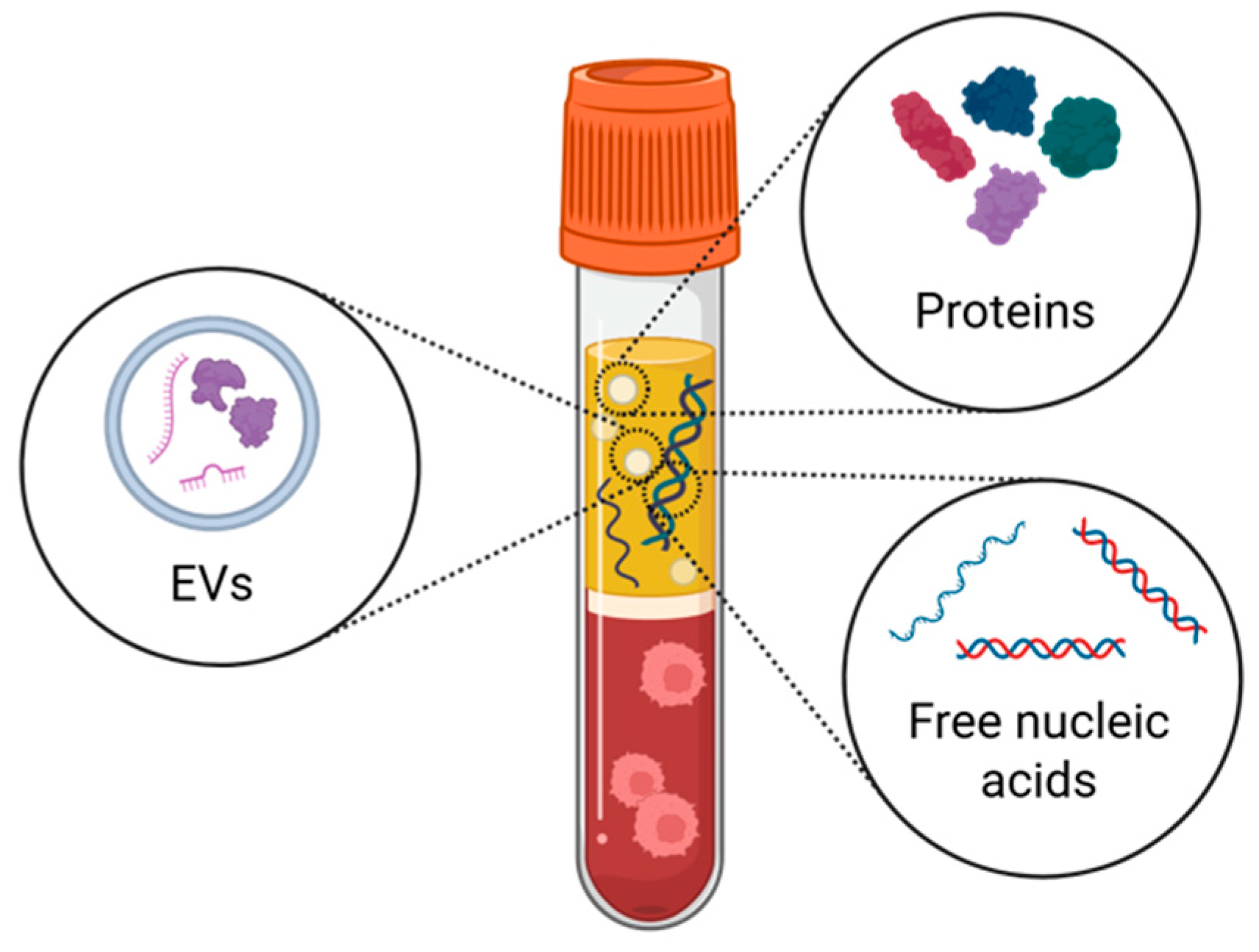
3. Beyond Free Circulating Proteins: Extracellular Vesicles and Cell-Free DNA

Different biological entities, like extracellular vesicles (EVs) and cell-free DNA (cfDNA), are also present in biofluids and carry specific information about the site of origin. Technical hurdles and poor reproducibility across laboratories still hamper their use as biomarkers, but their potential must be considered in view of the technical challenges that are likely to be overcome in the future.
  • Extracellular vesicles (EVs)
EVs are nanoparticles released by all cells and consist of a phospholipidic bilayer that protects the cargo, comprising proteins, nucleic acids, and metabolites, from degradation by proteases, nucleases, and other enzymes present in circulation. EVs are usually classified into exosomes, microvesicles, and apoptotic bodies according to the biogenesis pathway from which they derive [129]. However, the biophysical characteristics, such as size and density, or the structural and molecular content, cannot fully distinguish the different subpopulations. Recently, circulating EVs have gained attention for their potential use as disease biomarkers. Different lines of investigation focus on unraveling the cargo of brain EVs in physiological and pathological conditions as a tool to study CNS alterations. The bidirectional passage of EVs between the CNS and the bloodstream has been reported, but not mechanistically explained. A few markers have been identified as specifically associated with brain-derived EVs and used to isolate these species from peripheral biofluids like blood. These markers include L1 cell adhesion molecule (L1CAM), Neural Cell Adhesion Molecule (NCAM), glutamate ionotropic receptor AMPA type subunits 2 and 3 (GluR2/3), GFAP, glutamate receptor, ionotropic, N-methyl D-aspartate 2A (NMDAR2A), ATPase, Na+/K+ transporting, and alpha 3 polypeptide (ATP1A3) [130,131,132,133,134,135]. However, it still remains poorly understood how EVs can cross barriers, particularly the BBB [136,137].
Several studies analyzed EV passage through the BBB in vitro. Fluorescently labeled human serum-derived EVs pass through immortalized murine-derived bEnd.3 cells [138], and HEK293T-derived EVs cross the human brain microvascular endothelial cells (BMECs) only upon stimulation with Tumor Necrosis Factor-alpha (TNF-α) [139] or Lipopolysaccharide (LPS) [140]. These two observations suggest that inflammatory signals may exert an important influence on BBB permeability.
Preliminary observations suggest clathrin-mediated endocytosis and micropinocytosis as cellular mechanisms favoring EV transcytosis to explain how EVs pass through the BBB. However, how the process is initiated per se remains the subject of open investigations [139,141]. More complex systems with microfluidics, BMECs, extracellular matrix, primary astrocytes, and pericytes confirmed the transcellular transport of EVs through endothelial cells mediated by endocytosis [141]. In vitro models and BMEC cells are suitable tools to study the interplay between EVs and the BBB; nevertheless, considering the complex multicellular structure of the BBB, they may not clearly explain the whole process of EV passage through the barrier.
Nevertheless, there is evidence for EV crossings from the periphery to the CNS and vice versa, although in an unbalanced manner. In physiological conditions, the passage of EVs from the periphery to the CNS is indeed an exceptional event, considering that most EVs accumulate in the liver, kidney, and spleen [140]. Injecting zebrafish embryos with fluorescently labeled peripheral EVs collected from AD patients’ serum resulted in EVs internalized by neurons and glial cells [138]. Through the injection of radioactively labeled EVs collected from human erythrocytes in the peripheral circulatory system of mice, EVs entered the microglia cells during LPS-induced systemic inflammation [140].
Another unresolved question regards EV uptake selectivity in vivo in the CNS. EVs derived from up to ten cell lines were radiolabeled and injected intravenously in CD-1 mice. All of them were retrieved in the brain, showing that all EV types crossed the BBB. A 10-fold variation rate was reported among cell lines, suggesting that specific surface proteins may favor EV uptake in the CNS [142]. Treatments with LPS, wheat germ agglutinin (WGA), and mannose 6-phosphate (M6P) also induce EV uptake by stimulating the adsorptive transcytosis [142]. Furthermore, the use of transgenic mice, in which the hematopoietic cells express a Cre protein that is sorted in EVs circulating in the bloodstream and recipient cells express a construct that is activated by the Cre protein leading to the irreversible expression of an enhanced yellow fluorescent protein, showed unequivocally that bloodstream EVs can be uptaken by synaptically active neurons [143]. Accordingly, intranasally delivered EVs accumulated in brain regions up to 96 h after administration [144]. On the other hand, regarding the passage of brain EVs into peripheral biofluids, radioactively labeled EVs injected into the ventricles passed from the brain to the bloodstream [142], also supporting the existence of EV flows from the brain into circulation. Consequently, brain-derived EVs can be isolated from the blood as potential CNS biomarkers [145,146]. In ALS and FTD patients, brain-derived EVs enriched with neuronal markers (L1CAM+) were isolated from plasma samples, and their cargo contained TDP-43 and tau proteins [99]. Remarkably, FTD individuals with a tau pathology presented a significant enrichment of tau protein in neuronal EVs; similarly, FTD (and ALS) patients with TDP-43 pathology had increased TDP-43 levels in neuronal EVs, suggesting the use of neuronal EVs to predict specifically tau or TDP-43 pathology associated with FTD subtypes [99]. Also, the microRNA content is noted to be altered in L1CAM-positive EVs in ALS patients [147,148,149]. Still, the lack of consensus in different laboratories on the specific RNAs that are deregulated in brain-derived EVs in disease conditions highlights the need to further validate this approach before applying it in clinical practice.
  • Circulating cell-free DNA (cfDNA)
In 1948, Mandel and Metais demonstrated for the first time the presence of circulating cell-free nucleic acids in healthy donors’ blood [150,151]. These nucleic acids comprise cell-free RNA (cfRNA) and DNA (cfDNA) and circulating cell-free mitochondrial DNA (cf-mtDNA) [152,153]. cfDNA is double-stranded and binds to histone proteins, and single nucleosomes, with a 160–180 bp size, can be detected in bioliquids upon release from apoptotic cells [152,153]. After the discovery of free nucleic acids in biological fluids like blood and CSF, cfDNA has received special attention as a potential biomarker, especially in cancer and prenatal research. In healthy individuals, cfDNA is derived from normal metabolic activity of hematopoietic cells, as shown by the methylome profile and in vivo generation of genome-wide nucleosome occupancy maps [154,155]. In general, plasma cfDNA content in healthy people is less than 10ng/mL, but it increases not only in several chronic and acute disorders (such as autoimmune diseases, cancer, diabetes, sepsis and transplantation), but also in several physiological conditions, such as pregnancy due to fetal DNA release, or during aging because of reduced clearance [155,156,157].
The use of cfDNA as a valuable diagnostic and prognostic biomarker came first from the cancer field [157,158,159,160]. In cancers located in the CNS, cfDNA has become an essential diagnostic and predictive biomarker, considering that the biopsy is risky or inaccessible [156]. In these cases, cfDNA analysis in the CSF proved to be more sensitive than standard cytological analysis of tumor cells, making it a promising new diagnostic biomarker [161]. The amount of CSF cfDNAs is also predictive, being higher in patients with advanced CNS tumors [162], and it is inversely correlated with the survival of glioma patients [163,164].
Another interesting application regards the analysis of the methylation profile of the cfDNA, which could be more informative than investigating specific disease-associated mutations, especially in neurodegenerative disorders, where the sporadic forms are more frequent than the familial ones. The advantage is that, by assessing the methylation profile, the cell of origin of the cfDNA is specifically determined [165]. For example, the methylation profile data of 8 ALS patients and 8 controls demonstrated a significant increase in the level of muscle-derived cfDNA in ALS patients’ blood compared to controls [166]. The methylation profile of plasma-derived cfDNA makes it possible to discriminate patients with AD from people with mild cognitive impairment (MCI) and healthy donors based on the amount of neuronal-derived cfDNA [167]. Furthermore, patients with severe traumatic brain injury and cardiac arrest with neuronal damage and BBB disruption also show elevated levels of brain-derived cfDNA in the bloodstream [157].
Circulating mitochondrial cfDNA (cf-mtDNA) is also gaining attention in neurodegenerative disease research, as it is released during cell stress and necrosis. Cf-mtDNA is usually detected by amplifying mitochondrial genes, such as COX3, using droplet digital PCR (ddPCR) [168]. Mitochondrial DNA is elevated in PD patients’ serum and MS CSF [169], while contradictory results have been reported [170] in AD. Changes in the cf-mtDNA amount were found in patients with CNS tumors and ALS [171].
Due to its high sensitivity, the analysis of cfDNA could serve as an early biomarker in liquid biopsy for various disorders.
By combining its analysis with other biomarkers, including EVs, the ability to diagnose diseases and predict their progression could greatly improve (Figure 3). In this perspective, Mugoni et al. have developed the ONCE protocol, which allows simultaneous RNA analysis from EVs and cfDNA from the same plasma aliquot [172] (Figure 3).

4. Conclusions

The tight structure of the barriers that protect the tissues of the central nervous system poses a challenge to researchers in understanding and targeting the CNS. For this reason, it is essential to determine their structure and how they function to implement strategies and novel routes for diagnosing, monitoring, and treating neurological disorders such as ALS. CNS-derived biomarkers are certainly useful in tracking changes in the cells of the nervous system. In ALS, biomarkers are actively investigated as promising tools to assess unmet needs, e.g., the effect of drug treatments. Several validated biomarkers worldwide, such as NFL, are currently measured in people with ALS, but some drugs may affect their clearance and yield confusing results [173,174]. Other problems that arise related to the candidate biomarkers identified are linked to the lack of reproducibility and technical challenges. Therefore, protein and nucleic acid biomarkers are being continuously investigated with an increasing interest towards brain-derived EVs and cell-free DNA from neuronal origin. In addition to understanding the neuronal origin of the molecular content of biofluids, deeper research should be conducted into the mechanisms that favor the mobility of molecules, proteins, and EVs via the CNS and intestinal barriers.

Funding

We acknowledge the support of the MUR PNRR project iNEST—Interconnected Nord-Est Innovation Ecosystem (ECS00000043) funded by the NextGenerationEU. We acknowledge funding from Ministero della Salute project PERMEALS—PNRR-MAD-2022-12375731 (to A.C., N.T., A.R.). We acknowledge the European Union—Next Generation EU, Mission 4, Component 1, CUP E53D23019700001, project “MYSTICALS” (to J.M., A.C., N.T.). We acknowledge the Italian Ministero della Salute, Grant-No. RF-2016-02361616 (to J.M.). We thank Fondazione AriSLA for supporting the project EVTestInALS (to M.B., A.C.). AR acknowledges “Aldo Ravelli Center for Neurotechnology and Experimental Brain Therapeutics”, Università degli Studi di Milano. We acknowledge the support of the Next Generation EU through the MUR-PRIN 2022 project EV-PRINT 2022CS9H53 (to P.G.).

Institutional Review Board Statement

Not applicable.

Informed Consent Statement

Not applicable.

Data Availability Statement

Not applicable.

Conflicts of Interest

The authors declare no conflicts of interest.

Abbreviations

The following abbreviations are used in this manuscript:
CNScentral nervous system
BCSFBblood–cerebrospinal fluid barrier
BBBblood–brain barrier
BSCBblood–spinal cord barrier
GVBgut–vascular barrier
NUVneurovascular unit
BCRPATP-binding cassettes, breast cancer resistance protein
ABCG2ATP-binding cassette subfamily G member 2
CYP1B1cytochrome P450 family, with cytochrome P450 family 1 subfamily B
PDGF-βplatelet-derived growth factor
AQP4aquaporin 4
ADAlzheimer’s disease
PDParkinson’s disease
HDHuntington’s disease
ALSAmyotrophic Lateral Sclerosis
MSmultiple sclerosis
TDP-43TAR DNA-binding protein 43
COL6A1collagen type VI alpha 1 chain
SPP1secreted phosphoprotein 1
JAM-1junctional adhesion molecule 1
GFAPglial fibrillary acidic protein
EGCenteric glial cell
PSD95postsynaptic density protein 95
FMTfecal microbiota transplantation
SCFAsshort-chain fatty acids
CSFcerebrospinal fluid
ANPAtrial Natriuretic Peptide
CHIT1myeloid protein chitotriosidase-1
CHI3L1chitinase-3-like protein 1
HDGFL2hepatoma-derived growth factor-like protein 2
EVsextracellular vesicles
cfDNAcell-free DNA
L1CAML1 cell adhesion molecule
NCAMNeural Cell Adhesion Molecule
GluR2/3glutamate ionotropic receptor AMPA type subunits 2 and 3
NMDAR2Aglutamate receptor, ionotropic, N-methyl D-aspartate 2A
ATP1A3ATPase, Na+/K+ transporting, and alpha 3 polypeptide
BMECsbrain microvascular endothelial cells
TNF-αTumor Necrosis Factor-alpha
LPSLipopolysaccharide
WGAwheat germ agglutinin
M6Pmannose 6-phosphate
cfRNAcell-free RNA
cf-mtDNAcirculating cell-free mitochondrial DNA
MCImild cognitive impairment
ddPCRdroplet digital PCR
NFLneurofilament light polypeptide
NFHneurofilament heavy polypeptide
BMECsbrain microvascular endothelial cells
p75ECDneurotrophin receptor p75 extracellular domain
HSChemopoietic stem cell
CHI3L2Chitinase-3-like protein 2

References

  1. Engelhardt, B. Cluster: Barriers of the Central Nervous System. Acta Neuropathol. 2018, 135, 307–310. [Google Scholar] [CrossRef] [PubMed]
  2. Carloni, S.; Rescigno, M. The Gut-Brain Vascular Axis in Neuroinflammation. Semin. Immunol. 2023, 69, 101802. [Google Scholar] [CrossRef]
  3. Lun, M.P.; Monuki, E.S.; Lehtinen, M.K. Development and Functions of the Choroid Plexus–Cerebrospinal Fluid System. Nat. Rev. Neurosci. 2015, 16, 445–457. [Google Scholar] [CrossRef] [PubMed]
  4. Sakka, L.; Coll, G.; Chazal, J. Anatomy and Physiology of Cerebrospinal Fluid. Eur. Ann. Otorhinolaryngol. Head. Neck Dis. 2011, 128, 309–316. [Google Scholar] [CrossRef]
  5. Spector, R.; Johanson, C.E. The Mammalian Choroid Plexus. Sci. Am. 1989, 261, 68–74. [Google Scholar] [CrossRef] [PubMed]
  6. Ayre, J.R.; Bazira, P.J.; Abumattar, M.; Makwana, H.N.; Sanders, K.A. A New Classification System for the Anatomical Variations of the Human Circle of Willis: A Systematic Review. J. Anat. 2022, 240, 1187–1204. [Google Scholar] [CrossRef]
  7. Jones, J.D.; Castanho, P.; Bazira, P.; Sanders, K. Anatomical Variations of the Circle of Willis and Their Prevalence, with a Focus on the Posterior Communicating Artery: A Literature Review and Meta-analysis. Clin. Anat. 2021, 34, 978–990. [Google Scholar] [CrossRef]
  8. Wälchli, T.; Bisschop, J.; Carmeliet, P.; Zadeh, G.; Monnier, P.P.; De Bock, K.; Radovanovic, I. Shaping the Brain Vasculature in Development and Disease in the Single-Cell Era. Nat. Rev. Neurosci. 2023, 24, 271–298. [Google Scholar] [CrossRef]
  9. Winkler, E.A.; Bell, R.D.; Zlokovic, B.V. Central Nervous System Pericytes in Health and Disease. Nat. Neurosci. 2011, 14, 1398–1405. [Google Scholar] [CrossRef]
  10. Kadry, H.; Noorani, B.; Cucullo, L. A Blood–Brain Barrier Overview on Structure, Function, Impairment, and Biomarkers of Integrity. Fluids Barriers CNS 2020, 17, 69. [Google Scholar] [CrossRef]
  11. Komarova, Y.A.; Kruse, K.; Mehta, D.; Malik, A.B. Protein Interactions at Endothelial Junctions and Signaling Mechanisms Regulating Endothelial Permeability. Circ. Res. 2017, 120, 179–206. [Google Scholar] [CrossRef] [PubMed]
  12. Dauchy, S.; Dutheil, F.; Weaver, R.J.; Chassoux, F.; Daumas-Duport, C.; Couraud, P.; Scherrmann, J.; De Waziers, I.; Declèves, X. ABC Transporters, Cytochromes P450 and Their Main Transcription Factors: Expression at the Human Blood–Brain Barrier. J. Neurochem. 2008, 107, 1518–1528. [Google Scholar] [CrossRef]
  13. Bell, R.D.; Winkler, E.A.; Sagare, A.P.; Singh, I.; LaRue, B.; Deane, R.; Zlokovic, B.V. Pericytes Control Key Neurovascular Functions and Neuronal Phenotype in the Adult Brain and during Brain Aging. Neuron 2010, 68, 409–427. [Google Scholar] [CrossRef] [PubMed]
  14. Armulik, A.; Genové, G.; Mäe, M.; Nisancioglu, M.H.; Wallgard, E.; Niaudet, C.; He, L.; Norlin, J.; Lindblom, P.; Strittmatter, K.; et al. Pericytes Regulate the Blood–Brain Barrier. Nature 2010, 468, 557–561. [Google Scholar] [CrossRef] [PubMed]
  15. Peppiatt, C.M.; Howarth, C.; Mobbs, P.; Attwell, D. Bidirectional Control of CNS Capillary Diameter by Pericytes. Nature 2006, 443, 700–704. [Google Scholar] [CrossRef]
  16. Iliff, J.J.; Wang, M.; Liao, Y.; Plogg, B.A.; Peng, W.; Gundersen, G.A.; Benveniste, H.; Vates, G.E.; Deane, R.; Goldman, S.A.; et al. A Paravascular Pathway Facilitates CSF Flow Through the Brain Parenchyma and the Clearance of Interstitial Solutes, Including Amyloid β. Sci. Transl. Med. 2012, 4, 147ra111. [Google Scholar] [CrossRef]
  17. Ray, L.A.; Heys, J.J. Fluid Flow and Mass Transport in Brain Tissue. Fluids 2019, 4, 196. [Google Scholar] [CrossRef]
  18. Murlidharan, G.; Crowther, A.; Reardon, R.A.; Song, J.; Asokan, A. Glymphatic Fluid Transport Controls Paravascular Clearance of AAV Vectors from the Brain. JCI Insight 2016, 1, e88034. [Google Scholar] [CrossRef]
  19. Nielsen, S.; Arnulf Nagelhus, E.; Amiry-Moghaddam, M.; Bourque, C.; Agre, P.; Petter Ottersen, O. Specialized Membrane Domains for Water Transport in Glial Cells: High-Resolution Immunogold Cytochemistry of Aquaporin-4 in Rat Brain. J. Neurosci. 1997, 17, 171–180. [Google Scholar] [CrossRef]
  20. Rash, J.E.; Yasumura, T.; Hudson, C.S.; Agre, P.; Nielsen, S. Direct Immunogold Labeling of Aquaporin-4 in Square Arrays of Astrocyte and Ependymocyte Plasma Membranes in Rat Brain and Spinal Cord. Proc. Natl. Acad. Sci. USA 1998, 95, 11981–11986. [Google Scholar] [CrossRef]
  21. Bartanusz, V.; Jezova, D.; Alajajian, B.; Digicaylioglu, M. The Blood–Spinal Cord Barrier: Morphology and Clinical Implications. Ann. Neurol. 2011, 70, 194–206. [Google Scholar] [CrossRef] [PubMed]
  22. Jeong, J.-Y.; Kwon, H.-B.; Ahn, J.-C.; Kang, D.; Kwon, S.-H.; Park, J.A.; Kim, K.-W. Functional and Developmental Analysis of the Blood–Brain Barrier in Zebrafish. Brain Res. Bull. 2008, 75, 619–628. [Google Scholar] [CrossRef] [PubMed]
  23. Chopra, N.; Menounos, S.; Choi, J.P.; Hansbro, P.M.; Diwan, A.D.; Das, A. Blood-Spinal Cord Barrier: Its Role in Spinal Disorders and Emerging Therapeutic Strategies. NeuroSci 2021, 3, 1–27. [Google Scholar] [CrossRef] [PubMed]
  24. Winkler, E.A.; Sengillo, J.D.; Bell, R.D.; Wang, J.; Zlokovic, B.V. Blood–Spinal Cord Barrier Pericyte Reductions Contribute to Increased Capillary Permeability. J. Cereb. Blood Flow. Metab. 2012, 32, 1841–1852. [Google Scholar] [CrossRef]
  25. Sweeney, M.D.; Zhao, Z.; Montagne, A.; Nelson, A.R.; Zlokovic, B.V. Blood-Brain Barrier: From Physiology to Disease and Back. Physiol. Rev. 2019, 99, 21–78. [Google Scholar] [CrossRef]
  26. Chen, T.; Dai, Y.; Hu, C.; Lin, Z.; Wang, S.; Yang, J.; Zeng, L.; Li, S.; Li, W. Cellular and Molecular Mechanisms of the Blood–Brain Barrier Dysfunction in Neurodegenerative Diseases. Fluids Barriers CNS 2024, 21, 60. [Google Scholar] [CrossRef]
  27. Riva, N.; Domi, T.; Pozzi, L.; Lunetta, C.; Schito, P.; Spinelli, E.G.; Cabras, S.; Matteoni, E.; Consonni, M.; Bella, E.D.; et al. Update on Recent Advances in Amyotrophic Lateral Sclerosis. J. Neurol. 2024, 271, 4693–4723. [Google Scholar] [CrossRef]
  28. Bendotti, C.; Bonetto, V.; Pupillo, E.; Logroscino, G.; Al-Chalabi, A.; Lunetta, C.; Riva, N.; Mora, G.; Lauria, G.; Weishaupt, J.H.; et al. Focus on the Heterogeneity of Amyotrophic Lateral Sclerosis. Amyotroph. Lateral Scler. Front. Degener. 2020, 21, 485–495. [Google Scholar] [CrossRef]
  29. Schreiber, S.; Bernal, J.; Arndt, P.; Schreiber, F.; Müller, P.; Morton, L.; Braun-Dullaeus, R.C.; Valdés-Hernández, M.D.C.; Duarte, R.; Wardlaw, J.M.; et al. Brain Vascular Health in ALS Is Mediated through Motor Cortex Microvascular Integrity. Cells 2023, 12, 957. [Google Scholar] [CrossRef]
  30. Garbuzova-Davis, S.; Saporta, S.; Haller, E.; Kolomey, I.; Bennett, S.P.; Potter, H.; Sanberg, P.R. Evidence of Compromised Blood-Spinal Cord Barrier in Early and Late Symptomatic SOD1 Mice Modeling ALS. PLoS ONE 2007, 2, e1205. [Google Scholar] [CrossRef]
  31. Nicaise, C.; Mitrecic, D.; Demetter, P.; De Decker, R.; Authelet, M.; Boom, A.; Pochet, R. Impaired Blood–Brain and Blood–Spinal Cord Barriers in Mutant SOD1-Linked ALS Rat. Brain Res. 2009, 1301, 152–162. [Google Scholar] [CrossRef] [PubMed]
  32. Zhong, Z.; Deane, R.; Ali, Z.; Parisi, M.; Shapovalov, Y.; O’Banion, M.K.; Stojanovic, K.; Sagare, A.; Boillee, S.; Cleveland, D.W.; et al. ALS-Causing SOD1 Mutants Generate Vascular Changes Prior to Motor Neuron Degeneration. Nat. Neurosci. 2008, 11, 420–422. [Google Scholar] [CrossRef]
  33. Meister, S.; Storck, S.E.; Hameister, E.; Behl, C.; Weggen, S.; Clement, A.M.; Pietrzik, C.U. Expression of the ALS-Causing Variant hSOD1G93A Leads to an Impaired Integrity and Altered Regulation of Claudin-5 Expression in an in Vitro Blood–Spinal Cord Barrier Model. J. Cereb. Blood Flow. Metab. 2015, 35, 1112–1121. [Google Scholar] [CrossRef]
  34. Tang, J.; Kang, Y.; Zhou, Y.; Li, X.; Lan, J.; Wu, L.; Feng, X.; Peng, Y. ALS-Causing SOD1 Mutants Regulate Occludin Phosphorylation/Ubiquitination and Endocytic Trafficking via the ITCH/Eps15/Rab5 Axis. Neurobiol. Dis. 2021, 153, 105315. [Google Scholar] [CrossRef] [PubMed]
  35. Pan, Y.; Kagawa, Y.; Sun, J.; Turner, B.J.; Huang, C.; Shah, A.D.; Schittenhelm, R.B.; Nicolazzo, J.A. Altered Blood–Brain Barrier Dynamics in the C9orf72 Hexanucleotide Repeat Expansion Mouse Model of Amyotrophic Lateral Sclerosis. Pharmaceutics 2022, 14, 2803. [Google Scholar] [CrossRef] [PubMed]
  36. Miyazaki, K.; Ohta, Y.; Nagai, M.; Morimoto, N.; Kurata, T.; Takehisa, Y.; Ikeda, Y.; Matsuura, T.; Abe, K. Disruption of Neurovascular Unit Prior to Motor Neuron Degeneration in Amyotrophic Lateral Sclerosis. J. Neurosci. Res. 2011, 89, 718–728. [Google Scholar] [CrossRef]
  37. Nicaise, C.; Soyfoo, M.S.; Authelet, M.; De Decker, R.; Bataveljic, D.; Delporte, C.; Pochet, R. Aquaporin-4 Overexpression in Rat ALS Model. Anat. Rec. 2009, 292, 207–213. [Google Scholar] [CrossRef]
  38. Watanabe-Matsumoto, S.; Moriwaki, Y.; Okuda, T.; Ohara, S.; Yamanaka, K.; Abe, Y.; Yasui, M.; Misawa, H. Dissociation of Blood-Brain Barrier Disruption and Disease Manifestation in an Aquaporin-4-Deficient Mouse Model of Amyotrophic Lateral Sclerosis. Neurosci. Res. 2018, 133, 48–57. [Google Scholar] [CrossRef]
  39. Månberg, A.; Skene, N.; Sanders, F.; Trusohamn, M.; Remnestål, J.; Szczepińska, A.; Aksoylu, I.S.; Lönnerberg, P.; Ebarasi, L.; Wouters, S.; et al. Altered Perivascular Fibroblast Activity Precedes ALS Disease Onset. Nat. Med. 2021, 27, 640–646. [Google Scholar] [CrossRef]
  40. Aragón-González, A.; Shaw, A.C.; Kok, J.R.; Roussel, F.S.; Santos Souza, C.D.; Granger, S.M.; Vetter, T.; De Diego, Y.; Meyer, K.C.; Beal, S.N.; et al. C9ORF72 Patient-Derived Endothelial Cells Drive Blood-Brain Barrier Disruption and Contribute to Neurotoxicity. Fluids Barriers CNS 2024, 21, 34. [Google Scholar] [CrossRef]
  41. Garbuzova-Davis, S.; Haller, E.; Saporta, S.; Kolomey, I.; Nicosia, S.V.; Sanberg, P.R. Ultrastructure of Blood–Brain Barrier and Blood–Spinal Cord Barrier in SOD1 Mice Modeling ALS. Brain Res. 2007, 1157, 126–137. [Google Scholar] [CrossRef] [PubMed]
  42. Garbuzova-Davis, S.; Hernandez-Ontiveros, D.G.; Rodrigues, M.C.O.; Haller, E.; Frisina-Deyo, A.; Mirtyl, S.; Sallot, S.; Saporta, S.; Borlongan, C.V.; Sanberg, P.R. Impaired Blood–Brain/Spinal Cord Barrier in ALS Patients. Brain Res. 2012, 1469, 114–128. [Google Scholar] [CrossRef]
  43. Henkel, J.S.; Beers, D.R.; Wen, S.; Bowser, R.; Appel, S.H. Decreased mRNA expression of tight junction proteins in lumbar spinal cords of patients with ALS. Neurology 2009, 72, 1614–1616. [Google Scholar] [CrossRef]
  44. Saul, J.; Hutchins, E.; Reiman, R.; Saul, M.; Ostrow, L.W.; Harris, B.T.; Van Keuren-Jensen, K.; Bowser, R.; Bakkar, N. Global Alterations to the Choroid Plexus Blood-CSF Barrier in Amyotrophic Lateral Sclerosis. Acta Neuropathol. Commun. 2020, 8, 92. [Google Scholar] [CrossRef]
  45. Winkler, E.A.; Sengillo, J.D.; Sullivan, J.S.; Henkel, J.S.; Appel, S.H.; Zlokovic, B.V. Blood–Spinal Cord Barrier Breakdown and Pericyte Reductions in Amyotrophic Lateral Sclerosis. Acta Neuropathol. 2013, 125, 111–120. [Google Scholar] [CrossRef] [PubMed]
  46. Yamadera, M.; Fujimura, H.; Inoue, K.; Toyooka, K.; Mori, C.; Hirano, H.; Sakoda, S. Microvascular Disturbance with Decreased Pericyte Coverage Is Prominent in the Ventral Horn of Patients with Amyotrophic Lateral Sclerosis. Amyotroph. Lateral Scler. Front. Degener. 2015, 16, 393–401. [Google Scholar] [CrossRef] [PubMed]
  47. Li, J.-Y.; Cai, Z.-Y.; Sun, X.-H.; Shen, D.; Yang, X.-Z.; Liu, M.-S.; Cui, L.-Y. Blood–Brain Barrier Dysfunction and Myelin Basic Protein in Survival of Amyotrophic Lateral Sclerosis with or without Frontotemporal Dementia. Neurol. Sci. 2022, 43, 3201–3210. [Google Scholar] [CrossRef]
  48. Verde, F.; Ferrari, I.; Maranzano, A.; Ciusani, E.; Torre, S.; Milone, I.; Colombo, E.; Doretti, A.; Peverelli, S.; Ratti, A.; et al. Relationship between Cerebrospinal Fluid/Serum Albumin Quotient and Phenotype in Amyotrophic Lateral Sclerosis: A Retrospective Study on 328 Patients. Neurol. Sci. 2023, 44, 1679–1685. [Google Scholar] [CrossRef]
  49. Brescia, P.; Rescigno, M. The Gut Vascular Barrier: A New Player in the Gut–Liver–Brain Axis. Trends Mol. Med. 2021, 27, 844–855. [Google Scholar] [CrossRef]
  50. Savidge, T.C.; Newman, P.; Pothoulakis, C.; Ruhl, A.; Neunlist, M.; Bourreille, A.; Hurst, R.; Sofroniew, M.V. Enteric Glia Regulate Intestinal Barrier Function and Inflammation Via Release of S-Nitrosoglutathione. Gastroenterology 2007, 132, 1344–1358. [Google Scholar] [CrossRef]
  51. Bush, T.G.; Savidge, T.C.; Freeman, T.C.; Cox, H.J.; Campbell, E.A.; Mucke, L.; Johnson, M.H.; Sofroniew, M.V. Fulminant Jejuno-Ileitis Following Ablation of Enteric Glia in Adult Transgenic Mice. Cell 1998, 93, 189–201. [Google Scholar] [CrossRef] [PubMed]
  52. Bonaz, B.; Bazin, T.; Pellissier, S. The Vagus Nerve at the Interface of the Microbiota-Gut-Brain Axis. Front. Neurosci. 2018, 12, 49. [Google Scholar] [CrossRef]
  53. Fan, Y.; Pedersen, O. Gut Microbiota in Human Metabolic Health and Disease. Nat. Rev. Microbiol. 2021, 19, 55–71. [Google Scholar] [CrossRef]
  54. Lynch, S.V.; Pedersen, O. The Human Intestinal Microbiome in Health and Disease. N. Engl. J. Med. 2016, 375, 2369–2379. [Google Scholar] [CrossRef]
  55. Gan, Y.; Chen, Y.; Zhong, H.; Liu, Z.; Geng, J.; Wang, H.; Wang, W. Gut Microbes in Central Nervous System Development and Related Disorders. Front. Immunol. 2024, 14, 1288256. [Google Scholar] [CrossRef] [PubMed]
  56. Chu, C.; Murdock, M.H.; Jing, D.; Won, T.H.; Chung, H.; Kressel, A.M.; Tsaava, T.; Addorisio, M.E.; Putzel, G.G.; Zhou, L.; et al. The Microbiota Regulate Neuronal Function and Fear Extinction Learning. Nature 2019, 574, 543–548. [Google Scholar] [CrossRef] [PubMed]
  57. Heijtz, R.D.; Wang, S.; Anuar, F.; Qian, Y.; Björkholm, B.; Samuelsson, A.; Hibberd, M.L.; Forssberg, H.; Pettersson, S. Normal Gut Microbiota Modulates Brain Development and Behavior. Proc. Natl. Acad. Sci. USA 2011, 108, 3047–3052. [Google Scholar] [CrossRef]
  58. Braniste, V.; Al-Asmakh, M.; Kowal, C.; Anuar, F.; Abbaspour, A.; Tóth, M.; Korecka, A.; Bakocevic, N.; Ng, L.G.; Kundu, P.; et al. The Gut Microbiota Influences Blood-Brain Barrier Permeability in Mice. Sci. Transl. Med. 2014, 6, 263ra158. [Google Scholar] [CrossRef]
  59. Yan, J.; Chen, H.; Zhang, Y.; Peng, L.; Wang, Z.; Lan, X.; Yu, S.; Yang, Y. Fecal Microbiota Transplantation Significantly Improved Respiratory Failure of Amyotrophic Lateral Sclerosis. Gut Microbes 2024, 16, 2353396. [Google Scholar] [CrossRef]
  60. Vázquez-Liébanas, E.; Mocci, G.; Li, W.; Laviña, B.; Reddy, A.; O’Connor, C.; Hudson, N.; Elbeck, Z.; Nikoloudis, I.; Gaengel, K.; et al. Mosaic Deletion of Claudin-5 Reveals Rapid Non-Cell-Autonomous Consequences of Blood-Brain Barrier Leakage. Cell Rep. 2024, 43, 113911. [Google Scholar] [CrossRef]
  61. Li, K.; Wei, S.; Hu, L.; Yin, X.; Mai, Y.; Jiang, C.; Peng, X.; Cao, X.; Huang, Z.; Zhou, H.; et al. Protection of Fecal Microbiota Transplantation in a Mouse Model of Multiple Sclerosis. Mediat. Inflamm. 2020, 2020, 2058272. [Google Scholar] [CrossRef] [PubMed]
  62. Sampson, T.R.; Debelius, J.W.; Thron, T.; Janssen, S.; Shastri, G.G.; Ilhan, Z.E.; Challis, C.; Schretter, C.E.; Rocha, S.; Gradinaru, V.; et al. Gut Microbiota Regulate Motor Deficits and Neuroinflammation in a Model of Parkinson’s Disease. Cell 2016, 167, 1469–1480.E12. [Google Scholar] [CrossRef]
  63. Xie, J.; Bruggeman, A.; De Nolf, C.; Vandendriessche, C.; Van Imschoot, G.; Van Wonterghem, E.; Vereecke, L.; Vandenbroucke, R.E. Gut Microbiota Regulates Blood-cerebrospinal Fluid Barrier Function and Aβ Pathology. EMBO J. 2023, 42, e111515. [Google Scholar] [CrossRef] [PubMed]
  64. Al, K.F.; Craven, L.J.; Gibbons, S.; Parvathy, S.N.; Wing, A.C.; Graf, C.; Parham, K.A.; Kerfoot, S.M.; Wilcox, H.; Burton, J.P.; et al. Fecal Microbiota Transplantation Is Safe and Tolerable in Patients with Multiple Sclerosis: A Pilot Randomized Controlled Trial. Mult. Scler. J.-Exp. Transl. Clin. 2022, 8, 20552173221086662. [Google Scholar] [CrossRef] [PubMed]
  65. Scheperjans, F.; Levo, R.; Bosch, B.; Lääperi, M.; Pereira, P.A.B.; Smolander, O.-P.; Aho, V.T.E.; Vetkas, N.; Toivio, L.; Kainulainen, V.; et al. Fecal Microbiota Transplantation for Treatment of Parkinson Disease: A Randomized Clinical Trial. JAMA Neurol. 2024, 81, 925–938. [Google Scholar] [CrossRef]
  66. Feng, R.; Zhu, Q.; Wang, A.; Wang, H.; Wang, J.; Chen, P.; Zhang, R.; Liang, D.; Teng, J.; Ma, M.; et al. Effect of Fecal Microbiota Transplantation on Patients with Sporadic Amyotrophic Lateral Sclerosis: A Randomized, Double-Blind, Placebo-Controlled Trial. BMC Med. 2024, 22, 566. [Google Scholar] [CrossRef]
  67. Mandrioli, J.; Amedei, A.; Cammarota, G.; Niccolai, E.; Zucchi, E.; D’Amico, R.; Ricci, F.; Quaranta, G.; Spanu, T.; Masucci, L. FETR-ALS Study Protocol: A Randomized Clinical Trial of Fecal Microbiota Transplantation in Amyotrophic Lateral Sclerosis. Front. Neurol. 2019, 10, 1021. [Google Scholar] [CrossRef]
  68. Kim, H.S.; Son, J.; Lee, D.; Tsai, J.; Wang, D.; Chocron, E.S.; Jeong, S.; Kittrell, P.; Murchison, C.F.; Kennedy, R.E.; et al. Gut- and Oral-Dysbiosis Differentially Impact Spinal- and Bulbar-Onset ALS, Predicting ALS Severity and Potentially Determining the Location of Disease Onset. BMC Neurol. 2022, 22, 62. [Google Scholar] [CrossRef]
  69. Spector, R.; Robert Snodgrass, S.; Johanson, C.E. A Balanced View of the Cerebrospinal Fluid Composition and Functions: Focus on Adult Humans. Exp. Neurol. 2015, 273, 57–68. [Google Scholar] [CrossRef]
  70. Buishas, J.; Gould, I.G.; Linninger, A.A. A Computational Model of Cerebrospinal Fluid Production and Reabsorption Driven by Starling Forces. Croat. Med. J. 2014, 55, 481–497. [Google Scholar] [CrossRef]
  71. Shen, D.; Artru, A.; Adkison, K. Principles and Applicability of CSF Sampling for the Assessment of CNS Drug Delivery and Pharmacodynamics. Adv. Drug Deliv. Rev. 2004, 56, 1825–1857. [Google Scholar] [CrossRef] [PubMed]
  72. Peng, S.; Liu, J.; Liang, C.; Yang, L.; Wang, G. Aquaporin-4 in Glymphatic System, and Its Implication for Central Nervous System Disorders. Neurobiol. Dis. 2023, 179, 106035. [Google Scholar] [CrossRef] [PubMed]
  73. Aspelund, A.; Antila, S.; Proulx, S.T.; Karlsen, T.V.; Karaman, S.; Detmar, M.; Wiig, H.; Alitalo, K. A Dural Lymphatic Vascular System That Drains Brain Interstitial Fluid and Macromolecules. J. Exp. Med. 2015, 212, 991–999. [Google Scholar] [CrossRef] [PubMed]
  74. Louveau, A.; Smirnov, I.; Keyes, T.J.; Eccles, J.D.; Rouhani, S.J.; Peske, J.D.; Derecki, N.C.; Castle, D.; Mandell, J.W.; Lee, K.S.; et al. Structural and Functional Features of Central Nervous System Lymphatic Vessels. Nature 2015, 523, 337–341. [Google Scholar] [CrossRef]
  75. Yankova, G.; Bogomyakova, O.; Tulupov, A. The Glymphatic System and Meningeal Lymphatics of the Brain: New Understanding of Brain Clearance. Rev. Neurosci. 2021, 32, 693–705. [Google Scholar] [CrossRef]
  76. Absinta, M.; Ha, S.-K.; Nair, G.; Sati, P.; Luciano, N.J.; Palisoc, M.; Louveau, A.; Zaghloul, K.A.; Pittaluga, S.; Kipnis, J.; et al. Human and Nonhuman Primate Meninges Harbor Lymphatic Vessels That Can Be Visualized Noninvasively by MRI. eLife 2017, 6, e29738. [Google Scholar] [CrossRef]
  77. Al-Diwani, A.; Provine, N.M.; Murchison, A.; Laban, R.; Swann, O.J.; Koychev, I.; Sheerin, F.; Da Mesquita, S.; Heslegrave, A.; Zetterberg, H.; et al. Neurodegenerative Fluid Biomarkers Are Enriched in Human Cervical Lymph Nodes. Brain 2025, 148, 394–400. [Google Scholar] [CrossRef]
  78. Abu-Rumeileh, S.; Scholle, L.; Mensch, A.; Großkopf, H.; Ratti, A.; Kölsch, A.; Stoltenburg-Didinger, G.; Conrad, J.; De Gobbi, A.; Barba, L.; et al. Phosphorylated Tau 181 and 217 Are Elevated in Serum and Muscle of Patients with Amyotrophic Lateral Sclerosis. Nat. Commun. 2025, 16, 2019. [Google Scholar] [CrossRef]
  79. Sturmey, E.; Malaspina, A. Blood Biomarkers in ALS : Challenges, Applications and Novel Frontiers. Acta Neuro. Scand. 2022, 146, 375–388. [Google Scholar] [CrossRef]
  80. Behzadi, A.; Pujol-Calderón, F.; Tjust, A.E.; Wuolikainen, A.; Höglund, K.; Forsberg, K.; Portelius, E.; Blennow, K.; Zetterberg, H.; Andersen, P.M. Neurofilaments Can Differentiate ALS Subgroups and ALS from Common Diagnostic Mimics. Sci. Rep. 2021, 11, 22128. [Google Scholar] [CrossRef]
  81. Steinacker, P.; Verde, F.; Fang, L.; Feneberg, E.; Oeckl, P.; Roeber, S.; Anderl-Straub, S.; Danek, A.; Diehl-Schmid, J.; Fassbender, K.; et al. Chitotriosidase (CHIT1) Is Increased in Microglia and Macrophages in Spinal Cord of Amyotrophic Lateral Sclerosis and Cerebrospinal Fluid Levels Correlate with Disease Severity and Progression. J. Neurol. Neurosurg. Psychiatry 2018, 89, 239–247. [Google Scholar] [CrossRef] [PubMed]
  82. Agnello, L.; Colletti, T.; Lo Sasso, B.; Vidali, M.; Spataro, R.; Gambino, C.M.; Giglio, R.V.; Piccoli, T.; Bivona, G.; La Bella, V.; et al. Tau Protein as a Diagnostic and Prognostic Biomarker in Amyotrophic Lateral Sclerosis. Euro J. Neurol. 2021, 28, 1868–1875. [Google Scholar] [CrossRef] [PubMed]
  83. Irwin, K.E.; Jasin, P.; Braunstein, K.E.; Sinha, I.R.; Garret, M.A.; Bowden, K.D.; Chang, K.; Troncoso, J.C.; Moghekar, A.; Oh, E.S.; et al. A Fluid Biomarker Reveals Loss of TDP-43 Splicing Repression in Presymptomatic ALS–FTD. Nat. Med. 2024, 30, 382–393. [Google Scholar] [CrossRef]
  84. Teunissen, C.E.; Petzold, A.; Bennett, J.L.; Berven, F.S.; Brundin, L.; Comabella, M.; Franciotta, D.; Frederiksen, J.L.; Fleming, J.O.; Furlan, R.; et al. A Consensus Protocol for the Standardization of Cerebrospinal Fluid Collection and Biobanking. Neurology 2009, 73, 1914–1922. [Google Scholar] [CrossRef]
  85. Alkire, K.; Collingwood, J. Physiology of Blood and Bone Marrow. Semin. Oncol. Nurs. 1990, 6, 99–108. [Google Scholar] [CrossRef] [PubMed]
  86. Krebs, H.A. Chemical Composition of Blood Plasma and Serum. Annu. Rev. Biochem. 1950, 19, 409–430. [Google Scholar] [CrossRef]
  87. Weiss, C.; Jelkmann, W. Functions of the Blood. In Human Physiology; Schmidt, R.F., Thews, G., Eds.; Springer: Berlin/Heidelberg, Germany, 1989; pp. 402–438. ISBN 978-3-642-73833-3. [Google Scholar]
  88. Pinho, S.; Frenette, P.S. Haematopoietic Stem Cell Activity and Interactions with the Niche. Nat. Rev. Mol. Cell Biol. 2019, 20, 303–320. [Google Scholar] [CrossRef]
  89. Benjamin, R.J.; McLaughlin, L.S. Plasma Components: Properties, Differences, and Uses. Transfusion 2012, 52, 9S–19S. [Google Scholar] [CrossRef]
  90. Donini, L.; Tanel, R.; Zuccarino, R.; Basso, M. Protein Biomarkers for the Diagnosis and Prognosis of Amyotrophic Lateral Sclerosis. Neurosci. Res. 2023, 197, 31–41. [Google Scholar] [CrossRef]
  91. Mondesert, E.; Delaby, C.; De La Cruz, E.; Kuhle, J.; Benkert, P.; Pradeilles, N.; Duchiron, M.; Morchikh, M.; Camu, W.; Cristol, J.-P.; et al. Comparative Performances of 4 Serum NfL Assays, pTau181, and GFAP in Patients With Amyotrophic Lateral Sclerosis. Neurology 2025, 104, e213400. [Google Scholar] [CrossRef]
  92. Verde, F.; Milone, I.; Maranzano, A.; Colombo, E.; Torre, S.; Solca, F.; Doretti, A.; Gentile, F.; Manini, A.; Bonetti, R.; et al. Serum Levels of Glial Fibrillary Acidic Protein in Patients with Amyotrophic Lateral Sclerosis. Ann. Clin. Transl. Neurol. 2023, 10, 118–129. [Google Scholar] [CrossRef] [PubMed]
  93. Kläppe, U.; Chamoun, S.; Shen, Q.; Finn, A.; Evertsson, B.; Zetterberg, H.; Blennow, K.; Press, R.; Samuelsson, K.; Månberg, A.; et al. Cardiac Troponin T Is Elevated and Increases Longitudinally in ALS Patients. Amyotroph. Lateral Scler. Front. Degener. 2022, 23, 58–65. [Google Scholar] [CrossRef] [PubMed]
  94. Vu, L.T.; Bowser, R. Fluid-Based Biomarkers for Amyotrophic Lateral Sclerosis. Neurotherapeutics 2017, 14, 119–134. [Google Scholar] [CrossRef]
  95. Feneberg, E.; Gray, E.; Ansorge, O.; Talbot, K.; Turner, M.R. Towards a TDP-43-Based Biomarker for ALS and FTLD. Mol. Neurobiol. 2018, 55, 7789–7801. [Google Scholar] [CrossRef] [PubMed]
  96. Kasai, T.; Kojima, Y.; Ohmichi, T.; Tatebe, H.; Tsuji, Y.; Noto, Y.; Kitani-Morii, F.; Shinomoto, M.; Allsop, D.; Mizuno, T.; et al. Combined Use of CSF NfL and CSF TDP-43 Improves Diagnostic Performance in ALS. Ann. Clin. Transl. Neurol. 2019, 6, 2489–2502. [Google Scholar] [CrossRef]
  97. Matsuura, S.; Tatebe, H.; Higuchi, M.; Tokuda, T. Validation of a Newly Developed Immunoassay for TDP-43 in Human Plasma. Heliyon 2024, 10, e24672. [Google Scholar] [CrossRef]
  98. Casiraghi, V.; Milone, I.; Brusati, A.; Peverelli, S.; Doretti, A.; Poletti, B.; Maderna, L.; Morelli, C.; Ticozzi, N.; Silani, V.; et al. Quantification of Serum TDP-43 and Neurofilament Light Chain in Patients with Amyotrophic Lateral Sclerosis Stratified by UNC13A Genotype. J. Neurol. Sci. 2024, 466, 123210. [Google Scholar] [CrossRef]
  99. Chatterjee, M.; Özdemir, S.; Fritz, C.; Möbius, W.; Kleineidam, L.; Mandelkow, E.; Biernat, J.; Doğdu, C.; Peters, O.; Cosma, N.C.; et al. Plasma Extracellular Vesicle Tau and TDP-43 as Diagnostic Biomarkers in FTD and ALS. Nat. Med. 2024, 30, 1771–1783. [Google Scholar] [CrossRef]
  100. Ogobuiro, I.; Tuma, F. Physiology, Renal. In StatPearls; StatPearls Publishing: Treasure Island, FL, USA, 2025. [Google Scholar]
  101. Hopsort, G.; Latapie, L.; Groenen Serrano, K.; Loubière, K.; Tzedakis, T. Deciphering the Human Urine Matrix: A New Approach to Simultaneously Quantify the Main Ions and Organic Compounds by Ion Chromatography/Mass Spectrometry (IC-MS). Anal. Bioanal. Chem. 2023, 415, 5337–5352. [Google Scholar] [CrossRef]
  102. Karagiannidis, A.G.; Theodorakopoulou, M.P.; Pella, E.; Sarafidis, P.A.; Ortiz, A. Uromodulin Biology. Nephrol. Dial. Transplant. 2024, 39, 1073–1087. [Google Scholar] [CrossRef]
  103. Julian, B.A.; Suzuki, H.; Suzuki, Y.; Tomino, Y.; Spasovski, G.; Novak, J. Sources of Urinary Proteins and Their Analysis by Urinary Proteomics for the Detection of Biomarkers of Disease. Proteom. Clin. Apps 2009, 3, 1029–1043. [Google Scholar] [CrossRef] [PubMed]
  104. Shao, C.; Zhao, M.; Chen, X.; Sun, H.; Yang, Y.; Xiao, X.; Guo, Z.; Liu, X.; Lv, Y.; Chen, X.; et al. Comprehensive Analysis of Individual Variation in the Urinary Proteome Revealed Significant Gender Differences. Mol. Cell. Proteom. 2019, 18, 1110–1122. [Google Scholar] [CrossRef]
  105. Nagaraj, N.; Mann, M. Quantitative Analysis of the Intra- and Inter-Individual Variability of the Normal Urinary Proteome. J. Proteome Res. 2011, 10, 637–645. [Google Scholar] [CrossRef]
  106. Parikh, P.C.; Souza, S.D.; Obeid, W. Changes in the Composition of Urine over Six Hours Using Urine Dipstick Analysis and Automated Microscopy. BMC Nephrol. 2025, 26, 11. [Google Scholar] [CrossRef]
  107. Shepheard, S.R.; Wuu, J.; Cardoso, M.; Wiklendt, L.; Dinning, P.G.; Chataway, T.; Schultz, D.; Benatar, M.; Rogers, M.-L. Urinary P75ECD: A Prognostic, Disease Progression, and Pharmacodynamic Biomarker in ALS. Neurology 2017, 88, 1137–1143. [Google Scholar] [CrossRef] [PubMed]
  108. Rogers, M.-L.; Schultz, D.W.; Karnaros, V.; Shepheard, S.R. Urinary Biomarkers for Amyotrophic Lateral Sclerosis: Candidates, Opportunities and Considerations. Brain Commun. 2023, 5, fcad287. [Google Scholar] [CrossRef]
  109. Alhajj, M.; Babos, M. Physiology, Salivation. In StatPearls; StatPearls Publishing: Treasure Island, FL, USA, 2025. [Google Scholar]
  110. Carpenter, G.H. The Secretion, Components, and Properties of Saliva. Annu. Rev. Food Sci. Technol. 2013, 4, 267–276. [Google Scholar] [CrossRef]
  111. Veerman, E.C.I.; Van Den Keybus, P.A.M.; Vissink, A.; Amerongen, A.V.N. Human Glandular Salivas: Their Separate Collection and Analysis. Eur. J. Oral. Sci. 1996, 104, 346–352. [Google Scholar] [CrossRef] [PubMed]
  112. Humphrey, S.P.; Williamson, R.T. A Review of Saliva: Normal Composition, Flow, and Function. J. Prosthet. Dent. 2001, 85, 162–169. [Google Scholar] [CrossRef]
  113. Scott, B.J.; Hassanwalia, R.; Linden, R.W. The Masticatory–Parotid Salivary Reflex in Edentulous Subjects. J. Oral. Rehabil. 1998, 25, 28–33. [Google Scholar] [CrossRef]
  114. Carlomagno, C.; Banfi, P.I.; Gualerzi, A.; Picciolini, S.; Volpato, E.; Meloni, M.; Lax, A.; Colombo, E.; Ticozzi, N.; Verde, F.; et al. Human Salivary Raman Fingerprint as Biomarker for the Diagnosis of Amyotrophic Lateral Sclerosis. Sci. Rep. 2020, 10, 10175. [Google Scholar] [CrossRef] [PubMed]
  115. Sjoqvist, S.; Otake, K. Saliva and Saliva Extracellular Vesicles for Biomarker Candidate Identification-Assay Development and Pilot Study in Amyotrophic Lateral Sclerosis. Int. J. Mol. Sci. 2023, 24, 5237. [Google Scholar] [CrossRef] [PubMed]
  116. Cao, Z.; Wu, Y.; Liu, G.; Jiang, Y.; Wang, X.; Wang, Z.; Feng, T. α-Synuclein in Salivary Extracellular Vesicles as a Potential Biomarker of Parkinson’s Disease. Neurosci. Lett. 2019, 696, 114–120. [Google Scholar] [CrossRef]
  117. Zhou, L.; Zhao, S.Z.; Koh, S.K.; Chen, L.; Vaz, C.; Tanavde, V.; Li, X.R.; Beuerman, R.W. In-Depth Analysis of the Human Tear Proteome. J. Proteom. 2012, 75, 3877–3885. [Google Scholar] [CrossRef]
  118. Von Thun Und Hohenstein-Blaul, N.; Funke, S.; Grus, F.H. Tears as a Source of Biomarkers for Ocular and Systemic Diseases. Exp. Eye Res. 2013, 117, 126–137. [Google Scholar] [CrossRef]
  119. Gachon, A.-M.; Richard, J.; Dastugue, B. Human Tears: Normal Protein Pattern and Individual Protein Determinations in Adults. Curr. Eye Res. 1982, 2, 301–308. [Google Scholar] [CrossRef]
  120. Gachon, A.M.; Verrelle, P.; Betail, G.; Dastugue, B. Immunological and Electrophoretic Studies of Human Tear Proteins. Exp. Eye Res. 1979, 29, 539–553. [Google Scholar] [CrossRef] [PubMed]
  121. Tiffany, J.M. Tears in Health and Disease. Eye 2003, 17, 923–926. [Google Scholar] [CrossRef]
  122. Pieczyński, J.; Szulc, U.; Harazna, J.; Szulc, A.; Kiewisz, J. Tear Fluid Collection Methods: Review of Current Techniques. Eur. J. Ophthalmol. 2021, 31, 2245–2251. [Google Scholar] [CrossRef]
  123. Hagan, S.; Martin, E.; Enríquez-de-Salamanca, A. Tear Fluid Biomarkers in Ocular and Systemic Disease: Potential Use for Predictive, Preventive and Personalised Medicine. EPMA J. 2016, 7, 15. [Google Scholar] [CrossRef]
  124. Scholl, L.-S.; Demleitner, A.F.; Riedel, J.; Adachi, S.; Neuenroth, L.; Meijs, C.; Tzeplaeff, L.; Gomes, L.C.; Galhoz, A.; Cordts, I.; et al. Identification and Validation of a Tear Fluid-Derived Protein Biomarker Signature in Patients with Amyotrophic Lateral Sclerosis. Res. Sq. 2025. [Google Scholar] [CrossRef]
  125. Khanna, R.K.; Catanese, S.; Mortemousque, G.; Dupuy, C.; Lefevre, A.; Emond, P.; Beltran, S.; Gissot, V.; Pisella, P.-J.; Blasco, H.; et al. Metabolomics of Basal Tears in Amyotrophic Lateral Sclerosis: A Cross-Sectional Study. Ocul. Surf. 2024, 34, 363–369. [Google Scholar] [CrossRef]
  126. Gagliardi, D.; Rizzuti, M.; Masrori, P.; Saccomanno, D.; Del Bo, R.; Sali, L.; Meneri, M.; Scarcella, S.; Milone, I.; Hersmus, N.; et al. Exploiting the Role of CSF NfL, CHIT1, and miR-181b as Potential Diagnostic and Prognostic Biomarkers for ALS. J. Neurol. 2024, 271, 7557–7571. [Google Scholar] [CrossRef]
  127. Varghese, A.M.; Ghosh, M.; Bhagat, S.K.; Vijayalakshmi, K.; Preethish-Kumar, V.; Vengalil, S.; Chevula, P.-C.-R.; Nashi, S.; Polavarapu, K.; Sharma, M.; et al. Chitotriosidase, a Biomarker of Amyotrophic Lateral Sclerosis, Accentuates Neurodegeneration in Spinal Motor Neurons through Neuroinflammation. J. Neuroinflam. 2020, 17, 232. [Google Scholar] [CrossRef]
  128. Rosén, C.; Mitre, B.; Nellgård, B.; Axelsson, M.; Constantinescu, R.; Andersen, P.M.; Dalla, K.; Blennow, K.; Nilsson, G.; Zetterberg, H.; et al. High Levels of Neurofilament Light and YKL-40 in Cerebrospinal Fluid Are Related to Poor Outcome in ALS. J. Neurol. Sci. 2024, 463, 123112. [Google Scholar] [CrossRef]
  129. Basso, M.; Bonetto, V. Extracellular Vesicles and a Novel Form of Communication in the Brain. Front. Neurosci. 2016, 10, 127. [Google Scholar] [CrossRef]
  130. Bravo-Miana, R.D.C.; Arizaga-Echebarria, J.K.; Otaegui, D. Central Nervous System-Derived Extracellular Vesicles: The next Generation of Neural Circulating Biomarkers? Transl. Neurodegener. 2024, 13, 32. [Google Scholar] [CrossRef] [PubMed]
  131. Gomes, D.E.; Witwer, K.W. L1CAM-associated Extracellular Vesicles: A Systematic Review of Nomenclature, Sources, Separation, and Characterization. J. Extracell. Biol. 2022, 1, e35. [Google Scholar] [CrossRef]
  132. Janas, A.M.; Sapoń, K.; Janas, T.; Stowell, M.H.B.; Janas, T. Exosomes and Other Extracellular Vesicles in Neural Cells and Neurodegenerative Diseases. Biochim. Biophys. Acta-Biomembr. 2016, 1858, 1139–1151. [Google Scholar] [CrossRef]
  133. Ter-Ovanesyan, D.; Whiteman, S.; Gilboa, T.; Kowal, E.J.; Trieu, W.; Iyer, S.; Budnik, B.; Babila, C.M.; Heimberg, G.; Burgess, M.W.; et al. Identification of Markers for the Isolation of Neuron-Specific Extracellular Vesicles. bioRxiv 2024. [Google Scholar] [CrossRef]
  134. Tian, C.; Stewart, T.; Hong, Z.; Guo, Z.; Aro, P.; Soltys, D.; Pan, C.; Peskind, E.R.; Zabetian, C.P.; Shaw, L.M.; et al. Blood Extracellular Vesicles Carrying Synaptic Function- and Brain-related Proteins as Potential Biomarkers for Alzheimer’s Disease. Alzheimer’s Dement. 2023, 19, 909–923. [Google Scholar] [CrossRef] [PubMed]
  135. You, Y.; Zhang, Z.; Sultana, N.; Ericsson, M.; Martens, Y.A.; Sun, M.; Kanekiyo, T.; Ikezu, S.; Shaffer, S.A.; Ikezu, T. ATP1A3 as a Target for Isolating Neuron-Specific Extracellular Vesicles from Human Brain and Biofluids. Sci. Adv. 2023, 9, eadi3647. [Google Scholar] [CrossRef]
  136. Ramos-Zaldívar, H.M.; Polakovicova, I.; Salas-Huenuleo, E.; Corvalán, A.H.; Kogan, M.J.; Yefi, C.P.; Andia, M.E. Extracellular Vesicles through the Blood–Brain Barrier: A Review. Fluids Barriers CNS 2022, 19, 60. [Google Scholar] [CrossRef]
  137. Torrini, F.; Gil-Garcia, M.; Cardellini, J.; Frigerio, R.; Basso, M.; Gori, A.; Arosio, P. Monitoring Neurodegeneration through Brain-Derived Extracellular Vesicles in Biofluids. Trends Pharmacol. Sci. 2025, 46, 468–479. [Google Scholar] [CrossRef] [PubMed]
  138. Counil, H.; Silva, R.O.; Rabanel, J.; Zaouter, C.; Haddad, M.; Ben Khedher, M.R.; Brambilla, D.; Fülöp, T.; Patten, S.A.; Ramassamy, C. Brain Penetration of Peripheral Extracellular Vesicles from Alzheimer’s Patients and Induction of Microglia Activation. J. Extracell. Biol. 2025, 4, e70027. [Google Scholar] [CrossRef]
  139. Chen, C.C.; Liu, L.; Ma, F.; Wong, C.W.; Guo, X.E.; Chacko, J.V.; Farhoodi, H.P.; Zhang, S.X.; Zimak, J.; Ségaliny, A.; et al. Elucidation of Exosome Migration Across the Blood–Brain Barrier Model In Vitro. Cel. Mol. Bioeng. 2016, 9, 509–529. [Google Scholar] [CrossRef] [PubMed]
  140. Matsumoto, J.; Stewart, T.; Sheng, L.; Li, N.; Bullock, K.; Song, N.; Shi, M.; Banks, W.A.; Zhang, J. Transmission of α-Synuclein-Containing Erythrocyte-Derived Extracellular Vesicles across the Blood-Brain Barrier via Adsorptive Mediated Transcytosis: Another Mechanism for Initiation and Progression of Parkinson’s Disease? Acta Neuropathol. Commun. 2017, 5, 71. [Google Scholar] [CrossRef]
  141. Morad, G.; Carman, C.V.; Hagedorn, E.J.; Perlin, J.R.; Zon, L.I.; Mustafaoglu, N.; Park, T.-E.; Ingber, D.E.; Daisy, C.C.; Moses, M.A. Tumor-Derived Extracellular Vesicles Breach the Intact Blood–Brain Barrier via Transcytosis. ACS Nano 2019, 13, 13853–13865. [Google Scholar] [CrossRef]
  142. Banks, W.A.; Sharma, P.; Bullock, K.M.; Hansen, K.M.; Ludwig, N.; Whiteside, T.L. Transport of Extracellular Vesicles across the Blood-Brain Barrier: Brain Pharmacokinetics and Effects of Inflammation. Int. J. Mol. Sci. 2020, 21, 4407. [Google Scholar] [CrossRef]
  143. Kur, I.-M.; Prouvot, P.-H.; Fu, T.; Fan, W.; Müller-Braun, F.; Das, A.; Das, S.; Deller, T.; Roeper, J.; Stroh, A.; et al. Neuronal Activity Triggers Uptake of Hematopoietic Extracellular Vesicles In Vivo. PLoS Biol. 2020, 18, e3000643. [Google Scholar] [CrossRef]
  144. Perets, N.; Betzer, O.; Shapira, R.; Brenstein, S.; Angel, A.; Sadan, T.; Ashery, U.; Popovtzer, R.; Offen, D. Golden Exosomes Selectively Target Brain Pathologies in Neurodegenerative and Neurodevelopmental Disorders. Nano Lett. 2019, 19, 3422–3431. [Google Scholar] [CrossRef]
  145. Rufino-Ramos, D.; Lule, S.; Mahjoum, S.; Ughetto, S.; Cristopher Bragg, D.; Pereira De Almeida, L.; Breakefield, X.O.; Breyne, K. Using Genetically Modified Extracellular Vesicles as a Non-Invasive Strategy to Evaluate Brain-Specific Cargo. Biomaterials 2022, 281, 121366. [Google Scholar] [CrossRef] [PubMed]
  146. Choi, Y.; Park, J.H.; Jo, A.; Lim, C.-W.; Park, J.-M.; Hwang, J.W.; Lee, K.S.; Kim, Y.-S.; Lee, H.; Moon, J. Blood-Derived APLP1+ Extracellular Vesicles Are Potential Biomarkers for the Early Diagnosis of Brain Diseases. Sci. Adv. 2025, 11, eado6894. [Google Scholar] [CrossRef]
  147. Katsu, M.; Hama, Y.; Utsumi, J.; Takashina, K.; Yasumatsu, H.; Mori, F.; Wakabayashi, K.; Shoji, M.; Sasaki, H. MicroRNA Expression Profiles of Neuron-Derived Extracellular Vesicles in Plasma from Patients with Amyotrophic Lateral Sclerosis. Neurosci. Lett. 2019, 708, 134176. [Google Scholar] [CrossRef]
  148. Banack, S.A.; Dunlop, R.A.; Cox, P.A. An miRNA Fingerprint Using Neural-Enriched Extracellular Vesicles from Blood Plasma: Towards a Biomarker for Amyotrophic Lateral Sclerosis/Motor Neuron Disease. Open Biol. 2020, 10, 200116. [Google Scholar] [CrossRef]
  149. Dunlop, R.A.; Banack, S.A.; Cox, P.A. L1CAM Immunocapture Generates a Unique Extracellular Vesicle Population with a Reproducible miRNA Fingerprint. RNA Biol. 2023, 20, 140–148. [Google Scholar] [CrossRef]
  150. Mandel, P.; Metais, P. Nuclear Acids In Human Blood Plasma. C. R. Seances Soc. Biol. Fil. 1948, 142, 241–243. [Google Scholar]
  151. Kustanovich, A.; Schwartz, R.; Peretz, T.; Grinshpun, A. Life and Death of Circulating Cell-Free DNA. Cancer Biol. Ther. 2019, 20, 1057–1067. [Google Scholar] [CrossRef]
  152. Giacona, M.B.; Ruben, G.C.; Iczkowski, K.A.; Roos, T.B.; Porter, D.M.; Sorenson, G.D. Cell-Free DNA in Human Blood Plasma: Length Measurements in Patients with Pancreatic Cancer and Healthy Controls. Pancreas 1998, 17, 89–97. [Google Scholar] [CrossRef]
  153. Rumore, P.; Muralidhar, B.; Lin, M.; Lai, C.; Steinman, C.R. Haemodialysis as a Model for Studying Endogenous Plasma DNA: Oligonucleosome-like Structure and Clearance. Clin. Exp. Immunol. 2008, 90, 56–62. [Google Scholar] [CrossRef] [PubMed]
  154. Snyder, M.W.; Kircher, M.; Hill, A.J.; Daza, R.M.; Shendure, J. Cell-Free DNA Comprises an In Vivo Nucleosome Footprint That Informs Its Tissues-Of-Origin. Cell 2016, 164, 57–68. [Google Scholar] [CrossRef]
  155. Moss, J.; Magenheim, J.; Neiman, D.; Zemmour, H.; Loyfer, N.; Korach, A.; Samet, Y.; Maoz, M.; Druid, H.; Arner, P.; et al. Comprehensive Human Cell-Type Methylation Atlas Reveals Origins of Circulating Cell-Free DNA in Health and Disease. Nat. Commun. 2018, 9, 5068. [Google Scholar] [CrossRef]
  156. Gaitsch, H.; Franklin, R.J.M.; Reich, D.S. Cell-Free DNA-Based Liquid Biopsies in Neurology. Brain 2023, 146, 1758–1774. [Google Scholar] [CrossRef]
  157. Lehmann-Werman, R.; Neiman, D.; Zemmour, H.; Moss, J.; Magenheim, J.; Vaknin-Dembinsky, A.; Rubertsson, S.; Nellgård, B.; Blennow, K.; Zetterberg, H.; et al. Identification of Tissue-Specific Cell Death Using Methylation Patterns of Circulating DNA. Proc. Natl. Acad. Sci. USA 2016, 113, E1826–E1834. [Google Scholar] [CrossRef]
  158. Vasioukhin, V.; Anker, P.; Maurice, P.; Lyautey, J.; Lederrey, C.; Stroun, M. Point Mutations of the N-ras Gene in the Blood Plasma DNA of Patients with Myelodysplastic Syndrome or Acute Myelogenous Leukaemia. Br. J. Haematol. 1994, 86, 774–779. [Google Scholar] [CrossRef]
  159. Stroun, M.; Anker, P.; Maurice, P.; Lyautey, J.; Lederrey, C.; Beljanski, M. Neoplastic Characteristics of the DNA Found in the Plasma of Cancer Patients. Oncology 1989, 46, 318–322. [Google Scholar] [CrossRef]
  160. Bettegowda, C.; Sausen, M.; Leary, R.J.; Kinde, I.; Wang, Y.; Agrawal, N.; Bartlett, B.R.; Wang, H.; Luber, B.; Alani, R.M.; et al. Detection of Circulating Tumor DNA in Early- and Late-Stage Human Malignancies. Sci. Transl. Med. 2014, 6, 224ra24. [Google Scholar] [CrossRef]
  161. Liu, A.P.Y.; Smith, K.S.; Kumar, R.; Paul, L.; Bihannic, L.; Lin, T.; Maass, K.K.; Pajtler, K.W.; Chintagumpala, M.; Su, J.M.; et al. Serial Assessment of Measurable Residual Disease in Medulloblastoma Liquid Biopsies. Cancer Cell 2021, 39, 1519–1530.E4. [Google Scholar] [CrossRef]
  162. Wang, Y.; Springer, S.; Zhang, M.; McMahon, K.W.; Kinde, I.; Dobbyn, L.; Ptak, J.; Brem, H.; Chaichana, K.; Gallia, G.L.; et al. Detection of Tumor-Derived DNA in Cerebrospinal Fluid of Patients with Primary Tumors of the Brain and Spinal Cord. Proc. Natl. Acad. Sci. USA 2015, 112, 9704–9709. [Google Scholar] [CrossRef]
  163. Iser, F.; Hinz, F.; Hoffmann, D.C.; Grassl, N.; Güngör, C.; Meyer, J.; Dörner, L.; Hofmann, L.; Kelbch, V.; Göbel, K.; et al. Cerebrospinal Fluid cfDNA Sequencing for Classification of Central Nervous System Glioma. Clin. Cancer Res. 2024, 30, 2974–2985. [Google Scholar] [CrossRef]
  164. Miller, A.M.; Shah, R.H.; Pentsova, E.I.; Pourmaleki, M.; Briggs, S.; Distefano, N.; Zheng, Y.; Skakodub, A.; Mehta, S.A.; Campos, C.; et al. Tracking Tumour Evolution in Glioma through Liquid Biopsies of Cerebrospinal Fluid. Nature 2019, 565, 654–658. [Google Scholar] [CrossRef]
  165. Robichaud, P.-P.; Arseneault, M.; O’Connell, C.; Ouellette, R.J.; Morin, P.J. Circulating Cell-Free DNA as Potential Diagnostic Tools for Amyotrophic Lateral Sclerosis. Neurosci. Lett. 2021, 750, 135813. [Google Scholar] [CrossRef]
  166. Caggiano, C.; Celona, B.; Garton, F.; Mefford, J.; Black, B.L.; Henderson, R.; Lomen-Hoerth, C.; Dahl, A.; Zaitlen, N. Comprehensive Cell Type Decomposition of Circulating Cell-Free DNA with CelFiE. Nat. Commun. 2021, 12, 2717. [Google Scholar] [CrossRef]
  167. Pollard, C.; Aston, K.; Emery, B.R.; Hill, J.; Jenkins, T. Detection of Neuron-Derived cfDNA in Blood Plasma: A New Diagnostic Approach for Neurodegenerative Conditions. Front. Neurol. 2023, 14, 1272960. [Google Scholar] [CrossRef]
  168. Trumpff, C.; Michelson, J.; Lagranha, C.J.; Taleon, V.; Karan, K.R.; Sturm, G.; Lindqvist, D.; Fernström, J.; Moser, D.; Kaufman, B.A.; et al. Stress and Circulating Cell-Free Mitochondrial DNA: A Systematic Review of Human Studies, Physiological Considerations, and Technical Recommendations. Mitochondrion 2021, 59, 225–245. [Google Scholar] [CrossRef]
  169. Wojtkowska, M.; Karczewska, N.; Pacewicz, K.; Pacak, A.; Kopeć, P.; Florczak-Wyspiańska, J.; Popławska-Domaszewicz, K.; Małkiewicz, T.; Sokół, B. Quantification of Circulating Cell-Free DNA in Idiopathic Parkinson’s Disease Patients. Int. J. Mol. Sci. 2024, 25, 2818. [Google Scholar] [CrossRef]
  170. Takousis, P.; Devonshire, A.S.; Redshaw, N.; Von Baumgarten, L.; Whale, A.S.; Jones, G.M.; Fernandez-Gonzalez, A.; Martin, J.; Foy, C.A.; Alexopoulos, P.; et al. A Standardised Methodology for the Extraction and Quantification of Cell-Free DNA in Cerebrospinal Fluid and Application to Evaluation of Alzheimer’s Disease and Brain Cancers. New Biotechnol. 2022, 72, 97–106. [Google Scholar] [CrossRef]
  171. Li, J.; Gao, C.; Wang, Q.; Liu, J.; Xie, Z.; Zhao, Y.; Yu, M.; Zheng, Y.; Lv, H.; Zhang, W.; et al. Elevated Serum Circulating Cell-free Mitochondrial DNA in Amyotrophic Lateral Sclerosis. Eur. J. Neurol. 2024, 31, e16493. [Google Scholar] [CrossRef]
  172. Mugoni, V.; Ciani, Y.; Quaini, O.; Tomasini, S.; Notarangelo, M.; Vannuccini, F.; Marinelli, A.; Leonardi, E.; Pontalti, S.; Martinelli, A.; et al. Integrating Extracellular Vesicle and Circulating Cell-free DNA Analysis Using a Single Plasma Aliquot Improves the Detection of HER2 Positivity in Breast Cancer Patients. J. Extracell. Biol. 2023, 2, e108. [Google Scholar] [CrossRef]
  173. Gentile, J.E.; Heiss, C.; Corridon, T.L.; Mortberg, M.A.; Fruhwürth, S.; Guzman, K.; Grötschel, L.; Chan, K.; Herring, N.C.; Janicki, T.; et al. Evidence That Minocycline Treatment Confounds the Interpretation of Neurofilament as a Biomarker. Brain Commun. 2025, 7, fcaf175. [Google Scholar] [CrossRef]
  174. A Virata, M.C.; Catahay, J.A.; Lippi, G.; Henry, B.M. Neurofilament Light Chain: A Biomarker at the Crossroads of Clarity and Confusion for Gene-Directed Therapies. Neurodegener. Dis. Manag. 2024, 14, 227–239. [Google Scholar] [CrossRef] [PubMed]
Figure 1. Representation of the main physical barriers controlling the passage of molecules and biological entities from and to the nervous system. The cell composition of the blood–brain barrier (BBB) and the blood–spinal cord barrier (BSCB) is illustrated on the left. On the right are the illustrations of the blood–CSF barrier (BSCFB) and the gut–vascular barrier (GVB). The illustration was realized with Biorender ©.
Cells 14 00848 g001
Figure 2. General representation of the biological complexity of the human biofluids (CSF, blood, urine, saliva, tears) used for biomarker discovery and monitoring. Illustration realized with Biorender©. Urine collection is generally considered the least invasive, while CSF collection is the most invasive.
Cells 14 00848 g002
Figure 3. The potential of combining a single blood sample to analyze genomic material, proteins, extracellular vesicles, and metabolites. Illustration realized with Biorender ©.
Cells 14 00848 g003
Disclaimer/Publisher’s Note: The statements, opinions and data contained in all publications are solely those of the individual author(s) and contributor(s) and not of MDPI and/or the editor(s). MDPI and/or the editor(s) disclaim responsibility for any injury to people or property resulting from any ideas, methods, instructions or products referred to in the content.

