Article

Physiological Oxygen Levels in the Microenvironment Program Ex Vivo-Generated Conventional Dendritic Cells Toward a Tolerogenic Phenotype

1
Laboratory of Experimental Hematology, Vaccine & Infectious Disease Institute (VAXINFECTIO), Faculty of Medicine and Health Sciences, University of Antwerp, 2610 Antwerp, Belgium
2
Health Department, Flemish Institute for Technological Research (VITO), 2400 Mol, Belgium
3
Center for Cell Therapy and Regenerative Medicine, Antwerp University Hospital, 2650 Edegem, Belgium
4
Flow Cytometry and Sorting Core Facility (FACSUA), University of Antwerp, 2610 Antwerp, Belgium
5
Gamete Research Centre, Laboratory of Veterinary Physiology and Biochemistry, Department of Veterinary Sciences, University of Antwerp, 2610 Antwerp, Belgium
6
Department of Theriogenology, Faculty of Veterinary Medicine, Cairo University, Giza 3725005, Egypt
*
Author to whom correspondence should be addressed.
Cells 2025, 14(10), 736; https://doi.org/10.3390/cells14100736 (registering DOI)
Submission received: 5 April 2025 / Revised: 13 May 2025 / Accepted: 14 May 2025 / Published: 18 May 2025
(This article belongs to the Special Issue Cellular and Molecular Mechanisms in Immune Regulation)

Abstract

:
Dendritic cells (DCs) are critical regulators of immune homeostasis, balancing tolerance and immunity through antigen presentation and T cell modulation. While the influence of hypoxia (<2% O2) on DC function in pathological settings is well-documented, the impact of physiological O2 levels remains underexplored. This study investigates the role of physioxia (4% O2) in programming mature DCs toward a tolerogenic phenotype compared to atmospheric conditions (21% O2) typically present in in vitro assays. DC cultures generated under 4% O2 exhibited a reduced monocyte-to-DC transformation rate, increased lactate production, a semi-mature surface marker profile, and increased surface expression of the tolerance-associated marker ILT4. T cell priming was altered only when atmospheric DCs were co-cultured under physioxia, suggesting an O2-dependent threshold for immunostimulatory capacity. These findings highlight the complexity of O2-dependent mechanisms in DC-T cell interactions, revealing a delicate balance between tolerance and immunogenicity. Our results underscore the need for physiologically relevant O2 conditions in DC research to better reflect in vivo behavior and inform immunotherapy design. Overall, this study advances understanding of how microenvironmental cues shape DC biology, with implications for immune tolerance, autoimmunity, and cancer immunotherapy.

1. Introduction

Dendritic cells (DCs) are professional antigen-presenting cells that serve as a critical link between the innate and adaptive immune systems. Derived from CD34+ bone marrow-resident hematopoietic stem cells, they are strategically distributed throughout the body to perform immune surveillance and modulate immune responses [1,2,3]. Under homeostatic conditions, DCs capture self-antigens and harmless environmental antigens (e.g., commensal microbes, apoptotic cells) in peripheral tissues such as the skin, lungs, and gut [4]. Immature (i) DCs, characterized by high endocytic capacity, low antigen presentation capacity, low expression of co-stimulatory molecules, and minimal cytokine secretion, present these antigens in a non-inflammatory context to T cells in secondary lymphoid organs [5,6,7,8]. This process promotes peripheral tolerance by inducing regulatory T cells (Tregs) or silencing self-reactive T cells, thereby preventing autoimmune responses. During inflammation, danger signals such as damage-associated molecular patterns (DAMPs) or pathogen-associated molecular patterns (PAMPs) trigger DCs into maturation [9,10,11]. This involves upregulation of antigen presentation via the major histocompatibility complex, co-stimulatory molecules (e.g., CD80, CD86), and chemokine receptors, enabling their efficient migration to secondary lymphoid organs [12,13,14]. Mature (m)DCs subsequently present processed antigens to T cells, initiating adaptive immune responses tailored to the detected pathogen [15,16]. Depending on the specific microenvironmental signals they receive, mDCs can promote tolerance or immunogenicity [17,18,19,20]. For example, in a tolerogenic context, inhibitory signals such as the programmed death-ligand (PD-L)1/2—programmed death-1 (PD-1), CD155—T cell immunoreceptor with immunoglobulin and ITIM domains (TIGIT), and CD80/CD86—cytotoxic T lymphocyte-associated protein-4 (CTLA-4) interactions, combined with secretion of anti-inflammatory cytokines (e.g., IL-10), suppress effector T cell activation and support Treg induction [21,22,23,24]. In an immunogenic context, CD80 and CD86 interact with CD28 on T cells and, in combination with pro-inflammatory cytokines like IL-12, drive effector T cell activation and differentiation, promoting robust immune responses to pathogens [25,26]. Through the integration of cytokine secretion profiles and surface marker expression, mDCs can prime Tregs for immune tolerance or activate T helper cell subsets such as Th1, Th2, and Th17 [27,28]. These subsets coordinate responses against intracellular pathogens, extracellular parasites, or mucosal infections, respectively. The ability of DCs to balance tolerance and immunogenicity is crucial for maintaining immune homeostasis, as an overactive response may lead to allergies or autoimmunity, whereas insufficient activation could result in uncontrolled infections or tumor progression. Homeostatic immune responses aim to prevent unwanted immune activation while maintaining vigilance for potential threats.
These diverse functional outcomes are mediated by different DC subsets, each of which plays a specialized role in shaping immune responses. DC subsets include conventional (c)DCs, plasmacytoid DCs, and monocyte-derived (mo)DCs [29]. cDCs are further classified into cDC1 and cDC2 based on their lineage and function, with cDC1 excelling in cross-presentation and cytotoxic T cell activation, while cDC2 specializes in activating T helper cells. pDCs play a critical role in antiviral immunity through the production of type I interferons, enhancing the immune response to viral infections. moDCs, often generated ex vivo for autologous immunotherapies, can also differentiate from monocytes in vivo under inflammatory conditions, highlighting the plasticity of the immune system and its ability to adapt to pathological states. Together, these subsets provide the functional versatility required for DCs to balance immune responses across diverse physiological and pathological scenarios.
In both health and disease, the tissue microenvironment plays a crucial role in guiding immune functions. The microenvironment is composed of various factors, including cell interactions, cytokines, nutrients, metabolites, pH, and oxygen (O2) levels, all of which collectively influence cellular behavior and function [30,31,32]. In a homeostatic state, these components are tightly regulated to maintain normal tissue function and immune balance. However, in disease states such as cancer, chronic inflammation, and ischemic conditions, homeostatic mechanisms become dysregulated. This dysregulation imposes significant metabolic and physiological challenges, leading to altered immune responses and pathological outcomes [33,34,35,36]. Among the various factors in the tissue microenvironment, O2 availability plays a pivotal role in shaping the metabolic and functional states of immune cells, including DCs. Physiological O2 levels range from 4–14% in the blood, 3–9% in well-vascularized tissues, and as low as 0.5% in lymphoid organs [37,38,39,40,41]. In contrast, atmospheric O2 levels (21%) typically used in cell culture represent a hyperoxic state and do not reflect in vivo environments [41,42,43,44,45,46,47,48]. While the role of O2 in the regulation of DC function has been extensively studied in pathological states such as tumors, wounds, and inflamed tissues, characterized by hypoxia (<2%) [49], its specific role in immune homeostasis remains underexplored.
This study investigates whether physiological O2 levels (4%) program DCs toward a tolerogenic phenotype, thereby contributing to immune homeostasis. To explore the role of O2 in DC biology, we applied 4% O2 during the DC generation period to mimic the O2 conditions typically encountered in peripheral tissues. We assessed the resulting cells in terms of their phenotype, mitochondrial function, and capacity to activate allogeneic T cells. Recognizing and incorporating physiologically relevant O2 conditions into experimental designs may enhance our understanding of how DCs operate in their natural microenvironment and support the clinical translation of DC-based therapies. Ultimately, gaining deeper insights into how O2 shapes DC biology will advance our understanding of immune responses in both health and disease.

2. Materials and Methods

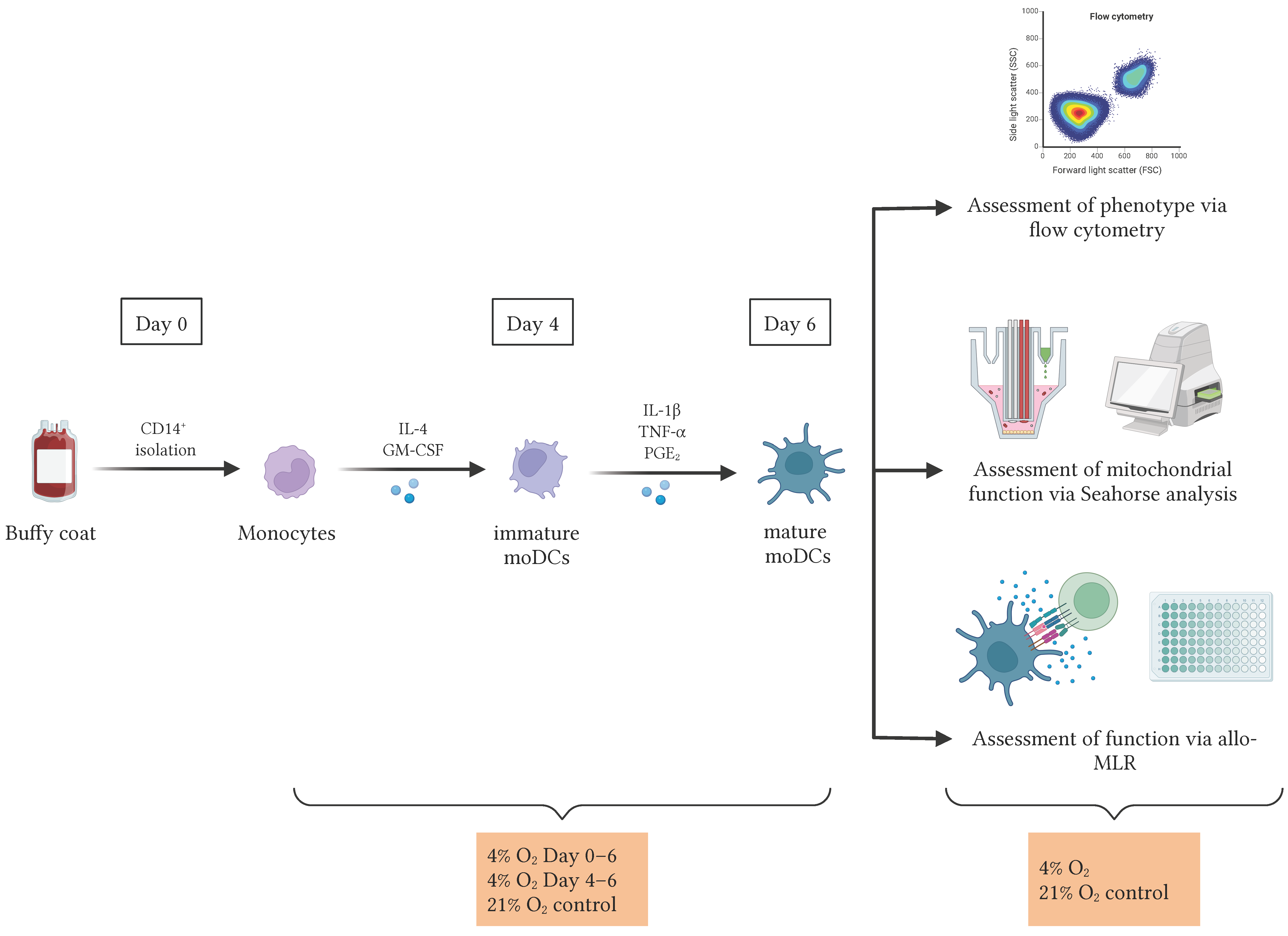
2.1. Cells and Culture Conditions

Buffy coats derived from healthy donor whole blood collections were purchased from the Red Cross Flanders (Mechelen, Belgium) and processed as described previously [50], with each biological replicate, denoted as n, representing an independent donor. Peripheral blood mononuclear cells were isolated using Ficoll-Paque density gradient centrifugation (GE Healthcare, Diegem, Belgium) followed by isolation of CD14+ monocytes using CD14-microbead-based immunomagnetic separation (CD14 reagent; Miltenyi Biotec, Leiden, The Netherlands). To generate DCs, purified CD14+ cells were resuspended in Iscove’s Modified Dulbecco’s Medium (IMDM; Thermo Fisher Scientific, Merelbeke, Belgium), supplemented with 2% human AB serum (hAB; Thermo Fisher Scientific), 250 IU/mL interleukin (IL)-4 (Miltenyi Biotec), and 200 IU/mL granulocyte-macrophage colony-stimulating factor (GM-CSF; Gentaur, Kampenhout, Belgium) at day 0 at a density of 1 × 10⁶ cells/mL and cultured for a period of six days in culture flasks (Greiner Bio-One, Vilvoorde, Belgium). On day 4, a combination of pro-inflammatory cytokines, consisting of 1000 IU/mL IL-1β (Miltenyi Biotec), 1000 IU/mL tumor necrosis factor (TNF)-α (Miltenyi Biotec), and 2.5 µg/mL prostaglandin (PG)E2 (Pfizer, Puurs, Belgium) was added to the culture for a 48-h maturation stimulus. Cells were incubated at 37 °C in a humidified atmosphere with 5% CO2 under either 21% or 4% O2, using a Whitley H45 HEPA Hypoxystation (Don Whitley Scientific, Bingley, UK) for the latter (Figure 1). On day 6, mature moDCs were harvested for additional experiments.
The fraction of CD14 cells, comprising peripheral blood lymphocytes (PBLs), was cryopreserved for further use at a concentration of 50 × 106 cells/mL in fetal bovine serum (FBS; Thermo Fisher Scientific) supplemented with 10% dimethyl sulfoxide (DMSO; Merck Life Science, Hoeilaart, Belgium) and 4% glucose (Laboratoires Sterop, Brussel, Belgium). The aliquots were frozen in Corning CoolCell LX cell-freezing containers (Corning, Lasne, Belgium) at −80 °C.

2.2. Immunophenotyping of DCs

The quality of the cells was evaluated by assessing viability using flow cytometry with propidium iodide staining (Thermo Fisher Scientific) after the harvest on day 6. Additionally, DC surface marker expression was assessed through phenotypical analysis. Following a 5-min blocking step (Human TruStain FcX; BioLegend, Amsterdam, The Netherlands), cells were stained with two distinct antibody panels, both of which included a viability dye (LIVE/DEAD fixable Near-Infrared Dead Cell Stain; Thermo Fisher Scientific) and a TruStain monocyte blocker (BioLegend) to avoid unspecific binding of DCs to tandem fluorophores. The first panel was designed to assess DC identity and maturation, and contained the following fluorochrome-conjugated monoclonal antibodies: anti-CD209-phycoerythrin-cyanine7 (PE-Cy7; BioLegend), anti-human leukocyte antigen (HLA)-DR-Kiravia-Blue-520 (BioLegend), anti-CD14-Brilliant-Violet-711 (BV711; BioLegend), anti-CD83-BV785 (BioLegend), anti-CD80-allophycocyanin (APC, BioLegend), and anti-CD86-BV605 (BioLegend). The second panel focused on the analysis of tolerance-associated surface markers and included: anti-CD209-PE (BioLegend), anti-CD11c-PerCP/Cyanine5.5 (BioLegend), anti-B7 homolog 3 (B7-H3)-PE/Dazzle 594 (BioLegend), anti-programmed death-ligand 1 (PD-L1)-BV711 (BioLegend), anti-CD40-BV650 (BioLegend), and anti-immunoglobulin-like transcript 4 (ILT4)-APC (BioLegend). In addition, staining for ILT3 (anti-ILT3-PE; BioLegend) was performed separately in combination with the same viability dye. The concentrations used for each antibody are listed in Supplementary Table S1. Non-specific background staining was assessed using isotype-matched control antibodies or fluorescence-minus-one (FMO) controls, as appropriate. Compensations were set up with compensation beads (UltraComp eBeads Plus Compensation Beads, Thermo Fisher Scientific) according to the manufacturer’s instructions. Per condition, 1 × 10⁵ cells were washed in sheath buffer (BD FACSFlow Sheath Fluid, BD Biosciences, Erembodegem, Belgium) supplemented with 0.1% bovine serum albumin (BSA; Merck Life Science) and 0.05% sodium azide (Merck Life Science), stained in a volume of 100 µL for 15 min at 4 °C, and washed once more before data acquisition on a NovoCyte Quanteon flow cytometer (Agilent Technologies, Diegem, Belgium), collecting 104 events per sample based on forward scatter (FSC) and side scatter (SSC) properties. All median fluorescence intensity (MFI) and percentage expression values were calculated from viable cells, as determined by exclusion of the L/D marker. Control populations consistently exhibited >99% CD209 and HLA-DR double-positivity, confirming their DC identity. Gating strategy and representative plots for all markers are provided in Supplementary Figure S1.
In addition, Mitotracker dyes were used for mitochondrial labeling of DCs: Mitotracker Green FM fluorescein isothiocyanate (FITC), Mitotracker Red FM CMXRos PE, and MitoSOX Red PE (Table S1). Mitochondrial labeling was performed in single stains in combination with a LIVE/DEAD fixable Near-Infrared Dead Cell Stain. All Mitotracker dyes are products of Thermo Fisher Scientific. Per condition, 1 × 10⁵ cells were washed in serum-free phosphate-buffered saline (PBS; Thermo Fisher Scientific), stained in a volume of 100 µL for 30 min at 37 °C, and washed once more before data acquisition as described above. MFI values were directly recorded from viable cells, gated as described in Supplementary Figure S1.

2.3. Lactate Measurement

To assess lactate production, 10 µL of cell culture supernatant was collected on days 2, 4, and 6 of the DC culture and promptly frozen at −20 °C until further analysis. Lactate concentrations were measured using a StatStrip Express Lactate device in combination with StatStrip Lactate test strips, according to the manufacturer’s instructions (Nova Biomedical, Boxtel, The Netherlands).

2.4. Seahorse Assay for Mitochondrial Function

Mitochondrial function was assessed on day 6 of DC culture using the Agilent Seahorse XFp Cell Mito Stress Test. Sensor cartridges were hydrated overnight with sterile water, starting the day before the assay. On the day of measurement, DCs were harvested and assessed for viability as described in Section 2.2. Hydrated sensor cartridges were then loaded with Seahorse XF Calibrant and incubated for at least 45 min at 37 °C in a non-CO2 incubator.
Meanwhile, DCs were resuspended in Seahorse XF RPMI assay medium (pH 7.4) supplemented with 10 mM glucose, 1 mM pyruvate, and 2 mM glutamine, and seeded into poly-D-lysine (PDL)-coated Seahorse XFp miniplates that had been pre-equilibrated to 37 °C in a non-CO2 incubator. Depending on the condition-specific viability, a total of 5 × 10⁴–2 × 10⁵ cells were plated per well. Plates were centrifuged briefly (300× g, 1 min) to ensure cell adherence.
Following drug loading of the sensor cartridges with 1.5 µM oligomycin (adenosine triphosphate (ATP) synthase inhibitor), 1.5 µM carbonyl cyanide-p-trifluoromethoxyphenylhydrazone (FCCP; uncoupling agent), and 0.5 µM rotenone/antimycin A (complex I/III inhibitors), the cartridges were placed into the Seahorse XF Mini Bioanalyzer for calibration. During this time, the cell plates were kept at 37 °C in a non-CO2 incubator. Once calibration was complete, the cell plate was inserted into the analyzer, and measurements of oxygen consumption rate (OCR) were taken before and after each compound injection. These were used to calculate key mitochondrial parameters reflecting different aspects of mitochondrial respiration, including basal respiration, ATP-linked respiration, proton leak, maximal respiration, spare respiratory capacity, non-mitochondrial respiration, and coupling efficiency. Each condition was tested in technical triplicates. Data were analyzed using the Agilent Seahorse Analytics software (version 1.0.0-749) and normalized to the number of total living DCs per well. All Seahorse-specific equipment and reagents were obtained from Agilent Technologies.

2.5. Allogeneic Mixed Lymphocyte Reaction (Allo-MLR)

To determine the T cell stimulatory capacity of DCs, allogeneic PBLs were co-cultured with DCs in a 10:1 ratio in 24-well plates (Greiner Bio-One), with the number of cells seeded based on live cell counts. PBLs stimulated with 1 µg/mL phytohaemagglutinin (PHA; Merck Life Science) served as a positive control. Furthermore, unstimulated allogeneic PBLs without DCs served as a negative control. Additionally, PBLs from the same donor as the DCs were included to control for potential effects of O2 deprivation on T cells in the absence of allogeneic stimulation (referred to as the PBL-only control in the results section). The co-cultures were performed in IMDM supplemented with 5% hAB and maintained for five days at 37 °C in a humidified atmosphere with 5% CO2 under either 21% or 4% O2 (Figure 1). After this incubation period, the supernatants were collected, frozen, and stored at −20 °C for subsequent testing. As a measure of the T cell stimulatory capacity of DCs, the levels of interferon (IFN)-γ were quantified using a commercially available enzyme-linked immunosorbent assay (ELISA; PeproTech, London, UK) according to the manufacturer’s instructions. Each sample was tested in triplicate. Plates were read by measuring absorbance at 405 nm using a Victor3 multilabel plate reader (PerkinElmer, Mechelen, Belgium) and interpolated to the concentration (pg/mL) using MS Office Excel.

2.6. Immunophenotyping of PBLs

To compare the stimulatory capacities of atmospheric and physioxic DCs to elicit allogeneic T cell responses not only based on IFN-γ secretion but also on the surface expression of co-stimulatory ligands and other markers, PBLs were stained with two different antibody panels on the last day of the allo-MLR following a 5-min blocking step (Human TruStain FcX). The first panel was used to distinguish different T cell subsets and contained the following fluorochrome-conjugated monoclonal antibodies: anti-CD45RA-Pacific-Blue, anti-CD8-BV510, anti-CD27-BV605, anti-CD57-BV785, anti-CD4-FITC, anti-CCR7-PE, anti-PD-1-PE-Cy7, anti-CD28-APC, anti-CD3-Spark-Red-718, and LIVE/DEAD fixable Near-Infrared Dead Cell Stain (Table S1). The second panel for T cell exhaustion markers contained the following fluorochrome-conjugated monoclonal antibodies: anti-PD-L2-BV421, anti-CD8-BV510, anti-TIGIT-BV605, anti-TIM-3-BV711, anti-CTLA-4-BV785, anti-CD4-FITC, anti-CD96-PE, anti-LAG-3-PE-Cy7, anti-PD-L1-APC, anti-CD3-Spark-Red-718, and LIVE/DEAD fixable Near-Infrared Dead Cell Stain (Table S1). All antibodies are products of BioLegend, except for CCR7 (Bio-Techne, Abingdon, UK). Samples were prepared, stained, and acquired as described in Section 2.2, with 5 × 10⁴ live cells recorded per condition. Gating strategy and representative plots for all markers are provided in Supplementary Figure S2.

2.7. Statistical Analysis

Flow cytometric data were analyzed using the FlowJo 10.10.0 software (FlowJo, TreeStar Inc., Ashland, OR, USA). Values are presented as the median, unless otherwise indicated, with the interquartile range (IQR) expressed as median (Q25–Q75). Statistical analyses were performed using GraphPad Prism 10.10.2, with the respective tests outlined in the figure legends. Each dataset was initially tested for normality using the Shapiro-Wilk test. For comparisons between two groups, a two-tailed paired t-test was used for normally distributed data, and the Wilcoxon matched-pairs signed rank test for non-parametric analysis. When comparing three groups, data that did not pass the normality test were analyzed using the Kruskal-Wallis test with Dunn’s multiple comparisons due to the inability of the Friedman test for paired data to accommodate datasets with missing values. Normally distributed data were analyzed using a one-way ANOVA (non-paired) with Šídák’s multiple comparisons test. For analyses involving multiple levels of grouping, a mixed-effects model with the Geisser-Greenhouse correction and Šídák’s multiple comparisons test was employed. Volcano plots for the PBL phenotyping data were generated to visualize fold changes against the Benjamini-Hochberg-adjusted p-values derived from two-tailed paired t-tests. A fold change > 1.0 was classified as upregulation, while a fold change < 1.0 was classified as downregulation. A p-value < 0.05, corresponding to a −log (adjusted p-value) of 1.30, was considered statistically significant.

3. Results

3.1. Physiological O2 Levels During the Generation Period Resulted in a Decreased Transformation of Monocytes into DCs

To understand how O2 influences DC generation and phenotype under physiological conditions, we examined the impact of 4% O2, referred to hereafter as physioxia, on the generation of mature moDCs in comparison to 21% O2, which will be referred to as atmospheric O2 levels or control conditions. We employed two physioxic culture conditions: physioxia during the entire 6-day culture period (4% O2 days 0–6) and physioxia only during the maturation phase between days 4 and 6 (4% O2 D4–6), the latter serving as a maturation-phase-specific comparator to contextualize the effects of continuous low-O2 exposure. These were compared to continuous culture under 21% O2 (Figure 1). Our findings demonstrated a significant reduction in the monocyte-to-mDC transformation rate (control: 34.3%, IQR: 26.9–42.4%; 4% O2 D4–6: 33.4%, IQR: 31.4–40.4%; 4% O2 D0–6: 24.0%, IQR: 9.3–25.9%) under continuous physioxia compared to atmospheric O2 levels (p < 0.0001). This was calculated as the number of viable cells on day 6 excluding CD209CD14+ monocytes, relative to the number of viable monocytes seeded on day 0. CD14 downregulation and CD209 upregulation was used to distinguish successfully transformed moDCs from undifferentiated monocytes, in line with established phenotypic criteria. The same effect was observed regarding cell viability (p < 0.0001) (Figure 2A). In contrast, no significant differences were observed when comparing control conditions to physioxic maturation conditions. Furthermore, the percentage of monocytes (CD209CD14+) was significantly increased upon a full physioxic culture period compared to control conditions (p < 0.0001), whereas no such increase was observed when the cells were exposed to 4% O2 only during the maturation phase (Figure 2B). Taken together, these results suggest that physioxic culture conditions negatively affect DC generation from monocytes when applied throughout the entire culture period.

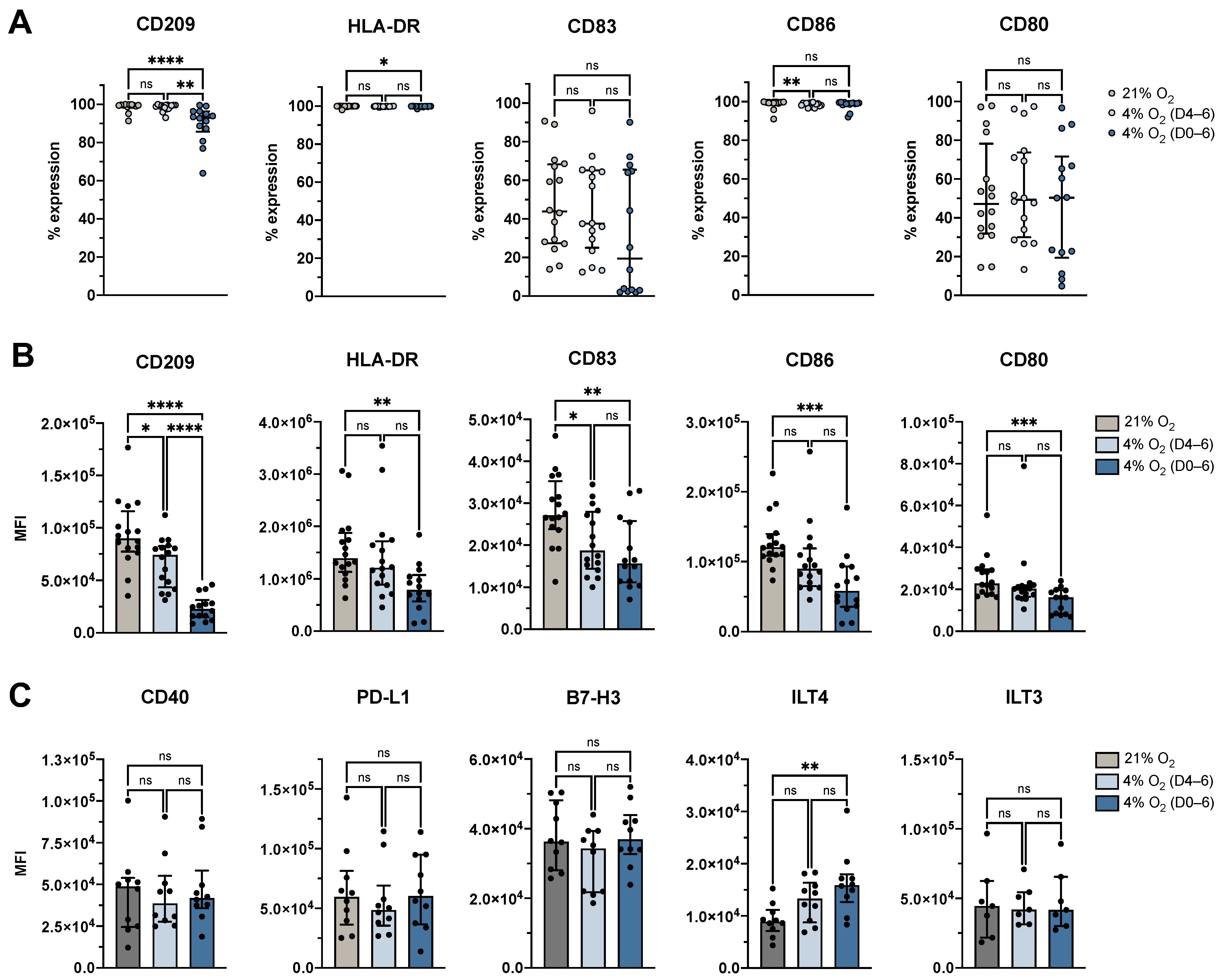
3.2. Physiological O2 Levels During the Generation Period Resulted in Decreased Surface Expression of Identity and Maturation Markers, and Increased Expression of Tolerance-Associated Marker ILT4

The generation of mDCs from monocytes is typically marked by downregulation of CD14 and upregulation of DC identity and co-stimulatory markers, including CD209, HLA-DR, CD80, CD86, and CD83. To assess the phenotypic status of the cells generated under different O2 conditions, we analyzed surface marker expression profiles indicative of this immunophenotypic progression. Our study revealed that the proportion of moDCs expressing CD209 and HLA-DR remained high under both physioxic conditions (Figure 3A), indicating preserved cellular integrity (CD209: control: 99.5%, IQR: 99.1–99.8%; 4% O2 D4–6: 99.2%, IQR: 97.9–99.6%; 4% O2 D0–6: 93.1%, IQR: 85.7–96.3%; HLA-DR: control: 100.0%, IQR: 99.9–100.0%; 4% O2 D4–6: 99.9%, IQR: 99.7–100.0%; 4% O2 D0–6: 99.9%, IQR: 99.7–99.9%). However, significant reductions in the expression levels of CD209 (p < 0.0001) and HLA-DR (p = 0.003), as measured by MFI, were observed under continuous physioxic conditions compared to atmospheric O2 levels, with significant differences between physioxic maturation and controls for CD209 (p = 0.0104) but not for HLA-DR (Figure 3B).
When examining the maturation markers, we found that the proportion of moDCs expressing CD83 and CD80 was unaffected by physioxia. However, their expression levels were significantly reduced under continuous physioxia compared to atmospheric O2 levels (CD83: p = 0.0031, CD80: p = 0.0009). Additionally, a significant reduction in CD83 expression was observed under physioxic maturation (p = 0.0344). CD86 was uniformly expressed across all conditions (control: 99.6%, IQR: 99.3–99.8%; 4% O2 D4–6: 98.9%, IQR: 97.8–99.3%; 4% O2 D0–6: 99.1%, IQR: 98.5–99.4%; Figure 3A), although expression levels were significantly downregulated under continuous physioxia (p = 0.0002), similar to the other maturation markers (Figure 3B).
To further investigate the effect of O2 levels on tolerance-associated markers, we assessed the expression of CD40, PD-L1, B7-H3, ILT3, and ILT4 on moDCs cultured under atmospheric O2, physioxic maturation (4% O2 D4–6), or continuous physioxia (4% O2 D0–6). Among the markers tested, only ILT4 showed a statistically significant difference, with expression levels significantly upregulated under continuous physioxia compared to atmospheric O2 (p = 0.0051; Figure 3C). No significant differences were observed for CD40, PD-L1, B7-H3, or ILT3 (Figure 3C).

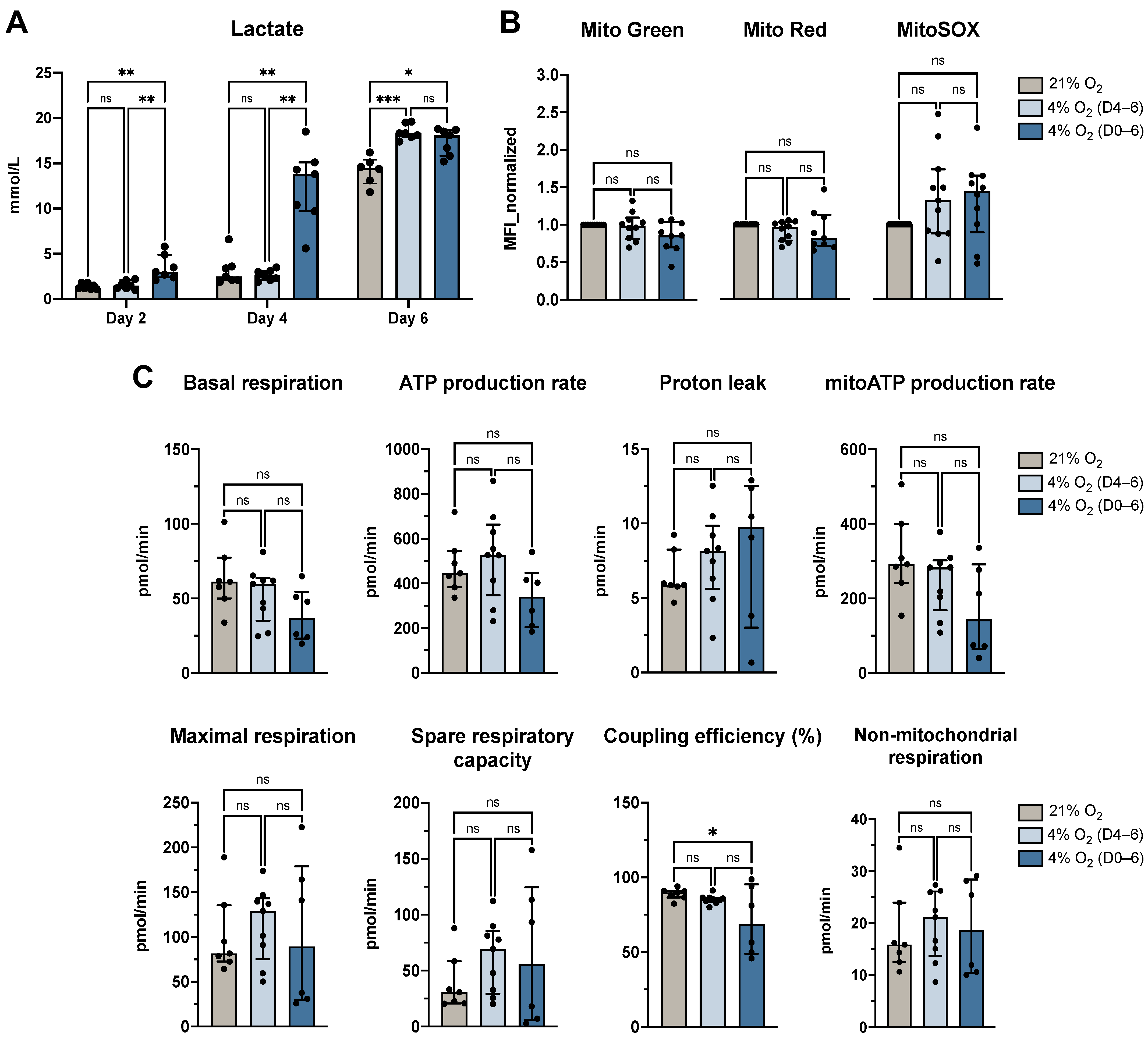
3.3. Physiological O2 Levels During the Generation Period Increased Lactate Secretion but Largely Preserved Mitochondrial Function

To assess the impact of physiological O2 levels on DC metabolism, lactate secretion and mitochondrial parameters were evaluated under atmospheric (21% O2) and physioxic (4% O2) conditions. While glycolytic activity, as indicated by lactate production, was elevated under physioxia, mitochondrial function remained mostly unaffected (Figure 4).
Lactate measurements revealed a significant increase in secretion by moDCs cultured under continuous physioxia compared to atmospheric O2 levels on day 2 (p = 0.0063) and day 4 (p = 0.0023; Figure 4A). By day 6, lactate levels were significantly higher in both physioxic culture conditions compared to control cells (D4–6: p = 0.0002, D0–6: p = 0.0163).
Mitochondrial characteristics were assessed using fluorescent dyes (Mitotracker Green, Red, and MitoSOX Red) and Seahorse metabolic flux analysis. Fluorescence-based measurements indicated no significant differences in mitochondrial mass, membrane potential, or mitochondrial superoxide levels between atmospheric and physioxic conditions (Figure 4B), suggesting preserved mitochondrial integrity under both conditions. In parallel, the Seahorse Mito Stress Test evaluated parameters including basal respiration, ATP production rate, proton leak, maximal respiration, spare respiratory capacity, and non-mitochondrial respiration. Most parameters showed no significant differences between physioxic and normoxic culture conditions, aligning with the fluorescence-based findings. Notably, a modest but statistically significant decrease in coupling efficiency was observed under continuous physioxia compared to atmospheric O2 (p = 0.0489; Figure 4C), indicating reduced efficiency in ATP production relative to total O2 consumption. Although there was an observed trend towards lower basal respiration and mitochondrial ATP production rate under physioxic conditions, these differences were not statistically significant. Together, these results suggest that while physioxia may slightly reduce respiratory efficiency, mitochondrial health remains uncompromised, and any shifts in ATP production are minimal.

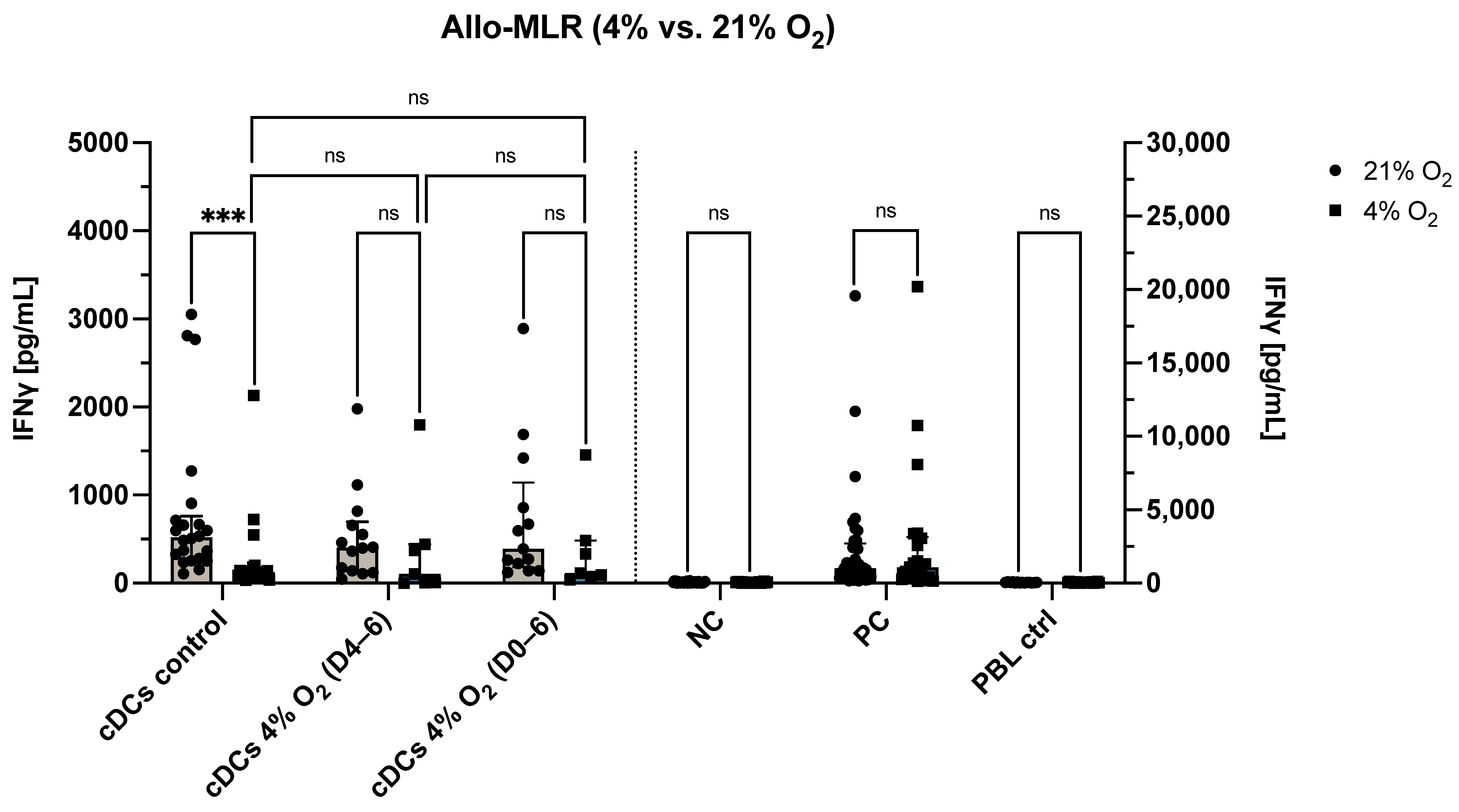
3.4. O2 Levels During the Allo-MLR Modulate T Cell Responses

To investigate the influence of physiological O2 levels on DC function in terms of their capacity to stimulate allogeneic T cell responses, we conducted mixed lymphocyte reactions (allo-MLR) under both atmospheric (21% O2) and physioxic (4% O2) conditions, evaluating IFN-γ secretion and phenotypic changes of peripheral blood lymphocytes (PBLs) after five days of co-culture (Figure 5 and Figure 6).
As a first step, the effect of O2 levels during the allo-MLR itself was assessed. DCs cultured under atmospheric O2 were co-cultured with allogeneic PBLs in either a 21% or 4% O2 allo-MLR. Under physioxic allo-MLR conditions, these control DCs induced significantly lower levels of IFN-γ secretion compared to atmospheric conditions (p < 0.0001), suggesting that low O2 leads to T cell hyporesponsiveness in this context (Figure 5). In contrast, no significant differences in IFN-γ secretion were observed when comparing 21% and 4% O2 allo-MLRs for any of the following: negative control, positive control, PBL-only control, moDCs matured under physioxia (4% O2 D4–6), or moDCs cultured under continuous physioxia (4% O2 D0–6). These findings suggest that the reduced IFN-γ levels seen under physioxia may be specific to the combination of atmospheric moDCs and low O2 during T cell activation, rather than a general suppression of PBL responsiveness under physioxic conditions.
Next, the impact of O2 exposure during DC generation was examined under physioxic allo-MLR conditions. Specifically, IFN-γ secretion was compared across all three DC culture conditions—atmospheric (21% O2), physioxic maturation (4% O2 D4–6), and continuous physioxia (4% O2 D0–6)—in a 4% O2 allo-MLR. In this setting, no significant differences were observed between the groups (Figure 5), suggesting that preconditioning DCs under physioxia does not markedly alter their IFN-γ-inducing capacity in allogeneic settings.
To gain further insights into how O2 tension affects T cell phenotypes beyond IFN-γ secretion, flow cytometric profiling was performed on PBLs after five days of allo-MLR. For this, cells were analyzed for memory and exhaustion markers on CD4⁺ and CD8⁺ subsets.
As a baseline comparison, PBLs co-cultured with control DCs (cultured entirely under atmospheric conditions) were analyzed after allo-MLR under either 21% or 4% O2. In the CD4⁺ population, significant reductions in MFI were observed for CCR7, TIGIT, and CD45RA under physioxia (Figure 6A). In the CD8⁺ population, MFI of CD57, TIGIT, CD45RA, PD-L2, TIM-3, LAG-3, CTLA-4, CD96, PD-L1, and CCR7 were all significantly decreased under physioxic conditions (Figure 6B). In terms of percentage marker expression, CD4⁺ cells showed significantly reduced frequencies of CD27⁺CD28⁻, PD-L1⁺, TIGIT⁺, CTLA-4⁺, TIM-3⁺, LAG-3⁺, and CD45RA⁻CCR7⁺ cells (central memory T cells), while CD8⁺ cells showed a decrease in LAG-3⁺, PD-L1⁺, and CCR7⁺ cells, and a corresponding increase in CD27⁺ cells (Figure 6C,D). The control conditions (NC, PC, PBL-only) exhibited minor changes in this setup, limited to MFI shifts in a few markers without significant alterations in the percentage of marker-expressing cells (Figure S3), in line with the IFN-γ secretion data.
Next, the influence of O2 exposure during DC maturation was assessed. When comparing DCs exposed to physioxia during maturation (4% O2 D4–6) to control DCs (21% O2) in a 4% O2 allo-MLR, no significant differences were observed in the expression of any markers on either CD4+ or CD8+ T cells.
Finally, the effects of continuous physioxia (4% O2 D0–6) were assessed relative to atmospheric DCs in a 4% O2 allo-MLR. For CD4⁺ cells, only CD45RA showed a significant reduction in MFI upon physioxic preconditioning (p = 0.034), while no significant differences were observed in CD8+ cells. Marker expression frequencies remained unchanged in both T cell subsets.
These results reveal broad downregulation of key exhaustion and memory markers across both CD4⁺ and CD8⁺ T cell subsets along with reduced IFN-γ secretion in response to physioxic allo-MLRs. On the other hand, preconditioning DCs under physioxia alone had limited impact on T cell phenotypes and IFN-γ secretion levels.

4. Discussion

Understanding how O2 levels influence both DC phenotype and their downstream T cell interactions is crucial for designing more effective immunotherapies and for studying tolerance mechanisms in transplantation and autoimmunity. While most in vitro studies rely on atmospheric O2 levels (21%), these are far from the physiological conditions that DCs encounter in vivo. Previous research has predominantly focused on exploring DC biology in pathological hypoxia (<2% O2), relevant for cancer, bacterial infections, arthritis, wound healing, and inflammatory lesions [49]. However, little is known about how physiological O2 levels affect immune homeostasis. DCs continuously sample antigens in the steady state but avoid full maturation unless activated by PAMPs or DAMPs. The shift from tolerogenic to immunogenic DCs occurs upon encountering inflammatory signals, ensuring that immune responses are mounted only in the presence of a true threat. Understanding the tolerogenic role of DCs under homeostatic conditions has significant implications. Dysregulation of tolerogenic DCs can lead to loss of tolerance and autoimmunity. In addition, tumor microenvironments often exploit tolerogenic mechanisms to suppress DC function and evade immune responses. The main goal of this study was to understand how homeostatic immune responses are generated by mimicking the in vivo conditions of physiological O2 levels (4% O2) and comparing them to hyperoxia (21% O2) typically present in in vitro cell culture. We demonstrated that culturing ex vivo-generated moDCs under physiological O2 levels leads to reduced monocyte-to-mDC transformation rate and programs the resulting cells toward a tolerogenic phenotype, characterized by increased secretion of lactate, a semi-mature surface marker profile, and altered T cell priming. This suggests a critical role for O2 in maintaining DC homeostasis, with potential implications for both steady-state and pathological conditions. The observed effects were most pronounced when physioxia was applied throughout the entire culture period, while exposure during the maturation phase only had subtler effects. In vivo, DCs can differentiate from monocytes in inflammatory conditions often characterized by hypoxia [29]. Our experimental setting using physioxia is intended to mimic a homeostatic or steady-state environment. The observed reduction in monocyte-to-mDC transformation rate under physioxia suggests that the metabolic demands in this condition may limit monocyte commitment to the DC lineage in the absence of inflammatory cues, aligning with the physiological scenario under non-inflammatory conditions. The observed reduction in viability under physioxia should be carefully interpreted alongside functional outcomes of the viable DC population. Notably, our assessment on day 6 via PI exclusion reflects the total cell pool before distinguishing between differentiated moDCs and remaining monocytes based on extensive phenotypic profiling. Thus, physioxia affected both overall cell survival and monocyte-to-DC transformation, the latter confirmed by DC lineage markers. This raises the question of why viability decreased under conditions that were intended to mimic physiological O2 levels. Monocytes were isolated from human blood under standard atmospheric conditions and were then immediately cultured under physioxia. This shift does not mirror the gradual or tissue-specific O2 exposure that cells would experience in vivo and highlights the difficulty of mimicking physiological conditions in vitro.
Interestingly, while the percentage of cells expressing key surface molecules remained high (CD209, HLA-DR, CD86) or unchanged (CD80, CD83) under physioxia, their expression levels (as indicated by MFI) were significantly reduced. This discrepancy suggests that although DC identity was preserved, the density of the marker expression on the cell surface declined, which indicates a collective impairment of DC-mediated T cell activation by weakening of co-stimulation (CD86, CD80, CD83), antigen presentation (HLA-DR), and antigen uptake (CD209). Of note, phenotypic profiling was performed on the total viable cell population, capturing not only differentiated DCs but also cells that did not fully commit to the DC lineage. As such, the observed shifts reflect both phenotypic modulation of DCs and impaired monocyte-to-mDC transformation efficiency under physioxia.
The next step for homeostatic DCs in vivo is their potential interaction with T cells under steady-state conditions, where tissue O2 levels are considerably lower than atmospheric. To investigate their functionality in this context, we assessed the capacity of physioxia-conditioned DCs to activate allogeneic T cells under reduced O2 conditions. We observed no differences in the ability of physioxia-conditioned or atmospheric DCs to induce IFN-γ secretion or to alter the phenotype of PBLs under these conditions. This may suggest that physiological O2 levels alone do not significantly contribute to shaping immune homeostasis—though the complexity of in vivo interactions cannot be fully recapitulated in this system. However, when we introduced a shift from 21% to 4% O2 during the T cell activation phase, IFN-γ secretion was reduced in control moDC co-cultures. In parallel, we observed a broad downregulation of markers associated with co-inhibitory signaling, memory differentiation, and effector maturation across both CD4+ and CD8+ subsets. This suggests that low O2 levels at the site of T cell priming may impair effector cytokine production and modulate the quality of T cell responses. Importantly, no difference in IFN-γ secretion was observed between 21% and 4% O2 in the absence of DCs, highlighting that the O2 levels did not have a direct effect on T cell function itself but rather attenuated the immunostimulatory capacity of ex vivo-generated DCs during their interaction with T cells. Rather than indicating a classical exhausted phenotype, which would typically involve upregulation of checkpoint molecules and co-inhibitory receptors, the observed profile suggests a hyporesponsive or metabolically restrained T cell state. This is characterized by insufficient activation to induce either effector function or regulatory feedback mechanisms. Markers involved in terminal differentiation (CD57) and lymphoid homing and memory formation (CCR7, CD45RA) were likewise reduced, along with several immune checkpoints and regulatory receptors (PD-1, PD-L1, PD-L2, TIGIT, TIM-3, CTLA-4, LAG-3, CD96). Importantly, our results highlight that immune responses cannot be defined by the absence or presence of individual markers alone but must be instead understood as an integrated functional and phenotypic state. It is important to note that this experimental setup, in which DCs are generated under atmospheric O2 and T cells are activated under physioxic conditions, does not reflect a physiological scenario, as both cell types would naturally co-localize within the same O2 microenvironment in vivo. These insights have implications for immunotherapeutic applications, where ex vivo-generated DCs are administered into low-O2 tissue environments. Atmospheric O2 levels are standard during in vitro quality assessments of such cell products, which may not reflect their functional potential in vivo. Our results indicate that physioxia shapes the DC-T cell crosstalk—both in terms of inflammatory cytokine output and T cell phenotypic profile—in a way that does not support robust effector responses. This highlights the need to consider O2 as a critical environmental regulator of immune activation thresholds.
To better understand the mechanisms underlying the induction of a tolerogenic state under physioxia, we examined cellular metabolism as a potential driver of this phenotype. Immunometabolism is increasingly recognized as a key regulator of immune cell fate, not just in terms of energy production, but also through the integration of environmental signals into functional outcomes. As outlined in our recent review, shifts in cellular respiration stemming from the surrounding O2 environment can influence whether DCs adopt an immunogenic or tolerogenic profile [49]. This affects features such as cytokine secretion, antigen presentation, and T cell priming. On the metabolic level, we observed that physioxia enhanced lactate production, consistent with a shift toward aerobic glycolysis—also known as the Warburg effect [51]—a hallmark associated with tolerogenic DCs. This metabolite has been shown to influence immune regulation by promoting tolerogenic DC functions, such as the induction of regulatory T cells and suppression of pro-inflammatory cytokines [52,53,54,55,56]. Despite elevated lactate levels, mitochondrial integrity and respiration remained largely intact in physioxia-conditioned DCs. This suggests a metabolic rewiring rather than a collapse of mitochondrial function and may act in concert with the downregulation of co-stimulatory surface molecules, altered cytokine secretion pattern, and changes in antigen uptake or processing capacity to maintain a non-immunogenic profile under steady-state conditions.
Our findings contrast with a study by Futalan et al. (2011), which reported that generation under 5% O2 did not influence yield, phenotype, or T cell activation of moDCs [57]. Several factors may explain this discrepancy. First, the O2 level in our study was slightly lower (4% O2), which may have crossed a functional threshold affecting cellular metabolism and protein expression. Second, the cell isolation methods differed: while we used CD14+ immunomagnetic separation to enrich for monocytes, Futalan et al. used adherence-based selection, which may yield a more heterogeneous starting population and influence subsequent differentiation. Furthermore, differences in maturation stimuli, although similar in composition, could have further contributed to the divergence in results. Lastly, our study maintained tighter control of O2 levels throughout the entire experimental setup and also compared different O2 levels during the allo-MLR, whereas Futalan et al. performed their co-cultures under atmospheric O2 conditions. These subtle yet important distinctions highlight the sensitivity of DC generation to microenvironmental parameters and underscore the need for standardization when comparing studies using physiological O2 levels.
In summary, DCs are central regulators of immune homeostasis, balancing tolerance and immunity through their interaction with T cells and their response to microenvironmental cues. The tolerogenic phenotype of DCs under homeostatic conditions is influenced by cytokines, metabolic signals, and O2 levels. Our findings point toward a potential link between O2-sensitive metabolic reprogramming and the establishment of a semi-mature, tolerogenic DC state by limiting robust inflammatory activation, advancing our understanding of immune regulation in health and disease. Physiological O2 levels may act as a stabilizing factor in steady-state conditions, whereas the hyperoxic conditions typically used in in vitro studies may drive DCs toward an inflammatory state, potentially misrepresenting their behavior in vivo. Recognizing atmospheric O2 as a state that may drive immunogenicity offers a valuable perspective for future studies seeking to bridge the gap between in vitro models and in vivo immune regulation. Future research should ensure that both DCs and their interacting immune cells are studied under physiologically relevant O2 conditions to more accurately reflect in vivo scenarios.

Supplementary Materials

The following supporting information can be downloaded at: https://www.mdpi.com/article/10.3390/cells14100736/s1, Table S1: Antibody concentrations; Figure S1: Representative gating strategy for analysis of DC phenotype; Figure S2: Representative gating strategy for analysis of PBL phenotype; Figure S3: Phenotypic analysis of CD4⁺ and CD8⁺ T cell subsets on day 5 of the allo-MLR under the three control conditions.

Author Contributions

Conceptualization, A.P. and N.C.; methodology, A.P., M.V., M.V.D., A.D., S.P., H.D.R. and W.F.A.M.; validation, A.P.; formal analysis, A.P.; investigation, A.P., M.V., M.V.D., A.D., S.P., H.D.R. and W.F.A.M.; data curation, A.P.; writing—original draft preparation, A.P.; writing—review and editing, A.P., Z.N.B. and N.C.; visualization, A.P.; supervision, N.C.; project administration, A.P. and N.C.; funding acquisition, Z.N.B. and N.C. All authors have read and agreed to the published version of the manuscript.

Funding

This project has received funding from the European Union’s Horizon 2020 research and innovation programme under the Marie Skłodowska-Curie grant agreement No. 860003 (A.P.), and the Special Research Fund (BOF) of the University of Antwerp (DOCPRO1, PS ID 49061; A.P.). Additional support was provided by the Industrial Research Fund (IOF) of the University of Antwerp (SEP, PS ID 53192; N.C.) and the FWO Hercules infrastructure fund (I010524N). M.V. holds a doctoral scholarship from the BOF (DOCPRO4, PS ID 5114). M.V.D. holds a doctoral fellowship funded by the Flemish Institute for Technological Research (VITO). A.D. receives funding from the Research Foundation—Flanders (FWO—Vlaanderen) under grant agreement No. T002523N. S.P. is supported through the University Hospital Antwerp (UZA) budget 21HMT17, allocated to cancer research. The hypoxia workstation was funded by the FWO—Vlaanderen (G040120N). The Seahorse Bioanalyzer was funded by a BOF infrastructure grant from the University of Antwerp (FN 543600001).

Institutional Review Board Statement

The use of buffy coats for scientific research was approved by the Ethische Commissie Onderzoek UZ/KU Leuven on 20 March 2019, with approval code S62549.

Informed Consent Statement

Anonymized buffy coats were provided by the Blood Transfusion Center of the Red Cross; written informed consent for research use was obtained from all subjects. The study followed the Tenets of Helsinki.

Data Availability Statement

The data presented in this study are available on request from the corresponding author.

Acknowledgments

The authors would like to thank FACSUA, the Flow Cytometry and Cell Sorting Core Facility of the University of Antwerp, for providing access to their equipment and technical support. We also acknowledge the Center for Oncological Research (CORE) for access to the hypoxia workstation, and the Gamete Research Centre (GRC) of the University of Antwerp for use of their Seahorse Bioanalyzer.

Conflicts of Interest

The authors declare no conflict of interest.

Abbreviations

allo-MLRAllogeneic mixed lymphocyte reaction
ATPAdenosine triphosphate
BSABovine serum albumin
CCR7C-C chemokine receptor 7
CDCluster of differentiation
moDCsMonocyte-derived dendritic cells
CO2Carbon dioxide
CTLA-4Cytotoxic T-lymphocyte-associated protein 4
DAMPsDamage-associated molecular patterns
DCsDendritic cells
DMSODimethyl sulfoxide
FBSFetal bovine serum
FCCPCarbonyl cyanide-p-trifluoromethoxyphenylhydrazone
FMOFluorescence-minus-one
FSCForward scatter
GM-CSFGranulocyte-macrophage colony-stimulating factor
hABHuman AB serum
HLA-DRHuman leukocyte antigen-DR
iDCsImmature dendritic cells
ILInterleukin
ILTImmunoglobulin-like transcript
IFN-γInterferon-gamma
IQRInterquartile range
LAG-3Lymphocyte activation gene 3
mDCsMature dendritic cells
MFIMean fluorescence intensity
moDCsMonocyte-derived dendritic cells
nsNot significant
O2Oxygen
OCROxygen consumption rate
PAMPsPathogen-associated molecular patterns
PBLsPeripheral blood lymphocytes
PBSPhosphate-buffered saline
PD-1Programmed death-1
PD-L1/L2Programmed death-ligand 1/2
PDLPoly-D-lysine
PGE2Prostaglandin E2
PIPropidium iodide
SSCSide scatter
ThT helper cell subset
TIGITT cell immunoreceptor with immunoglobulin and ITIM domain
TNFTumor necrosis factor
TregsRegulatory T cells

References

  1. Caux, C.; Massacrier, C.; Vanbervliet, B.; Dubois, B.; Durand, I.; Cella, M.; Lanzavecchia, A.; Banchereau, J. CD34+ hematopoietic progenitors from human cord blood differentiate along two independent dendritic cell pathways in response to granulocyte-macrophage colony-stimulating factor plus tumor necrosis factor alpha: II. Functional analysis. Blood 1997, 90, 1458–1470. [Google Scholar] [CrossRef] [PubMed]
  2. Cravens, P.D.; Melkus, M.W.; Padgett-Thomas, A.; Islas-Ohlmayer, M.; Del P. Martin, M.; Garcia, J.V. Development and activation of human dendritic cells in vivo in a xenograft model of human hematopoiesis. Stem Cells 2005, 23, 264–278. [Google Scholar] [CrossRef]
  3. Patente, T.A.; Pinho, M.P.; Oliveira, A.A.; Evangelista, G.C.M.; Bergami-Santos, P.C.; Barbuto, J.A.M. Human dendritic cells: Their heterogeneity and clinical application potential in cancer immunotherapy. Front. Immunol. 2018, 9, 3176. [Google Scholar] [CrossRef]
  4. Wilson, N.S.; El-Sukkari, D.; Villadangos, J.A. Dendritic cells constitutively present self antigens in their immature state in vivo and regulate antigen presentation by controlling the rates of MHC class II synthesis and endocytosis. Blood 2004, 103, 2187–2195. [Google Scholar] [CrossRef] [PubMed]
  5. Steinman, R.M.; Swanson, J. The endocytic activity of dendritic cells. J. Exp. Med. 1995, 182, 283–288. [Google Scholar] [CrossRef]
  6. Skelton, L.; Cooper, M.; Murphy, M.; Platt, A. Human immature monocyte-derived dendritic cells express the G protein-coupled receptor GPR105 (KIAA0001, P2Y14) and increase intracellular calcium in response to its agonist, uridine diphosphoglucose. J. Immunol. 2003, 171, 1941–1949. [Google Scholar] [CrossRef] [PubMed]
  7. Trombetta, E.S.; Mellman, I. Cell biology of antigen processing in vitro and in vivo. Annu. Rev. Immunol. 2005, 23, 975–1028. [Google Scholar] [CrossRef] [PubMed]
  8. Gouwy, M.; Struyf, S.; Leutenez, L.; Pörtner, N.; Sozzani, S.; Van Damme, J. Chemokines and other GPCR ligands synergize in receptor-mediated migration of monocyte-derived immature and mature dendritic cells. Immunobiology 2014, 219, 218–229. [Google Scholar] [CrossRef]
  9. Hemmi, H.; Akira, S. TLR signalling and the function of dendritic cells. Chem. Immunol. Allergy 2005, 86, 120–135. [Google Scholar] [CrossRef]
  10. Tang, D.; Kang, R.; Coyne, C.B.; Zeh, H.J.; Lotze, M.T. PAMPs and DAMPs: Signal 0s that spur autophagy and immunity. Immunol. Rev. 2012, 249, 158–175. [Google Scholar] [CrossRef]
  11. Cerboni, S.; Gentili, M.; Manel, N. Diversity of pathogen sensors in dendritic cells. Adv. Immunol. 2013, 120, 211–237. [Google Scholar] [CrossRef]
  12. López, C.B.; García-Sastre, A.; Williams, B.R.G.; Moran, T.M. Type I interferon induction pathway, but not released interferon, participates in the maturation of dendritic cells induced by negative-strand RNA viruses. J. Infect. Dis. 2003, 187, 1126–1136. [Google Scholar] [CrossRef] [PubMed]
  13. McIlroy, D.; Tanguy-Royer, S.; Le Meur, N.; Guisle, I.; Royer, P.-J.; Léger, J.; Meflah, K.; Grégoire, M. Profiling dendritic cell maturation with dedicated microarrays. J. Leukoc. Biol. 2005, 78, 794–803. [Google Scholar] [CrossRef] [PubMed]
  14. Dalod, M.; Chelbi, R.; Malissen, B.; Lawrence, T. Dendritic cell maturation: Functional specialization through signaling specificity and transcriptional programming. EMBO J. 2014, 33, 1104–1116. [Google Scholar] [CrossRef] [PubMed]
  15. Allenspach, E.J.; Lemos, M.P.; Porrett, P.M.; Turka, L.A.; Laufer, T.M. Migratory and lymphoid-resident dendritic cells cooperate to efficiently prime naive CD4 T cells. Immunity 2008, 29, 795–806. [Google Scholar] [CrossRef]
  16. Bourque, J.; Hawiger, D. Immunomodulatory Bonds of the Partnership between Dendritic Cells and T Cells. Crit. Rev. Immunol. 2018, 38, 379–401. [Google Scholar] [CrossRef]
  17. Steinman, R.M.; Hawiger, D.; Liu, K.; Bonifaz, L.; Bonnyay, D.; Mahnke, K.; Iyoda, T.; Ravetch, J.; Dhodapkar, M.; Inaba, K.; et al. Dendritic cell function in vivo during the steady state: A role in peripheral tolerance. Ann. N. Y. Acad. Sci. 2003, 987, 15–25. [Google Scholar] [CrossRef]
  18. Cools, N.; Ponsaerts, P.; Van Tendeloo, V.F.I.; Berneman, Z.N. Balancing between immunity and tolerance: An interplay between dendritic cells, regulatory T cells, and effector T cells. J. Leukoc. Biol. 2007, 82, 1365–1374. [Google Scholar] [CrossRef]
  19. Novak, N.; Bieber, T. 2. Dendritic cells as regulators of immunity and tolerance. J. Allergy Clin. Immunol. 2008, 121, S370–S374. [Google Scholar] [CrossRef]
  20. Nam, J.-H.; Lee, J.-H.; Choi, S.-Y.; Jung, N.-C.; Song, J.-Y.; Seo, H.-G.; Lim, D.-S. Functional ambivalence of dendritic cells: Tolerogenicity and immunogenicity. Int. J. Mol. Sci. 2021, 22, 4430. [Google Scholar] [CrossRef]
  21. Lozano, E.; Dominguez-Villar, M.; Kuchroo, V.; Hafler, D.A. The TIGIT/CD226 axis regulates human T cell function. J. Immunol. 2012, 188, 3869–3875. [Google Scholar] [CrossRef] [PubMed]
  22. Machicote, A.; Belén, S.; Baz, P.; Billordo, L.A.; Fainboim, L. Human CD8+HLA-DR+ Regulatory T Cells, Similarly to Classical CD4+Foxp3+ Cells, Suppress Immune Responses via PD-1/PD-L1 Axis. Front. Immunol. 2018, 9, 2788. [Google Scholar] [CrossRef]
  23. Dilek, N.; Poirier, N.; Hulin, P.; Coulon, F.; Mary, C.; Ville, S.; Vie, H.; Clémenceau, B.; Blancho, G.; Vanhove, B. Targeting CD28, CTLA-4 and PD-L1 costimulation differentially controls immune synapses and function of human regulatory and conventional T-cells. PLoS ONE 2013, 8, e83139. [Google Scholar] [CrossRef]
  24. Tekguc, M.; Wing, J.B.; Osaki, M.; Long, J.; Sakaguchi, S. Treg-expressed CTLA-4 depletes CD80/CD86 by trogocytosis, releasing free PD-L1 on antigen-presenting cells. Proc. Natl. Acad. Sci. USA 2021, 118, e2023739118. [Google Scholar] [CrossRef]
  25. Fuse, S.; Obar, J.J.; Bellfy, S.; Leung, E.K.; Zhang, W.; Usherwood, E.J. CD80 and CD86 control antiviral CD8+ T-cell function and immune surveillance of murine gammaherpesvirus 68. J. Virol. 2006, 80, 9159–9170. [Google Scholar] [CrossRef] [PubMed]
  26. Koorella, C.; Nair, J.R.; Murray, M.E.; Carlson, L.M.; Watkins, S.K.; Lee, K.P. Novel regulation of CD80/CD86-induced phosphatidylinositol 3-kinase signaling by NOTCH1 protein in interleukin-6 and indoleamine 2,3-dioxygenase production by dendritic cells. J. Biol. Chem. 2014, 289, 7747–7762. [Google Scholar] [CrossRef]
  27. Sakaguchi, S.; Yamaguchi, T.; Nomura, T.; Ono, M. Regulatory T cells and immune tolerance. Cell 2008, 133, 775–787. [Google Scholar] [CrossRef] [PubMed]
  28. Iwasaki, A.; Medzhitov, R. Control of adaptive immunity by the innate immune system. Nat. Immunol. 2015, 16, 343–353. [Google Scholar] [CrossRef]
  29. Collin, M.; Bigley, V. Human dendritic cell subsets: An update. Immunology 2018, 154, 3–20. [Google Scholar] [CrossRef]
  30. Buck, M.D.; Sowell, R.T.; Kaech, S.M.; Pearce, E.L. Metabolic instruction of immunity. Cell 2017, 169, 570–586. [Google Scholar] [CrossRef]
  31. Kim, J. Regulation of immune cell functions by metabolic reprogramming. J. Immunol. Res. 2018, 2018, 8605471. [Google Scholar] [CrossRef] [PubMed]
  32. Kedia-Mehta, N.; Finlay, D.K. Competition for nutrients and its role in controlling immune responses. Nat. Commun. 2019, 10, 2123. [Google Scholar] [CrossRef]
  33. Gottfried, E.; Kunz-Schughart, L.A.; Ebner, S.; Mueller-Klieser, W.; Hoves, S.; Andreesen, R.; Mackensen, A.; Kreutz, M. Tumor-derived lactic acid modulates dendritic cell activation and antigen expression. Blood 2006, 107, 2013–2021. [Google Scholar] [CrossRef] [PubMed]
  34. Okajima, F. Regulation of inflammation by extracellular acidification and proton-sensing GPCRs. Cell. Signal. 2013, 25, 2263–2271. [Google Scholar] [CrossRef]
  35. Cao, L.; Shi, X.; Chang, H.; Zhang, Q.; He, Y. pH-Dependent recognition of apoptotic and necrotic cells by the human dendritic cell receptor DEC205. Proc. Natl. Acad. Sci. USA 2015, 112, 7237–7242. [Google Scholar] [CrossRef]
  36. Hosonuma, M.; Yoshimura, K. Association between pH regulation of the tumor microenvironment and immunological state. Front. Oncol. 2023, 13, 1175563. [Google Scholar] [CrossRef]
  37. Steurer, J.; Hoffmann, U.; Dür, P.; Russi, E.; Vetter, W. Changes in arterial and transcutaneous oxygen and carbon dioxide tensions during and after voluntary hyperventilation. Respiration 1997, 64, 200–205. [Google Scholar] [CrossRef] [PubMed]
  38. Caldwell, C.C.; Kojima, H.; Lukashev, D.; Armstrong, J.; Farber, M.; Apasov, S.G.; Sitkovsky, M.V. Differential effects of physiologically relevant hypoxic conditions on T lymphocyte development and effector functions. J. Immunol. 2001, 167, 6140–6149. [Google Scholar] [CrossRef]
  39. Chow, D.C.; Wenning, L.A.; Miller, W.M.; Papoutsakis, E.T. Modeling pO2 distributions in the bone marrow hematopoietic compartment. II. Modified Kroghian models. Biophys. J. 2001, 81, 685–696. [Google Scholar] [CrossRef]
  40. Carreau, A.; El Hafny-Rahbi, B.; Matejuk, A.; Grillon, C.; Kieda, C. Why is the partial oxygen pressure of human tissues a crucial parameter? Small molecules and hypoxia. J. Cell. Mol. Med. 2011, 15, 1239–1253. [Google Scholar] [CrossRef]
  41. Keeley, T.P.; Mann, G.E. Defining physiological normoxia for improved translation of cell physiology to animal models and humans. Physiol. Rev. 2019, 99, 161–234. [Google Scholar] [CrossRef] [PubMed]
  42. Chapple, S.J.; Keeley, T.P.; Mastronicola, D.; Arno, M.; Vizcay-Barrena, G.; Fleck, R.; Siow, R.C.M.; Mann, G.E. Bach1 differentially regulates distinct Nrf2-dependent genes in human venous and coronary artery endothelial cells adapted to physiological oxygen levels. Free Radic. Biol. Med. 2016, 92, 152–162. [Google Scholar] [CrossRef] [PubMed]
  43. Ferguson, D.C.J.; Smerdon, G.R.; Harries, L.W.; Dodd, N.J.F.; Murphy, M.P.; Curnow, A.; Winyard, P.G. Altered cellular redox homeostasis and redox responses under standard oxygen cell culture conditions versus physioxia. Free Radic. Biol. Med. 2018, 126, 322–333. [Google Scholar] [CrossRef]
  44. Stuart, J.A.; Fonseca, J.; Moradi, F.; Cunningham, C.; Seliman, B.; Worsfold, C.R.; Dolan, S.; Abando, J.; Maddalena, L.A. How Supraphysiological Oxygen Levels in Standard Cell Culture Affect Oxygen-Consuming Reactions. Oxid. Med. Cell. Longev. 2018, 2018, 8238459. [Google Scholar] [CrossRef] [PubMed]
  45. Timpano, S.; Guild, B.D.; Specker, E.J.; Melanson, G.; Medeiros, P.J.; Sproul, S.L.J.; Uniacke, J. Physioxic human cell culture improves viability, metabolism, and mitochondrial morphology while reducing DNA damage. FASEB J. 2019, 33, 5716–5728. [Google Scholar] [CrossRef] [PubMed]
  46. Shin, D.-Y.; Huang, X.; Gil, C.-H.; Aljoufi, A.; Ropa, J.; Broxmeyer, H.E. Physioxia enhances T-cell development ex vivo from human hematopoietic stem and progenitor cells. Stem Cells 2020, 38, 1454–1466. [Google Scholar] [CrossRef]
  47. Sies, H.; Belousov, V.V.; Chandel, N.S.; Davies, M.J.; Jones, D.P.; Mann, G.E.; Murphy, M.P.; Yamamoto, M.; Winterbourn, C. Defining roles of specific reactive oxygen species (ROS) in cell biology and physiology. Nat. Rev. Mol. Cell Biol. 2022, 23, 499–515. [Google Scholar] [CrossRef]
  48. Igarashi, K.J.; Kucinski, I.; Chan, Y.Y.; Tan, T.-K.; Khoo, H.M.; Kealy, D.; Bhadury, J.; Hsu, I.; Ho, P.Y.; Niizuma, K.; et al. Physioxia improves the selectivity of hematopoietic stem cell expansion cultures. Blood Adv. 2023, 7, 3366–3377. [Google Scholar] [CrossRef]
  49. Peter, A.; Berneman, Z.N.; Cools, N. Cellular respiration in dendritic cells: Exploring oxygen-dependent pathways for potential therapeutic interventions. Free Radic. Biol. Med. 2025, 227, 536–556. [Google Scholar] [CrossRef]
  50. Van Delen, M.; Janssens, I.; Dams, A.; Roosens, L.; Ogunjimi, B.; Berneman, Z.N.; Derdelinckx, J.; Cools, N. Tolerogenic Dendritic Cells Induce Apoptosis-Independent T Cell Hyporesponsiveness of SARS-CoV-2-Specific T Cells in an Antigen-Specific Manner. Int. J. Mol. Sci. 2022, 23, 15201. [Google Scholar] [CrossRef]
  51. Vaupel, P.; Multhoff, G. Revisiting the Warburg effect: Historical dogma versus current understanding. J. Physiol. 2021, 599, 1745–1757. [Google Scholar] [CrossRef] [PubMed]
  52. Caronni, N.; Simoncello, F.; Stafetta, F.; Guarnaccia, C.; Ruiz-Moreno, J.S.; Opitz, B.; Galli, T.; Proux-Gillardeaux, V.; Benvenuti, F. Downregulation of membrane trafficking proteins and lactate conditioning determine loss of dendritic cell function in lung cancer. Cancer Res. 2018, 78, 1685–1699. [Google Scholar] [CrossRef] [PubMed]
  53. Marin, E.; Bouchet-Delbos, L.; Renoult, O.; Louvet, C.; Nerriere-Daguin, V.; Managh, A.J.; Even, A.; Giraud, M.; Vu Manh, T.P.; Aguesse, A.; et al. Human Tolerogenic Dendritic Cells Regulate Immune Responses through Lactate Synthesis. Cell Metab. 2019, 30, 1075–1090.e8. [Google Scholar] [CrossRef]
  54. Raychaudhuri, D.; Bhattacharya, R.; Sinha, B.P.; Liu, C.S.C.; Ghosh, A.R.; Rahaman, O.; Bandopadhyay, P.; Sarif, J.; D’Rozario, R.; Paul, S.; et al. Lactate Induces Pro-tumor Reprogramming in Intratumoral Plasmacytoid Dendritic Cells. Front. Immunol. 2019, 10, 1878. [Google Scholar] [CrossRef] [PubMed]
  55. Kanemaru, H.; Mizukami, Y.; Kaneko, A.; Tagawa, H.; Kimura, T.; Kuriyama, H.; Sawamura, S.; Kajihara, I.; Makino, K.; Miyashita, A.; et al. A mechanism of cooling hot tumors: Lactate attenuates inflammation in dendritic cells. iScience 2021, 24, 103067. [Google Scholar] [CrossRef]
  56. Sanmarco, L.M.; Rone, J.M.; Polonio, C.M.; Fernandez Lahore, G.; Giovannoni, F.; Ferrara, K.; Gutierrez-Vazquez, C.; Li, N.; Sokolovska, A.; Plasencia, A.; et al. Lactate limits CNS autoimmunity by stabilizing HIF-1α in dendritic cells. Nature 2023, 620, 881–889. [Google Scholar] [CrossRef]
  57. Futalan, D.; Huang, C.-T.; Schmidt-Wolf, I.G.H.; Larsson, M.; Messmer, D. Effect of oxygen levels on the physiology of dendritic cells: Implications for adoptive cell therapy. Mol. Med. 2011, 17, 910–916. [Google Scholar] [CrossRef]
Figure 1. Workflow for the assessment of physioxic culture conditions in moDCs. DCs were differentiated from monocytes in the presence of IL-4 and GM-CSF and matured with the pro-inflammatory cytokines IL-1β, TNF-α, and PGE2. Cultures were divided into three experimental groups: (1) continuous culture under 21% O2 (atmospheric O2 levels), (2) continuous culture under 4% O2 (days 0–6), and (3) 4% O2 during the maturation phase only (days 4–6). The latter was included to provide a reference for late-stage low-O2 exposure and to support interpretation of the sustained low-O2 exposure group. On day 6, cell phenotype was analyzed by flow cytometry, mitochondrial function was assessed using a Seahorse assay, and immunostimulatory capacity was evaluated in an allo-MLR performed at either 4% or 21% O2. This setup was designed to better mimic the physiological O2 levels found in human tissues. Abbreviations used: moDCs, monocyte-derived dendritic cells; IL, interleukin; GM-CSF, granulocyte-macrophage colony-stimulating factor; TNF-α, tumor necrosis factor-alpha; PGE2, prostaglandin E2; O2, oxygen; allo-MLR, allogeneic mixed lymphocyte reaction.
Cells 14 00736 g001
Figure 2. Generation of moDCs under 4% O2 resulted in decreased viability and increased percentage of monocytes. (A) Viability of the total cell population on day 6 determined via PI exclusion, prior to DC phenotyping (n = 20). (B) Monocyte population represented as the percentage of CD209CD14+ cells (n = 16). All cells were cultured under atmospheric O2 levels, 4% O2 during the entire generation phase (D0–6), or 4% O2 only during the maturation phase (D4–6). Data are shown as median with interquartile range. Statistical analyses were performed using an ordinary one-way ANOVA (A) or the Kruskal-Wallis test with Dunn’s post-hoc analysis (B). Statistical significance is denoted by *, where *** represents p < 0.001 and **** represents p < 0.0001. Abbreviations used: moDCs, monocyte-derived dendritic cells; O2, oxygen; D0–6, days 0 to 6; D4–6, days 4 to 6; PI, propidium iodide; ANOVA, analysis of variance; ns, not significant.
Cells 14 00736 g002
Figure 3. DC generation under 4% O2 resulted in a decreased surface expression of identity and maturation markers, and an increase of tolerance marker ILT4. (A) The percentage of expression for the markers CD209, HLA-DR, CD83, CD86, and CD80 (n = 16). (B) The expression levels of the markers CD209, HLA-DR, CD83, CD86, and CD80 presented as MFI of the positive population (n = 16). (C) The expression levels of the markers CD40, PD-L1, B7-H3, ILT4, and ILT3 presented as MFI of the positive population (n = 7 for ILT3, n = 10 for all other markers). Cells depicted in all graphs were cultured under atmospheric O2 levels, 4% O2 (D4–6), or 4% O2 (D0–6). Data are shown as median with interquartile range. Statistical analyses were performed using an ordinary one-way ANOVA ((A): CD83, CD80; (B): CD209, CD83; (C): CD40, B7-H3, ILT4, ILT3) or the Kruskal-Wallis test with Dunn’s post-hoc analysis ((A): CD209, HLA-DR, CD86; (B): HLA-DR, CD86, CD80; (C): PD-L1). Statistical significance is denoted by *, where * represents p < 0.05, ** represents p < 0.01, *** represents p < 0.001, and **** represents p < 0.0001. Abbreviations used: O2, oxygen; D0–6, days 0 to 6; D4–6, days 4 to 6; MFI, mean fluorescence intensity; ANOVA, analysis of variance; CD, cluster of differentiation; HLA-DR, human leukocyte antigen—DR isotype; PD-L1, programmed death-ligand 1; B7-H3, B7 homolog 3; ILT, immunoglobulin-like transcript; ns, not significant.
Cells 14 00736 g003
Figure 4. DC generation under 4% O2 increased lactate production but did not alter mitochondrial function. (A) Lactate production by moDCs (n = 7). (B) Mitochondrial stainings as indicated by Mitotracker Green (mitochondrial mass), Mitotracker Red (membrane potential), and MitoSOX Red (superoxide production) presented as normalized MFI values (n = 11). (C) Seahorse Mito Stress Test analysis of moDCs (n = 9). Mitochondrial parameters shown include basal respiration (OCR under baseline conditions), ATP production rate (portion of OCR used for ATP synthesis, inferred from the drop after oligomycin addition), proton leak (non-ATP-linked OCR following ATP synthase inhibition), mitoATP production rate (absolute ATP output via oxidative phosphorylation), maximal respiration (peak OCR following FCCP addition), spare respiratory capacity (difference between maximal and basal respiration), coupling efficiency (proportion of basal OCR coupled to ATP production), and non-mitochondrial respiration (residual OCR after inhibition of mitochondrial complexes I and III by rotenone and antimycin A). All cells were cultured under atmospheric O2 levels, 4% O2 (D4–6), or 4% O2 (D0–6). Data are shown as median with interquartile range. Statistical analyses were performed using a mixed-effects model with the Geisser-Greenhouse correction (A), an ordinary one-way ANOVA ((B): Mito Green, Mito Red; (C): basal respiration, ATP production rate, proton leak, mitoATP production rate, maximal respiration, coupling efficiency), or the Kruskal-Wallis test with Dunn’s post-hoc analysis ((B): MitoSOX; (C): spare respiratory capacity, non-mitochondrial respiration). Statistical significance is denoted by *, where * represents p < 0.05, ** represents p < 0.01, and *** represents p < 0.001. Abbreviations used: O2, oxygen; D0–6, days 0 to 6; D4–6, days 4 to 6; moDCs, monocyte-derived dendritic cells; MFI, mean fluorescence intensity; ANOVA, analysis of variance; OCR, oxygen consumption rate; ATP, adenosine triphosphate; FCCP, carbonyl cyanide-p-trifluoromethoxyphenylhydrazone; ns, not significant.
Cells 14 00736 g004
Figure 5. O2 levels during the allo-MLR modulate T cell responses. IFN-γ secretion (pg/mL) by PBLs stimulated with control moDCs (cultured at 21% O2), moDCs exposed to physioxia during maturation (4% O2 D4–6), or moDCs cultured under continuous physioxia (4% O2 D0–6), as well as NC, PC (PHA-stimulated PBLs), and PBL-only control (autologous PBLs), in 5-day allo-MLRs conducted at either 21% or 4% O2. Data are shown as median with interquartile range. Comparisons for the three moDC conditions were performed using the Wilcoxon matched-pairs signed rank test. Controls were analyzed using a mixed-effects model with Geisser-Greenhouse correction and Šídák’s multiple comparisons test. Statistical significance is denoted by *, where *** represents p < 0.001. Abbreviations used: O2, oxygen; moDCs, monocyte-derived dendritic cells; D0–6, days 0 to 6; D4–6, days 4 to 6; allo-MLR, allogeneic mixed lymphocyte reaction; IFN-γ, interferon-gamma; PBLs, peripheral blood lymphocytes; NC, negative control; PC, positive control; PHA, phytohemagglutinin; ns, not significant.
Cells 14 00736 g005
Figure 6. O2 levels during the allo-MLR modulate T cell phenotypes. Phenotypic analysis of CD4⁺ and CD8⁺ T cell subsets was performed on day 5 of the allo-MLR to evaluate the impact of O2 tension during T cell activation. Control moDCs cultured at atmospheric O2 levels were used to stimulate PBLs in either 4% or 21% O2 allo-MLRs. Significant changes reflect the effect of physioxia (4% O2) compared to atmospheric conditions (21% O2). Values represent either MFI (A,B) or percentage of expression (C,D) in CD4⁺ or CD8⁺ T cell subsets. Data are presented as −log (Benjamini-Hochberg-adjusted p-value; derived from two-tailed paired t-tests) versus log (fold change). Statistically significant differences (p < 0.05) are highlighted in red (downregulation) or green (upregulation) (n = 7). Abbreviations used: O2, oxygen; moDCs, monocyte-derived dendritic cells; D0–6, days 0 to 6; D4–6, days 4 to 6; allo-MLR, allogeneic mixed lymphocyte reaction; PBLs, peripheral blood lymphocytes; CD, cluster of differentiation; MFI, mean fluorescence intensity; TIGIT, T cell immunoreceptor with Ig and ITIM domains; CCR7, C-C chemokine receptor type 7; LAG-3, lymphocyte activation gene 3; PD-L1/2, programmed death-ligand 1/2; CTLA-4, cytotoxic T-lymphocyte-associated protein 4; TIM-3, T cell immunoglobulin and mucin-domain containing-3; PD-1, programmed cell death protein 1.
Cells 14 00736 g006
Disclaimer/Publisher’s Note: The statements, opinions and data contained in all publications are solely those of the individual author(s) and contributor(s) and not of MDPI and/or the editor(s). MDPI and/or the editor(s) disclaim responsibility for any injury to people or property resulting from any ideas, methods, instructions or products referred to in the content.

