Review

PANoptosis as a Two-Edged Sword in Colorectal Cancer: A Pathogenic Mechanism and Therapeutic Opportunity

Immunology Division, Department of Internal Medicine and Hematology, Semmelweis University, 1088 Budapest, Hungary
*
Authors to whom correspondence should be addressed.
Cells 2025, 14(10), 730; https://doi.org/10.3390/cells14100730
Submission received: 6 April 2025 / Revised: 14 May 2025 / Accepted: 16 May 2025 / Published: 16 May 2025
(This article belongs to the Collection Molecular and Cellular Mechanisms of Cancers: Colorectal Cancer)

Abstract

The examination of PANoptosis in colorectal cancer is particularly important, as many tumor cells can evade apoptotic cell death while continuing to proliferate through inflammatory mediators and creating an immunosuppressive environment. The PANoptosome functions as a regulatory complex that unites proteins governing pyroptotic, apoptotic, and necroptotic pathways, rather than allowing distinct death pathways to compete. The expression and functional status of key molecules within the PANoptosome, such as ZBP1, RIPK1, RIPK3, CASP8, and ASC, may influence tumor viability and immune detection. The tumorigenic impact of PANoptosis is complex and predominantly manifests through chronic inflammation, immune response modulation, and changes in the tumor microenvironment. PANoptosis also aids in the defense against colon cancer by directly eradicating tumor cells and modifying the cellular environment. The expression profile of PANoptosis components may possess prognostic and predictive significance. The therapeutic ramifications of PANoptosis in colorectal cancer are now being investigated through many avenues. It provides an opportunity to develop targeted therapeutic techniques. In contrast, it may also be pertinent in conjunction with immunotherapy, as PANoptosis signifies an immunogenic type of cell death and may consequently enhance the anti-tumor immune response. A thorough comprehension of how these parameters influence PANoptosis is crucial for practical implementation.

1. Introduction

Colorectal carcinoma (CRC) ranks among the foremost causes of cancer mortality globally, with inflammatory mechanisms, the tumor microenvironment, and the dysregulation of apoptosis significantly contributing to its growth and progression [1]. Multiple treatment options exist for CRC, encompassing chemotherapy and immunotherapy [2]. Regrettably, clinical outcomes continue to be suboptimal due to tumor heterogeneity, hereditary characteristics, and many risk factors [3,4]. About 25% of CRC patients are discovered at an advanced stage, and nearly 50% of those diagnosed at an early stage subsequently develop metastatic disease [5,6]. The 5-year survival rate for individuals with minimal metastatic lesions is 40% after surgical resection and systemic therapy, while for those with advanced metastatic CRC, the survival rate significantly decreases (approx. 20%) [7,8]. Given the low survival rate of CRC patients, there is a need to establish a precise classification of the disease that might enhance the prediction of patient outcomes and responses to immunotherapy and chemotherapy, thereby forming tailored treatment regimens and increasing prognoses.
Programmed cell death (PCD) is conventionally categorized into five primary forms: apoptosis, autophagy, necroptosis, pyroptosis, and ferroptosis [9]. A recent study has revealed a novel, integrated form of cell death that amalgamates the pathways of pyroptosis, apoptosis, and necroptosis, termed PANoptosis [10]. The PANoptosome, a multifunctional protein complex, facilitates PANoptosis by simultaneously activating caspases, gasdermin D, and necroptotic kinases [11].
The functions of apoptosis, pyroptosis, and necroptosis in oncology research are areas of vigorous discussion. The investigation of PANoptosis in CRC is notably significant since numerous tumor cells can circumvent apoptotic cell death while sustaining proliferation via inflammatory mediators and fostering an immunosuppressive milieu [12]. The expression and functional status of critical molecules comprising the PANoptosome, including ZBP1, RIPK1, RIPK3, CASP8, and ASC, can have a bidirectional impact on tumor survival, immune recognition, or tumor cell death [13]. The existence or nonexistence of these compounds may hold prognostic significance for treatment, particularly in immunotherapeutic strategies.
Considering the dual function of PANoptosis in either inhibiting or facilitating colorectal cancer, comprehensive knowledge of its mechanisms could reveal essential elements of tumor biology and pave the way for innovative diagnostic and therapeutic approaches in an era increasingly characterized by precision oncology. Targeted manipulation of PANoptosis, such as triggering immunogenic cell death in tumor cells, may provide novel therapeutic avenues for colorectal cancer treatment.
This review aims to elucidate the pathogenic and therapeutic roles of PANoptosis in CRC, with an emphasis on preclinical and clinical relevance.

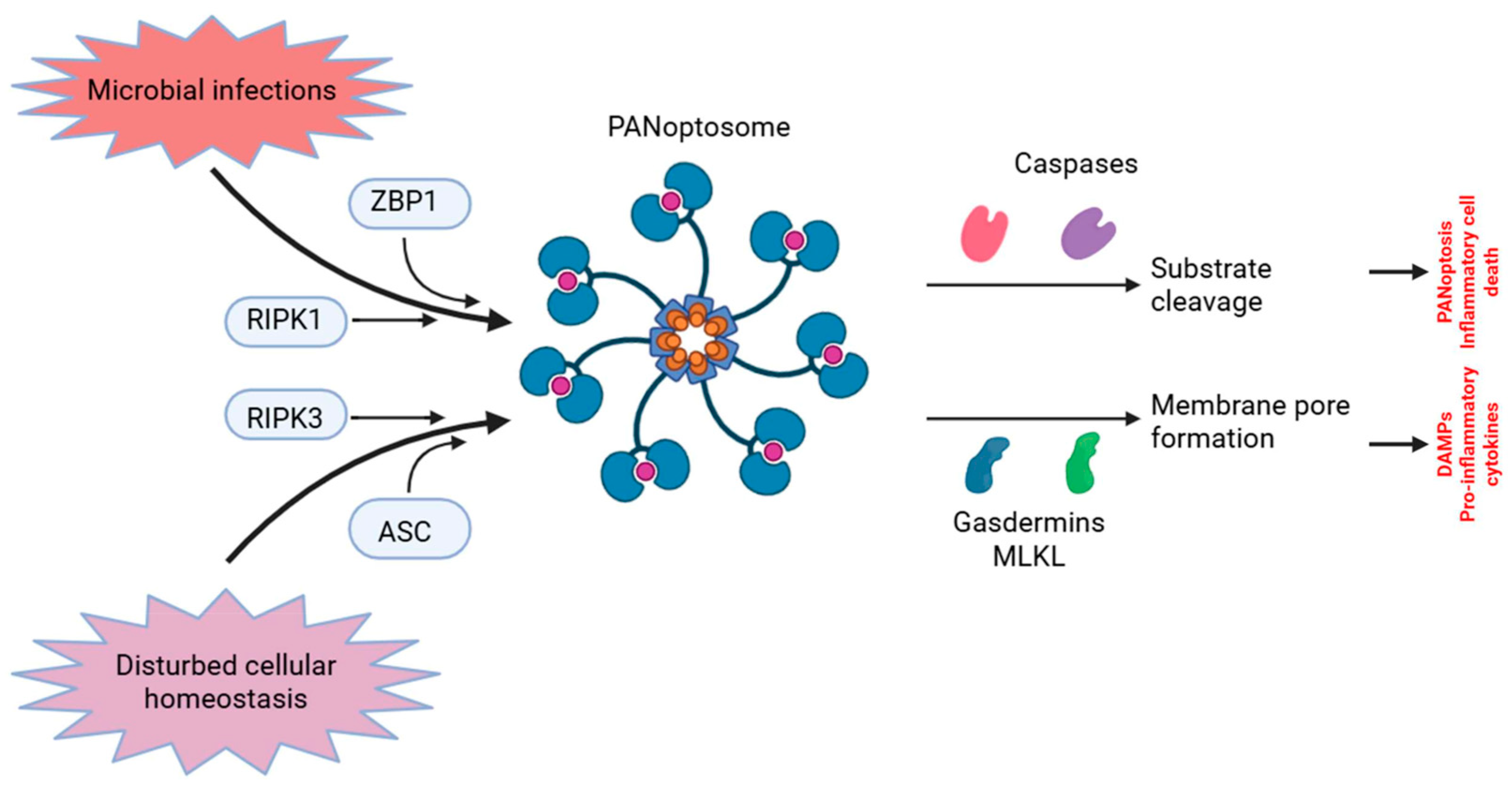
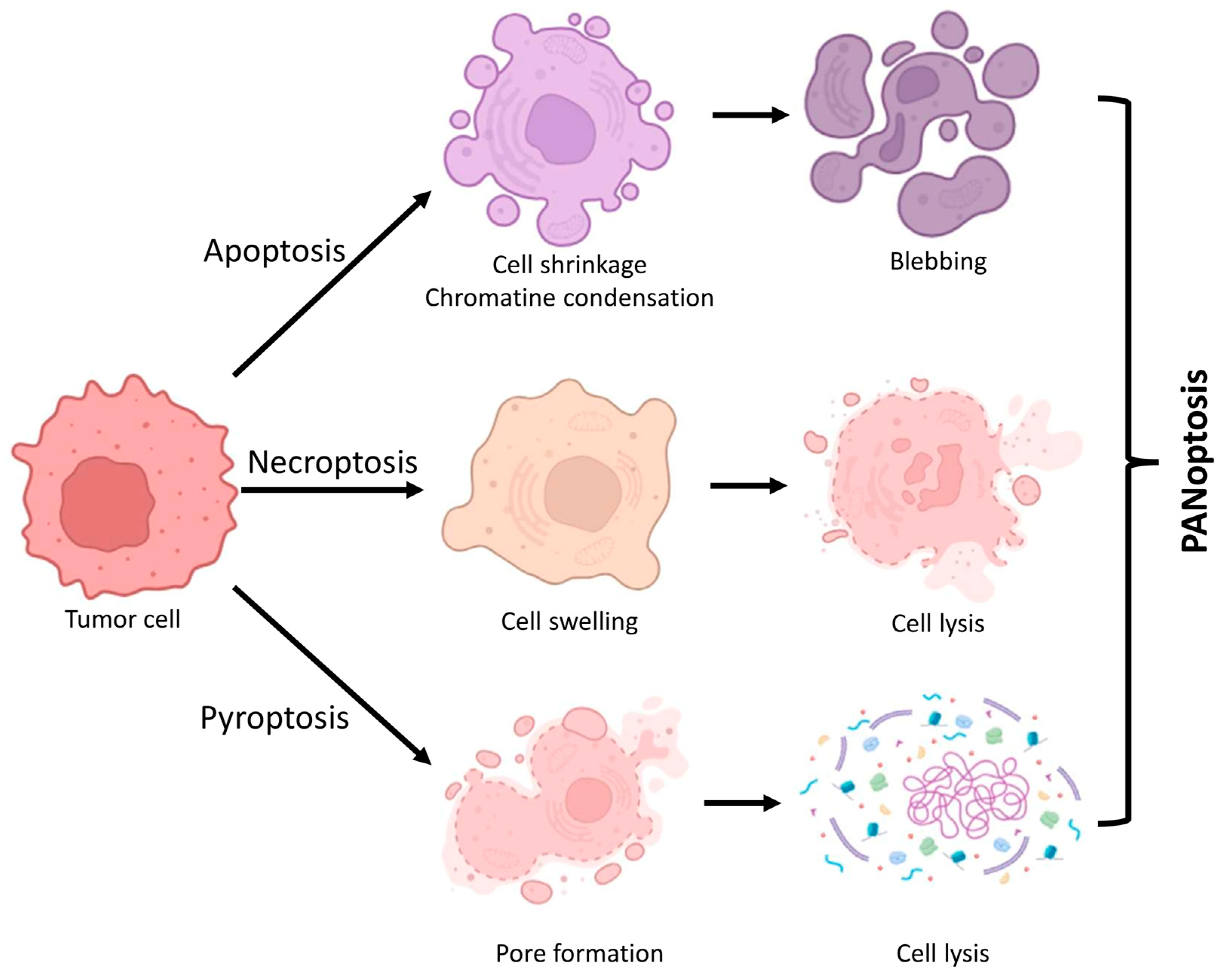
2. Fundamental Characteristics and Processes of PANoptosis

PANoptosis is a multifaceted, inflammatory PCD that encompasses aspects of apoptosis, necroptosis, and pyroptosis; however, it cannot be solely classified as any one of these forms [14,15]. The mechanism is governed by a multi-protein complex known as the PANoptosome, which comprises essential components from all three cell death pathways.
It is crucial to differentiate PANoptosis from other forms of cell death. Apoptosis is a non-inflammatory, so-called immunologically silent form of cell death characterized by the activation of caspases (e.g., CASP3, -8, and -9) and is associated with phenomena such as cell disintegration (blebbing) [16]. RIPK1-RIPK3-MLKL is involved in necroptosis, an inflammatory form of cell death that leads to the cell membrane becoming permeable [17]. Pyroptosis is a form of inflammatory cell death characterized by the activation of inflammatory caspases (e.g., CASP1, -4, -5, and -11) and results in the creation of gasdermin pores [14]. The PANoptosome orchestrates PANoptosis, which integrates the mechanisms of apoptosis, necroptosis, and pyroptosis, typically in response to a singular stimulus. This type of cellular demise is particularly significant in infections, autoimmune disorders, and tumors, as it initiates a complicated immunological response [18]. The activation of PANoptosis involves a coordinated action of multiple molecular players, as detailed below.
The PANoptosome is a supramolecular signaling complex that unites essential components from pyroptotic, apoptotic, and necroptotic pathways [19]. Pathogen-associated molecular patterns (PAMPs), damage-associated molecular patterns (DAMPs), or cytokine signaling (e.g., tumor necrosis factor/TNF/, IFNs, and IL-1) initiate the assembly [20].
The assembly of the PANoptosome is mostly reliant on scaffold proteins, including Z-DNA binding protein 1 (ZBP1), apoptosis-associated speck-like protein containing a CARD (ASC), and receptor-interacting serine/threonine-protein kinase 3 (RIPK3) [21,22]. Upon assembly, the PANoptosome binds and activates numerous cell death effectors, such as caspases (CASP1, -3, and -8), gasdermins (GSDMD, GSDME), and mixed lineage kinase domain-like pseudokinase (MLKL), leading to a coordinated execution of cell death pathways [23].
Caspases, RIPK3, gasdermins, and ZBP1 are essential activating molecules [24]. When activated by the inflammasome, caspase-1 cleaves the pro-inflammatory cytokines IL-1β and IL-18, and GSDMD, which leads to the formation of pyroptotic pores [24]. CASP3 and CASP7 are quintessential executors of apoptosis, cleaving substrates such as poly(ADP-ribose) polymerase (PARP) [25]. CASP8 operates at the intersection of apoptosis and necroptosis. It can either cause apoptosis through death receptors or inhibit necroptosis by blocking RIPK3/MLKL activation [25]. RIPK3 is a crucial regulator of necroptosis that interacts with RIPK1 and MLKL [26]. In the absence or inhibition of caspase-8, RIPK3 phosphorylates MLKL, resulting in plasma membrane rupture and necroptotic cell death [27]. In PANoptosis, RIPK3 interacts with ZBP1, promoting the assembly of the PANoptosome [28]. GSDMD is cleaved by caspase-1 or caspase-11, resulting in the formation of membrane holes indicative of pyroptosis. GSDME is cleaved by CASP3, transforming apoptosis into secondary pyroptosis and enhancing inflammatory responses [29]. ZBP1 functions as a crucial PANoptotic sensor in reaction to viral infections [30]. It associates with Z-form nucleic acids, attracting RIPK3 and initiating PANoptosome formation. It can induce all three types of cell death by activating RIPK3, CASP8, and CASP1 [30] (Figure 1).
The equilibrium among these molecules dictates the prevailing mechanism of cellular demise. Under typical circumstances, caspase-8 inhibits necroptosis and pyroptosis, thereby averting severe inflammation [31]. During infections or cytokine storms, PANoptosis functions as a fail-safe mechanism to eradicate diseased or malignant cells [32]. Table 1 delineates the principal molecular constituents implicated in PANoptosis pathways.

3. Synergistic and Evasive Functional Effects of the PANoptosome as a Central Integrative Hub

It is evident that PANoptosis incorporates elements from several cell death mechanisms, with synergies and overlaps facilitating multiple routes to attain the ultimate biological response. Instead of separate death pathways competing with each other, the PANoptosome works as a regulatory complex that brings together proteins that control pyroptotic, apoptotic, and necroptotic processes (Figure 2). Scaffold proteins, including ZBP1, RIPK3, and ASC, facilitate the recruitment and activation of necessary effectors. Depending on the cellular setting (e.g., infection, cytokine storm, or malignancy), many molecules are activated to trigger overlapping pathways of cell death [33]. In viral infections, the detection of cytosolic viral nucleic acids by sensors such as ZBP1 or AIM2 can result in the robust assembly of PANoptosomes and the demise of inflammatory cells [34]. In contrast, the expression or activation state of PANoptosis regulators (e.g., ZBP1, RIPK3, and caspase-8) can be altered by factors such as hypoxia, metabolic stress, and DNA damage in the tumor microenvironment, potentially suppressing or reprogramming PANoptosome formation [35]. Consequently, the availability of upstream signals, post-translational modifications, and interaction partners that regulate PANoptosome activation and outcome is determined by the cellular context. If one pathway is obstructed, PANoptosis guarantees cell death by transitioning to other mechanisms [36].
The majority of the redundancy provided by PANoptosis is intended for fail-safe functionality. Apoptosis is inhibited in numerous malignancies and viral infections [37,38,39,40,41,42]. In these instances, pyroptosis and necroptosis serve as compensatory mechanisms for the removal of compromised cells [43,44,45].
Another important goal or consequence of functional synergism is to increase inflammation. In contrast to apoptosis, which is typically immunologically silent [46], PANoptosis provides a robust immune response through the activation of DAMPs, cytokines, and the inflammasome [32].
PANoptosis enhances host defense and facilitates effective pathogen eradication. Numerous infections strive to obstruct a specific type of cell death; however, PANoptosis mitigates the effect by concurrently activating several pathways. In mammals, innate immunity serves as the primary defense against viral infections. After getting infected, host pattern recognition receptor (PRR) systems sense viral PAMPs. Toll-like receptors (TLRs), C-type lectin receptors (CLRs), NOD-like receptors (NLRs), RIG-I-like receptors (RLRs), and AIM2-like receptors (ALRs) comprise these systems. The identification of viral PAMPs by PRRs initiates the activation of innate immune signaling pathways, including the transcription factor NF-κB and MAPK signaling, leading to the production of inflammatory cytokines and interferons, thereby preparing the immune response [47]. The activation of PRRs frequently results in diverse types of cellular death. The eradication of infected host cells through PCD death pathways is essential for halting viral dissemination. Conversely, at the organismal level, cellular apoptosis can exacerbate disease pathogenesis during viral infections; inflammatory byproducts (e.g., DAMPs, alarmins, supplementary PAMPs, and inflammatory cytokines) are released from necrotic cells, leading to a cytokine storm, organ damage, and death [48,49,50]. Consequently, the time and degree of cell death activation are meticulously and intricately regulated under optimal conditions, largely to safeguard the host [51].
The primary objective is to eradicate the infected cells to ensure the host’s survival. Intracellular pathogens, including viruses (e.g.,: influenza A virus, murine hepatitis virus, vesicular stomatitis virus, herpes simplex virus 1, −2, or human cytomegalovirus), can circumvent this by obstructing or redirecting the pathways for host cell death, facilitating their own proliferation [52,53,54,55,56,57]. The virus-induced suppression of a specific PCD pathway may evolutionarily foster the development of mechanisms that enhance alternative cell death executors and effectors via a shared signaling framework. This highlights the significance of comprehending the interconnections among PCD pathways.
Not only viruses, but also bacteria, have evolved mechanisms to bypass the immune response to PANoptosis, thus facilitating their survival and spread [58]. Shigella concurrently manipulates the interplay of apoptosis, necroptosis, and pyroptosis to undermine the host’s integrated defenses. Shigella disrupts these pathways by targeting CASP8 and RIPK1/3. Bacterial effector proteins, such as OspC1 and OspD3, inhibit apoptosis and necroptosis, respectively. OspC1 inhibits CASP8, preventing apoptosis while simultaneously facilitating RIPK1/RIPK3 signaling, which promotes necroptosis via the activation of MLKL. Conversely, OspD3 proficiently inhibits necroptosis by facilitating the degradation of RIPK1 and RIPK3, thereby disrupting their interaction with apoptosis [59]. Moreover, Shigella obstructs CASP4 activation via OspC3, thereby preventing pyroptosis [60]. Additional effectors, including the virulence factor VirA and the invasion plasmid gene D, further inhibit apoptosis via distinct pathways [61,62,63].
These processes underscore the intricate tactics utilized by pathogens to exploit host cell signaling and ensure survival. By evading scheduled cell death pathways, pathogens not only improve their own survival but also facilitates the endurance of the infection, presenting a considerable challenge to host immune responses. PANoptosis is not merely the presence of apoptosis, necroptosis, and pyroptosis; it is a highly integrated and synergistic process in which molecular effectors from distinct pathways cross-activate and enhance each other.
PANoptosis also contributes to the host’s antitumor immune defense by inducing a highly inflammatory form of cell death [13]. This process promotes the release of DAMPs and pro-inflammatory cytokines, enhancing dendritic cell activation, antigen presentation, and cytotoxic T cell priming [13]. Unlike apoptosis alone, PANoptosis can bypass tumor resistance by simultaneously activating multiple death pathways, leading to effective tumor cell elimination. The resulting inflammatory microenvironment recruits innate and adaptive immune cells, further amplifying immune responses [13]. Moreover, components of the PANoptosome, such as ZBP1 and AIM2, act as innate immune sensors that detect tumor-derived stress signals, initiating cell death and inflammation [21]. PANoptosis may also synergize with immunotherapies by converting immunologically “cold” tumors into “hot,” inflamed ones, thereby improving therapeutic responsiveness [64].
This integrated process guarantees strong immune activation, efficient pathogen or tumor cell elimination, and a reliable cell death response in inflammatory and disease contexts.

4. PANoptosis in Colorectal Tumor Development

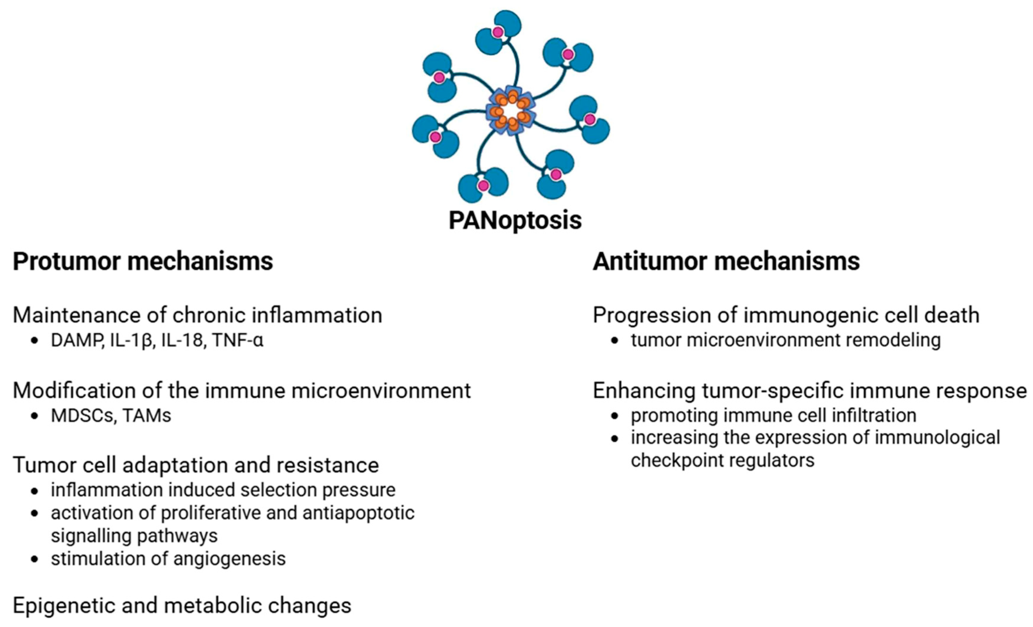
4.1. Protumor Effects

The tumorigenic effect of PANoptosis is intricate and primarily occurs through chronic inflammation, immune response regulation, and alterations in the cellular environment (Figure 3). DAMPs and pro-inflammatory cytokines (e.g., IL-1β, IL-18, and TNF-α) generated during PANoptosis can provoke persistent inflammation, thereby facilitating tumorigenesis [65]. IL-1β and IL-18 can activate the NF-κB and STAT3 signaling pathways, thereby promoting cell proliferation and the production of anti-apoptotic proteins in colonic tumors [65]. The elevated production of reactive oxygen species (ROS) during chronic inflammation may result in mutations in the DNA of colonic epithelial cells [66,67,68]. These findings may partially elucidate the heightened risk of CRC in chronic inflammatory colitis.
PANoptosis can also modify the tumor immune microenvironment. Myeloid-derived suppressor cells (MDSCs) are pivotal in the advancement of colitis-associated CRC [69]. MDSCs facilitate tumor growth by augmenting angiogenesis, fostering chronic inflammation, and establishing a tumor microenvironment (TME) that inhibits immune system function [70]. Tumor-associated macrophages (TAMs) fulfill their critical functions by enhancing tumor proliferation, invasion, and migration; promoting angiogenesis; inhibiting antitumor immunity; altering metabolic profiles; and engaging with colonic microbiota [71]. The molecular components of PANoptosis can stimulate the immunomodulatory activities of MDSCs and TAMs, thereby diminishing the anti-tumor immune response [72,73,74,75,76,77,78].
Long-lasting chronic inflammation can lead to T cell exhaustion in colorectal cancer [79,80]. In this case, tumor antigens and inhibitory receptors like PD-1, PD-L1, CTLA-4, LAG-3, and Tim-3 are present [79,80]. This is especially true in MSI-H colorectal cancers with a high immunoscore [81]. T-cell exhaustion can result from the cytokines released during inflammatory cell death, which in turn reduces the elimination of tumor cells [82,83]. In the chronic inflammatory environment generated by PANoptosis, inflammation can even form an immunosuppressive tumor microenvironment [78,84].
PANoptosis-generated inflammatory cytokines in the TME may impose selection pressure on tumor cells, facilitating the survival of resistant clones and advancing disease progression. Elevated levels of IL-6 have been linked to resistance against chemotherapeutic treatments in CRC. IL-6 may stimulate the STAT3 signaling pathway, hence enhancing tumor cell survival and proliferation, which contributes to the emergence of resistant clones [85]. The persistent presence of TNF-α in the TME may enhance tumor cell resistance to apoptosis. This cytokine can stimulate the NF-κB pathway, thereby enhancing the production of anti-apoptotic genes, which in turn leads to the selective survival of resistant cells [86,87]. Prolonged exposure to IL-1β may enhance the invasiveness of colon cancer cells and facilitate treatment resistance. IL-1β-induced inflammatory responses may facilitate the adaptability of tumor cells to adverse environments, promoting the emergence of resistant clones [88].
While PANoptosis seeks to eradicate infected or damaged cells, it is conceivable that certain cancer cells may persist, triggering alternative signaling pathways that enhance proliferation and anti-apoptotic processes. Inflammatory cytokines generated during PANoptosis, including IL-1β and IL-18, can stimulate the NF-κB and STAT3 signaling pathways in adjacent cells. These pathways facilitate cellular survival and proliferation by augmenting the expression of anti-apoptotic proteins, including Bcl-2 and Bcl-xL [89]. Growth factors generated in an inflammatory milieu, such as VEGF and TGF-β, may also facilitate the proliferation of surviving cells and inhibit apoptosis [90,91]. These substances may activate the ERK/MAPK and PI3K/Akt signaling pathways, which are crucial for the regulation of cell growth and survival [90,91]. The consequence of all this is that tumor cells that survive in an inflammatory environment may become more resistant to immune responses and therapies.
Inflammatory cell death, especially pyroptosis, can substantially influence the metabolism of adjacent cells, notably enhancing glycolysis, which may facilitate tumor proliferation in colorectal cancer [92]. Inflammatory cytokines generated during pyroptosis, including IL-1β and IL-6, can activate the STAT3 and NF-κB signaling pathways in adjacent cells [93]. These pathways can enhance the production of glycolytic enzymes, thereby augmenting glucose absorption and metabolism in tumor cells [94]. Enhanced glycolysis facilitates the Warburg effect, whereby glucose is converted to lactate even in the presence of oxygen and pyruvate does not enter the oxidative phosphorylation pathway. Thus, despite the availability of oxygen, tumor cells produce energy predominantly via glycolysis. This metabolic alteration also facilitates accelerated cell division and tumor proliferation in colorectal cancer [95].
Epigenetic modifications, such as DNA methylation and histone alterations, that transpire during inflammation may exert enduring impacts on the behavior of tumor cells, particularly those of colon cancer. In chronic inflammatory situations, epigenetic modifications may enhance oncogene expression [96,97,98]. Consequently, focused manipulation of PANoptosis, such as anti-inflammatory methods and inflammasome inhibitors, may provide a viable therapeutic approach in anticancer treatment.

4.2. Antitumor Effects

PANoptosis contributes to the defense against colon cancer by directly eliminating tumor cells and altering the tumor microenvironment.
PANoptosis contributes to the progression of immunogenic cell death. In PANoptosis, tumor cells experience lysis, resulting in the release of cellular proteins, such as tumor cell antigens and DAMPs [32,99]. These chemicals indicate the existence of atypical cells to the immune system. Furthermore, PANoptosis stimulates inflammasomes, leading to the synthesis and secretion of inflammatory cytokines, including IL-1β and IL-18, via caspases [100]. These cytokines augment the immune response, facilitating anti-tumor activity. The liberated antigens and cytokines draw and stimulate immune cells, including dendritic cells, T cells, and NK cells. The heightened presence and activity of immune cells facilitates the immunogenic remodeling of the tumor microenvironment [101]. Recent studies indicate that the interplay of specific cytokines, including TNF and interferon-gamma (IFN-γ), might trigger PANoptosis in neoplastic cells, potentially resulting in decreased tumor size in preclinical models [102,103]. The findings indicate that PANoptosis may directly facilitate the eradication of colon cancer cells.
PANoptosis, like other regulated cell death mechanisms, can effectively generate highly immunogenic tumors. Research has shown that immunogenic PANoptotic cell death can alter an immunosuppressive environment and enhance innate immune responses. This is achieved by enhancing dendritic cell maturation and macrophage polarization via the generation of DAMPs [104]. Targeting PANoptotic cell death not only prevents immune evasion but also establishes a feedback loop for immunological activation, crucial for surmounting resistance in treatment-resistant cancers. Moreover, PANoptosis is positively correlated with the invasion of immune cells, including CD4+ T cells, CD8+ T cells, and NK cells, in the TME. This enhances the tumor-specific immune response. PANoptotic characteristics exhibit a positive connection with immune checkpoint markers such as CD4, CD274, CCL2, CXCR4, and LAG-3 [105,106]. PANoptosis significantly contributes to the immune response against tumors by promoting immune cell infiltration, enhancing tumor immunogenicity, and increasing the expression of immunological checkpoint regulators.
Further investigation into the relationships between these immunological variables and PANoptosis may yield innovative strategies to improve cancer therapy. The interplay between immunological elements and PANoptosis in cancer treatment presents significant therapeutic and prognostic potential.

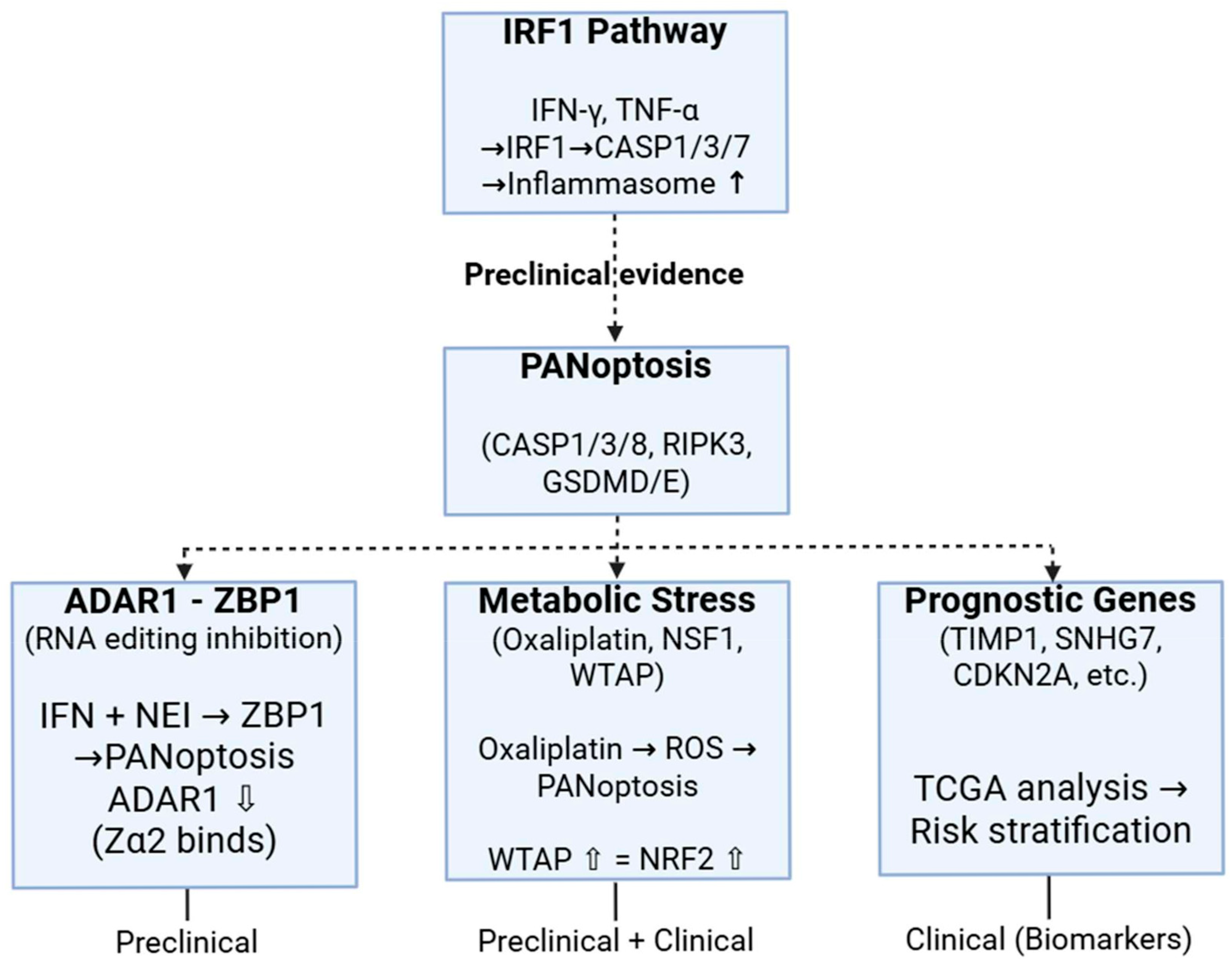
5. Preclinical and Clinical Dimensions of PANoptosis in Colorectal Cancer

Interferon regulatory factor 1 (IRF1) is a transcription factor that conducts the function of interferons. It contributes to inflammation, innate and adaptive immunity, and tumor surveillance. IRF1 modulates the gene expression of guanylate-binding proteins, inducible nitric oxide synthase, and caspase-1 [107,108,109], which have roles in numerous inflammatory disorders. IRF1 has been demonstrated to facilitate the activation of the NLRP3 and AIM2 inflammasomes in response to microbial infection [109,110,111]. For instance, the detection of bacterial DNA by cGAS, followed by cGAS/STING-mediated type I IFN-dependent production of IRF1, stimulates the expression of guanylate-binding proteins. This results in the intracellular eradication of bacteria and the release of DNA [109]. Additionally, in fungal infections, the C-type lectin receptor pathway activates both MAPK and NF-κB signaling, resulting in the induction of IRF1 and the priming of inflammasomes. TLR signaling via adaptor molecules MyD88 and TRIF facilitates the effective activation of IRF1, which then stimulates IRGB10 expression, ultimately leading to antifungal action [110]. These inflammasomes have been linked to the emergence of preventive mechanisms against colon cancer [112,113,114]. The investigation of IRF1’s role in carcinogenesis through PANoptosis revealed that IRF1 substantially decreases CRC incidence in mice, and the induction of PANoptosis successfully inhibits AOM/DSS-induced colon cancer. Nonetheless, Irf1-deficient animals exhibited an atypical vulnerability to colitis-associated colon cancers [115]. The comparison of the production of pro-inflammatory cytokines and the death of colonic epithelial cells in Irf1-deficient mice compared to wild-type mice showed that there was no significant difference in the production of cytokines. However, the death of colon cells was significantly lower in Irf1-deficient mice. The decrease in colon cell mortality was linked to diminished activation of caspase-3 and -7, as well as a reduction in pyroptosis and necroptosis. All of these are supported by IRF1-regulated production of TNF-α and IFN-γ. This indicates that IRF1, as an upstream regulator of PANoptosis, promotes PANoptosis in colonic epithelial cells, hence safeguarding them from cancer [115]. Moreover, the loss of IRF1 in subepithelial myofibroblasts and fibroblasts may also facilitate enhanced colorectal carcinogenesis in whole-body knockout mice. The findings indicate that IRF1 could serve as a viable target in the modulation of many PCD pathways.
The PANoptosis sensor ZBP1 triggers cell death, while the RNA editor ADAR1 (adenosine deaminase acting on RNA 1) preserves a balance between cell death and survival [116]. The interplay between ADAR1 and ZBP1 is significant in the modulation of cellular death in neoplasms. The combination of IFN and NEI (nuclear export inhibitor) can stimulate ZBP1-mediated PANoptosis. ADAR1, however, obstructs this process by engaging with the Zα2 structural domain of ZBP1; hence, it prevents the interaction between ZBP1 and RIPK3 [116,117]. Adar1 knockout mice exhibit resistance to CRC growth, which can be counteracted by the deletion of the Zα2 domain of ZBP1 [116,118]. This evidence indicates that ADAR1 suppresses ZBP1-mediated PANoptosis and exerts a protumorigenic function.
Metabolic reprogramming is a defining hallmark of cancer cells [119]. Metabolic enzymes are crucial to metabolic reprogramming [120]. Their abnormal expression is intricately linked to carcinogenesis, tumor development, and chemotherapeutic sensitivity [120]. Iron–sulfur (Fe–S) clusters serve as essential cofactors for Fe–S proteins and participate in numerous physiological activities, including iron homeostasis, energy metabolism, and lipid production [121]. Fe–S clusters are markedly elevated during the fast proliferation of cancer cells [122]. In vivo CRISPR-Cas9 screening of metabolic enzyme genes revealed that the deletion of the rate-limiting enzyme in Fe–S cluster biogenesis, cysteine desulfurase (Nfs1), enhances the efficacy of antitumor therapy by elevating intracellular oxidative stress-induced PANoptosis, in conjunction with oxaliplatin [123]. NFS1 S293 phosphorylation-dependent inhibition of PANoptosis occurs following oxaliplatin therapy. The elevated expression of NFS1 in colorectal cancer patients correlates with unfavorable prognosis [124].
It has been discovered that the depletion of Wilms tumor 1-associating protein (WTAP; a m6A methyltransferase) in CRC cells caused PANoptosis in response to oxaliplatin treatment by increasing intracellular oxidative stress [125]. Additionally, the treatment with oxaliplatin increased the expression of WTAP, which, in turn, prevented PANoptosis by maintaining the expression of nuclear factor erythroid-2-related factor 2 (NRF2), a significant antioxidant response element, through a m6A-dependent mechanism. Furthermore, clinical data analysis of The Cancer Genome Atlas (TCGA) database and patient cohort studies have shown that high WTAP expression in CRC patients is associated with a poor prognosis and diminished benefit from standard chemotherapy [125].
A recent study utilized TCGA and GEO datasets to find differential lncRNAs linked to metastasis and PANoptosis in CRC [126]. Differentially expressed lncRNAs were utilized to establish a lncRNA–miRNA–mRNA network, and the functional and prognostic implications of these lncRNAs were further examined by various bioinformatics methods. The authors found the PANoptosis-related lncRNA SNHG7 linked to CRC metastases, chemoresistance, and prognosis. Consequently, lncRNA SNHG7 has been proposed as a possible predictive biomarker and therapeutic target for CRC [126].
Also using the TCGA database, the mRNA expression dataset was profiled in 404 CRC cases [127]. After identifying key genes associated with PANoptosis, a prognostic model (i.e., TIMP1, CDKN2A, CAMK2B, and TLR3) was developed that showed high predictive accuracy for CRC prognosis. A significant association was found between high PANoptosis risk scores and worse survival outcomes [127]. The finding highlights the potential of these genes as biomarkers for the diagnosis and prognosis of CRC.
Another study identified key PANoptosis-related genes (i.e., BCL10, CDKN2A, DAPK1, PYGM, and TIMP1) associated with CRC progression using multiple datasets [128]. They then defined the pathogenic regions of these genes and explored their relationship with the immune microenvironment and chemotherapeutic drug sensitivity, tumor progression genes, single-cell subpopulations, signaling pathways, transcription factor regulation, and miRNA regulatory networks in CRC. A total of 146 miRNAs have been identified through in silico analysis as modulating PANoptosis via post-transcriptional regulation of the BCL10, PYGM, CDKN2A, DAPK1, and TIMP1 genes; however, none have been validated in vitro or in vivo [128]. By using these complex results, they successfully constructed a new prognostic nomogram model for CRC. Several datasets have demonstrated the clinical relevance and prognostic value of key genes [128].
Table 2 presents a summary of the activities of RNA types that govern PANoptosis in colorectal carcinogenesis.
The analysis of differentially expressed genes linked to PANoptosis indicated a reduction in UNC5D gene expression in HCT116, HT29, and SW480 colon cancer cell lines [129]. An analysis of the TCGA database indicated that diminished expression of UNC5D correlates with inferior survival in CRC [129].
As new discoveries about PANoptosis and its involvement in colorectal cancer continue to grow, it is becoming clear that PANoptosis could play an important role in predicting outcomes and treating the disease. Figure 4 illustrates the schematic representation of PANoptosis pathways and their regulation in colorectal cancer.

Expression of Main Components of PANoptosis in Colorectal Cancer

The principal elements of PANoptosis—proteins implicated in the formation of the PANoptosome—may be expressed in various tumors, including colorectal cancer, and their function can be dual: either anti-tumor or supportive of tumor survival, contingent upon the setting. The schematic representation of key molecular players in PANoptosis and their overlap in colorectal cancer is visualized in Figure 5.
The ZBP1 gene is often expressed at higher levels in colon cancer cells and colon cancer, especially in situations involving inflammation like colitis-associated cancer [130,131,132]. The expression of ZBP1 may activate the PANoptosome by enlisting more adaptors, hence affecting the progression of colon cancer.
The expression of RIPK1 and RIPK3 is variable in CRC, absent in certain tumors and overactivated in others [133,134]. The lack of RIPK3 may correlate with a poorer prognosis [135,136].
CASP8 is often mutated or epigenetically suppressed in colorectal cancer [137,138,139]. In TRAIL-resistant DLD1 colon cancer cells, reduced stability and expedited degradation of caspase-8 have been seen [140]. Decreased levels of CASP8 may facilitate necroptosis or PANoptosis cell death [141,142].
In colon cancer, ASC levels or activity may be elevated in an inflammatory microenvironment [143,144,145]. The activation of ASC may trigger PANoptosis through inflammasome-associated caspases, such as CASP1.
The expression profile of PANoptosis components may hold prognostic and predictive value. It offers a chance to formulate focused therapy strategies, such as combination modulators of inflammasomes and necroptosis. Conversely, it may also be relevant in conjunction with immunotherapy, as PANoptosis represents an immunogenic variant of cell death and may hence augment the anti-tumor immune response.

6. Therapeutic Strategies and Targets Based on PANoptosis in Colorectal Cancer

The therapeutic implications of PANoptosis in CRC are now being explored through many avenues, primarily relying on preclinical models and expression data.
PANoptosis is an immunogenic form of cell death; it creates an inflammatory environment and promotes the activation of the anti-tumor immune response. A therapeutic target may be to sensitize tumor cells with agents that induce PANoptosis through activation of ZBP1, CASP8, RIPK3, or other key components [146,147,148]. This trait may be particularly important for tumors that are resistant to apoptosis (e.g., p53-mutant CRC) [149].
Pro-inflammatory cytokines (e.g., IL-1β and IL-18) and DAMP molecules generated during PANoptosis may enhance the efficacy of anti-PD-1/PD-L1 or anti-CTLA-4 immunotherapies [150]. Animal models have demonstrated enhanced T cell infiltration and tumor regression following the administration of PANoptosis inducers [151].
ZBP1 agonists are not yet available, but ZBP1 may be self-activated in inflammatory settings, e.g., in IBD-associated colorectal carcinoma [152,153]. In CRC patients, chemotherapy triggers the emergence of dsRNA and damaged DNA, which are detected by ZBP1 in normal colorectal tissues, indicating that ZBP1-mediated PANoptosis is activated by chemotherapy in these tissues. This process exacerbates the deleterious side effects of chemotherapy due to DNA damage. Consequently, ZBP1 could serve as a viable therapeutic target to alleviate chemotherapy-induced side effects [154].
Drugs can affect RIPK3 and CASP8. In certain colorectal cancers, CASP8 is inactivated, which could lead to the amplification of necroptosis and PANoptosis pathways [155]. This may represent a prospective therapeutic window for anticancer treatment.
A recent study examined the production of a BiFe2O4@Ag nanocomposite utilizing Chlorella vulgaris extract and its cytotoxic effects on colon cancer cells [156]. The nanocomposite treatment resulted in a substantial upregulation of the CASP8, BAX, and BCL2 genes. Moreover, caspase-3 activity was elevated, and apoptotic morphological alterations were noted [156].
The second mitochondria-derived activator of caspases (SMAC) protein suppresses IAP activity. Numerous small molecules that mimic SMAC function have been synthesized in the past two decades [157]. Inhibition of XIAP by SMAC mimetics promotes caspase-3 activation, whereas the inhibition of cIAPs enables the establishment of the RIPK1-dependent platform for caspase-8 activation that governs cell death. Consequently, SMAC mimetics constitute a promising therapeutic strategy to enhance the sensitivity of apoptosis-resistant malignancies to chemotherapy [157,158]. The genomic heterogeneity of colorectal malignancies raises the question of whether certain consensus molecular subtypes (CMS) may exhibit greater susceptibility to IAP inhibition than others. It has been demonstrated that IAP inhibition is a potential modulator of responses to oxaliplatin/5-FU in colorectal tumors of the CMS1 subtype and may also have promise in the CMS2 subtype [158].
RIPK3 may facilitate tumor proliferation in certain malignancies that are RIPK3-positive or overexpressing RIPK3 [26]. RIPK3 inhibitors can impede the emergence and advancement of malignant tumors. In the case of insufficient diet, the upregulation of RIPK3 is inhibited by the tumor suppressor gene TSC1 [159]. The reduction of gut microbiota through antibiotics or probiotics may diminish RIPK3 activation and expression. This process can lead to reduced intestinal epithelial cell necrosis and mitigated colonic inflammation [159].
OSW-1, extracted from the bulbs of Ornithogalum saundersiae, demonstrates significant anticancer efficacy [160]. The inhibitory impact of OSW-1 on colorectal cancer has been established by both in vitro and in vivo studies. In colon cancer cells, OSW-1 has been demonstrated to cause necroptosis through the RIPK1/RIPK3/MLKL signaling pathway, with this action mediated by the RIPK1-p62/SQSTM1 complex [161].
Necrostatin-1 is a particular inhibitor of necroptosis that functions by blocking the interaction between receptor-interacting protein (RIP)1 and RIP3 [162]. The efficacy of necrostatin-1 as an anti-inflammatory and antitumorigenic agent was examined in animal models of DSS-induced colitis and colitis-associated cancer [163]. Necrostatin-1 markedly diminished the clinical and histological severity of colitis. The injection of Necrostatin-1 reduced the elevation of RIP1 and RIP3 while augmenting the expression of caspase-8 in DSS-induced colitis. Furthermore, necrostatin-1 administration diminished the synthesis of pro-inflammatory cytokines and the release of extracellular HMGB1 in HT29 colon cancer cells undergoing active necroptosis. Moreover, the treatment of necrostatin-1 markedly inhibited tumor growth and progression via suppressing JNK/c-Jun signaling [163].
Potential anticancer therapeutic targets affecting molecules and processes involved in PANoptosis are summarized in Figure 6.

7. Future Perspectives

The capacity of PANoptosis to inhibit or mitigate cancer is increasingly recognized, and the proteins and regulatory elements governing PANoptosomes may be associated with tumor pathophysiology [164].
However, a number of technological challenges remain to be solved in investigating the role of PANoptosis in CRC. The task includes, in part, a precise understanding of the bypass mechanisms caused by complex and redundant signaling pathways, as well as mapping the different biological responses of cancer cells without and with TME [165].
Cancer cell heterogeneity leads to disparate responses to PANoptosis within the same tumor, attributable to variations in transcriptional activity, oxidative stress response, and antioxidant defenses [166]. Such variability hampers the formulation of universal medicines, as efficacy may vary among cancer types and even patients with identical cancers.
A significant future research avenue should focus on the correlation between PANoptosis and colorectal cancer stem cells (CSCs). Currently, we possess no significant data about the impact of PANoptosis on cancer cell resilience to stress and medicines. The influence of the tumor/inflammatory microenvironment, shaped by PANoptosis, on the survival and multiplication of cancer stem cells requires thorough investigation. Investigating the specific involvement of PANoptosis processes in the treatment resistance of CSCs is equally crucial.
Also, the complex mechanism of PANoptosis implies that the therapeutic risks and unwanted effects of manipulating PANoptosis need to be understood in detail.
The processes of PANoptosis and cell death encompass many intermolecular interactions. A comprehensive understanding of how these factors affect PANoptosis is essential for practical application.

Author Contributions

Conceptualization, G.M. and F.S.; writing—original draft preparation, F.S. and G.M.; writing—review and editing, F.S. and G.M.; visualization, F.S.; supervision, G.M. and F.S. All authors have read and agreed to the published version of the manuscript.

Funding

This research received no external funding.

Institutional Review Board Statement

Not applicable.

Data Availability Statement

No new data were created.

Conflicts of Interest

The authors declare no conflicts of interest.

Abbreviations

ADAR1: Adenosine deaminase acting on RNA-1; AIM2: Absent In Melanoma 2; Akt: protein kinase B; ALR: AIM2-like receptor; AOM: azoxymethane; ASC: Apoptosis-associated speck-like protein containing a CARD; Bcl-2: B-cell lymphoma 2; Bcl-xL: B-cell lymphoma-extra large; CASP: caspase; CCL2: Chemokine CC motif ligand 2; CDKN2A/B: cyclin-dependent kinase inhibitor 2A/B; cGAS: cyclic GMP-AMP synthase; cIAP: Cellular inhibitor of apoptosis; CLR: C-type lectin receptor; CMS: consensus molecular subtypes; CRC: colorectal cancer; CRISPR: clustered regularly interspaced short palindromic repeats; CTLA-4: Cytotoxic T-lymphocyte associated protein 4; CXCR4: C-X-C chemokine receptor type 4; CSC: cancer stem cell; DAMP: danger-associated molecular pattern; DAPK: Death-associated protein kinase 1; DNA: Deoxyribonucleic acid; dsRNA: double-stranded RNA; DSS: dextran sulfate sodium; ERK: extracellular signal-regulated kinase; GEO: Gene Expression Omnibus; GSDND/E: gasdermin D/E; HMGB1: high-mobility group protein 1; IFN: interferon; IL: interleukin; IRF1: Interferon regulatory factor 1; JNK: c-Jun N-terminal kinase; LAG-3: Lymphocyte-activation gene 3; lncRNA: long non-coding RNA; MAPK: Mitogen-activated protein kinase; MDSC: myeloid-derived suppressor cell; miRNA: microRNA; MLKL: Mixed lineage kinase domain like pseudokinase; mRNA: messenger RNA; MSI-H: Microsatellite instability-high; NEI: nuclear export inhibitor; NF-kB: nuclear factor-kB; Nfs1: cysteine desulfurase; NLR: NOD-like receptor; NLRP3: NLR family pyrin domain containing 3; NRF2: nuclear factor erythroid-2-related factor 2; OspC1/3: outer Shigella protein C1/3; OspD3: outer Shigella protein D3; OSW-1: Orsaponin 1; p53: Tumor protein P53; PAMP: pathogen-associated molecular pattern; PARP: Poly (ADP-ribose) polymerase; PCD: programmed cell death; PD-1: Programmed cell death protein 1; PD-L1: Programmed Death-Ligand 1; PI3K: Phosphoinositide 3-kinase; PRR: pattern recognition receptor; PYGM: glycogen phosphorylase, muscle associated; RIP1/3: Receptor interacting protein 1/3; RIPK1/3: Receptor-interacting serine/threonine-protein kinase 1/3; RLR: Rig-like receptor; RNA: Ribonucleic acid; ROS: reactive oxygen species; SMAC: second mitochondria-derived activator of caspases; SNHG7: Small Nucleolar RNA Host Gene 7; SQSTM1: Sequestosome 1; STAT3: Signal transducer and activator of transcription 3; STING: Stimulator of interferon genes; TAM: tumor-associated macrophage; TGCA: The Cancer Genome Atlas; TGF: transforming growth factor; Tim-3: T-cell immunoglobulin and mucin-domain containing-3; TIMP1: metallopeptidase inhibitor 1; TLR: Toll-like receptor; TME: tumor microenvironment; TNF: tumor necrosis factor; TRAIL: TNF-related apoptosis-inducing ligand; TSC1: tuberous sclerosis complex 1; UNC5D: Unc-5 Netrin Receptor D; VEGF: vascular endothelial growth factor; VirA: virulence factor A; WTAP: Wilms tumor 1-associating protein; XIAP: X-Linked Inhibitory Apoptosis Protein; ZBP1: Z-DNA-binding protein 1.

References

  1. Yan, L.; Shi, J.; Zhu, J. Cellular and molecular events in colorectal cancer: Biological mechanisms, cell death pathways, drug resistance and signalling network interactions. Discov. Oncol. 2024, 15, 294. [Google Scholar] [CrossRef] [PubMed]
  2. Dekker, E.; Tanis, P.J.; Vleugels, J.L.A.; Kasi, P.M.; Wallace, M.B. Colorectal cancer. Lancet 2019, 394, 1467–1480. [Google Scholar] [CrossRef] [PubMed]
  3. Sagaert, X.; Vanstapel, A.; Verbeek, S. Tumor Heterogeneity in Colorectal Cancer: What Do We Know So Far? Pathobiology 2018, 85, 72–84. [Google Scholar] [CrossRef]
  4. Sasaki, N.; Clevers, H. Studying cellular heterogeneity and drug sensitivity in colorectal cancer using organoid technology. Curr. Opin. Genet. Dev. 2018, 52, 117–122. [Google Scholar] [CrossRef]
  5. Pinsky, P.F.; Doroudi, M. Colorectal Cancer Screening. JAMA 2016, 316, 1715. [Google Scholar] [CrossRef]
  6. Ma, J.-Y.; Wang, Y.-X.; Zhao, Z.-Y.; Xiong, Z.-Y.; Zhang, Z.-L.; Cai, J.; Guo, J.-W. Identification of key programmed cell death genes for predicting prognosis and treatment sensitivity in colorectal cancer. Front. Oncol. 2024, 14, 1483987. [Google Scholar] [CrossRef]
  7. Davidson, B.; Gurusamy, K.; Corrigan, N.; Croft, J.; Ruddock, S.; Pullan, A.; Brown, J.; Twiddy, M.; Birtwistle, J.; Morris, S.; et al. Liver resection surgery compared with thermal ablation in high surgical risk patients with colorectal liver metastases: The LAVA international RCT. Health Technol. Assess. 2020, 24, 1–38. [Google Scholar] [CrossRef]
  8. Hornbech, K.; Ravn, J.; Steinbrüchel, D.A. Outcome after pulmonary metastasectomy: Analysis of 5 years consecutive surgical resections 2002–2006. J. Thorac. Oncol. 2011, 6, 1733–1740. [Google Scholar] [CrossRef]
  9. Tang, D.; Kang, R.; Berghe, T.V.; Vandenabeele, P.; Kroemer, G. The molecular machinery of regulated cell death. Cell Res. 2019, 29, 347–364. [Google Scholar] [CrossRef]
  10. Malireddi, R.K.S.; Kesavardhana, S.; Kanneganti, T.D. ZBP1 and TAK1: Master Regulators of NLRP3 Inflammasome/Pyroptosis, Apoptosis, and Necroptosis (PAN-optosis). Front. Cell. Infect. Microbiol. 2019, 9, 406. [Google Scholar] [CrossRef]
  11. Shi, C.; Cao, P.; Wang, Y.; Zhang, Q.; Zhang, D.; Wang, Y.; Wang, L.; Gong, Z. PANoptosis: A Cell Death Characterized by Pyroptosis, Apoptosis, and Necroptosis. J. Inflamm. Res. 2023, 16, 1523–1532. [Google Scholar] [CrossRef] [PubMed]
  12. Morana, O.; Wood, W.; Gregory, C.D. The Apoptosis Paradox in Cancer. Int. J. Mol. Sci. 2022, 23, 1328. [Google Scholar] [CrossRef]
  13. Gao, L.; Shay, C.; Teng, Y. Cell death shapes cancer immunity: Spotlighting PANoptosis. J. Exp. Clin. Cancer Res. 2024, 43, 168. [Google Scholar] [CrossRef]
  14. Pandey, A.; Li, Z.; Gautam, M.; Ghosh, A.; Man, S.M. Molecular mechanisms of emerging inflammasome complexes and their activation and signaling in inflammation and pyroptosis. Immunol. Rev. 2025, 329, e13406. [Google Scholar] [CrossRef]
  15. Wang, H.; Feng, X.; He, H.; Li, L.; Wetn, Y.; Liu, X.; He, B.; Hual, S.; Sun, S. Crosstalk between autophagy and other forms of programmed cell death. Eur. J. Pharmacol. 2025, 995, 177414. [Google Scholar] [CrossRef]
  16. Vogler, M.; Braun, Y.; Smith, V.M.; Westhoff, M.-A.; Pereira, R.S.; Pieper, N.M.; Anders, M.; Callens, M.; Vervliet, T.; Abbas, M.; et al. The BCL2 family: From apoptosis mechanisms to new advances in targeted therapy. Signal Transduct. Target. Ther. 2025, 10, 91. [Google Scholar] [CrossRef]
  17. Hao, M.-Y.; Li, H.-J.; Han, H.-S.; Chu, T.; Wang, Y.-W.; Si, W.-R.; Jiang, Q.-Y.; Wu, D.-D. Recent advances in the role of gasotransmitters in necroptosis. Apoptosis 2025, 30, 616–635. [Google Scholar] [CrossRef]
  18. Nadella, V.; Kanneganti, T.D. Inflammasomes and their role in PANoptosomes. Curr. Opin. Immunol. 2024, 91, 102489. [Google Scholar] [CrossRef]
  19. Liu, K.; Wang, M.; Li, D.; Duong, N.T.D.; Liu, Y.; Mal, J.; Xin, K.; Zhou, Z. PANoptosis in autoimmune diseases interplay between apoptosis, necrosis, and pyroptosis. Front. Immunol. 2024, 15, 1502855. [Google Scholar] [CrossRef]
  20. Sundaram, B.; Tweedell, R.E.; Prasanth Kumar, S.; Kanneganti, T.D. The NLR family of innate immune and cell death sensors. Immunity 2024, 57, 674–699. [Google Scholar] [CrossRef]
  21. Pandeya, A.; Kanneganti, T.D. Therapeutic potential of PANoptosis: Innate sensors, inflammasomes, and RIPKs in PANoptosomes. Trends Mol. Med. 2024, 30, 74–88. [Google Scholar] [CrossRef] [PubMed]
  22. You, Y.-P.; Yan, L.; Ke, H.-Y.; Li, Y.-P.; Shi, Z.-J.; Zhou, Z.-Y.; Yang, H.-Y.; Yuan, T.; Gan, Y.-Q.; Lu, N.; et al. Baicalin inhibits PANoptosis by blocking mitochondrial Z-DNA formation and ZBP1-PANoptosome assembly in macrophages. Acta Pharmacol. Sin. 2025, 46, 430–447. [Google Scholar] [CrossRef] [PubMed]
  23. Yuan, T.; Yang, H.-Y.; Li, Y.-P.; Shi, Z.-J.; Zhou, Z.-Y.; You, Y.-P.; Ke, H.-Y.; Yan, L.; Xu, L.-H.; Ouyang, D.-Y.; et al. Scutellarin inhibits inflammatory PANoptosis by diminishing mitochondrial ROS generation and blocking PANoptosome formation. Int. Immunopharmacol. 2024, 139, 112710. [Google Scholar] [CrossRef]
  24. Song, K.; Wu, Y.; Tan, S. Caspases in PANoptosis. Curr. Res. Transl. Med. 2025, 73, 103502. [Google Scholar] [CrossRef]
  25. Sahoo, G.; Samal, D.; Khandayataray, P.; Murthy, M.K. A Review on Caspases: Key Regulators of Biological Activities and Apoptosis. Mol. Neurobiol. 2023, 60, 5805–5837. [Google Scholar] [CrossRef]
  26. Zhou, Y.; Xiang, Y.; Liu, S.; Li, C.; Dong, J.; Kong, X.; Ji, X.; Cheng, X.; Zhang, L. RIPK3 signaling and its role in regulated cell death and diseases. Cell Death Discov. 2024, 10, 200. [Google Scholar] [CrossRef]
  27. Ye, K.; Chen, Z.; Xu, Y. The double-edged functions of necroptosis. Cell Death Dis. 2023, 14, 163. [Google Scholar] [CrossRef]
  28. Karki, R.; Kanneganti, T.D. PANoptosome signaling and therapeutic implications in infection: Central role for ZBP1 to activate the inflammasome and PANoptosis. Curr. Opin. Immunol. 2023, 83, 102348. [Google Scholar] [CrossRef]
  29. Dai, Z.; Liu, W.C.; Chen, X.Y.; Wang, X.; Li, J.L.; Zhang, X. Gasdermin D-mediated pyroptosis: Mechanisms, diseases, and inhibitors. Front. Immunol. 2023, 14, 1178662. [Google Scholar] [CrossRef]
  30. Oh, S.; Lee, S. Recent advances in ZBP1-derived PANoptosis against viral infections. Front. Immunol. 2023, 14, 1148727. [Google Scholar] [CrossRef]
  31. Orning, P.; Lien, E. Multiple roles of caspase-8 in cell death, inflammation, and innate immunity. J. Leukoc. Biol. 2021, 109, 121–141. [Google Scholar] [CrossRef] [PubMed]
  32. Pandian, N.; Kanneganti, T.D. PANoptosis: A Unique Innate Immune Inflammatory Cell Death Modality. J. Immunol. 2022, 209, 1625–1633. [Google Scholar] [CrossRef] [PubMed]
  33. Gao, X.; Ma, C.; Liang, S.; Chen, M.; He, Y.; Lei, W. PANoptosis: Novel insight into regulated cell death and its potential role in cardiovascular diseases (Review). Int. J. Mol. Med. 2024, 54, 74. [Google Scholar] [CrossRef]
  34. Zhu, L.; Qi, Z.; Zhang, H.; Wang, N. Nucleic Acid Sensor-Mediated PANoptosis in Viral Infection. Viruses 2024, 16, 966. [Google Scholar] [CrossRef]
  35. Wang, L.; Zhu, Y.; Zhang, L.; Guo, L.; Wang, X.; Pan, Z.; Jiang, X.; Wu, F.; He, G. Mechanisms of PANoptosis and relevant small-molecule compounds for fighting diseases. Cell Death Dis. 2023, 14, 851. [Google Scholar] [CrossRef]
  36. Wang, Y.; Kanneganti, T.D. From pyroptosis, apoptosis and necroptosis to PANoptosis: A mechanistic compendium of programmed cell death pathways. Comput. Struct. Biotechnol. J. 2021, 19, 4641–4657. [Google Scholar] [CrossRef]
  37. Tian, R.; Song, H.; Li, J.; Yuan, T.; Liu, J.; Wang, Y.; Li, Y.; Song, X. PINCH-1 promotes tumor growth and metastasis by enhancing DRP1-mediated mitochondrial fission in head and neck squamous cell carcinoma. Cancer Biol. Ther. 2025, 26, 2477365. [Google Scholar] [CrossRef]
  38. Zhu, X.; Feng, Z.; Peng, X.; Di, T.; Li, Y.; Bai, J.; Ma, T.; Li, L.; Zhang, L. Threonine and tyrosine kinase promotes multiple myeloma progression by regulating regucalcin expression. Exp. Cell Res. 2025, 446, 114454. [Google Scholar] [CrossRef]
  39. Cui, Y.; Cao, X.; Zhang, Y.; Fu, C.; Li, D.; Sun, Y.; Zhang, Y.; Xu, T.; Tsukamoto, T.; Cao, D.; et al. Protein phosphatase 1 regulatory subunit 15 A (PPP1R15A) promoted the progression of gastric cancer by activating cell autophagy under energy stress. J. Exp. Clin. Cancer Res. 2025, 44, 52. [Google Scholar] [CrossRef]
  40. Yingsunthonwattana, W.; Sangsuriya, P.; Supungul, P.; Tassanakajon, A. Litopenaeus vannamei heat shock protein 90 (LvHSP90) interacts with white spot syndrome virus protein, WSSV322, to modulate hemocyte apoptosis during viral infection. Fish Shellfish. Immunol. 2024, 151, 109695. [Google Scholar] [CrossRef]
  41. Fu, Y.; Zhang, J.; Cheng, W.; Cheng, X.; Lu, L.; Gui, L.; Jiang, Y.; Zhang, Y.; Xu, D. miR-124 mediates the expression of ccBax to regulate Cyprinid herpesvirus 2 (CyHV-2)-induced apoptosis and viral replication. J. Fish Dis. 2023, 46, 743–749. [Google Scholar] [CrossRef] [PubMed]
  42. Lei, J.; Hu, D.; Xue, S.; Mao, F.; Obeng, E.; Quan, Y.; Yu, W. HN1L is essential for cell growth and survival during nucleopolyhedrovirus infection in silkworm, Bombyx mori. PLoS ONE 2019, 14, e0216719. [Google Scholar] [CrossRef] [PubMed]
  43. Duan, X.; Shi, J.; Hou, R.; Huang, Y.; Wang, C.; Du, H. The necroptosis-related lncRNA ENSG00000253385.1 promotes the progression of esophageal squamous cell carcinoma by targeting the miR-16-2-3p/VDAC1 axis. Sci. Rep. 2025, 15, 2650. [Google Scholar] [CrossRef]
  44. Cong, L.; Liu, X.; Bai, Y.; Qin, Q.; Zhao, L.; Shi, Y.; Bai, Y.; Guo, Z. Melatonin alleviates pyroptosis by regulating the SIRT3/FOXO3α/ROS axis and interacting with apoptosis in Atherosclerosis progression. Biol. Res. 2023, 56, 62. [Google Scholar] [CrossRef]
  45. Yuan, S.; Wang, Y.; Li, Z.; Chen, X.; Song, P.; Chen, A.; Qu, Z.; Wen, S.; Liu, H.; Zhu, X. Gasdermin D is involved in switching from apoptosis to pyroptosis in TLR4-mediated renal tubular epithelial cells injury in diabetic kidney disease. Arch. Biochem. Biophys. 2022, 727, 109347. [Google Scholar] [CrossRef]
  46. Bock, F.J.; Riley, J.S. When cell death goes wrong: Inflammatory outcomes of failed apoptosis and mitotic cell death. Cell Death Differ. 2023, 30, 293–303. [Google Scholar] [CrossRef]
  47. Carty, M.; Guy, C.; Bowie, A.G. Detection of Viral Infections by Innate Immunity. Biochem. Pharmacol. 2021, 183, 114316. [Google Scholar] [CrossRef]
  48. Karki, R.; Kanneganti, T.D. The ‘cytokine storm’: Molecular mechanisms and therapeutic prospects. Trends Immunol. 2021, 42, 681–705. [Google Scholar] [CrossRef]
  49. Fajgenbaum, D.C.; June, C.H. Cytokine Storm. N. Engl. J. Med. 2020, 383, 2255–2273. [Google Scholar] [CrossRef]
  50. Karki, R.; Sharma, B.R.; Tuladhar, S.; Williams, E.P.; Zalduondo, L.; Samir, P.; Zheng, M.; Sundaram, B.; Banoth, B.; Malireddi, R.K.S.; et al. Synergism of TNF-α and IFN-γ Triggers Inflammatory Cell Death, Tissue Damage, and Mortality in SARS-CoV-2 Infection and Cytokine Shock Syndromes. Cell 2021, 184, 149–168.e17. [Google Scholar] [CrossRef]
  51. Nguyen, L.N.; Kanneganti, T.D. PANoptosis in Viral Infection: The Missing Puzzle Piece in the Cell Death Field. J. Mol. Biol. 2022, 434, 167249. [Google Scholar] [CrossRef] [PubMed]
  52. Kuriakose, T.; Man, S.M.; Malireddi, R.K.S.; Karki, R.; Ketsavardhana, S.; Place, D.E.; Neale, G.; Vogel, P.; Kanneganti, T.-D. ZBP1/DAI is an innate sensor of influenza virus triggering the NLRP3 inflammasome and programmed cell death pathways. Sci. Immunol. 2016, 1, aag2045. [Google Scholar] [CrossRef]
  53. Zheng, M.; Williams, E.P.; Malireddi, R.K.S.; Karki, R.; Banoth, B.; Burton, A.; Webby, R.; Channappanavar, R.; Jonsson, C.B.; Kanneganti, T.-D. Impaired NLRP3 inflammasome activation/pyroptosis leads to robust inflammatory cell death via caspase-8/RIPK3 during coronavirus infection. J. Biol. Chem. 2020, 295, 14040–14052. [Google Scholar] [CrossRef]
  54. Christgen, S.; Zheng, M.; Kesavardhana, S.; Karki, R.; Malireddi, R.K.S.; Banoth, B.; Place, D.E.; Briard, B.; Sharma, B.R.; Tuladhar, S.; et al. Identification of the PANoptosome: A Molecular Platform Triggering Pyroptosis, Apoptosis, and Necroptosis (PANoptosis). Front. Cell. Infect. Microbiol. 2020, 10, 237. [Google Scholar] [CrossRef]
  55. Huang, Z.; Wu, S.-Q.; Liang, Y.; Zhou, X.; Chen, W.; Li, L.; Wu, J.; Zhuang, Q.; Chen, C.; Li, J.; et al. RIP1/RIP3 binding to HSV-1 ICP6 initiates necroptosis to restrict virus propagation in mice. Cell Host Microbe 2015, 17, 229–242. [Google Scholar] [CrossRef]
  56. Guo, H.; Omoto, S.; Harris, P.A.; Finger, J.N.; Bertin, J.; Gough, P.J.; Kaliser, W.J.; Mocarski, E.S. Herpes simplex virus suppresses necroptosis in human cells. Cell Host Microbe 2015, 17, 243–251. [Google Scholar] [CrossRef]
  57. Fletcher-Etherington, A.; Nobre, L.; Nightingale, K.; Antrobus, R.; Nichols, J.; Davison, A.J.; Stanton, R.J.; Weekes, M.P. Human cytomegalovirus protein pUL36: A dual cell death pathway inhibitor. Proc. Natl. Acad. Sci. USA. 2020, 117, 18771–18779. [Google Scholar] [CrossRef]
  58. He, X.; Jiang, X.; Guo, J.; Sun, H.; Yang, J. PANoptosis in Bacterial Infections: A Double-Edged Sword Balancing Host Immunity and Pathogenesis. Pathogens 2025, 14, 43. [Google Scholar] [CrossRef]
  59. Ashida, H.; Sasakawa, C.; Suzuki, T. A unique bacterial tactic to circumvent the cell death crosstalk induced by blockade of caspase-8. EMBO J. 2020, 39, e104469. [Google Scholar] [CrossRef]
  60. Kobayashi, T.; Ogawa, M.; Sanada, T.; Mimuro, H.; Kim, M.; Ashida, H.; Akakura, R.; Yoshida, M.; Kawalec, M.; Reichhart, J.-M.; et al. The Shigella OspC3 effector inhibits caspase-4, antagonizes inflammatory cell death, and promotes epithelial infection. Cell Host Microbe 2013, 13, 570–583. [Google Scholar] [CrossRef]
  61. Günther, S.D.; Fritsch, M.; Seeger, J.M.; Schiffmann, L.M.; Snipas, S.J.; Coutelle, M.; Kufer, T.A.; Higgins, P.G.; Hornung, V.; Bernardini, M.L.; et al. Cytosolic Gram-negative bacteria prevent apoptosis by inhibition of effector caspases through lipopolysaccharide. Nat. Microbiol. 2020, 5, 354–367. [Google Scholar] [CrossRef] [PubMed]
  62. Bergounioux, J.; Elisee, R.; Prunier, A.-L.; Donnadieu, F.; Sperandio, B.; Sansonetti, P.; Arbibe, L. Calpain activation by the Shigella flexneri effector VirA regulates key steps in the formation and life of the bacterium’s epithelial niche. Cell Host Microbe 2012, 11, 240–252. [Google Scholar] [CrossRef] [PubMed]
  63. Ramel, D.; Lagarrigue, F.; Pons, V.; Mounier, J.; Dupuis-Coronas, S.; Chicanne, G.; Sansonetti, P.J.; Gaits-Iacovoni, F.; Tronchère, H.; Payrastre, B. Shigella flexneri infection generates the lipid PI5P to alter endocytosis and prevent termination of EGFR signaling. Sci. Signal. 2011, 4, ra61. [Google Scholar] [CrossRef] [PubMed]
  64. Imani, S.; Farghadani, R.; Roozitalab, G.; Maghsoudloo, M.; Emadi, M.; Moradi, A.; Abedi, B.; Kaboli, P.J. Reprogramming the breast tumor immune microenvironment: Cold-to-hot transition for enhanced immunotherapy. J. Exp. Clin. Cancer Res. 2025, 44, 131. [Google Scholar] [CrossRef]
  65. Li, J.; Huang, L.; Zhao, H.; Yan, Y.; Lu, J. The Role of Interleukins in Colorectal Cancer. Int. J. Biol. Sci. 2020, 16, 2323–2339. [Google Scholar] [CrossRef]
  66. Kay, J.; Thadhani, E.; Samson, L.; Engelward, B. Inflammation-induced DNA damage, mutations and cancer. DNA Repair. 2019, 83, 102673. [Google Scholar] [CrossRef]
  67. Meira, L.B.; Bugni, J.M.; Green, S.L.; Lee, C.-W.; Pang, B.; Borenshtein, D.; Rickman, B.H.; Rogers, A.B.; Moroski-Erkul, C.A.; McFaline, J.L.; et al. DNA damage induced by chronic inflammation contributes to colon carcinogenesis in mice. J. Clin. Investig. 2008, 118, 2516–2525. [Google Scholar] [CrossRef]
  68. Mangerich, A.; Knutson, C.G.; Parry, N.M.; Muthupalani, S.; Ye, W.; Prestwich, E.; Cui, L.; McFaline, J.L.; Mobley, M.; Ge, Z.; et al. Infection-induced colitis in mice causes dynamic and tissue-specific changes in stress response and DNA damage leading to colon cancer. Proc. Natl. Acad. Sci. USA. 2012, 109, E1820–E1829. [Google Scholar] [CrossRef]
  69. Wang, K.; Wang, Y.; Yin, K. Role played by MDSC in colitis-associated colorectal cancer and potential therapeutic strategies. J. Cancer Res. Clin. Oncol. 2024, 150, 243. [Google Scholar] [CrossRef]
  70. Ke, Z.; Wang, C.; Wu, T.; Wang, W.; Yang, Y.; Dai, Y. PAR2 deficiency enhances myeloid cell-mediated immunosuppression and promotes colitis-associated tumorigenesis. Cancer Lett. 2020, 469, 437–446. [Google Scholar] [CrossRef]
  71. Wang, H.; Tian, T.; Zhang, J. Tumor-Associated Macrophages (TAMs) in Colorectal Cancer (CRC): From Mechanism to Therapy and Prognosis. Int. J. Mol. Sci. 2021, 22, 8470. [Google Scholar] [CrossRef]
  72. Traughber, C.A.; Deshpande, G.M.; Neupane, K.; Bhandari, N.; Khan, M.R.; McMullen, M.R.; Swaidani, S.; Opoku, E.; Muppala, S.; Smith, J.D.; et al. Myeloid-cell-specific role of Gasdermin D in promoting lung cancer progression in mice. iScience 2023, 26, 106076. [Google Scholar] [CrossRef]
  73. Yan, G.; Zhao, H.; Zhang, Q.; Zhou, Y.; Wu, L.; Lei, J.; Wang, X.; Zhang, J.; Zhang, X.; Zheng, L.; et al. A RIPK3-PGE2 Circuit Mediates Myeloid-Derived Suppressor Cell-Potentiated Colorectal Carcinogenesis. Cancer Res. 2018, 78, 5586–5599. [Google Scholar] [CrossRef]
  74. Jayakumar, A.; Bothwell, A.L.M. RIPK3-Induced Inflammation by I-MDSCs Promotes Intestinal Tumors. Cancer Res. 2019, 79, 1587–1599. [Google Scholar] [CrossRef]
  75. Zhang, N.; Gao, X.; Zhang, W.; Xiong, J.; Cao, X.; Fu, Z.F.; Cui, M. JEV Infection Induces M-MDSC Differentiation Into CD3+ Macrophages in the Brain. Front. Immunol. 2022, 13, 838990. [Google Scholar] [CrossRef]
  76. Herbert, A.; Balachandran, S. Z-DNA enhances immunotherapy by triggering death of inflammatory cancer-associated fibroblasts. J. Immunother. Cancer 2022, 10, e005704. [Google Scholar] [CrossRef]
  77. Ge, Y.; Jiang, L.; Yang, C.; Dong, Q.; Tang, C.; Xu, Y.; Zhong, X. Interactions between tumor-associated macrophages and regulated cell death: Therapeutic implications in immuno-oncology. Front. Oncol. 2024, 14, 1449696. [Google Scholar] [CrossRef]
  78. Zhang, X.; Tang, B.; Luo, J.; Yang, Y.; Weng, Q.; Fang, S.; Zhao, Z.; Tu, J.; Chen, M.; Ji, J. Cuproptosis, ferroptosis and PANoptosis in tumor immune microenvironment remodeling and immunotherapy: Culprits or new hope. Mol. Cancer. 2024, 23, 255. [Google Scholar] [CrossRef]
  79. Seidel, J.A.; Otsuka, A.; Kabashima, K. Anti-PD-1 and Anti-CTLA-4 Therapies in Cancer: Mechanisms of Action, Efficacy, and Limitations. Front. Oncol. 2018, 8, 86. [Google Scholar] [CrossRef]
  80. Fang, L.; Liu, K.; Liu, C.; Wang, X.; Ma, W.; Xu, W.; Wu, J.; Sun, C. Tumor accomplice: T cell exhaustion induced by chronic inflammation. Front. Immunol. 2022, 13, 979116. [Google Scholar] [CrossRef]
  81. Koi, M.; Tseng-Rogenski, S.S.; Carethers, J.M. Inflammation-associated microsatellite alterations: Mechanisms and significance in the prognosis of patients with colorectal cancer. World J. Gastrointest. Oncol. 2018, 10, 1–14. [Google Scholar] [CrossRef]
  82. Jiang, Y.; Li, Y.; Zhu, B. T-cell exhaustion in the tumor microenvironment. Cell Death Dis. 2015, 6, e1792. [Google Scholar] [CrossRef]
  83. Cai, Y.; Xiao, H.; Zhou, Q.; Lin, J.; Liang, X.; Xu, W.; Cao, Y.; Zhang, X.; Wang, H. Comprehensive Analyses of PANoptosome with Potential Implications in Cancer Prognosis and Immunotherapy. Biochem. Genet. 2025, 63, 331–353. [Google Scholar] [CrossRef]
  84. Wen, Y.; Zhu, Y.; Zhang, C.; Yang, X.; Gao, Y.; Li, M.; Yang, H.; Liu, T.; Tang, H. Chronic inflammation, cancer development and immunotherapy. Front. Pharmacol. 2022, 13, 1040163. [Google Scholar] [CrossRef]
  85. Hu, F.; Song, D.; Yan, Y.; Huang, C.; Shen, C.; Lan, J.; Chen, Y.; Liu, A.; Wu, Q.; Sun, L.; et al. IL-6 regulates autophagy and chemotherapy resistance by promoting BECN1 phosphorylation. Nat. Commun. 2021, 12, 3651. [Google Scholar] [CrossRef]
  86. Han, J.; Soletti, R.C.; Sadarangani, A.; Sridevi, P.; Ramirez, M.E.; Eckmann, L.; Borges, H.L.; Wang, J.Y. Nuclear expression of β-catenin promotes RB stability and resistance to TNF-induced apoptosis in colon cancer cells. Mol. Cancer Res. 2013, 11, 207–218. [Google Scholar] [CrossRef]
  87. Wei, W.; Wang, J.; Huang, P.; Gou, S.; Yu, D.; Zong, L. Tumor necrosis factor-α induces proliferation and reduces apoptosis of colorectal cancer cells through STAT3 activation. Immunogenetics 2023, 75, 161–169. [Google Scholar] [CrossRef]
  88. Li, Y.; Wang, L.; Pappan, L.; Galliher-Beckley, A.; Shi, J. IL-1β promotes stemness and invasiveness of colon cancer cells through Zeb1 activation. Mol. Cancer. 2012, 11, 87. [Google Scholar] [CrossRef]
  89. Dong, Y.; Chen, J.; Chen, Y.; Liu, S. Targeting the STAT3 oncogenic pathway: Cancer immunotherapy and drug repurposing. Biomed. Pharmacother. 2023, 167, 115513. [Google Scholar] [CrossRef]
  90. Deng, Z.; Fan, T.; Xiao, C.; Tian, H.; Zheng, Y.; Li, C.; He, J. TGF-β signaling in health, disease, and therapeutics. Signal Transduct. Target. Ther. 2024, 9, 61. [Google Scholar] [CrossRef]
  91. Stefani, C.; Miricescu, D.; Stanescu-Spinu, I.-I.; Nica, R.I.; Greabu, M.; Totan, A.R.; Jinga, M. Growth Factors, PI3K/AKT/mTOR and MAPK Signaling Pathways in Colorectal Cancer Pathogenesis: Where Are We Now? Int. J. Mol. Sci. 2021, 22, 10260. [Google Scholar] [CrossRef]
  92. Wang, S.; He, H.; Qu, L.; Shen, Q.; Dai, Y. Dual roles of inflammatory programmed cell death in cancer: Insights into pyroptosis and necroptosis. Front. Pharmacol. 2024, 15, 1446486. [Google Scholar] [CrossRef]
  93. Wang, H.; Wang, T.; Yan, S.; Tang, J.; Zhang, Y.; Wang, L.; Xu, H.; Tu, C. Crosstalk of pyroptosis and cytokine in the tumor microenvironment: From mechanisms to clinical implication. Mol. Cancer. 2024, 23, 268. [Google Scholar] [CrossRef]
  94. Jin, B.; Miao, Z.; Pan, J.; Zhang, Z.; Yang, Y.; Zhou, Y.; Jin, Y.; Niu, Z.; Xu, Q. The emerging role of glycolysis and immune evasion in ovarian cancer. Cancer Cell Int. 2025, 25, 78. [Google Scholar] [CrossRef]
  95. Zhong, X.; He, X.; Wang, Y.; Hu, Z.; Huang, H.; Zhao, S.; Wei, P.; Li, D. Warburg effect in colorectal cancer: The emerging roles in tumor microenvironment and therapeutic implications. J. Hematol. Oncol. 2022, 15, 160. [Google Scholar] [CrossRef]
  96. Claycombe, K.J.; Brissette, C.A.; Ghribi, O. Epigenetics of inflammation, maternal infection, and nutrition. J. Nutr. 2015, 145, 1109S–1115S. [Google Scholar] [CrossRef]
  97. Da Silva, M.L.R.; De Albuquerque, B.H.D.R.; Allyrio, T.A.D.M.F.; De Almeida, V.D.; Cobucci, R.N.D.O.; Bezerra, F.L.; Andrade, V.S.; Lanza, D.C.F.; De Azevedo, J.C.V.; De Araújo, J.M.G.; et al. The role of HPV-induced epigenetic changes in cervical carcinogenesis (Review). Biomed. Rep. 2021, 15, 60. [Google Scholar] [CrossRef]
  98. Yang, Z.H.; Dang, Y.Q.; Ji, G. Role of epigenetics in transformation of inflammation into colorectal cancer. World J. Gastroenterol. 2019, 25, 2863–2877. [Google Scholar] [CrossRef]
  99. Cai, H.; Lv, M.; Wang, T. PANoptosis in cancer, the triangle of cell death. Cancer Med. 2023, 12, 22206–22223. [Google Scholar] [CrossRef]
  100. Zhu, P.; Ke, Z.R.; Chen, J.X.; Li, S.J.; Ma, T.L.; Fan, X.L. Advances in mechanism and regulation of PANoptosis: Prospects in disease treatment. Front. Immunol. 2023, 14, 1120034. [Google Scholar] [CrossRef]
  101. Meyiah, A.; Khan, F.I.; Alfaki, D.A.; Murshed, K.; Raza, A.; Elkord, E. The colorectal cancer microenvironment: Preclinical progress in identifying targets for cancer therapy. Transl. Oncol. 2025, 53, 102307. [Google Scholar] [CrossRef]
  102. Najafi-Fard, S.; Petruccioli, E.; Farroni, C.; Petrone, L.; Vanini, V.; Cuzzi, G.; Salmi, A.; Altera, A.M.G.; Navarra, A.; Alonzi, T.; et al. Evaluation of the immunomodulatory effects of interleukin-10 on peripheral blood immune cells of COVID-19 patients: Implication for COVID-19 therapy. Front. Immunol. 2022, 13, 984098. [Google Scholar] [CrossRef]
  103. Li, H.; Ni, H.; Li, Y.; Zhou, A.; Qin, X.; Li, Y.; Che, L.; Mo, H.; Qin, C.; Li, J. Tumors cells with mismatch repair deficiency induce hyperactivation of pyroptosis resistant to cell membrane damage but are more sensitive to co-treatment of IFN-γ and TNF-α to PANoptosis. Cell Death Discov. 2024, 10, 227. [Google Scholar] [CrossRef]
  104. Zhou, L.; Lyu, J.; Liu, F.; Su, Y.; Feng, L.; Zhang, X. Immunogenic PANoptosis-Initiated Cancer Sono-Immune Reediting Nanotherapy by Iteratively Boosting Cancer Immunity Cycle. Adv. Mater. 2024, 36, e2305361. [Google Scholar] [CrossRef]
  105. Yi, X.; Li, J.; Zheng, X.; Xu, H.; Liao, D.; Zhang, T.; Weti, Q.; Li, H.; Peng, J.; Ai, J. Construction of PANoptosis signature: Novel target discovery for prostate cancer immunotherapy. Mol. Ther. Nucleic Acids. 2023, 33, 376–390. [Google Scholar] [CrossRef]
  106. Xiong, Y. The emerging role of PANoptosis in cancer treatment. Biomed. Pharmacother. 2023, 168, 115696. [Google Scholar] [CrossRef]
  107. Kamijo, R.; Harada, H.; Matsuyama, T.; Bosland, M.; Gerecitano, J.; Shapiro, D.; Le, J.; Koh, S.I.; Kimura, T.; Green, S.J.; et al. Requirement for transcription factor IRF-1 in NO synthase induction in macrophages. Science 1994, 263, 1612–1615. [Google Scholar] [CrossRef]
  108. Bouker, K.B.; Skaar, T.C.; Riggins, R.B.; Harburger, D.S.; Fernandez, D.R.; Zwart, A.; Wang, A.; Clarke, R. Interferon regulatory factor-1 (IRF-1) exhibits tumor suppressor activities in breast cancer associated with caspase activation and induction of apoptosis. Carcinogenesis 2005, 26, 1527–1535. [Google Scholar] [CrossRef]
  109. Man, S.M.; Karki, R.; Malireddi, R.K.S.; Neale, G.; Vogel, P.; Yamamoto, M.; Lamkanfi, M.; Kanneganti, T.-D. The transcription factor IRF1 and guanylate-binding proteins target activation of the AIM2 inflammasome by Francisella infection. Nat. Immunol. 2015, 16, 467–475. [Google Scholar] [CrossRef]
  110. Briard, B.; Karki, R.; Malireddi, R.K.S.; Bhattacharya, A.; Place, D.E.; Mavuluri, J.; Peters, J.L.; Vogel, P.; Yamamoto, M.; Kanneganti, T.-D. Fungal ligands released by innate immune effectors promote inflammasome activation during Aspergillus fumigatus infection. Nat. Microbiol. 2019, 4, 316–327. [Google Scholar] [CrossRef]
  111. Zheng, M.; Kanneganti, T.D. The regulation of the ZBP1-NLRP3 inflammasome and its implications in pyroptosis, apoptosis, and necroptosis (PANoptosis). Immunol. rev. 2020, 297, 26–38. [Google Scholar] [CrossRef]
  112. Karki, R.; Kanneganti, T.D. Diverging inflammasome signals in tumorigenesis and potential targeting. Nat. Rev. Cancer. 2019, 19, 197–214. [Google Scholar] [CrossRef]
  113. Karki, R.; Man, S.M.; Kanneganti, T.D. Inflammasomes and Cancer. Cancer Immunol. Res. 2017, 5, 94–99. [Google Scholar] [CrossRef]
  114. Sharma, B.R.; Kanneganti, T.D. Inflammasome signaling in colorectal cancer. Transl. Res. 2023, 252, 45–52. [Google Scholar] [CrossRef]
  115. Karki, R.; Sharma, B.R.; Lee, E.; Banoth, B.; Malireddi, R.S.; Samir, P.; Tuladhar, S.; Mummareddy, H.; Burton, A.R.; Vogel, P.; et al. Interferon regulatory factor 1 regulates PANoptosis to prevent colorectal cancer. JCI Insight 2020, 5, e136720. [Google Scholar] [CrossRef]
  116. Karki, R.; Sundaram, B.; Sharma, B.R.; Lee, S.; Malireddi, R.S.; Nguyen, L.N.; Christgen, S.; Zheng, M.; Wang, Y.; Samir, P.; et al. ADAR1 restricts ZBP1-mediated immune response and PANoptosis to promote tumorigenesis. Cell Rep. 2021, 37, 109858. [Google Scholar] [CrossRef]
  117. Karki, R.; Kanneganti, T.D. ADAR1 and ZBP1 in innate immunity, cell death, and disease. Trends Immunol. 2023, 44, 201–216. [Google Scholar] [CrossRef]
  118. Hu, S.B.; Li, J.B. RNA editing and immune control: From mechanism to therapy. Curr. Opin. Genet. Dev. 2024, 86, 102195. [Google Scholar] [CrossRef]
  119. Faubert, B.; Solmonson, A.; DeBerardinis, R.J. Metabolic reprogramming and cancer progression. Science 2020, 368, eaaw5473. [Google Scholar] [CrossRef]
  120. Pavlova, N.N.; Zhu, J.; Thompson, C.B. The hallmarks of cancer metabolism: Still emerging. Cell Metab. 2022, 34, 355–377. [Google Scholar] [CrossRef]
  121. Ali, V.; Nozaki, T. Iron-sulphur clusters, their biosynthesis, and biological functions in protozoan parasites. Adv. Parasitol. 2013, 83, 1–92. [Google Scholar] [CrossRef] [PubMed]
  122. Zhang, M.; Liu, Z.; Le, Y.; Gu, Z.; Zhao, H. Iron-Sulfur Clusters: A Key Factor of Regulated Cell Death in Cancer. Oxidative Med. Cell Longev. 2022, 2022, 7449941. [Google Scholar] [CrossRef]
  123. Lin, J.-F.; Hu, P.-S.; Wang, Y.-Y.; Tan, Y.-T.; Yu, K.; Liao, K.; Wu, Q.-N.; Li, T.; Meng, Q.; Lin, J.-Z.; et al. Phosphorylated NFS1 weakens oxaliplatin-based chemosensitivity of colorectal cancer by preventing PANoptosis. Signal Transduct. Target. Ther. 2022, 7, 54. [Google Scholar] [CrossRef]
  124. Majewska, J.; Ciesielski, S.J.; Schilke, B.; Kominek, J.; Blenska, A.; Delewski, W.; Song, J.-Y.; Marszalek, J.; Craig, E.A.; Dutkiewicz, R. Binding of the chaperone Jac1 protein and cysteine desulfurase Nfs1 to the iron-sulfur cluster scaffold Isu protein is mutually exclusive. J. Biol. Chem. 2013, 288, 29134–29142. [Google Scholar] [CrossRef] [PubMed]
  125. Tan, Y.-T.; Li, T.; Wang, R.-B.; Liu, Z.-K.; Ma, M.-Y.; Huang, R.-Z.; Mo, H.-Y.; Luo, S.-Y.; Lin, J.-F.; Xu, R.-H.; et al. WTAP weakens oxaliplatin chemosensitivity of colorectal cancer by preventing PANoptosis. Cancer Lett. 2024, 604, 217254. [Google Scholar] [CrossRef]
  126. Huang, J.; Jiang, S.; Liang, L.; He, H.; Liu, Y.; Cong, L.; Jiang, Y. Analysis of PANoptosis-Related LncRNA-miRNA-mRNA Network Reveals LncRNA SNHG7 Involved in Chemo-Resistance in Colon Adenocarcinoma. Front. Oncol. 2022, 12, 888105. [Google Scholar] [CrossRef]
  127. Zhao, T.; Zhang, X.; Liu, X.; Jiang, X.; Chen, S.; Li, H.; Ji, H.; Wang, S.; Liang, Q.; Ni, S.; et al. Characterizing PANoptosis gene signature in prognosis and chemosensitivity of colorectal cancer. J. Gastrointest. Oncol. 2024, 15, 2129–2144. [Google Scholar] [CrossRef]
  128. Yu, X.; Shao, Y.; Dong, H.; Zhang, X.; Ye, G. Biological function and potential application of PANoptosis-related genes in colorectal carcinogenesis. Sci. Rep. 2024, 14, 20672. [Google Scholar] [CrossRef]
  129. Zhang, M.; Li, W.; Zhao, Y.; Qi, L.; Xiao, Y.; Liu, D.; Peng, T. Molecular characterization analysis of PANoptosis-related genes in colorectal cancer based on bioinformatic analysis. PLoS ONE. 2024, 19, e0307651. [Google Scholar] [CrossRef]
  130. Wang, S.; Song, A.; Xie, J.; Wang, Y.-Y.; Wang, W.-D.; Zhang, M.-J.; Wu, Z.-Z.; Yang, Q.-C.; Li, H.; Zhang, J.; et al. Fn-OMV potentiates ZBP1-mediated PANoptosis triggered by oncolytic HSV-1 to fuel antitumor immunity. Nat. Commun. 2024, 15, 3669. [Google Scholar] [CrossRef]
  131. Wang, X.; Sun, R.; Chan, S.; Meng, L.; Xu, Y.; Zuo, X.; Wang, Z.; Hu, X.; Han, Q.; Dai, L.; et al. PANoptosis-based molecular clustering and prognostic signature predicts patient survival and immune landscape in colon cancer. Front. Genet. 2022, 13, 955355. [Google Scholar] [CrossRef] [PubMed]
  132. Wang, J.-M.; Yang, J.; Xia, W.-Y.; Wang, Y.-M.; Zhu, Y.-B.; Huang, Q.; Feng, T.; Xie, L.-S.; Li, S.-H.; Liu, S.-Q.; et al. Comprehensive Analysis of PANoptosis-Related Gene Signature of Ulcerative Colitis. Int. J. Mol. Sci. 2023, 25, 348. [Google Scholar] [CrossRef] [PubMed]
  133. Lin, P.; Lin, C.; He, R.; Chen, H.; Teng, Z.; Yao, H.; Liu, S.; Hoffman, R.M.; Ye, J.; Zhu, G. TRAF6 regulates the abundance of RIPK1 and inhibits the RIPK1/RIPK3/MLKL necroptosis signaling pathway and affects the progression of colorectal cancer. Cell Death Dis. 2023, 14, 6. [Google Scholar] [CrossRef] [PubMed]
  134. Zhao, Q.; Guo, J.; Cheng, X.; Liao, Y.; Bi, Y.; Gong, Y.; Zhang, X.; Guo, Y.; Wang, X.; Yu, W.; et al. RIPK3 Suppresses the Progression of Spontaneous Intestinal Tumorigenesis. Front. Oncol. 2021, 11, 664927. [Google Scholar] [CrossRef]
  135. Zhang, Z.; Ju, F.; Chen, F.; Wu, H.; Chen, J.; Zhong, J.; Shao, L.; Zheng, S.; Wang, L.; Xue, M. GDC-0326 Enhances the Effects of 5-Fu in Colorectal Cancer Cells by Inducing Necroptotic Death. Onco Targets Ther. 2021, 14, 2519–2530. [Google Scholar] [CrossRef]
  136. Conev, N.V.; Kashlov, Y.K.; Dimitrova, E.G.; Bogdanova, M.K.; Chaushev, B.G.; Radanova, M.A.; Petrov, D.P.; Georgiev, K.D.; Bachvarov, C.H.; Todorov, G.N.; et al. RIPK3 expression as a potential predictive and prognostic marker in metastatic colon cancer. Clin. Investig. Med. 2019, 42, E31–E38. [Google Scholar] [CrossRef]
  137. Kim, H.S.; Soung, Y.H.; Park, W.S.; Kim, S.Y.; Lee, J.H.; Park, J.Y.; Cho, Y.G.; Kim, C.J.; Jeong, S.W.; Nam, S.W.; et al. Inactivating mutations of caspase-8 gene in colorectal carcinomas. Gastroenterology 2003, 125, 708–715. [Google Scholar] [CrossRef]
  138. Cacina, C.; Turan Sürmen, S.; Arıkan, S.; Pençe, S.; Yaylım, İ. The influence of CASP8 D302H gene variant in colorectal cancer risk and prognosis. Turk. J. Biochem. 2023, 48, 234–238. [Google Scholar] [CrossRef]
  139. Sun, T.; Gao, Y.; Tan, W.; Ma, S.; Shi, Y.; Yao, J.; Guo, Y.; Yang, M.; Zhang, X.; Zhang, Q.; et al. A six-nucleotide insertion-deletion polymorphism in the CASP8 promoter is associated with susceptibility to multiple cancers. Nat. Genet. 2007, 39, 605–613. [Google Scholar] [CrossRef]
  140. Zhang, L.; Zhu, H.; Teraishi, F.; Davis, J.J.; Guo, W.; Fan, Z.; Fang, B. Accelerated degradation of caspase-8 protein correlates with TRAIL resistance in a DLD1 human colon cancer cell line. Neoplasia 2005, 7, 594–602. [Google Scholar] [CrossRef]
  141. Melo-Lima, S.; Celeste Lopes, M.; Mollinedo, F. Necroptosis is associated with low procaspase-8 and active RIPK1 and -3 in human glioma cells. Oncoscience 2014, 1, 649–664. [Google Scholar] [CrossRef] [PubMed]
  142. Jiang, M.; Qi, L.; Li, L.; Wu, Y.; Song, D.; Li, Y. Caspase-8: A key protein of cross-talk signal way in “PANoptosis” in cancer. Int. J. Cancer 2021, 149, 1408–1420. [Google Scholar] [CrossRef] [PubMed]
  143. Yokoyama, T.; Sagara, J.; Guan, X.; Masumoto, J.; Takeoka, M.; Komiyama, Y.; Miyata, K.; Higuchi, K.; Taniguchi, S. Methylation of ASC/TMS1, a proapoptotic gene responsible for activating procaspase-1, in human colorectal cancer. Cancer Lett. 2003, 202, 101–108. [Google Scholar] [CrossRef]
  144. Ohtsuka, T.; Liu, X.-F.; Koga, Y.; Kitajima, Y.; Nakafusa, Y.; Ha, C.-W.; Lee, S.W.; Miyazaki, K. Methylation-induced silencing of ASC and the effect of expressed ASC on p53-mediated chemosensitivity in colorectal cancer. Oncogene 2006, 25, 1807–1811. [Google Scholar] [CrossRef] [PubMed]
  145. Riojas, M.A.; Guo, M.; Glöckner, S.C.; Machida, E.O.; Baylin, S.B.; Ahuja, N. Methylation-induced silencing of ASC/TMS1, a pro-apoptotic gene, is a late-stage event in colorectal cancer. Cancer Biol. Ther. 2007, 6, 1710–1716. [Google Scholar] [CrossRef]
  146. Zhang, T.; Yin, C.; Fedorov, A.; Qiao, L.; Bao, H.; Beknazarov, N.; Wang, S.; Gautam, A.; Williams, R.M.; Crawford, J.C.; et al. ADAR1 masks the cancer immunotherapeutic promise of ZBP1-driven necroptosis. Nature 2022, 606, 594–602. [Google Scholar] [CrossRef]
  147. Gong, Z.; Jia, Q.; Guo, J.; Li, C.; Xu, S.; Jin, Z.; Chu, H.; Wan, Y.Y.; Zhu, B.; Zhou, Y. Caspase-8 contributes to an immuno-hot microenvironment by promoting phagocytosis via an ecto-calreticulin-dependent mechanism. Exp. Hematol. Oncol. 2023, 12, 7. [Google Scholar] [CrossRef]
  148. Chen, K.-S.; Manoury-Battais, S.; Kanaya, N.; Vogiatzi, I.; Borges, P.; Kruize, S.J.; Chen, Y.-C.; Lin, L.Y.; Rossignoli, F.; Mendonca, N.C.; et al. An inducible RIPK3-driven necroptotic system enhances cancer cell-based immunotherapy and ensures safety. J. Clin. Investig. 2024, 135, e181143. [Google Scholar] [CrossRef]
  149. Ocansey, D.K.W.; Qian, F.; Cai, P.; Ocansey, S.; Amoah, S.; Qian, Y.; Mao, F. Current evidence and therapeutic implication of PANoptosis in cancer. Theranostics 2024, 14, 640–661. [Google Scholar] [CrossRef]
  150. Liu, J.; Hong, M.; Li, Y.; Chen, D.; Wu, Y.; Hu, Y. Programmed Cell Death Tunes Tumor Immunity. Front. Immunol. 2022, 13, 847345. [Google Scholar] [CrossRef]
  151. Cai, Y.; Xiao, H.; Xue, S.; Li, P.; Zhan, Z.; Lin, J.; Song, Z.; Liu, J.; Xu, W.; Zhou, Q.; et al. Integrative analysis of immunogenic PANoptosis and experimental validation of cinobufagin-induced activation to enhance glioma immunotherapy. J. Exp. Clin. Cancer Res. 2025, 44, 35. [Google Scholar] [CrossRef] [PubMed]
  152. Hao, Y.; Yang, B.; Yang, J.; Shi, X.; Yang, X.; Zhang, D.; Zhao, D.; Yan, W.; Chen, L.; Zheng, H.; et al. ZBP1: A Powerful Innate Immune Sensor and Double-Edged Sword in Host Immunity. Int. J. Mol. Sci. 2022, 23, 10224. [Google Scholar] [CrossRef]
  153. Wang, R.; Li, H.; Wu, J.; Cai, Z.-Y.; Li, B.; Ni, H.; Qiu, X.; Chen, H.; Liu, W.; Yang, Z.-H.; et al. Gut stem cell necroptosis by genome instability triggers bowel inflammation. Nature 2020, 580, 386–390. [Google Scholar] [CrossRef]
  154. Wang, F.; Li, K.; Wang, W.; Hui, J.; He, J.; Cai, J.; Ren, W.; Zhao, Y.; Song, Q.; He, Y.; et al. Sensing of endogenous retroviruses-derived RNA by ZBP1 triggers PANoptosis in DNA damage and contributes to toxic side effects of chemotherapy. Cell Death Dis. 2024, 15, 779. [Google Scholar] [CrossRef]
  155. Zou, J.; Xia, H.; Zhang, C.; Xu, H.; Tang, Q.; Zhu, G.; Li, J.; Bi, F. Casp8 acts through A20 to inhibit PD-L1 expression: The mechanism and its implication in immunotherapy. Cancer sci. 2021, 112, 2664–2678. [Google Scholar] [CrossRef]
  156. Golrokh, F.J.; Tolami, H.F.; Ghanbarirad, M.; Mahmoudi, A.; Tabassi, N.R.; Alkinani, T.A.; Taramsari, S.M.; Aghajani, S.; Taati, H.; Akbari, F.; et al. Apoptosis induction in colon cancer cells (SW480) by BiFe2O4@Ag nanocomposite synthesized from Chlorella vulgaris extract and evaluation the expression of CASP8, BAX and BCL2 genes. J. Trace Elem. Med. Biol. 2024, 83, 127369. [Google Scholar] [CrossRef]
  157. Gyrd-Hansen, M.; Meier, P. IAPs: From caspase inhibitors to modulators of NF-kappaB, inflammation and cancer. Nat. Rev. Cancer 2010, 10, 561–574. [Google Scholar] [CrossRef]
  158. Fichtner, M.; Bozkurt, E.; Salvucci, M.; McCann, C.; McAllister, K.A.; Halang, L.; Düssmann, H.; Kinsella, S.; Crawford, N.; Sessler, T.; et al. Molecular subtype-specific responses of colon cancer cells to the SMAC mimetic Birinapant. Cell Death Dis. 2020, 11, 1020. [Google Scholar] [CrossRef]
  159. Xie, Y.; Zhao, Y.; Shi, L.; Li, W.; Chen, K.; Li, M.; Chen, X.; Zhang, H.; Li, T.; Matsuzawa-Ishimoto, Y.; et al. Gut epithelial TSC1/mTOR controls RIPK3-dependent necroptosis in intestinal inflammation and cancer. J. Clin. Investig. 2020, 130, 2111–2128. [Google Scholar] [CrossRef]
  160. Zhan, Z.; Liu, Z.; Lai, J.; Zhang, C.; Chen, Y.; Huang, H. Anticancer Effects and Mechanisms of OSW-1 Isolated From Ornithogalum saundersiae: A Review. Front. Oncol. 2021, 11, 747718. [Google Scholar] [CrossRef]
  161. Wang, N.; Li, C.Y.; Yao, T.F.; Kang, X.D.; Guo, H.S. OSW-1 triggers necroptosis in colorectal cancer cells through the RIPK1/RIPK3/MLKL signaling pathway facilitated by the RIPK1-p62/SQSTM1 complex. World J. Gastroenterol. 2024, 30, 2155–2174. [Google Scholar] [CrossRef] [PubMed]
  162. Cao, L.; Mu, W. Necrostatin-1 and necroptosis inhibition: Pathophysiology and therapeutic implications. Pharmacol. Res. 2021, 163, 105297. [Google Scholar] [CrossRef] [PubMed]
  163. Liu, Z.-Y.; Wu, B.; Guo, Y.-S.; Zhou, Y.-H.; Fu, Z.-G.; Xu, B.-Q.; Li, J.-H.; Jing, L.; Jiang, J.-L.; Tang, J.; et al. Necrostatin-1 reduces intestinal inflammation and colitis-associated tumorigenesis in mice. Am. J. Cancer Res. 2015, 5, 3174–3185. [Google Scholar]
  164. Cui, Z.; Li, Y.; Bi, Y.; Li, W.; Piao, J.; Ren, X. PANoptosis: A new era for anti-cancer strategies. Life Sci. 2024, 359, 123241. [Google Scholar] [CrossRef]
  165. Malireddi, R.K.S.; Karki, R.; Sundaram, B.; Kancharana, B.; Lee, S.; Samir, P.; Kanneganti, T.-D. Inflammatory Cell Death, PANoptosis, Mediated by Cytokines in Diverse Cancer Lineages Inhibits Tumor Growth. Immunohorizons 2021, 5, 568–580. [Google Scholar] [CrossRef]
  166. Zhuang, L.; Sun, Q.; Huang, S.; Hu, L.; Chen, Q. A comprehensive analysis of PANoptosome to prognosis and immunotherapy response in pan-cancer. Sci. Rep. 2023, 13, 3877. [Google Scholar] [CrossRef]
Figure 1. The process of PANoptosome assembly and the initiation of PANoptosis. In response to cellular damage, sensors can identify the disruption and trigger the development of the PANoptosome. PANoptosomes possess the capacity to integrate many elements from previously distinct cell death processes. The activation of caspases, gasdermins, MLKL, and others results in substrate cleavage and the development of cell membrane pores, facilitating the release of DAMPs and pro-inflammatory cytokines. ASC: apoptosis-associated speck-like protein containing a caspase activation and recruitment domain; DAMP: danger-associated molecular pattern; RIPK: receptor-interacting serine/threonine protein kinase; MLKL: mixed lineage kinase domain-like pseudokinase; ZBP1: Z-DNA-binding protein 1. Figure was partly created with https://biorender.com/ (accessed on 5 April 2025).
Cells 14 00730 g001
Figure 2. Cellular attributes of PANoptosis. PANoptosis is a type of inflammatory programmed cell death that incorporates molecular components of apoptosis, necroptosis, and pyroptosis. The schematic image illustrates the cellular changes associated with the primary kinds of cell death. Figure was partly created with https://biorender.com/ (accessed on 5 April 2025).
Cells 14 00730 g002
Figure 3. The protumor and antitumor effects of PANoptosis in colorectal cancer. DAMP: danger-associated molecular pattern; IL: interleukin; TNF: tumor necrosis factor; MDSC: myeloid-derived suppressor cell; TAM: tumor-associated macrophage. Figure was partly created with https://biorender.com/ (accessed on 5 April 2025).
Cells 14 00730 g003
Figure 4. Schematic overview of PANoptosis pathways and their modulation in colorectal cancer. Upstream regulatory pathways—including IRF1 signaling, ADAR1-ZBP1 interactions, and metabolic stress—integrate to activate PANoptosis via the PANoptosome complex. Preclinical and clinical evidence highlights the therapeutic and prognostic relevance of targeting PANoptosis-related mechanisms in CRC. ADAR1: Adenosine deaminase acting on RNA-1; CASP: caspase; CDKN2A: cyclin-dependent kinase inhibitor 2A; GSDM: gasdermin; IFN: interferon; IRF1: Interferon regulatory factor 1; NSF1: cysteine desulfurase; NRF2: nuclear factor erythroid-2-related factor 2; RIPK: Receptor-interacting serine/threonine-protein kinase; ROS: reactive oxygen species; SNHG7: Small Nucleolar RNA Host Gene 7; TGCA: The Cancer Genome Atlas; TIMP1: metallopeptidase inhibitor 1; TNF: tumor necrosis factor; WTAP: Wilms tumor 1-associating protein; ZBP1: Z-DNA-binding protein 1; ↑ or ↓: enhanced or suppressed function. Figure was partly created with https://biorender.com/ (accessed on 12 May 2025).
Cells 14 00730 g004
Figure 5. Key molecular players and crosstalk in PANoptosis in colorectal cancer. The diagram highlights the core components of apoptosis (CASP8), pyroptosis (ASC, CASP1), and necroptosis (RIPK1, RIPK3), with ZBP1 acting as a central upstream regulator. These molecules can exhibit either tumor-promoting or tumor-suppressive roles depending on the tumor microenvironment and inflammatory context. ASC: Apoptosis-associated speck-like protein containing a CARD; CASP: caspase; CRC: colorectal cancer; RIPK: Receptor-interacting serine/threonine-protein kinase; ZBP1: Z-DNA-binding protein 1; ↑ or ↓: enhanced or suppressed function. Figure was partly created with https://biorender.com/ (accessed on 12 May 2025).
Cells 14 00730 g005
Figure 6. Potential PANoptosis-related therapeutic targets and mechanisms in colorectal cancer. ZBP1: Z-DNA-binding protein 1; SMAC: second mitochondria-derived activator of caspases; OSW-1: Orsaponin 1; DAMP: danger-associated molecular pattern; IL: interleukin; RIPK: receptor-interacting serine/threonine protein kinase; MLKL: mixed lineage kinase domain-like pseudokinase; CASP: caspase. Figure was partly created with https://biorender.com/ (accessed on 6 April 2025).
Cells 14 00730 g006
Table 1. Summary of key molecular components involved in PANoptosis pathways.
PathwayKey MoleculesRole in PANoptosisReference
ApoptosisCASP3, CASP7, CASP8, and CASP9CASP3/7 execute apoptosis; CASP8 initiates apoptosis or inhibits necroptosis[16,25]
NecroptosisRIPK1, RIPK3, and MLKLRIPK1/RIPK3 activate MLKL; MLKL disrupts membrane; inhibited by CASP8[17,26,27]
PyroptosisCASP1, CASP4, CASP5, CASP11, ASC, and GSDMDCASP1 activates IL-1β/IL-18 and cleaves GSDMD; ASC scaffolds inflammasome formation[14,24]
PANoptosome scaffoldZBP1, RIPK3, and ASCZBP1 senses stress/DNA and recruits RIPK3, ASC, and CASP8 to form the PANoptosome[21,22,28,30]
Executioners shared across pathwaysCASP1, CASP3, CASP8, GSDMD, GSDME, and MLKLMediate final membrane disruption and cell death[23,24,29]
Table 2. The role of regulatory RNAs and mRNAs in the process of PANoptosis in CRC.
lncRNA/miRNA or mRNAFunction/RoleReference
lncRNA SNHG7Associated with CRC metastasis, chemoresistance, and prognosis; proposed as a predictive biomarker and therapeutic target.[126]
miR-33ab; miR-34ac; miR-101; miR-187Affect PANoptosis in CRC through post-transcriptional regulation of the BCL10 gene.[128]
miR-15abc; miR-31; miR-133abc; miR-191Influence PANoptosis in CRC via post-transcriptional modulation of the CDKN2A gene.[128]
miR-23abc; miR-181abc; miR-217; miR-455-5p; Modulate PANoptosis in CRC through post-transcriptional regulation of the DAPK1 gene.[128]
miR-18ab; miR-19ab; miR-141; miR218Affect PANoptosis in CRC via post-transcriptional modulation of the TIMP1 gene.[128]
miR-1ab; miR-145; miR-193; miR210Influence PANoptosis in CRC through post-transcriptional modulation of the PYGM gene.[128]
TIMP1 (mRNA)Part of a PANoptosis-related prognostic model; associated with poorer survival in CRC.[127,128]
CDKN2A (mRNA)Involved in CRC prognosis and progression; linked to immune microenvironment and drug sensitivity.[127,128]
CAMK2B (mRNA)Component of a PANoptosis-based prognostic model; contributes to CRC survival prediction.[127]
TLR3 (mRNA)Included in the prognostic model; high PANoptosis risk score correlates with worse CRC survival.[127]
BCL10 (mRNA)PANoptosis-related gene associated with CRC progression; involved in immune response and drug sensitivity.[128]
DAPK1 (mRNA)Plays a role in CRC and PANoptosis; potentially involved in signaling and therapeutic response.[128]
PYGM (mRNA)PANoptosis gene linked to CRC progression; shows prognostic value.[128]
Disclaimer/Publisher’s Note: The statements, opinions and data contained in all publications are solely those of the individual author(s) and contributor(s) and not of MDPI and/or the editor(s). MDPI and/or the editor(s) disclaim responsibility for any injury to people or property resulting from any ideas, methods, instructions or products referred to in the content.

