

Differential Upregulation of Th1/Th17-Associated Proteins and PD-L1 in Granulomatous Mycosis Fungoides
 , ,
, , 
 , and
, and 
Abstract
1. Introduction
2. Materials and Methods
2.1. Patient Selection
2.2. Immunohistochemistry (IHC) Analysis
2.3. Statistical Analysis
3. Results
3.1. Patient Baseline Characteristics
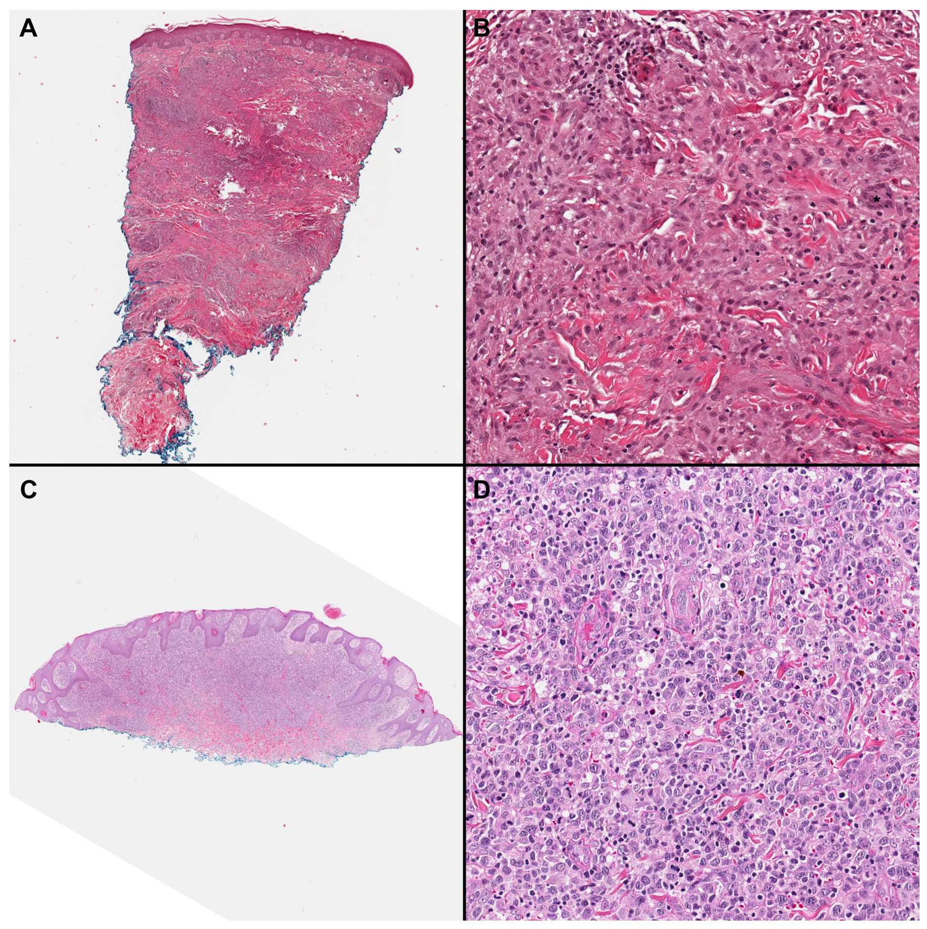
3.2. GMF Group
3.3. MFLCT Group
3.4. Comparison of Baseline Characteristics between GMF and MFLCT
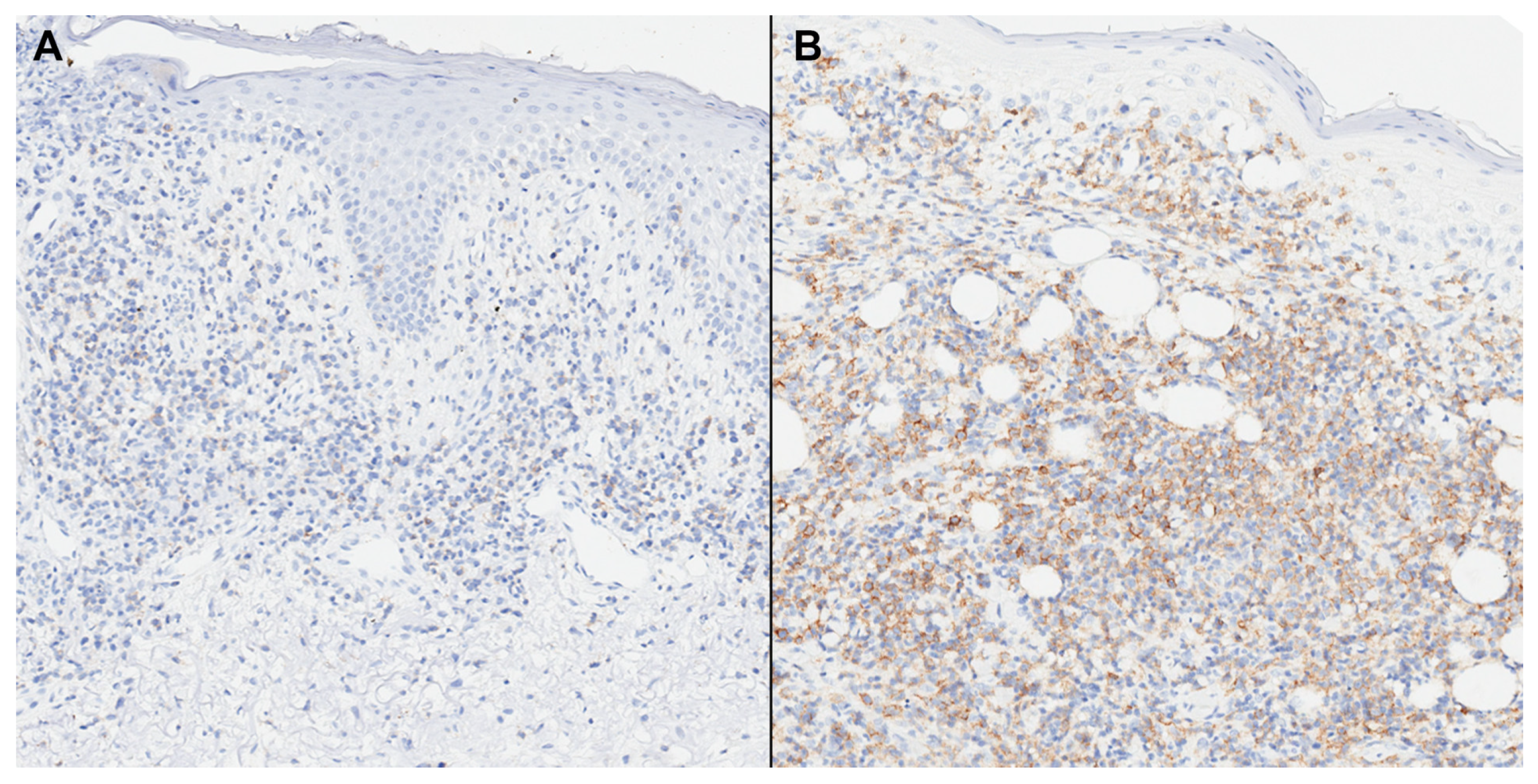
3.5. Upregulation of Th1/Th17 and ICI Markers in GFM
3.6. Staging and Correlation with Outcomes
4. Discussion
5. Conclusions
Supplementary Materials
Author Contributions
Funding
Institutional Review Board Statement
Informed Consent Statement
Data Availability Statement
Conflicts of Interest
References
- Kempf, W.; Ostheeren-Michaelis, S.; Paulli, M.; Lucioni, M.; Wechsler, J.; Audring, H.; Assaf, C.; Rüdiger, T.; Willemze, R.; Meijer, C.J.; et al. Granulomatous Mycosis Fungoides and Granulomatous Slack Skin: A Multicenter Study of the Cutaneous Lymphoma Histopathology Task Force Group of the European Organization for Research and Treatment of Cancer (Eortc). Arch. Dermatol. 2008, 14, 1609–1617. [Google Scholar] [CrossRef] [PubMed]
- Lenskaya, V.; de Moll, E.H.; Hussein, S.; Phelps, R.G. Subtlety of Granulomatous Mycosis Fungoides: A Retrospective Case Series Study and Proposal of Helpful Multimodal Diagnostic Approach with Literature Review. Am. J. Dermatopathol. 2022, 44, 559–567. [Google Scholar] [CrossRef] [PubMed]
- Virmani, P.; Myskowski, P.L.; Pulitzer, M. Unusual Variants of Mycosis Fungoides. Diagn. Histopathol. 2016, 22, 142–151. [Google Scholar] [CrossRef] [PubMed]
- Gallardo, F.; García-Muret, M.P.; Servitje, O.; Estrach, T.; Bielsa, I.; Salar, A.; Abella, E.; Barranco, C.; Pujol, R.M. Cutaneous Lymphomas Showing Prominent Granulomatous Component: Clinicopathological Features in a Series of 16 Cases. J. Eur. Acad. Dermatol. Venereol. 2009, 23, 639–647. [Google Scholar] [CrossRef] [PubMed]
- Pulitzer, M.; Myskowski, P.L.; Horwitz, S.M.; Querfeld, C.; Connolly, B.; Li, J.; Murali, R. Mycosis Fungoides with Large Cell Transformation: Clinicopathological Features and Prognostic Factors. Pathology 2014, 46, 610–616. [Google Scholar] [CrossRef] [PubMed]
- Talpur, R.; Sui, D.; Gangar, P.; Dabaja, B.S.; Duvic, M. Retrospective Analysis of Prognostic Factors in 187 Cases of Transformed Mycosis Fungoides. Clin. Lymphoma Myeloma Leuk. 2016, 16, 49–56. [Google Scholar] [CrossRef] [PubMed]
- Li, J.Y.; Pulitzer, M.P.; Myskowski, P.L.; Dusza, S.W.; Horwitz, S.; Moskowitz, A.; Querfeld, C. A Case-Control Study of Clinicopathologic Features, Prognosis, and Therapeutic Responses in Patients with Granulomatous Mycosis Fungoides. J. Am. Acad. Dermatol. 2013, 69, 366–374. [Google Scholar] [CrossRef] [PubMed]
- Saravia, J.; Chapman, N.M.; Chi, H. Helper T Cell Differentiation. Cell. Mol. Immunol. 2019, 16, 634–643. [Google Scholar] [CrossRef]
- Amador, C.; Greiner, T.C.; Heavican, T.B.; Smith, L.M.; Galvis, K.T.; Lone, W.; Bouska, A.; D’Amore, F.; Pedersen, M.B.; Pileri, S.; et al. Reproducing the Molecular Subclassification of Peripheral T-Cell Lymphoma-Nos by Immunohistochemistry. Blood 2019, 134, 2159–2170. [Google Scholar] [CrossRef]
- Tristão, F.S.M.; Rocha, F.A.; Carlos, D.; Ketelut-Carneiro, N.; Souza, C.O.S.; Milanezi, C.M.; Silva, J.S. Th17-Inducing Cytokines Il-6 and Il-23 Are Crucial for Granuloma Formation During Experimental Paracoccidioidomycosis. Front. Immunol. 2017, 8, 949. [Google Scholar] [CrossRef]
- Ten Berge, B.; Paats, M.S.; Bergen, I.M.; van den Blink, B.; Hoogsteden, H.C.; Lambrecht, B.N.; Hendriks, R.W.; Kleinjan, A. Increased Il-17a Expression in Granulomas and in Circulating Memory T Cells in Sarcoidosis. Rheumatology 2012, 51, 37–46. [Google Scholar] [CrossRef] [PubMed]
- Miyagaki, T.; Sugaya, M.; Suga, H.; Kamata, M.; Ohmatsu, H.; Fujita, H.; Asano, Y.; Tada, Y.; Kadono, T.; Sato, S. Il-22, but Not Il-17, Dominant Environment in Cutaneous T-Cell Lymphoma. Clin. Cancer Res. 2011, 17, 7529–7538. [Google Scholar] [CrossRef] [PubMed]
- Ishida, M.; Hotta, M.; Takikita-Suzuki, M.; Kojima, F.; Okabe, H. Cd8-Positive Granulomatous Mycosis Fungoides: A Case Report with Review of the Literature. J. Cutan. Pathol. 2010, 37, 1072–1076. [Google Scholar] [CrossRef] [PubMed]
- Ishida, M.; Okabe, H. Reactive Lymphoid Follicles with Germinal Centers in granulomatous Mycosis Fungoides: A Case Report with Review of the Literature. J. Cutan. Pathol. 2013, 40, 284–285. [Google Scholar] [CrossRef]
- Sidiropoulou, P.; Tsaoutou, K.; Constantinou, A.; Marinos, L.; Voudouri, D.; Iliakis, T.; Kanellis, G.; Pouliou, E.; Stratigos, A.; Nikolaou, V. New Insights into Granulomatous Mycosis Fungoides (Gmf): A Single-Center Experience. Eur. J. Cancer 2021, 156 (Suppl. 1), S69–S70. [Google Scholar] [CrossRef] [PubMed]
- Kogut, M.; Hadaschik, E.; Grabbe, S.; Andrulis, M.; Enk, A.; Hartschuh, W. Granulomatous Mycosis Fungoides, a Rare Subtype of Cutaneous T-Cell Lymphoma. JAAD Case Rep. 2015, 1, 298–302. [Google Scholar] [CrossRef]
- Kempf, W.; Mitteldorf, C. Cutaneous Lymphomas: New Entities and Rare Variants. Pathologe 2015, 36, 62–69. [Google Scholar] [CrossRef]
- Pousa, C.M.F.D.L.; Nery, N.S.; Mann, D.; Obadia, D.L.; Alves, M.D.F.G.S. Granulomatous Mycosis Fungoides—A Diagnostic Challenge. Bras. Dermatol. 2015, 90, 554–556. [Google Scholar] [CrossRef]
- Du, J.; Zhang, Y.; Liu, D.; Zhu, G.; Zhang, Q. Hodgkin’s Lymphoma with Marked Granulomatous Reaction: A Diagnostic Pitfall. Int. J. Clin. Exp. Pathol. 2019, 12, 2772–2774. [Google Scholar]
- Noto, G.; Pravatà, G.; Miceli, S.; Aricò, M. Granulomatous Slack Skin: Report of a Case Associated with Hodgkin’s Disease and a Review of the Literature. Br. J. Dermatol. 1994, 131, 275–279. [Google Scholar] [CrossRef]
- Van Wilpe, S.; Koornstra, R.; Den Brok, M.; De Groot, J.W.; Blank, C.; De Vries, J.; Gerritsen, W.; Mehra, N. Lactate Dehydrogenase: A Marker of Diminished Antitumor Immunity. Oncoimmunology 2020, 9, 1731942. [Google Scholar] [CrossRef] [PubMed]
- Jour, G.; Aung, P.P.; Merrill, E.D.; Curry, J.L.; Tetzlaff, M.T.; Nagarajan, P.; Ivan, D.; Prieto, V.G.; Duvic, M.; Miranda, R.N.; et al. Differential Expression of Ccr4 in Primary Cutaneous Gamma/Delta (Γ/Δ) T Cell Lymphomas and Mycosis Fungoides: Significance for Diagnosis and Therapy. J. Dermatol. Sci. 2018, 89, 88–91. [Google Scholar] [CrossRef] [PubMed]
- Iqbal, J.; Wright, G.; Wang, C.; Rosenwald, A.; Gascoyne, R.D.; Weisenburger, D.D.; Greiner, T.C.; Smith, L.; Guo, S.; Wilcox, R.A.; et al. Gene Expression Signatures Delineate Biological and Prognostic Subgroups in Peripheral T-Cell Lymphoma. Blood 2014, 123, 2915–2923. [Google Scholar] [CrossRef] [PubMed]
- Iqbal, J.; Wilcox, R.; Naushad, H.; Rohr, J.; Heavican, T.B.; Wang, C.; Bouska, A.; Fu, K.; Chan, W.C.; Vose, J.M. Genomic Signatures in T-Cell Lymphoma: How Can These Improve Precision in Diagnosis and Inform Prognosis? Blood Rev. 2016, 30, 89–100. [Google Scholar] [CrossRef] [PubMed]
- Facco, M.; Cabrelle, A.; Teramo, A.; Olivieri, V.; Gnoato, M.; Teolato, S.; Ave, E.; Gattazzo, C.; Fadini, G.P.; Calabrese, F.; et al. Sarcoidosis Is a Th1/Th17 Multisystem Disorder. Thorax 2011, 66, 144–150. [Google Scholar] [CrossRef] [PubMed]
- Larousserie, F.; Pflanz, S.; Coulomb-L’Herminé, A.; Brousse, N.; Kastelein, R.; Devergne, O. Expression of Il-27 in Human Th1-Associated Granulomatous Diseases. J. Pathol. 2004, 202, 164–171. [Google Scholar] [CrossRef] [PubMed]
- Krejsgaard, T.; Litvinov, I.V.; Wang, Y.; Xia, L.; Willerslev-Olsen, A.; Koralov, S.B.; Kopp, K.L.; Bonefeld, C.M.; Wasik, M.A.; Geisler, C.; et al. Elucidating the Role of Interleukin-17f in Cutaneous T-Cell Lymphoma. Blood 2013, 122, 943–950. [Google Scholar] [CrossRef]
- Geginat, J.; Paroni, M.; Maglie, S.; Alfen, J.S.; Kastirr, I.; Gruarin, P.; De Simone, M.; Pagani, M.; Abrignani, S. Plasticity of Human Cd4 T Cell Subsets. Front. Immunol. 2014, 5, 630. [Google Scholar] [CrossRef]
- Kołkowski, K.; Jolanta, G.; Berenika, O.; Monika, Z.; Nowicki, R.J.; Małgorzata, S.-W. Interleukin-17 Genes Polymorphisms Are Significantly Associated with Cutaneous T-Cell Lymphoma Susceptibility. Acta Derm. Venereol. 2022, 102, adv00777. [Google Scholar] [CrossRef]
- Dobos, G.; Lazaridou, I.; de Masson, A. Mycosis Fungoides and Sézary Syndrome: Microenvironment and Cancer Progression. Cancers 2023, 15, 746. [Google Scholar] [CrossRef]
- Roccuzzo, G.; Giordano, S.; Fava, P.; Pileri, A.; Guglielmo, A.; Tonella, L.; Sanlorenzo, M.; Ribero, S.; Fierro, M.T.; Quaglino, P. Immune Check Point Inhibitors in Primary Cutaneous T-Cell Lymphomas: Biologic Rationale, Clinical Results and Future Perspectives. Front. Oncol. 2021, 11, 733770. [Google Scholar] [CrossRef] [PubMed]
- Beygi, S.; Fernandez-Pol, S.; Duran, G.; Wang, E.B.; Stehr, H.; Zehnder, J.L.; Ramchurren, N.; Fling, S.P.; Cheever, M.A.; Weng, W.-K.; et al. Pembrolizumab in Mycosis Fungoides with Pd-L1 Structural Variants. Blood Adv. 2021, 5, 771–774. [Google Scholar] [CrossRef] [PubMed]
- Pileri, A.; Tabanelli, V.; Fuligni, F.; Agostinelli, C.; Guglielmo, A.; Sabattini, E.; Grandi, V.; Pileri, S.A.; Pimpinelli, N. Pd-1 and Pd-L1 Expression in Mycosis Fungoides and Sézary Syndrome. Ital. J. Dermatol. Venerol. 2022, 157, 355–362. [Google Scholar] [CrossRef] [PubMed]
- Khodadoust, M.S.; Rook, A.H.; Porcu, P.; Foss, F.; Moskowitz, A.J.; Shustov, A.; Shanbhag, S.; Sokol, L.; Fling, S.P.; Ramchurren, N.; et al. Pembrolizumab in Relapsed and Refractory Mycosis Fungoides and Sézary Syndrome: A Multicenter Phase Ii Study. J. Clin. Oncol. 2020, 38, 20–28. [Google Scholar] [CrossRef] [PubMed]
- Zhang, L.; Zhang, M.; Xu, J.; Li, S.; Chen, Y.; Wang, W.; Yang, J.; Li, S.; Gu, M. The Role of the Programmed Cell Death Protein-1/Programmed Death-Ligand 1 Pathway, Regulatory T Cells and T Helper 17 Cells in Tumor Immunity: A Narrative Review. Ann. Transl. Med. 2020, 8, 1526. [Google Scholar] [CrossRef] [PubMed]
- Esfahani, K.; Miller, W.H., Jr. Reversal of Autoimmune Toxicity and Loss of Tumor Response by Interleukin-17 Blockade. N. Engl. J. Med. 2017, 376, 1989–1991. [Google Scholar] [CrossRef]


| Cohort, n = 49 (%) | GMF, n = 28 (%) | MFLCT, n = 21 (%) | p-Value | |
|---|---|---|---|---|
| Age (yrs) | 0.067 | |||
| Median | 60 | 58 | 61 | |
| Range | 21–77 | 21–67 | 34–77 | |
| Sex | 0.356 | |||
| Male | 29 (59) | 15 (54) | 14 (67) | |
| Female | 20 (41) | 13 (46) | 7 (33) | |
| Ethnicity | 0.540 | |||
| White | 35 (71) | 19 (68) | 16 (76) | |
| Black | 11 (23) | 8 (28) | 3 (14) | |
| Hispanic | 2 (4) | 1 (4) | 1 (5) | |
| Unknown | 1 (2) | 0 (0) | 1 (5) | |
| Previous history of malignancy | 11 (23) | 6 (21) | 5 (23) | 0.843 |
| History of previous treatments | 46 (93) | 25 (89) | 21 (100) | 0.250 |
| LDH, median (range), IU/L | 543 (128–1035) | 501 (128–836) | 583 (338–1035) | 0.042 |
| β2M, median (range), mcg/mL | 2.7 (1.6–4.8) | 2.45 (2.4–4.8) | 3.15 (1.6–4.5) | 0.828 |
| Clinical presentation | 0.897 | |||
| Papules | 5 (10) | 2 (7) | 3 (14) | >0.999 |
| Patch | 34 (69) | 18 (64) | 16 (76) | 0.665 |
| Plaques | 37 (75) | 19 (67) | 18 (25) | 0.969 |
| Tumor | 16 (32) | 7 (25) | 9 (42) | 0.582 |
| Clinical Stage at Diagnosis | 0.036 | |||
| I | 13 (26) | 11 (39) | 1 (5) | |
| II | 16 (32) | 8 (29) | 8 (38) | |
| III | 0 (0) | 0 (0) | 0 (0) | |
| IV | 13 (26) | 4 (14) | 4 (19) | |
| Unknown | 7 (16) | 5 (18) | 8 (38) | |
| Status at last follow-up | N/A | |||
| ANED | 2 (4) | 1 (3) | 1 (4) | |
| AWD | 17 (34) | 15 (53) | 2 (8) | |
| DOD | 30 (62) | 12 (44) | 18 (88) |
| Antibody | Number of Positive Cases | |||
|---|---|---|---|---|
| Cohort (%) | GMF (%) | MFLCT (%) | p-Value | |
| CD1a | 0/1 (0) | 0/1 (0) | N/A | N/A |
| CD2 | N/A | N/A | N/A | N/A |
| CD3 | 32/32 (100) | 24/24 (100) | 8/8 (100) | >0.999 |
| CD4 | 29/31 (93) | 22/23 (95) | 7/8 (87.5) | 0.455 |
| CD5 | N/A | N/A | N/A | N/A |
| CD7 | 6/22 (27) | 4/19 (21) | 2/3 (66) | 0.168 |
| CD8 * | 6/30 (25) | 3/23 (13) | 3/7 (42) | 0.120 |
| CD20 | 0/7 (0) | 0/5 (0) | 0/2 (0) | >0.999 |
| CD25 | 12/25 (48) | 3/8 (37.5) | 9/17 (53) | 0.672 |
| CD30 | 37/40 (92) | 17/19 (89) | 20/21 (95) | 0.596 |
| Median% (range) | 50 (1–100) | 4 (1–50) | 80 (50–100) | <0.0001 |
| EBER | 0/3 (0) | 0/2 (0) | 0/1 (0) | >0.999 |
| TCRBF1 | 3/3 (100) | 3/3 (100) | N/A | N/A |
| TCRG | 0/3 (0) | 0/3 (0) | N/A | N/A |
| Cohort | GMF | MFLCT | ||
|---|---|---|---|---|
| Tbet (%) | 0.017 | |||
| Mean (SD) | 18 (0.17) | 22 (0.16) | 13 (0.19) | |
| Median | 15 | 20 | 0 | |
| Intensity, n | 0.252 | |||
| 1+ | 1 | 0 | 1 | |
| 2+ | 4 | 3 | 1 | |
| 3+ | 28 | 21 | 7 | |
| GATA3 (%) | 0.422 | |||
| Mean (SD) | 42 (0.29) | 40 (0.26) | 46 (0.33) | |
| Median | 40 | 40 | 40 | |
| Intensity, n | 0.453 | |||
| 1+ | 4 | 1 | 3 | |
| 2+ | 7 | 4 | 3 | |
| 3+ | 31 | 18 | 13 | |
| RORγT (%) | 0.001 | |||
| Mean (SD) | 13 (0.15) | 17 (0.12) | 8 (0.16) | |
| Median | 10 | 15 | 0 | |
| Intensity, n | 0.518 | |||
| 1+ | 6 | 4 | 2 | |
| 2+ | 9 | 8 | 1 | |
| 3+ | 13 | 11 | 2 | |
| Foxp3 (%) | 0.112 | |||
| Mean (SD) | 15 (0.13) | 17 (0.12) | 13 (0.15) | |
| Median | 10 | 15 | 10 | |
| Intensity, n | 0.113 | |||
| 1+ | 1 | 1 | 0 | |
| 2+ | 2 | 0 | 2 | |
| 3+ | 34 | 23 | 11 | |
| PD-1 (%) | 0.589 | |||
| Mean (SD) | 22 (0.26) | 22 (0.25) | 21 (0.29) | |
| Median | 10 | 10 | 10 | |
| Intensity, n | 0.031 | |||
| 1+ | 0 | 0 | 0 | |
| 2+ | 6 | 6 | 0 | |
| 3+ | 23 | 12 | 11 | |
| PD-L1 (%) | 0.011 | |||
| Mean (SD) | 16 (0.24) | 19 (0.22) | 12 (0.26) | |
| Median | 10 | 10 | 0 | |
| Intensity, n | 0.814 | |||
| 1+ | 0 | 0 | 0 | |
| 2+ | 6 | 5 | 1 | |
| 3+ | 19 | 15 | 4 |
| Variables | Univariate Analysis (p Value) |
|---|---|
| Sex | 0.547 |
| Ethnicity | 0.880 |
| Previous History of malignancy | 0.371 |
| LDH | 0.0555 |
| Β2 microglobulin | 0.071 |
| %Tbet (<15% vs. ≥ 15%) | 0.410 |
| %GATA3 (<40% vs. ≥ 40%) | 0.675 |
| %PD-1 (<10% vs. ≥ 10%) | 0.562 |
| %PD-L1 (<10% vs. ≥ 10%) | 0.787 |
| Age | 0.017 |
| Clinical Stage at Diagnosis | <0.001 |
| Group (GMF vs. MFLCT) | 0.018 |
| %RORγT (<10% vs. ≥ 10%) | 0.006 |
| %Foxp3 (<10% vs. ≥ 10%) | 0.002 |
Disclaimer/Publisher’s Note: The statements, opinions and data contained in all publications are solely those of the individual author(s) and contributor(s) and not of MDPI and/or the editor(s). MDPI and/or the editor(s) disclaim responsibility for any injury to people or property resulting from any ideas, methods, instructions or products referred to in the content. |
© 2024 by the authors. Licensee MDPI, Basel, Switzerland. This article is an open access article distributed under the terms and conditions of the Creative Commons Attribution (CC BY) license (https://creativecommons.org/licenses/by/4.0/).
Share and Cite
Marques-Piubelli, M.L.; Navarrete, J.; Ledesma, D.A.; Hudgens, C.W.; Lazcano, R.N.; Alani, A.; Huen, A.; Duvic, M.; Nagarajan, P.; Aung, P.P.; et al. Differential Upregulation of Th1/Th17-Associated Proteins and PD-L1 in Granulomatous Mycosis Fungoides. Cells 2024, 13, 419. https://doi.org/10.3390/cells13050419
Marques-Piubelli ML, Navarrete J, Ledesma DA, Hudgens CW, Lazcano RN, Alani A, Huen A, Duvic M, Nagarajan P, Aung PP, et al. Differential Upregulation of Th1/Th17-Associated Proteins and PD-L1 in Granulomatous Mycosis Fungoides. Cells. 2024; 13(5):419. https://doi.org/10.3390/cells13050419
Chicago/Turabian StyleMarques-Piubelli, Mario L., Jesus Navarrete, Debora A. Ledesma, Courtney W. Hudgens, Rossana N. Lazcano, Ali Alani, Auris Huen, Madeleine Duvic, Priyadharsini Nagarajan, Phyu P. Aung, and et al. 2024. "Differential Upregulation of Th1/Th17-Associated Proteins and PD-L1 in Granulomatous Mycosis Fungoides" Cells 13, no. 5: 419. https://doi.org/10.3390/cells13050419
APA StyleMarques-Piubelli, M. L., Navarrete, J., Ledesma, D. A., Hudgens, C. W., Lazcano, R. N., Alani, A., Huen, A., Duvic, M., Nagarajan, P., Aung, P. P., Wistuba, I. I., Curry, J. L., Miranda, R. N., & Torres-Cabala, C. A. (2024). Differential Upregulation of Th1/Th17-Associated Proteins and PD-L1 in Granulomatous Mycosis Fungoides. Cells, 13(5), 419. https://doi.org/10.3390/cells13050419