Review

Inflammation as a Regulator of the Airway Surface Liquid pH in Cystic Fibrosis

by
Tayyab Rehman
1,* and
Michael J. Welsh
2,3,*
1
Department of Internal Medicine, University of Michigan, Ann Arbor, MI 48109, USA
2
Departments of Internal Medicine and Molecular Physiology and Biophysics, Pappajohn Biomedical Institute, Roy J. and Lucille A. Carver College of Medicine, University of Iowa, Iowa City, IA 52242, USA
3
Howard Hughes Medical Institute, University of Iowa, Iowa City, IA 52242, USA
*
Authors to whom correspondence should be addressed.
Cells 2023, 12(8), 1104; https://doi.org/10.3390/cells12081104
Submission received: 9 January 2023 / Revised: 5 April 2023 / Accepted: 6 April 2023 / Published: 7 April 2023
(This article belongs to the Special Issue The Role of Airway Epithelial Cells in Health and Disease)

Abstract

:
The airway surface liquid (ASL) is a thin sheet of fluid that covers the luminal aspect of the airway epithelium. The ASL is a site of several first-line host defenses, and its composition is a key factor that determines respiratory fitness. Specifically, the acid–base balance of ASL has a major influence on the vital respiratory defense processes of mucociliary clearance and antimicrobial peptide activity against inhaled pathogens. In the inherited disorder cystic fibrosis (CF), loss of cystic fibrosis transmembrane conductance regulator (CFTR) anion channel function reduces HCO3 secretion, lowers the pH of ASL (pHASL), and impairs host defenses. These abnormalities initiate a pathologic process whose hallmarks are chronic infection, inflammation, mucus obstruction, and bronchiectasis. Inflammation is particularly relevant as it develops early in CF and persists despite highly effective CFTR modulator therapy. Recent studies show that inflammation may alter HCO3 and H+ secretion across the airway epithelia and thus regulate pHASL. Moreover, inflammation may enhance the restoration of CFTR channel function in CF epithelia exposed to clinically approved modulators. This review focuses on the complex relationships between acid–base secretion, airway inflammation, pHASL regulation, and therapeutic responses to CFTR modulators. These factors have important implications for defining optimal ways of tackling CF airway inflammation in the post-modulator era.

1. Introduction

The airway surface liquid (ASL) is a thin layer of fluid that covers the luminal aspect of the airway epithelium [1,2,3]. The ASL thus forms a point of contact with the environment and is a site of several first-line host defenses. Antimicrobial peptides within ASL disrupt microbial cell membrane integrity, and thereby kill inhaled pathogens [4]. Gel-forming mucins, secreted into ASL, engage inhaled particles and pathogens, and the coordinated beating of cilia removes them from the lungs (mucociliary clearance) [5]. Neutrophils and macrophages phagocytose microbes that settle within ASL or kill them by extruding a meshwork of chromatin fibers [6,7]. Reactive oxygen species, released into ASL, suppress bacterial growth [8,9], and secreted purinergic nucleotides regulate ASL volume [10,11]. Several ASL factors (e.g., lysozyme, LL-37) show antiviral activity [12], and extracellular proteases alter the infectivity of respiratory viruses [13,14]. ASL composition critically influences these processes, and thus determines respiratory fitness. For the purpose of this review, we focus on the acid–base balance of ASL as a key parameter that controls host defense properties. Other factors that have been hypothesized to influence ASL properties include increased activity of the epithelial Na+ channels [15] and DNA accumulation [16,17].
The abnormal acidification of ASL initiates a pathologic process in the airways, and for some lung disorders may provide a therapeutic target [18]. A preeminent example is cystic fibrosis (CF), an inherited disorder caused by mutations in the cystic fibrosis transmembrane conductance regulator (CFTR) gene [19,20]. The protein encoded by this gene forms an anion channel that conducts Cl and HCO3 across the apical membrane of several epithelia, including airway epithelia [21,22]. CFTR mutations eliminate or markedly reduce anion flow, thereby decreasing HCO3 secretion and lowering the pH of ASL (pHASL). The abnormally acidic ASL impairs, at least in part, an array of first-line host defenses (Figure 1) [23,24,25,26,27,28,29]. With time, CF airways develop bacterial and viral infection and inflammation. Inflammation in turn can alter acid–base secretion and pHASL. In recent years, the development of CFTR modulators has dramatically improved respiratory outcomes in people with CF [30,31,32]. However, airway inflammation persists in most people taking modulators. Importantly, inflammation can modify the response of CF airway epithelia to CFTR modulators. The complex interplay of these factors has important implications for how we approach CF airway inflammation in the post-modulator era. This review begins by outlining the cellular and molecular mechanisms that control pHASL. We focus on the relationships between inflammation, pHASL regulation, and responses to CFTR modulators. We conclude by exploring the relevance of these findings for targeting CF airway inflammation.

2. H+ and HCO3 Transporters Control pHASL

The balance of acid (H+) and base (HCO3) secretion across the apical membrane determines pHASL [36,37]. In the proximal (cartilaginous) airways, the non-gastric H+/K+-ATPase (ATP12A) is the main route for H+ secretion, and CFTR is the main route for HCO3 secretion. Non-CFTR mechanisms such as calcium-activated Cl channels (CaCC) and solute carrier family 26 (SLC26) transporters are also capable of secreting HCO3, but their overall contribution is small compared to CFTR (Figure 2) [36,38,39,40]. In contrast, in the distal (small) airways, ATP12A is absent, and the vacuolar H+-ATPase (V-ATPase) substitutes as the main H+-secreting mechanism [41]. Both CFTR and CaCC mediate HCO3 secretion across small airway epithelia [42].
Two distinct mechanisms provide HCO3 to apical HCO3 channels and transporters. First is the Na+-HCO3 cotransport (NBC) activity located at the basolateral membrane [43,44,45]. NBC activity is mediated by solute carrier 4 (SLC4) family transporters, several of which are expressed in airway epithelia. These transporters employ the favorable Na+ gradient to cotransport HCO3 into the cytosol with a stoichiometry varying between 1:1 to 1:3, depending on the NBC isoform. However, molecular mechanisms regulating basolateral NBC activity in human airway epithelia remain poorly understood.
Another source of secreted HCO3 is the CO2 hydration reaction [21,46]. The rate-limiting step in this reaction, i.e., the conversion of reactants H2O and CO2 to carbonic acid, is catalyzed by carbonic anhydrase (CA) enzymes. Once generated, carbonic acid readily dissociates into HCO3 and H+. Several CA isoforms, both membrane-associated and cytoplasmic, are expressed in airway epithelia [40,47]. However, their contribution to overall HCO3 secretion is small relative to that of NBCs.
Figure 2 shows a simple model of channels, transporters, and enzymes that control pHASL in airway epithelia. For clarity, we do not show the basolateral Na+/H+ exchanger (NHE1) or Cl/HCO3 exchanger (AE2), which play important roles in intracellular pH regulation. Nor do we show the Na+/K+-ATPase, and Na+ and K+ channels that influence HCO3 flow indirectly by establishing and altering electrochemical gradients. Readers interested in these transport mechanisms are directed to excellent published reviews [36,37,43,48,49,50].

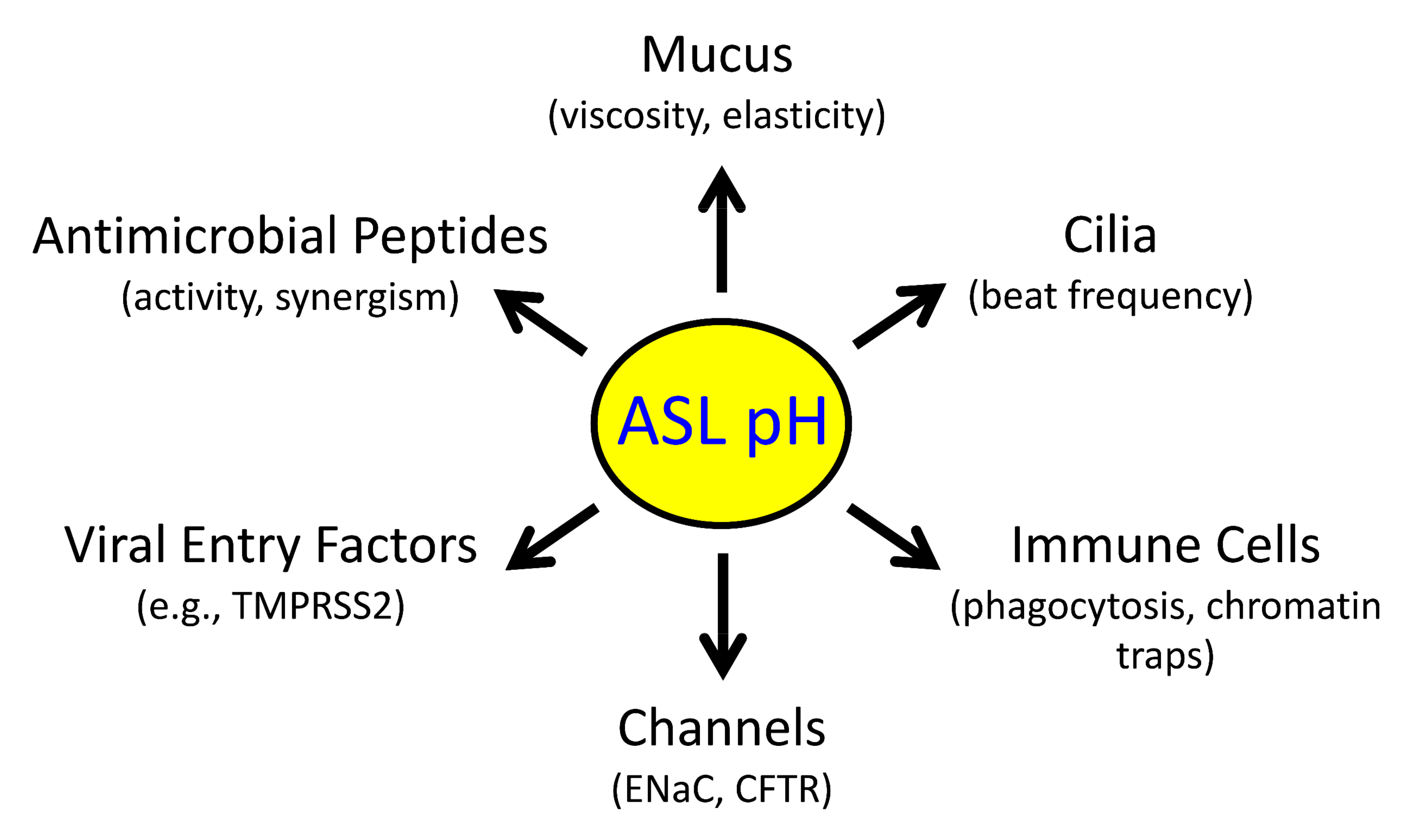
2.1. Reduced pHASL Disrupts Host Defenses

The normal pHASL is mildly acidic (e.g., 6.9–7.1 in human lower airways) relative to the interstitial fluid [7.4], though it varies considerably between individuals as well as between studies [1,36,37]. Recent reviews provide excellent tables summarizing pHASL variability between studies, including wild type versus CF [36,37]. Absolute pHASL measurements are influenced by technique, model system and airway region. It is also interesting to point out that pH is measured on log scale and a 3/10 increase in pH reflects a doubling of [H+].
Notwithstanding challenges involved in comparing results between studies, recent reports have identified several genetic changes in humans and/or animal models that alter pHASL (Figure 2). These studies provide critical insights into acid–base transport mechanisms that control pHASL and thus airway host defenses.
In humans with CF, the loss of CFTR-mediated HCO3 secretion leaves H+ secretion unbalanced, and hence lowers pHASL [51]. An abnormally acidic pHASL impairs mucociliary clearance, antimicrobial peptide activity against inhaled pathogens, and phagocytic cell function [23,24,27]. Raising pHASL at least partially rescues these impairments and identifies epithelial acid–base transporters as potential therapeutic targets.
The involvement of pHASL in airway host defense has also been tested by disrupting acid–base transporters other than CFTR. For instance, inhibiting basolateral NBC activity in human airway epithelia with normal CFTR channels lowers pHASL [52]. Interestingly, CFTR disruption in mice fails to produce spontaneous airway disease [53,54]. This is partly due to increased expression of non-CFTR HCO3 transporters and lack of expression of the non-gastric H+-pump (ATP12A) in murine airways (see below) [55]. As a result, pHASL in CF mice is the same as in non-CF mice. However, inhibiting basolateral NBC activity in freshly excised mouse tracheae reduces HCO3 secretion [52]. Moreover, mice lacking the main NBC isoform (SLC4A4−/−) show an airway phenotype marked by thick, adherent mucus and reduced mucociliary clearance, changes reminiscent of human CF airway disease.
Mutations in carbonic anhydrase isoform 12 (CA12) also phenocopy the loss of CFTR channel activity in human airways [56]. People with CA12 mutations show chronic coughs, airway colonization, bronchiectasis, and elevated sweat [Cl]. Interestingly, CA12 localizes at the apical membrane of airway epithelia. This expression pattern is physiologically relevant because proximal airway CO2 concentration fluctuates during tidal breathing [57]. Thus, pHASL rises during inhalation as airway lumen fills up with inhaled air (low CO2 concentration) but falls during exhalation as alveolar gases (higher CO2 concentration) enter the airways. Tidal pHASL oscillations enhance the epithelial host defense against bacteria. In CF, the amplitude of pHASL oscillations is reduced, which suggests a potential mechanism linking CFTR loss with impaired host defense.
In addition to HCO3 secretion, H+ secretion also controls pHASL. As noted above, mouse airways lack ATP12A expression and pHASL in CF mice is not reduced [55]. However, exogenous expression of ATP12A in CF mice increases H+ secretion, lowers pHASL, and impairs host defenses.
ASL buffers resist changes in pHASL when H+ ions are added or removed. The main ASL buffer is HCO3 [58]. Accordingly, in Calu-3 epithelia, forskolin stimulation increases and CFTR knockdown decreases buffering capacity of apically secreted fluid. Mucins are negatively charged molecules that can also bind H+ and thus contribute to ASL buffering [59,60]. This effect is dependent on mucin concentration. Mucus accumulation lowers the amplitude of ventilatory pHASL oscillations [57], and dampened oscillations reduce antibacterial host defense, thus providing a potential mechanism by which mucus accumulation may increase susceptibility to respiratory infections.
Together, the studies highlighted above point to the vital role of HCO3 and H+ transport mechanisms in controlling pHASL. Abnormally acidic pHASL, due to either reduced HCO3 secretion or unbalanced/increased H+ secretion, impairs critical first-line airway host defenses and predisposes to processes such as inflammation and chronic bacterial colonization. In the next section, we review studies that investigate how pHASL in CF might change with the development of inflammation and progression of airway disease.

2.2. pHASL Changes with CF Airway Disease Progression

In vitro studies in human airway epithelia show that pHASL is abnormally acidic in CF [51,55,61], but in vivo studies reveal mixed results. Some studies show a lower pHASL in CF [62,63], whereas others report no difference between CF and non-CF individuals [64,65]. This discrepancy is intriguing, given that CFTR-mediated HCO3 secretion is decreased in both in vitro and in vivo studies.
Most babies with CF develop airway inflammation over the first year of life [66,67,68,69,70]; in some cases, they also develop respiratory infections. Inflammation may induce ASL alkalinization through CFTR-independent mechanisms, and thus conceal the loss of CFTR-mediated HCO3 secretion. Studying initial stages of human CF airway disease might help separate effects due to CFTR loss from those due to inflammation. As inflammation develops early in CF, this approach requires studying ASL from babies soon after birth. In one small pilot study, pHASL in newborn CF babies (<four weeks of age) was lower compared with non-CF babies [71]. However, when the investigators studied the same cohort at three months of age, pHASL in CF babies had increased and did not differ from pHASL in non-CF babies. These findings are intriguing but require validation in a larger, independent cohort.
Several observations from the CF pig model might be relevant here: newborn CF piglets lack airway inflammation and infection and have an abnormally acidic pHASL [23,72]. In contrast, three-week-old CF piglets have airway inflammation, and their pHASL is higher than at birth and not different from the non-CF pHASL. Based on these observations, it may be predicted that CF-like inflammation in non-CF airways will further increase pHASL, and that inflamed non-CF airways will have a higher pHASL than inflamed CF airways. However, this prediction remains untested, partly because experimentally reproducing CF-like inflammation in vivo remains a challenge.
Respiratory viral infections also develop early in CF and are often encountered during infancy [73]. CF epithelia show multiple defects in antiviral immunity including reduced ASL-mediated neutralization of respiratory viruses [12], impaired interferon signaling [74], and lower levels of nitric oxide and hypothiocyanite [9,75,76]. However, whether pHASL influences susceptibility to respiratory viruses or antiviral host defense is not clear. Conversely, whether viral infection or viral-induced inflammation change pHASL also remains unknown.
CF airway inflammation is known to alter levels of airway antimicrobial peptides including defensins and LL-37 [77]. In addition to killing bacteria, antimicrobial peptides also modulate inflammation. For example, β-defensins interact with chemokine receptor 6 expressed on lymphocytes and dendritic cells and recruit these cells to sites of infection [78]. LL-37 directly binds LPS extracted from Pseudomonas aeruginosa and thus reduces IL-8 production from monocytes [79]. It is interesting to consider whether, similar to its effect on bacterial killing, pHASL also influences the immunomodulatory effects of antimicrobial peptides. Future studies in this area may reveal additional mechanisms linking reduced HCO3 secretion and abnormal pHASL regulation in CF with impaired host defenses.

2.3. Inflammatory Cytokines Regulate pHASL

In the absence of rigorous in vivo assessments, the effects of inflammation on pHASL may be investigated in vitro by applying CF-relevant inflammatory stimuli to primary cultures of differentiated human airway epithelia. The cytokine interleukin-17 (IL-17) is an evolutionarily conserved molecule that drives neutrophilic airway inflammation [80,81]. In targeting the airway epithelium, IL-17 acts synergistically with other cytokines such as tumor necrosis factor-α (TNFα), IL-1β, etc. [82]. These proinflammatory molecules are increased in established CF airway disease [83,84]. Some studies have also evaluated the effects of IL-4 and IL-13, which are relevant to asthma and allergic bronchopulmonary aspergillosis, conditions that often affect CF individuals [47,85,86,87]. These in vitro studies have provided key insights into the cellular and molecular mechanisms by which inflammation might regulate pHASL (Figure 3). In the following section, we review salient findings from these studies.

2.4. H+ Secretion

Inflammation may alter acid secretion into ASL. One study reported increased expression of ATP12A in CF airways with established disease [88]. Exposure of airway epithelia to IL-4 or IL-13 also increased ATP12A expression, and thus H+ secretion [47,86]. TNFα exposure had a similar effect, but IL-17 alone or combined IL-17/TNFα did not change H+ secretion [40]. Importantly, inhibiting ATP12A with apical ouabain decreases H+ secretion, lowers ASL viscosity, and increases bacterial killing [51,55,86]. Efforts to identify safer agents that reduce ATP12A activity are underway.
Loss of CFTR function also affects distal (small) airways. Small airway epithelia lack ATP12A, but instead use V-ATPase to secrete H+. Effects of inflammation and infection on small airway H+ secretion remain relatively unexplored. P. aeruginosa infection may reduce V-ATPase-mediated H+ secretion [89,90], or acidify ASL via apically expressed monocarboxylate transporter 2, a H+-lactate cotransporter [91]. Interestingly, Li et al. showed that pHASL regulates membrane localization of V-ATPase in porcine small airway epithelia [41]. At neutral pHASL (7.4), the V-ATPase subunit ATP6V0D2 localizes in the apical membrane of small airway secretory cells. However, at a lower extracellular pH of 6.8, ATP6V0D2 translocates into cytosol. V-ATPase may thus alter its apical membrane location and activity to prevent large changes in pHASL. Additional studies are needed to understand H+ secretion and pHASL regulation in small airway epithelia under both basal and inflamed conditions.

2.5. HCO3 Secretion

2.5.1. CFTR-Mediated HCO3 Secretion

Several inflammatory cytokines have been shown to alkalinize pHASL. IL-17/TNFα, IL-4, IL-13, and IL-1β increase CFTR expression and activity, and increased CFTR-mediated anion secretion improves respiratory host defenses [40,47,92,93]. In CF epithelia, this component of host response is missing due to mutated, non-functional CFTR proteins. In contrast to above-mentioned cytokines, TGF-β reduces CFTR expression and transport activities in airway epithelia [94,95]. In vivo effects of inflammation are likely to be complex given that several cytokines elevated in CF airways target airway epithelium, alter HCO3 transport, and thus modify pHASL.

2.5.2. Non-CFTR-Mediated HCO3 Secretion

An array of cytokines (IL-17/TNFα, IL-4, and IL-13) induce non-CFTR HCO3 secretion across airway epithelia [39,47,85,92,96]. This is achieved through pendrin, an apical Cl/HCO3 exchanger, encoded by the gene SLC26A4. Several aspects of this transport process are noteworthy. First, pendrin is minimally expressed in airway epithelia under basal conditions but is strongly upregulated by inflammatory cytokines. Second, pendrin is an electroneutral exchanger that does not mediate net anion secretion or absorption or change membrane potential. Third, in the absence of functional CFTR channels, pendrin alone can drive ASL alkalinization, though greater alkalinization is achieved with pendrin plus CFTR [39,40]. Fourth, some reports indicate structural or functional interactions between CFTR and pendrin, resulting in the increased activity of both [85,97]. Although these transporters are coexpressed in secretory cells, and studies in heterologous expression systems are supportive, more evidence is needed to establish their significance. Potentiating pendrin-mediated HCO3 secretion is a promising strategy for alkalinizing ASL and might be particularly relevant to airway inflammatory disorders.

2.5.3. Paracellular HCO3 Shunt

In addition to secretion by airway epithelial cells, HCO3 can also move between the cells. Very few studies have explored the contribution of the paracellular pathway to pHASL, though it is often mentioned in the context of inflammation. A recent report showed that the paracellular pathway is as permeable to HCO3 as it is to Cl [98]. Under basal conditions, pHASL (6.6) is lower than the pH of the interstitial fluid (7.4) and the paracellular HCO3 flux is towards the lumen; however, the paracellular HCO3 flux decreases or even reverses as pHASL approaches or rises higher than the pH of the interstitial fluid. The paracellular pathway thus acts as a HCO3 shunt that may oppose increased cellular HCO3 secretion in inflamed airway epithelia [99]. Whether paracellular HCO3 permeability can be modulated to support ASL alkalinization is an interesting question and requires further investigation.

2.6. Other Regulatory Mechanisms

Inflammatory cytokines regulate diverse cellular mechanisms involved in HCO3 secretion. In addition to changes in apical HCO3 transporters, cytokines such as IL-17/TNFα, IL-13, or IL-4 also increase transcripts of several carbonic anhydrase and NBC isoforms [39,47]. Additional cytoplasmic mechanisms that regulate epithelial HCO3 secretion also change in the presence of cytokines. The WNK (with-no-lysine [K]) kinases are master-regulators of pancreatic HCO3 secretion [100]. As reported recently, these kinases also control HCO3 secretion across CF and non-CF airway epithelia [101]. Secretory cells, key HCO3 secreting cells in airway epithelia, express two WNK isoforms, WNK1 and WNK2. Reducing WNK kinase activity increases HCO3 secretion, raises pHASL, and enhances CF epithelial host defenses. At a mechanistic level, WNK kinases regulate intracellular [Cl] through their control of the basolateral Na+-K+-2Cl cotransporter (NKCC1) (Figure 4). Inhibiting WNK lowers intracellular [Cl], which in turn may act as a signaling ion to stimulate HCO3 transport [102,103]. It is of note that combined IL-17/TNFα reduce WNK2 expression and raise pHASL and inhibiting residual WNK kinase activity further alkalinizes ASL. Future investigations may reveal additional mechanisms that regulate HCO3 and H+ secretion in inflamed airway epithelia.

3. Airway Inflammation and CFTR Modulators Influence Each Other

The natural history of CF airway disease has changed markedly with the widespread use of highly effective CFTR modulator therapy (HEMT) [104,105,106,107,108]. CFTR modulators are small-molecule drugs used to restore anion channel activity to mutated CFTR proteins. These agents include potentiators which increase the open-state probability of CFTR channels at the apical membrane, and correctors which increase the processing of misfolded CFTR proteins. A remarkably striking proof of the real-world effectiveness of these agents is the precipitous decline in new lung transplants for CF individuals coinciding with the availability of HEMT for > 90% of people with CF [109,110].
Inflammation is highly prevalent in CF individuals taking HEMT, which makes the relationship between airway inflammation and CFTR modulators critically important. The nature of this relationship is two-fold. On the one hand, starting HEMT results in either no change or a decrease in airway inflammatory markers [111,112,113]. On the other, inflammatory mediators that are elevated in CF airways enhance the restoration of CFTR channel function in response to CFTR modulators [39,114,115]. Interestingly, CFTR modulators further alkalinize pHASL in IL-17/TNFα-treated CF epithelia, but not in control CF epithelia [39]. Moreover, baseline sputum inflammatory markers (IL-8, IL-1β, neutrophil elastase) positively correlate with CFTR modulator-induced lung function improvements [39]. These effects are consistent with robust clinical improvements observed in most CF individuals after starting HEMT [109].
It is interesting to speculate as to the long-term effects of HEMT on pHASL. As noted above, pendrin is minimally expressed under basal conditions but markedly upregulated by inflammation. In inflamed CF airways, HEMT may further influence pHASL through at least two mechanisms: (a) by adding functional CFTR channels; and (b) by changing the level of inflammation and perhaps pendrin expression. Over time, how these factors might influence net HCO3 secretion and the relative contribution of CFTR and pendrin is not clear and is an interesting question for future studies.

4. Optimal Anti-Inflammatory Strategy in CF Is Unclear

Airway inflammation and bacterial colonization persist even after prolonged use of HEMT [116,117]. Considerable evidence suggests that inflammation may contribute to CF airway pathology [118]. However, the reports reviewed above indicate that inflammation may also have at least two beneficial effects: (1) it increases pHASL that may partially rescue host defense, and (2) it increases the response to CFTR modulators. Some studies suggest that cellular pathways activated by inflammation intersect with those involved in CFTR biogenesis [119]. It follows that the use of non-specific anti-inflammatory agents may limit ASL alkalinization and the restoration of CFTR channel activity with HEMT.
In the past, anti-inflammatory therapies in CF have produced mixed results. High-dose ibuprofen is currently the only strategy recommended by treatment guidelines, but it is not widely pursued in real-world settings due to issues with compliance and side-effects [120,121,122,123]. Other agents such as corticosteroids, azithromycin, etc., have not proved consistently effective. In airway epithelia, glucocorticoids reduce anion secretion [124] and CFTR mRNA expression [125]. Systemic steroids are well known to induce hyperglycemia, which may increase ASL glucose concentration, lower pHASL, and promote bacterial survival [91]. Considering these complex, inconsistent, and potentially harmful effects, the optimal strategy to target residual airway inflammation in people taking HEMT remains unclear. For future anti-inflammatory agents entering preclinical or clinical evaluation, reducing inflammation-mediated tissue damage without compromising HCO3 secretion, CFTR expression, and responses to CFTR modulators are all desirable features.

5. Conclusions

A well-orchestrated host response to inhaled particles and pathogens is essential for respiratory fitness. In CF, impaired HCO3 secretion and an abnormally acidic pHASL disrupt innate mucosal host defenses and initiate airway pathology. Persistent environmental challenges lead to chronic inflammation, a complex process with both protective and pathogenic effects. These effects are particularly important in the setting of bacterial or fungal colonization. Inflammation and infection persist after long-term HEMT and thus remain relevant concerns in the post-modulator era. However, the optimum anti-inflammatory approach for people taking HEMT remains unclear. One goal for future research might be to reveal targets and strategies that limit tissue damage but preserve HCO3 secretion and therapeutic responses to CFTR modulators. This goal may be pursued using a combination of in vitro and in vivo approaches. In vitro models may help achieve a better understanding of signaling pathways involved in acid–base secretion, CFTR biogenesis, and their relationship to inflammation. In vivo CF models that develop airway inflammation and have mutations amenable to CFTR modulators may prove useful in evaluating the safety and efficacy of novel anti-inflammatory agents.

Author Contributions

Conceptualization, T.R. and M.J.W.; writing—original draft preparation, T.R.; writing—review and editing, T.R. and M.J.W. All authors have read and agreed to the published version of the manuscript.

Funding

T.R. was supported by the Cystic Fibrosis Foundation Harry Shwachman Clinical Investigator Award. M.J.W. is an investigator of the Howard Hughes Medical Institute. Funding of work from T.R. and M.J.W. was supported in part by NIH research awards HL091842 and HL152960.

Institutional Review Board Statement

Not applicable.

Informed Consent Statement

Not applicable.

Data Availability Statement

Not applicable.

Conflicts of Interest

The authors declare no conflict of interest.

References

  1. Widdicombe, J.H. Airway Epithelium; Morgan & Claypool: San Rafael, CA, USA, 2013. [Google Scholar]
  2. Widdicombe, J.H.; Widdicombe, J.G. Regulation of human airway surface liquid. Respir. Physiol. 1995, 99, 3–12. [Google Scholar] [CrossRef] [PubMed]
  3. Haq, I.J.; Gray, M.A.; Garnett, J.P.; Ward, C.; Brodlie, M. Airway surface liquid homeostasis in cystic fibrosis: Pathophysiology and therapeutic targets. Thorax 2016, 71, 284–287. [Google Scholar] [CrossRef] [Green Version]
  4. Laube, D.M.; Yim, S.; Ryan, L.K.; Kisich, K.O.; Diamond, G. Antimicrobial peptides in the airway. Curr. Top. Microbiol. Immunol. 2006, 306, 153–182. [Google Scholar] [CrossRef]
  5. Ermund, A.; Trillo-Muyo, S.; Hansson, G.C. Assembly, Release, and Transport of Airway Mucins in Pigs and Humans. Ann. Am. Thorac. Soc. 2018, 15 (Suppl. S3), S159–S163. [Google Scholar] [CrossRef]
  6. Downey, D.G.; Bell, S.C.; Elborn, J.S. Neutrophils in cystic fibrosis. Thorax 2009, 64, 81–88. [Google Scholar] [CrossRef] [Green Version]
  7. Cheng, O.Z.; Palaniyar, N. NET balancing: A problem in inflammatory lung diseases. Front. Immunol. 2013, 4, 1. [Google Scholar] [CrossRef] [PubMed] [Green Version]
  8. Fragoso, M.A.; Fernandez, V.; Forteza, R.; Randell, S.H.; Salathe, M.; Conner, G.E. Transcellular thiocyanate transport by human airway epithelia. J. Physiol. 2004, 561 Pt 1, 183–194. [Google Scholar] [CrossRef]
  9. Moskwa, P.; Lorentzen, D.; Excoffon, K.J.; Zabner, J.; McCray, P.B., Jr.; Nauseef, W.M.; Dupuy, C.; Banfi, B. A novel host defense system of airways is defective in cystic fibrosis. Am. J. Respir. Crit. Care Med. 2007, 175, 174–183. [Google Scholar] [CrossRef] [PubMed] [Green Version]
  10. Lazarowski, E.R.; Tarran, R.; Grubb, B.R.; van Heusden, C.A.; Okada, S.; Boucher, R.C. Nucleotide release provides a mechanism for airway surface liquid homeostasis. J. Biol. Chem. 2004, 279, 36855–36864. [Google Scholar] [CrossRef] [PubMed] [Green Version]
  11. Van Heusden, C.; Grubb, B.R.; Button, B.; Lazarowski, E.R. Airway Epithelial Nucleotide Release Contributes to Mucociliary Clearance. Life 2021, 11, 430. [Google Scholar] [CrossRef]
  12. Berkebile, A.R.; Bartlett, J.A.; Abou Alaiwa, M.; Varga, S.M.; Power, U.F.; McCray, P.B., Jr. Airway Surface Liquid Has Innate Antiviral Activity That Is Reduced in Cystic Fibrosis. Am. J. Respir. Cell Mol. Biol. 2020, 62, 104–111. [Google Scholar] [CrossRef]
  13. Laporte, M.; Naesens, L. Airway proteases: An emerging drug target for influenza and other respiratory virus infections. Curr. Opin. Virol. 2017, 24, 16–24. [Google Scholar] [CrossRef] [PubMed]
  14. Hoffmann, M.; Kleine-Weber, H.; Schroeder, S.; Kruger, N.; Herrler, T.; Erichsen, S.; Schiergens, T.S.; Herrler, G.; Wu, N.H.; Nitsche, A.; et al. SARS-CoV-2 Cell Entry Depends on ACE2 and TMPRSS2 and Is Blocked by a Clinically Proven Protease Inhibitor. Cell 2020, 181, 271–280.e8. [Google Scholar] [CrossRef]
  15. Hill, D.B.; Button, B.; Rubinstein, M.; Boucher, R.C. Physiology and pathophysiology of human airway mucus. Physiol. Rev. 2022, 102, 1757–1836. [Google Scholar] [CrossRef]
  16. Voynow, J.A.; Rubin, B.K. Mucins, mucus, and sputum. Chest 2009, 135, 505–512. [Google Scholar] [CrossRef] [PubMed]
  17. Rubin, B.K. Mucus structure and properties in cystic fibrosis. Paediatr. Respir. Rev. 2007, 8, 4–7. [Google Scholar] [CrossRef] [PubMed]
  18. Stoltz, D.A.; Meyerholz, D.K.; Welsh, M.J. Origins of cystic fibrosis lung disease. N. Engl. J. Med. 2015, 372, 1574–1575. [Google Scholar] [CrossRef] [PubMed] [Green Version]
  19. Cutting, G.R. Cystic fibrosis genetics: From molecular understanding to clinical application. Nat. Rev. Genet. 2015, 16, 45–56. [Google Scholar] [CrossRef] [Green Version]
  20. Quinton, P.M. Physiological basis of cystic fibrosis: A historical perspective. Physiol. Rev. 1999, 79 (Suppl. S1), S3–S22. [Google Scholar] [CrossRef] [Green Version]
  21. Smith, J.J.; Welsh, M.J. cAMP stimulates bicarbonate secretion across normal, but not cystic fibrosis airway epithelia. J. Clin. Investig. 1992, 89, 1148–1153. [Google Scholar] [CrossRef] [Green Version]
  22. Poulsen, J.H.; Fischer, H.; Illek, B.; Machen, T.E. Bicarbonate conductance and pH regulatory capability of cystic fibrosis transmembrane conductance regulator. Proc. Natl. Acad. Sci. USA 1994, 91, 5340–5344. [Google Scholar] [CrossRef] [Green Version]
  23. Pezzulo, A.A.; Tang, X.X.; Hoegger, M.J.; Abou Alaiwa, M.H.; Ramachandran, S.; Moninger, T.O.; Karp, P.H.; Wohlford-Lenane, C.L.; Haagsman, H.P.; van Eijk, M.; et al. Reduced airway surface pH impairs bacterial killing in the porcine cystic fibrosis lung. Nature 2012, 487, 109–113. [Google Scholar] [CrossRef] [PubMed] [Green Version]
  24. Tang, X.X.; Ostedgaard, L.S.; Hoegger, M.J.; Moninger, T.O.; Karp, P.H.; McMenimen, J.D.; Choudhury, B.; Varki, A.; Stoltz, D.A.; Welsh, M.J. Acidic pH increases airway surface liquid viscosity in cystic fibrosis. J. Clin. Investig. 2016, 126, 879–891. [Google Scholar] [CrossRef] [PubMed] [Green Version]
  25. Gustafsson, J.K.; Ermund, A.; Ambort, D.; Johansson, M.E.; Nilsson, H.E.; Thorell, K.; Hebert, H.; Sjovall, H.; Hansson, G.C. Bicarbonate and functional CFTR channel are required for proper mucin secretion and link cystic fibrosis with its mucus phenotype. J. Exp. Med. 2012, 209, 1263–1272. [Google Scholar] [CrossRef] [Green Version]
  26. Clary-Meinesz, C.; Mouroux, J.; Cosson, J.; Huitorel, P.; Blaive, B. Influence of external pH on ciliary beat frequency in human bronchi and bronchioles. Eur. Respir. J. 1998, 11, 330–333. [Google Scholar] [CrossRef] [Green Version]
  27. Khan, M.A.; Philip, L.M.; Cheung, G.; Vadakepeedika, S.; Grasemann, H.; Sweezey, N.; Palaniyar, N. Regulating NETosis: Increasing pH Promotes NADPH Oxidase-Dependent NETosis. Front. Med. 2018, 5, 19. [Google Scholar] [CrossRef] [PubMed]
  28. Garland, A.L.; Walton, W.G.; Coakley, R.D.; Tan, C.D.; Gilmore, R.C.; Hobbs, C.A.; Tripathy, A.; Clunes, L.A.; Bencharit, S.; Stutts, M.J.; et al. Molecular basis for pH-dependent mucosal dehydration in cystic fibrosis airways. Proc. Natl. Acad. Sci. USA 2013, 110, 15973–15978. [Google Scholar] [CrossRef] [Green Version]
  29. Abou Alaiwa, M.H.; Reznikov, L.R.; Gansemer, N.D.; Sheets, K.A.; Horswill, A.R.; Stoltz, D.A.; Zabner, J.; Welsh, M.J. pH modulates the activity and synergism of the airway surface liquid antimicrobials beta-defensin-3 and LL-37. Proc. Natl. Acad. Sci. USA 2014, 111, 18703–18708. [Google Scholar] [CrossRef] [Green Version]
  30. Middleton, P.G.; Taylor-Cousar, J.L. Development of elexacaftor–tezacaftor–ivacaftor: Highly effective CFTR modulation for the majority of people with Cystic Fibrosis. Expert Rev. Respir. Med. 2021, 15, 723–735. [Google Scholar] [CrossRef]
  31. Myerburg, M.; Pilewski, J.M. CFTR Modulators to the Rescue of Individuals with Cystic Fibrosis and Advanced Lung Disease. Am. J. Respir. Crit. Care Med. 2021, 204, 7–9. [Google Scholar] [CrossRef]
  32. Volkova, N.; Moy, K.; Evans, J.; Campbell, D.; Tian, S.; Simard, C.; Higgins, M.; Konstan, M.W.; Sawicki, G.S.; Elbert, A.; et al. Disease progression in patients with cystic fibrosis treated with ivacaftor: Data from national US and UK registries. J. Cyst. Fibros. 2020, 19, 68–79. [Google Scholar] [CrossRef] [PubMed]
  33. Wang, Y.; Lam, C.S.; Wu, F.; Wang, W.; Duan, Y.; Huang, P. Regulation of CFTR channels by HCO(3)--sensitive soluble adenylyl cyclase in human airway epithelial cells. Am. J. Physiol. Cell Physiol. 2005, 289, C1145–C1151. [Google Scholar] [CrossRef] [Green Version]
  34. Baudouin-Legros, M.; Hamdaoui, N.; Borot, F.; Fritsch, J.; Ollero, M.; Planelles, G.; Edelman, A. Control of basal CFTR gene expression by bicarbonate-sensitive adenylyl cyclase in human pulmonary cells. Cell. Physiol. Biochem. 2008, 21, 75–86. [Google Scholar] [CrossRef] [PubMed]
  35. Kreutzberger, A.J.B.; Sanyal, A.; Saminathan, A.; Bloyet, L.M.; Stumpf, S.; Liu, Z.; Ojha, R.; Patjas, M.T.; Geneid, A.; Scanavachi, G.; et al. SARS-CoV-2 requires acidic pH to infect cells. Proc. Natl. Acad. Sci. USA 2022, 119, e2209514119. [Google Scholar] [CrossRef]
  36. Zajac, M.; Dreano, E.; Edwards, A.; Planelles, G.; Sermet-Gaudelus, I. Airway Surface Liquid pH Regulation in Airway Epithelium Current Understandings and Gaps in Knowledge. Int. J. Mol. Sci. 2021, 22, 3384. [Google Scholar] [CrossRef]
  37. Fischer, H.; Widdicombe, J.H. Mechanisms of acid and base secretion by the airway epithelium. J. Membr. Biol. 2006, 211, 139–150. [Google Scholar] [CrossRef] [PubMed] [Green Version]
  38. Saint-Criq, V.; Gray, M.A. Role of CFTR in epithelial physiology. Cell Mol. Life Sci. 2017, 74, 93–115. [Google Scholar] [CrossRef] [PubMed] [Green Version]
  39. Rehman, T.; Karp, P.H.; Tan, P.; Goodell, B.J.; Pezzulo, A.A.; Thurman, A.L.; Thornell, I.M.; Durfey, S.L.; Duffey, M.E.; Stoltz, D.A.; et al. Inflammatory cytokines TNF-alpha and IL-17 enhance the efficacy of cystic fibrosis transmembrane conductance regulator modulators. J. Clin. Investig. 2021, 131, e150398. [Google Scholar] [CrossRef] [PubMed]
  40. Rehman, T.; Thornell, I.M.; Pezzulo, A.A.; Thurman, A.L.; Romano Ibarra, G.S.; Karp, P.H.; Tan, P.; Duffey, M.E.; Welsh, M.J. TNFalpha and IL-17 alkalinize airway surface liquid through CFTR and pendrin. Am. J. Physiol. Cell Physiol. 2020, 319, C331–C344. [Google Scholar] [CrossRef] [PubMed]
  41. Li, X.; Villacreses, R.; Thornell, I.M.; Noriega, J.; Mather, S.; Brommel, C.M.; Lu, L.; Zabner, A.; Ehler, A.; Meyerholz, D.K.; et al. V-Type ATPase Mediates Airway Surface Liquid Acidification in Pig Small Airway Epithelial Cells. Am. J. Respir. Cell Mol. Biol. 2021, 65, 146–156. [Google Scholar] [CrossRef]
  42. Shamsuddin, A.K.; Quinton, P.M. Native small airways secrete bicarbonate. Am. J. Respir. Cell Mol. Biol. 2014, 50, 796–804. [Google Scholar] [CrossRef] [PubMed] [Green Version]
  43. Bridges, R.J. Mechanisms of bicarbonate secretion: Lessons from the airways. Cold Spring Harb. Perspect. Med. 2012, 2, a015016. [Google Scholar] [CrossRef] [PubMed] [Green Version]
  44. Devor, D.C.; Singh, A.K.; Lambert, L.C.; DeLuca, A.; Frizzell, R.A.; Bridges, R.J. Bicarbonate and chloride secretion in Calu-3 human airway epithelial cells. J. Gen. Physiol. 1999, 113, 743–760. [Google Scholar] [CrossRef] [PubMed] [Green Version]
  45. Kreindler, J.L.; Peters, K.W.; Frizzell, R.A.; Bridges, R.J. Identification and membrane localization of electrogenic sodium bicarbonate cotransporters in Calu-3 cells. Biochim. Biophys. Acta 2006, 1762, 704–710. [Google Scholar] [CrossRef] [PubMed] [Green Version]
  46. Supuran, C.T. Carbonic anhydrases—An overview. Curr. Pharm. Des. 2008, 14, 603–614. [Google Scholar] [CrossRef]
  47. Gorrieri, G.; Scudieri, P.; Caci, E.; Schiavon, M.; Tomati, V.; Sirci, F.; Napolitano, F.; Carrella, D.; Gianotti, A.; Musante, I.; et al. Goblet Cell Hyperplasia Requires High Bicarbonate Transport To Support Mucin Release. Sci. Rep. 2016, 6, 36016. [Google Scholar] [CrossRef]
  48. Alper, S.L. Molecular physiology and genetics of Na+-independent SLC4 anion exchangers. J. Exp. Biol. 2009, 212 Pt 11, 1672–1683. [Google Scholar] [CrossRef] [Green Version]
  49. Hollenhorst, M.I.; Richter, K.; Fronius, M. Ion transport by pulmonary epithelia. J. Biomed. Biotechnol. 2011, 2011, 174306. [Google Scholar] [CrossRef] [Green Version]
  50. Xu, H.; Ghishan, F.K.; Kiela, P.R. SLC9 Gene Family: Function, Expression, and Regulation. Compr. Physiol. 2018, 8, 555–583. [Google Scholar] [CrossRef]
  51. Simonin, J.; Bille, E.; Crambert, G.; Noel, S.; Dreano, E.; Edwards, A.; Hatton, A.; Pranke, I.; Villeret, B.; Cottart, C.H.; et al. Airway surface liquid acidification initiates host defense abnormalities in Cystic Fibrosis. Sci. Rep. 2019, 9, 6516. [Google Scholar] [CrossRef] [Green Version]
  52. Saint-Criq, V.; Guequen, A.; Philp, A.R.; Villanueva, S.; Apablaza, T.; Fernandez-Moncada, I.; Mansilla, A.; Delpiano, L.; Ruminot, I.; Carrasco, C.; et al. Inhibition of the sodium-dependent HCO3(-) transporter SLC4A4, produces a cystic fibrosis-like airway disease phenotype. eLife 2022, 11, e75871. [Google Scholar] [CrossRef]
  53. Rosen, B.H.; Chanson, M.; Gawenis, L.R.; Liu, J.; Sofoluwe, A.; Zoso, A.; Engelhardt, J.F. Animal and model systems for studying cystic fibrosis. J. Cyst. Fibros. 2018, 17, S28–S34. [Google Scholar] [CrossRef] [Green Version]
  54. McCarron, A.; Donnelley, M.; Parsons, D. Airway disease phenotypes in animal models of cystic fibrosis. Respir. Res. 2018, 19, 54. [Google Scholar] [CrossRef] [Green Version]
  55. Shah, V.S.; Meyerholz, D.K.; Tang, X.X.; Reznikov, L.; Abou Alaiwa, M.; Ernst, S.E.; Karp, P.H.; Wohlford-Lenane, C.L.; Heilmann, K.P.; Leidinger, M.R.; et al. Airway acidification initiates host defense abnormalities in cystic fibrosis mice. Science 2016, 351, 503–507. [Google Scholar] [CrossRef] [PubMed] [Green Version]
  56. Lee, M.; Vecchio-Pagan, B.; Sharma, N.; Waheed, A.; Li, X.; Raraigh, K.S.; Robbins, S.; Han, S.T.; Franca, A.L.; Pellicore, M.J.; et al. Loss of carbonic anhydrase XII function in individuals with elevated sweat chloride concentration and pulmonary airway disease. Hum. Mol. Genet. 2016, 25, 1923–1933. [Google Scholar] [CrossRef] [PubMed] [Green Version]
  57. Kim, D.; Liao, J.; Scales, N.B.; Martini, C.; Luan, X.; Abu-Arish, A.; Robert, R.; Luo, Y.; McKay, G.A.; Nguyen, D.; et al. Large pH oscillations promote host defense against human airways infection. J. Exp. Med. 2021, 218, e20201831. [Google Scholar] [CrossRef]
  58. Kim, D.; Liao, J.; Hanrahan, J.W. The buffer capacity of airway epithelial secretions. Front. Physiol. 2014, 5, 188. [Google Scholar] [CrossRef] [Green Version]
  59. Holma, B. Influence of buffer capacity and pH-dependent rheological properties of respiratory mucus on health effects due to acidic pollution. Sci. Total Environ. 1985, 41, 101–123. [Google Scholar] [CrossRef]
  60. Holma, B.; Hegg, P.O. pH- and protein-dependent buffer capacity and viscosity of respiratory mucus. Their interrelationships and influence on health. Sci. Total Environ. 1989, 84, 71–82. [Google Scholar] [CrossRef] [PubMed]
  61. Coakley, R.D.; Grubb, B.R.; Paradiso, A.M.; Gatzy, J.T.; Johnson, L.G.; Kreda, S.M.; O'Neal, W.K.; Boucher, R.C. Abnormal surface liquid pH regulation by cultured cystic fibrosis bronchial epithelium. Proc. Natl. Acad. Sci. USA 2003, 100, 16083–16088. [Google Scholar] [CrossRef] [Green Version]
  62. Tate, S.; MacGregor, G.; Davis, M.; Innes, J.A.; Greening, A.P. Airways in cystic fibrosis are acidified: Detection by exhaled breath condensate. Thorax 2002, 57, 926–929. [Google Scholar] [CrossRef] [Green Version]
  63. Song, Y.; Salinas, D.; Nielson, D.W.; Verkman, A.S. Hyperacidity of secreted fluid from submucosal glands in early cystic fibrosis. Am. J. Physiol. Cell Physiol. 2006, 290, C741–C749. [Google Scholar] [CrossRef]
  64. McShane, D.; Davies, J.C.; Davies, M.G.; Bush, A.; Geddes, D.M.; Alton, E.W. Airway surface pH in subjects with cystic fibrosis. Eur. Respir. J. 2003, 21, 37–42. [Google Scholar] [CrossRef]
  65. Schultz, A.; Puvvadi, R.; Borisov, S.M.; Shaw, N.C.; Klimant, I.; Berry, L.J.; Montgomery, S.T.; Nguyen, T.; Kreda, S.M.; Kicic, A.; et al. Airway surface liquid pH is not acidic in children with cystic fibrosis. Nat. Commun. 2017, 8, 1409. [Google Scholar] [CrossRef] [Green Version]
  66. Sly, P.D.; Brennan, S.; Gangell, C.; de Klerk, N.; Murray, C.; Mott, L.; Stick, S.M.; Robinson, P.J.; Robertson, C.F.; Ranganathan, S.C.; et al. Lung disease at diagnosis in infants with cystic fibrosis detected by newborn screening. Am. J. Respir. Crit. Care Med. 2009, 180, 146–152. [Google Scholar] [CrossRef] [PubMed]
  67. Sly, P.D.; Gangell, C.L.; Chen, L.; Ware, R.S.; Ranganathan, S.; Mott, L.S.; Murray, C.P.; Stick, S.M.; Investigators, A.C. Risk factors for bronchiectasis in children with cystic fibrosis. N. Engl. J. Med. 2013, 368, 1963–1970. [Google Scholar] [CrossRef] [PubMed] [Green Version]
  68. Khan, T.Z.; Wagener, J.S.; Bost, T.; Martinez, J.; Accurso, F.J.; Riches, D.W. Early pulmonary inflammation in infants with cystic fibrosis. Am. J. Respir. Crit. Care Med. 1995, 151, 1075–1082. [Google Scholar] [CrossRef]
  69. Balough, K.; McCubbin, M.; Weinberger, M.; Smits, W.; Ahrens, R.; Fick, R. The relationship between infection and inflammation in the early stages of lung disease from cystic fibrosis. Pediatr. Pulmonol. 1995, 20, 63–70. [Google Scholar] [CrossRef]
  70. Ranganathan, S.C.; Hall, G.L.; Sly, P.D.; Stick, S.M.; Douglas, T.A.; Australian Respiratory Early Surveillance Team for Cystic Fibrosis (AREST-CF). Early Lung Disease in Infants and Preschool Children with Cystic Fibrosis. What Have We Learned and What Should We Do about It? Am. J. Respir. Crit. Care Med. 2017, 195, 1567–1575. [Google Scholar] [CrossRef] [PubMed]
  71. Abou Alaiwa, M.H.; Beer, A.M.; Pezzulo, A.A.; Launspach, J.L.; Horan, R.A.; Stoltz, D.A.; Starner, T.D.; Welsh, M.J.; Zabner, J. Neonates with cystic fibrosis have a reduced nasal liquid pH; a small pilot study. J. Cyst. Fibros. 2014, 13, 373–377. [Google Scholar] [CrossRef] [Green Version]
  72. Stoltz, D.A.; Meyerholz, D.K.; Pezzulo, A.A.; Ramachandran, S.; Rogan, M.P.; Davis, G.J.; Hanfland, R.A.; Wohlford-Lenane, C.; Dohrn, C.L.; Bartlett, J.A.; et al. Cystic fibrosis pigs develop lung disease and exhibit defective bacterial eradication at birth. Sci. Transl. Med. 2010, 2, 29ra31. [Google Scholar] [CrossRef] [Green Version]
  73. Deschamp, A.R.; Hatch, J.E.; Slaven, J.E.; Gebregziabher, N.; Storch, G.; Hall, G.L.; Stick, S.; Ranganathan, S.; Ferkol, T.W.; Davis, S.D. Early respiratory viral infections in infants with cystic fibrosis. J. Cyst. Fibros. 2019, 18, 844–850. [Google Scholar] [CrossRef] [Green Version]
  74. Xu, W.; Zheng, S.; Goggans, T.M.; Kiser, P.; Quinones-Mateu, M.E.; Janocha, A.J.; Comhair, S.A.; Slee, R.; Williams, B.R.; Erzurum, S.C. Cystic fibrosis and normal human airway epithelial cell response to influenza a viral infection. J. Interferon Cytokine Res. 2006, 26, 609–627. [Google Scholar] [CrossRef]
  75. Zheng, S.; De, B.P.; Choudhary, S.; Comhair, S.A.; Goggans, T.; Slee, R.; Williams, B.R.; Pilewski, J.; Haque, S.J.; Erzurum, S.C. Impaired innate host defense causes susceptibility to respiratory virus infections in cystic fibrosis. Immunity 2003, 18, 619–630. [Google Scholar] [CrossRef] [PubMed] [Green Version]
  76. Zheng, S.; Xu, W.; Bose, S.; Banerjee, A.K.; Haque, S.J.; Erzurum, S.C. Impaired nitric oxide synthase-2 signaling pathway in cystic fibrosis airway epithelium. Am. J. Physiol. Lung Cell. Mol. Physiol. 2004, 287, L374–L381. [Google Scholar] [CrossRef] [PubMed]
  77. Chen, C.I.; Schaller-Bals, S.; Paul, K.P.; Wahn, U.; Bals, R. Beta-defensins and LL-37 in bronchoalveolar lavage fluid of patients with cystic fibrosis. J. Cyst. Fibros. 2004, 3, 45–50. [Google Scholar] [CrossRef] [Green Version]
  78. Yang, D.; Chertov, O.; Bykovskaia, S.N.; Chen, Q.; Buffo, M.J.; Shogan, J.; Anderson, M.; Schroder, J.M.; Wang, J.M.; Howard, O.M.; et al. Beta-defensins: Linking innate and adaptive immunity through dendritic and T cell CCR6. Science 1999, 286, 525–528. [Google Scholar] [CrossRef]
  79. Scott, A.; Weldon, S.; Buchanan, P.J.; Schock, B.; Ernst, R.K.; McAuley, D.F.; Tunney, M.M.; Irwin, C.R.; Elborn, J.S.; Taggart, C.C. Evaluation of the ability of LL-37 to neutralise LPS in vitro and ex vivo. PLoS ONE 2011, 6, e26525. [Google Scholar] [CrossRef] [Green Version]
  80. McAleer, J.P.; Kolls, J.K. Mechanisms controlling Th17 cytokine expression and host defense. J. Leukoc. Biol. 2011, 90, 263–270. [Google Scholar] [CrossRef] [PubMed] [Green Version]
  81. Stoppelenburg, A.J.; Salimi, V.; Hennus, M.; Plantinga, M.; Huis in 't Veld, R.; Walk, J.; Meerding, J.; Coenjaerts, F.; Bont, L.; Boes, M. Local IL-17A potentiates early neutrophil recruitment to the respiratory tract during severe RSV infection. PLoS ONE 2013, 8, e78461. [Google Scholar] [CrossRef]
  82. McAllister, F.; Henry, A.; Kreindler, J.L.; Dubin, P.J.; Ulrich, L.; Steele, C.; Finder, J.D.; Pilewski, J.M.; Carreno, B.M.; Goldman, S.J.; et al. Role of IL-17A, IL-17F, and the IL-17 receptor in regulating growth-related oncogene-alpha and granulocyte colony-stimulating factor in bronchial epithelium: Implications for airway inflammation in cystic fibrosis. J. Immunol. 2005, 175, 404–412. [Google Scholar] [CrossRef] [PubMed] [Green Version]
  83. Tan, H.L.; Regamey, N.; Brown, S.; Bush, A.; Lloyd, C.M.; Davies, J.C. The Th17 pathway in cystic fibrosis lung disease. Am. J. Respir. Crit. Care Med. 2011, 184, 252–258. [Google Scholar] [CrossRef] [PubMed] [Green Version]
  84. Karpati, F.; Hjelte, F.L.; Wretlind, B. TNF-alpha and IL-8 in consecutive sputum samples from cystic fibrosis patients during antibiotic treatment. Scand. J. Infect. Dis. 2000, 32, 75–79. [Google Scholar] [CrossRef] [PubMed]
  85. Kim, D.; Huang, J.; Billet, A.; Abu-Arish, A.; Goepp, J.; Matthes, E.; Tewfik, M.A.; Frenkiel, S.; Hanrahan, J.W. Pendrin Mediates Bicarbonate Secretion and Enhances Cystic Fibrosis Transmembrane Conductance Regulator Function in Airway Surface Epithelia. Am. J. Respir. Cell Mol. Biol. 2019, 60, 705–716. [Google Scholar] [CrossRef]
  86. Lennox, A.T.; Coburn, S.L.; Leech, J.A.; Heidrich, E.M.; Kleyman, T.R.; Wenzel, S.E.; Pilewski, J.M.; Corcoran, T.E.; Myerburg, M.M. ATP12A promotes mucus dysfunction during Type 2 airway inflammation. Sci. Rep. 2018, 8, 2109. [Google Scholar] [CrossRef] [Green Version]
  87. Haggie, P.M.; Phuan, P.W.; Tan, J.A.; Zlock, L.; Finkbeiner, W.E.; Verkman, A.S. Inhibitors of pendrin anion exchange identified in a small molecule screen increase airway surface liquid volume in cystic fibrosis. FASEB J. 2016, 30, 2187–2197. [Google Scholar] [CrossRef] [Green Version]
  88. Scudieri, P.; Musante, I.; Caci, E.; Venturini, A.; Morelli, P.; Walter, C.; Tosi, D.; Palleschi, A.; Martin-Vasallo, P.; Sermet-Gaudelus, I.; et al. Increased expression of ATP12A proton pump in cystic fibrosis airways. JCI Insight 2018, 3, e123616. [Google Scholar] [CrossRef] [PubMed] [Green Version]
  89. Kong, F.; Young, L.; Chen, Y.; Ran, H.; Meyers, M.; Joseph, P.; Cho, Y.H.; Hassett, D.J.; Lau, G.W. Pseudomonas aeruginosa pyocyanin inactivates lung epithelial vacuolar ATPase-dependent cystic fibrosis transmembrane conductance regulator expression and localization. Cell. Microbiol. 2006, 8, 1121–1133. [Google Scholar] [CrossRef]
  90. Ran, H.; Hassett, D.J.; Lau, G.W. Human targets of Pseudomonas aeruginosa pyocyanin. Proc. Natl. Acad. Sci. USA 2003, 100, 14315–14320. [Google Scholar] [CrossRef] [Green Version]
  91. Garnett, J.P.; Kalsi, K.K.; Sobotta, M.; Bearham, J.; Carr, G.; Powell, J.; Brodlie, M.; Ward, C.; Tarran, R.; Baines, D.L. Hyperglycaemia and Pseudomonas aeruginosa acidify cystic fibrosis airway surface liquid by elevating epithelial monocarboxylate transporter 2 dependent lactate-H(+) secretion. Sci. Rep. 2016, 6, 37955. [Google Scholar] [CrossRef] [Green Version]
  92. Simoes, F.B.; Kmit, A.; Amaral, M.D. Cross-talk of inflammatory mediators and airway epithelium reveals the cystic fibrosis transmembrane conductance regulator as a major target. ERJ Open Res. 2021, 7, 00247–2021. [Google Scholar] [CrossRef]
  93. Gray, T.; Coakley, R.; Hirsh, A.; Thornton, D.; Kirkham, S.; Koo, J.S.; Burch, L.; Boucher, R.; Nettesheim, P. Regulation of MUC5AC mucin secretion and airway surface liquid metabolism by IL-1beta in human bronchial epithelia. Am. J. Physiol. Lung Cell Mol. Physiol. 2004, 286, L320–L330. [Google Scholar] [CrossRef] [PubMed] [Green Version]
  94. Snodgrass, S.M.; Cihil, K.M.; Cornuet, P.K.; Myerburg, M.M.; Swiatecka-Urban, A. Tgf-beta1 inhibits Cftr biogenesis and prevents functional rescue of DeltaF508-Cftr in primary differentiated human bronchial epithelial cells. PLoS ONE 2013, 8, e63167. [Google Scholar] [CrossRef] [PubMed] [Green Version]
  95. Kim, M.D.; Bengtson, C.D.; Yoshida, M.; Niloy, A.J.; Dennis, J.S.; Baumlin, N.; Salathe, M. Losartan ameliorates TGF-beta1-induced CFTR dysfunction and improves correction by cystic fibrosis modulator therapies. J. Clin. Investig. 2022, 132, e155241. [Google Scholar] [CrossRef]
  96. Adams, K.M.; Abraham, V.; Spielman, D.; Kolls, J.K.; Rubenstein, R.C.; Conner, G.E.; Cohen, N.A.; Kreindler, J.L. IL-17A induces Pendrin expression and chloride-bicarbonate exchange in human bronchial epithelial cells. PLoS ONE 2014, 9, e103263. [Google Scholar] [CrossRef] [Green Version]
  97. Gray, M.A. Bicarbonate secretion: It takes two to tango. Nat. Cell Biol. 2004, 6, 292–294. [Google Scholar] [CrossRef] [PubMed]
  98. Thornell, I.M.; Rehman, T.; Pezzulo, A.A.; Welsh, M.J. Paracellular bicarbonate flux across human cystic fibrosis airway epithelia tempers changes in airway surface liquid pH. J. Physiol. 2020, 598, 4307–4320. [Google Scholar] [CrossRef]
  99. Parker, M.D. Soda stream modifies airway fluid. J. Physiol. 2020, 598, 4143–4144. [Google Scholar] [CrossRef] [PubMed]
  100. Lee, M.G.; Ohana, E.; Park, H.W.; Yang, D.; Muallem, S. Molecular mechanism of pancreatic and salivary gland fluid and HCO3 secretion. Physiol. Rev. 2012, 92, 39–74. [Google Scholar] [CrossRef] [Green Version]
  101. Rehman, T.; Karp, P.H.; Thurman, A.L.; Mather, S.E.; Jain, A.; Cooney, A.L.; Sinn, P.L.; Pezzulo, A.A.; Duffey, M.E.; Welsh, M.J. WNK Inhibition Increases Surface Liquid pH and Host Defense in Cystic Fibrosis Airway Epithelia. Am. J. Respir. Cell Mol. Biol. 2022, 67, 491–502. [Google Scholar] [CrossRef]
  102. Luscher, B.P.; Vachel, L.; Ohana, E.; Muallem, S. Cl(-) as a bona fide signaling ion. Am. J. Physiol. Cell Physiol. 2020, 318, C125–C136. [Google Scholar] [CrossRef]
  103. Shcheynikov, N.; Son, A.; Hong, J.H.; Yamazaki, O.; Ohana, E.; Kurtz, I.; Shin, D.M.; Muallem, S. Intracellular Cl- as a signaling ion that potently regulates Na+/HCO3- transporters. Proc. Natl. Acad. Sci. USA 2015, 112, E329–E337. [Google Scholar] [CrossRef] [Green Version]
  104. Ramsey, B.W.; Davies, J.; McElvaney, N.G.; Tullis, E.; Bell, S.C.; Drevinek, P.; Griese, M.; McKone, E.F.; Wainwright, C.E.; Konstan, M.W.; et al. A CFTR potentiator in patients with cystic fibrosis and the G551D mutation. N. Engl. J. Med. 2011, 365, 1663–1672. [Google Scholar] [CrossRef] [PubMed] [Green Version]
  105. Keating, D.; Marigowda, G.; Burr, L.; Daines, C.; Mall, M.A.; McKone, E.F.; Ramsey, B.W.; Rowe, S.M.; Sass, L.A.; Tullis, E.; et al. VX-445-Tezacaftor-Ivacaftor in Patients with Cystic Fibrosis and One or Two Phe508del Alleles. N. Engl. J. Med. 2018, 379, 1612–1620. [Google Scholar] [CrossRef] [PubMed]
  106. Middleton, P.G.; Mall, M.A.; Drevinek, P.; Lands, L.C.; McKone, E.F.; Polineni, D.; Ramsey, B.W.; Taylor-Cousar, J.L.; Tullis, E.; Vermeulen, F.; et al. Elexacaftor-Tezacaftor-Ivacaftor for Cystic Fibrosis with a Single Phe508del Allele. N. Engl. J. Med. 2019, 381, 1809–1819. [Google Scholar] [CrossRef] [PubMed]
  107. Barry, P.J.; Mall, M.A.; Alvarez, A.; Colombo, C.; de Winter-de Groot, K.M.; Fajac, I.; McBennett, K.A.; McKone, E.F.; Ramsey, B.W.; Sutharsan, S.; et al. Triple Therapy for Cystic Fibrosis Phe508del-Gating and -Residual Function Genotypes. N. Engl. J. Med. 2021, 385, 815–825. [Google Scholar] [CrossRef]
  108. Mall, M.A.; Mayer-Hamblett, N.; Rowe, S.M. Cystic Fibrosis: Emergence of Highly Effective Targeted Therapeutics and Potential Clinical Implications. Am. J. Respir. Crit. Care Med. 2020, 201, 1193–1208. [Google Scholar] [CrossRef]
  109. Burgel, P.R.; Durieu, I.; Chiron, R.; Ramel, S.; Danner-Boucher, I.; Prevotat, A.; Grenet, D.; Marguet, C.; Reynaud-Gaubert, M.; Macey, J.; et al. Rapid Improvement after Starting Elexacaftor-Tezacaftor-Ivacaftor in Patients with Cystic Fibrosis and Advanced Pulmonary Disease. Am. J. Respir. Crit. Care Med. 2021, 204, 64–73. [Google Scholar] [CrossRef]
  110. Martin, C.; Legeai, C.; Regard, L.; Cantrelle, C.; Dorent, R.; Carlier, N.; Kerbaul, F.; Burgel, P.R. Major Decrease in Lung Transplantation for Patients with Cystic Fibrosis in France. Am. J. Respir. Crit. Care Med. 2022, 205, 584–586. [Google Scholar] [CrossRef]
  111. Hisert, K.B.; Heltshe, S.L.; Pope, C.; Jorth, P.; Wu, X.; Edwards, R.M.; Radey, M.; Accurso, F.J.; Wolter, D.J.; Cooke, G.; et al. Restoring Cystic Fibrosis Transmembrane Conductance Regulator Function Reduces Airway Bacteria and Inflammation in People with Cystic Fibrosis and Chronic Lung Infections. Am. J. Respir. Crit. Care Med. 2017, 195, 1617–1628. [Google Scholar] [CrossRef]
  112. Harris, J.K.; Wagner, B.D.; Zemanick, E.T.; Robertson, C.E.; Stevens, M.J.; Heltshe, S.L.; Rowe, S.M.; Sagel, S.D. Changes in Airway Microbiome and Inflammation with Ivacaftor Treatment in Patients with Cystic Fibrosis and the G551D Mutation. Ann. Am. Thorac. Soc. 2020, 17, 212–220. [Google Scholar] [CrossRef] [PubMed]
  113. McNally, P.; Butler, D.; Karpievitch, Y.V.; Linnane, B.; Ranganathan, S.; Stick, S.M.; Hall, G.L.; Schultz, A. Ivacaftor and Airway Inflammation in Preschool Children with Cystic Fibrosis. Am. J. Respir. Crit. Care Med. 2021, 204, 605–608. [Google Scholar] [CrossRef] [PubMed]
  114. Gentzsch, M.; Cholon, D.M.; Quinney, N.L.; Martino, M.E.B.; Minges, J.T.; Boyles, S.E.; Guhr Lee, T.N.; Esther, C.R., Jr.; Ribeiro, C.M.P. Airway Epithelial Inflammation In Vitro Augments the Rescue of Mutant CFTR by Current CFTR Modulator Therapies. Front. Pharmacol. 2021, 12, 628722. [Google Scholar] [CrossRef]
  115. Ribeiro, C.M.P.; Gentzsch, M. Impact of Airway Inflammation on the Efficacy of CFTR Modulators. Cells 2021, 10, 3260. [Google Scholar] [CrossRef]
  116. Keown, K.; Brown, R.; Doherty, D.F.; Houston, C.; McKelvey, M.C.; Creane, S.; Linden, D.; McAuley, D.F.; Kidney, J.C.; Weldon, S.; et al. Airway Inflammation and Host Responses in the Era of CFTR Modulators. Int. J. Mol. Sci. 2020, 21, 6379. [Google Scholar] [CrossRef]
  117. Harvey, C.; Weldon, S.; Elborn, S.; Downey, D.G.; Taggart, C. The Effect of CFTR Modulators on Airway Infection in Cystic Fibrosis. Int. J. Mol. Sci. 2022, 23, 3513. [Google Scholar] [CrossRef] [PubMed]
  118. Roesch, E.A.; Nichols, D.P.; Chmiel, J.F. Inflammation in cystic fibrosis: An update. Pediatr. Pulmonol. 2018, 53, S30–S50. [Google Scholar] [CrossRef] [Green Version]
  119. Bitam, S.; Pranke, I.; Hollenhorst, M.; Servel, N.; Moquereau, C.; Tondelier, D.; Hatton, A.; Urbach, V.; Sermet-Gaudelus, I.; Hinzpeter, A.; et al. An unexpected effect of TNF-alpha on F508del-CFTR maturation and function. F1000Research 2015, 4, 218. [Google Scholar] [CrossRef] [Green Version]
  120. Lands, L.C.; Dauletbaev, N. High-Dose Ibuprofen in Cystic Fibrosis. Pharmaceuticals 2010, 3, 2213–2224. [Google Scholar] [CrossRef] [Green Version]
  121. Lands, L.C.; Stanojevic, S. Oral non-steroidal anti-inflammatory drug therapy for lung disease in cystic fibrosis. Cochrane Database Syst. Rev. 2019, 9, CD001505. [Google Scholar] [CrossRef]
  122. Konstan, M.W.; Byard, P.J.; Hoppel, C.L.; Davis, P.B. Effect of high-dose ibuprofen in patients with cystic fibrosis. N. Engl. J. Med. 1995, 332, 848–854. [Google Scholar] [CrossRef] [PubMed]
  123. Mogayzel, P.J., Jr.; Naureckas, E.T.; Robinson, K.A.; Mueller, G.; Hadjiliadis, D.; Hoag, J.B.; Lubsch, L.; Hazle, L.; Sabadosa, K.; Marshall, B.; et al. Cystic fibrosis pulmonary guidelines. Chronic medications for maintenance of lung health. Am. J. Respir. Crit. Care Med. 2013, 187, 680–689. [Google Scholar] [CrossRef] [PubMed]
  124. Urbach, V.; Verriere, V.; Grumbach, Y.; Bousquet, J.; Harvey, B.J. Rapid anti-secretory effects of glucocorticoids in human airway epithelium. Steroids 2006, 71, 323–328. [Google Scholar] [CrossRef] [PubMed]
  125. Laube, M.; Bossmann, M.; Thome, U.H. Glucocorticoids Distinctively Modulate the CFTR Channel with Possible Implications in Lung Development and Transition into Extrauterine Life. PLoS ONE 2015, 10, e0124833. [Google Scholar] [CrossRef] [Green Version]
Figure 1. pHASL influences respiratory host defenses. The acid–base balance of ASL controls key first-line airway host defenses including secreted antimicrobial peptide activity and synergism against inhaled bacteria; mucus viscosity and elasticity; ciliary beat frequency (CBF)#; innate immune cell activities such as phagocytosis and extracellular killing of microbes through release of chromatin; activities of apical channels (e.g., acidic pHASL inhibits short palate lung and nasal epithelial clone 1-mediated inhibition of ENaC, promoting increased Na+ absorption [28]; extracellular HCO3 concentration, sensed by soluble adenylyl cyclase, regulates CFTR expression [33,34]); and entry of respiratory viruses into airway epithelial cells (e.g., pH-dependent entry of SARS-CoV-2 in TMPRSS2-expressing cells [35])##. ASL = airway surface liquid, ENaC = epithelial Na+ channel, CFTR = cystic fibrosis transmembrane conductance regulator, TMPRSS2 = transmembrane serine protease 2. #The mechanism by which pHASL alters CBF is not clear. In one study, CBF in bronchial cells increased as extracellular pH increased from 6 to 7.5 [26]. However, pH outside this range reduced CBF. Interestingly, the effect was less prominent in small airway ciliated cells. ##Most studies of airway physiology use proximal (large) airway cells. As CF airway disease involves distal (small) airways, regional differences in pHASL regulation and host defense mechanisms require further attention.
Cells 12 01104 g001
Figure 2. Acid–base transporters that control pHASL differ between human and mouse airways. Models show key transport mechanisms that determine pHASL in human and mouse airway epithelia. Left panel (human): (A) a model of non-CF airway epithelium with ASL overlying the apical membrane; (B) loss of CFTR-mediated HCO3 secretion resulting in a lower pHASL; (C) inhibition of basolateral NBC diminishes HCO3 secretion and lowers pHASL despite intact apical CFTR channels. Right panel (mouse): (D) a model of non-CF (wild type) mouse airway epithelium. Note the absence of ATP12A and the expression of non-CFTR (CaCC) HCO3 channels; (E,F) in contrast to humans, loss of CFTR fails to lower pHASL in CF mice, providing one explanation for lack of spontaneous airway disease. However, exogenous ATP12A expression increases H+ secretion and lowers CF mouse pHASL; (G) SLC4A4−/− mice phenocopy human CF. For simplicity, only the chief acid–base transport mechanisms controlling pHASL are shown. We do not show Na+/H+ exchangers, Cl/HCO3 exchangers, Na+ and K+ channels, or Na+/K+-ATPase, which may also influence the movement of acid–base equivalents into or out of ASL. See legend and text for details. CA = carbonic anhydrase.
Cells 12 01104 g002
Figure 3. Inflammatory cytokines (IL-17/TNFα) regulate pHASL in human CF and non-CF airway epithelia. Control CF epithelia lack CFTR-mediated HCO3 secretion and have a lower pHASL than control non-CF epithelia. IL-17/TNFα upregulate pendrin, an apical Cl/HCO3 exchanger, and thereby increase CF pHASL. Non-CF epithelia exposed to IL-17/TNFα have a higher pHASL compared to similarly treated CF epithelia. Interestingly, restoring CFTR channel function in IL-17/TNFα-treated CF epithelia further increases pHASL. Thus, maximal ASL alkalinization response involves two apical HCO3 transporters, CFTR and pendrin. Modified from Rehman et al. [39] and reproduced with permission.
Cells 12 01104 g003
Figure 4. WNK kinases regulate HCO3 versus Cl secretion across human airway epithelia. Model shows the with-no-lysine [K] (WNK) kinase signaling pathway in human CF airway epithelia lacking functional CFTR. Left panel: WNK1 and WNK2 signal via intermediate Ste20/SPS1-related proline-alanine-rich protein kinase/oxidative stress responsive 1 kinase (SPAK/OSR1) to regulate basolateral Na+-K+-2Cl cotransporter (NKCC1) activity. Right panel: reducing WNK activity, inhibiting NKCC1, or removing Cl from basolateral solution lower the intracellular [Cl]. At the same time, these interventions also increase HCO3 secretion and alkalinize ASL. Higher pHASL improves epithelial host defenses which are otherwise impaired in CF. The mechanism by which WNK kinases and intracellular [Cl] regulate apical and/or basolateral HCO3 transporters (CaCC, pendrin, NBC) remain unknown. Modified from Rehman et al. [101] and reproduced with permission. Copyright © 2022 American Thoracic Society. All rights reserved. The American Journal of Respiratory Cell and Molecular Biology is an official journal of the American Thoracic Society.
Cells 12 01104 g004
Disclaimer/Publisher’s Note: The statements, opinions and data contained in all publications are solely those of the individual author(s) and contributor(s) and not of MDPI and/or the editor(s). MDPI and/or the editor(s) disclaim responsibility for any injury to people or property resulting from any ideas, methods, instructions or products referred to in the content.

