Review

Human and Pig Pluripotent Stem Cells: From Cellular Products to Organogenesis and Beyond

1
Stem Cell & Regenerative Medicine Consortium, School of Biomedical Sciences, Li Ka Shing Faculty of Medicine, The University of Hong Kong, Hong Kong, China
2
Institute of Farm Animal Genetics, Friedrich-Loeffler-Institut, Mariensee, 31535 Neustadt am Rübenberge, Germany
3
Center for Translational Stem Cell Biology, Hong Kong, China
*
Author to whom correspondence should be addressed.
Cells 2023, 12(16), 2075; https://doi.org/10.3390/cells12162075
Submission received: 23 June 2023 / Revised: 7 August 2023 / Accepted: 10 August 2023 / Published: 16 August 2023
(This article belongs to the Special Issue iPS Cells (iPSCs) for Modelling and Treatment of Human Diseases 2022)

Abstract

:
Pluripotent stem cells (PSCs) are important for studying development and hold great promise in regenerative medicine due to their ability to differentiate into various cell types. In this review, we comprehensively discuss the potential applications of both human and pig PSCs and provide an overview of the current progress and challenges in this field. In addition to exploring the therapeutic uses of PSC-derived cellular products, we also shed light on their significance in the study of interspecies chimeras, which has led to the creation of transplantable human or humanized pig organs. Moreover, we emphasize the importance of pig PSCs as an ideal cell source for genetic engineering, facilitating the development of genetically modified pigs for pig-to-human xenotransplantation. Despite the achievements that have been made, further investigations and refinement of PSC technologies are necessary to unlock their full potential in regenerative medicine and effectively address critical healthcare challenges.

1. Introduction

Pluripotent stem cells (PSCs) possess extensive development potential and propagation abilities. They are derived from preimplantation or early post-implantation embryos, or reprogrammed from somatic cells. PSCs from various species, such as mice [1], humans [2], pigs [3], and cattle [4], have been established. Generally, PSCs can be categorized into primed and naïve PSC states, corresponding to the post-implantation and preimplantation epiblasts. In recent years, other new types of PSCs have been reported, such as expanded or extended potential stem cells (EPSCs) [3,4,5,6,7,8]. Generally, these PSCs are maintained in a medium cocktail modulating the developmentally important signaling pathways to minimize downstream differentiation but can generate various cell types when re-introduced into preimplantation embryos (in the case of mouse embryonic stem cells), or by directed or random differentiation in vitro. The PSCs have the ability to autonomously form structures resembling the blastocyst, called blastoid.
Human and pig pluripotent stem cells have emerged as promising tools in regenerative medicine. Compared to mice, pigs offer a more relevant animal model due to their genetic and physiological similarities to humans. Cellular products derived from human PSCs have shown potential in treating advanced-stage diabetes [9] and macular degeneration [10,11]. However, challenges such as immune compatibility, long-term functionality, and oncogenic transformation need to be carefully addressed for their clinical application.
In addition, the integration of human PSCs into pig embryos [12,13,14] and the use of pig PSCs for generating humanized pig organs through blastocyst complementation [15] or blastoid formation have opened up new avenues for organ transplantation. Although these approaches hold promise, they also pose numerous challenges that must be addressed to ensure their successful implementation. Overcoming issues such as apoptosis [16,17], developmental disparity [15,18], and pace variation [12,13,14] are crucial in order to overcome these hurdles.
Furthermore, pig PSCs provide a distinct and valuable platform for producing genetically modified animals specifically for xenotransplantation purposes. When compared to pig embryos or fibroblasts as cell sources for genetic engineering and the creation of humanized pigs, pig stem cells possess unique advantages. Pig stem cells exhibit higher efficiency in gene editing and offer the possibility of animal generation through various potential methods [3,19,20], such as germline transmission in chimera, cloning, and blastoid formation. Although these methods are still being validated, they hold significant potential in utilizing pig stem cells for xenotransplantation purposes.
This review aims to explore the application of human and pig PSCs in regenerative medicine, highlighting their potential in producing cellular products, tissues, and genetically modified animals, thereby underscoring their significant impact in the field.

2. Cellular Products

Human-induced pluripotent stem cells (iPSCs) and pluripotent stem cells (PSCs) can provide an almost limitless source of seeding cells for therapeutic purposes. For degenerative diseases, there is no curative therapy available. Therefore, researchers are exploring the possibility of replacing dysfunctional cells with stem cell-derived differentiated cells, with the aim of restoring physiological function and ensuring long-term safety in the human body. Currently, numerous trials are underway, testing human PSC-derived cellular products for clinical use. In this section, we will primarily discuss two regenerative medicine cases.

2.1. Advanced Stage Diabetes

For both type 1 and type 2 diabetes, at an advanced stage, pancreatic β cells are either completely destroyed or exhausted, leading to uncontrolled hyperglycemia. The transplantation or replacement of pancreatic β cells is a promising approach to restoring islet function and endogenous insulin secretion. Researchers have already successfully derived β cells with insulin secretion ability from human iPSC-derived definitive endoderm to pancreatic progenitors, by stepwise modulating signaling pathways, such as Activin A, SHH, FGF, NOTCH, and IGF [21]. Using these cells, in 2021, a VS-8803 trial was conducted in three T1DM patients, and all showed a good response with controlled blood glucose levels and good tolerance and safety [9]. Despite the safety and efficacy of VS-8803, the widespread clinical application of PSC-derived β cells still faces many challenges. For example, immune tolerance is not satisfactory, and without a long-term immunosuppressive regimen, human PSC-derived β cells will be attacked by the immune system. Encapsulating stem cell-derived β cells with a semi-permeable membrane allows hormone secretion while avoiding direct contact with immune cells [22]; alternatively, using PSCs expressing immune checkpoint CTLA4-Ig and PD-L1 or those universal and off-the-shelf PSCs may reduce immune cell attack [22]. The immune-tolerant β cells derived from iPSCs would facilitate their widespread use for the treatment of diabetes and other diseases.

2.2. Macular Degeneration

Another example of human PSC application is age-related degenerative disease. Macular degeneration (AMD), a leading cause of blindness in old people, is marked by dystrophy and dysfunction of the retinal pigment epithelium. A cell-based therapeutic approach is to replace the aged retinal pigment epithelium cells with the normal ones.
Recently, researchers succeeded in deriving retinal pigment epithelium from human PSCs. To achieve this, hPSCs were first differentiated to the neuroectodermal lineage by WNT, TGF β and BMP signaling inhibition. After several days of neural induction, the differentiation medium was switched to an RPE induction recipe, activating the SMAD2, and GSK3 β [23].
This human PSC-derived RPE has been tested in several clinical trials [10,11]. The current evidence from rodents [3,24], swine [10,11], rabbits [25], non-human primates [26], and human clinical trials [11,27,28] shows the medium-term safety and efficacy, which improve partial retina function and vision-related quality of life with no evidence of adverse events, such as immune rejections, tumor formations, or graft failure [11,27]. In the case of the allogeneic transplantation of HLA-mismatched hPSC-derived RPE, researchers also showed a 2-year survival of RPE with no evidence of immune reaction [11].
However, to achieve long-term treatment targets, challenges remain. Similar to all cell replacement therapies, there are concerns about immune rejection [29], long-term survival [30], aging and dysfunction, and the oncogenic transformation of transplanted cells [31,32,33,34].
On the other hand, the transplanted RPE may benefit from a privileged immune environment compared to cases like diabetes, due to the protection provided by the blood–retina barrier that does not recognize foreign antigens [29]. However, some studies have shown T-cell and NK-cell responses to human ESC-derived RPE [31], raising concerns about long-term immune compatibility. One approach to address this issue is the genetic removal of HLA classes I and II, allowing human ESC-RPE to escape T-cell attacks and reduce in vivo rejection [31].
Another challenge is to ensure the long-term functionality of PSC-derived RPE derived from stem cells. In order to reduce RPE apoptosis and oxidative stress during the peri-transplantation window, it is suggested to overexpress key factors related to normal function, such as pigment epithelium-derived factor (PEDF), anti-apoptotic genes like BCL-2 and XIAP, or anti-inflammatory genes like IL-10.
Lastly, concerns have been raised about oncogenic transformations in all stem cell-based clinical products [31,32,33,34]. After long-term culture in vitro, cancer-related mutations can occur and accumulate at a rate 40-fold higher than in vivo cases [33,35]. To mitigate the risk of mutations, it is important to use early passage seed cells, regularly check cell morphology to identify transformed or unhealthy cells, and carefully examine genomic stability and molecular properties, RNA sequencing, or whole genome sequencing before transplantation [31]. Further studies are required to assess the biocompatibility of different methods for improving cellular product transplantation applications.

3. Human or Humanized Pig Organ Generation

In addition to cell products for regenerative medicine, recent studies have shown the potential to generate transplantable organs or tissues using human or large animal-derived PSCs (Figure 1).
Pigs, in particular, possess unique advantages for generating humanized organs and applications in regenerative medicine. Unlike non-human primates, they offer greater availability, lower maintenance costs, and fewer ethical concerns. Additionally, compared to bovine and sheep organs, pigs’ heart valves have long been used in clinical settings [36], thus exhibiting better immune tolerance and biosafety. Moreover, their genetic closeness as well as physiological and metabolic similarities to humans further support their utility in these applications.
Various approaches can be employed to generate human or humanized pig organs. One approach involves creating human–pig chimeras by integrating human PSCs into pre-implantation pig embryos [12,13,14], allowing for the further development and maturation of human cells in the pig recipient. This could potentially result in the harvesting of human tissues from chimeric piglets. Another approach involves using pig–pig chimeras [15], where humanized pig PSCs are integrated into pig embryos and undergo further development. Also, taking advantage of the full developmental potential of human and porcine PSC, it is also possible to produce a human organoid or humanized porcine organoid in a culture dish. Moreover, humanized pig PSCs could also serve as nuclear donors to generate fully humanized pigs via cloning [19,20]. In addition to cloning, pig blastoids generated from pig PSCs may offer a promising technology for obtaining genetically modified pigs for organ transplantation. However, these new approaches are still at various developing stages and need time to address technical challenges and regulatory hurdles before clinical applications.

3.1. Chimera Formation in Rodents PSC

The term “chimera” originally comes from Greek mythology, referring to a monster with a lion’s head, goat’s body, and snail’s tail. In biology, a chimera refers to a living organism with a mixed genetic background. In 1968, Richard Gardner et al. pioneered the mouse–mouse chimera assay by injecting mouse ESCs into a mouse blastocyst, resulting in mosaicism with pigmented eyes and coat color, demonstrating the contribution of mouse ESCs to various tissues [37]. Building on this principle, researchers began exploring interspecies chimeras with the goal of generating functional organs from another species, which was inspired by the concept of blastocyst complementation, where wild-type ESCs are injected into a host embryo with a specific gene mutation that causes developmental defects in a particular organ. The injected ESCs take advantage of the developmental niche provided by the host embryo and complement the agenesis defects. For example, when mESCs were injected into pdx1 null rat embryos that lacked a pancreas, they efficiently formed a rat-sized pancreas with almost all cells derived from mouse donor cells [38]. These mESC-derived islets were then xenotransplanted into diabetic mice, effectively rescuing the disease and maintaining normal blood glucose levels without immune rejection. Since 2010, there have been numerous examples of mammalian organ generation using the blastocyst complementation principle, including mouse–mouse, rat–rat, mouse–rat, rat–mouse, or pig–pig chimeras. These experiments have shown clear evidence of generating functional and therapeutic organs, such as vascular/blood structures [39], kidney [40], heart [41], lung [42], functional thymus [43], pancreas [38,44], liver [45], retina [46], inner ears [47], germ cells [48], and more, which can be xenotransplanted into diseased animal models.

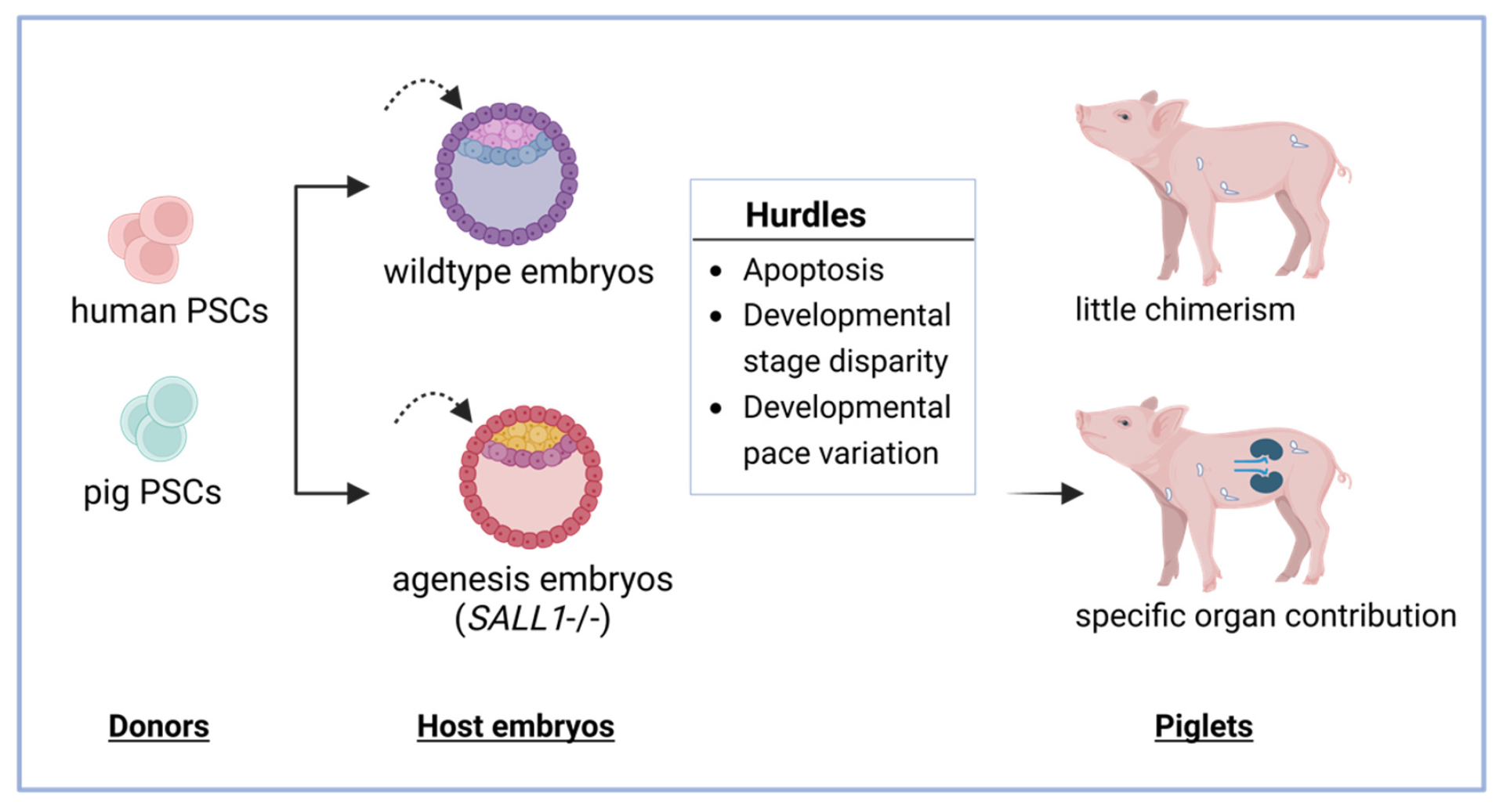
3.2. Chimera Formation in Animals Using Human and Pig PSC

The aforementioned successes open the feasibility of generating human organs via interspecies chimeras. However, the efficient integration of human PSCs into mouse, rat, rabbit, or large-animal (such as cattle or pig) blastocysts has proven to be challenging, still with limited success (Table 1).

3.2.1. Apoptosis

One of the major obstacles is the tendency of human PSCs to undergo apoptosis when injected into early embryos [16,17]. To find the underlying mechanisms, Wu Jun and colleagues developed an in vitro co-culture model to study cell–cell competition in an interspecies context, using pluripotent stem cells from both humans and mice [17]. Through transcriptome analysis, the authors found that NF- κ B signaling played a crucial role in inducing apoptosis in human cells when co-cultured with mouse cells.
Apart from apoptosis during cell competition, cell aging is another hurdle. Evidence showed that the poor quality of human PSCs in vitro was accompanied by progressively shortening telomeres, which was associated with cellular aging [12,16,17], self-renewal, and pluripotency [50,51]. Besides modifying key elements of NF- κ B signaling, it might be helpful to either inactivate P65, P53 or the transgenic expression of MYD55, BMI1, BCL2 in human PSCs [12,13,16,17,52]. To avoid cellular aging, we can temporarily express ZSCAN4 [53] or inhibit mTOR signaling [54]. Those strategies could potentially help enhance fitness and improve chimerism.

3.2.2. Developmental Disparity

The second challenge was the developmental disparity between PSCs and host embryos. It is speculated that the microenvironment of the host embryo would eventually exclude the unmatched donor PSCs. For example, in intraspecies pig–pig chimeras, when porcine EpiESCs (derived from post-implantation embryos) were injected into porcine pre-implantation blastocysts, rarely any chimerism was observed after further in vivo development at E10 [15]. This is likely due to the fact that the donor pig EpiESCs and the host pig blastocyst were at different developmental time points, preventing proper synchronization between the cells and the host embryos. Similar phenomena have been observed in mouse EpiESCs, which showed low chimerism in mouse pre-implantation embryos, and similarly, mESCs also exhibited poor survival in post-implantation embryos [18]. Therefore, when introducing primed human PSCs, which resemble the post-implantation mouse epiblast stage [55], to a host embryo from a pre-implantation stage, low chimerism is expected due to this developmental disparity. On the other hand, naïve hPSCs have a more open chromatin structure and epiblast pigenetic signature, making them more developmentally plastic and easier to mature within the host embryo. Therefore, most human PSC chimeras have been tested using naïve or intermediate human cells, which exhibit higher efficiency [14]. However, large-scale analysis has revealed that naïve human PSCs are genome-wide hypomethylated [8,56], and their imprinting pattern is abnormal [57]. This loss of imprinting may prove to be carcinogenic and problematic for clinical applications.
Another type of human PSC, called human expanded potential stem cells (human EPSCs), may be an alternative developmental match for pre-implantation host embryos. These cells have been reported to have a higher DNA methylation level and are considered to be developmentally more primitive [3,4,6]. Given their genomic stability and molecular features, EPSCs may potentially serve as an alternative donor cell source for interspecies chimeras. In this regard, human EPSCs have been used for in vitro chimera experiments, including with mouse and monkey early-stage embryos [5,58]. Subsequent analysis of these chimeric embryos following in vitro culture has revealed embryonic and extraembryonic contributions from human EPSCs. However, further extensive testing and evaluation of the competency and chimerism of human EPSCs in extended culture or in vivo development are required.

3.2.3. Developmental Pace Variations

The third difficulty lies in the variations in developmental pace. Although the expression of lineage marker genes and regulatory networks shows cross-species conservation in general, the timing of developmental events exhibits species-specific variation.
In mouse embryos, the first lineage segregation occurs early at E3.0. In contrast, in human and pig embryos, the separation of the inner cell mass (ICM) and trophectoderm (TE) occurs relatively late at around E5.0 to E6.0, followed immediately by the second wave of fate determination and the emergence of the epiblast and hypoblast at around E6.0. Consequently, mouse embryos progress much more rapidly than human and pig embryos, making them unsuitable as host embryos.
On the other hand, pigs are genetically close to humans and exhibit physiological and metabolic similarities, making them a potentially ideal host animal for generating human organs. The first published trial on human–pig chimeras was conducted in 2017 by Wu Jun et al., whereas human PSCs demonstrated chimeric contribution to monkey embryos until day 19 [14]. They also conducted two parallel experiments using rat naïve PSCs to test chimera contribution to two different host embryos: mouse embryos (developmentally close to rats) and pig embryos (developmentally distant from rats). The rat–mouse chimera showed good chimerism, while the rat–pig chimera showed no chimerism due to the phylogenetic distance. Currently, by using pigs as host embryos in combination with the blastocyst complementation principle, researchers have been able to generate humanized muscle and hematoendothelial cells from human PSCs [12,13].

4. Humanized Pig PSCs for Xenotransplantation

Apart from growing human PSCs in chimera to generate humanized organs, researchers are also trying to directly generate humanized pigs for organ transplantation. However, inter-species incompatibility poses a significant threat to future clinical xenotransplantation application. The long-term survival and normal function of xeno-grafts in the host have been greatly hindered by hyperacute rejection, acute vascular rejection, cellular rejection, and delayed xenograft rejection [59], which not only encompass the immune system but also elicit a very complex interaction of coagulation and inflammation response.

4.1. Brief History of Xenotransplantation

The history of xenotransplantation dates back to the 17th century with the successful blood transfusion from a lamb to a boy [60]. However, subsequent attempts using animal organs from bovine, goat, and pig proved largely ineffective and fatal. Despite the use of immunosuppressive drugs in the 1960s, chimpanzee-to-human heart transplants only survived for 90 min [61]. In the 1990s, researchers identified α Gal as a major antigen mediating pig-to-human rejection [62]. Therefore, with advancements in gene editing and cloning techniques, the first genetically modified pig was generated in 2003 [20,63]. In the following years, more important genetic modification strategies were put forward, and genetically engineered pigs were generated. In 2022, successful pig-to-human heart [64] and kidney transplants [65,66] were reported and showcased the promising potential of using humanized pigs for organ transplantation.

4.2. Gene Modification Strategies

Until now, some important genetic loci have been discovered and edited in pig cells to mitigate the undesirable rejection effects. For example, the xeno-derived glycan epitopes ( α Gal, NeuG5c, Sd(a)-like glycan) would strongly activate the preformed antibodies in the host, causing organ failure within several minutes to hours. Scientists engineered the glycan epitopes to create the triple knock-out pig grafts [67], attempting to reduce transplant rejection. More recently, gene editing tools have enabled additional genetic manipulations, such as genes involved in complement activation (human CD46, human CD55, and human CD59), coagulation regulation (human thrombomodulin (TBM), human endothelial protein C receptor (EPCR), and human vWF), endothelial protection (heme oxygenase-1 (HO-1)), and immune modulation (macrophage inhibition-human CD47, NK cell inhibition-HLA-E, and HLA-G). In addition to using genetic engineering to address immune rejections, there are potential genetic methods to reduce the risk of zoonotic transmission from pigs to humans. One such approach involves the successful elimination of all porcine endogenous viruses (PERVs) in pigs through genetic manipulation, resulting in PERV-free pigs suitable for xenotransplantation [68]. Based on these, scientists have successfully created pigs with various combinations of targeted genetic changes using genome-editing techniques [69,70].

4.3. Cell Sources for Xenotransplantation

The cell source for generating multi-gene modified pigs could be pig fibroblasts, pig embryos, and pig embryonic or pluripotent stem cells (Figure 2). There are pros and cons for each cell source to generate genetically modified pigs (Table 2).

4.3.1. Pig Embryos

The most direct approach involves gene editing in the pig zygotes. Exogenous gRNA and Cas9 protein can be introduced into pig zygotes through microinjection, electroporation, or lipofection [71], resulting in the generation of transgenic pigs with germline competency [58]. These offspring exhibit comparable health to their wildtype counterparts. However, there are certain limitations to this method. Firstly, it allows for only limited gene editing for xenotransplantation, thereby restricting the extent of genetic modifications that can be performed. Secondly, the analysis of gene editing outcomes can be challenging. The F0 founder pig may exhibit mosaicism across different tissues, leading to potential false positive or false negative results [72,73]. Additionally, crossbreeding F0 homozygous animals can sometimes result in undesired phenotypes in the F1 generation as observed in mouse studies [74]. Therefore, the direct gene editing of porcine zygotes can be time-consuming, less efficient and with unclear consequences.

4.3.2. Pig Fibroblasts

Subsequently, as pig somatic cell nuclear transfer (SCNT) technology advanced, researchers turned to gene editing fibroblasts followed by cloning to generate humanized pigs [58]. Currently, using fibroblasts for gene editing is the most common method to obtain humanized pigs (Figure 2) [58]. Fibroblasts are easy to handle and also allow for gene editing. Researchers have even successfully generated 3KO-9Tg pigs (knockout of three pig xeno-genes and insertion of nine human genes) with germline transmission using porcine fibroblasts as the gene-editing source cells [70]. However, gene editing analysis of fibroblasts requires serial dilution to ensure clear genotyping results due to the presence of cell–cell mixtures. Furthermore, fibroblasts have limitations in terms of passage numbers, low genome-editing efficiency, and limited capacity for precision and complex gene targeting. Additionally, edited fibroblast editing does not permit immune compatibility testing and it requires SCNT to generate pig endothelial cells and organs for in vitro immune compatibility assessment or direct in vivo transplantation in non-human primate (NHP) models. This SCNT-based approach is time-consuming for selecting optimal combinations of pig gene edits and human gene insertions, which critically delays progress in the field.

4.3.3. Pig Pluripotent Stem Cells

On the other hand, the emergence of pig embryonic or pluripotent stem cells (PSC) provided a promising donor cell source for xenotransplantation, addressing the above-mentioned limitations. Several types of pig PSCs are currently available, including pig EPSCs (expanded potential stem cells) [3], pig EpiSCs (epiblast-derived stem cells) [19], and pig EDSCs (embryonic disc stem cells) [20], derived from both pre-implantation and peri-implantation embryos. Notably, EPSCs are of preimplantation embryo origin, exhibit higher homologous recombination activity [75] and possess unlimited proliferation potential, enabling complex and precise gene editing. Additionally, EPSCs can differentiate into both embryonic and extra-embryonic tissues [3,15,20]. Furthermore, their ability to grow from single cells and form colonies facilitates unambiguous genotyping analysis and the generation of well-defined genetically modified stem cells.
These unique characteristics of pig pluripotent stem cells offer several potential applications. Firstly, they can serve as nuclear donors for generating cloned pigs. Secondly, they can potentially be utilized for generating humanized pig organs through blastocyst complementation and germline transmission in chimera assays. Lastly, pig stem cells can be used to create blastoids (artificial blastocyst counterparts) that can potentially be transferred into pig uteri to potentially generate pigs.

4.4. Generation of Humanized Pigs through Cloning Techniques

Pig PSCs are an ideal cell source in xenotransplantation. However, there is still little investigation related to their stem cell cloning efficiencies. Some very early studies showed that long-term cultured established mouse ESC lines could be used in generating cloned mice [70], which showed the cloning application of other species’ pluripotent stem cells, like the pig.
However, the road has not been easy for deriving cloned pigs from PSCs. Ten years ago, Lai et al. generated a total of 11,923 cloned embryos using pig iPSCs, but only resulted in 25 pregnancies at 24–26 days, and no cloned piglets were generated [76]. The reason behind this was probably due to the exogenous reprogramming factor gene expression in pig iPSCs, which hindered cloned embryo development.
More recently, pig PSCs derived from embryos have been generated, which could serve as nuclear donors for cloning at efficiencies similar to those using fibroblasts [15,20], where pig PSCs were pre-differentiated in a medium containing BMP4, SB431542, and FGF2 [15]. In the meantime, the epigenetic remodeling of the cloned embryo was found to improve the cleavage and blastocyst rates. Substances, such as scriptaid [77], abexinostat [78], vitamin C [79], trichostatin A (TSA) [80,81], cytochalasin B [20,81], Kdm4d/Kdm5b mRNA injection [81,82], and others could also be useful in PSC-based pig cloning. Furthermore, cell cycle synchronization by serum starvation or culture to confluency to arrest donor pig cells at the G0/G1 phase were found to further improve the cloning efficiency. Apart from this, the quality of the starting cells, particularly the donor cell chromosome stability, is an important factor. Checking the karyotyping and using early passage pig stem cells improved donor efficiency.

4.5. Acquisition of Humanized Pig Organs through Chimera Formation

As for using pig stem cells as donors for chimera assays to generate pig organs, many experiments are still under investigation. As stated above, scientists have already shown in mouse studies that mESC could give rise to fully functional mouse organs in organ agenesis mouse or rat embryos. It is foreseeable that through blastocyst complementation, genetically modified pig PSCs could also generate humanized pig organs in organ agenesis hosts. Until now, there have been no studies using pig multi-gene modified PSCs to generate humanized pig organs via blastocyst complementation in pig embryos. However, previous studies have paved the way for testing this idea. Scientists genetically knocked out SALL1 in pig zygotes which revealed no kidney phenotype [83], knocked out ETV2 which showed no endothelium [13], and removed HHEX which showed no liver phenotype [45]. Those pig genetically mutant agenesis embryos, combined with genetically edited pig PSCs, offer an attractive approach for the generation of humanized pig organs in transplantation.

4.6. Production of Humanized Pigs via Blastocyst-like Structure Development

Alternatively, the generation of humanized pigs via blastoid formation also holds great promise. Blastoids are stem cell-derived structures that resemble natural blastocysts, which can be generated by modulating specific signaling pathways in pluripotent stem cells. The first mouse blastoid was successfully created by Nicolas Rivron’s group in 2018 [84], by sequential aggregation of mESCs (mouse embryonic stem cells) and mTSCs (mouse trophoblast stem cells). These blastoids not only exhibited morphological similarities to E3.5 mouse blastocysts but also demonstrated functional capabilities by implanting into pseudo-pregnant mice and triggering endometrium decidualization.
Since then, rapid advancements have been made in blastoid generation [56,85,86,87,88]. It is now possible to directly reprogram somatic cells to blastoids [86] or even form a blastoid from a single type of pluripotent stem cell. Recent studies have achieved the generation of human blastoids with an efficiency of up to 70% through the triple inhibition of Hippo, Nodal, and Erk signaling pathways [87]. Although both mouse and human blastoids have limitations in their developmental potential, being unable to generate functional tissues or organs, these groundbreaking studies provide a promising outlook for regenerative medicine.
Looking ahead, the generation of pig blastoids from pig PSCs is plausible in the future. With further refinement of signaling pathways and culture conditions, it may be possible to develop multi-gene modified pig blastoids that can develop in an in vivo environment. While the authentic blastocyst model is still in its infancy, this innovative approach to generating living organisms holds tremendous potential for advancing developmental biology research and revolutionizing regenerative medicine.

5. Ethics and Other Concerns

There is a higher social acceptance of pig-related products for therapeutic purposes—they have a long history of domestication as livestock and serve as an important food source for human beings. This simple argument is, however, not a valid argument for xenotransplantation, as the research touches on many more ethical aspects. Therefore, xenotransplantation research is accompanied by ethical research to evaluate the perception of the different ethical groups regarding the use of pig organs. Pioneering work has been established by the German xenotransplantation consortium. In a survey of representative citizens, the group considered the benefits of xenotransplantation to outweigh the risks but called for strict regulatory measures [89]. Despite animals having been used in scientific research for a long time, the main ongoing concern is the welfare of these animals, ensuring that they live well, feel well, and function well [90]. Therefore, when manipulating the pig genome to create humanized pigs for xenotransplantation, it is essential to carefully assess gene modifications to avoid harming the animals’ physical and mental health.
When employing human PSCs for human-pig chimeras or directly utilizing humanized pig organs for regenerative medicine, we need to avoid human PSCs contributing to the neurons and germ cells. Efforts could be made to specifically eliminate the donor cell contribution in the brain in interspecies chimera [91]. Such scientific advances are expected to help circumvent the contribution of human cells to the pig brain or germ cells.
Again, the main argument for an overall positive assessment of stem cell research in general and xenotransplantation in particular in the societal context is primarily based on the societal obligation to help people with life-threatening conditions and to alleviate suffering where possible. The fact that research is well ahead of the public consciousness and that a broader societal debate is necessary and still lacking demands ongoing evaluation and regulation to ensure the responsible and safe implementation of using stem cell- and pig-related products in research and medicine. These research activities, including the generation of gene-edited pigs, must have ethical approval by an independent commission and abide by the local and international regulations.

6. Conclusions

The availability of PSCs from various species, particularly humans and pigs, has greatly expanded the potential applications of stem cells. Researchers have successfully derived a wide range of clinically relevant cell types from both human and pig PSCs. In addition, the generation of transplantable organs and tissues offers a potential solution to the global organ shortage. By utilizing techniques like chimera formation and blastocyst complementation, where stem cells are integrated into agenesis host embryos, it would be possible to generate human organs or humanized organs. Furthermore, pig PSCs are an ideal cell source for gene editing in xenotransplantation.
Overall, the advent in the generation of PSCs from multiple species has significantly expanded stem cell application possibilities, from generating specific cell types for therapeutic purposes to addressing organ shortages through innovative techniques like chimera formation and blastoids. However, these innovative approaches are still in their infancy and require further investigation.

Funding

Pentao Liu’s research is supported by Health@InnoHK and Hong Kong Research Grant Council.

Conflicts of Interest

The authors declare no conflict of interest.

References

  1. Evans, M.; Kaufman, M. Establishment in culture of pluripotential cells from mouse embryos. Nature 1981, 292, 154–156. [Google Scholar] [CrossRef] [PubMed]
  2. Thomson, J.; Itskovitz-Eldor, J.; Shapiro, S.; Waknitz, M.; Swiergiel, J.; Marshall, V.; Jones, J. Embryonic Stem Cell Lines Derived from Human Blastocysts. Science 1998, 282, 1145–1147. [Google Scholar] [CrossRef]
  3. Gao, X.; Nowak-Imialek, M.; Chen, X.; Chen, D.; Herrmann, D.; Ruan, D.; Chen, A.C.H.; Eckersley-Maslin, M.A.; Ahmad, S.; Lee, Y.L.; et al. Establishment of porcine and human expanded potential stem cells. Nat. Cell Biol. 2019, 21, 687–699. [Google Scholar] [CrossRef]
  4. Zhao, L.; Gao, X.; Zheng, Y.; Wang, Z.; Zhao, G.; Ren, J.; Zhang, J.; Wu, J.; Wu, B.; Chen, Y.; et al. Establishment of bovine expanded potential stem cells. Proc. Natl. Acad. Sci. USA 2021, 118, e2018505118. [Google Scholar] [CrossRef]
  5. Yang, Y.; Liu, B.; Xu, J.; Wang, J.; Wu, J.; Shi, C.; Xu, Y.; Dong, J.; Wang, C.; Lai, W.; et al. Derivation of Pluripotent Stem Cells with In Vivo Embryonic and Extraembryonic Potency. Cell 2017, 169, 243–257.e25. [Google Scholar] [CrossRef] [PubMed]
  6. Yang, J.; Ryan, D.; Wang, W.; Tsang, J.; Lan, G.; Masaki, H.; Gao, X.; Antunes, L.; Yu, Y.; Zhu, Z.; et al. Establishment of mouse expanded potential stem cells. Nature 2017, 550, 393–397. [Google Scholar] [CrossRef] [PubMed]
  7. Liu, B.; Chen, S.; Xu, Y.; Lyu, Y.; Wang, J.; Du, Y.; Sun, Y.; Liu, H.; Zhou, H.; Lai, W.; et al. Chemically defined and xeno-free culture condition for human extended pluripotent stem cells. Nat. Commun. 2021, 12, 3017. [Google Scholar] [CrossRef] [PubMed]
  8. Bayerl, J.; Ayyash, M.; Shani, T.; Manor, Y.; Gafni, O.; Kalma, Y.; Aguilera-Castrejon, A.; Zerbib, M.; Amir, H.; Sheban, D. Others Tripartite inhibition of SRC-WNT-PKC signalling consolidates human naïve pluripotency. bioRxiv 2020. [Google Scholar] [CrossRef]
  9. Markmann, J.; Naji, A.; Rickels, M.; Alba, M.; Marigowda, G.; Ross, L.; Wang, C.; Pagliuca, F.; Sanna, B.; Kean, L.; et al. 259-OR: Stem cell–derived, fully differentiated islet cells for type 1 diabetes. Diabetes 2022, 71, 259-OR. [Google Scholar] [CrossRef]
  10. Sharma, R.; Khristov, V.; Rising, A.; Jha, B.; Dejene, R.; Hotaling, N.; Li, Y.; Stoddard, J.; Stankewicz, C.; Wan, Q.; et al. Clinical-grade stem cell–derived retinal pigment epithelium patch rescues retinal degeneration in rodents and pigs. Sci. Transl. Med. 2019, 11, eaat5580. [Google Scholar] [CrossRef]
  11. Kashani, A.; Lebkowski, J.; Hinton, D.; Zhu, D.; Faynus, M.; Chen, S.; Rahhal, F.; Avery, R.; Salehi-Had, H.; Chan, C.; et al. Survival of an HLA-mismatched, bioengineered RPE implant in dry age-related macular degeneration. Stem Cell Rep. 2022, 17, 448–458. [Google Scholar] [CrossRef] [PubMed]
  12. Maeng, G.; Das, S.; Greising, S.; Gong, W.; Singh, B.; Kren, S.; Mickelson, D.; Skie, E.; Gafni, O.; Sorensen, J.; et al. Humanized skeletal muscle in MYF5/MYOD/MYF6-null pig embryos. Nat. Biomed. Eng. 2021, 5, 805–814. [Google Scholar] [CrossRef] [PubMed]
  13. Das, S.; Koyano-Nakagawa, N.; Gafni, O.; Maeng, G.; Singh, B.; Rasmussen, T.; Pan, X.; Choi, K.; Mickelson, D.; Gong, W.; et al. Generation of human endothelium in pig embryos deficient in ETV2. Nat. Biotechnol. 2020, 38, 297–302. [Google Scholar] [CrossRef]
  14. Wu, J.; Platero-Luengo, A.; Sakurai, M.; Sugawara, A.; Gil, M.; Yamauchi, T.; Suzuki, K.; Bogliotti, Y.; Cuello, C.; Valencia, M.; et al. Interspecies Chimerism with Mammalian Pluripotent Stem Cells. Cell 2017, 168, 473–486.e15. [Google Scholar] [CrossRef] [PubMed]
  15. Zhi, M.; Zhang, J.; Tang, Q.; Yu, D.; Gao, S.; Gao, D.; Liu, P.; Guo, J.; Hai, T.; Gao, J.; et al. Generation and characterization of stable pig pregastrulation epiblast stem cell lines. Cell Res. 2021, 32, 383–400. [Google Scholar] [CrossRef] [PubMed]
  16. Huang, K.; Zhu, Y.; Ma, Y.; Zhao, B.; Fan, N.; Li, Y.; Song, H.; Chu, S.; Ouyang, Z.; Zhang, Q.; et al. BMI1 enables interspecies chimerism with human pluripotent stem cells. Nat. Commun. 2018, 9, 4649. [Google Scholar] [CrossRef]
  17. Zheng, C.; Hu, Y.; Sakurai, M.; Pinzon-Arteaga, C.; Li, J.; Wei, Y.; Okamura, D.; Ravaux, B.; Barlow, H.; Yu, L.; et al. Cell competition constitutes a barrier for interspecies chimerism. Nature 2021, 592, 272–276. [Google Scholar] [CrossRef]
  18. Huang, Y.; Osorno, R.; Tsakiridis, A.; Wilson, V. In Vivo Differentiation Potential of Epiblast Stem Cells Revealed by Chimeric Embryo Formation. Cell Rep. 2012, 2, 1571–1578. [Google Scholar] [CrossRef]
  19. Zhong, C.; Li, R.; Belmonte, J. Transcriptomic profiling fuels the derivation of stable pig epiblast stem cells. Cell Res. 2022, 32, 329–330. [Google Scholar] [CrossRef]
  20. Kinoshita, M.; Kobayashi, T.; Planells, B.; Klisch, D.; Spindlow, D.; Masaki, H.; Bornelöv, S.; Stirparo, G.; Matsunari, H.; Uchikura, A.; et al. Pluripotent stem cells related to embryonic disc exhibit common self-renewal requirements in diverse livestock species. Development 2021, 148, dev199901. [Google Scholar] [CrossRef]
  21. Veres, A.; Faust, A.L.; Bushnell, H.L.; Engquist, E.N.; Kenty, J.H.-R.; Harb, G.; Poh, Y.-C.; Sintov, E.; Gürtler, M.; Pagliuca, F.W.; et al. Charting cellular identity during human in vitro β-cell differentiation. Nature 2019, 569, 368–373. [Google Scholar] [CrossRef]
  22. Rong, Z.; Wang, M.; Hu, Z.; Stradner, M.; Zhu, S.; Kong, H.; Yi, H.; Goldrath, A.; Yang, Y.; Xu, Y.; et al. An Effective Approach to Prevent Immune Rejection of Human ESC-Derived Allografts. Cell Stem Cell 2014, 14, 121–130. [Google Scholar] [CrossRef] [PubMed]
  23. Limnios, I.; Chau, Y.; Skabo, S.; Surrao, D.; O’Neill, H. Efficient differentiation of human embryonic stem cells to retinal pigment epithelium under defined conditions. Stem Cell Res. Ther. 2021, 12, 248. [Google Scholar] [CrossRef] [PubMed]
  24. Zhang, H.; Su, B.; Jiao, L.; Xu, Z.; Zhang, C.; Nie, J.; Gao, M.; Zhang, Y.; Jin, Z. Transplantation of GMP-grade human iPSC-derived retinal pigment epithelial cells in rodent model: The first pre-clinical study for safety and efficacy in China. Ann. Transl. Med. 2021, 9, 245. [Google Scholar] [CrossRef]
  25. Stanzel, B.; Liu, Z.; Somboonthanakij, S.; Wongsawad, W.; Brinken, R.; Eter, N.; Corneo, B.; Holz, F.; Temple, S.; Stern, J.; et al. Human RPE Stem Cells Grown into Polarized RPE Monolayers on a Polyester Matrix Are Maintained after Grafting into Rabbit Subretinal Space. Stem Cell Rep. 2014, 2, 64–77. [Google Scholar] [CrossRef]
  26. Liu, Z.; Parikh, B.; Tan, Q.; Wong, D.; Ong, K.; Yu, W.; Seah, I.; Holder, G.; Hunziker, W.; Tan, G.; et al. Surgical Transplantation of Human RPE Stem Cell-Derived RPE Monolayers into Non-Human Primates with Immunosuppression. Stem Cell Rep. 2021, 16, 237–251. [Google Scholar] [CrossRef]
  27. Schwartz, S.; Regillo, C.; Lam, B.; Eliott, D.; Rosenfeld, P.; Gregori, N.; Hubschman, J.; Davis, J.; Heilwell, G.; Spirn, M.; et al. Human embryonic stem cell-derived retinal pigment epithelium in patients with age-related macular degeneration and Stargardt’s macular dystrophy: Follow-up of two open-label phase 1/2 studies. Lancet 2015, 385, 509–516. [Google Scholar] [CrossRef]
  28. Cruz, L.; Fynes, K.; Georgiadis, O.; Kerby, J.; Luo, Y.; Ahmado, A.; Vernon, A.; Daniels, J.; Nommiste, B.; Hasan, S.; et al. Phase 1 clinical study of an embryonic stem cell–derived retinal pigment epithelium patch in age-related macular degeneration. Nat. Biotechnol. 2018, 36, 328–337. [Google Scholar] [CrossRef] [PubMed]
  29. Wang, L.; Zhang, H. Ocular barriers as a double-edged sword: Preventing and facilitating drug delivery to the retina. Drug Deliv. Transl. Res. 2022, 13, 547–567. [Google Scholar] [CrossRef] [PubMed]
  30. Petrus-Reurer, S.; Kumar, P.; Sánchez, S.; Aronsson, M.; André, H.; Bartuma, H.; Reyes, A.; Nandrot, E.; Kvanta, A.; Lanner, F. Preclinical safety studies of human embryonic stem cell-derived retinal pigment epithelial cells for the treatment of age-related macular degeneration. Stem Cells Transl. Med. 2020, 9, 936–953. [Google Scholar] [CrossRef] [PubMed]
  31. Petrus-Reurer, S.; Winblad, N.; Kumar, P.; Gorchs, L.; Chrobok, M.; Wagner, A.; Bartuma, H.; Lardner, E.; Aronsson, M.; Plaza Reyes, A.; et al. Generation of Retinal Pigment Epithelial Cells Derived from Human Embryonic Stem Cells Lacking Human Leukocyte Antigen Class I and II. Stem Cell Rep. 2020, 14, 648–662. [Google Scholar] [CrossRef]
  32. Kanemura, H.; Go, M.; Shikamura, M.; Nishishita, N.; Sakai, N.; Kamao, H.; Mandai, M.; Morinaga, C.; Takahashi, M.; Kawamata, S. Tumorigenicity Studies of Induced Pluripotent Stem Cell (iPSC)-Derived Retinal Pigment Epithelium (RPE) for the Treatment of Age-Related Macular Degeneration. PLoS ONE 2014, 9, e85336. [Google Scholar] [CrossRef] [PubMed]
  33. Kuijk, E.; Jager, M.; Roest, B.; Locati, M.; Hoeck, A.; Korzelius, J.; Janssen, R.; Besselink, N.; Boymans, S.; Boxtel, R.; et al. The mutational impact of culturing human pluripotent and adult stem cells. Nat. Commun. 2020, 11, 2493. [Google Scholar] [CrossRef] [PubMed]
  34. Hasaart, K.; Manders, F.; Ubels, J.; Verheul, M.; Roosmalen, M.; Groenen, N.; Oka, R.; Kuijk, E.; Sousa Lopes, S.; Boxtel, R. Human induced pluripotent stem cells display a similar mutation burden as embryonic pluripotent cells in vivo. iScience 2022, 25, 103736. [Google Scholar] [CrossRef] [PubMed]
  35. Avior, Y.; Lezmi, E.; Eggan, K.; Benvenisty, N. Cancer-Related Mutations Identified in Primed Human Pluripotent Stem Cells. Cell Stem Cell 2021, 28, 10–11. [Google Scholar] [CrossRef] [PubMed]
  36. Head, S.; Çelik, M.; Kappetein, A. Mechanical versus bioprosthetic aortic valve replacement. Eur. Heart J. 2017, 38, 2183–2191. [Google Scholar] [CrossRef]
  37. Gardner, R. Mouse Chimaeras obtained by the Injection of Cells into the Blastocyst. Nature 1968, 220, 596–597. [Google Scholar] [CrossRef]
  38. Yamaguchi, T.; Sato, H.; Kato-Itoh, M.; Goto, T.; Hara, H.; Sanbo, M.; Mizuno, N.; Kobayashi, T.; Yanagida, A.; Umino, A.; et al. Interspecies organogenesis generates autologous functional islets. Nature 2017, 542, 191–196. [Google Scholar] [CrossRef]
  39. Hamanaka, S.; Umino, A.; Sato, H.; Hayama, T.; Yanagida, A.; Mizuno, N.; Kobayashi, T.; Kasai, M.; Suchy, F.; Yamazaki, S.; et al. Generation of Vascular Endothelial Cells and Hematopoietic Cells by Blastocyst Complementation. Stem Cell Rep. 2018, 11, 988–997. [Google Scholar] [CrossRef]
  40. Goto, T.; Hara, H.; Sanbo, M.; Masaki, H.; Sato, H.; Yamaguchi, T.; Hochi, S.; Kobayashi, T.; Nakauchi, H.; Hirabayashi, M. Generation of pluripotent stem cell-derived mouse kidneys in Sall1-targeted anephric rats. Nat. Commun. 2019, 10, 451. [Google Scholar] [CrossRef]
  41. Coppiello, G.; Barlabé, P.; Moya-Jódar, M.; Abizanda, G.; Barreda, C.; Iglesias, E.; Linares, J.; Arellano-Viera, E.; Ruiz-Villalba, A.; Larequi, E.; et al. In vivo generation of heart and vascular system by blastocyst complementation. bioRxiv 2022. [Google Scholar] [CrossRef]
  42. Kitahara, A.; Ran, Q.; Oda, K.; Yasue, A.; Abe, M.; Ye, X.; Sasaoka, T.; Tsuchida, M.; Sakimura, K.; Ajioka, Y.; et al. Generation of Lungs by Blastocyst Complementation in Apneumic Fgf10-Deficient Mice. Cell Rep. 2020, 31, 107626. [Google Scholar] [CrossRef] [PubMed]
  43. Yamazaki, K.; Kubara, K.; Ishii, S.; Li, P.; Dairiki, R.; Hihara, T.; Ishizuka, Y.; Izumi, Y.; Kumai, M.; Kamisako, T.; et al. In vitro and in vivo functions of T cells produced in complemented thymi of chimeric mice generated by blastocyst complementation. Sci. Rep. 2022, 12, 3242. [Google Scholar] [CrossRef] [PubMed]
  44. Kobayashi, T.; Yamaguchi, T.; Hamanaka, S.; Kato-Itoh, M.; Yamazaki, Y.; Ibata, M.; Sato, H.; Lee, Y.-S.; Usui, J.-I.; Knisely, A.S.; et al. Generation of Rat Pancreas in Mouse by Interspecific Blastocyst Injection of Pluripotent Stem Cells. Cell 2010, 142, 787–799. [Google Scholar] [CrossRef] [PubMed]
  45. Ruiz-Estevez, M.; Crane, A.; Rodriguez-Villamil, P.; Ongaratto, F.; You, Y.; Steevens, A.; Hill, C.; Goldsmith, T.; Webster, D.; Sherry, L.; et al. Liver development is restored by blastocyst complementation of HHEX knockout in mice and pigs. Stem Cell Res. Ther. 2021, 12, 292. [Google Scholar] [CrossRef] [PubMed]
  46. Zhang, H.; Huang, J.; Li, Z.; Qin, G.; Zhang, N.; Hai, T.; Hong, Q.; Zheng, Q.; Zhang, Y.; Song, R.; et al. Rescuing ocular development in an anophthalmic pig by blastocyst complementation. EMBO Mol. Med. 2018, 10, e8861. [Google Scholar] [CrossRef] [PubMed]
  47. Steevens, A.; Griesbach, M.; You, Y.; Dutton, J.; Low, W.; Santi, P. Generation of inner ear sensory neurons using blastocyst complementation in a Neurog1+/−-deficient mouse. Stem Cell Res. Ther. 2021, 12, 122. [Google Scholar] [CrossRef]
  48. Kobayashi, T.; Goto, T.; Oikawa, M.; Sanbo, M.; Yoshida, F.; Terada, R.; Niizeki, N.; Kajitani, N.; Kazuki, K.; Kazuki, Y.; et al. Blastocyst complementation using Prdm14-deficient rats enables efficient germline transmission and generation of functional mouse spermatids in rats. Nat. Commun. 2021, 12, 1328. [Google Scholar] [CrossRef]
  49. Tan, T.; Wu, J.; Si, C.; Dai, S.; Zhang, Y.; Sun, N.; Zhang, E.; Shao, H.; Si, W.; Yang, P.; et al. Chimeric contribution of human extended pluripotent stem cells to monkey embryos ex vivo. Cell 2021, 184, 2020–2032.e14. [Google Scholar] [CrossRef]
  50. Wang, F.; Yin, Y.; Ye, X.; Liu, K.; Zhu, H.; Wang, L.; Chiourea, M.; Okuka, M.; Ji, G.; Dan, J.; et al. Molecular insights into the heterogeneity of telomere reprogramming in induced pluripotent stem cells. Cell Res. 2011, 22, 757–768. [Google Scholar] [CrossRef]
  51. Wang, H.; Zhang, K.; Liu, Y.; Fu, Y.; Gao, S.; Gong, P.; Wang, H.; Zhou, Z.; Zeng, M.; Wu, Z.; et al. Telomere heterogeneity linked to metabolism and pluripotency state revealed by simultaneous analysis of telomere length and RNA-seq in the same human embryonic stem cell. BMC Biol. 2017, 15, 114. [Google Scholar] [CrossRef] [PubMed]
  52. Masaki, H.; Kato-Itoh, M.; Takahashi, Y.; Umino, A.; Sato, H.; Ito, K.; Yanagida, A.; Nishimura, T.; Yamaguchi, T.; Hirabayashi, M.; et al. Inhibition of Apoptosis Overcomes Stage-Related Compatibility Barriers to Chimera Formation in Mouse Embryos. Cell Stem Cell 2016, 19, 587–592. [Google Scholar] [CrossRef]
  53. Zalzman, M.; Falco, G.; Sharova, L.; Nishiyama, A.; Thomas, M.; Lee, S.; Stagg, C.; Hoang, H.; Yang, H.; Indig, F.; et al. Zscan4 regulates telomere elongation and genomic stability in ES cells. Nature 2010, 464, 858–863. [Google Scholar] [CrossRef] [PubMed]
  54. Hu, Z.; Li, H.; Jiang, H.; Ren, Y.; Yu, X.; Qiu, J.; Stablewski, A.; Zhang, B.; Buck, M.; Feng, J. Transient inhibition of mTOR in human pluripotent stem cells enables robust formation of mouse-human chimeric embryos. Sci. Adv. 2020, 6, eaaz0298. [Google Scholar] [CrossRef] [PubMed]
  55. Weinberger, L.; Ayyash, M.; Novershtern, N.; Hanna, J. Dynamic stem cell states: Naive to primed pluripotency in rodents and humans. Nat. Rev. Mol. Cell Biol. 2016, 17, 155–169. [Google Scholar] [CrossRef]
  56. Li, J.; Zhu, Q.; Cao, J.; Liu, Y.; Lu, Y.; Sun, Y.; Li, Q.; Huang, Y.; Shang, S.; Bian, X.; et al. Cynomolgus monkey embryo model captures gastrulation and early pregnancy. Cell Stem Cell 2023, 30, 362–377.e7. [Google Scholar] [CrossRef]
  57. Keshet, G.; Benvenisty, N. Large-scale analysis of imprinting in naive human pluripotent stem cells reveals recurrent aberrations and a potential link to FGF signaling. Stem Cell Rep. 2021, 16, 2520–2533. [Google Scholar] [CrossRef]
  58. Yang, L.; Church, G.; Zhao, H.; Huang, L.; Gao, Y.; Wei, H.; Yang, G. Porcine germline genome engineering. Proc. Natl. Acad. Sci. USA 2021, 118, e2004836117. [Google Scholar] [CrossRef]
  59. Yang, Y.; Sykes, M. Xenotransplantation: Current status and a perspective on the future. Nat. Rev. Immunol. 2007, 7, 519–531. [Google Scholar] [CrossRef]
  60. Varki, A. Multiple changes in sialic acid biology during human evolution. Glycoconj. J. 2008, 26, 231–245. [Google Scholar] [CrossRef]
  61. Mercer, W.; Shields, M.; Amin, M.; Sauve, G.; Appella, E.; Romano, J.; Ullrich, S. Negative growth regulation in a glioblastoma tumor cell line that conditionally expresses human wild-type p53. Proc. Natl. Acad. Sci. USA 1990, 87, 6166–6170. [Google Scholar] [CrossRef] [PubMed]
  62. Cooper, D.; Koren, E.; Oriol, R. Oligosaccharides and Discordant Xenotransplantation. Immunol. Rev. 1994, 141, 31–58. [Google Scholar] [CrossRef] [PubMed]
  63. Petkov, S.; Glage, S.; Nowak-Imialek, M.; Niemann, H. Long-Term Culture of Porcine Induced Pluripotent Stem-Like Cells Under Feeder-Free Conditions in the Presence of Histone Deacetylase Inhibitors. Stem Cells Dev. 2016, 25, 386–394. [Google Scholar] [CrossRef] [PubMed]
  64. Mo, H.; Tian, F.; Ma, X.; Zhang, Y.; Zhang, C.; Zeng, W.; Zhang, Y.; Lin, Y. PDIA3 regulates trophoblast apoptosis and proliferation in preeclampsia via the MDM2/p53 pathway. Reproduction 2020, 160, 293–305. [Google Scholar] [CrossRef]
  65. Phelps, C.; Koike, C.; Vaught, T.; Boone, J.; Wells, K.; Chen, S.; Ball, S.; Specht, S.; Polejaeva, I.; Monahan, J.; et al. Production of α1, 3-Galactosyltransferase-Deficient Pigs. Science 2003, 299, 411–414. [Google Scholar] [CrossRef]
  66. Griesemer, A.; Hirakata, A.; Shimizu, A.; Moran, S.; Tena, A.; Iwaki, H.; Ishikawa, Y.; Schule, P.; Arn, J.; Robson, S.; et al. Results of Gal-Knockout Porcine Thymokidney Xenografts. Am. J. Transplant. 2009, 9, 2669–2678. [Google Scholar] [CrossRef]
  67. Shim, J.; Ko, N.; Kim, H.; Lee, Y.; Lee, J.; Jin, D.; Kim, H.; Choi, K. Human immune reactivity of GGTA1/CMAH/A3GALT2 triple knockout Yucatan miniature pigs. Transgenic Res. 2021, 30, 619–634. [Google Scholar] [CrossRef]
  68. Niu, D.; Wei, H.; Lin, L.; George, H.; Wang, T.; Lee, I.; Zhao, H.; Wang, Y.; Kan, Y.; Shrock, E.; et al. Inactivation of porcine endogenous retrovirus in pigs using CRISPR-Cas9. Science 2017, 357, 1303–1307. [Google Scholar] [CrossRef]
  69. Ma, D.; Hirose, T.; Lassiter, G.; Sasaki, H.; Rosales, I.; Coe, T.; Rickert, C.; Matheson, R.; Colvin, R.; Qin, W.; et al. Kidney transplantation from triple-knockout pigs expressing multiple human proteins in cynomolgus macaques. Am. J. Transplant. 2022, 22, 46–57. [Google Scholar] [CrossRef]
  70. Yue, Y.; Xu, W.; Kan, Y.; Zhao, H.; Zhou, Y.; Song, X.; Wu, J.; Xiong, J.; Goswami, D.; Yang, M.; et al. Extensive germline genome engineering in pigs. Nat. Biomed. Eng. 2020, 5, 134–143. [Google Scholar] [CrossRef]
  71. Le, Q.; Wittayarat, M.; Namula, Z.; Lin, Q.; Takebayashi, K.; Hirata, M.; Tanihara, F.; Do, L.; Otoi, T. Multiple gene editing in porcine embryos using a combination of microinjection, electroporation, and transfection methods. Vet. World 2022, 15, 2210–2216. [Google Scholar] [CrossRef] [PubMed]
  72. Mianné, J.; Codner, G.; Caulder, A.; Fell, R.; Hutchison, M.; King, R.; Stewart, M.; Wells, S.; Teboul, L. Analysing the outcome of CRISPR-aided genome editing in embryos: Screening, genotyping and quality control. Methods 2017, 121–122, 68–76. [Google Scholar] [CrossRef]
  73. Mackenzie, M.; Fower, A.; Allan, A.; Codner, G.; Bunton-Stasyshyn, R.; Teboul, L. Genotyping Genome-Edited Founders and Subsequent Generation. In Methods In Molecular Biology; Springer: Berlin/Heidelberg, Germany, 2023; pp. 103–134. [Google Scholar] [CrossRef]
  74. Yen, S.; Zhang, M.; Deng, J.; Usman, S.; Smith, C.; Parker-Thornburg, J.; Swinton, P.; Martin, J.; Behringer, R. Somatic mosaicism and allele complexity induced by CRISPR/Cas9 RNA injections in mouse zygotes. Dev. Biol. 2014, 393, 3–9. [Google Scholar] [CrossRef] [PubMed]
  75. Choi, E.; Yoon, S.; Koh, Y.; Seo, Y.; Kim, K. Maintenance of genome integrity and active homologous recombination in embryonic stem cells. Exp. Mol. Med. 2020, 52, 1220–1229. [Google Scholar] [CrossRef] [PubMed]
  76. Fan, N.; Chen, J.; Shang, Z.; Dou, H.; Ji, G.; Zou, Q.; Wu, L.; He, L.; Wang, F.; Liu, K.; et al. Piglets cloned from induced pluripotent stem cells. Cell Res. 2012, 23, 162–166. [Google Scholar] [CrossRef]
  77. Zhao, J.; Ross, J.; Hao, Y.; Spate, L.; Walters, E.; Samuel, M.; Rieke, A.; Murphy, C.; Prather, R. Significant Improvement in Cloning Efficiency of an Inbred Miniature Pig by Histone Deacetylase Inhibitor Treatment after Somatic Cell Nuclear Transfer1. Biol. Reprod. 2009, 81, 525–530. [Google Scholar] [CrossRef]
  78. Jin, L.; Zhu, H.; Guo, Q.; Li, X.; Zhang, Y.; Zhang, G.; Xing, X.; Xuan, M.; Luo, Q.; Yin, X.; et al. PCI-24781 can improve in vitro and in vivo developmental capacity of pig somatic cell nuclear transfer embryos. Biotechnol. Lett. 2016, 38, 1433–1441. [Google Scholar] [CrossRef]
  79. Huang, Y.; Tang, X.; Xie, W.; Zhou, Y.; Li, D.; Zhou, Y.; Zhu, J.; Yuan, T.; Lai, L.; Pang, D.; et al. Vitamin C enhances in vitro and in vivo development of porcine somatic cell nuclear transfer embryos. Biochem. Biophys. Res. Commun. 2011, 411, 397–401. [Google Scholar] [CrossRef]
  80. Guo, Z.; Lv, L.; Liu, D.; Fu, B. Effects of trichostatin A on pig SCNT blastocyst formation rate and cell number: A meta-analysis. Res. Vet. Sci. 2018, 117, 161–166. [Google Scholar] [CrossRef]
  81. Liu, Z.; Cai, Y.; Wang, Y.; Nie, Y.; Zhang, C.; Xu, Y.; Zhang, X.; Lu, Y.; Wang, Z.; Poo, M.; et al. Cloning of Macaque Monkeys by Somatic Cell Nuclear Transfer. Cell 2018, 172, 881–887.e7. [Google Scholar] [CrossRef]
  82. Liu, W.; Liu, X.; Wang, C.; Gao, Y.; Gao, R.; Kou, X.; Zhao, Y.; Li, J.; Wu, Y.; Xiu, W.; et al. Identification of key factors conquering developmental arrest of somatic cell cloned embryos by combining embryo biopsy and single-cell sequencing. Cell Discov. 2016, 2, 16010. [Google Scholar] [CrossRef]
  83. Watanabe, M.; Nakano, K.; Uchikura, A.; Matsunari, H.; Yashima, S.; Umeyama, K.; Takayanagi, S.; Sakuma, T.; Yamamoto, T.; Morita, S.; et al. Anephrogenic phenotype induced by SALL1 gene knockout in pigs. Sci. Rep. 2019, 9, 8016. [Google Scholar] [CrossRef] [PubMed]
  84. Rivron, N.; Frias-Aldeguer, J.; Vrij, E.; Boisset, J.; Korving, J.; Vivié, J.; Truckenmüller, R.; Oudenaarden, A.; Blitterswijk, C.; Geijsen, N. Blastocyst-like structures generated solely from stem cells. Nature 2018, 557, 106–111. [Google Scholar] [CrossRef] [PubMed]
  85. Yu, L.; Wei, Y.; Duan, J.; Schmitz, D.; Sakurai, M.; Wang, L.; Wang, K.; Zhao, S.; Hon, G.; Wu, J. Blastocyst-like structures generated from human pluripotent stem cells. Nature 2021, 591, 620–626. [Google Scholar] [CrossRef] [PubMed]
  86. Liu, X.; Tan, J.; Schröder, J.; Aberkane, A.; Ouyang, J.; Mohenska, M.; Lim, S.; Sun, Y.; Chen, J.; Sun, G.; et al. Modelling human blastocysts by reprogramming fibroblasts into iBlastoids. Nature 2021, 591, 627–632. [Google Scholar] [CrossRef]
  87. Kagawa, H.; Javali, A.; Khoei, H.; Sommer, T.; Sestini, G.; Novatchkova, M.; Reimer, Y.; Castel, G.; Bruneau, A.; Maenhoudt, N.; et al. Human blastoids model blastocyst development and implantation. Nature 2021, 601, 600–605. [Google Scholar] [CrossRef]
  88. Pinzón-Arteaga, C.; Wang, Y.; Wei, Y.; Li, L.; Orsi, A.; Scatolin, G.; Liu, L.; Sakurai, M.; Ye, J.; Yu, L.; et al. Bovine blastocyst like structures derived from stem cell cultures. bioRxiv 2022. [Google Scholar] [CrossRef] [PubMed]
  89. Kögel, J.; Marckmann, G. “Xenotransplantation challenges us as a society”. EMBO Rep. 2020, 21, e50274. [Google Scholar] [CrossRef]
  90. Fraser, D.; Weary, D.; Pajor, E.; Milligan, B. A Scientific Conception of Animal Welfare that Reflects Ethical Concerns. Anim. Welf. 1997, 6, 187–205. [Google Scholar] [CrossRef]
  91. Rashid, T.; Kobayashi, T.; Nakauchi, H. Revisiting the Flight of Icarus: Making Human Organs from PSCs with Large Animal Chimeras. Cell Stem Cell 2014, 15, 406–409. [Google Scholar] [CrossRef]
Figure 1. Organ generation from human or pig pluripotent stem cells. Human or pig pluripotent stem cells (PSCs) could generate functional organs via chimera formation. Combined with blastocyst complementation, specific organ contributions could be enriched. For example, SALL1 is important for kidney development; the donor PSCs would compensate for the host’s developmental defects (no kidney), specifically enriched in the kidney. The same principles could be applied to other organs.
Cells 12 02075 g001
Figure 2. Cell sources for generating genetically engineered pigs in xenotransplantation. Brightfield images depict the potential cell sources for gene editing and the generation of humanized pigs for xenotransplantation. Pig embryos (A), fibroblasts (B), and embryonic stem cells (EPSCs) (C) are shown as viable options for obtaining genetically engineered pigs.
Cells 12 02075 g002
Table 1. Summary of cross-species chimera using pluripotent stem cells.
AuthorDonor–Host
Relationship
Donor PSC TypeDonor Cell Culture ConditionHost Embryo TypeLineage ContributionPSC Contribution PercentageGermline Transmission
Yang, Y. et al. (2017) [5]Mouse–mouseMouse EPSCsLCDM mediumMouse 8C embryo, WTAt mouse E17.5, to both embryonic and extra-embryonic lineages1% to 19%Yes
Human–mouseHuman EPSCsLCDM mediumMouse 8C embryo, WTIn vitro cultured mouse blastocyst, to both ICM or TE part14.70%No
Wu, J. et al. (2017) [14]Rat–mouseRat naïve ESCrat ESC mediumMouse blastocysts3-week-old rat-mouse chimera, to brain, heart, intestine, kidney, lung, pancreas, stomach, liver etc, no germline reportedFrom 0.1% to 10% across
different tissues
Not reported
Rodent–pigMouse/Rat naïve ESCMouse/rat ESC mediumPig parthenogenesis blastocystAt pig E21-28, no contribution0%No
Human–pigNaïve or intermediate
human iPSC
Naïve culture: 2iLD-hiPSCs, NHSM-hiPSCs, 4i-hiPSCs, intermediate: FAC mediumPig parthenogenesis blastocystAt pig E21-28, all three germ layers. 25–45% embryos showed chimerism, among chimera embryos, 75% showed growth retardation.Not reportedNo
Human–cattle Cattle IVF blastocystIn vitro cultured cattle embryo at E9, 60–80% embryos with chimerism to ICM, not to TE.Not reportedNo
Huang, K. et al (2018) [16]Human–mouseHuman primed PSC, BMI1-overexpressionmTeSR1 mediumMouse morula or blastocystsAt mouse E10.5, all three germ layers7.50%No
Zhi, M. et al. (2020) [15]Human–mouseHuman naive PSCmTOR inhibition pretreated; in 2iL plus insulin mediumMouse blastocystsAt mouse E17.5, all three germ layers, red blood cells, liver, retina, not to germine0.1% to 4%No
Maeng, G. et al. (2021) [12]Human–pigHuman-primed WT and TP53-/- iPSCmTeSR1 or TeSR-E8 mediumPorcine MYF5-/-MYOD-/-
MYF6-/- cloned morula
At pig E27, preferred contribution to the myogenic lineages (muscle), not to neural and germline1:1000 to 1:100,000No
Das, S. et al. (2021) [13]Human–pigHuman WT or BCL2 overexpression iPSCmTeSR1Pig ETV2-null SCNT embryoAt pig E17-18, 81% embryos showed chimerism with BCL2-OE human cells; 52% with normal human cells. all endothelial cells with human origin1:2000 in BCL2-OE hiPSC, Less than 1:10,000 in WT hiPSCNo
Tan, T. et al. (2021) [49]Human–monkeyHuman EPSCsLCDM mediumMonkey in vitro cultured
embryos
In vitro culture until D19, to epiblast and hypoblast, undetected to trophoblast lineage (highest at D9 with 2%).D19, 6% epiblast; 4% to
hypoblast, 5% to trophoblast.
No
Zhi, M. et al. (2022) [15]Pig–pigPig EpiSC3i/LAF mediumPorcine SCNT blastocysts at E5At pig E10, no contribution0%No
Table 2. Comparisons among different cell sources to generating genetically modified pigs.
EmbryosFibroblastsEmbryonic Stem Cells
Gene editingOne time editingSimple editingComplex editing
GenotypingMosaicism in nature, unclear resultsRequire serial dilution, clear resultsSingle colony genotyping, clear results
Passage numberNo passage numberLimited, less than 20 passagesUnlimited passage number
Pig generationIn vivo productionSCNTSCNT/ Blastoid, remain further validation
Off-spring healthy statusHealthyReprogramming issuesUnknown
Disclaimer/Publisher’s Note: The statements, opinions and data contained in all publications are solely those of the individual author(s) and contributor(s) and not of MDPI and/or the editor(s). MDPI and/or the editor(s) disclaim responsibility for any injury to people or property resulting from any ideas, methods, instructions or products referred to in the content.

