Article

Potent New Targets for Autophagy Enhancement to Delay Neuronal Ageing

1
Department of Genetics, Eötvös Loránd University (ELTE), H-1117 Budapest, Hungary
2
Doctoral School of Biology, Institute of Biology, ELTE Eötvös Loránd University (ELTE), Pázmány Péter sétány 1/C, H-1117 Budapest, Hungary
3
ELKH-ELTE Genetic Research Group, H-1117 Budapest, Hungary
*
Author to whom correspondence should be addressed.
These authors contributed equally to this work.
Cells 2023, 12(13), 1753; https://doi.org/10.3390/cells12131753
Submission received: 15 March 2023 / Revised: 28 June 2023 / Accepted: 28 June 2023 / Published: 30 June 2023
(This article belongs to the Special Issue Autophagy in the Nervous System)

Abstract

:
Autophagy is a lysosomal-dependent degradation process of eukaryotic cells responsible for breaking down unnecessary and damaged intracellular components. Autophagic activity gradually declines with age due to genetic control, and this change contributes to the accumulation of cellular damage at advanced ages, thereby causing cells to lose their functionality and viability. This could be particularly problematic in post-mitotic cells including neurons, the mass destruction of which leads to various neurodegenerative diseases. Here, we aim to uncover new regulatory points where autophagy could be specifically activated and test these potential drug targets in neurodegenerative disease models of Drosophila melanogaster. One possible way to activate autophagy is by enhancing autophagosome–lysosome fusion that creates the autolysosome in which the enzymatic degradation happens. The HOPS (homotypic fusion and protein sorting) and SNARE (Snap receptor) protein complexes regulate the fusion process. The HOPS complex forms a bridge between the lysosome and autophagosome with the assistance of small GTPase proteins. Thus, small GTPases are essential for autolysosome maturation, and among these proteins, Rab2 (Ras-associated binding 2), Rab7, and Arl8 (Arf-like 8) are required to degrade the autophagic cargo. For our experiments, we used Drosophila melanogaster as a model organism. Nerve-specific small GTPases were silenced and overexpressed. We examined the effects of these genetic interventions on lifespan, climbing ability, and autophagy. Finally, we also studied the activation of small GTPases in a Parkinson’s disease model. Our results revealed that GTP-locked, constitutively active Rab2 (Rab2-CA) and Arl8 (Arl8-CA) expression reduces the levels of the autophagic substrate p62/Ref(2)P in neurons, extends lifespan, and improves the climbing ability of animals during ageing. However, Rab7-CA expression dramatically shortens lifespan and inhibits autophagy. Rab2-CA expression also increases lifespan in a Parkinson’s disease model fly strain overexpressing human mutant (A53T) α-synuclein protein. Data provided by this study suggests that Rab2 and Arl8 serve as potential targets for autophagy enhancement in the Drosophila nervous system. In the future, it might be interesting to assess the effect of Rab2 and Arl8 coactivation on autophagy, and it would also be worthwhile to validate these findings in a mammalian model and human cell lines. Molecules that specifically inhibit Rab2 or Arl8 serve as potent drug candidates to modulate the activity of the autophagic process in treating neurodegenerative pathologies. In the future, it would be reasonable to investigate which GAP enzyme can inhibit Rab2 or Arl8 specifically, but not affect Rab7, with similar medical purposes.

1. Introduction

In eukaryotic cells, there are two major forms of protein degradation, the ubiquitin–proteosome system, which degrades proteins that are labeled by a multiubiquitin signal [1], and the lysosome-dependent autophagic process that breaks down damaged and unnecessary cytoplasmic constituents including macromolecules and organelles [2]. Depending on the mechanism by which the cargo is delivered into the lysosomal compartment, three main types of autophagy can be distinguished: microautophagy, chaperone-mediated autophagy (CMA), and macroautophagy [3].
During macroautophagy (hereafter referred to as autophagy), a double-membrane structure called phagophore, or isolation membrane, forms in the cytoplasm around the materials destined to be degraded. The initial step of this process is termed vesicle nucleation. With the closure of the phagophore membrane, a vesicle-like structure is generated, called autophagosome, which later fuses with a lysosome to create an autolysosome. Alternatively, the autophagosome first fuses with an endosome to produce an amphisome, which also fuses with a lysosome. During lysosomal degradation, the enclosed cargo is degraded by acidic lysosomal hydrolases [4]. Because damaged macromolecules and organelles interfere with normal cellular functions, autophagy has a crucial cytoprotective role in eliminating such components. In addition, it also has important roles in macromolecule recycling, providing energy for cellular processes, and maintaining cellular homeostasis [5].
The hallmarks of ageing are the progressive deterioration of tissues and organs which causes the loss of physiological function and increased mortality rates. Nowadays, we can witness the increasing lifespan in developed societies, raising new challenges such as rising social and medical costs and finding treatment for age-related diseases. Experiments aiming for a better understanding of the mechanisms during ageing are vital for improving the health and quality of life of elderly populations [6]. The first evidence of autophagy playing a vital role in ageing was an experiment with Caenorhabditis elegans, which found that autophagy genes are essential for the lifespan extension of daf-2 long-lived worms [7]. It was demonstrated in several genetic models that the capacity of autophagy declines with age, leading to a significant accumulation of cellular damage at advanced ages [8,9]. Thus, the process plays an important role in slowing the cell’s age rate [10,11]. For example, in the nematode Caenorhabditis elegans, loss-of-function mutations in autophagy genes (Atg), including Atg1/unc-51 (uncoordinated), Atg18/atg18, and Atg6/bec-1 (beclin 1 homologue), cause progeria and lifespan shortening [7,8,9,10,11,12]. Furthermore, enhancing autophagy by overexpressing Atg8 or Atg5 genes extends the lifespan in the fruit fly Drosophila melanogaster and the mouse Mus musculus [13,14]. A decrease in the autophagy capacity is particularly significant for post-mitotic cells such as neurons, which lose their ability to divide, so it is impossible to dilute damaged constituents and reproduce the lost cells by cell division [15]. The decline in autophagic degradation often leads to the incidence of various neurodegenerative disorders [16,17].
Autophagy is regulated by several signaling pathways whose lifetime-dependent changes in activity can also affect both autophagy capacity and lifespan [9]. For example, autophagy is downstream of the TGF-β (transforming growth factor-beta) signaling pathway, which is known to be a potential catalyst for cellular senescence [18]. TGF-β mutant animals (such as daf-2) defective in the pathway display up to a twofold increase in lifespan [19]. In C. elegans, the FoxO-like transcription factor DAF-16 links the insulin/IGF-1 and TGF-β pathways to regulate reproductive growth [19]. Ageing is regulated through the TGF-β pathway via the translocation of cytosolic DAF-16 to the nucleus. Due to crosstalk between signaling pathways, the JNK (c-Jun N-terminal kinase) signaling pathway is also a regulator of FoxO and, through it, autophagy [20]. Furthermore, many molecules in the human body affect lifespan by changing their amount. Many of these molecules also play an autophagy regulatory role. These substrates are absorbed through our diet, while other molecules that affect our metabolism are produced inside our tissues. Spermidine is the most abundant polyamine in the human body, and its concentration decreases continuously over the lifespan [21]. Sperm cells contain a high concentration of spermidine, which prevents cell ageing and allows the long-term survival of the germ cell line. Spermidine homeostasis is influenced by nutrition. The intake of spermidine is mediated by gut microflora, endogenous biosynthesis, intercellular degradation, and the efficiency of transport systems [22]. Similar to other caloric deprivation mimetics, spermidine activates protein deacetylation and autophagy [23]. Spermidine treatment increases survival rates in yeast, nematodes, fruit flies, and human immune cells and reduces age-related mortality in mice. In ageing flies, spermidine treatment improved memory, an effect that correlates with increased autophagy in neuronal tissue [24]. Several other nutritional substances have autophagy-activating and lifespan-influencing roles, such as urolithins (e.g., Uro-A), which activate autophagy in the nervous system and have neuroprotective effects. Its antiaging potency is achieved by autophagy degradation of damaged mitochondria and inhibiting ER stress [25]. Ursolic acid is a substrate that also can be absorbed with food and has both antiaging and autophagy-activating benefits [26,27]. Melatonin is a hormone produced by the pineal gland, released at night, which plays a role in regulating circadian rhythms. Melatonin’s beneficial role in lifespan has been demonstrated in Drosophila and mouse experiments, and it has a neuroprotective role [28,29]. It can activate autophagy through TOR, and insulin/IGF-1 pathways [30]. The above examples show that the level of autophagy during ageing is a complex mechanism influenced by multiple factors, which is not only a consequence of the expression levels of autophagy genes and the state of the signaling pathways that regulate them; the amount of antiaging substances intake in the diet, the state of their metabolism, or the autophagy activators produced in the tissues can all influence both lifespan and autophagy levels.
In neurons, autophagosomes are formed at the distal end of axons (right at the presynaptic membrane) and transported along microtubules by motor proteins, such as dyneins, to the soma (retrograde transport) [31,32]. To recruit dynein, autophagosomes need to fuse with late endosomes, resulting in structures called amphisomes [33]. During the transport to the soma, colocalization of the key autophagy proteins LC3 (Light chain 3B) and LAMP1 (Lysosomal-associated membrane protein 1) occurs, and the acidification of autophagic structures is also evident, suggesting the fusion of amphisomes with lysosomes [34]. Taken together, these observations indicate that in neurons, autophagosomes undergo a spatially defined maturation process, which requires endocytosis and retrograde transport [34].
Conditional ablation of the core autophagy genes Atg5 and Atg7 in the nervous system of mice impairs neuronal function and causes progressive neurodegeneration [35]. Knockout studies have demonstrated a crucial role for autophagy in embryonic neurogenesis, and mutations in autophagy genes lead to developmental delay, cognitive impairment, and functional deficits in childhood [36]. Additionally, the neuronal development is also influenced by autophagy, for instance, neuronal plasticities [35] or the regulation of synaptic vesicle formation [36,37]. Autophagy can delay the ageing process commonly associated with its ability to eliminate damaged proteins. During ageing, the progressive decline of autophagy can generate protein accumulation in neurons, causing neurodegenerative diseases. This emphasizes the importance of autophagy in nondividing tissues such as neurons [38]. The accumulation of damaged DNA also accompanies ageing. Several disorders, including Werner and Bloom syndrome, are characterized by DNA repair enzyme deficiencies leading to accelerated ageing. Interestingly, despite being a mainly cytoplasmic process, autophagy also impacts genomic integrity and DNA repair [35]. Autophagy dysfunction is involved in the pathology of several neurodegenerative conditions [39]. In Parkinson’s disease (PD), the mutant α-synuclein, which CMA normally breaks down, strongly binds to the LAMP2A (lysosome-associated membrane protein 2A) transporter on the lysosome, and therefore it is unable to translocate into the lysosome [40]. It also inhibits the translocation of other proteins into the lysosomal lumen, thereby increasing the accumulation of defective cytoplasmic proteins in the cytosol [40]. Furthermore, mutations in proteins that are involved in PD, including Parkin, PINK1 (PTEN-induced kinase 1), and PARK7 (Parkinson’s disease protein 7), also affect autophagic degradation. In case of mitochondrial damage, these proteins can label the outer mitochondrial membrane proteins with ubiquitins. Thus, they degrade defective organelles by a selective autophagic process called mitophagy. However, in the absence of these proteins, mitophagy is blocked, making the cell more sensitive to oxidative stress. Reactive oxygen species (ROS) released into the cytoplasm eventually lead to neuronal cell loss. Dopaminergic neurons in a specific area of the midbrain, substantia nigra pars compacta, are particularly affected by this process [41].
Currently, neurodegenerative diseases are considered as fatal human pathologies. This problem creates a great demand for understanding the molecular basis of such diseases and finding new targets for their effective treatment. The risk of developing various neurodegenerative diseases gradually increases with age. Therefore, there is intensive research on small molecules (drug candidates) activating autophagy. Such autophagy inducers identified so far largely influence processes upstream of autophagy, often leading to undesired side effects. Most of these inducers target TOR (target of rapamycin) kinase, which integrates the cellular information on nutrient and growth factor availability [42,43]. In yeast, there are two TOR paralogs, TOR1 and TOR2, while in higher eukaryotes, there is one TOR protein that participates in two complexes, TORC1 (TOR complex 1) and TORC2 (TOR complex 2) [44]. The function of these TOR paralogs and complexes differs from each other. TOR1 and TORC1 are associated with autophagy, cell growth, protein synthesis, cellular metabolism, and cell cycle regulation, while TOR2 and TORC2 regulate the cytoskeletal system and lipid synthesis. Thus, TOR regulates various cellular processes in addition to autophagy, so manipulating its activity can lead to severe side effects.
During vesicle nucleation, the VPS34 (Vacuolar protein sorting 34) kinase complex marks vesicles of various origins with PI3P (Phosphatidylinositol 3-phosphate), thereby linking these structures to the autophagic pathway [45]. An antagonist of this step is the MTMR14 (myotubularin-related lipid phosphatase) protein, capable of dephosphorylating PI3P through its phosphatase activity, thereby inhibiting the initiation of autophagy [46]. We previously showed that two autophagy enhancers, AUTEN-67 (autophagy enhancer 67) and AUTEN-99, exert their autophagy-activating effect by inhibiting MTMR14 [47,48]. These drug candidates increase autophagic activity in cell cultures and various model organisms, including Drosophila, zebrafish, and mice [47,48]. In Drosophila, both AUTEN-67 and -99 can promote longevity and improve the ability of aged animals to climb up on the wall of glass vials. AUTEN treatment reduces the ageing deformities of the Drosophila indirect flying muscle and improves the animal’s flying ability [49]. Moreover, these AUTEN molecules significantly lower the amount of toxic protein aggregates found in Drosophila models of PD and Huntington’s disease (HD), thereby increasing the survival of affected neurons [48,50].
AUTEN-67 and -99 activate the early stages of the autophagic process, phagophore, and autophagosome formation, at which the PI3K (type III PtdIns3) kinase complex functions. Some studies suggest that inadequate degradation of the autophagosomal content may contribute to the formation and accumulation of toxic proteins causing neurodegenerative processes [51]. Our previous study demonstrated that autophagic capacity gradually decreased in the brains of flies during ageing. In aged animals, decreased levels of early markers of autophagy, e.g., FYVE (Fab-1, YGL023, Vps27, and EEA1 domain)- and Atg5-labeled structures, indicate that fewer autophagosomes are formed, but the increased amount of Atg8a-positive structures and accumulation of lipid-conjugated Atg8a (Atg8a-II) show that autophagy is also impaired after autophagosome formation (when the structure fuses with a lysosome or the autolysosomal content is digested enzymatically) [52]. Thus, autophagy gradually declines with age in brain neurons due to the cumulative effect of suppressed autophagosome formation and compromised autolysosomal function. This prompted us to identify novel protein targets, the modulation of which can promote acidic degradation after autophagosome formation. One possible way is to improve autophagosome–lysosome fusion to enhance autolysosome formation. The fusion process is regulated by HOPS and SNARE complexes, and small GTPase proteins [29]. Small GTPases have key functions in membrane trafficking, and are involved in membrane docking, recruitment of other proteins, and membrane fusion [53]. As a molecular switch, the GTP/GDP-binding of small GTPases strongly influences the binding of effectors to the autophagic membrane. Whether Rab binds to GTP or GDP, it is dependent on enzymes. The GEFs (guanine nucleotide exchange factors) promote GTP-binding and conversion of small GTPase into an activated, membrane-bound state [54]. Small GTPase is inactivated by GAP (GTPase-activating proteins) enzymes, which support the hydrolysis of GTP to GDP, resulting in an inactivated, cytosolic form of Rab. Rab2, Arl8, and Rab7 proteins are important in lysosomal degradation pathways [55,56,57]. Studies on yeast and Drosophila reported that defective Rab7, Arl8, or Rab2 cause the accumulation of autophagosomes [55,58].
Three small GTPases, Rab2, Rab7, and Arl8, have a prominent and evolutionarily conserved role in regulating lysosomal degradation. These proteins differ in membrane affinities and function in acidic degradation.
Rab2 is highly conserved among C. elegans, D. melanogaster, and humans [55]. It is located on late endosomes, Golgi vesicles, and lysosomes [59], and a study found that in Drosophila muscle cells, the protein can bind to the autophagosomal membrane, where it binds to the HOPS complex and enhances the fusion of autophagosomes with lysosomes [60]. Progranulin (PGRN) plays an important role in regulating the lysosome. In its absence, lysosomal abnormalities, LC3-II (Atg8-II), and p62 accumulation can be observed. In HEK293T cells, PGRN can physically interact with Rab2, thereby playing an important role in autophagosome–lysosome fusion [61]. Interaction between Rab2 and HOPS is present in Drosophila [62] and in cells where human Rab2A promotes an invasive program of breast cancers [63]. Rab2 also has an important role in autophagosome and endosome maturation, is vital for normal lysosomal function, and regulates retrograde and anterograde transports between the Golgi and endoplasmic reticulum [59,64]. In a mouse model, Rab2 was shown to activate ULK1/Atg1; hence, it regulates autophagy via the induction of the ULK1 kinase complex [55].
Rab7 is located in autophagosomal and late endosomal membranes [57], necessary for the autophagic vesicles’ transport along microtubules and the maturation of autophagosomes and endosomes [65,66]. The protein is involved in various cellular processes, including autophagy, apoptosis, phagocytosis, and cytoskeleton organization. However, unlike in yeast and Drosophila, Rab7 is required for autolysosome maturation (Rab7B KO cells have lower levels of autolysosome cathepsin B) rather than autophagosome–lysosome fusion. The divergence in mammalian models might be due to multiple Rab7 orthologs, which could have partially redundant functions [67,68].Similar to Rab2, Rab7 also has specific autophagy binding partners, such as Rubicon. Rubicon can only bind to the autophagosome in complex with Rab7. Thus, Rab7 is also a negative regulator of autophagy as a member of the complex with Rubicon [69]. In neurons, Rab7 functions during axonal retrograde transport, neuronal migration, trafficking, and signaling of the neurotrophin receptor [70].
Arl8 is a member of the Arf family of small GTPase proteins, conserved across many eukaryotic taxa, and can be found on the lysosomal membrane [71]. In human and Drosophila cells, the protein is essential for the optimal distribution of lysosomes [72]. Arl8 overexpression causes a peripheral distribution of lysosomes. Moreover, Arl8 plays a key role in the trafficking of components taken up by endocytosis to the lysosome, and the axonal transport of synaptic vesicle precursors [72]. In Drosophila, it is vital for axonal transport and endosomal function. Besides positioning lysosomes, Arl8 mediates endosome–lysosome and autophagosome–lysosome fusions by interacting with the HOPS complex [56]. Furthermore, Drosophila experiments have demonstrated that Rab2 and Arl8 are involved in the bidirectional axonal transport of amphisomes [67]. In macrophage cell cultures, Arl8 is required to form tubular lysosomes, which have significantly higher motility than punctate lysosomes [73].
Several research groups are investigating the role of small GTPases in neurodegenerative diseases. Drosophila Rab7 deficiency causes neurodegeneration [74]. Furthermore, overexpression of the wild-type form of Rab7 rescues the neuronal cell death phenotype in α-synuclein (A53T) mutant rats [75]. Overexpression of the wild-type form of Arl8 can further impair A53T-induced toxicity [76]. In contrast, the constitutively active form of Arl8 could rescue Aβ toxicity. Arl8 appears to be a susceptible point in neuronal transport processes in neurodegenerative diseases. Overexpression of a human mutant form of LRRK-2 (leucine-rich repeat kinase 2) involved in Parkinson’s disease causes accumulation of Arl8- and Rab3-positive vesicles at the terminal end of axons, leading to defects in axonal transport [77,78]. Arl8 also interferes with the lysosomal transport of axons in neurodegenerative lysosomal storage disorder Niemann–Pick disease type C (NPC).
In this study, we examined the effect of overexpressing constitutively (GTP-bound) active Rab2, Rab7, and Arl8 (Rab2-CA, Rab7-CA, and Arl8-CA) on the nervous system of Drosophila melanogaster. We aimed to test whether the activation of small GTPase proteins has a beneficial effect in this model system. We also inspected Rab2-CA and Arl8-CA overexpression in a PD model.

2. Materials and Methods

2.1. Animals

All tests were performed on a standard corn flour, sugar, and agar medium at 29 °C. We used the UAS-Gal4 system to express y1 w*; P{w[+mC] = UASp-YFP.Rab2.Q65L}02 (constitutively active—UAS-Rab2-CA; BDSC:9760), y1 w*; P{w[+mC] = UASp-YFP.Rab7.Q67L}19 (UAS-Rab7-CA#24103—BDSC:24103), y1 w[*]; P{w[+mC] = UASp-YFP.Rab7.Q67L}awd [14]/TM3, Sb1 (Rab7-CA#9779—BDSC:9779), P{w[+mC] = UASp-YFP.Rab7.Q67L}7, y1 w* (Rab7-CA#50785—BDSC:50785), w*; P{w[+mC] = UAS-Rab7.Q67L}2 (Rab7-CA#42707—BDSC:42707) specifically in dopaminergic and serotonergic neurons (Ddc-Gal4; BDSC:7010). The following Drosophila stocks were used for gene silencing: 40D-UAS (as an RNAi control—VDRC:60101), w1118; P{GD11800}v40337 (UAS-Rab7-RNAi—VDRC: 40337), w*, Arl8[NIG.7891R (UAS-Arl8-RNAi—VDRC: 7891R-2), P{KK107630}VIE-260B (UAS-Rab2-RNAi—VDRC: v105358). We also used y1 w*; P{w[+mC] = UAST-YFP.Rab2}l(3)neo3802/TM3, Sb1 (UAS-Rab2-WT—BDSC:23246) flies to compare the overexpression of wild-type Rab2 to the overexpression of a constitutively active form of Rab2 (Rab2 CA), and y* w*; P{UAS-Luc.VALIUM20}attP40 (UAS-Luc—gift of Tamás Lukácsovich [52]) to compensate 2 Gal4 sequences in a PD model w*; P{UAS-Hsap\SNCA.A53T} [15]. (UAS-A53T—BDSC:8148). y* w1118; 3xmCherry-Atg8a4–14 (mCherry-Atg8a) and w*; UASpArl8.Q67L (Arl8-CA) flies stocks were a gift from Gábor Juhász [79]. We also used w1118 isogenic flies as a control (w—BDSC: 5905).

2.2. Lifespan Assays

For this experiment, 150 animals per genotype (75 male and 75 female) were used. During the lifespan assay, animals were kept at 29 °C. We measured the number of dead flies daily and transferred the living animals into new tubes every second day.

2.3. Climbing Assay

The climbing tests were carried out as described in the publication [52]. In the climbing tests, we examined the sexes separately because there could be a significant difference between the climbing ability of males and females. We tested 10 animals in a single tube, with several parallel experiments per genotype. The average climbing ability of each point on the boxplots is shown for these parallel measurements. After sorting animals into climbing tubes with anesthesia, we provided them 1.5 h to regenerate at 29 °C before the test. In this experiment, the negative geotaxis reflex of the animals was examined [48]. We used 25 cm long glass vials to monitor the ability of animals to climb on the wall of these test tubes. We gave the animals 60 s to climb (and counted the animals at 20, 40, and 60 s that reached the bottom (6 cm) and the top (21 cm) line and we repeated the measurement 3 times with 30 min breaks between them. We measured the climbing ability at 7, 14, 21, and 28 days.

2.4. Microscopy

Fluorescent images were captured with a Zeiss Axioimager Z1 upright microscope (with objectives Plan-NeoFluar 10 × 0.3 NA, Plan-NeoFluar 40 × 0.75 NA and Plan-Apochromat 63 × 1.4 NA) equipped with ApoTome2. We used AxioVision 4.82 and ImageJ 1.52c software to examine and evaluate data.

2.5. Western Blotting

Protein samples of flies stem from female adult heads. For protein samples, solutions containing 2 mg protein were prepared using 15 female heads. The heads were homogenized in 48 μL lysis buffer and 48 μL mercaptoethanol-containing lysis buffer (BioRad, 1610737, Hercules, CA, USA). The lysis buffer contained the following components: 0.5% Tween 20, 25 mM Tris pH 7.5, 150 mM NaCl, 2 mM EDTA, 20 mM Nicotinamide, and 6.51 mL MQ water. Then, 15 μL samples were loaded on a 4–20% SDS-PAGE and blotted onto Immobilon P PVDF membrane (Millipore, IPVH00010, Burlington, MA, USA). Next, we performed blocking with 0.5% blocking reagent (Roche, 1096176, Basel, Switzerland) in PBST (PBS containing 0.1% Tween 20). Membranes were probed with the following antibodies: anti-Ref(2)P/p62 (rabbit, 1:2500) [80], alpha-Tub84B (mouse, 1:2500, Sigma, T6199, St. Louis, MI, USA), anti-Atg8a (rabbit, 1:2500) [81], anti-α-synuclein (mouse, 1:1000, BD Biosciences, 610787, Franklin Lakes, NJ, USA), anti-Arl8 (rabbit, 1:1000, DSHB, AB 2618258), anti-Rab7 (mouse, 1:1000, DSHB, AB 2722471), anti-Rab2 (rabbit, 1:200, Santa Cruz Biotechnology, Inc., FL-212-sc:28567, Dallas, TX, USA) [82]. The secondary antibodies, anti-rabbit IgG alkaline phosphatase (1:1000, Sigma, A3687), anti-mouse IgG alkaline phosphatase (1:1000, Sigma, A5153), were visualized by NBT-BCIP solution (1:50, Sigma, 72091). Western blot experiments were repeated at least three times. We evaluated the Western blot results using ImageJ software.

2.6. Immunohistochemistry

Female Drosophila heads (mouthparts were removed previously) were fixed in 4% formaldehyde for 45 min at room temperature, then we washed the samples 3 times for 30 min in PBST solution. After the last washing, the brains were dissected and blocked for 1 h in fetal bovine serum (solved in PBST). Primary antibody labeling (in blocking solution) was performed for 2 days at 4 °C with the following antibodies: antiubiquitin (mouse, 1:500, Sigma-Aldrich, ST1200) and anti-α-synuclein (1:1000, BD Biosciences, 610787). On the third day, the brains were washed 3 times for 20 min in PBST solution and incubated for an additional 20 min in blocking solution. Secondary antibody labeling was performed for 2 h at room temperature; secondary antibody: Alexa Fluor 488 goat anti-mouse lgG (1:500, Invitrogen, A11001, Waltham, MA, USA). Samples were washed 2 times in PBST and once in PBS. For microscopy, samples were covered with a 1:4 PBS: glycerol solution containing Hoechst (nucleus stain).

2.7. PCR and Quantitative PCR

Isolation of mRNA from female heads (for RNA isolation, 10 female heads were used for each sample) was performed according to the Direct-zol™ RNA MiniPrep kit (Zymo Research, R2050, Irvine, CA, USA) protocol, then cDNA was generated by RevertAid RT Reverse Transcription Kit (Thermo Scientific, K1691, Waltham, MA, USA). Quantitative real-time PCR reactions were performed in a Roche LightCycler 96 Instrument (Roche Molecular Systems) with FastStat Essential DNS Green Master kit (Roche, 06924204011). Actin5C mRNA level was used as an inner control. Forward (F) and reverse ® primers were as follows: Rab2 F: 5′-TGT CGC TAG CCA GTC ATC AT-3′ and R: 5′-CGT GTG ATA GAT CTG AAA GCC TC-3′, Rab7 F: 5′-GGT CAC AAT GCA GAT CTG GG-3′ and R: 5′-CGC CAG GAG TCG AGA TTC TT-3′, Arl8 F: 5′-CGT CAA TGT TAT TGC ATC CGG-3′ and R: 5′-TGA GTC CGG TTT CAT CGA GA-3′ actin5C F: 5′-GGA TAC TCC CGA CAC AA-3′ and R: 5′-GAG CAG CAA CTT CGT CA-3′. To determine relative mRNA levels, the results were corrected for the variation in actin5c mRNA (internal control) levels. The data were calculated according to the following method [83].

2.8. Statistical Analysis

For statistical analysis of climbing assays, lifespan measurements (mean lifespan), and fluorescence microscopy, results were determined using R Studio (Version 3.4.3). Distribution of samples (normal or not) was tested with Lilliefors test. If it was normal, F-test was performed to compare variances. In cases when variances were equal, a two-sample t-test was used, otherwise t-test for unequal variances was applied. In case of non-normal distribution, a Mann–Whitney U-test was performed. We calculated Pearson’s coefficients by Image J 1.52c, for evaluating the colocalization of mCherry-Atg8a and YFP-Rab2 particles. The logrank (Mantel-Cox) method was used for lifespan curve statistics, calculated with the SPSS17.0 program.Kaplan-Meier curves were created by SPSS17.0 program.

3. Results

3.1. Results

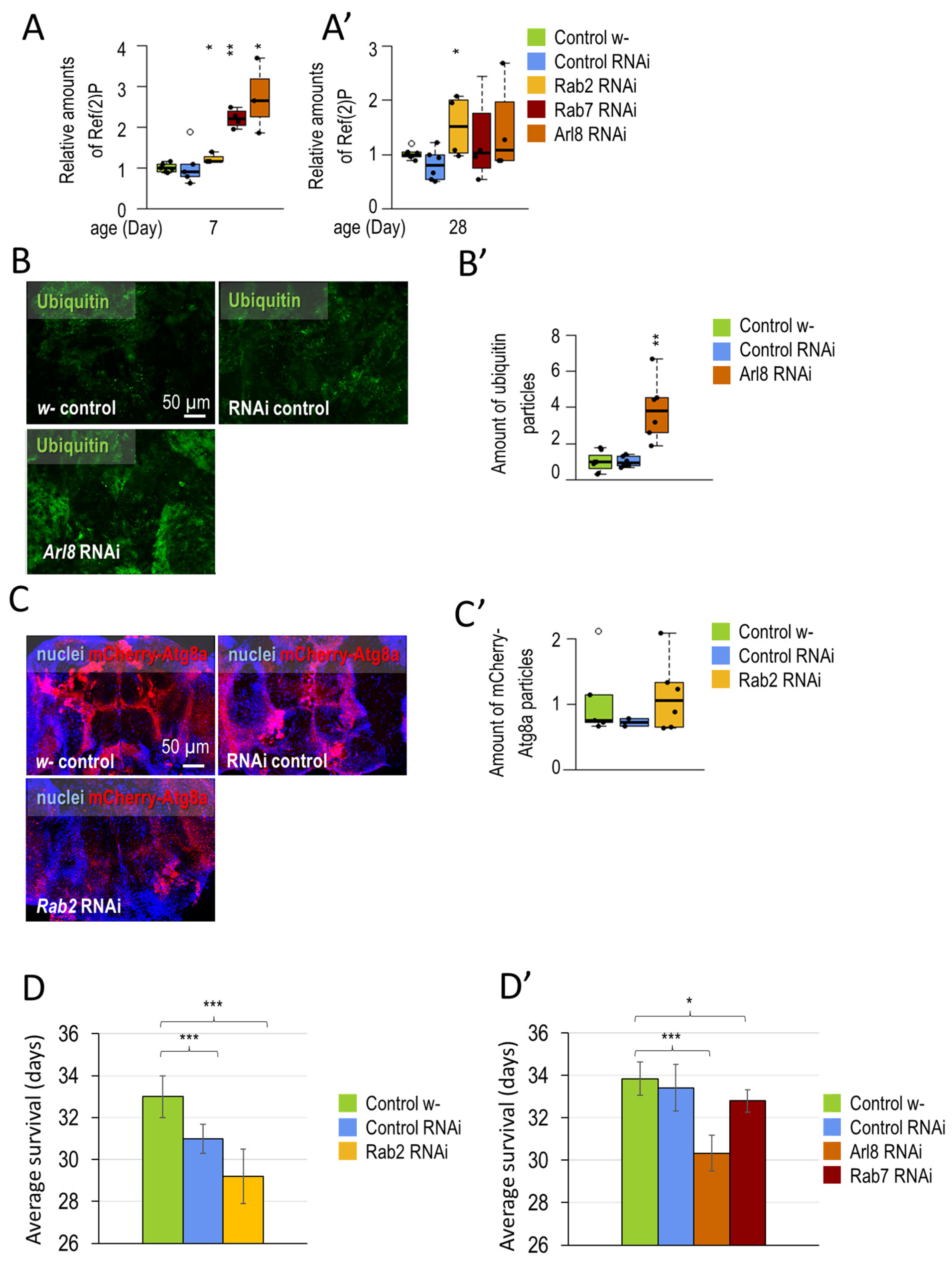
3.1.1. Small-GTPase Proteins Involved in the Lysosomal Degradation Pathway Are Required for Normal Longevity and Viability of Flies

In the Drosophila model system, some small GTPases are known to regulate cognitive and physiological abilities. For example, the overexpression of the small GTPase Rheb, which regulates TOR, results in elongated axon spines and cell bodies in the mushroom body. Increased levels of Rheb are also associated with memory impairment [84]. The lack of Ras-family Rin and Ric subfamily members shows neuronal morphology and survival abnormalities [85]. The photoreceptor cells of the compound eye derive from the nerve. The development of the R7 photoreceptor cell is regulated by Ras and Rap small GTPases [86]. Rho small GTPase is involved in the plasticity of the nervous system. It remodels actin-rich dendritic spines, in the absence of which, in invertebrates, learning and memory impairments occur [87].
In our experiments, we first examined whether the absence of small GTPases (similar to other small GTPases), which are essential in acid degradation, might cause damage to the nervous system that affects animal survival and behavior function. Our results show that the silencing of Rab2, Rab7, and Arl8 inhibits autophagy, reduces the lifespan of animals, and impairs their locomotor ability (Figure 1 and Figure 2).
Dopaminergic neurons (DNs) produce the neurotransmitter dopamine, the loss of which can lead to the development of PD. In our first measurements, we aimed to investigate the role of small GTPases on the physiological and cognitive abilities of Drosophila. All three small GTPases (Arl8, Rab2, Rab7) were silenced by RNA interference (RNAi). In a previous study, we showed that autophagic activity is not constant in Drosophila adult brains, with some neurons being more sensitive to changes in autophagy levels. Increasing autophagy in dopaminergic and serotonergic neurons (via inhibition of EDTP) may result in lifespan extension and improved cognitive function [52]. Therefore, we used Ddc-Gal4 expressed exclusively in dopaminergic and serotonergic neurons [88].
We measured the efficiency of the RNA interference strains by quantitative PCR and Western blot analysis (Figure A1A,B). The used Rab2-RNAi construct could reduce transcript and protein levels, and Arl8 and Rab7 silencing only reduced protein levels (translational inhibition). Autophagy was inhibited upon small-GTPase silencing. Ref(2)P/p62 is a substrate protein for autophagy, the amount of which is inversely proportional to the efficiency of autophagy [89]. In a Western blot analysis, Ref(2)P levels were elevated during Rab2, Rab7, and Arl8 silencing. Compared to the Atg8a-I and -II ratio in control samples, small GTPase silenced samples accumulated Atg8a-I soluble protein in aged samples. This result also suggests a failure in autophagy (Figure 1B and Figure A2A,A’). The ubiquitinated aggregates and cell organelles are also degraded by autophagy [90], which were labeled by immunohistochemistry and studied by fluorescence microscopy. Silencing of Rab2, Rab7, and Arl8 increased the number of ubiquitin-positive structures in Drosophila adult brains (Figure 1C,C’ and Figure A2B,B’). Finally, we examined the abundance of mCherry-Atg8a-positive structures by fluorescence microscopy (which indicates all autophagic structures from the phagophore to autolysosome). mCherry-positive autophagic structures accumulated in these three cases of gene silencing (Figure 1D,D’ and Figure A2C,C’).
Then, we examined the effect of silencing Rab2, Rab7, and Arl8 in the nervous system of Drosophila. Rab2-, Rab7-, and Arl8-RNAi in DNs shortened lifespan (Figure 2A,A’ and Figure A2D,D’). The Drosophila climbing assay can be used to investigate the fitness of animals and their response to a given stimulus. To measure the cognitive abilities of animals, it can be tested in a 25 cm long tube. By knocking down the tube, negative geotaxis (upward climbing) can be induced in the animals. From the speed of the climb, the response time of the animals to the stimulus can be inferred [48,50,52]. Silencing all three small-GTPases impaired the animals’ climbing ability (Figure 2B,B’). Our results suggest that small-GTPases are required for normal climbing ability and autophagy in Drosophila neurons. After this, we investigated the physiological and cell biological consequences of activating small-GTPase proteins.

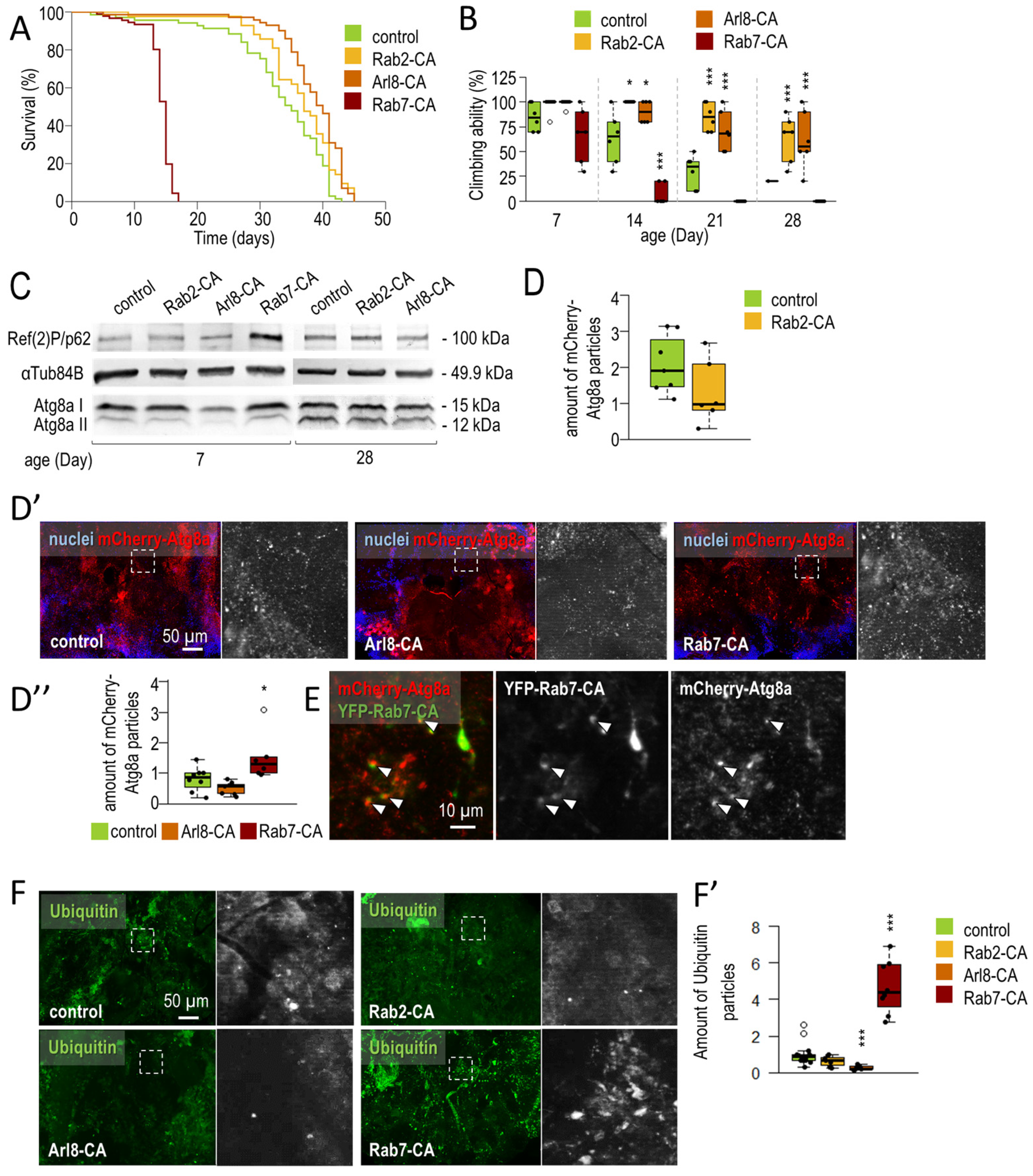
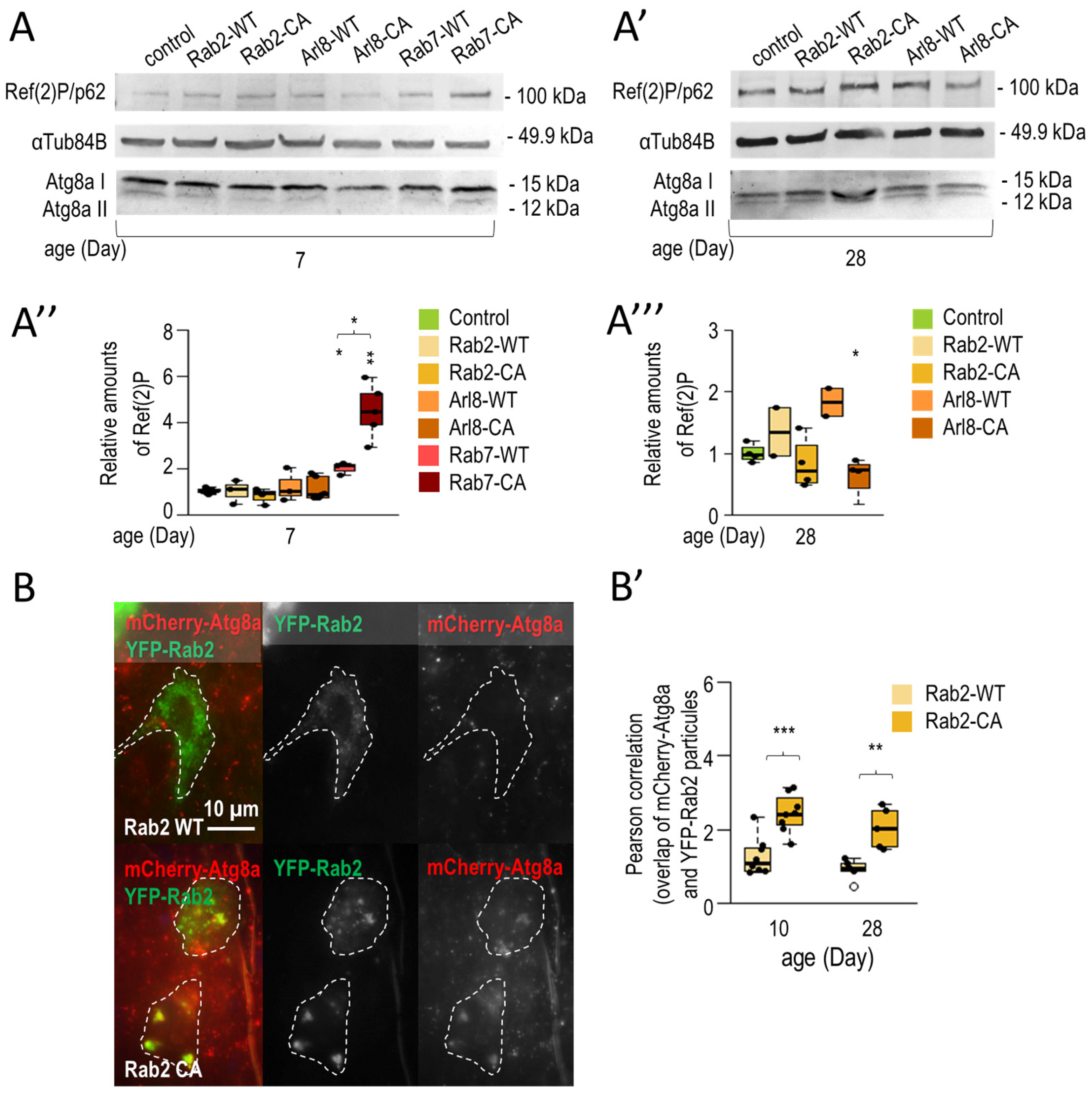
3.1.2. Rab2 and Arl8 Activation Extends Lifespan and Improves Climbing Ability

Following the silencing of small GTPase, we examined the effects of activating Rab2, Rab7, and Arl8 (overproduction of GTP-bound forms) on neuronal ageing (Figure 3). Our results show that activation of Rab2 and Arl8 enhances autophagy, whereas Rab7-CA inhibits acid degradation. Enhancement of autophagy may contribute to the increased lifespan and locomotor ability observed in animals. In contrast, Rab7-CA, which inhibits autophagy, is detrimental to the viability and motility of the animals (Figure 3).
The small GTPase activation was achieved by neuronal (dopaminergic and serotonergic specific) overexpression of constitutively active, continuously GTP-bound mutants (hereafter referred to as Rab2-CA, Rab7-CA, and Arl8-CA) [56,82,91]. We examined the effects of Rab2-CA, Arl8-CA, and Rab7-CA on lifespan, and found that the overexpression of activated forms of Rab2 and Arl8 significantly increased lifespan. In contrast, Rab7-CA significantly reduced lifespan relative to control (Figure 3A and Figure A3A). In aged animals, Rab2-CA and Arl8-CA improved, while Rab7-CA decreased, the ability of animals to climb compared to control (Figure 3B). Therefore, activated Rab2 and Arl8 exert a beneficial effect in aged flies. We also compared the effects of overexpressing constitutively active or wild-type forms of the investigated proteins and observed that only Rab2-CA and Arl8-CA have beneficial impacts compared to the WT forms. Rab7-WT and -CA were both detrimental, but Rab7-WT animals reached a longer lifespan (compared to age-matched Rab7-CA) (Figure A3B).
We compared the autophagy levels in samples isolated from the heads of small GTPase-WT and -CA animals. In young animals, Rab7-CA and Rab7-WT increased Ref(2)P, suggesting inhibition of autophagy. Rab7-overexpressing animals (both WT and CA) did not reach 28 days of age, so at this time, only Rab2 and Arl8 activation was compared to overexpression of wild-type forms. In older animals, only Rab2-CA and Arl8-CA decreased Ref(2)P levels, in contrast to the overexpression of -WT forms where a slight increase was observed in the relative amount of Ref(2)P (Figure A4A–A’’’). Climbing ability was also impaired in these animals, suggesting that overexpression of Rab2-WT and Arl8-WT is detrimental in older animals (Figure A3B).
The adverse effects of Rab7-CA could result from either the existence of activated Rab7 or the harmful expression of the protein. To address this issue, we compared the expression levels of Rab2-CA and Rab7-CA. Rab7#24103 (the originally used construct) showed significantly higher expression levels than Rab2-CA (Figure A3C,C’). The expression of three additional Rab7-CA alleles, Rab7-CA#9779, Rab7-CA#50785, and Rab7-CA#42707, were also assessed. Their expression levels were significantly lower than Rab7-CA#24103 and Rab2-CA (Figure A3C,C’). Despite these data, the neuronal expression of Rab7-CA#9779 and Rab7-CA#50785 alleles harmed the climbing ability of transgenic animals (Figure A3D). These results suggest that Rab7 activation may have a detrimental effect on the climbing ability of animals.
We also tested the effect of small GTPase activation on autophagic activity. Ref(2)P and Atg8a labeling were performed on protein samples isolated from heads by Western blotting. We observed a decrease in Ref(2)P levels in response to Rab2 and Arl8 activation, whereas increased Ref(2)P levels were noticed in the case of Rab7-CA (Figure 3C and Figure A3E,E’). Arl8-CA and Rab2-CA also led to a decrease in Atg8a-II levels compared to Atg8a-I (soluble form). For autophagic vesicle labeling, we used an mCherry-Atg8a reporter, which showed decreased Rab2-CA and Arl8-CA levels, but an accumulation in Rab7-CA levels, in brain samples (Figure 3D–D” and Figure A3F). In the presence of continuously elevated autophagy, Atg8a can also be considered a substrate for acid degradation, so decreased mCherry labeling and lowered Ref(2)P levels suggest autophagy activation. We next monitored the colocalization of the Rab7-CA marker with mCherry-Atg8a-positive structures and found several cases when green and red vesicles remained nonoverlapped (Figure 3E). These results indicate defects in autophagosome/late endosome–lysosome fusion. A similar colocalization feature to Rab7-CA was not observed between YFP-Rab2-CA and mCherry-At8a (Figure A4B,B’).
Finally, antiubiquitin labeling was used to investigate the amount of ubiquitinated structures in Drosophila brain samples. Both Rab2-CA and Arl8-CA were able to reduce, whereas Rab7 activation caused a significant increase in, the amount of ubiquitin-positive structures (Figure 3F,F’). These findings suggest that Rab2-CA and Arl8-CA can enhance the autophagic process in the Drosophila nervous system, and benefit neuronal functions in aged flies. Moreover, increasing the GTP-bound form of Rab7 in neurons can interfere with viability, which may result from, at least in part, the inhibition of autophagy.

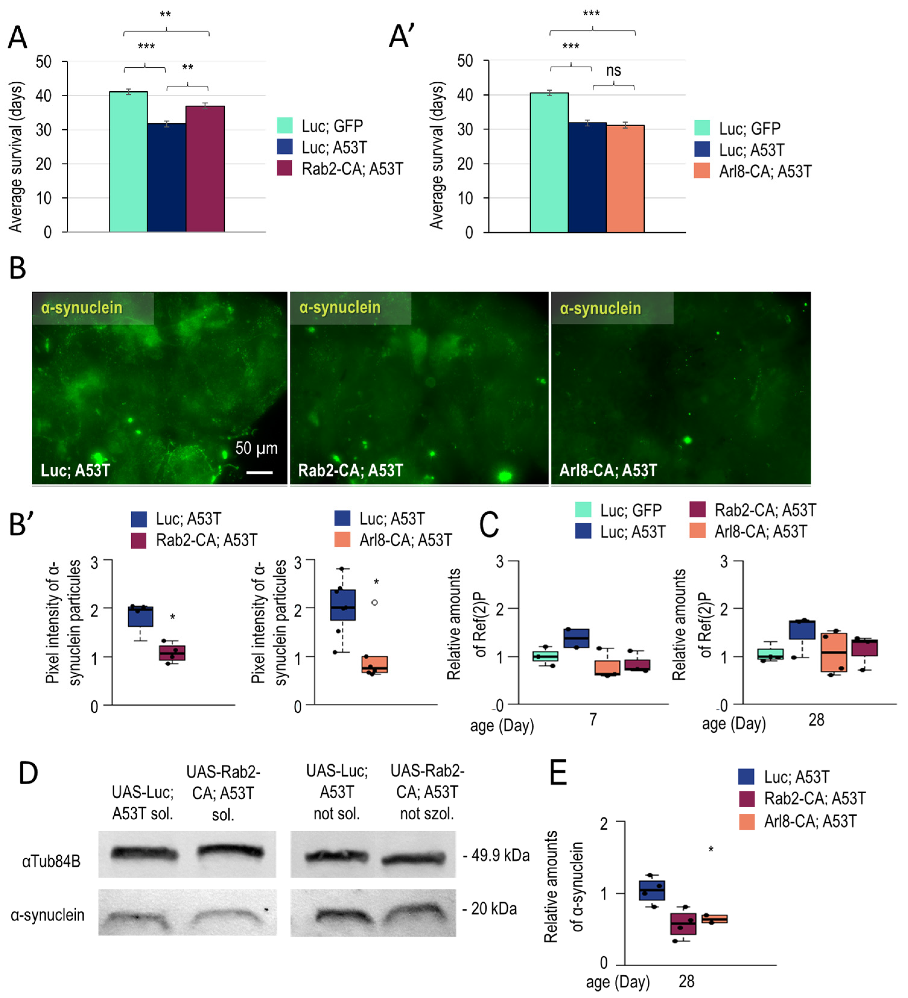
3.1.3. Rab2 Activation Promotes Longevity in a Parkinson’s Disease Model

The lack of autophagy has been associated with a number of neurodegenerative (ND) diseases. In contrast, enhancing acid degradation can benefit the survival of dying ND neurons, prolong the lifespan, and delay the onset of disease-associated dysfunctional symptoms. In the previous chapter, the induction of active forms of Rab2 and Arl8 stimulated autophagy function and had beneficial effects on animals during ageing (Figure 3). Therefore, it was considered worthwhile to investigate the effects of Rab2-CA and Arl8-CA in Parkinson’s disease (PD) models (Figure 4). Our results show that the neuronal activity of both Rab2 and Arl8 can enhance autophagy, stimulate the degradation of the toxic protein (that causes PD), and improve locomotor abilities in aged animals (Figure 4).
In humans, PD can result from an autosomal dominant mutation (e.g., A53T) of SCNA gene encoding α-synuclein protein [92]. The mutant protein is more likely to produce fibrils (A53T also triggers Tau fibrillation) than the wild-type one, thereby promoting disease progression [93]. We used Ddc-Gal4 driver to express Rab2-CA and Arl8-CA in dopaminergic and serotonergic neurons with an A53T background. UAS-Luc; UAS-A53T was used to create a control fly model of PD. According to these data, Rab2-CA increased survival and climbing in transgenic animals at advanced ages compared to age-matched control (Luc, A53T) (Figure 4A,B and Figure A5A). This indicates that the UAS-Luc; UAS-A53T transgene negatively influences climbing ability and lifespan, thereby successfully generating the PD model. In Arl8-CA animals, we did not observe a significant change in lifespan compared to PD control but noticed an improvement in the climbing of old animals (Figure 4A’,B’ and Figure A5A’).
Next, we examined changes in autophagic activity during ageing in flies serving as a model for PD. In these flies, Rab2-CA or Rab2-WT were overexpressed. We found that Ref(2)P/p62 levels increase in aged animals compared to young ones (Figure 4C). This implies that autophagy becomes compromised during ageing in transgenic animals. Contrary to this, overexpression of Rab2-CA enhanced autophagic activity compared to Rab2 WT-overexpressing flies (Figure 4C). Thus, wild-type overexpression of Rab2 alone does not induce A53T degradation (toxic A53T protein degradation requires Rab2-CA expression). In animals overexpressing Rab2-CA, the amount of lipid-bound Atg8a was reduced, suggesting an efficient degradation of the inner autophagosomal membrane (Figure 4C and Figure A5C). Similar to Rab2-CA, Arl8 activation also enhanced autophagy in aged animals. The amounts of Ref(2)P and Atg8a-II proteins were reduced compared to control PD (Luc; A53T) (Figure 4C’ and Figure A5C’). Thus, both Rab2-CA and Arl8-CA can activate autophagy in neurons of A53T mutant flies.
Finally, we also studied changes in the amount of toxic A53T protein by fluorescence microscopy and Western blotting (Figure 4D,D’,E and Figure A5B,B’,E). Human α-synuclein-specific antibody was used for labeling. Confocal imaging allows the detection of punctate structures (aggregates), whereas conventional imaging is more favorable for determining the soluble protein amount [94]. Results showed that Rab2 and Arl8 activation could reduce soluble and aggregated A53T amounts (Figure 4D,D’ and Figure A5B,B’). A Western blot analysis led to similar results. The overexpression of both small-GTPases-CA reduced A53T levels (Figure 4E and Figure A5E). For Rab2-CA, proteins were separated into soluble and nonsoluble fractions by centrifugation (Figure A5D) [95]. In both cases, a decrease in A53T abundance was observed in response to Rab2-CA. In summary, activation of Rab2 and Arl8 in neurons of flies serving as a model of PD can activate autophagy, decrease toxic A53T levels, and improve survival and movement abilities.

4. Discussion

In the present study, we investigated the effects of neuronal-specific silencing of Rab2, Rab7, and Arl8 on lifespan, climbing functions, and autophagic activity. Results we obtained show that silencing these small-GTPases harms animals’ survival and climbing. Furthermore, autophagy is inhibited in neurons overactivating these proteins (Figure 1, Figure 2 and Figure A2).
We revealed that overexpression of Rab2-CA and Arl8-CA each reduces the level of autophagy substrate Ref(2)P/p62 and ubiquitin proteins in dopaminergic and serotonergic neurons, and improves the movement of aged flies (Figure 3). The amounts of Ref(2)P and ubiquitin-positive structures were reversely altered in Rab7-CA samples compared to Rab2-CA and Arl8-CA. Thus, whereas the activation of Rab2 and Arl8 enhances autophagy, Rab7 impairs the degradation process. Furthermore, Rab7 activation appears to be detrimental to the animal’s climbing ability and viability (Figure 3 and Figure A3). The constitutively active form of Rab7 caused mCherry-Atg8a accumulation in the Drosophila brain. The colocalization of Rab7- and Atg8a-positive structures is impaired in Rab7-CA animals (Figure 3E). In HeLa cells, both Rab7 and Arl8 can bind to late endosomes [96]. The activation or inactivation of Rab7 affects the localization of Arl8 vesicles [97]. We hypothesized that Rab7 activation inhibits amphisome formation (amphisomal vesicles formed by the fusion of endosomes with autophagosomes). Autophagosomes forming at the end of axons undergo a retrograde transport into the soma. Prior to the transport, endosomes and autophagosomes fuse to create amphisomes. Finally, the autophagosomal cargo is degraded in the soma [31]. It can be assumed that autophagy inhibition and lifespan shortening in Rab7-CA animals are partly due to the lack of Rab7–Arl8 exchange on endosomes and inhibition of retrograde transport, respectively.
Overexpression of Rab2-CA in PD model animals (flies express the human A53T mutant α-synuclein) extends lifespan (Figure 4A). Both Rab2-CA and Arl8-CA improve climbing ability compared to PD control (Figure 4A–B’ and Figure A5A,A’). In addition, these hyperactivated small GTPases increase autophagy and degradation of toxic α-synuclein relative to the control group (Figure 4 and Figure A5). Cytoplasmic α-synuclein can be found in as soluble monomers, oligomers, and aggregates. The accumulation of α-synuclein aggregates is a characteristic feature of PD. However, soluble oligomers are toxic to the cell [98,99]. The degradation of α-synuclein dissolved in the cytoplasm was more significant than the elimination of α-synuclein-positive aggregates. In PD model animals expressing A53T mutation, only Rab2 and Arl8 activation was tested, and it was found to have positive effects on wild-type animals. Rab7-CA negatively affects viability and autophagy (Figure 3). In HEK293 cells, overexpressing Rab7 reduces the amount of mutant α-synuclein [100]. Furthermore, Rab7 overexpression rescues the climbing ability defective phenotype in a fly model of PD [100]. Although overexpression of Rab7-WT was detrimental to the climbing ability of the tested animals (Figure A3B), overexpression of Rab2-CA led to the opposite results, as only the latter was able to increase climbing ability in the affected animals. Similar results were also obtained when comparing Arl8-WT and Arl8-CA (Figure A3B). In this PD model, Rab-CA and Arl8-CA could trigger autophagy (Figure 4C,C’). It might be interesting to compare the effects of Rab7-CA and Rab7-WT overexpression between wild-type and PD model animals.
Although these small GTPases act as regulators of autophagy, they also function independently of acidic degradation. For example, Arl8 plays a role in controlling microtubule and vesicle transport [101]. The protein promotes the axonal transport of presynaptic cargo proteins, thereby preventing premature aggregation [102]. Rab2 is a Golgi-residential protein mediating ER-Golgi transport (in both anterograde and retrograde directions). Presynaptic precursors, established in the soma of neurons, are transported from the trans-Golgi network to synapses by Rab2. This process is essential for synapse formation and neurotransmission. It has also been shown that Rab2 functions upstream of Arl8 in the biogenesis of presynaptic neurons [103]. Rab7 is involved in tumor progression as it can be either an oncogenic or tumor suppressor, determined by environmental factors. It plays an essential role in chemoresistance to cisplatin by modulating the late endosomal pathway and lysosome biogenesis [104].
Data provided by this study suggests that Rab2 and Arl8 serve as potential targets for autophagy enhancement in the Drosophila nervous system. In the future, it might be interesting to assess the effect of Rab2 and Arl8 coactivation on autophagy, and it would also be worthwhile to validate these findings in a mammalian model and human cell lines. Molecules that specifically inhibit Rab2 or Arl8 serve as potent targets for drug candidates to modulate the activity of the autophagic process in treating neurodegenerative pathologies. In the future, it would be reasonable to investigate which GAP enzyme can inhibit Rab2 or Arl8 specifically, but not activate Rab7, with similar medical purposes.

Author Contributions

J.S. and F.K. designed and performed experiments on Drosophila samples, analyzed data, and wrote the manuscript. E.A.K. and G.F. performed experiments on Drosophila samples and analyzed data. T.V. and T.K. designed experiments, analyzed data, and wrote the manuscript. All authors have read and agreed to the published version of the manuscript.

Funding

This work was supported by the OTKA grants (Hungarian Scientific Research Fund) PD 143786 to T.K. T.K. was supported by the ÚNKP-22-5 New National Excellence Program of Culture and Innovation from the source of the National Research, Development and Innovation Fund and Bolyai János Research Scholarship. T.V. was supported by the OTKA grant K132439, and the Genetics Research Group ELKH/ELTE (01062). J.S. was supported by the Centre of Excellence, Hungary.

Informed Consent Statement

We confirm that the studies presented in the manuscript were carried out in compliance with the ARRIVE guidelines.

Data Availability Statement

The datasets used and/or analyzed during the current study available from the corresponding author on reasonable request.

Acknowledgments

Drosophila strains and reagents were kindly provided by Gábor Juhász and Tamás Lukácsovics. The authors also thank Ibtissem Benhamza, Daniella Baár, Regina Preisinger, Beatrix Supauer, and Erzsébet Gatyás for the excellent technical assistance.

Conflicts of Interest

The authors declare no conflict of interest.

Appendix A

Figure A1. Testing the effectiveness of RNA interference specific to small GTPases. (A) Quantitative PCR saturation curves are shown. Actin5C was used as an internal control. Bars show relative changes in mRNAs levels compared to control, and values were normalized to actin5C levels. (B) The effect of Rab7, Arl8, and Rab2 downregulation on protein levels was also investigated by Western blotting. Tub84B was as an internal control.
Cells 12 01753 g0a1
Figure A2. Silencing of small GTPases Rab2, Rab7, and Arl8 inhibits autophagy and limits lifespan. (A,A’) The plots show the relative change in the amount of Ref(2)P/p62 protein in response to small GTPase silencing. Samples are isolated from the heads of 7- and 28-day-old animals. (B,B’) Figures show antibody labelling and evaluation of ubiquitinated protein aggregates (green). (C,C’) The effect of Rab2-RNAi on the abundance of mCherry-Atg8a-positive (red) structures in the brains of adult females was tested. (D,D’) Graphs show the average survival data (Rab2-RNAi, Arl8-RNAi and Rab7-RNAi, as well as their corresponding controls). Silencing of small GTPases (Arl8, Rab2, Rab7) has a negative effect on lifespan. Boxes represent the most typical 50% of the samples, the line indicates the median, and upper and lower whiskers show the remaining 25–25% of the samples. Circles mark outliers. *: p < 0.05, **: p < 0.01, ***: p < 0.005; statistical analysis was performed as described in the Section 2.
Cells 12 01753 g0a2
Figure A3. Overexpression of the activated forms of Rab2 and Arl8 extends lifespan and climbing ability. (A) Graphs show the mean lifespan of animals (Rab2-CA, Arl8-CA, and Rab7-CA, as well as their corresponding controls). Arl8 and Rab2 activation prolongs lifespan, while Rab7 engagement significantly reduces lifespan. (B) The effects of wild-type (Rab2-WT, Arl8-WT, and Rab7-WT) and constitutively active (Rab2-CA, Arl8-CA, and Rab7-CA) forms of small GTPases on climbing ability were tested at different adult ages. Overexpressions were achieved only in dopaminergic and serotonergic neurons. Only Rab2-CA and Arl8-CA increase climbing ability. Climbing was measured at 20 seconds. (C,C’) Expression levels of different YFP-Rab2-CA constructs (Rab7-CA#24103, Rab7-CA#9779, Rab7-CA#50785, and Rab7-CA#42707) were assessed and compared to YFP-Rab2-CA in the brain of adult females. (D) Climbing ability of Rab7-CA (Rab7-CA#9779, Rab7-CA#50785) animals were investigated. These constructs were integrated into different genomic positions. Expression of all Rab7-CA constructs has a negative effect on movement ability. (E,E’) Quantitative changes in Ref(2)P protein were measured in samples isolated from the heads of female animals of different ages. For the corresponding Western blot images, see Figure 3C. (F) The effect of Rab2-CA on the abundance of mCherry-Atg8a-positive structures in the brains of adult female Drosophila. Rab2-CA reduces the number of mCherry-Atg8a structures The boxes represent the most typical 50% of the samples, the line indicates the median, and upper and lower whiskers show the remaining 25–25% of the samples. Circles mark outliers. *: p < 0.05, **: p < 0.01, ***: p < 0.005; statistical analysis was performed as described in Section 2.
Cells 12 01753 g0a3
Figure A4. Only the overexpression of constitutively active Rab2 and Arl8 can trigger autophagy. (AA’’’) We expressed wild-type (-WT) and constitutively active (-CA) small GTPase proteins in the central nervous system of animals (dopaminergic and serotonergic specific drivers). Proteins were isolated from the heads of young and old (7 and 21 days old) females. Anti-αTub84B was labeled as an internal control during Western blotting. Changes in autophagy were examined by anti-p62/Ref(2)P and anti-Atg8a labeling. (B,B’) Colocalization of mCherry-Atg8a (red) and YFP-Rab2 (wild-type—Rab2-WT and constitutively active—Rab2-CA) constructs was examined. YFP-Rab2 construct was expressed by a dopaminergic neuron (DN)-specific ple-Gal4. Green labeling indicates DN cells. The boxes represent the most typical 50% of the samples, the line indicates the median, and upper and lower whiskers show the remaining 25–25% of the samples. Circles mark outliers. *: p < 0.05, **: p < 0.01, ***: p < 0.005; statistical analysis was performed as described in Section 2.
Cells 12 01753 g0a4
Figure A5. Constitutively active forms of Rab2 and Arl8 enhance the degradation of human mutant α-synuclein. (A,A’) The effect of Rab2 and Arl8 activation on the lifespan of human α-synuclein mutants (A53T) is measured. Graphs show mean lifespan of Rab2-CA;A53T and Arl8-CA;A53T animals, as well as their corresponding controls (Luc;GFP was used as a healthy control and Luc;A53T was used as PD model). Rab2-CA increases lifespan in α-synuclein mutants. (B,B’) Levels of human mutant α-synuclein (A53T) were measured in different genotypes by conventional microscopy. (C) Changes in autophagy (in protein samples isolated from old and young heads) were measured through the relative levels of Ref(2)P by Western blot. The corsresponding Western blot images can be found in Figure 4C,C’. (D) Centrifugation was used to separate soluble and aggregated protein fractions in samples isolated from the head of female adults. Antihuman α-synuclein was used to label A53T proteins in the samples and αTub84B protein labeling was used as inner control. Rab2-CA increases the degradation of soluble and aggregated α-synuclein proteins. (E) Changes in toxic A53T levels were examined by Western blotting in animals expressing Rab2-CA and Arl8-CA proteins. See Figure 4E for the Western blot images associated with the diagrams. Boxes represent the most typical 50% of the samples, the line indicates the median, and upper, and lower whiskers show the remaining 25–25% of the samples. Circles mark outliers. *: p < 0.05, **: p < 0.01, ***: p < 0.005. Statistical analysis was performed as described in Section 2.
Cells 12 01753 g0a5

References

  1. Adams, J. The Proteasome: Structure, Function, and Role in the Cell. Cancer Treat. Rev. 2003, 29, 3–9. [Google Scholar] [CrossRef]
  2. Parzych, K.R.; Klionsky, D.J. An Overview of Autophagy: Morphology, Mechanism, and Regulation. Antioxid. Redox Signal. 2014, 20, 460–473. [Google Scholar] [CrossRef] [Green Version]
  3. Klionsky, D.J.; Petroni, G.; Amaravadi, R.K.; Baehrecke, E.H.; Ballabio, A.; Boya, P.; Pedro, J.M.B.; Cadwell, K.; Cecconi, F.; Choi, A.M.K.; et al. Autophagy in Major Human Diseases. EMBO J. 2021, 40, 108863. [Google Scholar] [CrossRef] [PubMed]
  4. Klionsky, D.J.; Codogno, P. The Mechanism and Physiological Function of Macroautophagy. J. Innate Immun. 2013, 5, 427–433. [Google Scholar] [CrossRef]
  5. Mizushima, N.; Komatsu, M. Autophagy: Renovation of Cells and Tissues. Cell 2011, 147, 728–741. [Google Scholar] [PubMed] [Green Version]
  6. Rae, M.J.; Butler, R.N.; Campisi, J.; De Grey, A.D.N.J.; Finch, C.E.; Gough, M.; Martin, G.M.; Vijg, J.; Perrott, K.M.; Logan, B.J. The Demographic and Biomedical Case for Late-Life Interventions in Aging. Sci. Transl. Med. 2010, 2, 40cm21. [Google Scholar] [CrossRef] [PubMed] [Green Version]
  7. Meléndez, A.; Tallóczy, Z.; Seaman, M.; Eskelinen, E.L.; Hall, D.H.; Levine, B. Autophagy Genes Are Essential for Dauer Development and Life-Span Extension in C. Elegans. Science 2003, 301, 1387–1391. [Google Scholar] [CrossRef] [Green Version]
  8. Donati, A.; Cavallini, G.; Paradiso, C.; Vittorini, S.; Pollera, M.; Gori, Z.; Bergamini, E. Age-Related Changes in the Autophagic Proteolysis of Rat Isolated Liver Cells: Effects of Antiaging Dietary Restrictions. J. Gerontol. Ser. A Biol. Sci. Med. Sci. 2001, 56, B375–B383. [Google Scholar] [CrossRef] [Green Version]
  9. Tóth, M.L.; Sigmond, T.; Borsos, É.; Barna, J.; Erdélyi, P.; Takács-Vellai, K.; Orosz, L.; Kovács, A.L.; Csikós, G.; Sass, M.; et al. Longevity Pathways Converge on Autophagy Genes to Regulate Life Span in Caenorhabditis Elegans. Autophagy 2008, 4, 330–338. [Google Scholar] [CrossRef] [Green Version]
  10. Vellai, T.; Takács-Vellai, K. Regulation of Protein Turnover by Longevity Pathways. In Advances in Experimental Medicine and Biology; Springer: Berlin/Heidelberg, Germany, 2010; Volume 694, pp. 69–80. [Google Scholar]
  11. Vellai, T. Autophagy Genes and Ageing. Cell Death Differ. 2009, 16, 94–102. [Google Scholar] [CrossRef] [Green Version]
  12. Sigmond, T.; Barna, J.; Tóth, M.L.; Takács-Vellai, K.; Pásti, G.; Kovács, A.L.; Vellai, T. Chapter 30 Autophagy in Caenorhabditis Elegans. In Methods in Enzymology; Elsevier: Amsterdam, The Netherlands, 2008; Volume 451, pp. 521–540. [Google Scholar]
  13. Simonsen, A.; Cumming, R.C.; Brech, A.; Isakson, P.; Schubert, D.R.; Finley, K.D. Promoting Basal Levels of Autophagy in the Nervous System Enhances Longevity and Oxidant Resistance in Adult Drosophila. Autophagy 2008, 4, 176–184. [Google Scholar] [CrossRef] [PubMed] [Green Version]
  14. Pyo, J.O.; Yoo, S.M.; Ahn, H.H.; Nah, J.; Hong, S.H.; Kam, T.I.; Jung, S.; Jung, Y.K. Overexpression of Atg5 in Mice Activates Autophagy and Extends Lifespan. Nat. Commun. 2013, 4, 1–9. [Google Scholar] [CrossRef] [PubMed] [Green Version]
  15. Campisi, J.; Warner, H.R. Aging in Mitotic and Post-Mitotic Cells. In Advances in Cell Aging and Gerontology; Gilchrest, B.A., Bohr, V.A., Eds.; Elsevier Science B.V.: Amsterdam, The Netherlands, 2001; pp. 1–16. [Google Scholar]
  16. Rubinsztein, D.C. The Roles of Intracellular Protein-Degradation Pathways in Neurodegeneration. Nature 2006, 443, 780–786. [Google Scholar] [CrossRef]
  17. Vellai, T.; Tóth, M.L.; Kovács, A.L. Janus-Faced Autophagy: A Dual Role of Cellular Self-Eating in Neurodegeneration? Autophagy 2007, 3, 461–463. [Google Scholar] [CrossRef] [Green Version]
  18. Tominaga, K.; Suzuki, H.I. TGF-β Signaling in Cellular Senescence and Aging-Related Pathology. Int. J. Mol. Sci. 2019, 20, 5002. [Google Scholar] [CrossRef] [Green Version]
  19. Shaw, W.M.; Luo, S.; Landis, J.; Ashraf, J.; Murphy, C.T. The C. Elegans TGF-β Dauer Pathway Regulates Longevity via Insulin Signaling. Curr. Biol. 2007, 17, 1635. [Google Scholar] [CrossRef] [PubMed] [Green Version]
  20. Oh, S.W.; Mukhopadhyay, A.; Svrzikapa, N.; Jiang, F.; Davis, R.J.; Tissenbaum, H.A. JNK Regulates Lifespan in Caenorhabditis Elegans by Modulating Nuclear Translocation of Forkhead Transcription Factor/DAF-16. Proc. Natl. Acad. Sci. USA 2005, 102, 4494. [Google Scholar] [CrossRef] [Green Version]
  21. Liu, J.K. Antiaging Agents: Safe Interventions to Slow Aging and Healthy Life Span Extension. Nat. Prod. Bioprospect. 2022, 12, 18. [Google Scholar] [CrossRef]
  22. Kiechl, S.; Pechlaner, R.; Willeit, P.; Notdurfter, M.; Paulweber, B.; Willeit, K.; Werner, P.; Ruckenstuhl, C.; Iglseder, B.; Weger, S.; et al. Higher Spermidine Intake Is Linked to Lower Mortality: A Prospective Population-Based Study. Am. J. Clin. Nutr. 2018, 108, 371–380. [Google Scholar] [CrossRef] [Green Version]
  23. Madeo, F.; Eisenberg, T.; Pietrocola, F.; Kroemer, G. Spermidine in Health and Disease. Science 2018, 359, eaan2788. [Google Scholar] [CrossRef] [Green Version]
  24. Gupta, V.K.; Scheunemann, L.; Eisenberg, T.; Mertel, S.; Bhukel, A.; Koemans, T.S.; Kramer, J.M.; Liu, K.S.Y.; Schroeder, S.; Stunnenberg, H.G.; et al. Restoring Polyamines Protects from Age-Induced Memory Impairment in an Autophagy-Dependent Manner. Nat. Neurosci. 2013, 16, 1453–1460. [Google Scholar] [CrossRef] [PubMed]
  25. Ahsan, A.; Zheng, Y.R.; Wu, X.L.; Tang, W.D.; Liu, M.R.; Ma, S.J.; Jiang, L.; Hu, W.W.; Zhang, X.N.; Chen, Z. Urolithin A-activated Autophagy but Not Mitophagy Protects against Ischemic Neuronal Injury by Inhibiting ER Stress in Vitro and in Vivo. CNS Neurosci. Ther. 2019, 25, 976. [Google Scholar] [CrossRef] [PubMed] [Green Version]
  26. Bakhtiari, N.; Hosseinkhani, S.; Tashakor, A.; Hemmati, R. Ursolic Acid Ameliorates Aging-Metabolic Phenotype through Promoting of Skeletal Muscle Rejuvenation. Med. Hypotheses 2015, 85, 1–6. [Google Scholar] [CrossRef] [PubMed]
  27. Lin, J.H.; Chen, S.Y.; Lu, C.C.; Lin, J.A.; Yen, G.C. Ursolic Acid Promotes Apoptosis, Autophagy, and Chemosensitivity in Gemcitabine-Resistant Human Pancreatic Cancer Cells. Phytother. Res. 2020, 34, 2053–2066. [Google Scholar] [CrossRef] [PubMed]
  28. Anisimov, V.N.; Alimova, I.N.; Baturin, D.A.; Popovich, I.G.; Zabezhinski, M.A.; Rosenfeld, S.V.; Manton, K.G.; Semenchenko, A.V.; Yashin, A.I. Dose-Dependent Effect of Melatonin on Life Span and Spontaneous Tumor Incidence in Female SHR Mice. Exp. Gerontol. 2003, 38, 449–461. [Google Scholar] [CrossRef] [PubMed]
  29. Izmaylov, D.M.; Obukhova, L.K. Geroprotector Effectiveness of Melatonin: Investigation of Lifespan of Drosophila melanogaster. Mech. Ageing Dev. 1999, 106, 233–240. [Google Scholar] [CrossRef]
  30. Luo, F.; Sandhu, A.F.; Rungratanawanich, W.; Williams, G.E.; Akbar, M.; Zhou, S.; Song, B.J.; Wang, X. Melatonin and Autophagy in Aging-Related Neurodegenerative Diseases. Int. J. Mol. Sci. 2020, 21, 7174. [Google Scholar] [CrossRef]
  31. Maday, S. Mechanisms of Neuronal Homeostasis: Autophagy in the Axon. Brain Res. 2016, 1649, 143–150. [Google Scholar] [CrossRef] [Green Version]
  32. Maday, S.; Holzbaur, E.L.F. Autophagosome Assembly and Cargo Capture in the Distal Axon. Autophagy 2012, 8, 858–860. [Google Scholar] [CrossRef] [Green Version]
  33. Cheng, X.T.; Zhou, B.; Lin, M.Y.; Cai, Q.; Sheng, Z.H. Axonal Autophagosomes Recruit Dynein for Retrograde Transport through Fusion with Late Endosomes. J. Cell Biol. 2015, 209, 377–386. [Google Scholar] [CrossRef] [Green Version]
  34. Maday, S.; Wallace, K.E.; Holzbaur, E.L.F. Autophagosomes Initiate Distally and Mature during Transport toward the Cell Soma in Primary Neurons. J. Cell Biol. 2012, 196, 407–417. [Google Scholar] [CrossRef]
  35. Hara, T.; Nakamura, K.; Matsui, M.; Yamamoto, A.; Nakahara, Y.; Suzuki-Migishima, R.; Yokoyama, M.; Mishima, K.; Saito, I.; Okano, H.; et al. Suppression of Basal Autophagy in Neural Cells Causes Neurodegenerative Disease in Mice. Nature 2006, 441, 885–889. [Google Scholar] [CrossRef]
  36. Maria Fimia, G.; Stoykova, A.; Romagnoli, A.; Giunta, L.; Di Bartolomeo, S.; Nardacci, R.; Corazzari, M.; Fuoco, C.; Ucar, A.; Schwartz, P.; et al. Ambra1 Regulates Autophagy and Development of the Nervous System. Nature 2007, 447, 1121–1125. [Google Scholar] [CrossRef] [PubMed] [Green Version]
  37. Reimer, R.J.; Schneider, K.; Leal-Ortiz, S.; Montenegro-Venegas, C.; Kim, S.A.; Garner, L.C.; Gundelfinger, E.D.; Reimer, R.J.; Garner, C.C. Bassoon Controls Presynaptic Autophagy through Atg5. Neuron 2017, 93, 897–913.e7. [Google Scholar] [CrossRef] [Green Version]
  38. Konstantinidis, G.; Tavernarakis, N. Molecular Basis of Neuronal Autophagy in Ageing: Insights from Caenorhabditis Elegans. Cells 2021, 10, 694. [Google Scholar] [CrossRef]
  39. Menzies, F.M.; Fleming, A.; Caricasole, A.; Bento, C.F.; Andrews, S.P.; Ashkenazi, A.; Füllgrabe, J.; Jackson, A.; Jimenez Sanchez, M.; Karabiyik, C.; et al. Autophagy and Neurodegeneration: Pathogenic Mechanisms and Therapeutic Opportunities. Neuron 2017, 93, 1015–1034. [Google Scholar] [CrossRef] [Green Version]
  40. Komatsu, M.; Waguri, S.; Chiba, T.; Murata, S.; Iwata, J.; Tanida, I.; Ueno, T.; Koike, M.; Uchiyama, Y.; Kominami, E.; et al. Loss of Autophagy in the Central Nervous System Causes Neurodegeneration in Mice. Nature 2006, 441, 880–884. [Google Scholar] [CrossRef] [PubMed]
  41. Cuervo, A.M.; Stefanis, L.; Fredenburg, R.; Lansbury, P.T.; Sulzer, D. Impaired Degradation of Mutant Alpha-Synuclein by Chaperone-Mediated Autophagy. Science 2004, 305, 1292–1295. [Google Scholar] [CrossRef] [PubMed]
  42. Jin, S.M.; Youle, R.J. PINK1- and Parkin-Mediated Mitophagy at a Glance. J. Cell Sci. 2012, 125, 795–799. [Google Scholar] [CrossRef] [Green Version]
  43. Laplante, M.; Sabatini, D.M. MTOR Signaling at a Glance. J. Cell Sci. 2009, 122, 3589–3594. [Google Scholar] [CrossRef] [Green Version]
  44. Vellai, T. How the Amino Acid Leucine Activates the Key Cell-Growth Regulator MTOR. Nature 2021, 596, 192–194. [Google Scholar] [CrossRef]
  45. Boutouja, F.; Stiehm, C.M.; Platta, H.W. MTOR: A Cellular Regulator Interface in Health and Disease. Cells 2019, 8, 18. [Google Scholar] [CrossRef] [PubMed] [Green Version]
  46. Glick, D.; Barth, S.; Macleod, K.F. Autophagy: Cellular and Molecular Mechanisms. J. Pathol. 2010, 221, 3–12. [Google Scholar] [CrossRef] [Green Version]
  47. Papp, D.; Kovács, T.; Billes, V.; Varga, M.; Tarnóci, A.; Hackler, L.; Puskás, L.G.; Liliom, H.; Tárnok, K.; Schlett, K.; et al. AUTEN-67, an Autophagy-Enhancing Drug Candidate with Potent Antiaging and Neuroprotective Effects. Autophagy 2016, 12, 273–286. [Google Scholar] [CrossRef] [PubMed] [Green Version]
  48. Kovács, T.; Billes, V.; Komlós, M.; Hotzi, B.; Manzéger, A.; Tarnóci, A.; Papp, D.; Szikszai, F.; Szinyákovics, J.; Rácz, Á.; et al. The Small Molecule AUTEN-99 (Autophagy Enhancer-99) Prevents the Progression of Neurodegenerative Symptoms. Sci. Rep. 2017, 7, 42014. [Google Scholar] [CrossRef] [PubMed] [Green Version]
  49. Komlós, M.; Szinyákovics, J.; Falcsik, G.; Sigmond, T.; Jezsó, B.; Vellai, T.; Kovács, T. The Small-Molecule Enhancers of Autophagy AUTEN-67 and -99 Delay Ageing in Drosophila Striated Muscle Cells. Int. J. Mol. Sci. 2023, 24, 8100. [Google Scholar] [CrossRef]
  50. Billes, V.; Kovács, T.; Hotzi, B.; Manzéger, A.; Tagscherer, K.; Komlós, M.; Tarnóci, A.; Pádár, Z.; Erdős, A.; Bjelik, A.; et al. AUTEN-67 (Autophagy Enhancer-67) Hampers the Progression of Neurodegenerative Symptoms in a Drosophila Model of Huntington’s Disease. J. Huntingt. Dis. 2016, 5, 133–147. [Google Scholar] [CrossRef] [PubMed] [Green Version]
  51. Button, R.W.; Roberts, S.L.; Willis, T.L.; Oliver Hanemann, C.; Luo, S. Accumulation of Autophagosomes Confers Cytotoxicity. J. Biol. Chem. 2017, 292, 13599–13614. [Google Scholar] [CrossRef] [Green Version]
  52. Kovács, T.; Szinyákovics, J.; Billes, V.; Murányi, G.; Varga, V.B.; Bjelik, A.; Légrádi, Á.; Szabó, M.; Sándor, S.; Kubinyi, E.; et al. A Conserved MTMR Lipid Phosphatase Increasingly Suppresses Autophagy in Brain Neurons during Aging. Sci. Rep. 2022, 12, 21817. [Google Scholar] [CrossRef]
  53. Stenmark, H. Rab GTPases as Coordinators of Vesicle Traffic. Nat. Rev. Mol. Cell Biol. 2009, 10, 513–525. [Google Scholar] [CrossRef]
  54. Stroupe, C. This Is the End: Regulation of Rab7 Nucleotide Binding in Endolysosomal Trafficking and Autophagy. Front. Cell Dev. Biol. 2018, 6, 129. [Google Scholar] [CrossRef] [PubMed] [Green Version]
  55. Ding, X.; Jiang, X.; Tian, R.; Zhao, P.; Li, L.; Wang, X.; Chen, S.; Zhu, Y.; Mei, M.; Bao, S.; et al. RAB2 Regulates the Formation of Autophagosome and Autolysosome in Mammalian Cells. Autophagy 2019, 15, 1774–1786. [Google Scholar] [CrossRef] [PubMed] [Green Version]
  56. Boda, A.; Lőrincz, P.; Takáts, S.; Csizmadia, T.; Tóth, S.; Kovács, A.L.; Juhász, G. Drosophila Arl8 Is a General Positive Regulator of Lysosomal Fusion Events. Biochim. Biophys. Acta Mol. Cell Res. 2019, 1866, 533–544. [Google Scholar] [CrossRef] [PubMed]
  57. Gutierrez, M.G.; Munafó, D.B.; Berón, W.; Colombo, M.I. Rab7 Is Required for the Normal Progression of the Autophagic Pathway in Mammalian Cells. J. Cell Sci. 2004, 117, 2687–2697. [Google Scholar] [CrossRef] [Green Version]
  58. Kirisako, T.; Baba, M.; Ishihara, N.; Miyazawa, K.; Ohsumi, M.; Yoshimori, T.; Noda, T.; Ohsumi, Y. Formation Process of Autophagosome Is Traced with Apg8/Aut7p in Yeast. J. Cell Biol. 1999, 147, 435–446. [Google Scholar] [CrossRef] [Green Version]
  59. Lund, V.K.; Madsen, K.L.; Kjaerulff, O. Drosophila Rab2 Controls Endosome-Lysosome Fusion and LAMP Delivery to Late Endosomes. Autophagy 2018, 14, 1520–1542. [Google Scholar] [CrossRef] [Green Version]
  60. Fujita, N.; Huang, W.; Lin, T.H.; Groulx, J.F.; Jean, S.; Nguyen, J.; Kuchitsu, Y.; Koyama-Honda, I.; Mizushima, N.; Fukuda, M.; et al. Genetic Screen in Drosophila Muscle Identifies Autophagy-Mediated T-Tubule Remodeling and a Rab2 Role in Autophagy. eLife 2017, 6, e23367. [Google Scholar] [CrossRef]
  61. Zhao, X.; Liberti, R.; Jian, J.; Fu, W.; Hettinghouse, A.; Sun, Y.; Liu, C.J. Progranulin Associates with Rab2 and Is Involved in Autophagosome-Lysosome Fusion in Gaucher Disease. J. Mol. Med. 2021, 99, 1639–1654. [Google Scholar] [CrossRef] [PubMed]
  62. Gillingham, A.K.; Sinka, R.; Torres, I.L.; Lilley, K.S.; Munro, S. Toward a Comprehensive Map of the Effectors of Rab GTPases. Dev. Cell 2014, 31, 358. [Google Scholar] [CrossRef] [PubMed] [Green Version]
  63. Kajiho, H.; Kajiho, Y.; Frittoli, E.; Confalonieri, S.; Bertalot, G.; Viale, G.; Di Fiore, P.P.; Oldani, A.; Garre, M.; Beznoussenko, G.V.; et al. RAB2A Controls MT1-MMP Endocytic and E-cadherin Polarized Golgi Trafficking to Promote Invasive Breast Cancer Programs. EMBO Rep. 2016, 17, 1061. [Google Scholar] [CrossRef] [Green Version]
  64. Lőrincz, P.; Mauvezin, C.; Juhász, G. Exploring Autophagy in Drosophila. Cells 2017, 6, 22. [Google Scholar] [CrossRef] [PubMed] [Green Version]
  65. Jäger, S.; Bucci, C.; Tanida, I.; Ueno, T.; Kominami, E.; Saftig, P.; Eskelinen, E.L. Role for Rab7 in Maturation of Late Autophagic Vacuoles. J. Cell Sci. 2004, 117, 4837–4848. [Google Scholar] [CrossRef] [PubMed] [Green Version]
  66. Takáts, S.; Glatz, G.; Szenci, G.; Boda, A.; Horváth, G.V.; Hegedűs, K.; Kovács, A.L.; Juhász, G. Non-Canonical Role of the SNARE Protein Ykt6 in Autophagosome-Lysosome Fusion. PLoS Genet. 2018, 14, e1007359. [Google Scholar] [CrossRef] [PubMed] [Green Version]
  67. Lund, V.K.; Lycas, M.D.; Schack, A.; Andersen, R.C.; Gether, U.; Kjaerulff, O. Rab2 Drives Axonal Transport of Dense Core Vesicles and Lysosomal Organelles. Cell Rep. 2021, 35, 108973. [Google Scholar] [CrossRef]
  68. Kuchitsu, Y.; Fukuda, M. Revisiting Rab7 Functions in Mammalian Autophagy: Rab7 Knockout Studies. Cells 2018, 7, 215. [Google Scholar] [CrossRef] [Green Version]
  69. Bhargava, H.K.; Tabata, K.; Byck, J.M.; Hamasaki, M.; Farrell, D.P.; Anishchenko, I.; DiMaio, F.; Im, Y.J.; Yoshimori, T.; Hurley, J.H. Structural Basis for Autophagy Inhibition by the Human Rubicon-Rab7 Complex. Proc. Natl. Acad. Sci. USA 2020, 117, 17003–17010. [Google Scholar] [CrossRef]
  70. Guerra, F.; Bucci, C. Multiple Roles of the Small GTPase Rab7. Cells 2016, 5, 34. [Google Scholar] [CrossRef] [Green Version]
  71. Oka, M.; Hashimoto, K.; Yamaguchi, Y.; Saitoh, S.I.; Sugiura, Y.; Motoi, Y.; Honda, K.; Kikko, Y.; Ohata, S.; Suematsu, M.; et al. Arl8b Is Required for Lysosomal Degradation of Maternal Proteins in the Visceral Yolk Sac Endoderm of Mouse Embryos. J. Cell Sci. 2017, 130, 3568–3577. [Google Scholar] [CrossRef] [PubMed] [Green Version]
  72. Rosa-Ferreira, C.; Sweeney, S.T.; Munro, S. The Small G Protein Arl8 Contributes to Lysosomal Function and Long-Range Axonal Transport in Drosophila. Biol. Open 2018, 7, bio035964. [Google Scholar] [CrossRef] [Green Version]
  73. Mrakovic, A.; Kay, J.G.; Furuya, W.; Brumell, J.H.; Botelho, R.J. Rab7 and Arl8 GTPases Are Necessary for Lysosome Tubulation in Macrophages. Traffic 2012, 13, 1667–1679. [Google Scholar] [CrossRef]
  74. Cherry, S.; Jin, E.J.; Özel, M.N.; Lu, Z.; Agi, E.; Wang, D.; Jung, W.-H.; Epstein, D.; Meinertzhagen, I.A.; Chan, C.-C.; et al. Charcot-Marie-Tooth 2B Mutations in Rab7 Cause Dosage-Dependent Neurodegeneration Due to Partial Loss of Function. eLife 2013, 2, e01064. [Google Scholar] [CrossRef]
  75. Szegö, E.M.; Van den Haute, C.; Höfs, L.; Baekelandt, V.; Van der Perren, A.; Falkenburger, B.H. Rab7 Reduces α-Synuclein Toxicity in Rats and Primary Neurons. Exp. Neurol. 2022, 347, 113900. [Google Scholar] [CrossRef]
  76. Griffin, E.F.; Caldwell, K.A.; Caldwell, G.A. Vacuolar Protein Sorting Protein 41 (VPS41) at an Intersection of Endosomal Traffic in Neurodegenerative Disease. Neural Regen. Res. 2019, 14, 1210–1212. [Google Scholar] [CrossRef]
  77. Griffin, E.F.; Yan, X.; Caldwell, K.A.; Caldwell, G.A. Distinct Functional Roles of Vps41-Mediated Neuroprotection in Alzheimer’s and Parkinson’s Disease Models of Neurodegeneration. Hum. Mol. Genet. 2018, 27, 4176–4193. [Google Scholar] [CrossRef]
  78. Inoshita, T.; Liu, J.Y.; Taniguchi, D.; Ishii, R.; Shiba-Fukushima, K.; Hattori, N.; Imai, Y. Parkinson Disease-Associated Leucine-Rich Repeat Kinase Regulates UNC-104-Dependent Axonal Transport of Arl8-Positive Vesicles in Drosophila. iScience 2022, 25, 105476. [Google Scholar] [CrossRef]
  79. Katheder, N.S.; Khezri, R.; O’Farrell, F.; Schultz, S.W.; Jain, A.; Schink, M.K.O.; Theodossiou, T.A.; Johansen, T.; Juhász, G.; Bilder, D.; et al. Microenvironmental Autophagy Promotes Tumour Growth. Nature 2017, 541, 417–420. [Google Scholar] [CrossRef] [PubMed] [Green Version]
  80. Pircs, K.; Nagy, P.; Varga, A.; Venkei, Z.; Erdi, B.; Hegedus, K.; Juhasz, G. Advantages and Limitations of Different P62-Based Assays for Estimating Autophagic Activity in Drosophila. PLoS ONE 2012, 7, e44214. [Google Scholar] [CrossRef] [PubMed]
  81. Takáts, S.; Nagy, P.; Varga, Á.; Pircs, K.; Kárpáti, M.; Varga, K.; Kovács, A.L.; Hegedűs, K.; Juhász, G. Autophagosomal Syntaxin17-Dependent Lysosomal Degradation Maintains Neuronal Function in Drosophila. J. Cell Biol. 2013, 201, 531–539. [Google Scholar] [CrossRef] [Green Version]
  82. Lőrincz, P.; Tóth, S.; Benkő, P.; Lakatos, Z.; Boda, A.; Glatz, G.; Zobel, M.; Bisi, S.; Hegedűs, K.; Takáts, S.; et al. Rab2 Promotes Autophagic and Endocytic Lysosomal Degradation. J. Cell Biol. 2017, 216, 1937–1947. [Google Scholar] [CrossRef] [PubMed] [Green Version]
  83. Pfaffl, M.W. A New Mathematical Model for Relative Quantification in Real-Time RT–PCR. Nucleic Acids Res. 2001, 29, e45. [Google Scholar] [CrossRef] [PubMed]
  84. Brown, H.L.D.; Kaun, K.R.; Edgar, B.A. The Small GTPase Rheb Affects Central Brain Neuronal Morphology and Memory Formation in Drosophila. PLoS ONE 2012, 7, e44888. [Google Scholar] [CrossRef] [Green Version]
  85. Shi, G.X.; Cai, W.; Andres, D.A. Rit Subfamily Small GTPases: Regulators in Neuronal Differentiation and Survival. Cell Signal. 2013, 25, 2060. [Google Scholar] [CrossRef] [PubMed] [Green Version]
  86. Mavromatakis, Y.E.; Tomlinson, A. The Role of the Small GTPase Rap in Drosophila R7 Photoreceptor Specification. Proc. Natl. Acad. Sci. USA 2012, 109, 3844–3849. [Google Scholar] [CrossRef] [PubMed] [Green Version]
  87. van der Voet, M.; Nijhof, B.; Oortveld, M.A.W.; Schenck, A. Drosophila Models of Early Onset Cognitive Disorders and Their Clinical Applications. Neurosci. Biobehav. Rev. 2014, 46 Pt 2, 326–342. [Google Scholar] [CrossRef] [PubMed] [Green Version]
  88. Riemensperger, T.; Issa, A.R.; Pech, U.; Coulom, H.; Nguyễn, M.V.; Cassar, M.; Jacquet, M.; Fiala, A.; Birman, S. A Single Dopamine Pathway Underlies Progressive Locomotor Deficits in a Drosophila Model of Parkinson Disease. Cell Rep. 2013, 5, 952–960. [Google Scholar] [CrossRef] [PubMed] [Green Version]
  89. Nagy, P.; Kárpáti, M.; Varga, Á.; Pircs, K.; Venkei, Z.; Takáts, S.; Varga, K.; Érdi, B.; Hegedus, K.; Juhász, G. Atg17/FIP200 Localizes to Perilysosomal Ref(2)P Aggregates and Promotes Autophagy by Activation of Atg1 in Drosophila. Autophagy 2014, 10, 453–467. [Google Scholar] [CrossRef] [PubMed] [Green Version]
  90. Pankiv, S.; Clausen, T.H.; Lamark, T.; Brech, A.; Bruun, J.A.; Outzen, H.; Øvervatn, A.; Bjørkøy, G.; Johansen, T. P62/SQSTM1 Binds Directly to Atg8/LC3 to Facilitate Degradation of Ubiquitinated Protein Aggregates by Autophagy. J. Biol. Chem. 2007, 282, 24131–24145. [Google Scholar] [CrossRef] [Green Version]
  91. Zhang, J.; Schulze, K.L.; Robin Hiesinger, P.; Suyama, K.; Wang, S.; Fish, M.; Acar, M.; Hoskins, R.A.; Bellen, H.J.; Scott, M.P. Thirty-One Flavors of Drosophila Rab Proteins. Genetics 2007, 176, 1307–1322. [Google Scholar] [CrossRef] [Green Version]
  92. Kurz, A.; Double, K.L.; Lastres-Becker, I.; Tozzi, A.; Tantucci, M.; Bockhart, V.; Bonin, M.; García-Arencibia, M.; Nuber, S.; Schlaudraff, F.; et al. A53T-Alpha-Synuclein Overexpression Impairs Dopamine Signaling and Striatal Synaptic Plasticity in Old Mice. PLoS ONE 2010, 5, e11464. [Google Scholar] [CrossRef]
  93. Kotzbauer, P.T.; Giasson, B.I.; Kravitz, A.V.; Golbe, L.I.; Mark, M.H.; Trojanowski, J.Q.; Lee, V.M.Y. Fibrillization of α-Synuclein and Tau in Familial Parkinson’s Disease Caused by the A53T α-Synuclein Mutation. Exp. Neurol. 2004, 187, 279–288. [Google Scholar] [CrossRef]
  94. Billes, V.; Kovács, T.; Manzéger, A.; Lőrincz, P.; Szincsák, S.; Regős, Á.; Kulcsár, P.I.; Korcsmáros, T.; Lukácsovich, T.; Hoffmann, G.; et al. Developmentally Regulated Autophagy Is Required for Eye Formation in Drosophila. Autophagy 2018, 14, 1499–1519. [Google Scholar] [CrossRef] [Green Version]
  95. Lo Piccolo, L.; Bonaccorso, R.; Onorati, M.C. Nuclear and Cytoplasmic Soluble Proteins Extraction from a Small Quantity of Drosophila’s Whole Larvae and Tissues. Int. J. Mol. Sci. 2015, 16, 12360. [Google Scholar] [CrossRef] [PubMed] [Green Version]
  96. Marwaha, R.; Arya, S.B.; Jagga, D.; Kaur, H.; Tuli, A.; Sharma, M. The Rab7 Effector PLEKHM1 Binds Arl8b to Promote Cargo Traffic to Lysosomes. J. Cell Biol. 2017, 216, 1051–1070. [Google Scholar] [CrossRef] [Green Version]
  97. Jongsma, M.L.; Bakker, J.; Cabukusta, B.; Liv, N.; van Elsland, D.; Fermie, J.; Akkermans, J.L.; Kuijl, C.; van der Zanden, S.Y.; Janssen, L.; et al. SKIP-HOPS Recruits TBC1D15 for a Rab7-to-Arl8b Identity Switch to Control Late Endosome Transport. EMBO J. 2020, 39, e102301. [Google Scholar] [CrossRef]
  98. Sharon, R.; Bar-Joseph, I.; Frosch, M.P.; Walsh, D.M.; Hamilton, J.A.; Selkoe, D.J. The Formation of Highly Soluble Oligomers of α-Synuclein Is Regulated by Fatty Acids and Enhanced in Parkinson’s Disease. Neuron 2003, 37, 583–595. [Google Scholar] [CrossRef] [PubMed] [Green Version]
  99. Winner, B.; Jappelli, R.; Maji, S.K.; Desplats, P.A.; Boyer, L.; Aigner, S.; Hetzer, C.; Loher, T.; Vilar, M.; Campioni, S.; et al. In Vivo Demonstration That α-Synuclein Oligomers Are Toxic. Proc. Natl. Acad. Sci. USA 2011, 108, 4194–4199. [Google Scholar] [CrossRef] [Green Version]
  100. Dinter, E.; Saridaki, T.; Nippold, M.; Plum, S.; Diederichs, L.; Komnig, D.; Fensky, L.; May, C.; Marcus, K.; Voigt, A.; et al. Rab7 Induces Clearance of α-Synuclein Aggregates. J. Neurochem. 2016, 138, 758–774. [Google Scholar] [CrossRef] [PubMed]
  101. Khatter, D.; Sindhwani, A.; Sharma, M. Arf-like GTPase Arl8: Moving from the Periphery to the Center of Lysosomal Biology. Cell. Logist. 2015, 5, e1086501. [Google Scholar] [CrossRef] [Green Version]
  102. Donaldson, J.G.; Jackson, C.L. ARF Family G Proteins and Their Regulators: Roles in Membrane Transport, Development and Disease. Nat. Rev. Mol. Cell Biol. 2011, 12, 362. [Google Scholar] [CrossRef]
  103. Götz, T.W.B.; Puchkov, D.; Lysiuk, V.; Lützkendorf, J.; Nikonenko, A.G.; Quentin, C.; Lehmann, M.; Sigrist, S.J.; Petzoldt, A.G. Rab2 Regulates Presynaptic Precursor Vesicle Biogenesis at the Trans-Golgi. J. Cell Biol. 2021, 220, e202006040. [Google Scholar] [CrossRef]
  104. Guerra, F.; Bucci, C. Role of the RAB7 Protein in Tumor Progression and Cisplatin Chemoresistance. Cancers 2019, 11, 1096. [Google Scholar] [CrossRef] [PubMed] [Green Version]
Figure 1. Inhibition of small GTPase proteins involved in controlling lysosomal degradation represses autophagy in neurons. (A) Roles of the small GTPase proteins Rab2, Rab7, and Arl8 during lysosome-dependent degradation pathways. (B) Western blot analysis shows autophagic activity in Rab2-, Arl8-, and Rab7-RNAi backgrounds. p62/Ref(2)P is a substrate of autophagy; thus, its levels inversely correlate with the autophagic flux. Atg8a has key roles during autophagy, as it regulates phagophore formation and closure, and is required for selective autophagy. Atg8a-I bands indicate soluble, while Atg8a-II bands represent membrane-bounded, form of the protein. αTub84B protein was used as an internal control. (C,C’) Ubiquitinated proteins and aggregates are digested with autophagy; therefore, the effect of small GTPase RNAi was also investigated with ubiquitin labelling (green). Silencing of Rab2 and Rab7 increases the amount of ubiquitinated aggregates (D,D’) The abundance of mCherry-Atg8a-positive (red) structures was observed in small GTPases RNAi background. The marker labels autophagic vesicles, forming phagophores, autophagosomes, amphisomes, and autolysosomes. Nuclei were stained by Hoechst dye (blue). Arl8 silencing increases the amount of mCherry-Atg8a structures. The white dashed line squares indicate the magnified detail in the fluorescence images. Boxes represent the most typical 50% of the samples, line indicates the median, and upper and lower whiskers show the remaining 25–25% of the samples. Circles mark outliers. **: p < 0.01, ***: p < 0.005; statistical analysis was performed as described in the Materials and Methods.
Cells 12 01753 g001
Figure 2. Neuronal silencing of small GTPases (Arl8, Rab2, Rab7) reduces lifespan and decreases climbing ability in Drosophila. (A,A’) Kaplan–Meier lifespan curves of Drosophila. Using a Ddc-Gal4 driver, Rab2, Arl8, and Rab7 were silenced only in dopaminergic and serotonergic neurons, and compared to isogenic w1118 and ON-target free RNAi controls (both controls were crossed with Ddc-Gal4). Neuronal silencing of small GTPases reduces lifespan. (B,B’) Plots represent climbing ability of animals silenced for a small GTPase. Climbing was measured at 20 s. The lack of small GTPases impairs the climbing ability of animals. The boxes represent the most typical 50% of the samples, the line indicates the median, and upper and lower whiskers show the remaining 25–25% of the samples. Circles mark outliers. *: p < 0.05, **: p < 0.01, ***: p < 0.005; statistical analysis was performed as described in the Section 2.
Cells 12 01753 g002
Figure 3. Neuron-specific activation of Rab2 and Arl8 extends lifespan and enhances autophagy. (A) Kaplan–Meier lifespan curves of Drosophila. Constitutively active forms of the small-GTPase proteins Rab2 (Rab2-CA), Arl8 (Arl8-CA), and Rab7 (Rab7-CA) in dopaminergic and serotonergic neurons were tested (using Ddc-Gal4 driver). Our results show that Rab2-CA and Arl8-CA increase, while Rab7-CA decreases lifespan. (B) Moving ability of Rab2-CA, Arl8-CA, and Rab-7-CA animals were observed at different adult ages. The activation of Rab2 and Arl8 oppositely varies the climbing ability of animals compared to Rab7 activation Climbing was measured at 20 s. (C) Western blot analysis showing p62/Ref(2) and Atg8a-II levels. Ref(2)P and Atg8a-II serve as substrates for autophagy; thus, their levels inversely correlate with autophagic capacity. αTub84B protein was used as internal control. For measurements, proteins were isolated from the head of young (7 days old) and old (28 days old) female flies. (DD’’) The abundance of mCherry-Atg8a-positive (red) foci refers to autophagic vesicles, measured in small-GTPase activated and control genetic backgrounds. Nuclei were stained by Hoechst dye (blue). Rab7-CA increases the amount of mCherry-Atg8a structures, while Rab2 and Arl8 activation decreases them. (E) Colocalization error can be found between the colocalization of YFP-Rab7-CA-(YFP-bounded protein—green) and mCherry-Atg8a-positive (red) structures. (F,F’) Ubiquitinated aggregates are digested by autophagy. Amounts of ubiquitinated proteins (green) were measured in adult brains with hyperactivated small-GTPase proteins. Rab2 and Arl8 activation decreased, whereas Rab7 activation increased, ubiquitin aggregation in Drosophila brains. For the measurement, animals were maintained at 29 °C during adulthood, and Ddc-Gal4 triggered the overexpression of constitutively active form of small-GTPase. The white dashed line squares indicate the magnified detail in the fluorescence images. Boxes represent the most typical 50% of the samples, the line indicates the median, and upper and lower whiskers show the remaining 25–25% of the samples. Circles mark outliers. *: p < 0.05, ***: p < 0.005; statistical analysis was performed as described in the Materials and Methods.
Cells 12 01753 g003
Figure 4. Rab2 hyperactivation promotes longevity and improves climbing ability in a Parkinson’s disease (PD) fly model. (A,A’) Kaplan–Meier graphs of Drosophila surviving ability in percentage. Rab2-CA and Arl8-CA were overexpressed in a background expressing the human A53T mutant α-synuclein, and compared with native (Luc; GFP) and disease (Luc; A53T) controls. Rab2 activation increased survival of A53T mutant animals (compared to disease control). (B,B’) Climbing ability was tested in PD model. Climbing was measured at 20, 40, and 60 s intervals, and changes are plotted on the diagrams (B) and (B’). Panel (B) shows climbing ability within 40 s, while diagram (B’) shows climbing ability within 20 s. The activation of Arl8 and Rab2 increases the climbing ability of A53T mutants. (C,C’) Autophagic activity was studied by Western blot. Atg8a-II and p62/Ref(2)P levels were identified. αTub84B was used as an internal control. (D,D’) Human mutant (A53T) α-synuclein (green) was stained by immunohistochemistry and observed by a fluorescent microscope. Arl8-CA and Rab2-CA increase the degradation of mutant α-synuclein protein. (E) A53T level in PD models was also measured by Western blotting. αTub84B was used as an internal control. Animals were maintained at 29 °C during the adulthood, and the constitutively active form of small-GTPase were triggered by Ddc-Gal4 driver. The white dashed line squares indicate the magnified detail in the fluorescence images. Boxes represent the most typical 50% of the samples, the line indicates the median, and upper and lower whiskers show the remaining 25–25% of the samples. Circles mark outliers. statistical analysis was performed as described in Section 2.
Cells 12 01753 g004
Disclaimer/Publisher’s Note: The statements, opinions and data contained in all publications are solely those of the individual author(s) and contributor(s) and not of MDPI and/or the editor(s). MDPI and/or the editor(s) disclaim responsibility for any injury to people or property resulting from any ideas, methods, instructions or products referred to in the content.

