Article

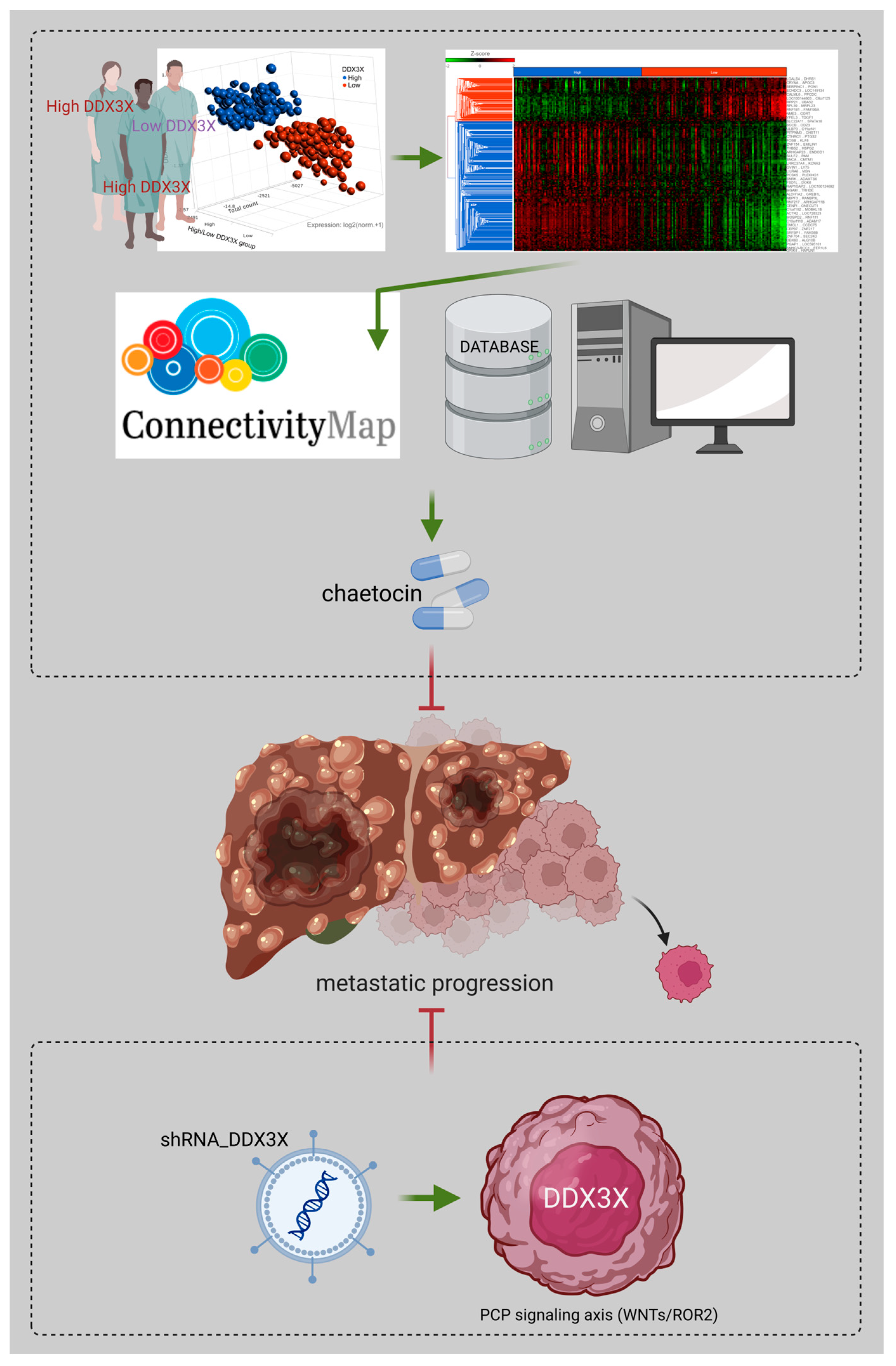
Perturbation Analysis of a Prognostic DDX3X-Mediated Gene Expression Signature Identifies the Antimetastatic Potential of Chaetocin in Hepatocellular Carcinoma

by
Tsung-Chieh Lin
1,2
1
Genomic Medicine Core Laboratory, Department of Medical Research and Development, Chang Gung Memorial Hospital, Linkou, Taoyuan City 333, Taiwan
2
Department of Biomedical Sciences, Chang Gung University, Taoyuan City 333, Taiwan
Cells 2023, 12(12), 1628; https://doi.org/10.3390/cells12121628
Submission received: 20 March 2023 / Revised: 5 June 2023 / Accepted: 12 June 2023 / Published: 14 June 2023

Abstract

:
ATP-dependent RNA helicase DDX3X, also known as DEAD (Asp-Glu-Ala-Asp) Box Polypeptide 3, X-Linked (DDX3X), is critical for RNA metabolism, and emerging evidence implicates ATP-dependent RNA helicase DDX3X’s participation in various cellular processes to modulate cancer progression. In this study, the clinical significance of DDX3X was addressed, and DDX3X was identified as a biomarker for poor prognosis. An exploration of transcriptomic data from 373 liver cancer patients from The Cancer Genome Atlas (TCGA) using Ingenuity Pathway Analysis (IPA) suggested an association between DDX3X expression and cancer metastasis. Lentiviral-based silencing of DDX3X in a hepatocellular carcinoma (HCC) cell line resulted in the suppression of cell migration and invasion. The molecular mechanism regarding ATP-dependent RNA helicase DDX3X in liver cancer progression had been addressed in many studies. I focused on the biological application of the DDX3X-mediated gene expression signature in cancer therapeutics. An investigation of the DDX3X-correlated expression signature via the L1000 platform of Connectivity Map (BROAD Institute) first identified a histone methyltransferase inhibitor, chaetocin, as a novel compound for alleviating metastasis in HCC. In this study, the prognostic value of DDX3X and the antimetastatic property of chaetocin are presented to shed light on the development of anti-liver cancer strategies.

1. Introduction

Emerging evidence indicates the critical regulatory role of DEAD (Asp-Glu-Ala-Asp) Box Polypeptide 3, X-Linked (DDX3X) in cancer progression. ATP-dependent RNA helicase DDX3X, a RNA helicase, is a DEAD-box family member, and has been reported to be involved in the splicing of pre-mRNA [1], RNA export [2], the transcription of RNA [3], and protein translation [4,5,6]. Due to its complex biological role in RNA metabolism, ATP-dependent RNA helicase DDX3X has gained increasing attention for its function in many types of cancer, and it regulates tumor progression in a complex manner. Furthermore, this complexity is further increased because ATP-dependent RNA helicase DDX3X generally exhibits its biological effects as components of multiprotein complexes [7]. The exact impacts of ATP-dependent RNA helicase DDX3X are affected by its interacting partners [8]. The overexpression of ATP-dependent RNA helicase DDX3X has been detected in hepatocellular carcinoma (HCC), and ATP-dependent RNA helicase DDX3X has been characterized as a critical gene in hepatocarcinogenesis [9]. In contrast, the loss of ATP-dependent RNA helicase DDX3X was found to lead to tumorigenesis due to a reduction in the expression of DNA repair factors in a mouse model [10]. Other studies revealed the loss of DDX3X expression in hepatocellular carcinoma tissue, and increased tumor cell proliferation because of the DDX3X silencing in hepatocellular carcinomas which were infected by hepatitis virus [3,11]. Hence, the conflicting roles of ATP-dependent RNA helicase DDX3X are inconsistent within the same type of cancer.
These conflicting results emphasize the urgent need for clarification. The prognostic role of DDX3X is a critical factor in determining whether DDX3X plays an oncogenic or tumor-suppressive role. In this study, I first aimed to characterize DDX3X’s role from the perspective of its prognostic significance in hepatoma patient survival. The current updated version of the liver cancer/hepatoma cohort, with more patients enrolled, was retrieved from The Cancer Genome Atlas (TCGA) database and reanalyzed. This analysis of the DDX3X-associated gene signature further suggested the potential value of studying its biological function in promoting hepatoma metastasis. Furthermore, ATP-dependent RNA helicase DDX3X has been proposed as a therapeutic target for cancers [12]. I also aimed to identify a novel use for a drug in reversing the DDX3X-mediated gene signature via a Connectivity Map (BROAD Institute) [13]. In this study, I demonstrated the prognostic value of DDX3X and the potential capability of a histone methyltransferase inhibitor, chaetocin, as a therapeutic target to shed light on the development of anti-liver cancer strategies.

2. Materials and Methods

2.1. TCGA Dataset Analysis

Gene expression in the TCGA liver hepatocellular carcinoma (LIHC) dataset (Dataset ID: TCGA.LIHC.sampleMap/HiSeqV2_PANCAN, version: 2017-10-13) was estimated by RNA-Seq (Illumina HiSeq), and these data were retrieved along with the associated clinical information for data analysis. The RNA-Seq read count was normalized and log2-transformed. A total of 373 HCC cases were divided into a DDX3X-high group and a DDX3X-low group based on the ranking assigned by the DDX3X expression level.

2.2. Ingenuity Pathway Analysis

The differential gene expression signatures in the HCC cohort were obtained after dividing the cohort into two groups with relatively high and low DDX3X levels. The gene signatures were further analyzed using Ingenuity® Pathway Analysis (IPA; QIAGEN, Hilden, Germany; https://digitalinsights.qiagen.com/products-overview/discovery-insights-portfolio/analysisand-visualization/qiagen-ipa/ (accessed on 1 November 2022)) according to the instructions provided. A list of relevant networks, upstream regulators and algorithmically generated mechanistic networks based on connectivity was obtained upon the comparison of the Ingenuity® Knowledge Database with the imported dataset. The canonical pathway analysis feature of IPA was also used to identify significant diseases and functions in rank order based on the altered gene signatures.

2.3. Cell Culture

Human hepatocellular carcinoma cell lines were purchased from Bioresource Collection and Research Center (Hsinchu, Taiwan). HA 22T/VGH and HA 59T/VGH cells were maintained in DMEM supplemented with 10% FBS, 0.1 mM NEAA, penicillin (100 units/mL), and streptomycin (100 µg/mL). C3A (HepG2/C3A) and HepG2 cells were maintained in MEM supplemented with 10% FBS, 0.1 mM NEAA, 1 mM sodium pyruvate, penicillin (100 units/mL), and streptomycin (100 µg/mL). The cells were incubated in a humidified atmosphere of 95% air and 5% CO2 at 37 °C. Chaetocin was purchased from Santa Cruz (CAS 28097-03-2, Dallas, TX, USA). The compound was dissolved in DMSO. In the chaetocin pretreatment assay, cancer cells were incubated with indicated concentrations of chaetocin for 48 h, and cells were then subjected to migration/invasion/wound-healing assay after removing chaetocin.

2.4. Lentivirus-Based shRNA Production and Infection

The lentiviral shRNA constructs were purchased from Thermo Scientific (Pittsburgh, PA, USA). Lentiviruses were produced via co-transfection of 293T cells with an shRNA-expressing plasmid, an envelope plasmid (pMD.G), and a packaging plasmid (pCMV-dR8.91) using calcium phosphate (Invitrogen, Carlsbad, CA, USA). The 293T cells were incubated for 18 h, followed by the replacement of the culture medium. The viral supernatants were harvested and titered at 48 and 72 h post-transfection. The cell monolayers were infected with the indicated lentivirus in the presence of polybrene and were further selected using puromycin.

2.5. Cell Migration and Invasion Assay

In vitro migration and invasion were investigated using a Transwell assay (Millipore, Bedford, MA, USA). For the invasion assay, transwell was additionally pre-coated with 35 µL of 3× diluted matrix matrigel (BD Biosciences Pharmingen, San Diego, CA, USA) for 30 min. A total of 2 × 10⁵ cells maintained in serum-free culture medium were added to the upper chamber of the device, and the lower chamber was filled with culture medium containing 10% FBS. After the indicated incubation periods, the cells remaining on the upper surface of the filter membrane were carefully removed using a cotton swab. The membrane was then fixed, stained with a 10× dilution of Giemsa solution (Merck, Darmstadt, Germany) and photographed. Cell motility was quantified by manually counting the cells in three random fields per filter membrane.

2.6. Wound Healing Assay

The wound healing assay was assessed using culture inserts (Ibidi, Martinsried, Germany). The culture inserts were transferred to plates. The cells were seeded at a density of 2 × 10⁵ cells/well and were allowed to attach. After incubation, the culture inserts were removed using sterile tweezers and washed with PBS. The plates were filled with culture medium supplemented with 2% serum to induce cell migration. The cells were photographed for quantification of closure of the exposed area. The denuded area closure was calculated by (Denuded distance 0h − Denuded distance Endpoint)/Denuded distance 0h.

2.7. Western Blot Analysis

Cells were lysed using RIPA buffer containing 50 mM Tris-HCl (pH 7.4), 150 mM NaCl, 1% Triton X-100, 0.25% sodium deoxycholate, 5 mM EDTA (pH 8.0), and 1 mM EGTA supplemented with protease and phosphatase inhibitors. After 20 min of lysis on ice, cell debris was removed via microcentrifugation and the supernatants were rapidly frozen. The protein concentration was measured via the Bradford method. In my experiments, equivalent samples containing 25–100 µg of protein were loaded onto an SDS-polyacrylamide gel, separated by electrophoresis, and electrophoretically transferred from the gel onto a polyvinylidene fluoride (PVDF) membrane (Millipore, Bedford, MA, USA). After blocking with 5% nonfat milk, the membrane was hybridized with specific primary antibodies overnight at 4 °C and subsequently incubated with a corresponding horseradish peroxidase-conjugated secondary antibody for 1 h. The relative levels of proteins on the membranes were determined using an ECL-Plus Detection Kit (PerkinElmer Life Sciences, Boston, MA, USA).

2.8. Statistical Analysis

Estimated survival rates were determined using the Kaplan–Meier method and were compared using the log-rank test. Student’s t test was performed for other statistical analyses. All data are shown as the mean ± S.D. values. The p values within the following ranges were considered significant: * p < 0.05, ** p < 0.01, and *** p < 0.001. The experiments were performed at least three times and shown by the representative.

3. Results

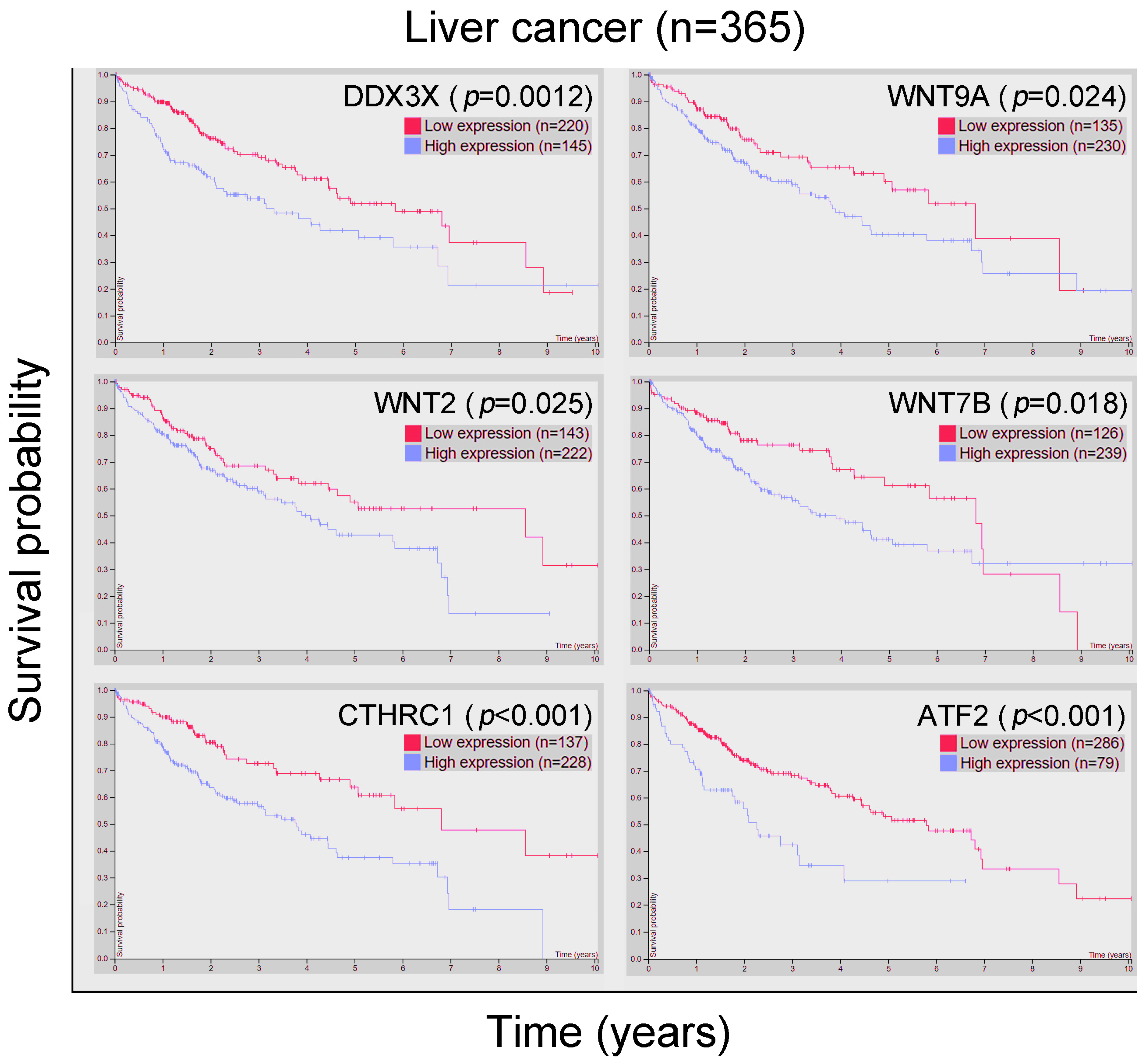
3.1. DDX3X Expression Is Significantly Correlated with Poor Outcomes in Liver Cancer, and the Significantly Differential Gene Signature Was Identified after Comparison between Patient Groups with High and Low DDX3X Levels

I first considered DDX3X’s clinical significance in cancer patient cohorts. A comprehensive pan-cancer study integrating cancer patients’ clinical data with RNA expression profile has been completed and released from the database: Human Protein Atlas (HPA) [14,15,16,17,18] and Kaplan–Meier plotter [19]. The prognostic data of DDX3X in different cancer types is listed and shown in Table 1. DDX3X expression predicted better survival rate in colorectal cancer, urothelial cancer, lung cancer and gastric cancer. However, DDX3X appeared to be an unfavorable prognostic biomarker in liver, pancreatic, breast and ovarian cancer. Among those cancer types, the up-to-date version of transcriptomic and clinical data of 373 liver cancer patients were analyzed. A scatter plot of the indicated expression patterns in the 373 patients is shown in Figure 1A.
The patients were divided into two groups with high or low DDX3X expression. The patients in the DDX3X-high group had a poor overall survival rate (p = 0.024, Figure 1B). Data from Cox regression analyses also indicated a trend toward DDX3X being useful as a prognostic factor (Table 2). These results suggest DDX3X’s prognostic value and indicate that further investigation regarding the molecular mechanisms altered by its overexpression is warranted.
In addition, a total of 2890 differentially expressed gene targets in the DDX3X-high group were selected and shown after hierarchical clustering analysis (Figure 1C and Supplementary Data Table S1).

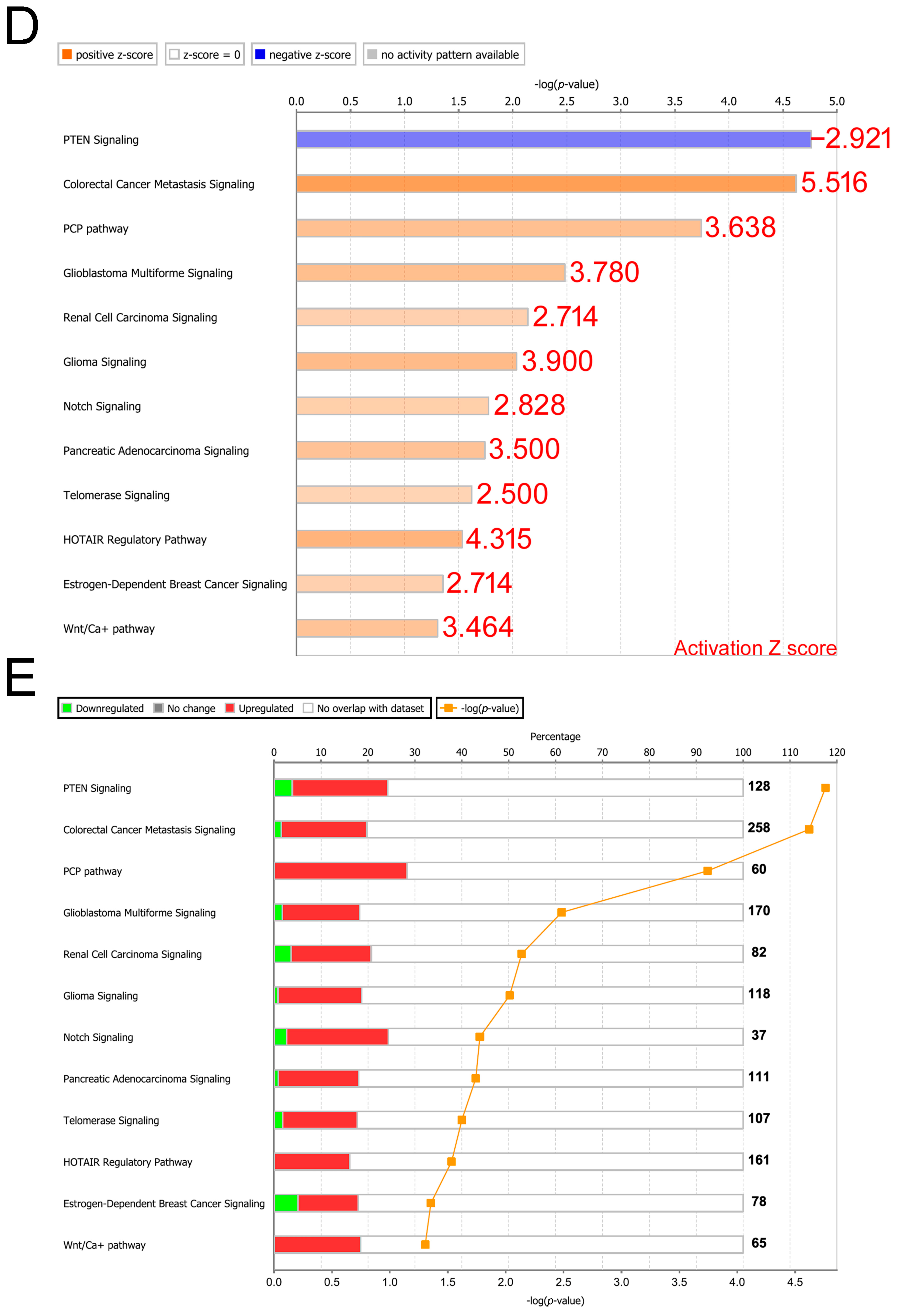
3.2. Knowledge-Based Analysis of Gene Signatures Reveals the Potential for Triggering Cancer Metastasis and Progression

The gene signature identified in the DDX3X-high group was analyzed with knowledge-based IPA. The results obtained with the canonical pathway module of IPA identified the significant pathways ranked by overlap (log p value), and the activation status of each signaling was determined by the transformed Z score (Activation Z score, Figure 1D). The similarity of the high DDX3X gene signature to those related to the activation of colorectal cancer metastasis and planar cell polarity (PCP) signaling was identified (Figure 1D,E). The inhibition of PTEN signaling was observed (Z score: −2.921), suggesting the activation of Akt (Figure 1D).
In addition, PCP pathway activation can lead to increased cancer proliferation and metastasis [20,21]. In Figure 2A, classic PCP signaling pathway was illustrated after IPA analysis of the gene signature observed in liver cancer patients with high DDX3X levels. Statistically highly expressed molecules including WNT9A, WNT2, WNT7B, ROR2, CTHRC1, DAMM1 and ATF2 were shown (Figure 2B). In a clinical setting, most of the aforementioned molecules were associated with poor overall survival in the liver cancer cohort (Figure 3). Moreover, the gene signature in the high DDX3X group was investigated by the analysis match module of IPA, which combined the curated and publicly available datasets. A significant similarity with those datasets annotated with the increased cell movement, migration of cells, cell movement of tumor cell lines and invasion of cells was detected (Table 3 and Figure 4). Hence, the transcriptomic data indicate the possibility that high DDX3X expression in liver cancer patients might trigger cancer progression, especially via metastasis, consistent with the observation of poor clinical outcomes (Figure 1B).

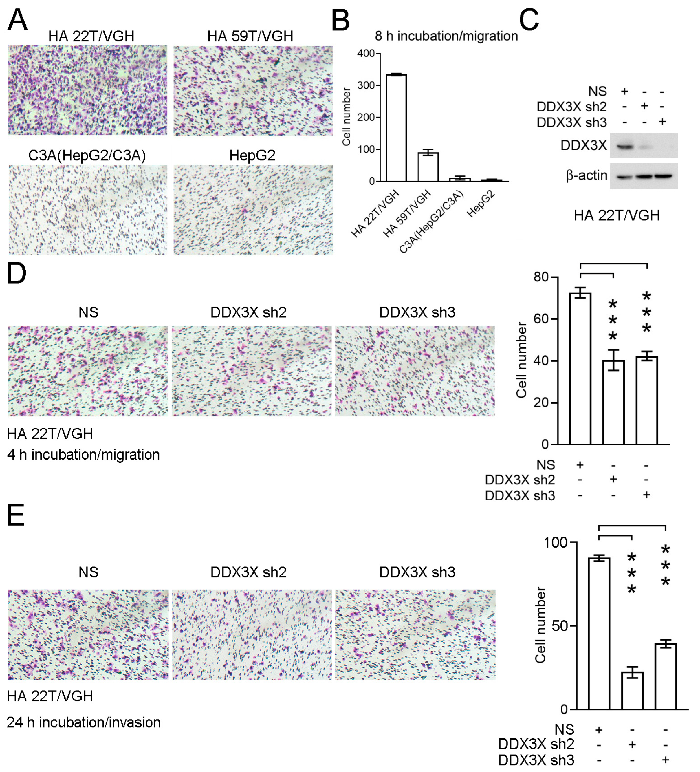
3.3. Knockdown of DDX3X Expression Inhibits Cell Migration and Invasion in a Liver Cancer Cell Line

Therefore, I focused on ATP-dependent RNA helicase DDX3X’s biological effect on modulating cancer cell migration. The relative cell migration levels were determined in an HCC cell line panel including HA 22T/VGH, HA 59T/VGH, C3A (HepG2/C3A) and HepG2 cells (Figure 5A). HA 22T/VGH exhibited significant malignant metastatic behavior (Figure 5B). ATP-dependent RNA helicase DDX3X expression was further stably silenced via the lentiviral transduction of two specific shRNAs in HA 22T/VGH cells (Figure 5C). Decreases in both cell migration and invasion were found after DDX3X silencing (p < 0.001, Figure 5D,E). Hence, roles of ATP-dependent RNA helicase DDX3X in regulating cancer cell migration and invasion are proposed.

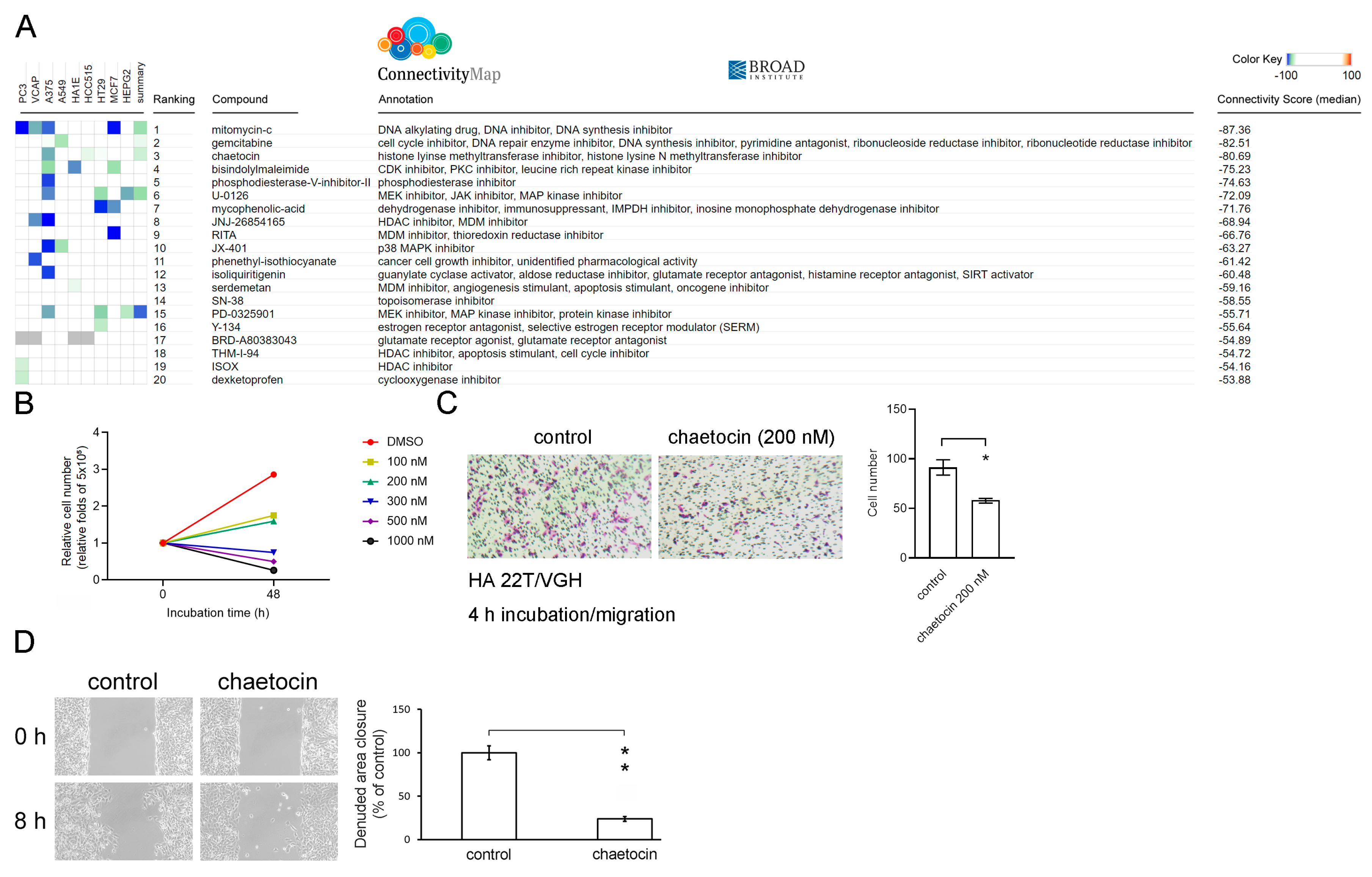
3.4. Chaetocin Reverses the High DDX3X Expression-Mediated Gene Signature and Suppresses Liver Cancer Cell Migration

I further performed in silico data analysis to identify suitable compounds for inhibiting HCC progression. The updates of the Connectivity Map containing over 1.3 million L1000 profiles have been released [13]. I compared the DDX3X perturbation analysis with those data in the L1000 platform of the Connectivity Map to screen potential therapeutic compounds that could reverse the gene expression signature detected in the HCC patient group with high DDX3X expression. Candidate compounds were listed according to the negative connectivity scores determined after perturbation analysis (Figure 6A). Mitomycin-c, gemcitabine and chaetocin were the top three candidates that appeared to possess the ability to reverse the gene signature observed in the DDX3X-high group. Both mitomycin-c and gemcitabine have been proven to reduce cancer cell metastasis [22,23]. Chaetocin is a fungal metabolite isolated from Chaetomium species fungi and shows various pharmacological and biological functions, including the ability to inhibit histone lysine methyltransferase activity [24]. Chaetocin was found to repress the self-renewal of bladder cancer stem cells [25] and to act as a sensitizer of apoptosis in glioblastoma [26]. However, the biological impact of chaetocin on cancer metastasis remains unknown. Chaetocin was first selected for the next tests of therapeutic capabilities to suppress HCC cell migration in this study. As shown in Figure 6B, chaetocin treatment resulted in a dose-dependent reduction in HA 22T/VGH cell proliferation. In addition, a sublethal concentration of 200 nM chaetocin was used for the treatment of HA 22T/VGH cells. The experimental results further showed chaetocin’s inhibitory effect on cancer cell migration (Figure 6C,D).

4. Conclusions

In this study, DDX3X was characterized as a poor prognostic biomarker for an HCC patient cohort, and correlations of high DDX3X level with cancer metastasis and the activation of PCP signaling in HCC cells were discovered. DDX3X knockdown in the HCC cell line results in the repression of cells’ migration and invasion ability. Furthermore, my study demonstrates for the first time a high-throughput drug screening-based characterization of chaetocin and reveals its potential antimetastatic effect, especially in HCC cells with high DDX3X expression levels, as illustrated in Figure 7.

5. Discussion

In this study, I characterized DDX3X’s clinical significance in predicting poor hepatoma patient survival based on RNA expression data. The experimental findings indicated that ATP-dependent RNA helicase DDX3X might potentially possess functions in regulating liver cancer progression. Hence, the next urgent need is to investigate the clinical significance including the associations with patient survival rate, TNM status and clinico-pathological features at a protein level in a patient cohort of HCC.
In this research study, I first observed the potentially therapeutic application of chaetocin for hepatocellular carcinoma in terms of precision medicine; that is, especially for those cancer patients displaying high DDX3X expression levels. Actually, ATP-dependent RNA helicase DDX3X appeared to be modulated by several compounds and cytokines via direct and/or indirect interactions. In liver hepatocellular cells, previous findings indicated that 5-HT treatment could augment 5-HT receptor 7-mediated DDX3X promoter activity as well as the induction of an innate immunity to abolish hepatitis B virus (HBV) infection [27]. Another research finding in hepatocellular HepG2 cells demonstrated that the addition of tazemetostat, SP2509, decitabine and trichostatin A led to the downregulation of DDX3X RNA [28]. Inhibiting ATP-dependent RNA helicase DDX3X’s ATP binding domain with the small molecule RK-33 was one of choices, and the effect was discovered to synergize with radiotherapy to reduce tumor cells’ proliferation in vitro and in vivo [29]. In a cancer-related study, RK-33 treatment was indicated to inactivate the WNT signaling axis and induce cell cycle G1 phase arrest, leading to cell apoptosis in lung cancer [30]. In addition, 1,3,4-thiadiazole is another ATPase activity inhibitor for ATP-dependent RNA helicase DDX3X, synthesized for blocking HIV-1-mediated effects [31]. The design of ATP-competitive inhibitors reported that FE15 and FE109 appeared to block helicase and the ATPase function of ATP-dependent RNA helicase DDX3X, along with having the capability to reduce the HIV viral load in peripheral blood mononuclear cells [32]. Furthermore, doxorubicin is considered to be one of the ATP-dependent RNA helicase DDX3X inhibitors via an in silico molecular docking approach which showed a potential interaction with common amino acid residues Tyr200/Thr201 and unique amino acid residue Thr198 via doxorubicin. The realistic function was also corroborated in oral squamous cell carcinoma H357 cells. DDX3X downregulation and the decreases in ATP hydrolysis, inorganic phosphate release and cancer proliferation were reported [33]. High throughput virtual screening was performed to identify ATP-dependent RNA helicase DDX3X inhibitors, in which Ketorolac salt had been characterized as a bioactive compound for its ability to bind with ATP-dependent RNA helicase DDX3X. The interaction further appeared to suppress the tumor growth in oral cancer [34]. In contrast to the aforementioned strategies, I first explored the drug candidates, focusing on the basis of comprehensively reversing DDX3X-dependent downstream gene signatures, and characterized chaetocin as one of potential targets for therapeutics in liver cancer. Nevertheless, several molecules related to upstream regulation and the direct interaction of ATP-dependent RNA helicase DDX3X might be noticed. AGR2 is a member of the protein disulfide isomerase family, which acts as a proto-oncogene. AGR2 was found to trigger cancer metastasis in animal models, and the interaction of ATP-dependent RNA helicase DDX3X with AGR2 has been characterized at protein level [35]. In addition, fibronectin 1 was uncovered to upregulate the expression of human DDX3X at an RNA level in cultured HUVEC cells in a microarray analysis [36]. Ginsenoside Rg3 stimulus-enhanced DDX3X upregulation and the Akt-p53-dependent DDX3X promoter transactivation were detected, along with the activation of innate immune response via the TBK1-IKKε-IRF3 pathway [37].
Chaetocin was identified based on the comparison of gene signatures derived from those cancer patients displaying high DDX3X levels with the datasets in the ConnectivityMap database (negative connectivity score, Figure 6A). The same gene signature was simultaneously analyzed via IPA. A significant signaling axis, the PCP pathway, was characterized according to the activation Z score (Figure 1D), which is a pivotal stimulus in triggering cancer progression [20,21]. Therefore, the PCP signaling axis might serve as a potential route inhibited by chaetocin to reverse DDX3X-mediated effects because of the overlap of similar downstream molecules.

Supplementary Materials

The following supporting information can be downloaded at: https://www.mdpi.com/article/10.3390/cells12121628/s1, Table S1: Supplementary data_Table S1.

Funding

This study was funded by the Chang Gung Memorial Hospital, Linkou, Taiwan (CMRPG3L1761) to Tsung-Chieh Lin and by the National Science and Technology Council, Taiwan (111-2314-B-182A-032-) to Tsung-Chieh Lin.

Data Availability Statement

All necessary data have been provided in this manuscript and Supplementary Data.

Acknowledgments

The author thanks the Genomic Medicine Core Laboratory, Chang Gung Memorial Hospital, Linkou, Taiwan, for their help.

Conflicts of Interest

The author declares no conflict of interest. The funders had no role in the design of the study; the collection, analyses, or interpretation of the data; the writing of the manuscript; or the decision to publish the results.

References

  1. Zhou, Z.; Licklider, L.J.; Gygi, S.P.; Reed, R. Comprehensive proteomic analysis of the human spliceosome. Nature 2002, 419, 182–185. [Google Scholar] [CrossRef] [PubMed]
  2. Yedavalli, V.S.; Neuveut, C.; Chi, Y.H.; Kleiman, L.; Jeang, K.T. Requirement of DDX3 DEAD box RNA helicase for HIV-1 Rev-RRE export function. Cell 2004, 119, 381–392. [Google Scholar] [CrossRef] [PubMed] [Green Version]
  3. Chao, C.H.; Chen, C.M.; Cheng, P.L.; Shih, J.W.; Tsou, A.P.; Lee, Y.H. DDX3, a DEAD box RNA helicase with tumor growth-suppressive property and transcriptional regulation activity of the p21waf1/cip1 promoter, is a candidate tumor suppressor. Cancer Res. 2006, 66, 6579–6588. [Google Scholar] [CrossRef] [PubMed] [Green Version]
  4. Chuang, R.Y.; Weaver, P.L.; Liu, Z.; Chang, T.H. Requirement of the DEAD-Box protein ded1p for messenger RNA translation. Science 1997, 275, 1468–1471. [Google Scholar] [CrossRef]
  5. Shih, J.W.; Tsai, T.Y.; Chao, C.H.; Wu Lee, Y.H. Candidate tumor suppressor DDX3 RNA helicase specifically represses cap-dependent translation by acting as an eIF4E inhibitory protein. Oncogene 2008, 27, 700–714. [Google Scholar] [CrossRef] [Green Version]
  6. Lee, C.S.; Dias, A.P.; Jedrychowski, M.; Patel, A.H.; Hsu, J.L.; Reed, R. Human DDX3 functions in translation and interacts with the translation initiation factor eIF3. Nucleic Acids Res. 2008, 36, 4708–4718. [Google Scholar] [CrossRef] [Green Version]
  7. Rocak, S.; Linder, P. DEAD-box proteins: The driving forces behind RNA metabolism. Nat. Rev. Mol. Cell Biol. 2004, 5, 232–241. [Google Scholar] [CrossRef]
  8. Fuller-Pace, F.V. DEAD box RNA helicase functions in cancer. RNA Biol. 2013, 10, 121–132. [Google Scholar] [CrossRef] [Green Version]
  9. Huang, J.S.; Chao, C.C.; Su, T.L.; Yeh, S.H.; Chen, D.S.; Chen, C.T.; Chen, P.J.; Jou, Y.S. Diverse cellular transformation capability of overexpressed genes in human hepatocellular carcinoma. Biochem. Biophys. Res. Commun. 2004, 315, 950–958. [Google Scholar] [CrossRef]
  10. Chan, C.H.; Chen, C.M.; Lee, Y.W.; You, L.R. DNA Damage, Liver Injury, and Tumorigenesis: Consequences of DDX3X Loss. Mol. Cancer Res. 2019, 17, 555–566. [Google Scholar] [CrossRef] [Green Version]
  11. Chang, P.C.; Chi, C.W.; Chau, G.Y.; Li, F.Y.; Tsai, Y.H.; Wu, J.C.; Wu Lee, Y.H. DDX3, a DEAD box RNA helicase, is deregulated in hepatitis virus-associated hepatocellular carcinoma and is involved in cell growth control. Oncogene 2006, 25, 1991–2003. [Google Scholar] [CrossRef] [Green Version]
  12. Bol, G.M.; Xie, M.; Raman, V. DDX3, a potential target for cancer treatment. Mol. Cancer 2015, 14, 188. [Google Scholar] [CrossRef] [Green Version]
  13. Subramanian, A.; Narayan, R.; Corsello, S.M.; Peck, D.D.; Natoli, T.E.; Lu, X.; Gould, J.; Davis, J.F.; Tubelli, A.A.; Asiedu, J.K.; et al. A Next Generation Connectivity Map: L1000 Platform and the First 1,000,000 Profiles. Cell 2017, 171, 1437–1452 e1417. [Google Scholar] [CrossRef]
  14. Uhlen, M.; Bjorling, E.; Agaton, C.; Szigyarto, C.A.; Amini, B.; Andersen, E.; Andersson, A.C.; Angelidou, P.; Asplund, A.; Asplund, C.; et al. A human protein atlas for normal and cancer tissues based on antibody proteomics. Mol. Cell. Proteom. 2005, 4, 1920–1932. [Google Scholar] [CrossRef] [PubMed] [Green Version]
  15. Uhlen, M.; Fagerberg, L.; Hallstrom, B.M.; Lindskog, C.; Oksvold, P.; Mardinoglu, A.; Sivertsson, A.; Kampf, C.; Sjostedt, E.; Asplund, A.; et al. Proteomics. Tissue-based map of the human proteome. Science 2015, 347, 1260419. [Google Scholar] [CrossRef] [PubMed]
  16. Thul, P.J.; Akesson, L.; Wiking, M.; Mahdessian, D.; Geladaki, A.; Ait Blal, H.; Alm, T.; Asplund, A.; Bjork, L.; Breckels, L.M.; et al. A subcellular map of the human proteome. Science 2017, 356, eaal3321. [Google Scholar] [CrossRef]
  17. Uhlen, M.; Oksvold, P.; Fagerberg, L.; Lundberg, E.; Jonasson, K.; Forsberg, M.; Zwahlen, M.; Kampf, C.; Wester, K.; Hober, S.; et al. Towards a knowledge-based Human Protein Atlas. Nat. Biotechnol. 2010, 28, 1248–1250. [Google Scholar] [CrossRef]
  18. Uhlen, M.; Zhang, C.; Lee, S.; Sjostedt, E.; Fagerberg, L.; Bidkhori, G.; Benfeitas, R.; Arif, M.; Liu, Z.; Edfors, F.; et al. A pathology atlas of the human cancer transcriptome. Science 2017, 357, eaan2507. [Google Scholar] [CrossRef] [PubMed] [Green Version]
  19. Gyorffy, B.; Schafer, R. Meta-analysis of gene expression profiles related to relapse-free survival in 1079 breast cancer patients. Breast Cancer Res. Treat. 2009, 118, 433–441. [Google Scholar] [CrossRef] [PubMed] [Green Version]
  20. Zheng, M.; Zhou, Q.; Liu, X.; Wang, C.; Liu, G. CTHRC1 overexpression promotes cervical carcinoma progression by activating the Wnt/PCP signaling pathway. Oncol. Rep. 2019, 41, 1531–1538. [Google Scholar] [CrossRef] [PubMed] [Green Version]
  21. Yang, X.M.; You, H.Y.; Li, Q.; Ma, H.; Wang, Y.H.; Zhang, Y.L.; Zhu, L.; Nie, H.Z.; Qin, W.X.; Zhang, Z.G.; et al. CTHRC1 promotes human colorectal cancer cell proliferation and invasiveness by activating Wnt/PCP signaling. Int. J. Clin. Exp. Pathol. 2015, 8, 12793–12801. [Google Scholar]
  22. Monch, D.; Koch, J.; Maass, A.; Janssen, N.; Murdter, T.; Renner, P.; Fallier-Becker, P.; Solass, W.; Schwab, M.; Dahlke, M.H.; et al. A human ex vivo coculture model to investigate peritoneal metastasis and innovative treatment options. Pleura. Peritoneum. 2021, 6, 121–129. [Google Scholar] [CrossRef]
  23. He, J.; Lin, X.; Meng, F.; Zhao, Y.; Wang, W.; Zhang, Y.; Chai, X.; Zhang, Y.; Yu, W.; Yang, J.; et al. A Novel Small Molecular Prostaglandin Receptor EP4 Antagonist, L001, Suppresses Pancreatic Cancer Metastasis. Molecules 2022, 27, 1209. [Google Scholar] [CrossRef]
  24. Jiang, H.; Li, Y.; Xiang, X.; Tang, Z.; Liu, K.; Su, Q.; Zhang, X.; Li, L. Chaetocin: A review of its anticancer potentials and mechanisms. Eur. J. Pharmacol. 2021, 910, 174459. [Google Scholar] [CrossRef]
  25. Yang, Z.; Wang, H.; Zhang, N.; Xing, T.; Zhang, W.; Wang, G.; Li, C.; Yu, C. Chaetocin Abrogates the Self-Renewal of Bladder Cancer Stem Cells via the Suppression of the KMT1A-GATA3-STAT3 Circuit. Front. Cell Dev. Biol. 2020, 8, 424. [Google Scholar] [CrossRef] [PubMed]
  26. Ozyerli-Goknar, E.; Sur-Erdem, I.; Seker, F.; Cingoz, A.; Kayabolen, A.; Kahya-Yesil, Z.; Uyulur, F.; Gezen, M.; Tolay, N.; Erman, B.; et al. The fungal metabolite chaetocin is a sensitizer for pro-apoptotic therapies in glioblastoma. Cell Death Dis. 2019, 10, 894. [Google Scholar] [CrossRef] [PubMed] [Green Version]
  27. Kang, L.J.; Nguyen, K.V.A.; Eom, S.; Choi, Y.J.; Nguyen, C.N.; Lee, J.; Kim, C.; Lee, S.; Lee, S.G.; Lee, J.H. Stimulating DDX3 expression by serotonin 5-HT receptor 7 through phosphorylation of p53 via the AC-PKA-ERK signaling pathway. J. Cell Biochem. 2019, 120, 18193–18208. [Google Scholar] [CrossRef] [PubMed]
  28. Zhang, Z.; Lei, A.; Xu, L.; Chen, L.; Chen, Y.; Zhang, X.; Gao, Y.; Yang, X.; Zhang, M.; Cao, Y. Similarity in gene-regulatory networks suggests that cancer cells share characteristics of embryonic neural cells. J. Biol. Chem. 2017, 292, 12842–12859. [Google Scholar] [CrossRef] [PubMed] [Green Version]
  29. Xie, M.; Vesuna, F.; Tantravedi, S.; Bol, G.M.; Heerma van Voss, M.R.; Nugent, K.; Malek, R.; Gabrielson, K.; van Diest, P.J.; Tran, P.T.; et al. RK-33 Radiosensitizes Prostate Cancer Cells by Blocking the RNA Helicase DDX3. Cancer Res. 2016, 76, 6340–6350. [Google Scholar] [CrossRef] [PubMed] [Green Version]
  30. Bol, G.M.; Vesuna, F.; Xie, M.; Zeng, J.; Aziz, K.; Gandhi, N.; Levine, A.; Irving, A.; Korz, D.; Tantravedi, S.; et al. Targeting DDX3 with a small molecule inhibitor for lung cancer therapy. EMBO Mol. Med. 2015, 7, 648–669. [Google Scholar] [CrossRef]
  31. Brai, A.; Ronzini, S.; Riva, V.; Botta, L.; Zamperini, C.; Borgini, M.; Trivisani, C.I.; Garbelli, A.; Pennisi, C.; Boccuto, A.; et al. Synthesis and Antiviral Activity of Novel 1,3,4-Thiadiazole Inhibitors of DDX3X. Molecules 2019, 24, 3988. [Google Scholar] [CrossRef] [Green Version]
  32. Radi, M.; Falchi, F.; Garbelli, A.; Samuele, A.; Bernardo, V.; Paolucci, S.; Baldanti, F.; Schenone, S.; Manetti, F.; Maga, G.; et al. Discovery of the first small molecule inhibitor of human DDX3 specifically designed to target the RNA binding site: Towards the next generation HIV-1 inhibitors. Bioorg. Med. Chem. Lett. 2012, 22, 2094–2098. [Google Scholar] [CrossRef]
  33. Botlagunta, M.; Kollapalli, B.; Kakarla, L.; Gajarla, S.P.; Gade, S.P.; Dadi, C.L.; Penumadu, A.; Javeed, S. In vitro anti-cancer activity of doxorubicin against human RNA helicase, DDX3. Bioinformation 2016, 12, 347–353. [Google Scholar] [CrossRef] [PubMed] [Green Version]
  34. Samal, S.K.; Routray, S.; Veeramachaneni, G.K.; Dash, R.; Botlagunta, M. Ketorolac salt is a newly discovered DDX3 inhibitor to treat oral cancer. Sci. Rep. 2015, 5, 9982. [Google Scholar] [CrossRef] [PubMed] [Green Version]
  35. Tiemann, K.; Garri, C.; Lee, S.B.; Malihi, P.D.; Park, M.; Alvarez, R.M.; Yap, L.P.; Mallick, P.; Katz, J.E.; Gross, M.E.; et al. Loss of ER retention motif of AGR2 can impact mTORC signaling and promote cancer metastasis. Oncogene 2019, 38, 3003–3018. [Google Scholar] [CrossRef] [PubMed]
  36. Orecchia, A.; Mettouchi, A.; Uva, P.; Simon, G.C.; Arcelli, D.; Avitabile, S.; Ragone, G.; Meneguzzi, G.; Pfenninger, K.H.; Zambruno, G.; et al. Endothelial cell adhesion to soluble vascular endothelial growth factor receptor-1 triggers a cell dynamic and angiogenic phenotype. FASEB J. 2014, 28, 692–704. [Google Scholar] [CrossRef] [PubMed]
  37. Choi, Y.J.; Kang, L.J.; Lee, S.G. Stimulation of DDX3 expression by ginsenoside Rg3 through the Akt/p53 pathway activates the innate immune response via TBK1/IKKepsilon/IRF3 signalling. Curr. Med. Chem. 2014, 21, 1050–1060. [Google Scholar] [CrossRef]
Figure 1. High DDX3X expression is associated with poor outcomes in the liver cancer cohort, and canonical pathway analysis of the DDX3X-dependent gene signature reveals a correlation with cancer metastasis, PCP and PTEN signaling. (A) A scatter plot of the indicated expression patterns in 373 liver cancer patients is shown. A total of 423 samples, including normal and tumor tissues, were retrieved from the TCGA database, and 50 adjacent normal samples were excluded prior to the analysis. The tumor samples were divided into the DDX3X-high and DDX3X-low groups based on the cut-off value around the median DDX3X gene expression (FPKM) in cohort. RNA-Seq data were retrieved from TCGA (Dataset ID: TCGA.LIHC.sampleMap/HiSeqV2_PANCAN, version: 2017-10-13). The RNA-Seq read count was normalized and log2 transformed. (B) Kaplan–Meier analysis revealed the overall survival of liver cancer patients in the DDX3X-high and DDX3X-low groups. (C) A total of 2890 differentially expressed gene targets in the DDX3X-high group were selected and are shown after hierarchical clustering (1.5-fold change, p value < 0.05). (D) Significant signaling pathways were ranked according to the −log p value. Activation and inactivation of the indicated signaling pathways were determined by the transformed Z score. (E) The percentage of genes overlapping in the indicated signaling pathways is shown. The numbers of upregulated and downregulated genes are displayed with red and green bars, respectively. The p values are shown on the orange line.
Cells 12 01628 g001aCells 12 01628 g001b
Figure 2. The activated PCP signaling pathway. (A) The signaling transduction of the PCP pathway is identified and represented after the analysis via IPA. Molecules of differential upregulation in the high DDX3X group were labeled in red. (B) Relative log2-transformed expressions of the principal molecules in PCP pathway are shown. The analysis was performed via Ingenuity® Pathway Analysis (IPA; QIAGEN, Hilden, Germany; https://digitalinsights.qiagen.com/products-overview/discovery-insights-portfolio/analysisand-visualization/qiagen-ipa/ (accessed on 1 November 2022)).
Cells 12 01628 g002
Figure 3. The associations of the PCP pathway-related gene signature in high DDX3X group with poor overall survival were analyzed. The correlation of indicated RNA levels with overall survival in liver cancer cohorts were studied and shown. Data were retrieved and analyzed from the HPA database (https://www.proteinatlas.org/ (accessed on 1 November 2022)).
Cells 12 01628 g003
Figure 4. Analysis match module of IPA reveals the potential function of ATP-dependent RNA helicase DDX3X in triggering cancer cell migration. The gene signature obtained from the high DDX3X group was explored with the analysis match module of IPA. The gene signature was, respectively, compared with the datasets in categories of cell movement (A), migration of cells (B), cell movement of tumor cell lines (C) and invasion of cells (D). The statistical significance and activation status of the gene signature in high DDX3X groups were shown by green dot.
Cells 12 01628 g004
Figure 5. Knockdown of DDX3X expression inhibits HCC cell migration. (A) The migration ability of the indicated liver cancer cell lines was evaluated using a Transwell assay. Cells were incubated for 8 h for the migration assay in Transwell devices, followed by fixation and counting. (B) The numbers of migrated cells are shown. (C) Stable knockdown of DDX3X was performed in HA 22T/VGH cells. Relative ATP-dependent RNA helicase DDX3X levels were evaluated by Western blotting. NS: non-silencing control (scrambled RNA). (D,E) The migration and invasion ability of the indicated HA 22T/VGH cells were investigated in Transwell devices. *** p < 0.001.
Cells 12 01628 g005
Figure 6. Via the L1000 platform of Connectivity Map, chaetocin is identified to reverse the high DDX3X expression-mediated gene signature, and chaetocin treatment suppresses HCC cell migration. (A) The gene signature identified in the DDX3X-high group versus the DDX3X-low group was analyzed via the L1000 platform of the Connectivity Map. The candidate drugs were ranked according to the connectivity score and are shown. (B) HA 22T/VGH cell numbers were determined using a trypan blue exclusion assay after 48 h of chaetocin pretreatment at the indicated concentrations. (C) HA 22T/VGH cell migration was evaluated using a Transwell assay of 4 h incubation and (D) with a wound-healing assay of 8 h incubation after 48 h of 200 nM chaetocin pretreatment. * p < 0.05, ** p < 0.01.
Cells 12 01628 g006
Figure 7. The illustration of targeting ATP-dependent RNA helicase DDX3X and reversing its downstream gene signatures-based chaetocin treatment in attenuating HCC metastatic progression.
Cells 12 01628 g007
Table 1. Prognostic significance of DDX3X in pan-cancer.
SymbolCancer TypePrognosisEndpointp ValueCase DatasetMethodProbe ID
DDX3XGlioma-Overall survivalN.S.153TCGARNA Seq
DDX3XThyroid Cancer-Overall survivalN.S.501TCGARNA Seq
DDX3XLung Cancer-Overall survivalN.S.994TCGARNA Seq
DDX3XColorectal CancerGoodOverall survival0.008597TCGARNA Seq
DDX3XHead and Neck Cancer-Overall survivalN.S.499TCGARNA Seq
DDX3XStomach Cancer-Overall survivalN.S.354TCGARNA Seq
DDX3XLiver CancerPoorOverall survival0.0012365TCGARNA Seq
DDX3XPancreatic CancerPoorOverall survival0.033176TCGARNA Seq
DDX3XRenal Cancer-Overall survivalN.S.877TCGARNA Seq
DDX3XUrothelial CancerGoodOverall survival0.029406TCGARNA Seq
DDX3XProstate Cancer-Overall survivalN.S.494TCGARNA Seq
DDX3XTestis Cancer-Overall survivalN.S.134TCGARNA Seq
DDX3XBreast cancerPoorOverall survival0.0241075TCGARNA Seq
DDX3XCervical Cancer-Overall survivalN.S.291TCGARNA Seq
DDX3XEndometrial Cancer-Overall survivalN.S.541TCGARNA Seq
DDX3XOvarian Cancer-Overall survivalN.S.373TCGARNA Seq
DDX3XMelanoma-Overall survivalN.S.102TCGARNA Seq
DDX3XBreast cancerPoorRelapse-free survival<0.0014929E-MTAB-365, E-TABM-43, GSE: 11121, 12093,Array201210_at
12276, 1456, 16391, 16446, 16716, 17705, 17907,
18728, 19615, 20194, 20271, 2034, 20685, 20711,
21653, 22093, 25066, 2603, 26971, 29044, 2990,
31448, 31519, 32646, 3494, 36771, 37946, 41998,
42568, 43358, 43365, 45255, 4611, 46184, 48390,
50948, 5327, 58812, 61304, 65194, 6532, 69031,
7390, 76275, 78958, 9195
DDX3XOvarian cancerPoorProgression-free survival<0.0011435GSE: 14764, 15622, 18520, 19829, 23554, 26193,Array201210_at
26712, 27651, 30161, 3149, 51373, 63885, 65986,RNA Seq
9891, TCGA (N = 565)
DDX3XLung cancerGoodOverall survival<0.0011925CAARRAY, GSE: 14814, 19188, 29013, 30219,Array201210_at
31210, 3141, 31908, 37745, 43580, 4573, 50081,RNA Seq
8894, TCGA (N = 133)
DDX3XGastric cancerGoodPost progression survival<0.001498GSE: 14210, 15459, 22377, 29272, 51105, 62254Array201210_at
Survival data were collected from databases The Human Protein Atlas, TCGA and Kaplan–Meier plotter. N.S.: no significance.
Table 2. Cox regression analysis of correlations of pathological stage, TNM prognostic factors and DDX3X expression with overall survival in 373 liver cancer patients.
VariableComparisonHR (95% CI)p Value
SexM:F0.800 (0.562–1.141)0.218
Stage3–4:1–22.485 (1.714–3.603)<0.001
TT3–4:T1–22.578 (1.812–3.667)<0.001
NN1:N02.012 (0.493–8.212)0.330
MM1:M04.055 (1.274–12.906)0.018
DDX3XHigh:Low1.485 (1.050–2.099)0.025
Abbreviations: F: female; M: male; HR: hazard ratio; CI: confidence interval. T: tumor; N: nodes; M: metastasis.
Table 3. Diseases and functions of dataset: DDX3X_high/low.
RankingCategoriesDiseases/Functions AnnotationPredicted Activation StateActivation z-Score
1Cellular MovementCell movementIncreased9.208
2Cellular MovementMigration of cellsIncreased8.506
3Cellular Assembly and Organization, Cellular Function and MaintenanceOrganization of cytoplasmIncreased8.472
4Cellular Assembly and Organization, Cellular Function and MaintenanceOrganization of cytoskeletonIncreased8.43
5Cellular MovementCell movement of tumor cell linesIncreased8.254
6Tissue MorphologyQuantity of cellsIncreased8.25
7Cell Morphology, Cellular Assembly and Organization, Cellular Function and MaintenanceFormation of cellular protrusionsIncreased8.077
8Cellular Assembly and Organization, Cellular Function and MaintenanceMicrotubule dynamicsIncreased7.699
9Cellular Development, Cellular Growth and Proliferation, Nervous System Development and Function, Tissue DevelopmentDevelopment of neuronsIncreased6.967
10Cellular MovementHoming of cellsIncreased6.705
11Cellular Function and MaintenanceCellular homeostasisIncreased6.55
12Cellular MovementInvasion of cellsIncreased6.461
13Cardiovascular System Development and FunctionDevelopment of vasculatureIncreased6.43
14Cardiovascular System Development and Function, Organismal DevelopmentAngiogenesisIncreased6.418
15Cellular MovementChemotaxisIncreased6.411
Disclaimer/Publisher’s Note: The statements, opinions and data contained in all publications are solely those of the individual author(s) and contributor(s) and not of MDPI and/or the editor(s). MDPI and/or the editor(s) disclaim responsibility for any injury to people or property resulting from any ideas, methods, instructions or products referred to in the content.

