Article

Lactate Activates Germline and Cleavage Embryo Genes in Mouse Embryonic Stem Cells

Institute of Reproductive Health, Tongji Medical College, Huazhong University of Science and Technology, Wuhan 430030, China
*
Author to whom correspondence should be addressed.
Cells 2022, 11(3), 548; https://doi.org/10.3390/cells11030548
Submission received: 11 January 2022 / Revised: 27 January 2022 / Accepted: 2 February 2022 / Published: 4 February 2022
(This article belongs to the Special Issue Germ Cells and Stem Cells in Regeneration and Repair)

Abstract

:
Lactate was recently found to mediate histone lysine lactylation and facilitate polarization of M1 macrophages, indicating its role in metabolic regulation of gene expression. During somatic cell reprogramming, lactate promotes histone lactylation of pluripotency genes and improves reprogramming efficiency. However, the function of lactate in cell fate control in embryonic stem cells (ESCs) remains elusive. In this study, we revealed that lactate supplementation activated germline genes in mouse ESCs. Lactate also induced global upregulation of cleavage embryo genes, such as members of the Zscan4 gene family. Further exploration demonstrated that lactate stimulated H3K18 lactylation accumulation on germline and cleavage embryo genes, which in turn promoted transcriptional elongation. Our findings indicated that lactate supplementation expanded the transcriptional network in mouse ESCs.

1. Introduction

Embryonic stem cells (ESCs), derived from inner cell mass of blastocysts, possess an unlimited capacity for proliferation, self-renewal, and multipotent differentiation [1], offering biologists a valuable experimental system to uncover the regulatory mechanism of pluripotency maintenance and differentiation during development. Furthermore, in vitro cultured ESCs occasionally overcome epigenetic barriers and transiently reach totipotent status with two-cell-like transcriptome and chromatin features [2,3,4]. Hence, in vitro cultured ESCs are also widely used to explore cell fate transition from pluripotency to totipotency [5,6,7].
Cellular metabolism is the most fundamental biological process satisfying energy demands to keep cells alive, and discrepancies in metabolism patterns reflect distinct cell status [8,9]. Progressing beyond crucial roles in energy homeostasis, the last decade has witnessed significant advances in our understanding of metabolic regulation of epigenetic modifications, gene expression, and cell fate change [10,11,12]. Like most rapidly dividing cells, pluripotent stem cells sustain high glycolysis to feed both cellular demands for building blocks such as nucleotides, phospholipids, and amino acids and energy needs [10,13]. Still, discrepant metabolic profiles associate with distinct pluripotent states, and cells reprogram their metabolic pattern during cell fate transition [14,15,16]. Since the discovery of acetyl-CoA as the substrate of histone acetyltransferases (HATs) to regulate histone acetylation in mammalian cells [17,18], increasing numbers of metabolic intermediates have been found to play pivotal roles in epigenetic modification. S-adenosylmethionine (SAM), a key intermediate of the one-carbon cycle, is the methyl donor for histone and DNA methylation reactions [8,19]. Deprivation of SAM causes downregulation of H3K4me3 and differentiation in ESCs [20,21]. α-ketoglutarate (αKG), an intermediate of the TCA cycle, is a required cofactor for both histone and DNA demethylase [10,13]. In ESCs, αKG promotes DNA and histone demethylation, activates the pluripotent gene, and maintains the pluripotent state [22,23]. Although an increasing number of metabolic intermediates have been found to play a regulatory role in epigenetic regulation of cell fate, the causal relationship between metabolic reprogramming and cell fate shift requires further investigation.
Lactate is an end product of glycolysis that is reused for gluconeogenesis in the liver. In tumor cells, the lactate produced by glycolysis facilitates the formation of an acidic tumor microenvironment, which reinforces cancer invasion and suppresses antitumor immunity [24,25]. Recent studies have identified a novel function for lactate whereby it is utilized for histone lysine lactylation to promote the transition from inflammatory to reparative macrophages and activate homeostatic gene expression, maintaining immune homeostasis [26,27]. Discovery of lactoyl-CoA (lactyl-CoA) in mammalian cells indicated that lactoyl-CoA generated by glucose metabolism was a possible biochemical link between lactate and histone lactylation in vivo [24,28]. A cell-free, recombinant chromatin-templated histone modification and transcription assay demonstrated that lactoyl-CoA activated transcription in a p53-dependent, p300-mediated manner [27], and deprivation of p300 reduced histone lactylation both in vitro and in vivo [27,29], indicating p300 as a potential histone lactyltransferase [24,30]. Moreover, lactate was found to play roles in lung fibrosis, cell differentiation and reprogramming, and lactylation of nonhistone proteins [24]. As an important metabolic intermediate, the role of lactate and lactate-induced histone lactylation in pluripotency maintenance and differentiation is still unclear. In this study, we uncovered the function of lactate and lactate-induced histone lactylation in cell fate control in mouse ESCs.

2. Materials and Methods

2.1. Cell Culture

The mouse ESC line AB2.2 was cultured on mouse embryonic fibroblast (MEF) feeder cells with ES cell medium supplemented with 15% fetal bovine serum (Hyclone, Logan, UT, USA, cat. no. SH30396.03), 1000 U/mL leukemia inhibitory factor (LIF, Millipore, Burlington, MA, USA, cat. no. ESG1107), 3 μM CHIR99021 (LC Laboratories, Woburn, MA, USA, cat. no. C-6556), and 1 μM PD0325901 (LC Laboratories, cat. no. P-9688). Fifty millimolar lactate (Sangon Biotech, Shanghai, China, cat. no. A604046) was added and maintained for 24 h before experimental procedures. Cells were cultured in a humidified chamber at 37 °C with 5% CO2. For culture of mouse ESC lines, the medium was refreshed daily, and cells were routinely passaged every 2 days.

2.2. Western Blot

Cells were lysed on ice with gentle stirring for 10 min in 0.5 mL of TBE buffer containing 0.5% Triton X-100 (Biosharp, Anhui, China. cat. no. BS084) (v/v) and 2 mM phenylmethylsulfonyl fluoride (PMSF)(Solarbio, Beijing, China. cat. no. P0100). The lysates were centrifuged at 6500× g for 10 min at 4 °C to remove the supernatant. Retained nuclei were washed twice using TEB and centrifuged as before. The nuclei were then resuspended in 0.2 N HCl at 4 °C overnight. Samples were centrifuged at 6500× g for 15 min at 4 °C to remove the nuclei debris the next day. Samples were subjected to Western blot analysis with the appropriate antibodies (anti-H4K8la, cat. no. PTM-1415; anti-H4K12la, cat. no. PTM-1411RM; and anti-H3K18la, cat. no. PTM-1406RM. PTM BIO, Hangzhou, China) after neutralizing HCl with 2M NaOH at 1/5 of the volume of the supernatant.

2.3. RNA Isolation and Quantitative RT-PCR (qRT-PCR)

Total RNA was extracted using a TRI reagent (Sigma, St. Louis, MO, USA, cat. no. T9424) following the manufacturer’s procedure. The purity and concentration of RNA samples were determined with a Nanodrop ND-2000 spectrophotometer (Thermo Fisher Scientific, Waltham, MA, USA). A reverse-transcriptional reaction was performed with a Hifair III 1st Strand cDNA Synthesis Kit (Yeasen, Shanghai, China. cat. no. 11139ES60) according to the manufacturer’s procedure. qRT-PCR was performed with SYBR green master mix (Yeasen, cat. no. 11203ES08) on a StepOnePlus™ Real-Time PCR System with Tower (Applied Biosystems, Foster City, CA, USA) according to the manufacturer’s instructions. For primer sequences, see Table S1.

2.4. ChIP-Seq

ChIP-seq was performed using a Hyperactive In-Situ ChIP Library Prep Kit for Illumina (pG-Tn5) (Vazyme, Nanjing, China, cat. no. TD901) according to the manufacturer’s procedure with the appropriate antibodies (anti-H3K18la, PTM-1406RM, and PTM BIO; anti-H3K4me3, 9727, and CST, Danvers, MA, USA; and anti-PolII, 39097, and active motif, Carlsbad, CA, USA).

2.5. RNA-Seq Dataset Analysis

Raw reads were processed with cutadapt v1.16 (https://cutadapt.readthedocs.io, accessed on 24 September 2021) to remove adapters and perform quality trimming with default parameters. Trimmed reads were mapped to the mouse genome (GENCODE release M23) using STAR (v2.5.1b) with default settings. Reads were counted in exons of the mouse genome (GENCODE release M23) using the STAR-quantMode GeneCounts setting. Differential expression of genes for all pairwise comparisons was assessed by DESeq2 v1.24.0 with internal normalization of reads to correct for library size and RNA composition bias. Differentially regulated genes in the DESeq2 analysis were defined as those that were more than two-fold increased or decreased with an adjusted p-value < 0.05. RSEM was used to calculate the FPKM value for each gene. Gene ontology was performed using Metascape (https://metascape.org, accessed on 12 October 2021) [31]. For GO term IDs, see Table S2 and S3. The enrichment bubble dot and heatmap were plotted on a website (http://www.bioinformatics.com.cn, accessed on 12 October 2021).

2.6. ChIP-Seq Dataset Analysis

Raw reads were processed with cutadapt v1.8.1 to remove adapters and perform quality trimming. Trimmed reads were mapped to the UCSC mm10 assembly using Bowtie2 with default parameters. Deeptools was used for normalization to draw a read density plot and heatmap from bigwig files for visualization of ChIP-seq data.

2.7. Microarray Dataset Analysis

Microarray datasets (GSE45181) were analyzed using GEO2R, an interactive web tool. Datasets GSM1098610, GSM1098612, and GSM1098614 were used as a control group, and datasets GSM1098611, GSM1098613, and GSM1098615 were used as a treatment group (Max knockdown).

2.8. Statistical Analysis

The two-tailed Student’s t-test and Wilcoxon rank sum test were used to calculate p values. Statistically significant values for p < 0.05, p < 0.01, and p < 0.001 are indicated by single, double, and triple asterisks, respectively.

3. Results

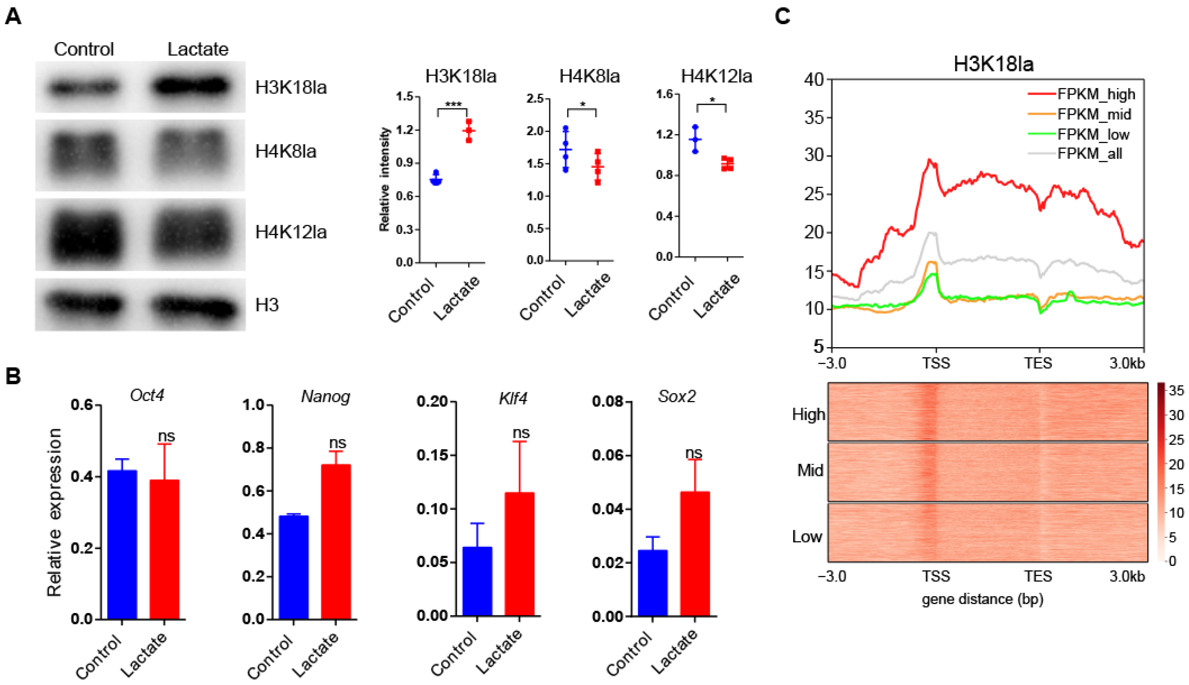
3.1. Lactate Supplementation Stimulated H3K18 Lactylation in Mouse ESCs

It was reported that exogenous lactate stimulates lactylation of histone lysine residues in cells [27]. Hence, we added 50 mM lactate to ES culture media to enhance histone lactylation. As expected, lactate supplementation significantly improved lactylation of H3K18 (H3K18la) after 24 h treatment. We found that H4K8la and H4K18la modifications were slightly weakened upon lactate supplementation, probably due to crosstalk among different histone lactylation modifications (Figure 1A). To reveal the effect of lactate supplementation on cell pluripotency, we examined the expression of pluripotent genes. The expression of pluripotent genes, such as Oct4, Nanog, and Sox2, was not affected upon lactate supplementation (Figure 1B). These results indicated that lactate supplementation strengthened H3K18la without affecting pluripotent cell identity of mouse ESCs. We further performed a ChIP-seq assay to illustrate the distribution of H3K18la in mouse ESCs, and examined the H3K18la signals at gene loci with different expression levels (only genes with FPKM >1 were analyzed) by RNA-seq. Interestingly, we noticed that genes expressed at higher levels had more a enriched H3K18la signal along the whole gene bodies compared to genes expressed at lower levels (Figure 1C). This result supported a positive correlation between H3K18la intensity and transcript abundance and indicated that many genes may be upregulated by lactate supplementation.

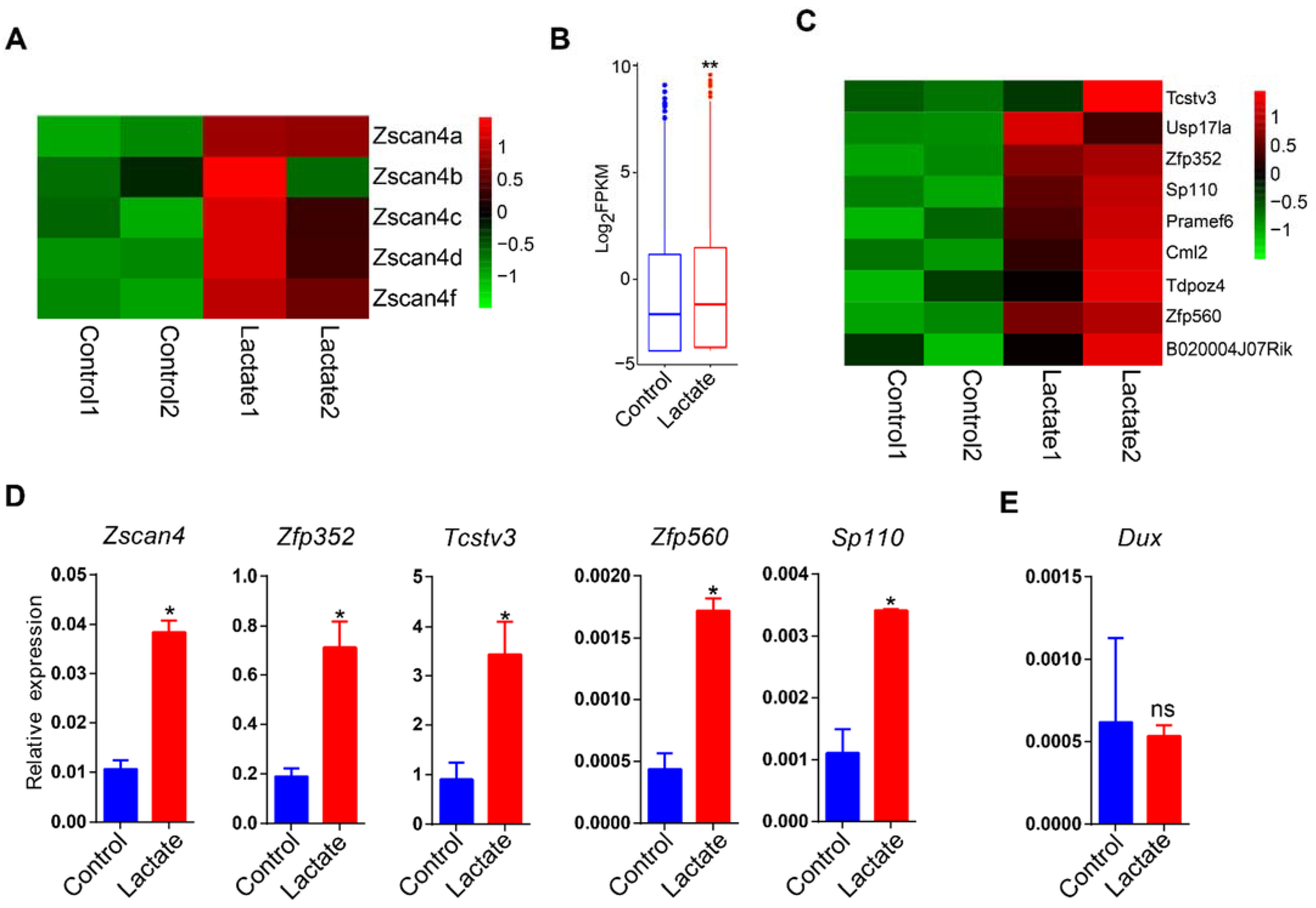
3.2. Lactate Supplementation Activated Germline Genes in mESCs

To study the potential influence of lactate supplementation on cell identity, we performed RNA-seq to uncover changes in the transcriptome. In general, there were 160 upregulated genes (|log2 fold-change| > 1, adjusted p-value < 0.05) and 145 downregulated genes (|log2 fold-change| < 1, adjusted p-value < 0.05) upon lactate supplementation (Figure 2A,B). Gene ontology analysis identified that there was enrichment in the downregulated genes of the response to virus, the CAMKK–AMPK signaling cascade, and the regulation of nervous system development. Remarkably, we noticed significant enrichment of the germline gene population in upregulated genes indicated by GO terms including ‘meiotic DNA double-strand break formation’ and ‘DNA methylation involved in gamete generation’ (Figure 2C). Correspondingly, the heatmap of RNA-seq data showed upregulation of a bulk of typical germline genes such as Dazl, Ddx4, Mael, Dmrt1, and Taf7l (Figure 2D). Upregulation of germline genes was also confirmed by qRT-PCR (Figure 2E).

3.3. Lactate Supplementation Induced Cleavage Embryo Genes in Mouse ESCs

In addition to germline genes, we also observed a significant upregulation of Zscan4 family genes (Figure 3A), a set of zygotic genome activation (ZGA) marker genes expressed specifically in two-cell mouse embryos and six to eight cell human embryos [32,33]. To further uncover the global expression change of ZGA genes, we reanalyzed the RNA-seq dataset to obtain upregulated genes in MERVL+Zscan4+ ESCs relative to MERVLZscan4 ESCs (|log2 fold-change| > 1, adjusted p-value < 0.05) for further analysis (Figure S1) [34]. A globally increased abundance of upregulated genes in MERVL+Zscan4+ ESCs was also observed in lactate-supplemented ESCs (Figure 3B,C). Upregulation of ZGA genes, such as Zscan4, Zfp352, Tcstv3, and Sp110, was verified by qRT-PCR (Figure 3D). However, the expression of Dux was not changed (Figure 3E), a key regulatory factor that activates hundreds of ZGA genes and retroviral elements [35]. These results correspond to a previous discovery that lactate raised the ratio of two-cell-like ESCs [14].

3.4. Sequential Activation of Germline and ZGA Genes in Mouse ESCs

To study the correlation between the expression of germline and ZGA genes, we analyzed the public transcriptome data in which ZGA and germline genes were activated. Zscan4c was reported to be a key activator of cleavage embryo genes in ESCs, so we analyzed RNA-seq data of mouse ESCs in which Zscan4c was overexpressed [36]. Expression of exogenous Zscan4c was almost 16 times higher than that of endogenous Zscan4c (Figure 4A). Activation of ZGA genes, such as Zscan4 family genes, Zfp352, and Dux, was observed after Zscan4c overexpression (Figure 4B), while activation of germline genes was not observed (Figure 4C). For transcriptional regulation of germ-cell-specific genes in mouse ESCs, Max was reported to be the key repressor by RNA interference screen, whose knockdown in ESCs resulted in a global upregulation of germ-cell-specific genes [37]. Therefore, we analyzed the microarray data in which Max was knocked down to activate germline genes in mouse ESCs [37]. Successful suppression of Max was confirmed (Figure 4D), and significant upregulation of germline genes, such as Ddx4, Dazl, and Mov10l1, was also observed upon Max knockdown (Figure 4E). In addition, we found a global upregulation of ZGA genes, such as Zscan4 family genes, Tcstv1, and Zfp352 (Figure 4F). These results suggested that germline gene activation stimulated ZGA genes in ESCs, while ZGA gene activation had little impact on germline gene expression.

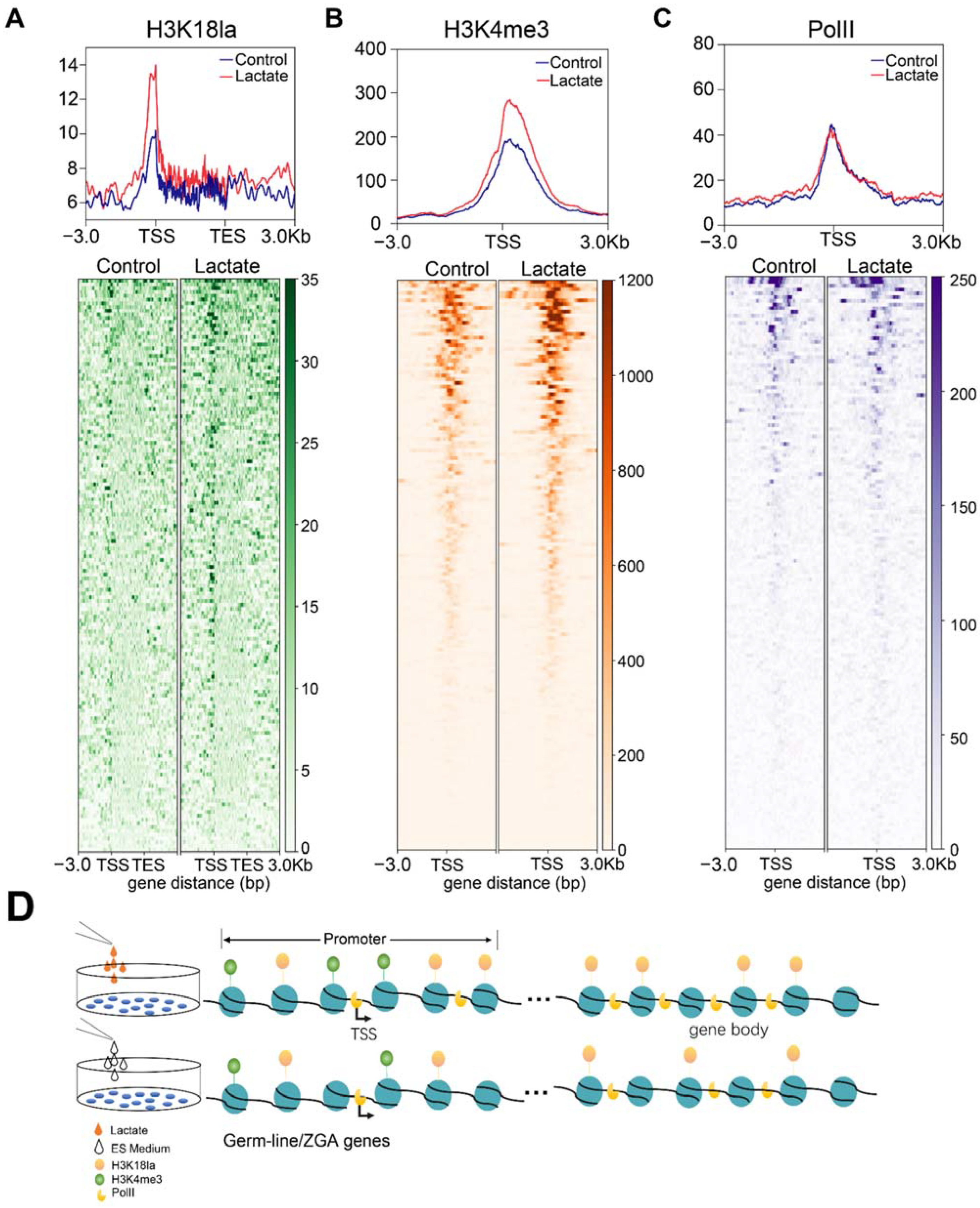
3.5. Lactate Supplementation Facilitated Transcriptional Elongation through Enhanced Histone Lactylation

To gain further mechanistic insights into regulation of germline and ZGA genes, we deciphered the occupancy landscape of H3K18la, H3K4me3, and RNA polymerase II (PolII) upon lactate supplementation. We found that there was remarkably increased deposition of H3K18la at the transcription start site (TSS), as well as a slightly increased accumulation at the gene body of upregulated genes (Figure 5A). Consistently, the intensity of H3K4me3, a marker of an active promoter, also increased at the TSS of upregulated genes (Figure 5B). Although no discrepant PolII localization around the TSS of upregulated genes was observed after lactate supplementation, increased PolII occupancy at the gene body was detected (Figure 5C). Our results indicated that lactate supplementation reinforced histone lactylation at the promoter and gene body and facilitated transcriptional elongation of target genes.

4. Discussion

In this study, we identified that lactate supplementation facilitated the expression of germline and ZGA genes through transcriptional elongation orchestration. In addition to recruitment and assembly of preinitiation complex (PIC), promoter-proximal pausing and release are other key steps in transcription regulation by PolII. While negative elongation factor (NELF) and DRB-sensitivity-inducing factor (DSIF) stabilize the paused Pol II, positive transcription elongation factor-b (P-TEFb) drives release of paused Pol II to begin productive elongation [38,39]. ELL is a component of the super elongation complex (SEC), which interacts directly with P-TEFb to promote elongation. p300 acetylates ELL and increases its stability [40]. Interestingly, p300 also acetylates both promoter-paused polymerase and gene-body-occupied polymerase, promoting PolII release and elongation efficiency [41]. Moreover, acetylation of promoter histone H3K18/K27 mediated by p300 stimulates the release of paused PolII and the recruitment of the super elongation complex (SEC), promoting productive elongation [42]. In this study, we observed accumulated PolII among the gene body but not on the promoter upon lactate supplementation. We suggest that H3K18la recruited transcription coactivators, possibly p300, and promoted transcription elongation. Additionally, we suggest that histone lactylation might facilitate PolII elongation by loosening nucleosome–DNA interaction and forming a platform for coactivators binding. There is a high distribution similarity between histone acetylation and lactylation among the genome [27]. Possible crosstalk between acetylation and lactylation warrants further study.
Eight to twelve percent of couples are infertile worldwide [43], and gametogenesis failure is one of the main causes. Therefore, establishment of an in vitro system for efficient generation of functional gametes is a promising approach for infertility treatment. Stem cells (SCs) including ESCs and iPSCs, possessing the ability to differentiate into all cell types, are valuable cell origins for in vitro differentiation toward germ cells. Though several methods and techniques have been identified to induce SCs’ differentiation into early germ cells, oocyte-like cells (OLCs), and male gametes [44,45,46,47], the efficiency is quite low. In our study, we discovered that lactate supplementation activates the expression of germline genes involved in DNA methylation and meiotic division in mESCs. We also found upregulation of a wealth of genes regulating piRNA metabolism, such as Piwil2, Mael, and Mov10l1. These results suggested that lactate supplementation facilitates differentiation of mouse ESCs toward germ cells. Whether and how lactate improves differentiation efficiency toward gametes warrants further study.
In ESCs, transient Zscan4 expression upregulated homologous recombination genes and facilitated telomere elongation by homologous recombination [48], improving the developmental potency of ES cells and restoring the differentiation potential of long-term cultured ES cells [3]. Knockdown of Zscan4 led to telomere shortening, genomic instability, and aneuploidy [48]. In addition, histone hyperacetylation and DNA demethylation were observed during transient Zscan4 expression [49]. Mechanism studies indicated that ZSCAN4 recruited TET2 through its SCAN domain to target locus and promoted DNA demethylation [50] while promoting degradation of UHRF1 and DNMT1 to suppress DNA methylation [51]. In this study, we observed a significant upregulation of the Zscan4 gene family and a global upregulation of ZGA genes upon lactate supplementation, and this may trigger other downstream events. Lactate is one of the major energy sources of cleavage embryos and promotes embryonic development to the morula stage [12,52]. Therefore, lactate may also regulate preimplantation development by activating ZGA and promoting telomere elongation and global DNA demethylation in early embryos.

5. Conclusions

In summary, we discovered for the first time that lactate supplementation activates the expression of germline and ZGA genes in mouse ESCs, especially the expression of germline genes and members of the Zscan4 gene family. We propose that lactate stimulates H3K18la accumulation at germline and ZGA genes, which recruit cofactors to facilitate transcriptional elongation.

Supplementary Materials

The following supporting information can be downloaded at: www.mdpi.com/article/10.3390/cells11030548/s1, Figure S1: Global expression comparison of upregulated genes between MERVL+Zscan4+ ESCs and MERVL-Zscan4- ESCs; Table S1: Primer sequences; Table S2: GO-Up regulated genes; Table S3: GO-Down regulated genes.

Author Contributions

L.-q.Z. conceived the idea and revised the manuscript. Q.T. performed the experiments and wrote the manuscript. All authors contributed to the article and approved the final manuscript.

Funding

This work was supported by the National Key R&D Program of China (2018YFC1004502 and 2018YFC1004001), the National Natural Science Foundation of China (NSFC 32170820, 31771661), and the program for HUST Academic Frontier Youth Team.

Institutional Review Board Statement

Not applicable.

Informed Consent Statement

Not applicable.

Data Availability Statement

RNA-seq and ChIP-seq data generated during this study were deposited to GEO database at December 2021: GSE191252, GSE192358.

Acknowledgments

We thank Lei Li (Institute of Zoology, Chinese Academy of Sciences, Beijing) for kindly providing the AB2.2 cell line. We also thank all members of Li-quan Zhou’s laboratory for help on this project.

Conflicts of Interest

The authors declare no conflict of interest.

References

  1. De Angeles, A.L.; Ferrari, F.; Xi, R.; Fujiwara, Y.; Benvenisty, N.; Deng, H.; Hochedlinger, K.; Jaenisch, R.; Lee, S.; Leitch, H.G.; et al. Hallmarks of pluripotency. Nature 2015, 525, 469–478. [Google Scholar] [CrossRef] [PubMed]
  2. Macfarlan, T.S.; Gifford, W.D.; Driscoll, S.; Lettieri, K.; Rowe, H.M.; Bonanomi, D.; Firth, A.; Singer, O.; Trono, D.; Pfaff, S.L. Embryonic stem cell potency fluctuates with endogenous retrovirus activity. Nature 2012, 487, 57–63. [Google Scholar] [CrossRef] [PubMed] [Green Version]
  3. Amano, T.; Hirata, T.; Falco, G.; Monti, M.; Sharova, L.V.; Amano, M.; Sheer, S.; Hoang, H.G.; Piao, Y.; Stagg, C.A.; et al. Zscan4 restores the developmental potency of embryonic stem cells. Nat. Commun. 2013, 4, 1966. [Google Scholar] [CrossRef] [PubMed] [Green Version]
  4. Bošković, A.; Eid, A.; Pontabry, J.; Ishiuchi, T.; Spiegelhalter, C.; Ram, E.V.R.; Meshorer, E.; Torres-Padilla, M.-E. Higher chromatin mobility supports totipotency and precedes pluripotency in vivo. Genes Dev. 2014, 28, 1042–1047. [Google Scholar] [CrossRef] [Green Version]
  5. Rodriguez-Terrones, D.; Gaume, X.; Ishiuchi, T.; Weiss, A.; Kopp, A.; Kruse, K.; Penning, A.; Vaquerizas, J.M.; Brino, L.; Torres-Padilla, M.-E. A molecular roadmap for the emergence of early-embryonic-like cells in culture. Nat. Genet. 2018, 50, 106–119. [Google Scholar] [CrossRef]
  6. Fu, X.D.; Wu, X.J.; Djekidel, M.N.; Zhang, Y. Myc and Dnmt1 impede the pluripotent to totipotent state transition in embryonic stem cells. Nat. Cell Biol. 2019, 21, 835–844. [Google Scholar] [CrossRef]
  7. Eckersley-Maslin, M.; Alda-Catalinas, C.; Blotenburg, M.; Kreibich, E.; Krueger, C.; Reik, W. Dppa2 and Dppa4 directly regulate the Dux-driven zygotic transcriptional program. Genes Dev. 2019, 33, 194–208. [Google Scholar] [CrossRef] [Green Version]
  8. Chisolm, D.A.; Weinmann, A.S. Connections Between Metabolism and Epigenetics in Programming Cellular Differentiation. Annu. Rev. Immunol. 2018, 36, 221–246. [Google Scholar] [CrossRef]
  9. TeSlaa, T.; Chaikovsky, A.C.; Lipchina, I.; Escobar, S.L.; Hochedlinger, K.; Huang, J.; Graeber, T.G.; Braas, D.; Teitell, M.A. Alpha-Ketoglutarate Accelerates the Initial Differentiation of Primed Human Pluripotent Stem Cells. Cell Metab. 2016, 24, 485–493. [Google Scholar] [CrossRef] [Green Version]
  10. Mathieu, J.; Ruohola-Baker, H. Metabolic remodeling during the loss and acquisition of pluripotency. Development 2017, 144, 541–551. [Google Scholar] [CrossRef] [Green Version]
  11. Ryall, J.G.; Cliff, T.; Dalton, S.; Sartorelli, V. Metabolic Reprogramming of Stem Cell Epigenetics. Cell Stem Cell 2015, 17, 651–662. [Google Scholar] [CrossRef] [PubMed] [Green Version]
  12. Kaneko, K.J. Metabolism of Preimplantation Embryo Development: A Bystander or an Active Participant? Curr. Top. Dev. Biol. 2016, 120, 259–310. [Google Scholar] [PubMed]
  13. Zhang, J.; Zhao, J.; Dahan, P.; Lu, V.; Zhang, C.; Li, H.; Teitell, M.A. Metabolism in Pluripotent Stem Cells and Early Mammalian Development. Cell Metab. 2018, 27, 332–338. [Google Scholar] [CrossRef] [PubMed] [Green Version]
  14. Rodriguez-Terrones, D.; Hartleben, G.; Gaume, X.; Eid, A.; Guthmann, M.; Iturbide, A.; Torres-Padilla, M. A distinct metabolic state arises during the emergence of 2-cell-like cells. EMBO Rep. 2020, 21, e48354. [Google Scholar] [CrossRef] [PubMed]
  15. Zhou, W.; Choi, M.; Margineantu, D.; Margaretha, L.; Hesson, J.; Cavanaugh, C.; Blau, C.A.; Horwitz, M.S.; Hockenbery, D.; Ware, C.; et al. HIF1 alpha induced switch from bivalent to exclusively glycolytic metabolism during ESC-to-EpiSC/hESC transition. EMBO J. 2012, 31, 2103–2116. [Google Scholar] [CrossRef] [PubMed]
  16. Zhang, J.; Khvorostov, I.; Hong, J.S.; Oktay, Y.; Vergnes, L.; Nuebel, E.; Wahjudi, P.N.; Setoguchi, K.; Wang, G.; Do, A.; et al. UCP2 regulates energy metabolism and differentiation potential of human pluripotent stem cells. EMBO J. 2011, 30, 4860–4873. [Google Scholar] [CrossRef] [Green Version]
  17. Wellen, K.E.; Hatzivassiliou, G.; Sachdeva, U.M.; Bui, T.V.; Cross, J.R.; Thompson, C.B. ATP-Citrate Lyase Links Cellular Metabolism to Histone Acetylation. Science 2009, 324, 1076–1080. [Google Scholar] [CrossRef] [Green Version]
  18. Moussaieff, A.; Rouleau, M.; Kitsberg, D.; Cohen, M.; Levy, G.; Barasch, D.; Nemirovski, A.; Shen-Orr, S.; Laevsky, I.; Amit, M.; et al. Glycolysis-Mediated Changes in Acetyl-CoA and Histone Acetylation Control the Early Differentiation of Embryonic Stem Cells. Cell Metab. 2015, 21, 392–402. [Google Scholar] [CrossRef] [Green Version]
  19. Kaelin, W.G.; McKnight, S.L. Influence of Metabolism on Epigenetics and Disease. Cell 2013, 153, 56–69. [Google Scholar] [CrossRef] [Green Version]
  20. Shiraki, N.; Shiraki, Y.; Tsuyama, T.; Obata, F.; Miura, M.; Nagae, G.; Aburatani, H.; Kume, K.; Endo, F.; Kume, S. Methionine Metabolism Regulates Maintenance and Differentiation of Human Pluripotent Stem Cells. Cell Metab. 2014, 19, 780–794. [Google Scholar] [CrossRef] [Green Version]
  21. Ng, S.-C.; Locasale, J.W.; Lyssiotis, C.A.; Zheng, Y.; Teo, R.Y.; Ratanasirintrawoot, S.; Zhang, J.; Onder, T.; Unternaehrer, J.J.; Zhu, H.; et al. Influence of Threonine Metabolism on S-Adenosylmethionine and Histone Methylation. Science 2013, 339, 222–226. [Google Scholar]
  22. Carey, B.W.; Finley, L.W.S.; Cross, J.R.; Allis, C.D.; Thompson, C.B. Intracellular alpha-ketoglutarate maintains the pluripotency of embryonic stem cells. Nature 2015, 518, 413–416. [Google Scholar] [CrossRef] [Green Version]
  23. Hwang, I.-Y.; Kwak, S.; Lee, S.; Kim, H.; Lee, S.E.; Kim, J.-H.; Kim, Y.A.; Jeon, Y.K.; Chung, D.H.; Jin, X.; et al. Psat1-Dependent Fluctuations in alpha-Ketoglutarate Affect the Timing of ESC Differentiation. Cell Metab. 2016, 24, 494–501. [Google Scholar] [CrossRef] [PubMed] [Green Version]
  24. Chen, A.-N.; Luo, Y.; Yang, Y.-H.; Fu, J.-T.; Geng, X.-M.; Shi, J.-P.; Yang, J. Lactylation, a Novel Metabolic Reprogramming Code: Current Status and Prospects. Front. Immunol. 2021, 12, 12. [Google Scholar] [CrossRef] [PubMed]
  25. Dai, X.; Lv, X.; Thompson, E.W.; Ostrikov, K. Histone lactylation: Epigenetic mark of glycolytic switch. Trends Genet. 2021, 38, 124–127. [Google Scholar] [CrossRef] [PubMed]
  26. Irizarry-Caro, R.A.; McDaniel, M.M.; Overcast, G.R.; Jain, V.G.; Troutman, T.D.; Pasare, C. TLR signaling adapter BCAP regulates inflammatory to reparatory macrophage transition by promoting histone lactylation. Proc. Natl. Acad. Sci. USA 2020, 117, 30628–30638. [Google Scholar] [CrossRef]
  27. Zhang, D.; Tang, Z.; Huang, H.; Zhou, G.; Cui, C.; Weng, Y.; Liu, W.; Kim, S.; Lee, S.; Perez-Neut, M.; et al. Metabolic regulation of gene expression by histone lactylation. Nature 2019, 574, 575–580. [Google Scholar] [CrossRef]
  28. Varner, E.L.; Trefely, S.; Bartee, D.; von Krusenstiern, E.; Izzo, L.; Bekeova, C.; O’Connor, R.S.; Seifert, E.L.; Wellen, K.E.; Meier, J.L.; et al. Quantification of lactoyl-CoA (lactyl-CoA) by liquid chromatography mass spectrometry in mammalian cells and tissues. Open Biol. 2020, 10, 200187. [Google Scholar] [CrossRef]
  29. Cui, H.C.; Xie, N.; Banerjee, S.; Ge, J.; Jiang, D.Y.; Dey, T.; Matthews, Q.L.; Liu, R.M.; Liu, G. Lung Myofibroblasts Promote Macrophage Profibrotic Activity through Lactate-induced Histone Lactylation. Am. J. Respir. Cell Mol. Biol. 2021, 64, 115–125. [Google Scholar] [CrossRef]
  30. Liberti, M.V.; Locasale, J.W. Histone Lactylation: A New Role for Glucose Metabolism. Trends Biochem. Sci. 2020, 45, 179–182. [Google Scholar] [CrossRef]
  31. Zhou, Y.; Zhou, B.; Pache, L.; Chang, M.; Khodabakhshi, A.H.; Tanaseichuk, O.; Benner, C.; Chanda, S.K. Metascape provides a biologist-oriented resource for the analysis of systems-level datasets. Nat. Commun. 2019, 10, 1523. [Google Scholar] [CrossRef] [PubMed]
  32. Falco, G.; Lee, S.-L.; Stanghellini, I.; Bassey, U.C.; Hamatani, T.; Ko, M.S. Zscan4: A novel gene expressed exclusively in late 2-cell embryos and embryonic stem cells. Dev. Biol. 2007, 307, 539–550. [Google Scholar] [CrossRef] [PubMed] [Green Version]
  33. Vassena, R.; Boue, S.; Gonzalez-Roca, E.; Aran, B.; Auer, H.; Veiga, A.; Belmonte, J.C.I. Waves of early transcriptional activation and pluripotency program initiation during human preimplantation development. Development 2011, 138, 3699–3709. [Google Scholar] [CrossRef] [PubMed] [Green Version]
  34. Eckersley-Maslin, M.A.; Svensson, V.; Krueger, C.; Stubbs, T.; Giehr, P.; Krueger, F.; Miragaia, R.J.; Kyriakopoulos, C.; Berrens, R.V.; Milagre, I.; et al. MERVL/Zscan4 Network Activation Results in Transient Genome-wide DNA Demethylation of mESCs. Cell Rep. 2016, 17, 179–192. [Google Scholar] [CrossRef] [PubMed] [Green Version]
  35. Hendrickson, P.G.; Dorais, J.A.; Grow, E.J.; Whiddon, J.L.; Lim, J.-W.; Wike, C.L.; Weaver, B.D.; Pflueger, C.; Emery, B.R.; Wilcox, A.L.; et al. Conserved roles of mouse DUX and human DUX4 in activating cleavage-stage genes and MERVL/HERVL retrotransposons. Nat. Genet. 2017, 49, 925–934. [Google Scholar] [CrossRef]
  36. Zhang, W.; Chen, F.; Chen, R.; Xie, D.; Yang, J.; Zhao, X.; Guo, R.; Zhang, Y.; Shen, Y.; Göke, J.; et al. Zscan4c activates endogenous retrovirus MERVL and cleavage embryo genes. Nucleic Acids Res. 2019, 47, 8485–8501. [Google Scholar] [CrossRef] [Green Version]
  37. Maeda, I.; Okamura, D.; Tokitake, Y.; Ikeda, M.; Kawaguchi, H.; Mise, N.; Abe, K.; Noce, T.; Okuda, A.; Matsui, Y. Max is a repressor of germ cell-related gene expression in mouse embryonic stem cells. Nat. Commun. 2013, 4, 1754. [Google Scholar] [CrossRef] [Green Version]
  38. Jonkers, I.; Lis, J.T. Getting up to speed with transcription elongation by RNA polymerase II. Nat. Rev. Mol. Cell Biol. 2015, 16, 167–177. [Google Scholar] [CrossRef] [Green Version]
  39. Zhou, Q.; Li, T.; Price, D.H. RNA Polymerase II Elongation Control. Annu. Rev. Biochem. 2012, 81, 119–143. [Google Scholar] [CrossRef] [Green Version]
  40. Basu, S.; Barad, M.; Yadav, D.; Nandy, A.; Mukherjee, B.; Sarkar, J.; Chakrabarti, P.; Mukhopadhyay, S.; Biswas, D. DBC1, p300, HDAC3, and Siah1 coordinately regulate ELL stability and function for expression of its target genes. Proc. Natl. Acad. Sci. USA 2020, 117, 6509–6520. [Google Scholar] [CrossRef] [Green Version]
  41. Schröder, S.; Herker, E.; Itzen, F.; He, D.; Thomas, S.; Gilchrist, D.; Kaehlcke, K.; Cho, S.; Pollard, K.S.; Capra, J.A.; et al. Acetylation of RNA Polymerase II Regulates Growth-Factor-Induced Gene Transcription in Mammalian Cells. Mol. Cell 2013, 52, 314–324. [Google Scholar] [CrossRef] [PubMed] [Green Version]
  42. Hsu, E.; Zemke, N.R.; Berk, A.J. Promoter-specific changes in initiation, elongation, and homeostasis of histone H3 acetylation during CBP/p300 inhibition. eLife 2021, 10, e63512. [Google Scholar] [CrossRef] [PubMed]
  43. Inhorn, M.C.; Patrizio, P. Infertility around the globe: New thinking on gender, reproductive technologies and global movements in the 21st century. Hum. Reprod. Update 2015, 21, 411–426. [Google Scholar] [CrossRef] [PubMed] [Green Version]
  44. Bharti, D.; Jang, S.-J.; Lee, S.-Y.; Lee, S.-L.; Rho, G.-J. In Vitro Generation of Oocyte Like Cells and Their In Vivo Efficacy: How Far We have been Succeeded. Cells 2020, 9, 557. [Google Scholar] [CrossRef] [Green Version]
  45. Sun, Y.-C.; Cheng, S.-F.; Sun, R.; Zhao, Y.; Shen, W. Reconstitution of Gametogenesis In Vitro: Meiosis Is the Biggest Obstacle. J. Genet. Genom. 2014, 41, 87–95. [Google Scholar] [CrossRef]
  46. Handel, M.A.; Eppig, J.J.; Schimenti, J.C. Applying “Gold Standards” to In-Vitro-Derived Germ Cells. Cell 2014, 157, 1257–1261. [Google Scholar] [CrossRef] [Green Version]
  47. Zhou, G.-B.; Meng, Q.-G.; Li, N. In Vitro Derivation of Germ Cells from Embryonic Stem Cells in Mammals. Mol. Reprod. Dev. 2010, 77, 586–594. [Google Scholar] [CrossRef]
  48. Zalzman, M.; Falco, G.; Sharova, L.V.; Nishiyama, A.; Thomas, M.; Lee, S.-L.; Stagg, C.A.; Hoang, H.G.; Yang, H.-T.; Indig, F.E.; et al. Zscan4 regulates telomere elongation and genomic stability in ES cells. Nature 2010, 464, 858–863. [Google Scholar] [CrossRef] [Green Version]
  49. Akiyama, T.; Xin, L.; Oda, M.; Sharov, A.A.; Amano, M.; Piao, Y.; Cadet, J.S.; Dudekula, D.; Qian, Y.; Wang, W.; et al. Transient bursts of Zscan4 expression are accompanied by the rapid derepression of heterochromatin in mouse embryonic stem cells. DNA Res. 2015, 22, 307–318. [Google Scholar] [CrossRef] [Green Version]
  50. Cheng, Z.-L.; Zhang, M.-L.; Lin, H.-P.; Gao, C.; Song, J.-B.; Zheng, Z.; Li, L.; Zhang, Y.; Shen, X.; Zhang, H.; et al. The Zscan4-Tet2 Transcription Nexus Regulates Metabolic Rewiring and Enhances Proteostasis to Promote Reprogramming. Cell Rep. 2020, 32, 107877. [Google Scholar] [CrossRef]
  51. Dan, J.; Rousseau, P.; Hardikar, S.; Veland, N.; Wong, J.; Autexier, C.; Chen, T. Zscan4 Inhibits Maintenance DNA Methylation to Facilitate Telomere Elongation in Mouse Embryonic Stem Cells. Cell Rep. 2017, 20, 1936–1949. [Google Scholar] [CrossRef] [PubMed] [Green Version]
  52. Brown, J.J.; Whittingham, D.G. The roles of pyruvate, lactate and glucose during preimplantation development of embryos from F1 hybrid mice in vitro. Development 1991, 112, 99–105. [Google Scholar] [CrossRef] [PubMed]
Figure 1. Lactate supplementation enhanced lactylation of H3K18 in mouse ESCs. (A) Western blotting of H3K18la, H4K8la, and H4K18la (left) and corresponding statistical analysis (right). Data are presented as means ± SD (n = 3, 4); *** p < 0.001; * p < 0.05; ns, p  >  0.05. Student’s t-test was used to calculate p-values. Histone H3 was used as the internal control. (B) qRT-PCR analysis of pluripotent genes in control and lactate-supplemented mouse ESCs. Data are presented as means ± SD (n = 3); ns, p > 0.05. Student’s t-test was used to calculate p-values. Actb gene was used as the internal control. (C) Average H3K18la enrichment along gene bodies and 3 kilobases (kb) upstream/downstream of gene bodies of the first third (high), the second third (mid), the posterior third (low), and all genes in WT ESC, sorted by FPKM (upper panel). Only genes with FPKM >1 were analyzed. Corresponding heatmap showing H3K18la enrichment along gene bodies and 3 kilobases (kb) upstream/downstream of gene bodies (lower panel).
Cells 11 00548 g001
Figure 2. Lactate supplementation activated germline genes in mouse ESCs. (A) Scatter plot comparing transcripts between control and lactate-supplemented mouse ESCs. Gene expression changes with |log2 fold-change| > 1 in lactate supplemented mouse ESCs are highlighted with red (upregulation) or blue (downregulation), respectively. (B) Heatmap of misregulated genes upon lactate supplementation. (C) Gene ontology analysis of enriched terms of up- and downregulated genes. (D) Heatmap of representative upregulated germline genes. (E) qRT-PCR analysis of germline genes in control and lactate-supplemented mouse ESCs. Data are presented as means ± SD (n = 3), * p < 0.05. Student’s t-test was used to calculate p-values. Actb was used as the internal control.
Cells 11 00548 g002
Figure 3. Lactate supplementation promoted expression of ZGA genes in mouse ESCs. (A) A heatmap of expression of Zscan4 family genes. (B) Global expression comparison of upregulated genes in MERVL+Zscan4+ ESCs between control and lactate-supplemented mouse ESCs. The Wilcoxon rank sum test was used to calculate p-values. ** p < 0.01. (C) Heatmap of representative upregulated ZGA genes. qRT-PCR analysis of ZGA genes (D) and Dux gene (E) in control and lactate-supplemented mouse ESCs. Data are presented as means ± SD (n = 3); * p < 0.05; ns, p >0.05. Student’s t-test was used to calculate p values. Actb was used as the internal control.
Cells 11 00548 g003
Figure 4. Sequential activation of germline genes and ZGA genes. (AC) Analysis of public RNA-seq data (GSE120998) for Zscan4c overexpression (A); ZGA gene expression change (B); germline gene expression change (C). (DF) Analysis of public microarray data (GSE45181) for Max downregulation (D); germline gene expression change (E); ZGA gene expression change (F).
Cells 11 00548 g004
Figure 5. Lactate supplementation facilitated transcriptional elongation through enhanced histone lactylation. (AC) Read density plot of H3K18la (A), H3K4me3 (B), and RNA PolII (C) signals at gene bodies and promoters of upregulated genes (upper panel), and corresponding heatmap at gene bodies and promoters (3 kb flanking TSSs) of upregulated genes (lower panel). (D) Schematic diagram of the mechanism by which lactate promotes expression of germline/ZGA genes.
Cells 11 00548 g005
Publisher’s Note: MDPI stays neutral with regard to jurisdictional claims in published maps and institutional affiliations.

