Article

Enteric Neural Network Assembly Was Promoted by Basic Fibroblast Growth Factor and Vitamin A but Inhibited by Epidermal Growth Factor

1
Department of Surgery, Chang Gung Children’s Hospital, College of Medicine, Chang Gung University, Taoyuan 333, Taiwan
2
Graduate Institute of Clinical Medicine, College of Medicine, National Taiwan University, Taipei 100, Taiwan
3
Pediatric Research Center, Chang Gung Children’s Hospital, College of Medicine, Chang Gung University, Taoyuan 333, Taiwan
*
Author to whom correspondence should be addressed.
Cells 2022, 11(18), 2841; https://doi.org/10.3390/cells11182841
Submission received: 6 August 2022 / Revised: 1 September 2022 / Accepted: 9 September 2022 / Published: 12 September 2022
(This article belongs to the Special Issue Neural Stem Cells: Developmental Mechanisms and Disease Modelling)

Abstract

:
Extending well beyond the original use of propagating neural precursors from the central nervous system and dorsal root ganglia, neurosphere medium (NSM) and self-renewal medium (SRM) are two distinct formulas with widespread popularity in enteric neural stem cell (ENSC) applications. However, it remains unknown what growth factors or nutrients are crucial to ENSC development, let alone whether the discrepancy in their components may affect the outcomes of ENSC culture. Dispersed enterocytes from murine fetal gut were nurtured in NSM, SRM or their modifications by selective component elimination or addition to assess their effects on ENSC development. NSM generated neuriteless neurospheres, whereas SRM, even deprived of chicken embryo extract, might wire ganglia together to assemble neural networks. The distinct outcomes came from epidermal growth factor, which inhibited enteric neuronal wiring in NSM. In contrast, basic fibroblast growth factor promoted enteric neurogenesis, gangliogenesis, and neuronal wiring. Moreover, vitamin A derivatives might facilitate neuronal maturation evidenced by p75 downregulation during ENSC differentiation toward enteric neurons to promote gangliogenesis and network assembly. Our results might help to better manipulate ENSC propagation and differentiation in vitro, and open a new avenue for the study of enteric neuronal neuritogenesis and synaptogenesis.

1. Introduction

Neurosphere medium (NSM) [1] and self-renewal medium (SRM) [2,3] employed to cultivate enteric neural stem cells (ENSCs) were developed from scratch and fundamentally differed in their components. NSM was prepared on the basis of Dulbecco’s Modified Eagle Medium/Nutrient Mixture F-12 (DMEM/F12) and supplemented with epidermal growth factor (EGF), basic fibroblast growth factor (bFGF), N2 and B27 supplements. Its prototype could be traced back to the protocol formulated for stem cells of the central nervous system (CNS) by Weiss’s group in 1986, consisting of DMEM/F12 and the supplements of N2-equivalent hormone and salt mixture in place of serum [4]. Later, this serum-free formula was supplemented with EGF [5,6] or bFGF [7]. Then, B27 was added to NSM for rapid and long-term growth of CNS precursors [8]. Since 2007, NSM or its relevant modifications had been widely used by Metzger et al. [9,10,11] and many other groups [1,12,13,14] to generate enteric neurospheres. With regard to SRM, its prototype was first put forth by Anderson’s team in 1999 for self-renewal and differentiation of post-migratory neural crest stem cells (NCSCs) from dorsal root ganglia (DRG) [15] as a low-glucose DMEM-based formula supplemented with chicken embryo extract (CEE), bFGF, N2, B27, and retinoic acid (RA). Bixby et al. appended insulin-like growth factor 1 (IGF-1) to Anderson’s NCSC formula to promote the NCSC survivals without affecting their differentiation [16]. In 2003, enteric neurospheres were first generated from dispersed enterocytes (referring to all the cells dissociated from intestine tissues) in this NCSC medium [17]. Later, Bixby’s formula was supplemented with neurobasal medium (NBM) and the term SRM was coined to describe its robust capacity to support self-renewal of murine NCSCs [3] or ENSCs [2].
Although NSM and SRM have extended well beyond their original use of propagating CNS and DRG precursors, and enjoyed widespread popularity in ENSC applications [12,13,18,19], nothing has been known about whether ENSCs shared neurotrophic factors and essential nutrients in common with CNS or DRG precursors given the difference in their embryological origin or developmental pathways of NCSCs [2,15,20]. In comparison with NSM, SRM necessitates additional supplements of CEE, RA, NBM, and IGF-1, but lacks EGF. However, the composition discrepancy between NSM and SRM did not reportedly bring any influences on the outcomes of ENSC culture, raising the question of whether these additional supplements or EGF were crucial to ENSC propagation. A better understanding of nutritional or neurotrophic requirements for ENSC development may help in manipulating ENSC proliferation and differentiation in vitro. It prompted us to assess the effects of NSM and SRM components on ENSC development. We disclosed that bFGF and RA helped in assembling enteric ganglia and neural networks in culture systems, whereas EGF precluded enteric ganglia from wiring together.

2. Materials and Methods

2.1. Mice

Inbred FVB/N mice were purchased from the National Laboratory Animal Center (Taipei, Taiwan) at the age of 6–8 weeks. Animals were housed in Animal Care Facility at Chang Gung Memorial Hospital (CGMH) under the standard guidelines from “Guide for the Care and Use of Laboratory Animals” and with the approval (IACUC2019091205 and 2021092201) of CGMH Committee on Animal Research. Females were caged with males in the afternoon and checked for vaginal plugs the following morning. The day of the plug detection was designated as day 0 of the pregnancy.

2.2. Harvest of Intestine in Fetal Mice

Under anesthesia with ketamine (100 mg/kg; Pfizer, NY City, USA) and xylazine (10 mg/kg; Bayer, Leverkusen, Germany), pregnant mice were subjected to midline laparotomy to expose uteri on their gestational day 14–15. All the fetuses were delivered through hysterotomy and immediately washed with saline. Following decapitation and laparotomy, fetal intestine was harvested and bathed in saline.

2.3. Culture of Enteric Neurospheres

Fetal gut was incubated in 1 mg/mL Collagenase/Dispase (Roche, Mannheim, Germany) in phosphate-buffered saline (PBS) for 15–30 min at 37 °C. Digested tissue was triturated and washed. Then, dissociated enterocytes of 2 × 10⁵/well were non-adherently grown in 6-well culture plates (TPP 92006; Trasadingen, Switzerland) flooded with 1.2 mL NSM, SRM or their relevant modifications. Serum-free NSM (100 mL) was prepared on the basis of 96 mL DMEM/F12 supplemented with EGF (20 ng/mL; PeproTech, London, UK), bFGF (20 ng/mL; PeproTech), 1 mL N2 (Gibco), 2 mL B27 (Gibco), and 1 mL penicillin-streptomycin (Gibco). Standard SRM of 100 mL was prepared by 50 mL low-glucose DMEM, 30 mL NBM (Gibco, Thermo Fisher Scientific, Waltham, MA, USA), 15 mL CEE, bFGF (20 ng/mL), IGF-1 (20 ng/mL; PeproTech), 1 mL N2, 2 mL B27, 100 µL RA (117 µM, Sigma-Aldrich, St. Louis, MO, USA), 1 mL penicillin-streptomycin, and 100 μL 2-mercaptoethanol (50 mM; Gibco). For component modifications, one component was eliminated from NSM or SRM. The medium volume deficit was replenished by its basal media of DMEM/F12 or low-glucose DMEM. Culture medium was replaced every 3–4 days.

2.4. Enumeration of Enteric Neurosphere Formation

Dissociated enterocytes were cultivated in SmartDish™ 6-well plates (STEMCELL Technologies, Vancouver, BC, Canada) as described above for 1 week. Then, neurospheres were enumerated under the 10× objective. STEMgrid™-6 counting grid (STEMCELL Technologies) was attached to the bottom of SmartDish™ 6-well plates. Five areas, of which each contained 4 grids of 2 mm × 2 mm, were marked for sphere enumeration. In each well, neurospheres generated on 20 selected grids were counted and classified by their maximal diameter of <50 μm, 50–100 μm, and >100 μm. Enteric neurospherogenesis was defined by the total neurosphere number per unit area (mm2).

2.5. Flow Cytometric Analyses for Neurons and ENSCs [21]

Colonies in culture wells were dissociated by pre-warmed (37 °C) 0.025% trypsin for 4 min, and then quenched by adding twice the volume of flow buffer (2% fetal bovine serum in PBS). Cell suspension was gently triturated by a microliter pipet to prepare a single cell suspension. Following PBS wash and centrifugation, cell pellets were resuspended in an appropriate volume for intracellular staining. Cells were fixed and permeabilized by BD Cytofix/CytopermTM Kit (BD Biosciences, Franklin Lakes, NJ, USA). Then, samples were subjected to staining with anti-Tubulin β3 (TUBB3, TUJ1, BioLegend, San Diego, CA, USA) or anti-p75 neurotrophin receptor (p75, expressed in ENSCs [16,18,19,22], ab8875, Abcam, Cambridge, UK) primary antibodies, followed by appropriated secondary antibodies conjugated with fluorescence. Following washing, cells were acquired by BD FACSCantoTM II and analyzed with BD FACSDiva software.

2.6. Immunofluorescence Staining for Enteric Neurons and Gliocytes

Cells grown on coverslips were first subjected to fixation and permeabilization with BD Cytofix/CytopermTM Kit. Then, samples were incubated with primary antibodies against TUBB3 and glial fibrillary acidic protein (GFAP, Poly28400, BioLegend), followed by fluorescence-conjugated secondary antibodies of rat anti-mouse IgG2a (BioLegend) and donkey anti-rabbit IgG (BioLegend). Counterstain for the nuclei was performed using diamidino-2-phenylindole (DAPI, Sigma-Aldrich). After washing, specimens were mounted with Dako fluorescence mounting medium (Agilent, Santa Clara, USA) and examined under a Leica confocal microscope.

2.7. Neural Network Assembly from Enteric Neurospheres

Enteric neurospheres generated in NSM were seeded on fibronectin-coated (human fibronectin of 10 μg/mL for 60 min; Sigma-Aldrich) coverslips placed in culture wells flooded with NSM, EGF-free NSM (NSM deprived of EGF), CEE-free SRM, and EGF-supplemented CEE-free SRM. On day 7, neural network assembly was photographed live and after Diff-Quik staining. The neuroconnectivity in the network assembled from a single neurosphere was graded by the number of network nodes (intersections of neurite bundles) [13] counted after Diff-Quik staining.

2.8. Statistical Analyses

All numerical data were shown in box plots. The equality of means was examined by Student’s t-test between two independent groups or by one-way analysis of variance (ANOVA) among three groups with post-hoc Fisher’s least significant difference (LSD) multiple comparisons. Differences were regarded as significant in all tests at p < 0.05.

3. Results

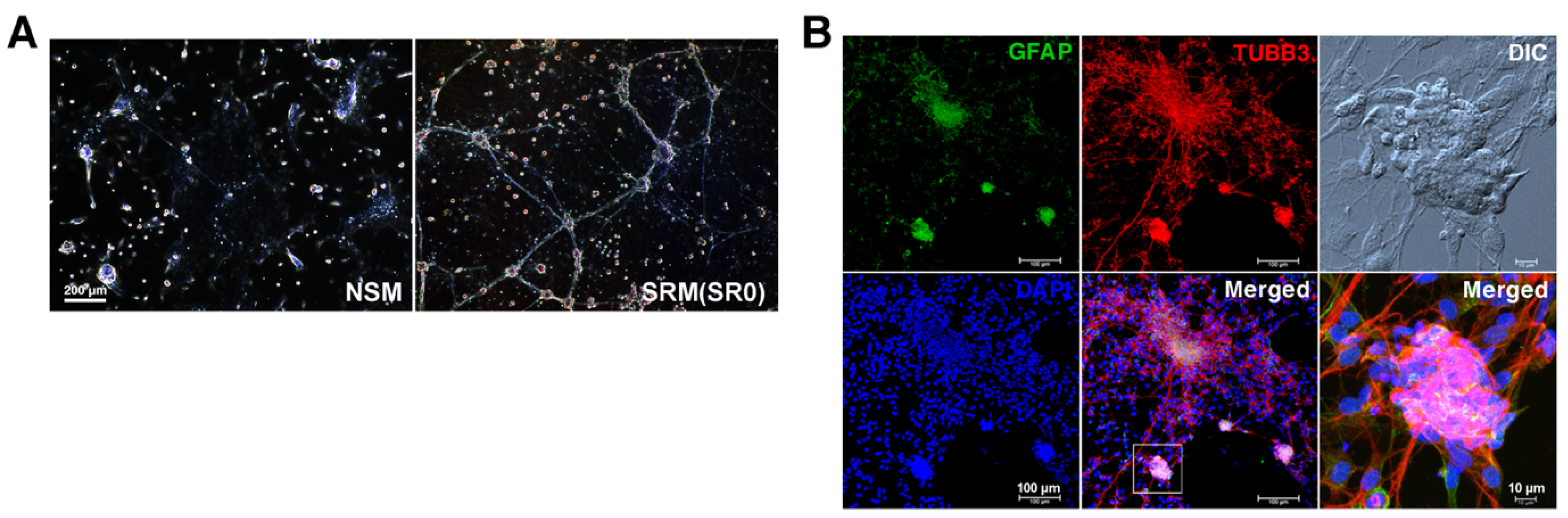
3.1. Formation of Enteric Neurospheres in NSM and SRM

Dispersed enterocytes were first nurtured non-adherently in NSM and SRM, respectively for a baseline assessment of their neurospherogenic outcome. It was found that both protocols could lead to the generation of enteric neurospheres. Notably, NSM generated neurospheres with the scarcity of neurite outgrowth, whereas SRM contributed to the assembly of enteric neural networks (Figure 1A) with ganglion-like (containing neurons and gliocytes) neurospheres wired together by neurite bundles (Figure 1B).

3.2. Assessing Component Effects on Enteric Neurospherogenesis in NSM

DMEN/F12-based NSM was supplemented with bFGF, EGF, N2, and B27 as a type of serum-free and chemically-defined nutrient medium. It permitted us to investigate the nutritional or neurotrophic requirements for neurospherogenesis (neuriteless gangliogenesis) by selective component elimination from NSM. Enteric neurospherogenesis was blocked following the deprivation of bFGF or B27, but not affected by the exclusion of EGF or N2 supplement from NSM (Figure 2A). It indicated the vital role of bFGF and B27 in the assembly of enteric neurospheres.

3.3. Assessing CEE Effects on Neural Network Assembly in SRM

Low-glucose DMEM-based SRM was supplemented with the serum analogue of CEE, the nutritional supplements of NBM, RA, B27, and N2, and the growth factors of bFGF and IGF-1. Given that neural networks developed in SRM rather than NSM, it called into question whether the biological fluid of CEE was crucial to neuronal wiring. Therefore, we examined whether the omission of CEE from SRM affected neural network assembly in vitro. CEE-free SRM (SR1) maintained the comparable fractions of p75+ ENSCs and TUBB3+ neurons to original CEE-supplemented SRM (SR0, Figure 2B) in resulting cell compositions after the 7-day culture, and retained the capacity to assemble enteric ganglia (Figure 2C) and neural networks (SR1, Figure 2D). Therefore, CEE was a superfluous supplement to neuronal differentiation and wiring in SRM.

3.4. Assessing Component Effects on Neural Network Assembly in CEE-Free SRM

CEE-free SRM (SR1) consisted of chemically-defined constituents, which permitted us to experimentally explore the relevance of nutrients or growth factors to neural network assembly. The selective exclusion of IGF-1 (SR3), NBM (SR7), RA (SR9) or N2 (SR10) supplement from CEE-free SRM (SR1) did not disturb neurogenesis (Figure 2E), nor did it affect gangliogenesis (Figure 2C) and neuronal wiring (Figure 2D). However, the selective elimination of bFGF (SR2) or B27 (SR8) from CEE-free SRM (SR1) significantly lessened neurogenesis (Figure 2E) and gangliogenesis (Figure 2C) to preclude neural network assembly (Figure 2D), highlighting the indispensability of bFGF and B27 to ENS network assembly.
To reappraise the neurotrophic effects of IGF-1 and bFGF on ENSCs, we prepared IGF-1/bFGF/CEE-free SRM (SR6) that was devoid of IGF-1 and bFGF. Sparse enteric gangliogenesis and neuronal wiring in SR6 could be enhanced by bFGF rather than IGF-1 supplementation (SR3 and SR2, respectively, Figure 2C,D), while the difference in neurogenesis between SR3 and SR6 did not reach statistical significance (Figure 2E). Moreover, bFGF addition to EGF-supplemented but IGF-1/bFGF/CEE-free SRM (SR5) promoted neurogenesis (SR4, Figure 2E) and gangliogenesis (SR4, Figure 2C) despite sparse neurite outgrowth (SR4, Figure 2D). Together, the results strengthened the role of bFGF rather than IGF-1 in facilitating ENSC neurogenesis, gangliogenesis, and network assembly.

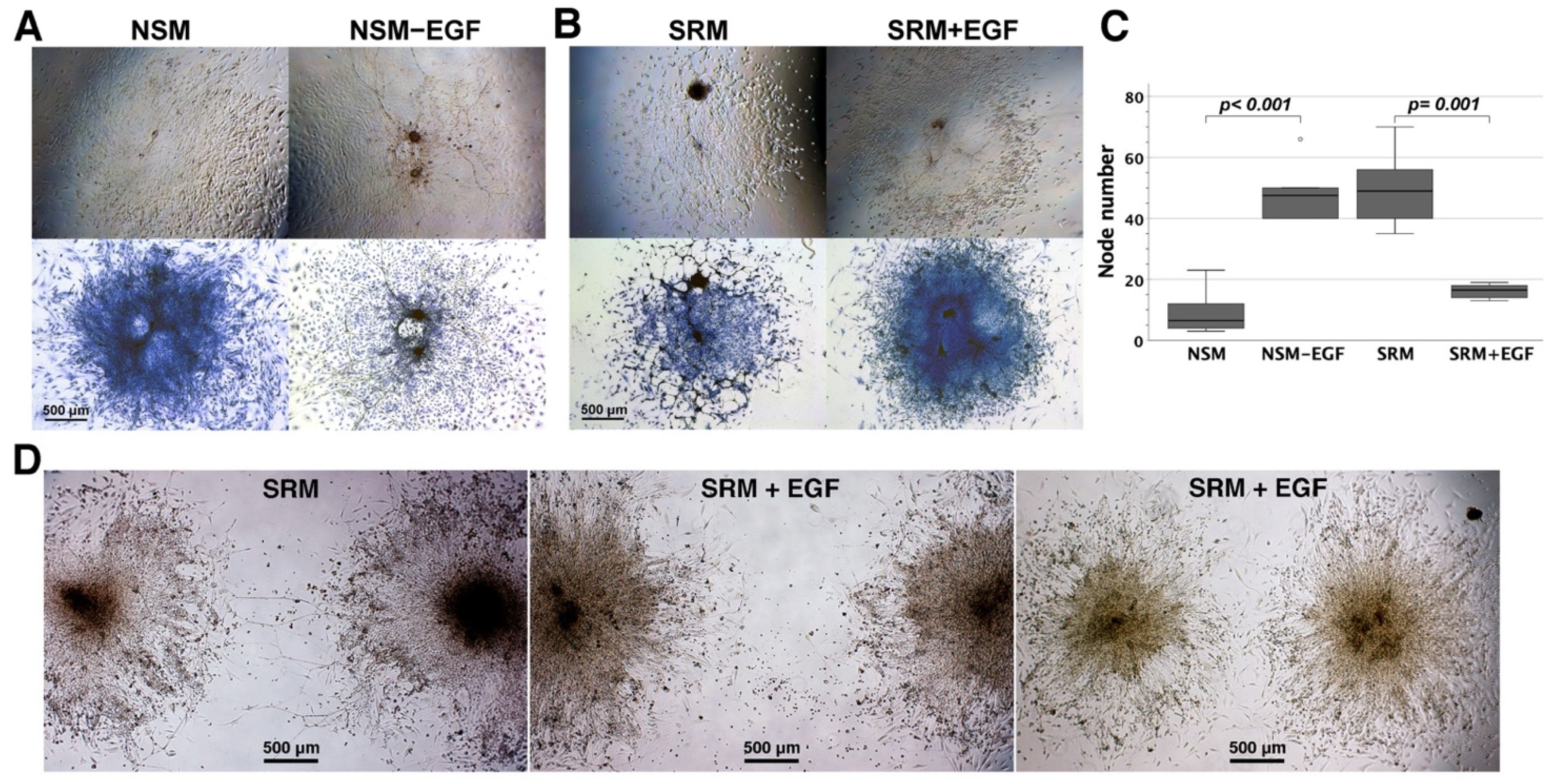
3.5. The Influence of EGF on Neural Network Assembly

EGF was not an essential ingredient for SRM, but rather a requisite for NSM. Enteric neurospherogenesis made no distinction as to whether NSM was supplemented with EGF or not (Figure 2A). However, EGF supplementation to IGF-1/CEE-free (SR3) and bFGF/IGF-1/CEE-free SRM (SR6) not only lessened neurogenesis (SR4 and SR5, respectively, Figure 2E), but also inhibited gangliogenesis (SR4 and SR5, respectively, Figure 2C) and neuronal wiring (SR4 and SR5, respectively, Figure 2D). Therefore, EGF precluded ENSCs from neurogenesis, gangliogenesis, and network assembly.
The influences of EGF on neural network assembly from enteric neurospheres were further investigated in NSM, EGF-free NSM, CEE-free SRM, and EGF-supplemented CEE-free SRM. EGF omission from NSM caused neural network assembly (Figure 3A), whereas EGF supplementation to CEE-free SRM inhibited neuronal wiring (Figure 3B). The neuroconnectivity in the assembled networks was quantified by the number of neurite intersections in the networks. The presence of EGF in media significantly lessened the neurite intersections of the network assembled from a single neurosphere (Figure 3C), reflecting sparse connectivity in neural circuits. EGF supplemented to CEE-free SRM also blocked neurite extension and neuroconnectivity between two individual neurospheres (Figure 3D). Therefore, EGF precluded enteric neurospheres from neurite outgrowth and wiring. These results further strengthened the inhibitory role of EGF in ENS network assembly.

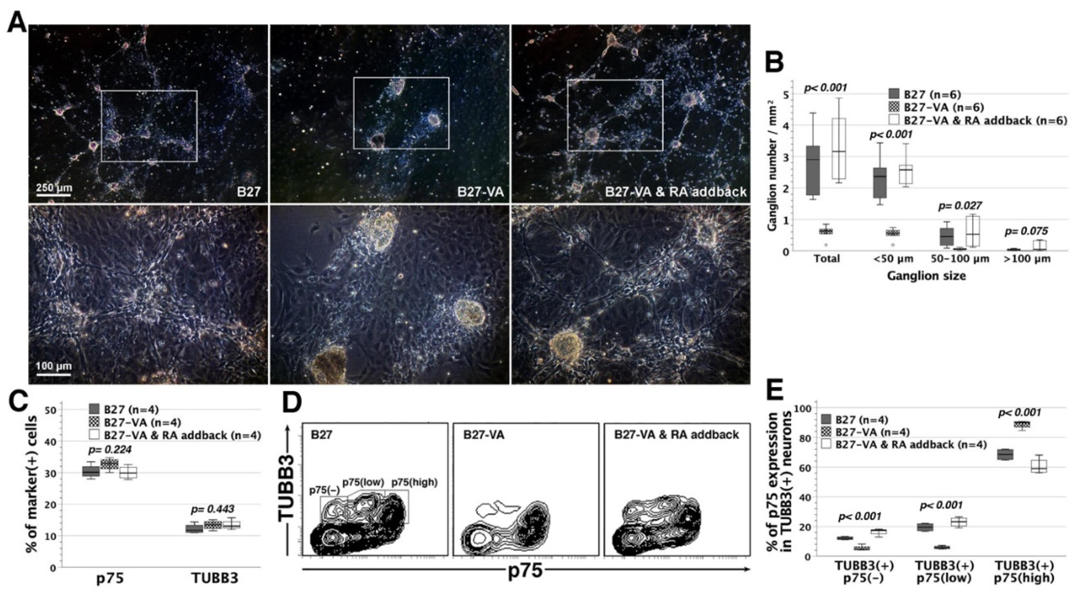
3.6. Reappraisal of RA Effects on Neural Network Assembly

Selective RA elimination from CEE-free SRM did not affect ENS network assembly. Given that B27 supplement contained vitamin A analogue of retinyl acetate [23], it might be premature to conclude the irrelevance of vitamin A to enteric neuronal wiring. As we know, vitamin A is a group of unsaturated nutritional organic compounds including preformed vitamin A (retinol and its esterified derivatives), retinal, and provitamin A carotenoids [24]. Both preformed vitamin A and provitamin A can be intracellularly metabolized to retinal and then RA (the active forms of vitamin A) to support the biological functions relevant to visual cycle, receptor binding, and the regulation of target gene expression [24]. Therefore, RA deprivation from CEE-free SRM (SR1) was unable to completely block biological pathways of vitamin A in RA/CEE-free SRM (SR9) given that the upstream retinyl acetate in B27 supplement was functionally equivalent to RA.
Based upon SR9 formula, we substituted B27 supplement minus vitamin A (B27-VA, without retinyl acetate) for the original B27 supplement. The complete absence of vitamin A derivatives in the B27-VA SR9 variant caused a significant loss of gangliogenic capacity and arrested neuronal wiring, whereas RA addback to B27-VA SR9 restored the assembly of ganglia and neural networks (Figure 4A,B). Notably, complete deprivation of vitamin A derivatives had no substantial influence on the fractions of p75+ ENSCs and TUBB3+ neurons (Figure 4C). Further cytometric analyses in a two-dimensional plot revealed that complete vitamin A deprivation retarded neuronal maturation with TUBB3+ neuron stagnation in a state of p75high expression during p75+ ENSC differentiation along a neuronal lineage [25] (Figure 4D). RA addback resumed the differentiation of TUBB3+p75high neurons toward TUBB3+p75low or TUBB3+p75 neurons (Figure 4D,E). Therefore, vitamin A derivatives might facilitate neuronal maturation in the process of neuronal differentiation, and promote the assembly of enteric ganglia and networks.

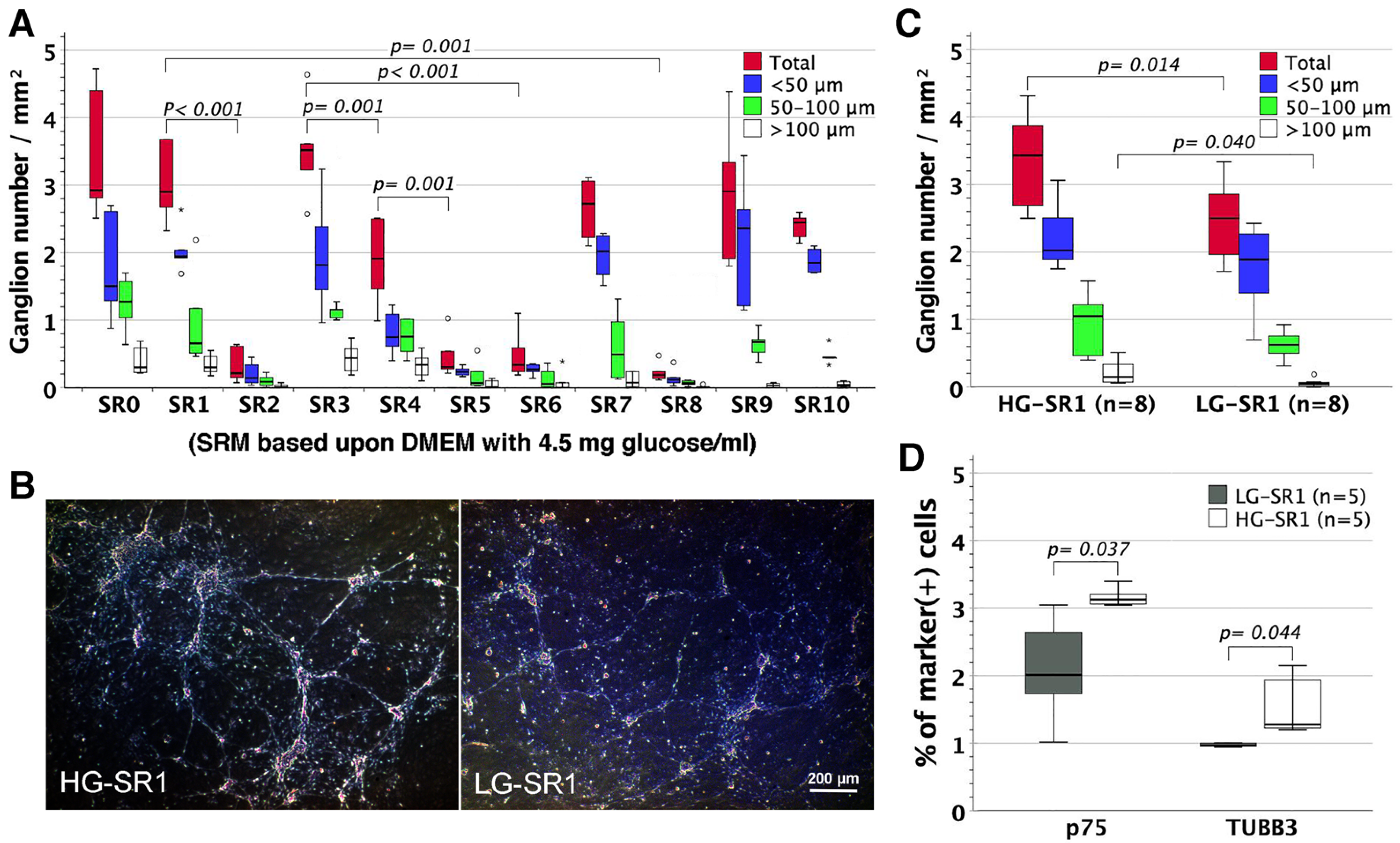
3.7. The Influence of Glucose Concentration on Neural Network Assembly

SRM was made of DMEM with 1.0 mg glucose/mL from the outset [15]. To examine the influences of glucose concentration on neuronal wiring, we substituted high-glucose (4.5 mg/mL) for low-glucose (1.0 mg/mL) DMEM in SRM and further subjected the high-glucose SRM to selective component eliminations as performed in original low-glucose SRM (Figure 2C). The assembly of enteric ganglia (Figure 5A) and networks (figures not shown) in high-glucose SRM benefitted significantly from bFGF and B27 rather than CEE, IGF-1, NBM, RA, and N2, essentially in line with what was observed in low-glucose SRM. However, high-glucose CEE-free SRM provided a higher yield of enteric ganglia (mainly sphere size >100 µm, Figure 5B,C), p75+ ENSCs, and TUBB3+ neurons (Figure 5D) than its low-glucose counterpart.

4. Discussion

Enteric nervous system (ENS) develops as a consequence of the migration, proliferation, and differentiation of the neural crest-derived ENSCs [26]. Nevertheless, little is known about what serum or humoral factors regulate or control ENSC proliferation and differentiation. The generation of ENS neurospheres and networks in vitro made the ENSC culture suited for the investigation of the nutritional or neurotrophic requirements for ENSC development, especially neural network assembly, but it would not be possible without a chemically-defined culture system. Up to the 1950s, cells or tissues grown in media would simply die without the supplement of biological fluids, such as blood sera, amniotic fluids or embryo extracts [27], which contained a variety of nutrients, hormones, growth factors or even metabolic wastes. The use of biological fluids came along with the drawbacks of enigmatic chemical compositions, lot-to-lot variations, and cytotoxic substances [28], resulting in chemically-undefined and inconsistent media to preclude researchers from formulating the requisite nutrients, hormones or growth factors for cell growth and development. It was not until 1973 that primary DRG neurons were successfully grown under serum-free conditions [29]. The supplement of special growth-promoting constituents of hormones, trace elements or defined molecules (such as transferrin) [30,31,32] led to groundbreaking advancement in the cultivation of neural cells [27], making it feasible to bring primary CNS cells into serum-free cultures [31].
Over the past two decades, ENSC culture had proved achievable in serum-free NSM [11,12,33], but required SRM to be supplemented with CEE [2,17,19]. However, we demonstrated that SRM sufficed to support ENS network assembly in vitro, independently of CEE. Therefore, it was superfluous to supplement SRM with CEE in the context of neuronal differentiation and wiring. By selective elimination of one specific ingredient from CEE-free SRM, we were able to assess the influences of the removed component on neurogenesis, gangliogenesis, and neuronal wiring. ENS network assembly was demonstrated to have relevance to bFGF and B27 rather than IGF-1, NBM, RA, and N2 in CEE-free SRM. The blockade of gangliogenesis following bFGF or B27 elimination from CEE-free SRM, to a certain extent, mirrored the failure of enteric neurospherogenesis in NSM deprived of bFGF or B27, suggesting the biologically relevant effects of bFGF and B27 on enteric neural cell aggregates in the form of ganglia or neurospheres.
NBM and B27 were developed by Brewer et al. for growing hippocampal neurons [23] and had widespread neuronal cell applications. In this study, NBM showed no substantial benefit for ENS network assembly despite its capacity to promote the outgrowth of murine NCSCs [3]. N2 supplement included insulin, transferrin, progesterone, putrescine, and selenium [32], and was actually part of 20 components in B27 supplement [23]. Therefore, selective N2 elimination from CEE-free SRM merely reduced the dosages of N2 components in media, and had no influence on ENS network assembly. B27 was demonstrated to affect ENS network assembly in vitro. Among its 20 ingredients, we identified vitamin A derivatives of retinyl acetate (or RA) as crucial to ENS network assembly. RA is the well-known signaling molecule for neuronal patterning and differentiation in CNS development [34], and also essential for ENS development [35] with relevance to RA effects on accelerating ENSC specification from neural crest derivatives [36], and orchestrating their migration [37] and colonization to the gut [38]. Our results further disclosed the vital role of RA in ENS neuronal differentiation in the context of neurite outgrowth and network assembly. In the absence of vitamin A derivatives, enteric TUBB3+ neurons were mostly neuriteless and stagnated in a p75high state. Since the p75 expression of developing enteric neurons declined as neuronal differentiation proceeded [25], these TUBB3+p75high enteric neurons might represent the less mature phenotype, unfavorable for gangliogenesis and neuronal wiring. Notably, RA addition restored neuronal maturation toward TUBB3+p75low/TUBB3+p75 neurons in the course of ENSC differentiation along a neuronal lineage, thereby promoting gangliogenesis, neurite outgrowth, and ENS network assembly. It was essentially in line with the promoting effects of RA on neuronal differentiation and neurite outgrowth of CNS [39,40] or DRG [34,41] stem cells, but in sharp contrast with the outcome reported by Sato et al., where RA promoted enteric neuronal differentiation but reduced neurite outgrowth [42]. Additional in-depth investigations into the influences of other B27 components on ENS network assembly may further define the signals that specify neuronal wiring, contributing to better understanding of the ENSC development.
Neural circuit formation relies upon the guidance of axonal growth cones that probe their surroundings for extracellular signals to mediate neurite outgrowth and synaptic connections. These signals involve different families of attractive or repulsive molecules, well-known as axon guidance cues [43]. There is compelling evidence that classic axon guidance cues regulate neural network assembly in collaboration with traditional growth factors [44]. Featuring the capacity of promoting stem cell proliferation, differentiation or survival, growth factors of bFGF, IGF-1, and EGF have been commonly supplemented into the protocols to propagate neural stem cells in neurosphere cultures. It was not until recent years that their later-phase role in neuronal morphogenesis, connectivity, and network assembly has increasingly attracted attention [44,45]. Both bFGF [46,47] and IGF-1 [48,49] were reported to promote CNS neurogenesis and neurite outgrowth. However, this study identified bFGF rather than IGF-1 as the potent neurotrophic factor for ENS network assembly in the context of promoting neurogenesis, gangliogenesis, and neuronal wiring despite the fact that IGF-1 supplementation might promote ENSC survivals [16]. These results were in keeping with the stimulatory effects of bFGF on neuronal differentiation and neurite outgrowth in neural crest-derived chromaffin cells [50], and also reflected the ENS morphological abnormalities of less neurons and smaller ganglia in bFGF-knockout mice [51]. As with bFGF, EGF was a potent mitogen [5] for CNS precursors [52] and exhibited neurotrophic activities of enhancing CNS neuronal survivals and neurite outgrowth in primary culture [53]. Although EGF might promote NCSC differentiation toward neurons and melanocytes [54], the loss of EGF receptors (EGFRs) contributed to massive axon outgrowth and branching of DRG neurons in Egfr null mutant mice [55]. This study further proved that EGF suppressed enteric neurogenesis, gangliogenesis, and network assembly. Together, they highlighted the inhibitory role of EGFR pathway in neurite outgrowth and branching in the later-phase development of NCSC-derived DRG and ENS neurons.
It has been well-recognized that EGF and bFGF affect neural network assembly directly by the alterations of growth cone cytoskeleton to modulate neurite extension [45,56], linked to invadopodia formation [57] or indirectly through non-neuronal cells, such as astrocytes [58], known as glia-growth cone interactions [59]. Moreover, neurite outgrowth might involve the interactions between cell adhesion molecules and EGF/FGF receptors [60,61]. Although it is clear now that both EGF [62] and bFGF [63] signal through receptor tyrosine kinases to trigger a variety of intracellular signaling cascades that regulate a series of biological processes in CNS or ENS development, the detailed molecular mechanisms underlying EGF or bFGF-mediated regulation of neurite guidance and outgrowth remain mostly shrouded in mystery [45] and await further experimental elucidation in the future. Notably, EGF exerted inhibitory effects on ENSC differentiation and development, not only in sharp contrast to its neurotrophic stimulation of cultured CNS neurons [53], but also diametrically opposed to bFGF, which was neurotrophic for ENSCs. It might explain the reason why ENSC-containing neurosphere isolation in SRM demanded EGF supplementation once neurosphere-like bodies appeared [17]. Therefore, the capacity of EGF to arrest enteric neuronal wiring made NSM particularly well-suited for ENSC enrichment in the form of neurospheres from enterocytes. Notably, isolated neurospheres in our laboratory could be maintained in NSM for at least 2–3 weeks without the loss of their capacity to assemble neural networks in SRM (unpublished data).
Energy metabolism plays an important role in neural stem cell function and fate [64]. The basal medium of SRM is low-glucose DMEM with 1.0 mg glucose/mL, whereas NSM contains glucose of 3.151 mg/mL (DMEM/F12 (1:1), (4.5 + 1.802)/2). High-glucose DMEM (4.5 mg/mL) had been used to prepare the formula for ENSC cultures [65]. Although nutrient media with higher glucose levels might hamper oxygen diffusion and enable anaerobic glycolysis with excessive lactate formation in cells, leading to neuronal cell damage [28], high-glucose SRM retained the capacity to assemble enteric neural networks in the bFGF- and B27-dependent patterns as observed in low-glucose SRM. Notably, high-glucose CEE-free SRM, comparing favorably in the gangliogenesis and neurogenesis with its low-glucose counterpart, remained ideally suited for ENS network assembly in vitro.

5. Conclusions

This study demonstrated the feasibility of assembling enteric neural networks in culture systems independently of complex and undefined biological fluids. The chemically-defined and consistent CEE-free SRM permitted us to investigate the nutritional or neurotrophic requirements for ENS network assembly. We identified bFGF and vitamin A as indispensable to the assembly of enteric ganglia and neural networks, whereas EGF, a CNS neurotrophic factor, proved inhibitory for enteric neurogenesis, gangliogenesis, and network assembly. Our results might help in better manipulating ENSC propagation and differentiation in vitro, and shedding light on the molecular pathways that regulate ENS network assembly.

Author Contributions

J.-C.C. conceptualized this study, acquired funding, analyzed the data, prepared the figures, and wrote the manuscript. W.Y. acquired funding, conducted the experiments, assisted in data analyses, and reviewed and edited the manuscript. L.-Y.T. and H.-L.C. conducted the experiments and assisted in animal surgery and care. All authors have read and agreed to the published version of the manuscript.

Funding

This project was supported by CMRPG3K0432 (J.-C.C.), CMRPG3J1112 (W.Y.), and CMRPG3M0191 (J.-C.C.) from Chang Gung Medical Foundation.

Institutional Review Board Statement

Ethical approval was obtained from Chang Gung Memorial Hospital Committee on Animal Research (IACUC2019091205 and IACUC2021092201).

Informed Consent Statement

Not applicable.

Data Availability Statement

All data are contained in this article.

Acknowledgments

Not applicable.

Conflicts of Interest

The authors declare no conflict of interest. The funders had no role in the design of the study; in the collection, analyses, or interpretation of data; in the writing of the manuscript, or in the decision to publish the results.

References

  1. Cooper, J.E.; McCann, C.J.; Natarajan, D.; Choudhury, S.; Boesmans, W.; Delalande, J.M.; Vanden Berghe, P.; Burns, A.J.; Thapar, N. In Vivo Transplantation of Enteric Neural Crest Cells into Mouse Gut; Engraftment, Functional Integration and Long-Term Safety. PLoS ONE 2016, 11, e0147989. [Google Scholar] [CrossRef] [PubMed]
  2. Joseph, N.M.; He, S.; Quintana, E.; Kim, Y.G.; Nunez, G.; Morrison, S.J. Enteric glia are multipotent in culture but primarily form glia in the adult rodent gut. J. Clin. Investig. 2011, 121, 3398–3411. [Google Scholar] [CrossRef] [PubMed]
  3. Pfaltzgraff, E.R.; Mundell, N.A.; Labosky, P.A. Isolation and culture of neural crest cells from embryonic murine neural tube. J. Vis. Exp. 2012, 64, e4134. [Google Scholar] [CrossRef] [PubMed]
  4. Weiss, S.; Pin, J.P.; Sebben, M.; Kemp, D.E.; Sladeczek, F.; Gabrion, J.; Bockaert, J. Synaptogenesis of cultured striatal neurons in serum-free medium: A morphological and biochemical study. Proc. Natl. Acad. Sci. USA 1986, 83, 2238–2242. [Google Scholar] [CrossRef]
  5. Reynolds, B.A.; Weiss, S. Generation of neurons and astrocytes from isolated cells of the adult mammalian central nervous system. Science 1992, 255, 1707–1710. [Google Scholar] [CrossRef]
  6. Reynolds, B.A.; Tetzlaff, W.; Weiss, S. A multipotent EGF-responsive striatal embryonic progenitor cell produces neurons and astrocytes. J. Neurosci. 1992, 12, 4565–4574. [Google Scholar] [CrossRef]
  7. Gritti, A.; Parati, E.A.; Cova, L.; Frolichsthal, P.; Galli, R.; Wanke, E.; Faravelli, L.; Morassutti, D.J.; Roisen, F.; Nickel, D.D.; et al. Multipotential stem cells from the adult mouse brain proliferate and self-renew in response to basic fibroblast growth factor. J. Neurosci. 1996, 16, 1091–1100. [Google Scholar] [CrossRef]
  8. Svendsen, C.N.; ter Borg, M.G.; Armstrong, R.J.; Rosser, A.E.; Chandran, S.; Ostenfeld, T.; Caldwell, M.A. A new method for the rapid and long term growth of human neural precursor cells. J. Neurosci. Methods 1998, 85, 141–152. [Google Scholar] [CrossRef]
  9. Metzger, M.; Conrad, S.; Skutella, T.; Just, L. RGMa inhibits neurite outgrowth of neuronal progenitors from murine enteric nervous system via the neogenin receptor in vitro. J. Neurochem. 2007, 103, 2665–2678. [Google Scholar] [CrossRef]
  10. Metzger, M.; Bareiss, P.M.; Danker, T.; Wagner, S.; Hennenlotter, J.; Guenther, E.; Obermayr, F.; Stenzl, A.; Koenigsrainer, A.; Skutella, T.; et al. Expansion and differentiation of neural progenitors derived from the human adult enteric nervous system. Gastroenterology 2009, 137, 2063–2073. [Google Scholar] [CrossRef]
  11. Metzger, M.; Caldwell, C.; Barlow, A.J.; Burns, A.J.; Thapar, N. Enteric nervous system stem cells derived from human gut mucosa for the treatment of aganglionic gut disorders. Gastroenterology 2009, 136, 2214–2225. [Google Scholar] [CrossRef] [PubMed]
  12. Hotta, R.; Stamp, L.A.; Foong, J.P.; McConnell, S.N.; Bergner, A.J.; Anderson, R.B.; Enomoto, H.; Newgreen, D.F.; Obermayr, F.; Furness, J.B.; et al. Transplanted progenitors generate functional enteric neurons in the postnatal colon. J. Clin. Investig. 2013, 123, 1182–1191. [Google Scholar] [CrossRef] [PubMed]
  13. McKeown, S.J.; Mohsenipour, M.; Bergner, A.J.; Young, H.M.; Stamp, L.A. Exposure to GDNF Enhances the Ability of Enteric Neural Progenitors to Generate an Enteric Nervous System. Stem Cell Rep. 2017, 8, 476–488. [Google Scholar] [CrossRef]
  14. Binder, E.; Natarajan, D.; Cooper, J.; Kronfli, R.; Cananzi, M.; Delalande, J.M.; McCann, C.; Burns, A.J.; Thapar, N. Enteric neurospheres are not specific to neural crest cultures: Implications for neural stem cell therapies. PLoS ONE 2015, 10, e0119467. [Google Scholar] [CrossRef] [PubMed]
  15. Morrison, S.J.; White, P.M.; Zock, C.; Anderson, D.J. Prospective identification, isolation by flow cytometry, and in vivo self-renewal of multipotent mammalian neural crest stem cells. Cell 1999, 96, 737–749. [Google Scholar] [CrossRef]
  16. Bixby, S.; Kruger, G.M.; Mosher, J.T.; Joseph, N.M.; Morrison, S.J. Cell-intrinsic differences between stem cells from different regions of the peripheral nervous system regulate the generation of neural diversity. Neuron 2002, 35, 643–656. [Google Scholar] [CrossRef]
  17. Bondurand, N.; Natarajan, D.; Thapar, N.; Atkins, C.; Pachnis, V. Neuron and glia generating progenitors of the mammalian enteric nervous system isolated from foetal and postnatal gut cultures. Development 2003, 130, 6387–6400. [Google Scholar] [CrossRef]
  18. Kruger, G.M.; Mosher, J.T.; Bixby, S.; Joseph, N.; Iwashita, T.; Morrison, S.J. Neural crest stem cells persist in the adult gut but undergo changes in self-renewal, neuronal subtype potential, and factor responsiveness. Neuron 2002, 35, 657–669. [Google Scholar] [CrossRef]
  19. Iwashita, T.; Kruger, G.M.; Pardal, R.; Kiel, M.J.; Morrison, S.J. Hirschsprung disease is linked to defects in neural crest stem cell function. Science 2003, 301, 972–976. [Google Scholar] [CrossRef]
  20. Heanue, T.A.; Pachnis, V. Enteric nervous system development and Hirschsprung’s disease: Advances in genetic and stem cell studies. Nat. Rev. Neurosci. 2007, 8, 466–479. [Google Scholar] [CrossRef]
  21. Menon, V.; Thomas, R.; Ghale, A.R.; Reinhard, C.; Pruszak, J. Flow cytometry protocols for surface and intracellular antigen analyses of neural cell types. J. Vis. Exp. 2014, 94, e52241. [Google Scholar] [CrossRef] [PubMed]
  22. Tian, D.H.; Qin, C.H.; Xu, W.Y.; Pan, W.K.; Zhao, Y.Y.; Zheng, B.J.; Chen, X.L.; Liu, Y.; Gao, Y.; Yu, H. Phenotypic and functional comparison of rat enteric neural crest-derived cells during fetal and early-postnatal stages. Neural Regen Res. 2021, 16, 2310–2315. [Google Scholar] [CrossRef] [PubMed]
  23. Brewer, G.J.; Torricelli, J.R.; Evege, E.K.; Price, P.J. Optimized survival of hippocampal neurons in B27-supplemented Neurobasal, a new serum-free medium combination. J. Neurosci Res. 1993, 35, 567–576. [Google Scholar] [CrossRef]
  24. Kam, R.K.; Deng, Y.; Chen, Y.; Zhao, H. Retinoic acid synthesis and functions in early embryonic development. Cell Biosci. 2012, 2, 11. [Google Scholar] [CrossRef] [PubMed]
  25. Young, H.M.; Ciampoli, D.; Hsuan, J.; Canty, A.J. Expression of Ret-, p75(NTR)-, Phox2a-, Phox2b-, and tyrosine hydroxylase-immunoreactivity by undifferentiated neural crest-derived cells and different classes of enteric neurons in the embryonic mouse gut. Dev. Dyn. 1999, 216, 137–152. [Google Scholar] [CrossRef]
  26. Burns, A.J.; Thapar, N. Neural stem cell therapies for enteric nervous system disorders. Nat. Rev. Gastroenterol. Hepatol. 2014, 11, 317–328. [Google Scholar] [CrossRef] [PubMed]
  27. Romijn, H.J. Development and advantages of serum-free, chemically defined nutrient media for culturing of nerve tissue. Biol. Cell 1988, 63, 263–268. [Google Scholar] [CrossRef]
  28. Romijn, H.J.; van Huizen, F.; Wolters, P.S. Towards an improved serum-free, chemically defined medium for long-term culturing of cerebral cortex tissue. Neurosci. Biobehav. Rev. 1984, 8, 301–334. [Google Scholar] [CrossRef]
  29. Luduena, M.A. The growth of spinal ganglion neurons in serum-free medium. Dev. Biol. 1973, 33, 470–476. [Google Scholar] [CrossRef]
  30. Snyder, E.Y.; Kim, S.U. Hormonal requirements for neuronal survival in culture. Neurosci. Lett. 1979, 13, 225–230. [Google Scholar] [CrossRef]
  31. Honegger, P.; Lenoir, D.; Favrod, P. Growth and differentiation of aggregating fetal brain cells in a serum-free defined medium. Nature 1979, 282, 305–308. [Google Scholar] [CrossRef] [PubMed]
  32. Bottenstein, J.E.; Sato, G.H. Growth of a rat neuroblastoma cell line in serum-free supplemented medium. Proc. Natl. Acad. Sci. USA 1979, 76, 514–517. [Google Scholar] [CrossRef] [PubMed]
  33. McCann, C.J.; Cooper, J.E.; Natarajan, D.; Jevans, B.; Burnett, L.E.; Burns, A.J.; Thapar, N. Transplantation of enteric nervous system stem cells rescues nitric oxide synthase deficient mouse colon. Nat. Commun. 2017, 8, 15937. [Google Scholar] [CrossRef] [PubMed]
  34. Maden, M. Retinoic acid in the development, regeneration and maintenance of the nervous system. Nat. Rev. Neurosci. 2007, 8, 755–765. [Google Scholar] [CrossRef]
  35. Niederreither, K.; Vermot, J.; Le Roux, I.; Schuhbaur, B.; Chambon, P.; Dolle, P. The regional pattern of retinoic acid synthesis by RALDH2 is essential for the development of posterior pharyngeal arches and the enteric nervous system. Development 2003, 130, 2525–2534. [Google Scholar] [CrossRef]
  36. Frith, T.J.R.; Gogolou, A.; Hackland, J.O.S.; Hewitt, Z.A.; Moore, H.D.; Barbaric, I.; Thapar, N.; Burns, A.J.; Andrews, P.W.; Tsakiridis, A.; et al. Retinoic Acid Accelerates the Specification of Enteric Neural Progenitors from In-Vitro-Derived Neural Crest. Stem Cell Rep. 2020, 15, 557–565. [Google Scholar] [CrossRef]
  37. Fu, M.; Sato, Y.; Lyons-Warren, A.; Zhang, B.; Kane, M.A.; Napoli, J.L.; Heuckeroth, R.O. Vitamin A facilitates enteric nervous system precursor migration by reducing Pten accumulation. Development 2010, 137, 631–640. [Google Scholar] [CrossRef]
  38. Uribe, R.A.; Hong, S.S.; Bronner, M.E. Retinoic acid temporally orchestrates colonization of the gut by vagal neural crest cells. Dev. Biol. 2018, 433, 17–32. [Google Scholar] [CrossRef]
  39. Brewer, G.J.; Torricelli, J.R. Isolation and culture of adult neurons and neurospheres. Nat. Protoc. 2007, 2, 1490–1498. [Google Scholar] [CrossRef]
  40. Dmetrichuk, J.M.; Carlone, R.L.; Spencer, G.E. Retinoic acid induces neurite outgrowth and growth cone turning in invertebrate neurons. Dev. Biol. 2006, 294, 39–49. [Google Scholar] [CrossRef] [Green Version]
  41. Clagett-Dame, M.; McNeill, E.M.; Muley, P.D. Role of all-trans retinoic acid in neurite outgrowth and axonal elongation. J. Neurobiol. 2006, 66, 739–756. [Google Scholar] [CrossRef] [PubMed]
  42. Sato, Y.; Heuckeroth, R.O. Retinoic acid regulates murine enteric nervous system precursor proliferation, enhances neuronal precursor differentiation, and reduces neurite growth in vitro. Dev. Biol. 2008, 320, 185–198. [Google Scholar] [CrossRef] [PubMed]
  43. Yu, T.W.; Bargmann, C.I. Dynamic regulation of axon guidance. Nat. Neurosci. 2001, 4, 1169–1176. [Google Scholar] [CrossRef]
  44. Short, C.A.; Onesto, M.M.; Rempel, S.K.; Catlett, T.S.; Gomez, T.M. Familiar growth factors have diverse roles in neural network assembly. Curr. Opin. Neurobiol. 2021, 66, 233–239. [Google Scholar] [CrossRef] [PubMed]
  45. Onesto, M.M.; Short, C.A.; Rempel, S.K.; Catlett, T.S.; Gomez, T.M. Growth Factors as Axon Guidance Molecules: Lessons From in vitro Studies. Front. Neurosci. 2021, 15, 678454. [Google Scholar] [CrossRef] [PubMed]
  46. Temple, S.; Qian, X. bFGF, neurotrophins, and the control or cortical neurogenesis. Neuron 1995, 15, 249–252. [Google Scholar] [CrossRef]
  47. Morrison, R.S.; Sharma, A.; de Vellis, J.; Bradshaw, R.A. Basic fibroblast growth factor supports the survival of cerebral cortical neurons in primary culture. Proc. Natl. Acad. Sci. USA 1986, 83, 7537–7541. [Google Scholar] [CrossRef] [PubMed]
  48. Nieto-Estevez, V.; Defterali, C.; Vicario-Abejon, C. IGF-I: A Key Growth Factor that Regulates Neurogenesis and Synaptogenesis from Embryonic to Adult Stages of the Brain. Front. Neurosci. 2016, 10, 52. [Google Scholar] [CrossRef]
  49. Nieto Guil, A.F.; Oksdath, M.; Weiss, L.A.; Grassi, D.J.; Sosa, L.J.; Nieto, M.; Quiroga, S. IGF-1 receptor regulates dynamic changes in neuronal polarity during cerebral cortical migration. Sci. Rep. 2017, 7, 7703. [Google Scholar] [CrossRef]
  50. Stemple, D.L.; Mahanthappa, N.K.; Anderson, D.J. Basic FGF induces neuronal differentiation, cell division, and NGF dependence in chromaffin cells: A sequence of events in sympathetic development. Neuron 1988, 1, 517–525. [Google Scholar] [CrossRef]
  51. Hagl, C.I.; Holland-Cunz, S.; Schafer, K.H. What do knockout models teach us about the enteric nervous system? Eur. J. Pediatr. Surg. 2003, 13, 170–175. [Google Scholar] [CrossRef] [PubMed]
  52. Reynolds, B.A.; Weiss, S. Clonal and population analyses demonstrate that an EGF-responsive mammalian embryonic CNS precursor is a stem cell. Dev. Biol. 1996, 175, 1–13. [Google Scholar] [CrossRef] [PubMed]
  53. Morrison, R.S.; Kornblum, H.I.; Leslie, F.M.; Bradshaw, R.A. Trophic stimulation of cultured neurons from neonatal rat brain by epidermal growth factor. Science 1987, 238, 72–75. [Google Scholar] [CrossRef] [PubMed]
  54. Garcez, R.C.; Teixeira, B.L.; Schmitt Sdos, S.; Alvarez-Silva, M.; Trentin, A.G. Epidermal growth factor (EGF) promotes the in vitro differentiation of neural crest cells to neurons and melanocytes. Cell. Mol. Neurobiol. 2009, 29, 1087–1091. [Google Scholar] [CrossRef] [PubMed]
  55. Maklad, A.; Nicolai, J.R.; Bichsel, K.J.; Evenson, J.E.; Lee, T.C.; Threadgill, D.W.; Hansen, L.A. The EGFR is required for proper innervation to the skin. J. Investig. Dermatol. 2009, 129, 690–698. [Google Scholar] [CrossRef] [PubMed]
  56. Szebenyi, G.; Dent, E.W.; Callaway, J.L.; Seys, C.; Lueth, H.; Kalil, K. Fibroblast growth factor-2 promotes axon branching of cortical neurons by influencing morphology and behavior of the primary growth cone. J. Neurosci. 2001, 21, 3932–3941. [Google Scholar] [CrossRef]
  57. Santiago-Medina, M.; Gregus, K.A.; Nichol, R.H.; O’Toole, S.M.; Gomez, T.M. Regulation of ECM degradation and axon guidance by growth cone invadosomes. Development 2015, 142, 486–496. [Google Scholar] [CrossRef]
  58. Liu, B.; Neufeld, A.H. Activation of epidermal growth factor receptors directs astrocytes to organize in a network surrounding axons in the developing rat optic nerve. Dev. Biol. 2004, 273, 297–307. [Google Scholar] [CrossRef]
  59. Rigby, M.J.; Gomez, T.M.; Puglielli, L. Glial Cell-Axonal Growth Cone Interactions in Neurodevelopment and Regeneration. Front. Neurosci. 2020, 14, 203. [Google Scholar] [CrossRef]
  60. Povlsen, G.K.; Berezin, V.; Bock, E. Neural cell adhesion molecule-180-mediated homophilic binding induces epidermal growth factor receptor (EGFR) down-regulation and uncouples the inhibitory function of EGFR in neurite outgrowth. J. Neurochem. 2008, 104, 624–639. [Google Scholar] [CrossRef]
  61. Garcia-Alonso, L.; Romani, S.; Jimenez, F. The EGF and FGF receptors mediate neuroglian function to control growth cone decisions during sensory axon guidance in Drosophila. Neuron 2000, 28, 741–752. [Google Scholar] [CrossRef]
  62. Romano, R.; Bucci, C. Role of EGFR in the Nervous System. Cells 2020, 9, 1887. [Google Scholar] [CrossRef] [PubMed]
  63. Guillemot, F.; Zimmer, C. From cradle to grave: The multiple roles of fibroblast growth factors in neural development. Neuron 2011, 71, 574–588. [Google Scholar] [CrossRef] [PubMed]
  64. Rafalski, V.A.; Brunet, A. Energy metabolism in adult neural stem cell fate. Prog. Neurobiol. 2011, 93, 182–203. [Google Scholar] [CrossRef]
  65. Suarez-Rodriguez, R.; Belkind-Gerson, J. Cultured nestin-positive cells from postnatal mouse small bowel differentiate ex vivo into neurons, glia, and smooth muscle. Stem Cells 2004, 22, 1373–1385. [Google Scholar] [CrossRef]
Figure 1. The formation of enteric neurospheres in NSM and SRM. Dispersed enterocytes (2 × 10⁵) from murine fetal gut were non-adherently nurtured in 6-well plates flooded with NSR or SRM. (A) On day 7, enteric neurospherogenesis was photographed live under a phase-contrast microscope. SRM supported the assembly of neural networks, whereas NSM generated discrete neurospheres with the scarcity of neurite outgrowth. (B) Neural networks assembled from dispersed enterocytes on coverslips in SRM-filled wells were subjected to immunofluorescence staining. The neurospheres, composed of TUBB3+ neurons and GFAP+ gliocytes, were structurally and morphologically linked to enteric ganglia and wired together by neuronal neurites to form neural networks (ganglionated plexuses). DIC: Differential interference contrast. SR0: Original SRM that contained CEE.
Cells 11 02841 g001
Figure 2. The influences of selective component elimination from NSM or SRM on the assembly of neurospheres or neural networks. Enterocytes (2 × 10⁵) were non-adherently grown in 6-well plates flooded with NSM, SRM or their modifications by selective component elimination. Enteric neurospheres (or ganglia) were enumerated and analyzed after 7-day cultivation. (A) Comparison was made to total neurosphere (neuriteless ganglion) number (the added numbers of neurospheres sized <50 μm, 50–100 μm, and >100 μm) between NSM and its indicated modifications (n = 4 for each group). Selective elimination of bFGF (NSM-bFGF) or B27 (NSM-B27) from NSM significantly reduced enteric neurospheres (t-test), whereas EGF or N2 omission (NSM-EGF or NSM-N2) made no change to neurospherogenesis. (B) Cultivated enterocytes in original SRM (SR0) and CEE-free SRM (SR1) were trypsinized, intracellularly stained with fluorescence-conjugated anti-p75 and anti-TUBB3, and analyzed by flow cytometry. SR0 and SR1 did not differ in the fractions of p75+ ENSCS and TUBB3+ neurons. (C) SRM was modified by selective elimination of one specific component or additional EGF supplementation as shown (n = 7 for each group). Comparison was made to total ganglion number of SR0 vs. SR1, SR1 vs. its indicated modifications (SR2, 3, 7–10) or two modified conditions that varied in only one supplement (SR3 vs. SR4, SR4 vs. SR5, SR5 vs. SR6, and SR2/SR3 vs. SR6). The p-values of <0.05 (t-test) were shown in the figure. Outliers (circle) and extremes (asterisk) were values of 1.5–3 times and more than 3 times interquartile ranges from the end of a box, respectively. (D) The images of ENSC development were taken live on day 7 in a representative set of experiments using the same batch of enterocytes. Day 7 cells in SRM and its relevant modifications (n = 6) were analyzed for TUBB3+ neurons by flow cytometry after intracellular staining. Comparison was made to TUBB3+ neuron fractions between the two groups as indicated above in (C).
Cells 11 02841 g002
Figure 3. The EGF effects on network assembly of enteric neurospheres. Enteric neurospheres generated in NSM were nurtured adherently in NSM, EGF-free NSM (NSM-EGF), CEE-free SRM (SRM), and EGF-supplemented CEE-free SRM (SRM + EGF) for 1 week. (A) NSM-EGF gave rise to neural network assembly in preference to NSM. The networks were photographed live (upper) and after Diff-Quik staining (lower). (B) SRM + EGF downregulated neuronal wiring in contrast with extensive neuronal wiring in SRM. (C) The neuroconnectivity in the network assembled from a single neurosphere was graded by the amount of neurite intersections after Diff-Quik staining (n = 6 for each group). Groups NSM-EGF and SRM compared favorably in the intersection number with groups NSM and SRM + EGF, respectively. Outliers (circle) were values of 1.5–3 times interquartile ranges from the end of a box. (D) The neurite outgrowth and connectivity between two individual neurospheres in groups SRM and SRM + EGF were examined on day 7. SRM might help in wiring neurospheres together (left panel). However, SRM + EGF blocked neurite extension from neurospheres and led to the absence of connectivity between two independent neurospheres (middle panel) even though there was a closer distance between two neurospheres (right panel). The images shown were representative of three independent experiments.
Cells 11 02841 g003
Figure 4. The effects of vitamin A derivatives on neuronal differentiation and wiring. As a modified version of the original B27 without vitamin A (retinyl acetate), B27 minus vitamin A (group B27-VA) was substituted for B27 to prepare RA/CEE-free SRM (SR9) that was completely free from vitamin A derivatives. (A,B) Group B27-VA had reduced gangliogenesis and lost the ability to support neuronal wiring, as compared with SR9 supplemented with original B27 (group B27), whereas RA addback to group B27-VA (group B27-VA & RA addback) restored gangliogenesis (ANOVA) and neuronal wiring. Outliers (circle) were values of 1.5–3 times interquartile ranges from the end of a box. (C) Flow cytometric analyses showed no significant difference in the fractions of p75+ ENSCs and TUBB3+ neurons among the three groups. (D) Contour plots addressed the variations of p75 expression in TUBB3+ enteric neurons of the three groups. (E) Group B27-VA exhibited significantly less TUBB3+p75 and TUBB3+p75low, but more TUBB3+p75high neurons than group B27. Group B27-VA & RA addback recovered the levels of TUBB3+p75, TUBB3+p75low, and TUBB3+p75high neurons.
Cells 11 02841 g004
Figure 5. Neural network assembly in high-glucose SRM and its relevant modifications. High-glucose (4.5 mg/mL) DMEM was substituted for low-glucose (1.0 mg/mL) DMEM to make up high-glucose SRM. Outliers (circle) and extremes (asterisk) were values of 1.5–3 times and more than 3 times interquartile ranges from the end of a box, respectively. (A) High-glucose SRM (n = 6 for each group) was modified in accordance with the rule previously used for low-glucose SRM in Figure 2C. The selective elimination of bFGF or B27 from high-glucose SRM proved to be most inimical to neural network assembly, identical to what was observed in low-glucose SRM. (B) High-glucose CEE-free SRM (HG-SR1) supported neural network assembly similar to low-glucose CEE-free SRM (LG-SR1). (C) HG-SR1 generated more ganglia than LG-SR1, especially ganglion size of >100 μm. (D) HG-SR1 compared favorably in the fraction of p75+ ENSCs and TUBB3+ neurons with LG-SR1.
Cells 11 02841 g005
Publisher’s Note: MDPI stays neutral with regard to jurisdictional claims in published maps and institutional affiliations.

