Article

Inflammation and Wasting of Skeletal Muscles in Kras-p53-Mutant Mice with Intraepithelial Neoplasia and Pancreatic Cancer—When Does Cachexia Start?
1
Institute of Anatomy and Cell Biology, Department of Medical Cell Biology, Philipps-University of Marburg, Robert-Koch-Str. 8, 35032 Marburg, Germany
2
Department of General, Visceral and Pedriatic Surgery, University Clinics, Georg-August University, Robert-Koch-Str. 40, 37075 Goettingen, Germany
3
Department of Visceral, Thoracic and Vascular Surgery, University Clinics of Giessen and Marburg, Baldinger Str., 35043 Marburg, Germany
4
Center for Endocrine Surgery, Schön Klinik Hamburg-Eilbek, Dehnhaide 120, 22081 Hamburg, Germany
*
Author to whom correspondence should be addressed.
These authors contributed equally to this work.
Cells 2022, 11(10), 1607; https://doi.org/10.3390/cells11101607
Submission received: 10 March 2022 / Revised: 28 April 2022 / Accepted: 6 May 2022 / Published: 11 May 2022
(This article belongs to the Special Issue Cancer-Induced Cachexia)

Abstract

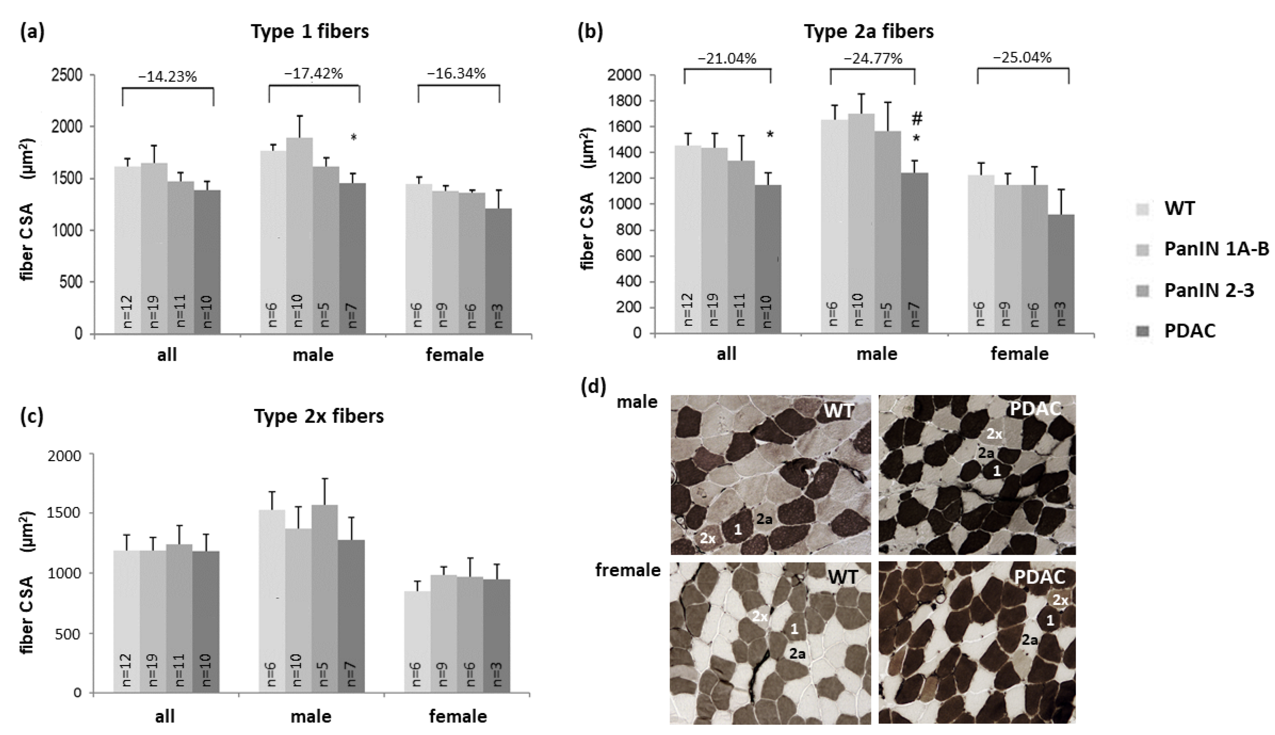
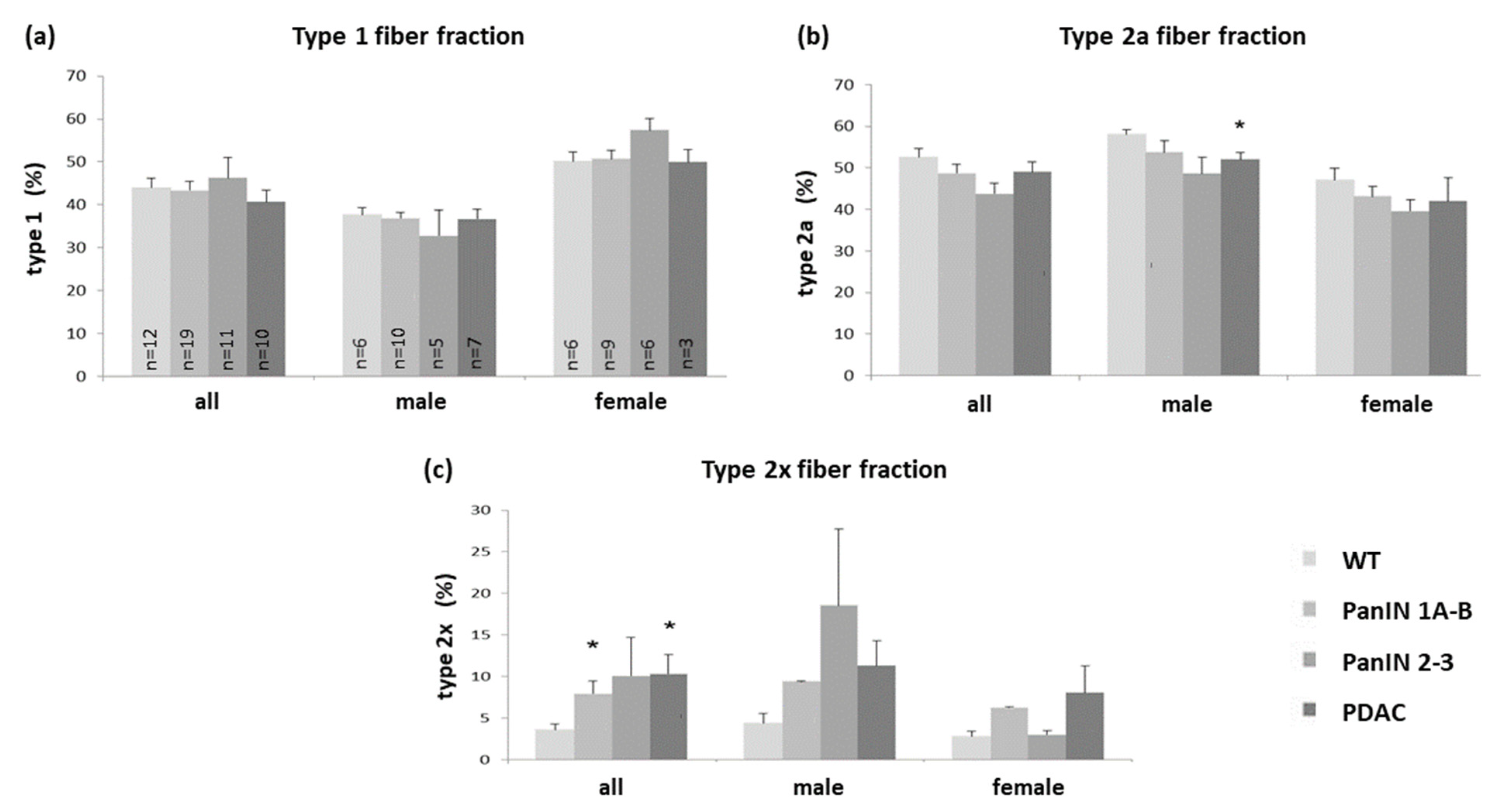
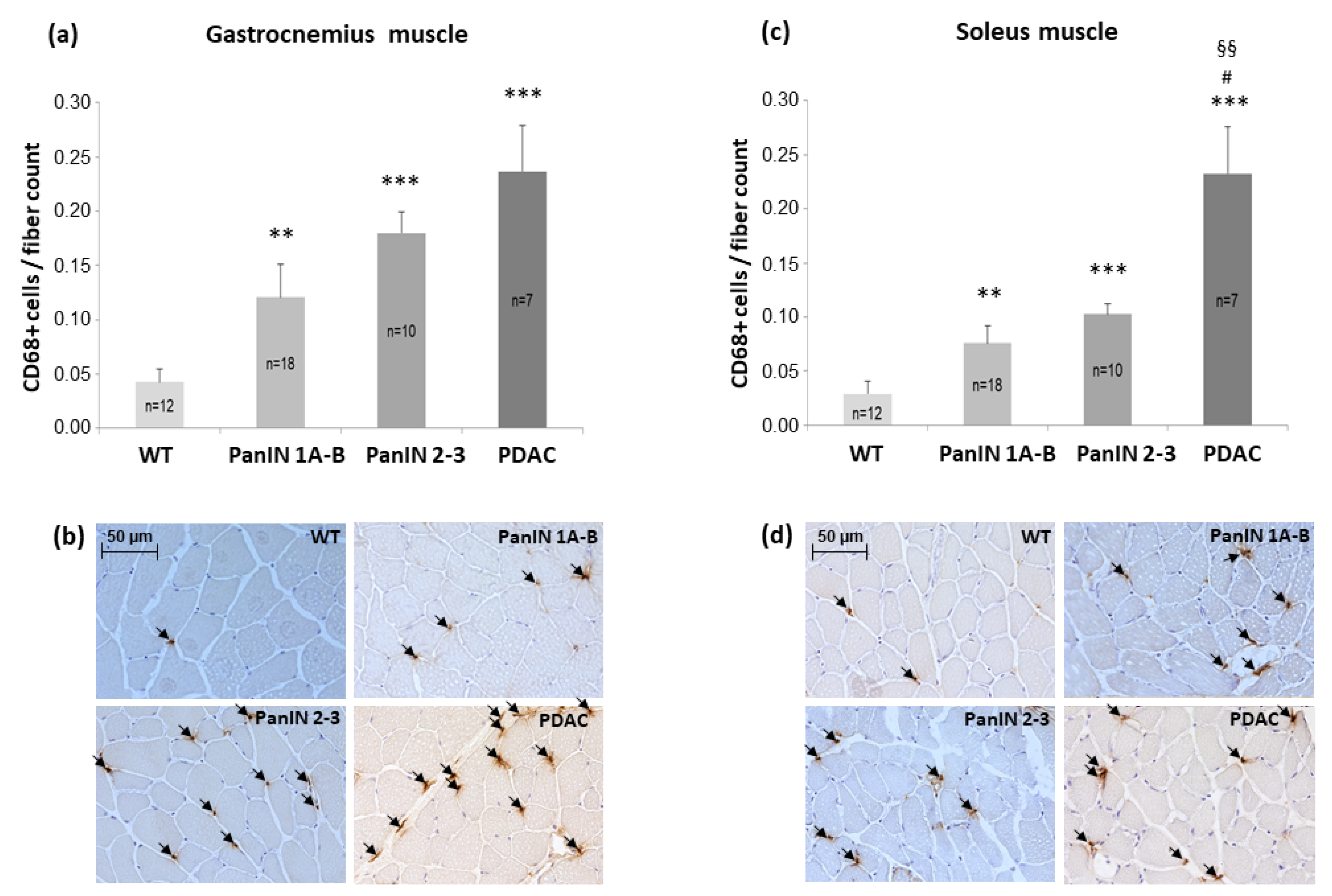
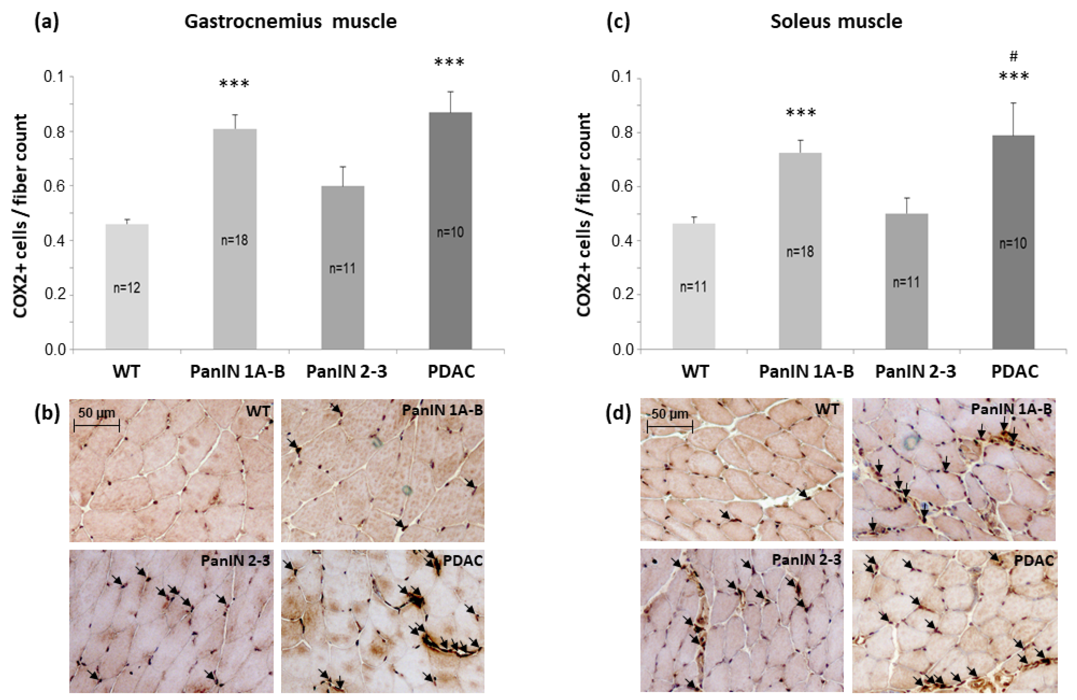
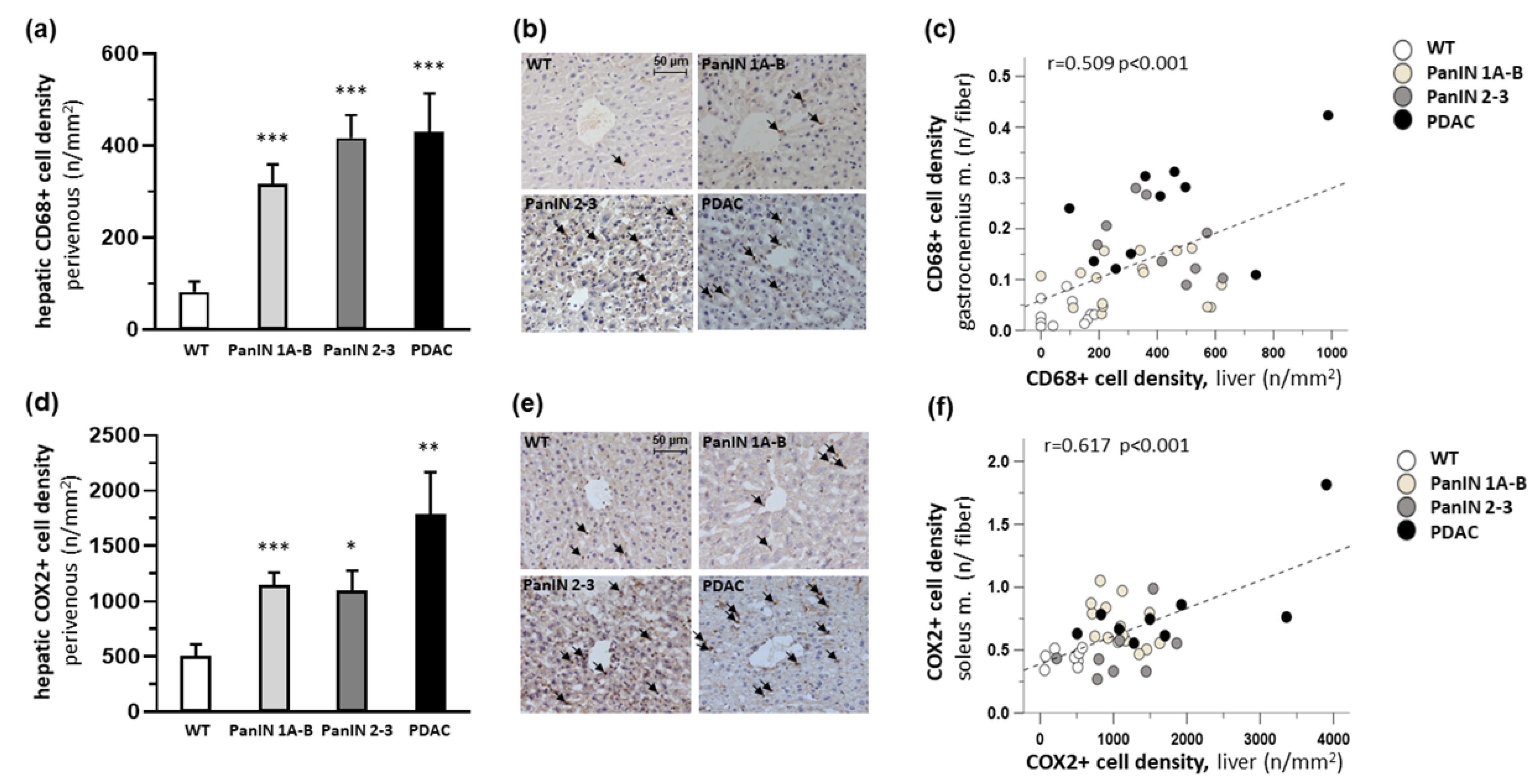
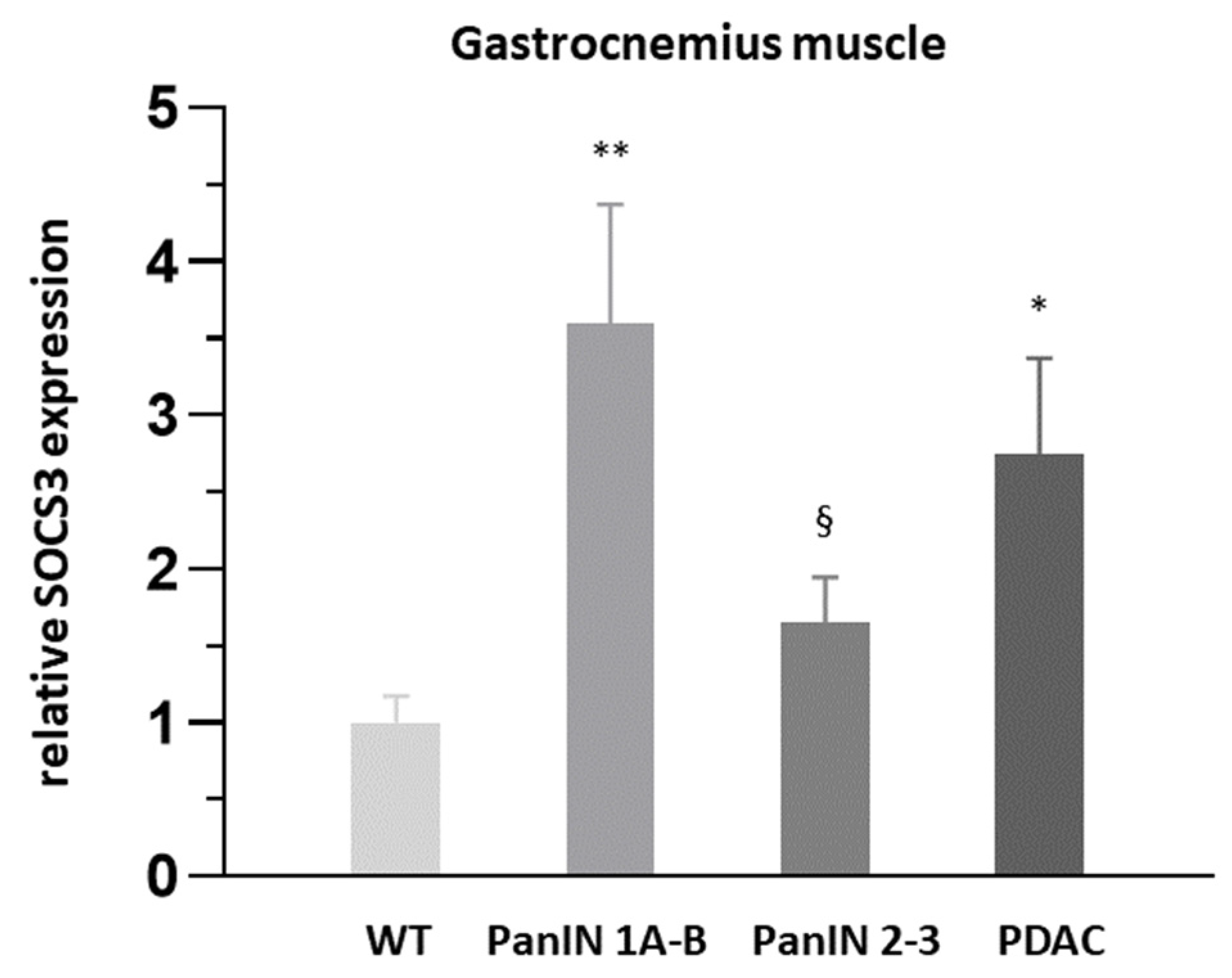
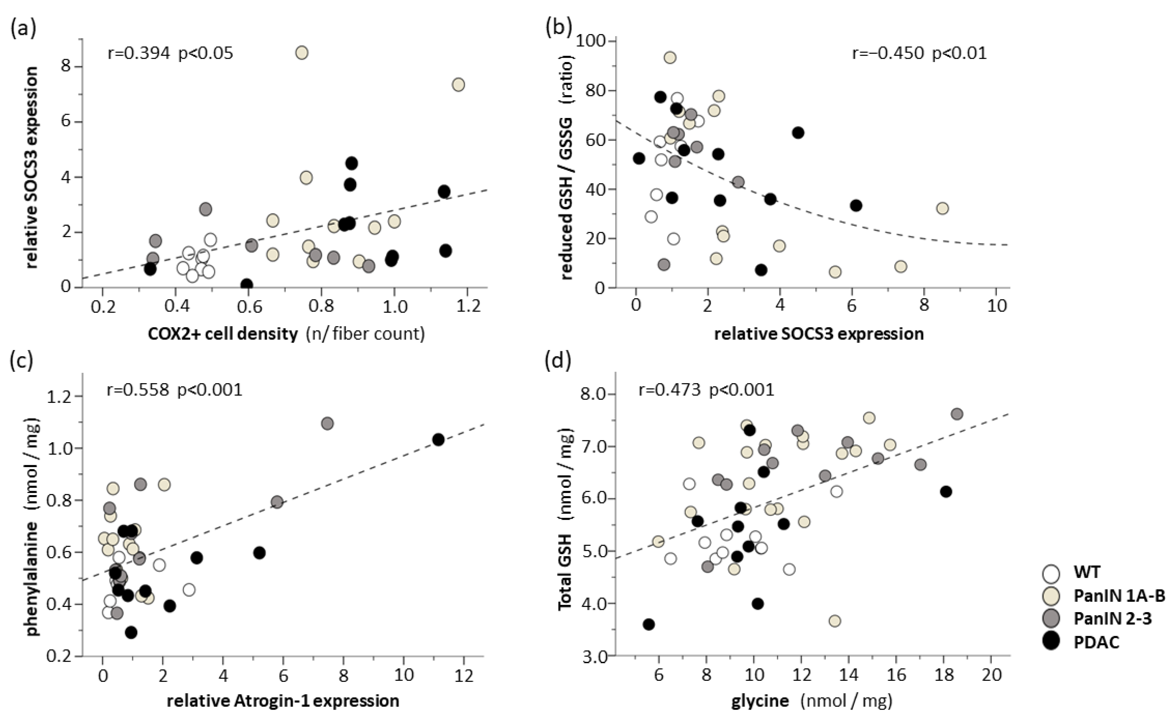
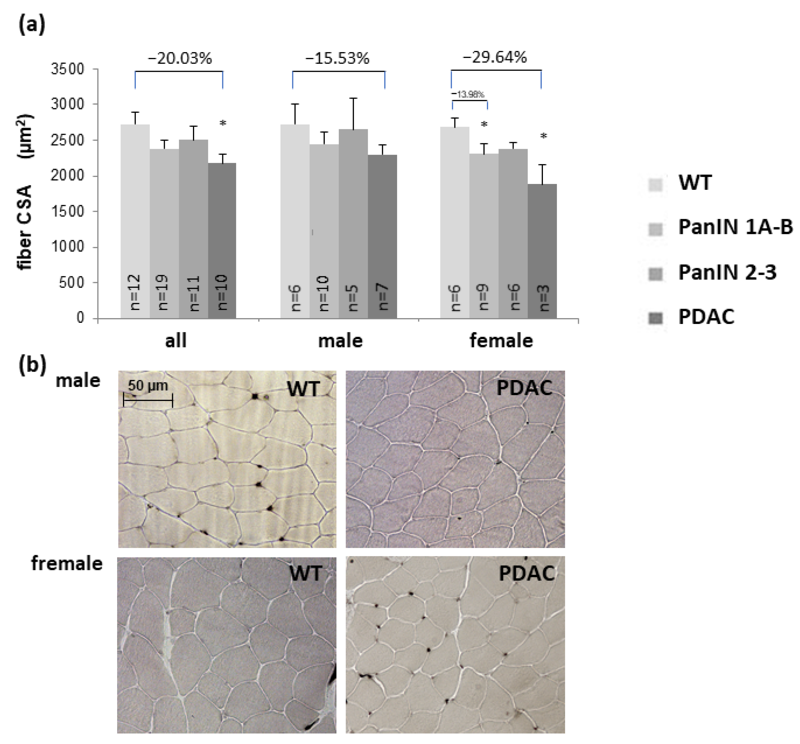
Skeletal muscle wasting critically impairs the survival and quality of life in patients with pancreatic ductal adenocarcinoma (PDAC). To identify the local factors initiating muscle wasting, we studied inflammation, fiber cross-sectional area (CSA), composition, amino acid metabolism and capillarization, as well as the integrity of neuromuscular junctions (NMJ, pre-/postsynaptic co-staining) and mitochondria (electron microscopy) in the hindlimb muscle of LSL-KrasG12D/+; LSL-TrP53R172H/+; Pdx1-Cre mice with intraepithelial-neoplasia (PanIN) 1-3 and PDAC, compared to wild-type mice (WT). Significant decreases in fiber CSA occurred with PDAC but not with PanIN 1-3, compared to WT: These were found in the gastrocnemius (type 2x: −20.0%) and soleus (type 2a: −21.0%, type 1: −14.2%) muscle with accentuation in the male soleus (type 2a: −24.8%, type 1: −17.4%) and female gastrocnemius muscle (−29.6%). Significantly higher densities of endomysial CD68+ and cyclooxygenase-2+ (COX2+) cells were detected in mice with PDAC, compared to WT mice. Surprisingly, CD68+ and COX2+ cell densities were also higher in mice with PanIN 1-3 in both muscles. Significant positive correlations existed between muscular and hepatic CD68+ or COX2+ cell densities. Moreover, in the gastrocnemius muscle, suppressor-of-cytokine-3 (SOCS3) expressions was upregulated >2.7-fold with PanIN 1A-3 and PDAC. The intracellular pools of proteinogenic amino acids and glutathione significantly increased with PanIN 1A-3 compared to WT. Capillarization, NMJ, and mitochondrial ultrastructure remained unchanged with PanIN or PDAC. In conclusion, the onset of fiber atrophy coincides with the manifestation of PDAC and high-grade local (and hepatic) inflammatory infiltration without compromised microcirculation, innervation or mitochondria. Surprisingly, muscular and hepatic inflammation, SOCS3 upregulation and (proteolytic) increases in free amino acids and glutathione were already detectable in mice with precancerous PanINs. Studies of initial local triggers and defense mechanisms regarding cachexia are warranted for targeted anti-inflammatory prevention.
Keywords: cancer cachexia; muscle atrophy; weight loss; sarcopenia; cytokines; gastrointestinal cancer cancer cachexia; muscle atrophy; weight loss; sarcopenia; cytokines; gastrointestinal cancer