Article

The Negative Regulative Roles of BdPGRPs in the Imd Signaling Pathway of Bactrocera dorsalis

State Key Laboratory of Agricultural Microbiology, Institute of Urban and Horticultural Entomology, College of Plant Science and Technology, Huazhong Agricultural University, Wuhan 430070, China
*
Author to whom correspondence should be addressed.
These authors contributed equally to this work.
Cells 2022, 11(1), 152; https://doi.org/10.3390/cells11010152
Submission received: 23 November 2021 / Revised: 23 December 2021 / Accepted: 24 December 2021 / Published: 4 January 2022
(This article belongs to the Collection Feature Papers in ‘Cellular Immunology’)

Abstract

:
Peptidoglycan recognition proteins (PGRPs) are key regulators in insects’ immune response, functioning as sensors to detect invading pathogens and as scavengers of peptidoglycan (PGN) to reduce immune overreaction. However, the exact function of PGRPs in Bactrocera dorsalis is still unclear. In this study, we identified and functionally characterized the genes BdPGRP-LB, BdPGRP-SB1 and BdPGRP-SC2 in B. dorsalis. The results showed that BdPGRP-LB, BdPGRP-SB1 and BdPGRP-SC2 all have an amidase-2 domain, which has been shown to have N-Acetylmuramoyl-l-Alanine amidase activity. The transcriptional levels of BdPGRP-LB and BdPGRP-SC2 were both high in adult stages and midgut tissues; BdPGRP-SB1 was found most abundantly expressed in the 2nd instar larvae stage and adult fat body. The expression of BdPGRP-LB and BdPGRP-SB1 and AMPs were significantly up-regulated after injury infected with Escherichia coli at different time points; however, the expression of BdPGRP-SC2 was reduced at 9 h, 24 h and 48 h following inoculation with E. coli. By injection of dsRNA, BdPGRP-LB, BdPGRP-SB1 and BdPGRP-SC2 were knocked down by RNA-interference. Silencing of BdPGRP-LB, BdPGRP-SB1 and BdPGRP-SC2 separately in flies resulted in over-activation of the Imd signaling pathway after bacterial challenge. The survival rate of the ds-PGRPs group was significantly reduced compared with the ds-egfp group after bacterial infection. Taken together, our results demonstrated that three catalytic PGRPs family genes, BdPGRP-LB, BdPGRP-SB1 and BdPGRP-SC2, are important negative regulators of the Imd pathway in B. dorsalis.

Graphical Abstract

1. Introduction

Insects come into contact with many kinds of pathogenic microorganisms from their habitat, and therefore insects have involved a strong innate immune system to resist microbial challenge. This system immediately responds against invading pathogens, and consists of cellular and humoral immune responses [1]. The activation of a series of antimicrobial defense mechanisms relies on a microbial sensing system of pattern-recognition receptors (PRRs) [2]. In insects, peptidoglycan recognition proteins (PGRPs) are a major class of PRRs that can recognize peptidoglycan (PGN), the specific component of the cell wall in both Gram-positive and Gram-negative bacteria [3,4]. PGN is a polymer with alternating N-acetylglucosamine and N-acetylmuramic acid residues that are cross-linked to each other by short peptide bridges; Gram-negative bacteria and Gram-positive Bacilli have DAP type PGN, unlike Gram-positive bacteria, which have Lys type PGN [5,6]. PGRP was first discovered in silkworms (Bombyx mori) in the late 1990s. PGRP was confirmed to have the ability to trigger a series of prophenoloxidase cascades after binding to different types of peptidoglycans [7]. With the progress of genome projects for different species, PGRP and its homologues have been identified in animals ranging from insects to mammals [8,9,10,11]. PGRPs are highly conserved from insects to mammals, which share a conserved 160 amino acid domain with similarities to the bacteriophage T7 lysozyme, a zinc-dependent amidase that hydrolyzes peptidoglycan [4].
Studies of PGRPs have focused on D. melanogaster. Drosophila has 13 PGRP genes which can encode 20 PGRP proteins; PGRPs can be divided into catalytic PGRPs and non-catalytic PGRPs according to their function [8]. Noncatalytic PGRPs (PGRP-SA, SD, LA, LC, LD, LE and LF) can only bind to peptidoglycan and lack amidase activity due to the absence of key cysteine residues for zinc binding, which are crucial for sensing of bacteria and activating immune pathways in the immune system. By contrast, catalytic PGRPs (PGRP-SC1a/b, SC2, LB and SB1/2) hydrolyze peptidoglycan by cleaving the amide bond between MurNAc and the peptidic bridge, leading to a termination of immune response [12,13]. The amidase PGRPs function as key immunoregulatory factors, regulating the immune response by cleaving peptidoglycan and existing directly as a bactericide [14]. In D. melanogaster, amidase PGRPs reduce the expression level of AMPs by degrading peptidoglycan and downregulating the immune response [13]. PGRP-LB deletion mutant and Pirk deletion mutant, and to a lesser extent PGRP-SC single deletion mutant flies showed reductions in mean lifespan compared to wild-type after Ecc15 (Erwinia carotovora carotovora 15) infection. The excessive death of null mutants was due to their own excessive immune response rather than the accumulation of conditional pathogens, which has been further confirmed in [13]. DmPGRP-LB with amidase can downregulate the immune response by converting the Gram negative PGN to non-immunostimulatory fragments [6]. DmPGRP-SC2 was inhibited by FOXO with age, leading to immune system disorders and intestinal microbial disorders [15]. DmPGRP-SB1 has an amidase activity against DAP-type PGN, while DmPGRP-SB1 and SB2 are, at most, only marginally involved in the regulation of the Imd pathway [13,16].
The oriental fruit fly Bactrocera dorsalis (Hendel) is a destructive polyphagous and invasive insect pest of tropical and subtropical fruits and vegetables [17]. Owing to its vast adaptability, high reproduction potential and invasive capacity, B. dorsalis has been one of the world’s most invasive and polyphagous pests of agriculture [18]. B. dorsalis larvae live in rotten fruits and are more likely to be exposed to pathogenic bacteria. Indeed, B. dorsalis is emerging as a good material for research into immunity [19,20] and the role of immunity in microbiota homeostasis [21]. Although the functions of PGRPs have been shown in a number of insects, especially in D. melanogaster [16,22,23,24,25] and in other insects such as Musca domestica, Sitophilus zeamais, Rhynchophorus ferrugineus [26,27,28] as well, there is no clear picture of the role of PGRPs in B. dorsalis.
In this study, we cloned BdPGRP-LB, one isoform of PGRP-SB (BdPGRP-SB1), and one isoform of PGRP-SC (BdPGRP-SC2). The expression profiles of the BdPGRP-LB, BdPGRP-SB1 and BdPGRP-SC2 genes in different developmental stages and adult tissues were examined by real-time quantitative polymerase chain reaction (qRT-PCR). We monitored the immune response of BdPGRPs after adults were infected with the Gram-negative bacteria E. coli, and revealed the important negative roles of the BdPGRP-LB, BdPGRP-SB1 and BdPGRP-SC2 genes in the Imd pathway of B. dorsalis using RNA interference methods.

2. Materials and Methods

2.1. Experimental Insects

B. dorsalis was collected from Guangzhou, China and reared more than 20 generations at the Institute of Urban and Horticultural Pests at Huazhong Agricultural University, Wuhan, as described by Li et al. [17]. The newly emerged adults were reared in cages under the following conditions: 28 ± 1 °C, 70–80% relative humidity, 12 h/12 h light/dark cycle; adults’ artificial diet contained 2.5% yeast extract, 7.5% sugar, 2.5% honey, 0.5% agar and 87% water; eggs and larvae were fed on bananas.

2.2. Cloning and Analysis of the BdPGRP Genes

Total RNA was extracted from B. dorsalis with RNAisoTM Plus reagent (TaKaRa, Otsu, Shiga, Japan) following the manufacturer’s instructions. Ten newly emerged adults of B. dorsalis with a sex ration at 1:1 were homogenized in 1mL RNAiso with a burnisher (Shanghai Jingxin Industrial Development Co., Ltd., Shanghai, China) at 70 Hz/s for 60 s at 10 s intervals. The purity of the RNA was analyzed using a NanoDrop 1000 Spectrophotometer (Thermo Fisher Scientific, Waltham, MA, USA) and the quality of RNA was tested by 1.0% agarose gel electrophoresis at voltage 120 V, 20 min in TAE buffer. First strand cDNA was synthesized from 1 μg RNA using the PrimeScript™ RT reagent Kit with gDNA Eraser Kit (TaKaRa). Then, the cDNA was served as template. The amplification of 3′- and 5′- cDNA ends of BdPGRP-SC2 was conducted with the 3′-Full RACE Core Set (Cat. # 6121) (TaKaRa, Otsu, Shiga, Japan) and 5′-Full RACE Kit (Cat. # 6122) (TaKaRa, Otsu, Shiga, Japan) according to the manufacturer’s instructions. Primers for RACE were designed according to the fragment sequence from transcriptome of B. dorsalis. The sequence of BdPGRP-LB and BdPGRP-SB1 were obtained from the NCBI database (Genebank: GAKP01019367; GAKP01007643). PCR conditions were 94 °C 3 min; 94 °C 30 s, 55 °C 30 s, 72 °C 60 s for 35 cycles; 72 °C 10 min. PCR was carried out in a volume of 25  μL consisting of 12.5 μL PCR Mix (Biomed, Beijing, China), 100  nM of each primer and 1 μg of cDNA. PCR products were purified with AxyPrep DNA Gel Extraction Kit (AXYGEN, Union City, CA, USA) and then cloned into pEASY-T1 Cloning Vector (TransGen, Beijing, China) and sequenced.
The nucleotide and protein sequences were analyzed with DNAMAN 6.0 (Lynnon Corporation, Quebec, QC, Canada). Nucleotide sequence alignment used the blast online tools (https://blast.ncbi.nlm.nih.gov/Blast.cgi (accessed on 18 January 2021)). Amino acid sequence alignment was analysed using DNAMAN software. The functional protein predictions were analyzed using online tools (http://smart.embl-heidelberg.de/smart/set_mode.cgi (accessed on 18 January 2021)). A phylogenic neighbour-joining (NJ) tree was constructed with the Mega7 software package (Mega, Auckland, New Zealand). The sequence data were transformed into a distance matrix. One thousand bootstraps were performed for the NJ tree to check the repeatability of the results.

2.3. Development Stage and Tissue Expression Profiles

The expression profile was analysed by qRT-PCR. Different development stages of B. dorsalis were collected: eggs, first instar larvae, second instar larvae, third instar larvae, early pupae (48 h after pupation), old pupae (48 h before eclosion), adults (sex ration at 1:1) before mating (2–3 days after eclosion), and adults (sex ration at 1:1) after mating (13–15 days after eclosion). For eggs, five independent cohorts of every 50 eggs were collected as biological replicates. For larvae, pupae, and adults, five independent cohorts of every ten individuals were collected as biological replicates. For different tissue collection, the adults (2–3 days after eclosion) were sterilized for 2–5 min in 75% alcohol, washed in DEPC-water three times and then dissected in phosphate buffer saline (137 mM NaCl, 2.7 mM KCl, 10 mM Na2HPO4, 2 mM KH2PO4, pH 7.4). The different tissues examined included the head, midgut, hindgut, Malpighian tubule, fat body, ovaries and testes. Five independent cohorts of every 30 flies were dissected and used as biological replicates. All samples were homogenized in 1 mL RNAisoTM Plus (TaKaRa, Otsu, Shiga, Japan) as described above, followed by RNA extraction and cDNA synthesis.

2.4. Bacterial Preparation and Infection Bioassays

Escherichia coli DH5α used in this experiment were stored in the Institute of Urban and Horticultural Entomology, Huazhong Agricultural University. A Gram-negative bacterium, E. coli has DAP type PGN, and the Imd pathway can be activated by DAP type PGN [29]. E. coli were cultivated in 400mL LB (Luria–Bertani) medium at 37 °C with shaking 220 r/min for 3–5 h until the concentration of OD 600 = 1 (~5 × 10⁸ colony-forming units (CFUs)), as previous described [21]. Then, the bacteria cultures were centrifuged at 3600× g for 5 min at room temperature and washed two times with phosphate buffer saline. For systemic infection, the bacteria pellets were resuspended in LB and adjusted to a certain concentration (OD600 = 400) for infection.
For infection bioassays, 250 newly emerged flies (within three days following eclosion) were collected in boxes. The glass needles which were prepared with a puller at heat level 60.8 (PC-10, Narishige, Tokyo, Japan) were used to dip into the bacteria pellet (OD600 = 400) or LB medium (the Control) for 30 s, and then the thorax of ice anaesthetized adult flies was inoculated and ten whole body samples were collected at 1 h, 3 h, 6 h, 9 h, 12 h, 24 h, 48 h after infection with a sex ratio of 1:1. The experiment was repeated three times.

2.5. Double Strain RNA Synthesis and RNAi

PCR amplification was carried out with primers of gene fragments containing T7 polymerase promoter (GGATCCTAATACGACTCACTATAGG). The egfp fragment which was used as a control was also amplified from Pub. nls. EGFP (Provided by Dr. Handler, USDA). The primers used to amplify the specific DNA fragments are listed in Table 1. PCR products were purified with an AxyPrep DNA Gel Extraction Kit (AXYGEN, USA) and then used as the template for double-stranded RNA synthesis by using a T7 RiboMAX™ Express RNAi System (Promega, Madison, WI, USA) as per the manufacturer’s instructions. The dsRNA pellet was resuspended in RNase-free water and quantified at 260 nm using a Nanodrop 1000 spectrophotometer (Thermo Fisher Scientific, Waltham, MA, USA). The quality of dsRNA was tested by 1.2% agarose gel electrophoresis at voltage 120 V, 20 min in TAE buffer.
Microinjection was performed using an Eppendorf micromanipulation system (Microinjector for cell biology, FemtoJet 5247, Hamburg, Germany). The injection condition was set to a Pi of 300  hpa and a Ti of 0.3 s. The needles for microinjection were made with a puller at heater level 60.8 (PC-10, Narishige, Tokyo, Japan) as previous described [21]. Each fly (three days after eclosion) was injected with 1 μL dsRNA at a concentration of 2000 ng/μL for the gene knockdown experiment. After injection, adult flies were transferred to a 17 cm × 8 cm × 7 cm plastic box and fed an artificial diet.

2.6. Investigation of the RNAi Off-Target Effect and RNAi Efficiency

Based on sequence similarity, numerous off-targets are predicted to occur in RNAi experiments [30]. BdPGRP-LB, BdPGRP-SB1 and BdPGRP-SC2 all belong to a PGRP family with high sequence homology. It is critical to investigate the RNAi off-target effect during PGRP gene RNAi experiments. To ensure the other PGRPs transcripts were not affected by one PGRP gene RNAi, the mRNA expression level was examined by qRT-PCR. qRT-PCR was performed with iQTM SYBR® Green Supermix (Bio-Rad, Berkeley, CA, USA) on Bio-Rad iQ5 (Bio-Rad, Berkeley, CA, USA). The 20 μL reactions contained 10 μL 2 × Master Mix, 2 μL cDNA (diluted 1:10), 0.8 μL 10 pmol forward and reverse primers and 6.4 μL double-distilled water. The PCR program was preincubated at 95 °C for 3 min followed by 40 cycles of denaturation at 95 °C for 10 s and annealing at 60 °C for 30 s. When BdPGRP-LB was knocked down at 24 h after RNAi, the expression of BdPGRP-SB1 and BdPGRP-SC2 was detected at the same time; when BdPGRP-SB1 was knocked down at 24 h after RNAi, the expression of BdPGRP-LB and BdPGRP-SC2 was detected at the same time; and when BdPGRP-SC2 was knocked down at 24 h after RNAi, the expression of BdPGRP-LB and BdPGRP-SB1 was detected at the same time.

2.7. The Effects of Knockdown of BdPGRPs on the Imd Pathway Response to Bacterial Challenge

To explore the effects of silencing PGRPs in the Imd pathway of B. dorsalis, E. coli were inoculated at 24 h after RNAi. There were three experimental groups: the control group was inoculated with LB medium 24 h after injection with ds-egfp; the ds-egfp group was infected with E. coli (OD600 = 400) after injection with ds-egfp; and the ds-PGRPs group was infected with E. coli (OD600 = 400) after injection with ds-PGRPs. Then, the expression of Dpt, a marker of Imd pathway activation, was detected at 6 h, 12 h, 24 h and 48 h after infection.

2.8. Survival Assay of B. dorsalis

24 h after dsRNA injection (separately or combined 3 BdPGRPs genes), insects that were alive in the control and treatment groups were individually challenged with E. coli by inoculation with bacteria resuspended in LB (Luria–Bertani) (refer to bacterial infection bioassays). Infected flies were placed into new boxes and these boxes into thermostatic incubator at 28 ± 1 °C, 70–80% relative humidity, 12 h/12 h light/dark cycle, and fed with artificial diet. The mortality of B. dorsalis adults was monitored daily and dead insects were recorded and removed from the boxes.

2.9. Quantitative Real-Time PCR

All tested samples of RNA were extracted with RNAisoTM Plus (TaKaRa) following the manufacturer’s instructions; refer to Section 2.2. The purity of the RNA was analyzed using a NanoDrop 1000 Spectrophotometer (Thermo Fisher Scientific Inc., USA) and the quality of RNA was tested by 1.0% agarose gel electrophoresis at voltage 120 V, 20 min in TAE buffer. cDNA was synthesized using the PrimeScript™ RT reagent Kit with gDNA Eraser (TaKaRa). The first-strand complementary DNA (cDNA) of each pool was synthesized from 1 μg of total RNA using a two-step cDNA synthesis kit (Takara) with the gDNA eraser to remove residual DNA contamination. qRT-PCR was performed with iQTM SYBR® Green Supermix (Bio-Rad, USA) on Bio-Rad iQ5 (Bio-Rad, USA). The 20 μL reactions contained 10 μL 2 × Master Mix, 2 μL cDNA (diluted 1:10), 0.8 μL 10 pmol forward and reverse primers and 6.4 μL double-distilled water. The PCR program was preincubated at 95 °C for 3 min, followed by 40 cycles of denaturation at 95 °C for 10 s and annealing at 60 °C for 30 s. Melting curve analysis was performed at the end of the program to confirm the specificity of the primers. BdRpl32 was chosen as the reference gene. To determine the amplification efficiencies, a standard curve was established for each primer pair with serial dilutions of cDNA (1/1, 1/10,1/100,1/1000,1/104, 1/105). Every sample had three technical replicates. The relative gene expression data were analyzed using a 2−ΔΔCT method and the data were normalized to the reference gene Rpl32 for mRNA expression analysis [31]. The qPCR primers are listed in Table 1.

2.10. Statistical Analyses

Comparisons between the means of two independent groups were performed with Student’s t-test, and multiple comparisons of results from experimental replicates were analyzed by one-way analysis of variance (ANOVA) and Turkey’s test using SPSS 16.0 (IBM Corporation, Somers, NY, USA). Survival statistical analysis was based on Log-rank (Mantel–Cox) test. The plots were handled with Excel (Microsoft, Redmond, WA, USA) and GraphPad Prism 7 (GraphPad Software Inc., San Diego, CA, USA).

3. Results

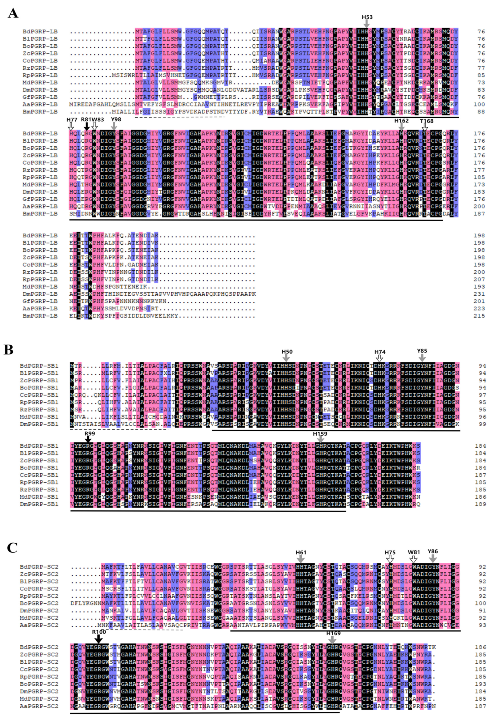
3.1. Sequence Features, Phylogenetic Tree and Functional Domain Prediction of PGRPs in B. dorsalis

A 564 bp nucleotide fragment of BdPGRP-SC2 was obtained by RACE; the GenBank accession number of the fragment of BdPGRP-SC2 is MW538960. The gene encoded a 188-amino acid protein. Both amino acid sequence alignment (Figure 1) and protein prediction results (Supplementary Figure S1) indicated that BdPGRP-LB, BdPGRP-SB1 and BdPGRP-SC2 all have a type 2 amidase domain, which has been shown to have N-acetylmuramoyl-L-alanine amidase activity. Amino acid sequence analysis showed that BdPGRP-LB, BdPGRP-SB1 and BdPGRP-SC2 all have conserved amino acid Arg, which is necessary for the recognition of DAP-type peptidoglycan [32]. BdPGRP-LB has three conserved histidines, H53, H77, H162, one conserved tryptophan, W83, one conserved tyrosine, Y98 and one conserved threonine, T168, which are required for Zn2+ binding and amidase activity (Figure 1A). BdPGRP-SB1 has conserved H50, H74, H159 and Y85 for amidase activity (Figure 1B). BdPGRP-SC2 has conserved H61, H75, H169, W81 and Y86 for Zn2+ binding and amidase activity (Figure 1C). These results indicate that BdPGRP-LB, BdPGRP-SB1 and BdPGRP-SC2 belong to the catalytic PGRPs. A phylogenetic tree was constructed to determine the evolutionary relationships with PGRPs from several other insect species of Diptera (Supplementary Figure S2). The results show that BdPGRP-LB, BdPGRP-SB1 and BdPGRP-SC2 from different species converge in a clade; respectively, this indicates that PGRP-LB, PGRP-SB1 and PGRP-SC2 evolved independently. In addition, all three BdPGRPs of B. dorsalis were closest to those of B. latifrons in evolution of the three genes (Supplementary Figure S2).

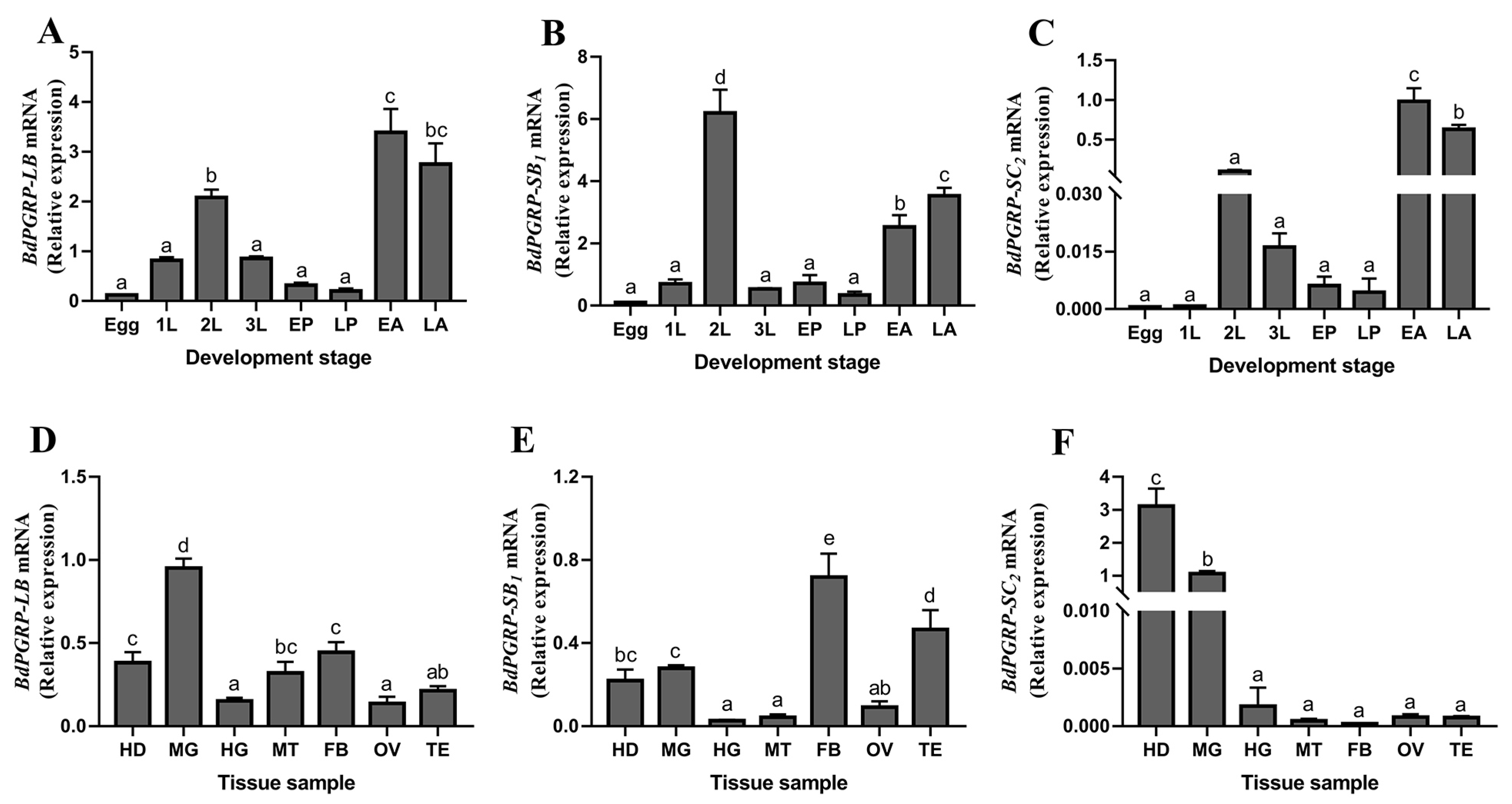
3.2. The Expression Profilings of BdPGRPs in B. dorsalis

qPCR was performed to detect the expression pattern of BdPGRP-LB, BdPGRP-SB1 and BdPGRP-SC2 in different development stages and in various tissues using the primers listed in Table 1. BdPGRPs can be detected across the life stage of B. dorsalis, and the expression levels of BdPGRP-LB, BdPGRP-SB1 and BdPGRP-SC2 were all highly expressed in the adult stage and in the second instar larvae stage (Figure 2A–C), and expressed weakly in the egg, 1st instar larvae, 3rd instar larvae and pupa stages (Figure 2A–C).
Tissue profiles of BdPGRP-SB1, BdPGRP-LB and BdPGRP-SC2 were also analyzed by qRT-PCR. In contrast to the weak expression observed in the hindgut and ovary, the BdPGRP-LB and BdPGRP-SB1 were primarily distributed in the head, midgut and fatbody (Figure 2D,E). BdPGRP-LB was also highly expressed in the Malpighian tubules (Figure 2D), which are vital immune response-related sites [33]. The high expression of BdPGRP-SB1 observed in the testis suggested they may have an important role in reproductive development of B. dorsalis (Figure 2E). Interestingly, the tissue specific expression indicated that BdPGRP-SC2 had higher expression levels in the head and midgut than in other tissues (Figure 2F). The varied expression of BdPGRP-LB, BdPGRP-SB1 and BdPGRP-SC2 in different developmental stage and tissues suggests that BdPGRPs may play distinct roles in B. dorsalis.

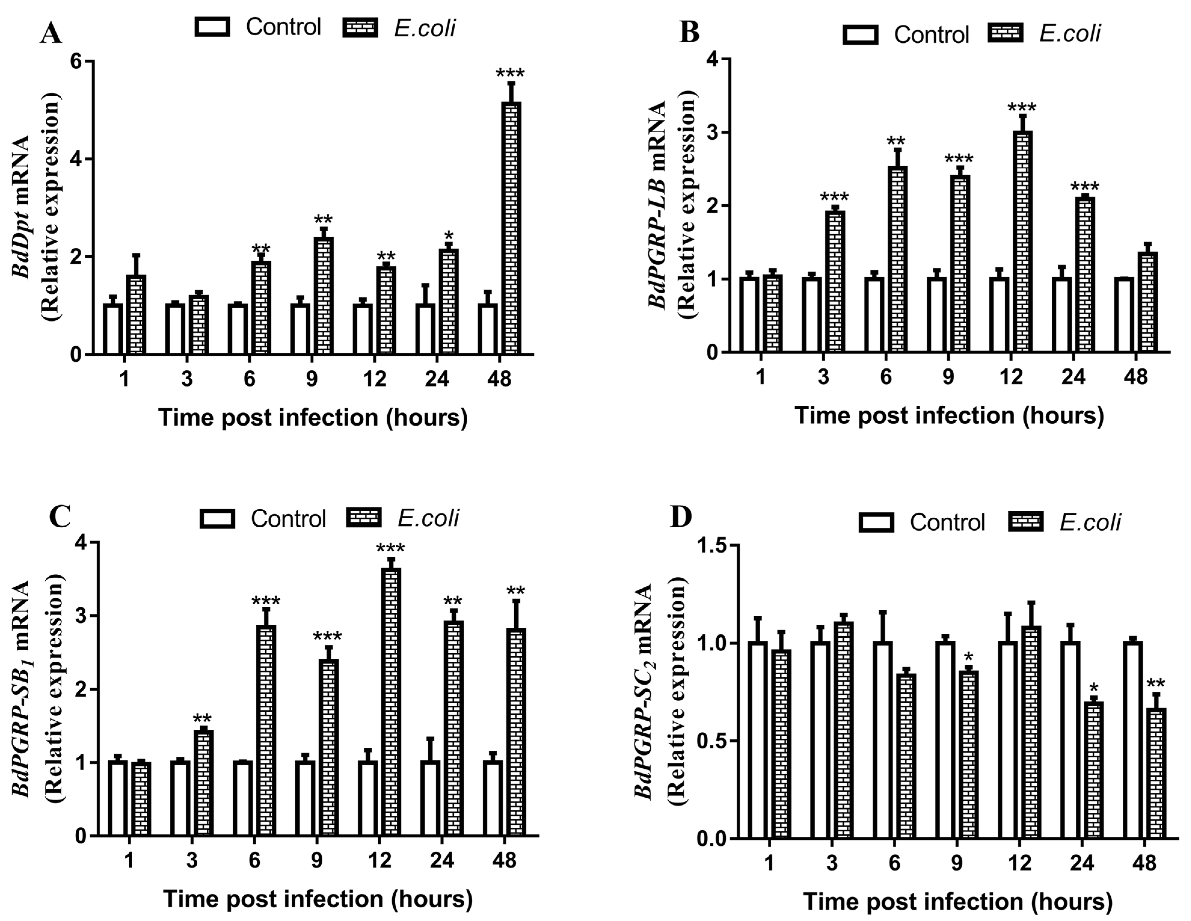
3.3. Responses of BdPGRPs to Systemic Bacterial Infection

To investigate the BdPGRPs response to bacterial challenge, the expression of BdPGRP-LB, BdPGRP-SB1 and BdPGRP-SC2 at 1 h, 3 h, 6 h, 9 h, 12 h, 24 h and 48 h after E. coli thorax inoculation was monitored in whole insects. The results showed that there was a significant increase of the expression of BdPGRP-LB and BdPGRP-SB1 during 3–24h infection, with a 1.90–2.99-fold and 1.40–3.62-fold increase, respectively (Figure 3B,C). Unexpectedly, there was a decrease of BdPGRP-SC2 at 9 h, 24 h and 48 h following inoculated with E. coli, and no response at other times post infection (Figure 3D). We also found an immune response of effector genes of the Imd pathway to E. coli infection; there was a 1.76–5.13-fold increase of the expression of the antimicrobial peptide gene Diptericin at 6h–48h post infection (Figure 3A). The relative expression of other antimicrobial peptide genes including AttacinA, AttacinB, AttacinC and Cecropin were also induced by E. coli infection, thus confirming the strong immunogenic nature of E. coli infection in B. dorsalis. (Supplementary Figure S3A–D). The above results indicate that inoculation with E. coli can immediately activate the immune response of the Imd pathway in B. dorsalis.

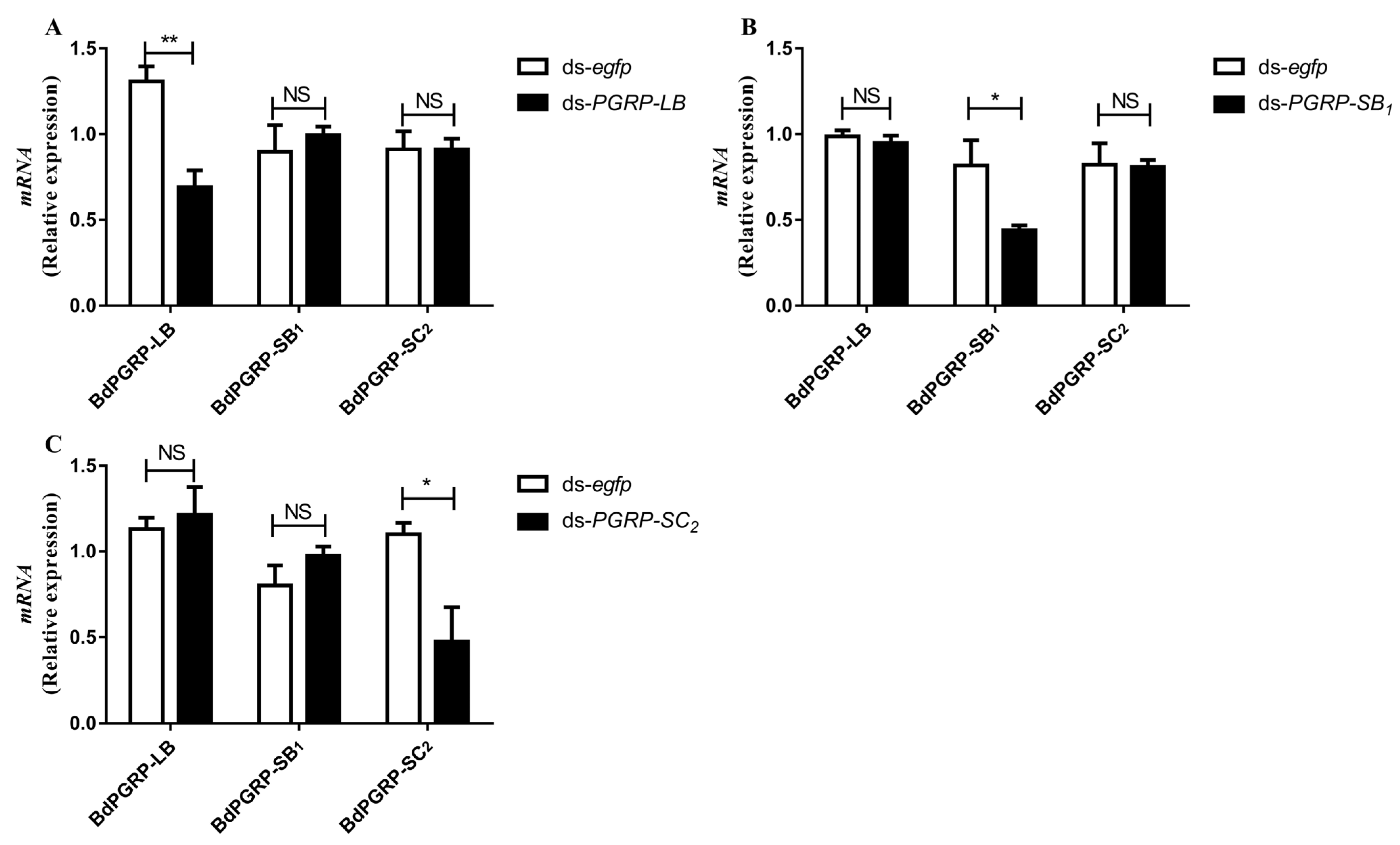
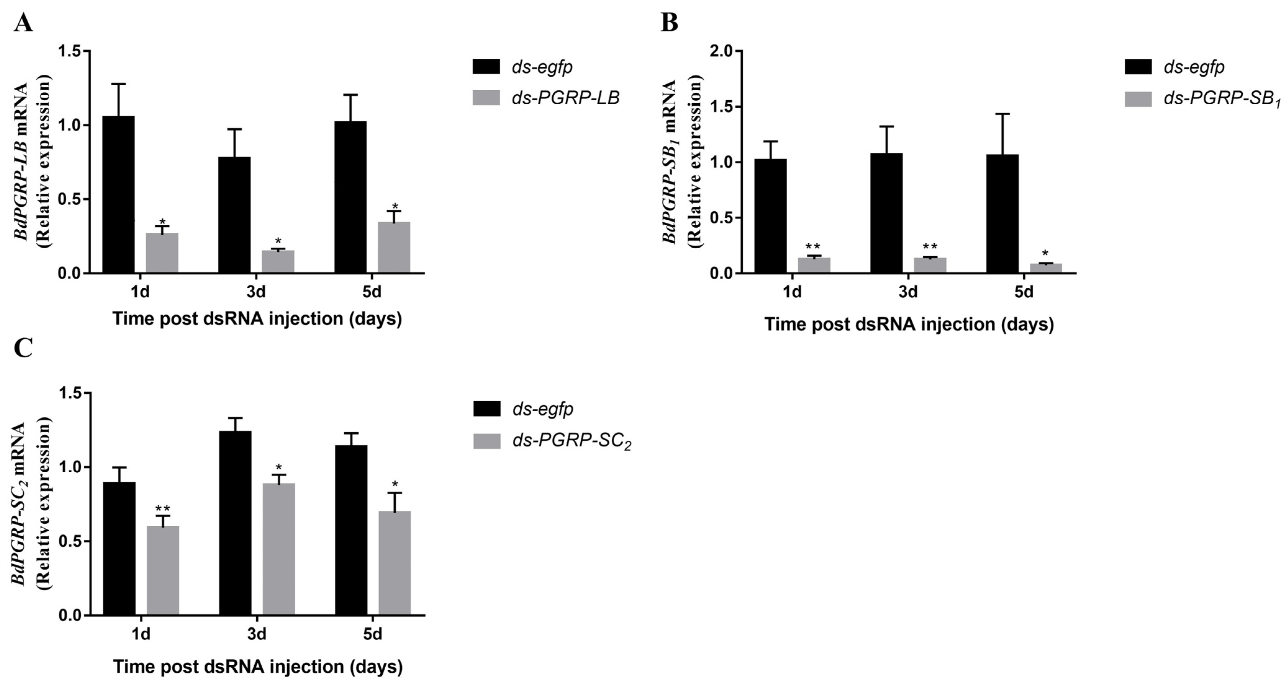
3.4. RNA Interference (RNAi) of BdPGRPs

Based on sequence similarity (Figure 1), numerous off-targets are predicted to occur in RNAi experiments [30]. To test whether potential off-target effects of ds-PGRPs exist, the off-target effect was analysed by qRT-PCR (Figure 4). When BdPGRP-LB RNAi was performed, the relative expression of BdPGRP-SB1 and BdPGRP-SC2 was also detected one day after dsRNA injection. Results showed that the expression of BdPGRP-SB1 and BdPGRP-SC2 were not affected by BdPGRP-LB knock down (Figure 4A). BdPGRP-SB1 RNAi did not affect the relative expression of BdPGRP-LB and BdPGRP-SC2 (Figure 4B), nor did BdPGRP-SC2 RNAi (Figure 4C). These results suggested that there was no off-target effect in the BdPGRPs knockdown experiment in B. dorsalis.
To evaluate the RNAi efficiency of BdPGRPs, we then monitored the expression of transcripts BdPGRP-LB, BdPGRP-SB1 and BdPGRP-SC2 in whole body samples at different days post-dsRNA injection (DPI). The results showed that the expression of BdPGRP-LB was significantly reduced, by 75.3%, 81.4% and 66.9% in the ds-BdPGRP-LB injection group at 1, 3 and 5 DPI when compared to the control ds-egfp group (Figure 5A); the expression of BdPGRP-SB1 was significantly reduced, by 87.4%, 88.2% and 93% in the ds-BdPGRP-SB1 dsRNA injection group at 1, 3 and 5 DPI (Figure 5B); and the expression of BdPGRP-SC2 was significantly reduced, by 33.7%, 28.5% and 39% in the ds-BdPGRP-SC2 dsRNA injection group at 1, 3 and 5 DPI (Figure 5C).

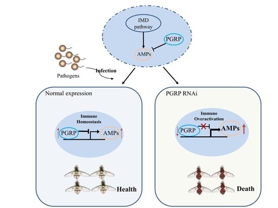
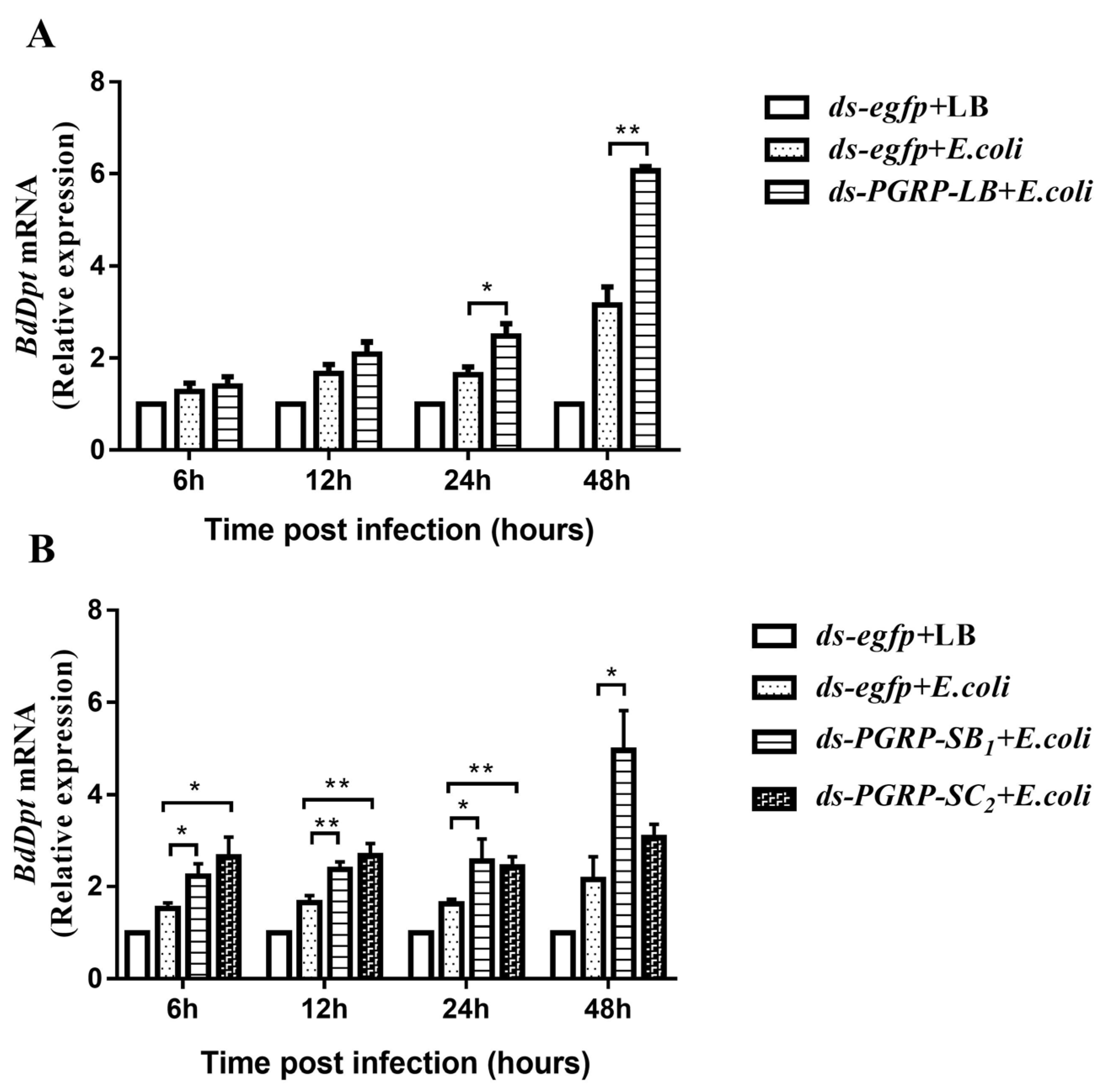
3.5. The Negative Regulatory Roles of BdPGRPs in Imd Pathway

To analyze the potential roles of BdPGRP-LB, BdPGRP-SB1 and BdPGRP-SC2 in the Imd pathway of B. dorsalis, we infected ds-egfp, ds-BdPGRP-LB, ds-BdPGRP-SB1 and ds-BdPGRP-SC2 treated flies with E. coli and measured Dpt transcript levels as a readout for Imd pathway activation. At 24 h and 48 h post E. coli infection, there was a 1.5 and 1.92-fold enhanced expression of Dpt in the infected ds-BdPGRP-LB group, respectively, compared to the infected ds-egfp group (Figure 6A). There was a 1.43–2.3-fold increase in expression of Dpt in the infected ds-BdPGRP-SB1 group compared to the infected ds-egfp group at 6, 12, 24 and 48 h post E. coli infection (Figure 6B). The knockdown of BdPGRP-SC2 led to a 1.74, 1.62 and 1.49-fold enhanced expression of Dpt at 6, 12 and 24 h post E. coli infection, respectively, as compared with infected ds-egfp group (Figure 6B). These results indicate that silencing of either BdPGRP-LB, BdPGRP-SB or BdPGRP-SC2 will induce overactivation of the Imd pathway upon bacterial infection, as all these three BdPGRPs perform negative regulatory roles in regulating AMPs gene expression in the Imd pathway of B. dorsalis.

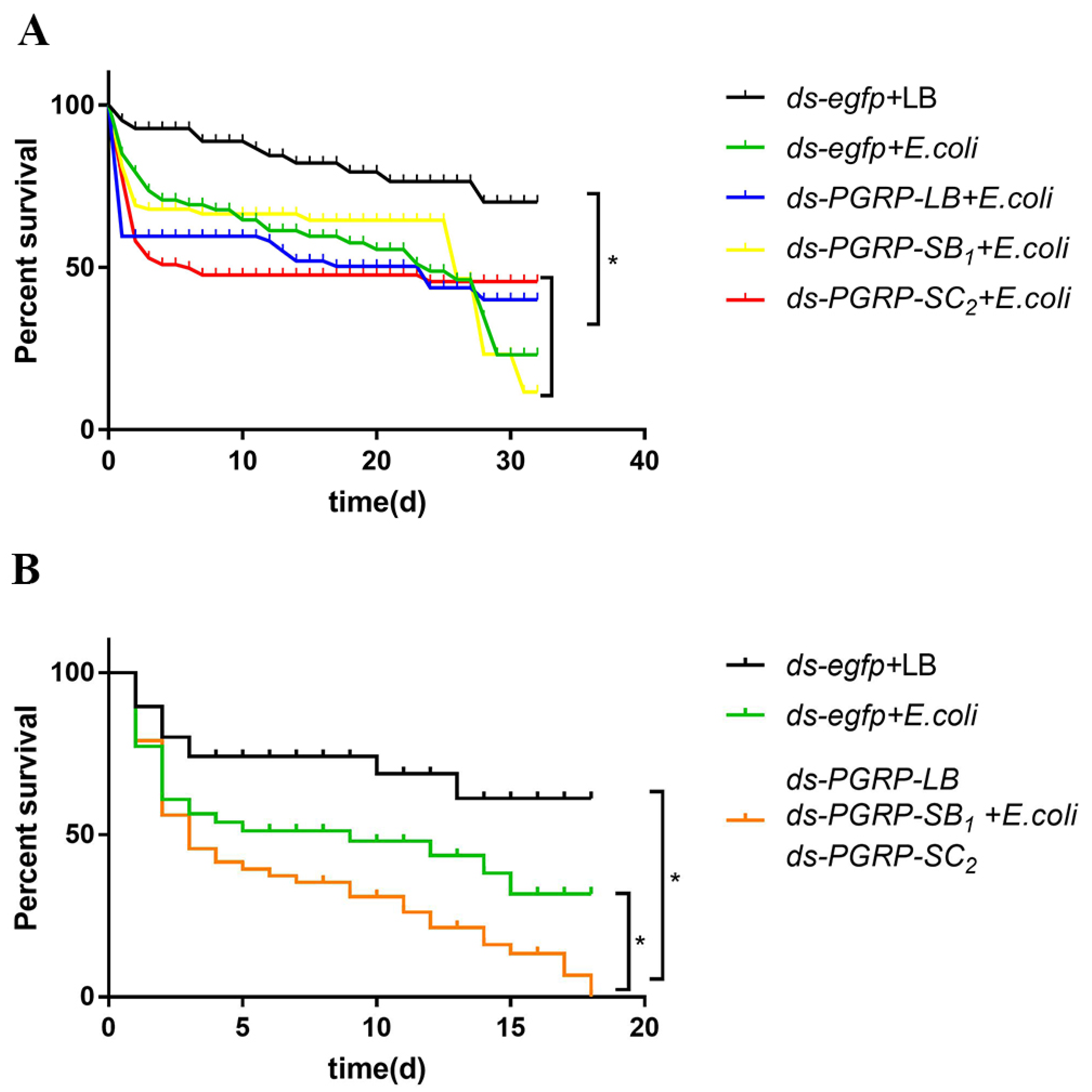
3.6. BdPGRPs RNAi Decreased Flies Survival Rate after Bacterial Challenge

After 24 h post dsRNA injection, flies that were alive in the control and treatment groups were individually challenged with E. coli by inoculation with bacteria in the thorax. From the results, we observed that the survival rate of infected flies was significantly lower than in the control group (Figure 7A). However, when BdPGRP-LB, BdPGRP-SB1 and BdPGRP-SC2 were silenced individually this did not affect survival status when compared with ds-egfp flies upon bacterial challenge (Figure 7A). Considering the functional redundancy of BdPGRPs in B. dorsalis, we knocked down of all three BdPGRPs. The results showed that the survival rate of the ds-BdPGRPs group was significantly reduced compared with the ds-egfp group after E. coli infection (Figure 7B). The median survival of ds-egfp group was nine days, while in the ds-BdPGRPs group it was shortened to three days.

4. Discussion

The PGRP family has been thoroughly studied in the last decade. PGRPs are evolutionally conserved proteins involved in the recognition and degradation of peptidoglycans, a cell wall component of bacteria [34]. PGRPs are involved in many immune processes ranging from initiation to termination of host immune activity; however, most research work has been concentrated in model animals such as in mice and Drosophila [12,23,35,36,37]. Here, we have characterized the immunological role of three PGRP family genes, BdPGRP-LB, BdPGRP-SB1 and BdPGRP-SC2, after inoculation with the Gram-negative bacterium E. coli in B. dorsalis. After applying RNAi methods to knock down BdPGRPs followed by Gram-negative bacterial infection, Dpt, a marker of IMD pathway activation, showed a significant increase compared with the ds-egfp group. The survival rate of the ds-BdPGRPs group was significantly reduced compared to the ds-egfp group after E. coli infection. Our results showed that BdPGRP-LB, BdPGRP-SB1 and BdPGRP-SC2 performed vital negative roles in regulating expression of AMPs in the Imd pathway of B. dorsalis, and maintaining the normal function of these three BdPGRPs is critical to host health when faced with bacterial challenge.
In this study, the results of the protein prediction indicated that BdPGRP-LB, BdPGRP-SB1 and BdPGRP-SC2 all have type 2 amidase domains, which suggests that BdPGRP-LB, BdPGRP-SB1 and BdPGRP-SC2 have amidase activity. PGRPs which have type 2 amidase domains have been confirmed to have important roles in innate immunity, not only in the model specie Drosophila [10] but in other insects such as Tenebrio Molitor [38], Anopheles gambiae [39], and Nilaparvata lugens [40]. This may indicate that the structure and function of PGRPs is highly conserved. All amidase-active PGRPs have a conserved Zn2+-binding site in the peptidoglycan-binding groove, which is also present in bacteriophage type 2 amidases and consists of two histidines, one tyrosine, and one cysteine [10]. The results on expression patterns showed that BdPGRP-LB and BdPGRP-SC2 were very highly expressed in the midgut of adults, while the BdPGRP-SB1 gene was mainly expressed in the fat body. The tissue expression profiling of BdPGRPs is similar with previous reports in other insects [8,10,26,41]. The fat body is the major tissue that generates AMPs to hemolymph in the systemic immune system, and intestinal epithelial cells produce AMPs to inhibit the overgrowth of pathogenic bacteria in gut lumen [6]. In the fat body of insects, characterized immune genes are induced by microbial infection and encode antimicrobial peptides which are then released into the hemolymph to defeat invading pathogens [42]. Updated research reveals that fat body tissues also synthesize and secrete some TEP and TOP peptides, which aid hemocyte phagocytosis [43]. Insects gut continually come in contact with microbiota, which generates a delicate intestinal immune response which must tolerate the presence of gut microbiota and dietary microorganisms while responding to and eliminating potential pathogens [13]. BdPGRP-LB, BdPGRP-SB1 and BdPGRP-SC2 all have highly conserved type 2 amidase domains and are highly expressed in immunocompetent tissues in B. dorsalis, indicating that BdPGRPs probably participate in the immune response of B. dorsalis, as in other insects.
In our study, injury infection with Gram-negative bacteria E. coli induced significantly higher transcript levels of BdPGRP-LB and BdPGRP-SB1 simultaneously. A similar immune expression of PGRP-LB has also been observed in Drosophila, where the expression of PGRP-LB was increased significantly following septic injury with E. carotovora [44]. PGRP-SB1 is strongly induced with injection of bacteria containing DAP-type PGN, which activates the Imd pathway [16]. Unexpectedly, there was a dramatic decrease in BdPGRP-SC2 expression upon systemic infection with E. coli. Similar results were observed in Musca domestica larvae; MdPGRP-SC cannot be induced when challenged by E. coli or S. aureus [26]. Based on the high expression of PGRP-SC in the guts of other insects [8,26], it is possible that PGRP-SC exerts its immune function in the gut. Stress stimulation can induce the transcription factor Foxo to help the host adapt to an adverse situation [45]. Guo et al. (2014) showed that chronic activation of the transcription factor Foxo reduces expression of PGRP-SC2 in Drosophila [15]. Therefore, it is plausible that the injury infection may induce the expression of BdFoxo, then decrease the expression of BdPGRP-SC2; however, further experiments are needed to reveal this phenomenon and its underlying mechanism. The knockdown of any BdPGRP-LB, BdPGRP-SB1 and BdPGRP-SC2 in flies will result in overactivation of the Imd signaling pathway upon bacterial challenge. The roles of BdPGRP-LB and BdPGRP-SC2 in B. dorsalis were consistent with previous findings in D. melanogaster that PGRP-LB and PGRP-SC2 act as important negative regulators of the Imd pathway [13,44,46,47]. After septic injury with the Gram-negative bacterium Erwinia carotovora carotovora 15 (Ecc15), PGRP-LB deletion mutant flies had stronger and more sustained immune response than wild-type flies as measured by the expression of the antibacterial peptide gene Diptericin (Dpt), a readout of the Imd pathway. In contrast with the Zaidman-Rémy et al. (2011) report in D. melanogaster that injection of Gram-negative bacteria Ecc15 did not affect the AMPs expression in PGRP-SB null mutant [16], the silencing of BdPGRP-SB1 in B. dorsalis induced enhanced expression of Dpt compared with the ds-egfp group after bacterial challenge. The discordance may be caused by different insect species having distinct catalytic PGRPs to regulate their systemic immune response. In Drosophila, three isoforms of PGRP-LB have two distinct functions; the PGRP-LBPC isoform is required to control the systemic response in the fat body, while PGRP-LBPA and PGRP-LBPD isoforms show the immune function only in gut [48]. In our results, we found that each of the three BdPGRPs performed its individual negative function in the systemic Imd pathway of B. dorsalis, because the immune phenotype caused by the absence of one of the three BdPGRPs cannot be compensated by the other two. Collectively, our results showed that these three PGRP family genes act as negative regulators in the systemic immune response of B. dorsalis by dampening the activation of the Imd pathway.
A tight balance between initiation and resolution in the control of inflammation is very important in animals, as both the absence and overactivity of immune response are harmful to the host [46]. Our results showed that the survival rate of the ds-BdPGRPs group was significant reduced compared with the ds-egfp group. However, the death events were mostly observed shortly after infection and mid-to-late post-infection. Death in the short term may be caused by an overreaction of the immune system [13], while death in the middle and late stages may be caused by excessive energy consumption in response to infection [49]. Noncatalytic PGRPs are crucial for the sensing of bacteria in insects such as in Drosophila, and catalytic PGRPs play a vital role in hydrolyzing peptidoglycan by cleaving the amide bond [13]. The bacterial infection induced the expression of PGRP-LB, PGRP-SB1 and PGRP-SC2 to degrade PGN and repress the activation of PGRP-LC, which reportedly is the major receptor of the Imd pathway [22], in order to ensure that the immune response is at an appropriate level. This negative regulation integrates into the sensitive immune regulation mechanism of insects, which keeps pathogenic bacteria below the level where they can cause harm and ensures that the host will not be harmed by an overactive immune response.

Supplementary Materials

The following supporting information can be downloaded at: https://www.mdpi.com/article/10.3390/cells11010152/s1, Figure S1: Prediction of BdPGRPs’ functional domains; Figure S2: Phylogenetic tree of peptidoglycan recognition proteins of B. dorsalis and other insects; Figure S3: Expression levels of AMPs in Imd pathway after E. coli challenge; Table S1: Primers used in supplementary experiments.

Author Contributions

H.Z., P.Z. and Z.Y. conceived and designed the project. P.Z., Z.Y. and S.B. performed experiments, and Z.Y. analysed data. Z.Y. and P.Z. made the graphs. P.Z. and S.B. reared the flies. H.Z., P.Z. and Z.Y. wrote the manuscript. All authors have read and agreed to the published version of the manuscript.

Funding

This work was supported by the National Natural Science Foundation of China (Nos. 31872931, 31572008 and 31801744), China Agriculture Research System of MOF and MARA (No. CARS-26).

Institutional Review Board Statement

Not applicable.

Informed Consent Statement

Not applicable.

Data Availability Statement

The data presented in this study are available within this artivle.

Conflicts of Interest

The authors declare no conflict of interest.

References

  1. Tzou, P.; De Gregorio, E.; Lemaitre, B. How Drosophila combats microbial infection: A model to study innate immunity and host-pathogen interactions. Curr. Opin. Microbiol. 2002, 5, 102–110. [Google Scholar] [CrossRef]
  2. Janeway, C.; Medzhitov, R. Innate immune recognition. Annu. Rev. Immunol. 2002, 20, 197–216. [Google Scholar] [CrossRef] [Green Version]
  3. Guan, R.; Mariuzza, R.A. Peptidoglycan recognition proteins of the innate immune system. Trends Microbiol. 2007, 15, 127–134. [Google Scholar] [CrossRef]
  4. Royet, J.; Dziarski, R. Peptidoglycan recognition proteins: Pleiotropic sensors and effectors of antimicrobial defences. Nat. Rev. Microbiol. 2007, 5, 264–277. [Google Scholar] [CrossRef] [PubMed]
  5. Mengin-Lecreulx, D.; Lemaitre, B. Structure and metabolism of peptidoglycan and molecular requirements allowing its detection by the Drosophila innate immune system. J. Endotoxin Res. 2005, 11, 105–111. [Google Scholar] [CrossRef] [Green Version]
  6. Lemaitre, B.; Hoffmann, J. The Host Defense of Drosophila melanogaster. Annu. Rev. Immunol. 2007, 25, 697–743. [Google Scholar] [CrossRef] [PubMed] [Green Version]
  7. Yoshida, H.; Kinoshita, K.; Ashida, M. Purification of a peptidoglycan recognition protein from hemolymph of the silkworm, Bombyx mori. J. Biol. Chem. 1996, 271, 13854–13860. [Google Scholar] [CrossRef] [PubMed] [Green Version]
  8. Werner, T.; Liu, G.; Kang, D.; Ekengren, S.; Steiner, H.; Hultmark, D. A family of peptidoglycan recognition proteins in the fruit fly Drosophila melanogaster. Proc. Natl. Acad. Sci. USA 2000, 97, 13772–13777. [Google Scholar] [CrossRef] [PubMed] [Green Version]
  9. Christophides, G.; Zdobnov, E.; Barillas-Mury, C.; Birney, E.; Blandin, S.; Blass, C.; Brey, P.; Collins, F.; Danielli, A.; Dimopoulos, G.; et al. Immunity-related genes and gene families in Anopheles gambiae. Science 2002, 298, 159–165. [Google Scholar] [CrossRef] [PubMed] [Green Version]
  10. Dziarski, R.; Gupta, D. The peptidoglycan recognition proteins (PGRPs). Genome Biol. 2006, 7, 232. [Google Scholar] [CrossRef] [Green Version]
  11. Wang, S.; Beerntsen, B.T. Insights into the different functions of multiple peptidoglycan recognition proteins in the immune response against bacteria in the mosquito, Armigeres subalbatus. Insect Biochem. Mol. Biol. 2013, 43, 533–543. [Google Scholar] [CrossRef] [PubMed]
  12. Buchon, N.; Broderick, N.A.; Lemaitre, B. Gut homeostasis in a microbial world: Insights from Drosophila melanogaster. Nat. Rev. Microbiol. 2013, 11, 615–626. [Google Scholar] [CrossRef] [PubMed] [Green Version]
  13. Paredes, J.C.; Welchman, D.P.; Poidevin, M.; Lemaitre, B. Negative Regulation by Amidase PGRPs Shapes the Drosophila Antibacterial Response and Protects the Fly from Innocuous Infection. Immunity. 2011, 35, 770–779. [Google Scholar] [CrossRef] [Green Version]
  14. Mellroth, P.; Karlsson, J.; Steiner, H. A scavenger function for a Drosophila peptidoglycan recognition protein. J. Biol. Chem. 2003, 278, 7059–7064. [Google Scholar] [CrossRef] [PubMed] [Green Version]
  15. Guo, L.; Karpac, J.; Tran, S.L.; Jasper, H. PGRP-SC2 Promotes Gut Immune Homeostasis to Limit Commensal Dysbiosis and Extend Lifespan. Cell 2014, 156, 109–122. [Google Scholar] [CrossRef] [Green Version]
  16. Zaidman-Rémy, A.; Poidevin, M.; Hervé, M.; Welchman, D.P.; Paredes, J.C.; Fahlander, C.; Steiner, H.; Mengin-Lecreulx, D.; Lemaitre, B. Drosophila Immunity: Analysis of PGRP-SB1 Expression, Enzymatic Activity and Function. PLoS ONE 2011, 6, e17231. [Google Scholar] [CrossRef] [PubMed]
  17. Li, X.; Zhang, M.; Zhang, H. RNA interference of four genes in adult Bactrocera dorsalis by feeding their dsRNAs. PLoS ONE 2011, 6, e17788. [Google Scholar] [CrossRef]
  18. Clarke, A.R.; Armstrong, K.F.; Carmichael, A.E.; Milne, J.R.; Raghu, S.; Roderick, G.K.; Yeates, D.K. Invasive phytophagous pests arising through a recent tropical evolutionary radiation: The Bactrocera dorsalis Complex of Fruit Flies. Annu. Rev. Entomol. 2005, 50, 293–319. [Google Scholar] [CrossRef] [Green Version]
  19. Li, Q.; Dong, X.; Zheng, W.; Zhang, H. The PLA2 gene mediates the humoral immune responses in Bactrocera dorsalis (Hendel). Dev. Comp. Immunol. 2017, 67, 293–299. [Google Scholar] [CrossRef]
  20. Dong, X.; Li, Q.; Zhang, H. The noa gene is functionally linked to the activation of the Toll/Imd signaling pathways in Bactrocera dorsalis (Hendel). Dev. Comp. Immunol. 2016, 55, 233–240. [Google Scholar] [CrossRef]
  21. Yao, Z.; Wang, A.; Li, Y.; Cai, Z.; Lemaitre, B.; Zhang, H. The dual oxidase gene BdDuox regulates the intestinal bacterial community homeostasis of Bactrocera dorsalis. ISME J. 2016, 10, 1037–1050. [Google Scholar] [CrossRef] [Green Version]
  22. Gottar, M.; Gobert, V.; Michel, T.; Belvin, M.; Duyk, G.; Hoffmann, J.; Ferrandon, D.; Royet, J. The Drosophila immune response against Gram-negative bacteria is mediated by a peptidoglycan recognition protein. Nature 2002, 416, 640–644. [Google Scholar] [CrossRef]
  23. Bischoff, V.; Vignal, C.; Boneca, I.G.; Michel, T.; Hoffmann, J.A.; Royet, J. Function of the Drosophila pattern-recognition receptor PGRP-SD in the detection of Gram-positive bacteria. Nat. Immunol. 2004, 5, 1175–1180. [Google Scholar] [CrossRef] [PubMed]
  24. Kaneko, T.; Yano, T.; Aggarwal, K.; Lim, J.H.; Ueda, K.; Oshima, Y.; Peach, C.; Erturk-Hasdemir, D.; Goldman, W.E.; Oh, B.H.; et al. PGRP-LC and PGRP-LE have essential yet distinct functions in the Drosophila immune response to monomeric DAP-type peptidoglycan. Nat. Immunol. 2006, 7, 715–723. [Google Scholar] [CrossRef] [PubMed]
  25. Kurata, S. Extracellular and intracellular pathogen recognition by Drosophila PGRP-LE and PGRP-LC. Int. Immunol. 2010, 22, 143–148. [Google Scholar] [CrossRef] [PubMed] [Green Version]
  26. Gao, Y.; Tang, T.; Gu, J.; Sun, L.; Gao, X.; Ma, X.; Wang, X.; Liu, F.; Wang, J. Downregulation of the Musca domestica peptidoglycan recognition protein SC (PGRP-SC) leads to overexpression of antimicrobial peptides and tardy pupation. Mol. Immunol. 2015, 67, 465–474. [Google Scholar] [CrossRef]
  27. Dawadi, B.; Wang, X.; Xiao, R.; Muhammad, A.; Hou, Y.; Shi, Z. PGRP-LB homolog acts as a negative modulator of immunity in maintaining the gut-microbe symbiosis of red palm weevil, Rhynchophorus ferrugineus Olivier. Dev. Comp. Immunol. 2018, 86, 65–77. [Google Scholar] [CrossRef]
  28. Maire, J.; Vincent-Monégat, C.; Balmand, S.; Vallier, A.; Hervé, M.; Masson, F.; Parisot, N.; Vigneron, A.; Anselme, C.; Perrin, J.; et al. Weevil pgrp-lb prevents endosymbiont TCT dissemination and chronic host systemic immune activation. Proc. Natl. Acad. Sci. USA 2019, 116, 5623–5632. [Google Scholar] [CrossRef] [PubMed] [Green Version]
  29. Leulier, F.; Parquet, C.; Pili-Floury, S.; Ryu, J.; Caroff, M.; Lee, W.; Mengin-Lecreulx, D.; Lemaitre, B. The Drosophila immune system detects bacteria through specific peptidoglycan recognition. Nat. Immunol. 2003, 4, 478–484. [Google Scholar] [CrossRef]
  30. Seinen, E.; Burgerhof, J.G.M.; Jansen, R.C.; Sibon, O.C.M. RNAi-induced off-target effects in Drosophila melanogaster: Frequencies and solutions. Brief. Funct. Genom. 2011, 10, 206–214. [Google Scholar] [CrossRef] [Green Version]
  31. Livak, K.J.; Schmittgen, T.D. Analysis of relative gene expression data using real-time quantitative PCR and the 2(-Delta Delta C(T)) Method. Methods 2001, 25, 402–408. [Google Scholar] [CrossRef]
  32. Lim, J.H.; Kim, M.S.; Kim, H.E.; Yano, T.; Oshima, Y.; Aggarwal, K.; Goldman, W.E.; Silverman, N.; Kurata, S.; Oh, B.H. Structural Basis for Preferential Recognition of Diaminopimelic Acid-type Peptidoglycan by a Subset of Peptidoglycan Recognition Proteins. J. Biol. Chem. 2006, 281, 8286–8295. [Google Scholar] [CrossRef] [Green Version]
  33. Beyenbach, K.; Skaer, H.; Dow, J. The developmental, molecular, and transport biology of Malpighian tubules. Annu. Rev. Entomol. 2010, 55, 351–374. [Google Scholar] [CrossRef]
  34. Kurata, S. Recognition of infectious non-self and activation of immune responses by peptidoglycan recognition protein (PGRP)-family members in Drosophila. Dev. Comp. Immunol. 2004, 28, 89–95. [Google Scholar] [CrossRef]
  35. Ferrandon, D.; Imler, J.; Hetru, C.; Hoffmann, J. The Drosophila systemic immune response: Sensing and signalling during bacterial and fungal infections. Nat. Rev. Immunol. 2007, 7, 862–874. [Google Scholar] [CrossRef] [PubMed]
  36. Saha, S.; Qi, J.; Wang, S.; Wang, M.; Li, X.; Kim, Y.G.; Núñez, G.; Gupta, D.; Dziarski, R. PGLYRP-2 and Nod2 are both required for peptidoglycan-induced arthritis and local inflammation. Cell Host Microbe 2009, 5, 137–150. [Google Scholar] [CrossRef] [PubMed] [Green Version]
  37. Bosco-Drayon, V.; Poidevin, M.; Boneca, I.G.; Narbonne-Reveau, K.; Royet, J.; Charroux, B. Peptidoglycan Sensing by the Receptor PGRP-LE in the Drosophila Gut Induces Immune Responses to Infectious Bacteria and Tolerance to Microbiota. Cell Host Microbe 2012, 12, 153–165. [Google Scholar] [CrossRef] [Green Version]
  38. Tindwa, H.; Patnaik, B.B.; Kim, D.H.; Mun, S.; Jo, Y.H.; Lee, B.L.; Lee, Y.S.; Kim, N.J.; Han, Y.S. Cloning, characterization and effect of TmPGRP-LE gene silencing on survival of Tenebrio molitor against Listeria monocytogenes infection. Int. J. Mol. Sci. 2013, 14, 22462–22482. [Google Scholar] [CrossRef]
  39. Mendes, C.; Felix, R.; Sousa, A.M.; Lamego, J.; Charlwood, D.; do Rosário, V.E.; Pinto, J.; Silveira, H. Molecular evolution of the three short PGRPs of the malaria vectors Anopheles gambiae and Anopheles arabiensis in East Africa. BMC Evol. Biol. 2010, 10, 9. [Google Scholar] [CrossRef] [PubMed] [Green Version]
  40. Bao, Y.Y.; Qu, L.Y.; Zhao, D.; Chen, L.B.; Jin, H.Y.; Xu, L.M.; Cheng, J.A.; Zhang, C.X. The genome- and transcriptome-wide analysis of innate immunity in the brown planthopper, Nilaparvata lugens. BMC Genom. 2013, 14, 160. [Google Scholar] [CrossRef] [Green Version]
  41. Wang, J.; Aksoy, S. PGRP-LB is a maternally transmitted immune milk protein that influences symbiosis and parasitism in tsetse’s offspring. Proc. Natl. Acad. Sci. USA 2012, 109, 10552–10557. [Google Scholar] [CrossRef] [Green Version]
  42. Tsakas, S.; Marmaras, V.J. Insect immunity and its signalling: An overview. Invertebr. Surviv. J. 2010, 7, 228–238. [Google Scholar]
  43. Li, S.; Yu, X.; Feng, Q. Fat Body Biology in the Last Decade. Annu. Rev. Entomol. 2019, 64, 315–333. [Google Scholar] [CrossRef] [PubMed]
  44. Zaidman-Rémy, A.; Hervé, M.; Poidevin, M.; Pili-Floury, S.; Kim, M.-S.; Blanot, D.; Oh, B.-H.; Ueda, R.; Mengin-Lecreulx, D.; Lemaitre, B. The Drosophila Amidase PGRP-LB Modulates the Immune Response to Bacterial Infection. Immunity 2006, 24, 463–473. [Google Scholar] [CrossRef] [PubMed] [Green Version]
  45. Eijkelenboom, A.; Burgering, B.M. FOXOs: Signalling integrators for homeostasis maintenance. Nature Reviews Molecular Cell Biology 2013, 14, 83–97. [Google Scholar] [CrossRef] [PubMed]
  46. Schneider, D.; Bischoff, V.; Vignal, C.; Duvic, B.; Boneca, I.G.; Hoffmann, J.A.; Royet, J. Downregulation of the Drosophila Immune Response by Peptidoglycan-Recognition Proteins SC1 and SC2. PLoS Pathog. 2006, 2, e14. [Google Scholar]
  47. Charroux, B.; Capo, F.; Kurz, C.L.; Peslier, S.; Chaduli, D.; Viallat-Lieutaud, A.; Royet, J. Cytosolic and Secreted Peptidoglycan-Degrading Enzymes in Drosophila Respectively Control Local and Systemic Immune Responses to Microbiota. Cell Host Microbe 2018, 23, 215–228. [Google Scholar] [CrossRef] [Green Version]
  48. Dziarski, R.; Gupta, D. A balancing act: PGRPs preserve and protect. Cell Host Microbe 2018, 23, 149–151. [Google Scholar] [CrossRef] [Green Version]
  49. Dolezal, T.; Krejcova, G.; Bajgar, A.; Nedbalova, P.; Strasser, P. Molecular regulations of metabolism during immune response in insects. Insect Biochem. Mol. Biol. 2019, 109, 31–42. [Google Scholar] [CrossRef]
Figure 1. Amino acid sequence alignment of BdPGRPs with that of homologous genes in other insect species. (A) Multiple alignments of PGRP-LB. BdPGRP-LB was aligned with Bactrocera latifrons PGRP-LB (XP_018789449.1), Bactrocera oleae PGRP-LB (XP_014091181.2), Zeugodacus cucurbitae PGRP-LB (XP_011197144.1), Ceratitis capitate PGRP-LB (XP_004518089.1), Rhagoletis zephyria PGRP-LB (XP_017470705.1), Rhagoletis pomonella PGRP-LB (XP_036322481.1), Aedes aegypti PGRP-LB (XP_021709443.1), Drosophila melanogaster PGRP-LB (NP_731575.1), Bombyx mori PGRP-LB (XP_012548100.1), Musca domestica PGRP-LB (XP_005180889.1), and Glossina fuscipes PGRP-LB (ACI22620.1). (B) Multiple alignments of PGRP-SB. BdPGRP-SB1 was aligned with B. latifrons PGRP-SB (XP_018789286.1), B. oleae PGRP-SB (XP_014099773.1), Z. cucurbitae PGRP-SB (XP_011181375.1), C. capitate PGRP-SB (XP_004537949.1), R. zephyria PGRP-SB (XP_017486043.1), R. pomonella PGRP-SB (XP_036336342.1), D. melanogaster PGRP-SB (CAD89135.1), M. domestica PGRP-SB (NP_001295929.1), and B. mori PGRP-SB (XP_004929843.1). (C) Multiple alignments of PGRP-SC2. BdPGRP-SC2 was aligned with B. latifrons PGRP-SC2 (XP_018798904.1), B. oleae PGRP-SC2 (XP_014085196.2), C. capitate PGRP-SC2 (XP_004520319.1), Z. cucurbitae PGRP-SC2 (XP_011180165.1), R. pomonella PGRP-SC2 (XP_036334551.1), M. domestica PGRP-SC2 (XP_005184140.3), D. melanogaster PGRP-SC2 (CAD89184.1), A. aegypti PGRP-SC2 (XP_011492940.1), and B. mori PGRP-SC2 (XP_004929814.1). The identical amino acids are shown against a black background; 75% conserved amino acids are shown against a pink background; 50% conserved amino acids are shown against a blue background. The signal peptides are indicated by dashed lines. The amidase domains are indicated by solid lines. Black arrows indicate the amino acid residues required for the recognition of DAP-type peptidoglycan. Grey arrows indicate the amino acid residues required for Zn2+ binding. White arrows indicate the amino acid residues required for amidase activity.
Cells 11 00152 g001
Figure 2. Expression profiles of BdPGRPs in B.dorsalis. (A) Relative expression of BdPGRP-LB at different development stages. (B) Relative expression of BdPGRP-SB1 at different development stages. (C) Relative expression of BdPGRP-SC2 at different development stages. (D) Relative expression of BdPGRP-LB from different tissue samples. (E) Relative expression of BdPGRP-SB1 from different tissue samples. (F) Relative expression of BdPGRP-SC2 from different tissue samples. B. dorsalis was collected at various developmental stages: 1 L, 1st instar larvae; 2 L, 2nd instar larvae; 3 L, 3rd instar larvae; EP, early pupal stage; LP, late pupal stage; EA, newly emergence adults; LA, late adult stage. Different adult tissues were collected: HD, head; MG, midgut; HG, hindgut; MT, Malpighian tube; FB, fatbody; OV, ovary; TE, testis. Multiple comparisons were carried out with one-way analysis of variance (ANOVA) and Turkey’s test in SPSS 16.0. Different lower-case letters indicate a significant difference at the level of p < 0.05 and a confidence interval of 95%. The relative gene expression data were analyzed using a 2−ΔΔCT method and the data were normalized to reference gene Rpl32.
Cells 11 00152 g002
Figure 3. Responses of Dpt and BdPGRPs to opportunistic pathogen E. coli challenges. Relative expression of Dpt (A), BdPGRP-LB (B), BdPGRP-SB1 (C), and BdPGRP-SC2 (D) after infection with E. coli at different time points, respectively. The data are expressed as mean ± SEM and the mean refers to the average of four biological replicates for each sample. Statistical analysis was based on Student’s t-test. * p < 0.05; ** p < 0.01; *** p < 0.001. The relative gene expression data were analyzed using a 2−ΔΔCT method and the data were normalized to reference gene Rpl32.
Cells 11 00152 g003
Figure 4. Off-target detection after dsRNA injection. (A) Influence of silencing BdPGRP-LB on expression of BdPGRP-SB1 and BdPGRP-SC2. (B) Influence of silencing BdPGRP- SB1 on expression of BdPGRP-LB and BdPGRP-SC2. (C) Influence of silencing BdPGRP- SC2 on expression of BdPGRP-LB and BdPGRP-SB1. All error bars represent the SEM of the mean of three independent biological replicates. Statistical analysis was based on Student’s t-test. * p < 0.05; ** p < 0.01; NS, no significant difference; p > 0.05. The relative gene expression data were analyzed using a 2−ΔΔCT method and the data were normalized to reference gene Rpl32.
Cells 11 00152 g004
Figure 5. RNA interference efficiency of BdPGRPs. Relative expression of PGRP-LB (A), PGRP-SB1 (B), and PGRP-SC2 (C) after dsRNA injection at different time points with whole body samples. All error bars represent the SEM of the mean of three independent biological replicates. Statistical analysis was based on Student’s t-test. * p < 0.05; ** p < 0.01. The relative gene expression data were analyzed using a 2−ΔΔCT method and the data were normalized to reference gene Rpl32.
Cells 11 00152 g005
Figure 6. Antimicrobial peptide gene expression in BdPGRPs RNAi flies after bacterial challenges. (A,B) Injury infection with E. coli induced a higher Diptericin (Dpt) expression in BdPGRPs RNAi flies than in the ds-egfp dsRNA injection flies. The data are expressed as the mean ± SEM, and the mean refers to the average of at least three replicates for each sample. Statistical analysis was based on Student’s t-test. * p < 0.05; ** p < 0.01. The relative gene expression data were analyzed using a 2−ΔΔCT method and the data were normalized to reference gene Rpl32.
Cells 11 00152 g006
Figure 7. Survival rate of B. dorsalis after BdPGRPs RNAi followed by E. coli infection. (A) Three BdPGRPs were knocked down separately. (B) Three BdPGRPs were knocked down at the same time. Statistical analysis was based on Log-rank (Mantel–Cox) test (* p < 0.05).
Cells 11 00152 g007
Table 1. Primers used in RT-PCR and qRT-PCR.
PrimerSequence (from 5′ to 3′)Purpose
PGRP-SC2 5′RACE outerCCTTAGCGGCAGCAATCTRACE
PGRP-SC2 5′RACE innerCCACGACCCTCATACACTRACE
PGRP-SC2 3′RACE outerGCAAGTGTATGAGGGTCGRACE
PGRP-SC2 3′RACE innerTTACTGCTCCACCCAAACRACE
QPGRP-LB FGCGTGGCTGGAATGACATTGqRT-PCR
QPGRP-LB RCGGTCATTGTATTTGGGCGCqRT-PCR
QPGRP-SB FTGGCATTGTCTTCATCGGCAqRT-PCR
QPGRP-SB RCAGATAACCCTTTTGCACCGCqRT-PCR
QPGRP-SC2 FGGGTCGTGGTTGGAGTACAGqRT-PCR
QPGRP-SC2 RGATCTGAGCGGCTGTTGGAAqRT-PCR
QRpL32 FCCCGTCATATGCTGCCAACTqRT-PCR
QRpL32 RGCGCGCTCAACAATTTCCTTqRT-PCR
QDiptericin FGCATAGATTTGAGCCTTGACACACqRT-PCR
QDiptericin RGCCATATCGTCCGCCCAAATqRT-PCR
PGRP-LB T7FGGATCCTAATACGACTCACTATAGGATGCCCAGCGCCTGTTACdsRNA synthesis
PGRP-LB T7RGGATCCTAATACGACTCACTATAGGTGCGGCCACGTCGTAATCdsRNA synthesis
PGRP-SB T7 FGGATCCTAATACGACTCACTATAGGTGTTTTGCGCTCAGGATCCAdsRNA synthesis
PGRP-SB T7RGGATCCTAATACGACTCACTATAGGTGGCCCAGCAGTGTGTAATTdsRNA synthesis
PGRP-SC2 T7 FGGATCCTAATACGACTCACTATAGGGGCTTTCAAGACTTTCCTCdsRNA synthesis
PGRP-SC2 T7RGGATCCTAATACGACTCACTATAGGAACCACGACCCTCATACACdsRNA synthesis
EGFP T7LGGATCCTAATACGACTCACTATAGGACGTAAACGGCCACAAGTTCdsRNA synthesis
EGFP T7RGGATCCTAATACGACTCACTATAGGAAGTCGTGCTGCTTAATGTGdsRNA synthesis
Primers starting with Q were used for qRT-PCR; the underlined sections indicate T7 polymerase promoter.
Publisher’s Note: MDPI stays neutral with regard to jurisdictional claims in published maps and institutional affiliations.

