Review

Molecular Markers and Targets in Melanoma

1
Department of Pathology, Hospital Clínic of Barcelona, University of Barcelona, Villarroel 170, 08036 Barcelona, Spain
2
August Pi i Sunyer Biomedical Research Institute (IDIBAPS), Rosselló 149, 08036 Barcelona, Spain
3
Department of Medical Oncology, Hospital Clínic of Barcelona, University of Barcelona, Villarroel 170, 08036 Barcelona, Spain
4
Department of Medical Oncology, Althaia Xarxa Assistencial Universitària de Manresa, Dr. Joan Soler, 1–3, 08243 Manresa, Spain
*
Author to whom correspondence should be addressed.
Cells 2021, 10(9), 2320; https://doi.org/10.3390/cells10092320
Submission received: 28 May 2021 / Revised: 28 August 2021 / Accepted: 1 September 2021 / Published: 5 September 2021

Abstract

:
Melanoma develops as a result of several genetic alterations, with UV radiation often acting as a mutagenic risk factor. Deep knowledge of the molecular signaling pathways of different types of melanoma allows better characterization and provides tools for the development of therapies based on the intervention of signals promoted by these cascades. The latest World Health Organization classification acknowledged the specific genetic drivers leading to melanoma and classifies melanocytic lesions into nine distinct categories according to the associate cumulative sun damage (CSD), which correlates with the molecular alterations of tumors. The largest groups are melanomas associated with low-CSD or superficial spreading melanomas, characterized by frequent presentation of the BRAFV600 mutation. High-CSD melanomas include lentigo maligna type and desmoplastic melanomas, which often have a high mutation burden and can harbor NRAS, BRAFnon-V600E, or NF1 mutations. Non-CSD-associated melanomas encompass acral and mucosal melanomas that usually do not show BRAF, NRAS, or NF1 mutations (triple wild-type), but in a subset may have KIT or SF3B1 mutations. To improve survival, these driver alterations can be treated with targeted therapy achieving significant antitumor activity. In recent years, relevant improvement in the prognosis and survival of patients with melanoma has been achieved, since the introduction of BRAF/MEK tyrosine kinase inhibitors and immune checkpoint inhibitors. In this review, we describe the current knowledge of molecular pathways and discuss current and potential therapeutic targets in melanoma, focusing on their clinical relevance of development.

Graphical Abstract

1. Introduction

1.1. Epidemiology

Melanoma is the most aggressive and deadly skin cancer. Its incidence has increased steadily in the last decades, especially in the Caucasian population, posing a heightened challenge to the global healthcare system [1,2]. Relevant geographical variations exist, depending on the clinical phenotype, the genetic background of individuals, and the extent of ultraviolet (UV) radiation exposure [3]. Currently, it is one of the most frequent cancers in fair-skinned people, especially those with blond or red hair, who have light-colored eyes. Unlike other solid tumors, melanoma mainly affects young and middle-aged people [4]. Melanoma-related mortality has increased in parallel with the increase in the incidence rate over the years, reaching a mortality rate of one in four deaths [5]. Nevertheless, the therapeutic landscape of unresectable stage III and IV melanoma has been revolutionized by immunotherapies and targeted therapies. Both strategies have shown markedly improved survival compared with the use of chemotherapy (ChT) regimens [6]. Melanoma mortality has decreased significantly since the US Food and Drug Administration (FDA) approved ipilimumab in 2011, the first immune checkpoint inhibitor (ICI) to improve survival in the advanced setting [7,8], and vemurafenib, a v-raf murine sarcoma viral oncogene homolog B1 (BRAF) tyrosine kinase inhibitor, first in class [9,10].

1.2. Risk Factors

Melanoma develops from cutaneous melanocytes, located in the basal layer of the epidermis. UV radiation represents a major contributor to cutaneous melanomagenesis through its harmful effects on the skin and direct DNA damage [11], and it triggers the acceleration of tumorigenesis. Intense and intermittent sun exposure, as well as exposure to UV-A rays from artificial sources, has also been linked to an increased risk of melanoma development [12].
Host risk factors, such as the number of nevi, both congenital or acquired, genetic susceptibility, and a family history of melanoma, are relevant risk factors for the development of melanoma. About 25% of cutaneous melanomas arise from a nevus [13]. Polymorphisms of the melanocortin 1 receptor (MC1R) gene represent the most relevant gene for susceptibility to melanoma [14].
A family history of melanoma is present in 5–15% of patients with cutaneous melanoma, but true hereditary melanoma due to a transmitted genetic mutation is less common, such as familial atypical multiple mole-melanoma (FAMMM) syndrome and its variant, melanoma-astrocytoma syndrome. Germline mutations in cyclin-dependent kinase inhibitor 2A (CDKN2A) and, less common, mutations in cyclin-dependent kinase 4 (CDK4) are the most frequent genetic abnormalities identified in these families [15]. Other inherited conditions, such as xeroderma pigmentosum, familial retinoblastoma, Lynch syndrome type II, and Li–Fraumeni cancer syndrome, may also be related to an increased risk of melanoma development [16].

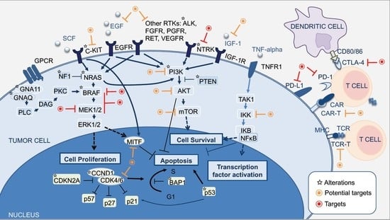
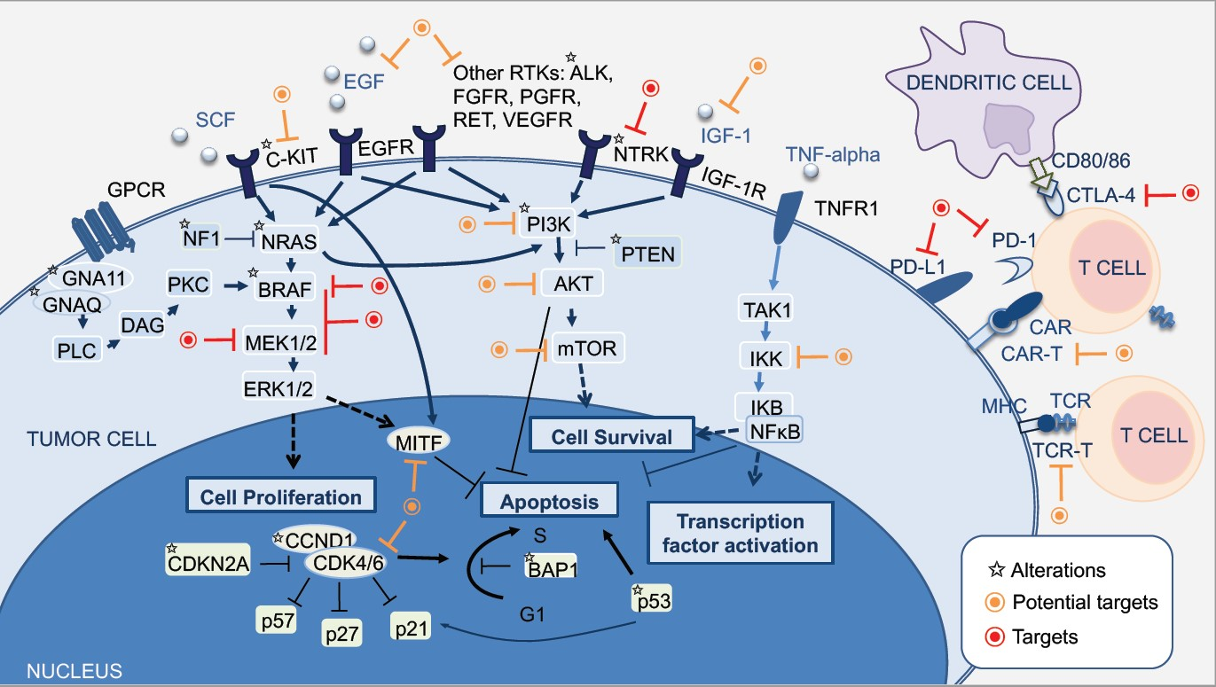
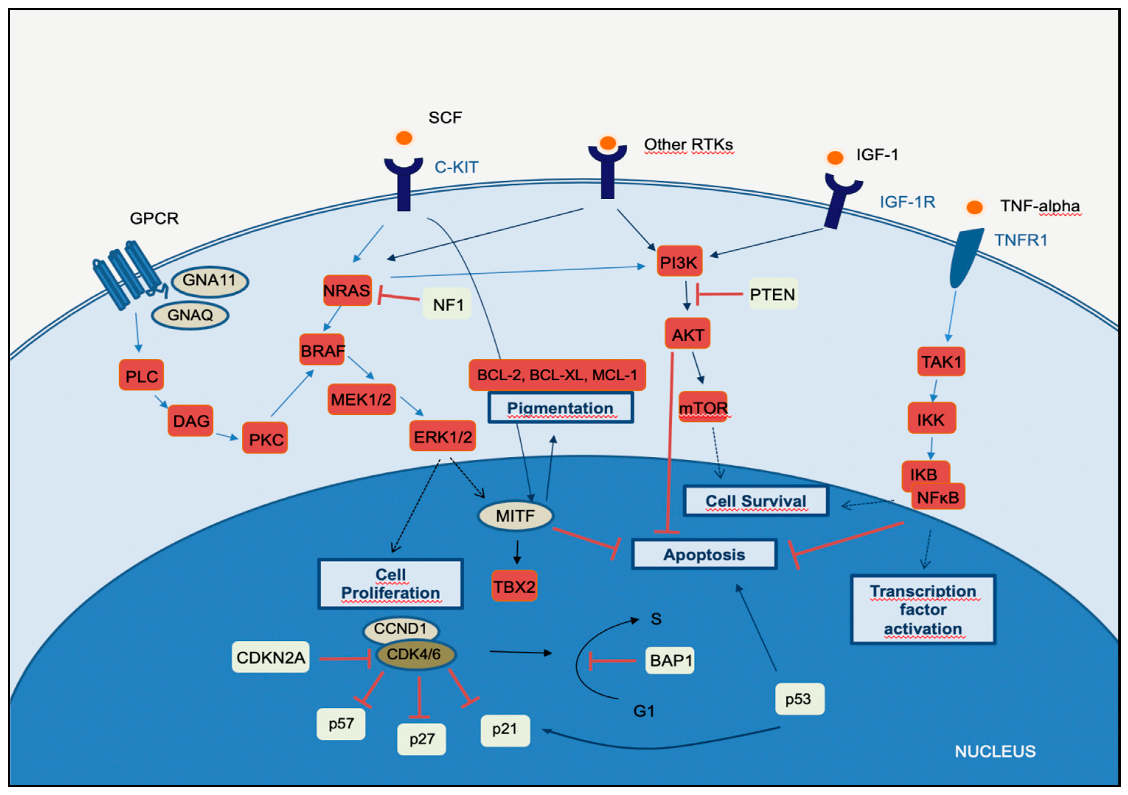
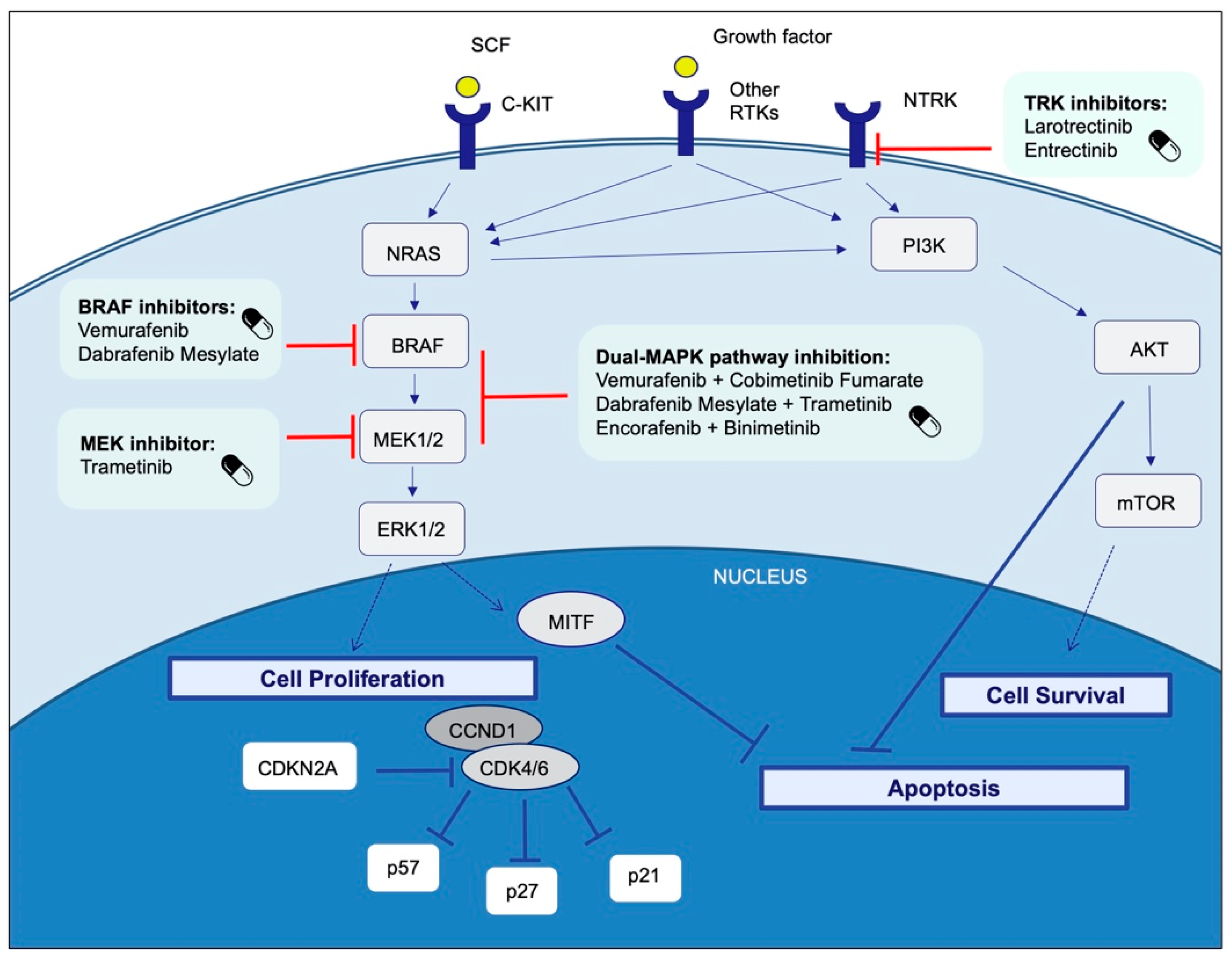
2. Molecular Pathways of Melanoma Development

Cancer results from uncontrolled cellular growth of malignant tumor cells caused by a combination of genetic alterations that lead to neoplastic transformation and escape from the inhibitory signals. Several steps in this process are known as the hallmark of cancers [17].
Several key molecular pathways have been discovered to be involved in the onset, proliferation, survival, progression, and invasion. In this section, we summarize the major signaling pathways that are currently known to be dysregulated and involved in melanoma disease.

2.1. MAPK Pathway

Melanomagenesis occurs after mutational events that produce signaling pathways critical for cell survival. Mitogen-activated protein kinase (MAPK) is a signal transduction pathway, involved in a variety of physiological programs, such as cell proliferation, differentiation, development, migration, apoptosis, and transformation, and is the most relevant in the development of melanoma (Figure 1) [18]. The MAPK pathway is activated by the binding of a growth factor to a receptor tyrosine kinase (RTK) on the cell surface and stimulates the guanosine triphosphatases (GTPase) activity of RAS. The signal propagates through the RAF, mitogen-activated protein kinase kinase 1 (MAP2K1), and extracellular signal-related kinase (ERK) cascade, which enters the nucleus to activate transcription factors and promote the cell cycle (Figure 1) [18].
The MAPK, PI3K, and NFκB pathways intersect significantly in melanoma pathogenesis. Briefly, in the MAPK-ERK pathway, stimulation of GPCR results in activation of PLC. This promotes DAG and then activates PKC, which stimulates the MAPK pathway. Receptor tyrosin kinases (RTKs) are activated by binding of extracellular growth factor ligands and activate the tyrosine kinase activity of the cytoplasmic domain receptor, starting the cascade of signals. Activated RAS activates the protein kinase activity of RAF isoforms (RAF1, BRAF, ARAF). Each RAF isoform possesses a distinct capacity to activate MEK, with BRAF being the strongest activator. MEK phosphorylates and activates downstream proteins, such as ERK1 and ERK2. ERK can translocate to the nucleus and phosphorylate different transcription factors, which leads to the control of cell cycle progression. MITF is a target of ERK and controls the production of the pigment melanin, cell cycling, and survival. The binding of the ligand to KIT (SCF) results in activation of the MAPK and PI3K pathways. In the PI3K-AKT pathway, ligand binding to the RTK leads to dimerization and autophosphorylation of the receptor and activation. Activated RTK recruits PI3K to the plasma membrane. PI3K activates AKT, whereas PTEN antagonizes this process. PI3K may also be activated by GPCR, IGF-1R, and RAS. Both ERK and AKT activate the mTOR-signaling pathway, which mediates cell survival and proliferation. In the TNFR pathway (canonical NF-κB pathway), binding of the TNF-alpha cytokine to its receptor TNFR1 results in TAK1 activation. TAK1 leads to the aggregation of a downstream kinase complex, the IKK complex. Phosphorylation of IκB by the IKK complex results in the release of NFκB. NFκB translocates to the nucleus and activates genes involved in cell survival and anti-apoptosis.
Fourteen MAPKs have been identified in mammals, and these kinases are typically divided into three main subfamilies: ERKs, c-Jun N-terminal kinases (JNKs), and P38 kinases. Each of these MAPKs is activated through phosphorylation by an MAPK kinase (MAP2K), which in turn is activated by an MAPKK kinase (MAP3K) [18]. The ERK pathway is the best-characterized MAPK pathway, which has a relevant role in the development and progression of melanoma. On this MAPK axis, the role of MAP3K is played by the RAF family of serine/threonine kinases, which is characterized by an RAS/GTP-binding domain. RAS proteins vHa-ras Harvey rat sarcoma viral oncogene homolog (HRAS), NRAS, and v-Ki-ras2 Kirsten rat sarcoma viral oncogene homolog (KRAS) are small GTPases located in the plasma membrane that act as activators in several pathways, apart from MAPK. Additionally, activation signals via RAS on the inner surface of the cell membrane increase ERK activity. Consequently, there is an increase in cellular proliferation, greater cell survival, and resistance to apoptosis. Activated ERK can also induce the metastatic potential of melanoma through the expression of integrins that promote tumor invasion [19].
In melanoma, dysregulated MAPK signaling and sustained ERK activation can eventually lead to cascade hyperactivity and subsequent cell proliferation, survival, invasion, metastasis, and angiogenesis. The BRAF gene is frequently mutated in several cancers, and BRAFV600 is the most common mutation of the skin. Mutated BRAFV600 leads to elevated BRAF kinase activity and sustained activation of downstream targets, in addition to unresponsive negative feedback mechanisms [20]. The mutant KRASQ61, the most frequent mutation of KRAS in melanoma, leads to an important decrease in its intrinsic hydrolytic activity and a sustained active state of KRAS. Mutations in other molecules may also lead to RAS overstimulation, such as loss-of-function mutations in neurofibromin 1 (NF1). In most melanomas with altered NF1, a loss-of-function mutation is found, in which neurofibromin loses its ability to inactivate RAS and promotes stimulation of the RAF and its downstream targets, leading to stimulation of the MAPK pathway and consequent cell proliferation and survival [21].
Telomerase reverse transcriptase (TERT) promoter mutations frequently occur in melanoma and, according to The Cancer Genome Atlas (TCGA) data, mainly in the mutated subtypes BRAF (75% of cases), RAS (72% of cases), and NF1 (83% of cases), suggesting a link between MAPK activation and TERT expression. The active MAPK pathway promotes phosphorylation and activation of the ETS1 transcription factor by ERK (the mutated TERT promoter bears ETS-binding sites) [22].

2.2. PI3K-AKT Pathway

The phosphatidylinositol-3-kinases (PI3Ks) comprise a family of lipid kinases with regulatory roles in many cellular mechanisms, including cell survival and growth, differentiation, proliferation, transcription, and translation. The pathway transduces signals from a variety of growth factors and cytokines and is the major downstream effector of RTKs and G-protein-coupled receptors (GPCRs) (Figure 1). Activated PI3K leads to the formation of phosphatidylinositol-3,4,5-trisphosphate (PIP3) through phosphorylation of phosphatidylinositol-4,5-diphosphate (PIP2) in the plasma membrane. PIP3 is essential for the recruitment of the serine-threonine protein kinase AKT to the plasma membrane. AKT is crucial in this signaling pathway, transmitting signals by phosphorylating different downstream effector targets [23]. Once AKT is phosphorylated and fully activated, it turns on a major downstream effector of the PI3K pathway, inhibiting or activating a variety of targets and regulating important cellular processes, such as apoptosis, DNA repair, cell cycle, glucose metabolism, cell growth, motility, invasion, and angiogenesis. The main target of AKT is the mammalian target of rapamycin (mTOR), which has a central role in the PI3K-AKT pathway and cancer disease. mTOR plays a crucial part in regulating cell growth and proliferation by monitoring nutrient availability, cellular energy, oxygen levels, and mitogenic signals.
PI3K-AKT signaling has negative regulators, to control any persistent and long-term activation. A major regulator of PI3K-AKT signaling is the tumor suppressor phosphatase and tensin homolog (PTEN), which antagonizes the PI3K activity through its intrinsic lipid phosphatase activity, converting PIP3 back to PIP2. Loss of PTEN results in constitutive activation of AKT and has been largely associated with tumor development in malignant melanoma. Indeed, PTEN loss has been shown to be predictive of shorter overall survival (OS) [24,25].
The PI3K signaling cascade is upregulated in different types of cancer, including melanoma. More than two-thirds of primary and metastatic melanomas show high levels of phosphorylated AKT, suggesting that this alteration is an early event in melanoma pathogenesis. Oncogenic events that activate PI3K-AKT may include mutations or copy number variations in certain components of the pathway. RAS gene mutations and mutated or amplified expression of RTK may also hyperactivate the PI3K-AKT pathway [20]. Mutations in the mTOR gene are present in approximately 10% of melanomas, and this molecular event leads to shorter survival and worse prognosis [26]. PI3K-AKT signaling may also be activated in melanoma due to loss of function of the negative regulator PTEN, which occurs in 10–30% of cutaneous melanomas, leading to constitutive activation of the PI3K pathway. Interestingly, PTEN gene alterations are mutually exclusive with NRAS mutations, and approximately 20% of melanomas with loss of PTEN function also have BRAFV600E mutations [27].

2.3. CDKN2A, Cell Cycle, and Apoptosis Regulation

The CDKN2A gene encodes two proteins, p16CDKN2A and p14CDKN2A, which have a tumor suppressor function. The cyclin proteins bind and activate CDKs, which has catalytic kinase activity. Several cyclin/CDK complexes have been identified that functionally act in different cell cycle phases: in the pre-replicative stage (G1), DNA duplication (S), and promotion of progression through the S phase to mitosis (Figure 1) [28]. p16CDKN2A and p14CDKN2A proteins have an inhibitory function, interfering with the activity of the cyclin/CDK complexes. p16CDKN2A inhibits the cyclin D1 (CCND1)/CDK4 complex, which, in turn, phosphorylates pRb and allows progression through the G1–S checkpoint. p14CDKN2A is an antagonist of the mouse double minute 2 homolog (MDM2) protein. This protein degrades p53 and eliminates p53 control of cell growth. The p14CDKN2A protein inhibits the oncogenic actions of MDM2 by blocking its actions on p53 [28]. p53 is a transcription factor that functions as a major negative regulator of cell proliferation and survival. Inactivation of the TP53 gene results in intracellular accumulation of genetic damage, which promotes melanoma development and progression. TP53 can be inactivated through silencing or mutation, the latter occurring most frequently in high-cumulative solar damage-associated (CSD-associated) melanomas [29].
Somatic impairment of the CDKN2A gene in melanoma can occur by genetic deletions, inactivated mutations, or promoter hypermethylation and leads to a decrease of the function of p16CDKN2A and/or p14CDKN2A proteins, with consequent loss of cell cycle control. This situation is associated with a higher melanoma invasion potential and metastases [30].
As mentioned above, mutation of the CDKN2A gene at the germline level is the most frequent genetic alteration in patients with a strong familial history of melanoma. In addition, variants of the MC1R gene increase the melanoma risk in CDKN2A mutation carriers [31].
The CCND1 and CDK4 genes are found to be altered in a minority of melanomas, representing less than 5%, and depend on the melanoma type. CCND1 gene amplifications affect about 30% of acral melanomas, 11% of lentigo maligna melanomas, and 6% of superficial spreading melanomas. CDK4 gene amplification is frequently found in acral and mucosal melanomas [32].

2.4. MITF Pathway

The microphthalmia-associated transcription factor (MITF) acts as a master regulator of melanocyte development, function, and survival by modulating differentiation and cell cycle progression genes [33]. It is involved in the differentiation and maintenance of melanocytes and modulates melanocyte differentiation and pigmentation (Figure 1). In melanomas, MITF can behave as an oncogene, and in approximately 20% of melanomas, it amplifies and promotes the proliferation of tumor cells. Its amplification correlates with a worse prognosis and a lower OS and ChT resistance [33]. MITF is activated by the MAPK and cAMP pathways and regulates the transcription of three major pigmentation enzymes (TYR, TYRP1, and DCT) [34]. In melanoma, ERK activity stimulated by BRAF is associated with MITF ubiquitin-dependent degradation. BRAF can modulate intracellular MITF protein through two opposite mechanisms. On the one hand, it can degrade the MITF protein; on the other hand, BRAF can stimulate transcription factors that increase the expression of the MITF protein. About 10–15% of melanomas harbor the BRAF mutation along with MITF amplification, suggesting that additional mechanisms are involved in ERK-dependent degradation of MITF.

2.5. NFκB Pathway

The nuclear factor-kappaB (NFκB) is a pleiotropic transcription factor that regulates several genes involved in many critical pathways and, in addition to immune and inflammatory responses, participates in physiological conditions, development, and cancer initiation and progression [35]. There are five members of the NFκB family, which are distinguished by their Rel homology domain, the portion of the protein that controls DNA binding, dimerization, and interactions with IκB proteins: RelA (p65), RelB, c-Rel, p100/p52, and p105/p50 [36]. Most IκB proteins bind and inactivate NFκB, through the retention of NFκB in the cytoplasm. Cytoplasmic NFκB complexes remain transcriptionally inactive until the cell is stimulated. Activated NFκB translocates to the nucleus, where it binds to target DNA loci and induces transcription of a variety of target genes involved in cell survival and anti-apoptosis. UV irradiation promotes the inflammatory response and cytokine production in skin cells, and many of these cytokines have NFκB as their downstream target/effector. Sustained activity of NFκB may lead to exacerbated expression of pro-inflammatory mediators, leading to tissue damage that may evolve into organ dysfunction and eventually cancer. The canonical (classical) NFκB pathway is mainly activated by tumor necrosis factor (TNF)-alpha, IL-1, and Toll-like receptors: TNFR, IL-1R, and TLR, respectively (Figure 1) [37]. NFκB activation may also occur due to deregulations in upstream MAPK and PI3K-AKT signaling pathways through different mechanisms. In melanoma cells, these alterations lead to an increase in proliferation and resistance to apoptosis [38].

2.6. WNT Pathway

The WNT proteins compose a family of 19 glycoproteins that act through a variety of receptors to stimulate distinct intracellular sub-pathways. These pathways are involved in development, cell growth, migration, and differentiation. WNT signaling can be the canonical or non-canonical type. The canonical type includes the intracellular transcriptional co-activator β-catenin as a central component [39]. This pathway can be activated by WNT proteins, such as WNT1/WNT3A, through binding to Frizzled receptors (FRZD1-7) and co-receptors, lipoprotein receptor (LRP) 5 (LRP5) and LRP6, that stimulate intracellular signaling to finally regulate β-catenin stability and transcription. Stimulation of canonical WNT signaling activates and prevents β-catenin from degradation by inhibiting glycogen synthase kinase 3β (GSK3β). Blocking the degradation of β-catenin leads to its stabilization in the cytosol, allowing its translocation in the nucleus and association with transcription factors, such as lymphoid-enhanced transcription factor (LEF) and T cell transcription factor (TCF) [39]. Due to the binding of β-catenin to LEF/TCF, these factors become transcriptional activators, and β-catenin-containing complexes control the expression of several WNT target genes, including c-MYC and CCND1. Regulation of c-MYC, a well-characterized proto-oncogene, through the canonical WNT pathway involves the control of cancer cell metabolism. c-MYC functions as a transcription factor by binding to several target genes, many of which are involved in cell cycle control, including CDKs, and CDK inhibitors. In addition, canonical WNT signaling can cooperate with MAPK signaling to regulate MITF expression and activity, which is associated with melanoma cell proliferation. WNT signaling can also be activated through various non-canonical pathways, independent of β-catenin, which are less characterized [40].

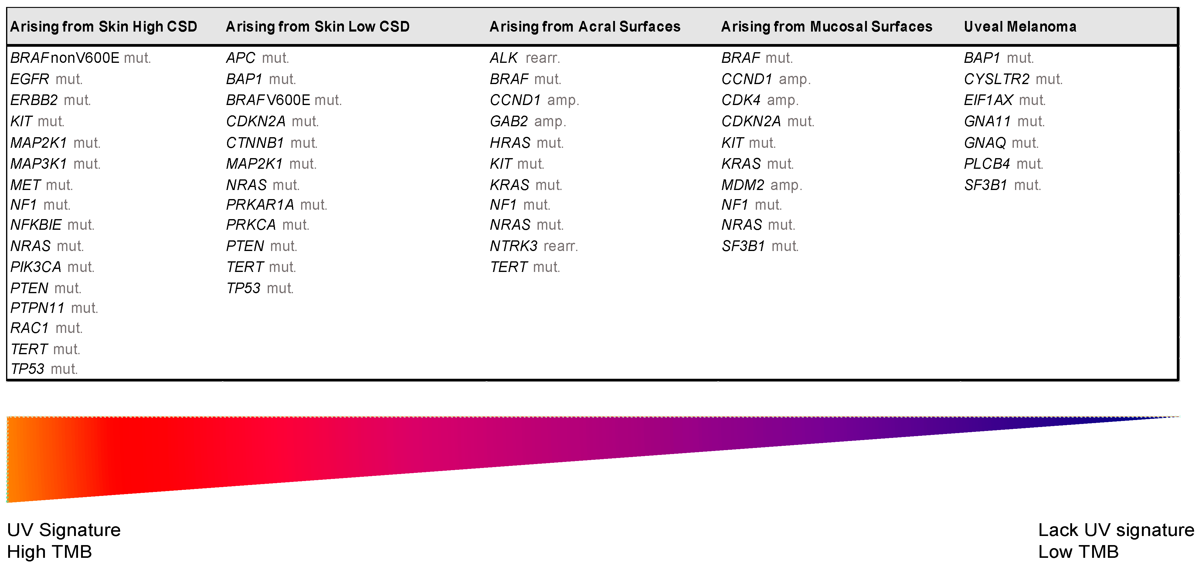
3. The Integration of Histology and Molecular Diagnostics of Melanoma

Despite recent molecular advances in melanoma characterization, paramount to diagnosis of a melanocytic skin lesion is the integration of several histopathological criteria with the clinical features. In many cases, general morphological criteria for atypia are often the subject of disagreement and inter-observer variability, especially in non-conventional lesions [41]. The World Health Organization (WHO) recognizes these challenges and incorporate the known molecular pathways in the latest WHO melanocytic tumor classification, introducing the concept of “intermediate” lesions. As stated in a recent review on the topic, this multidimensional classification showed that the view of melanocytic tumors as either benign or malignant might no longer be the proper approach [42]. Thus, WHO 2018 indicates nine categories/pathways leading to melanoma, each with specific genetic drivers (Table 1). Furthermore, melanomas can be clustered in three main subtypes, according to the degree of CSD (Table 1 and Figure 2) [43]. The largest group are melanomas associated with low-CSD or superficial spreading melanomas, which often arise on the trunk and proximal areas of the extremities. The most frequent molecular alteration in these melanomas is the BRAFV600E mutation [44]. In addition, TERT promoter mutations and CDKN2A mutations are also found in the majority of cases. PTEN and TP53 are commonly observed in advanced tumors. Lentigo maligna and desmoplastic melanomas are considered tumors associated with high-CSD. These melanomas arise on heavily sun-damaged skin, such as the face or hands, and affect older people. Molecularly, they often have a high mutation load and may harbor NRAS, BRAF non-V600E, or NF1 mutations. TERT promoter mutations and CDKN2A are also frequently found in these melanomas, and KIT mutations are found in a subset of cases. Interestingly, the number of mutations increases with the CSD grade (Figure 2), and desmoplastic melanomas harbor the highest tumor mutation burden. The category of “low to non-UV exposure/CSD” melanomas includes Spitz melanomas, acral melanomas, mucosal melanomas, melanomas developed from congenital nevi and blue nevi, and uveal melanomas. These melanomas rarely harbor BRAF, NRAS, or NF1 mutations (triple wild-type) [43]. A subset of acral and mucosal melanomas may have KIT mutations, in addition to gene amplifications and structural rearrangements, most frequently of the CCND1 gene and SF3B1. Therefore, genomic studies have subsequently exemplified that acral and mucosal melanomas are biologically distinct from their cutaneous counterparts at sun-exposed sites. Spitz melanomas show a particular oncogenic signaling pathway involving tyrosine kinase or serine-threonine kinase fusions, and melanomas in blue nevus and uveal melanomas are characterized by GNA11 or GNAQ mutations [44].
Certainly, to reduce diagnostic uncertainties and maintain a diagnostic approach based on the WHO 2018 classification, histological assessment should be accompanied by basic immunohistochemistry (IHC) and molecular tests. Recent recommendations of the European Society of Pathology, the European Organization for Research and Treatment of Cancer, and the EURACAN for the diagnosis of intermediate melanocytic proliferations and melanoma variants indicate that most pathology laboratories should perform basic IHC tests, such as: HMB-45; SOX10; MITF, tyrosinase, MART-1; P16; Ki-67/MIB1; BAP1 (BRCA1-associated protein 1); β-catenin; PRAME; and at least one molecular method to detect BRAF codon 600 and NRAS mutations [42]. The most difficult cases that require complementary studies should be analyzed in specialized referral centers, where laboratories can determine a higher grade in a given lesion or the identification of molecular targets that can benefit from targeted therapy.

4. Therapeutic Targets and Current Treatment Strategies in Advanced Melanoma Patients

The progressive understanding of the molecular pathways of melanoma has enabled the development of successful immunotherapies and targeted therapies for unresectable stage III and IV melanoma. This section describes the most important targeted therapies for melanoma. Some of them have been approved for clinical use, or are in clinical trials or preclinical research (Figure 3 and Table 2 and Table 3).

4.1. BRAF

BRAF is the most important therapeutic target and the most frequent genetic alteration in cutaneous melanoma, affecting 40–60% of cases [20]. The most frequently found BRAF mutation is V600E, which affects about 80% of BRAF-mutated melanomas; V600K involves 15% of cases; and V600R/M/D/G mutations are found in about 5% of cases [45]. The BRAFV600E mutation is frequently associated with the superficial spreading subtype, younger patient age, and non-CSD skin sites, such as the trunk and proximal areas of the extremities. In contrast, V600K mutations correlate with CSD skin sites, such as the head and neck, and patients of older age [46].
The presence of BRAF mutations in nevi supports the hypothesis that activation of the RAF/MEK/ pathway is an early event in melanoma development [47].
The FDA approved BRAFV600E/K inhibitors, MEK1/2 inhibitors, and dual-MAPK pathway inhibition with a combination of BRAF and MEK inhibitors for patients with BRAFV600E/K mutation-positive unresectable or metastatic melanoma. Furthermore, a combination of dabrafenib plus trametinib is approved for adjuvant treatment of resected stage III BRAFV600E/K mutant melanoma. On the other hand, larotrectinib and entrectinib are approved for patients with solid tumors that have NTRK gene fusions.

4.1.1. BRAF and MEK Inhibitors

Selective BRAF inhibitors have demonstrated remarkable clinical activity in melanoma patients carrying BRAFV600 mutations [93], such as vemurafenib [9,57], first in class. Its FDA approval in this setting was based on the results of the BRIM3 phase III trial, in which patients treated with vemurafenib had an OS and progression-free survival (PFS) of 13.6 and 5.3 months, compared to dacarbazine-treated patients, who had 9.7 and 1.6 months, respectively [9] (Table 2). Additionally, another selective BRAF inhibitor (BRAFi), dabrafenib, showed efficacy in this group of patients in the BREAK3 phase III trial, in which patients treated with dabrafenib had an OS and PFS of 20 and 6.9 months, whereas those treated with standard chemotherapy had 15.6 and 2.7 months, respectively [53]. Based on these findings, the FDA also approved dabrafenib as a first-line treatment for unresectable or metastatic melanomas carrying the BRAFV600 mutation.
Importantly, BRAFi leads to characteristic side effects, including photosensitivity, which can limit treatment, and the rapid development of cutaneous squamous cell carcinoma (cuSCC) or other keratinocytic secondary neoplasias, which are thought to arise due to the paradoxical activation of the MAPK pathway in keratinocytes that are wild-type for BRAF but present upstream RAS activation in chronically damaged skin [94]. Nonetheless, patients can develop resistance through upregulation of RTKs or NRAS [95,96]. Preclinical data showed that BRAFi-resistant cells were sensitive to MEK inhibitors (MEKi) [97]. Thus, the combination of BRAF and MEK inhibitors (BRAFi plus MEKi) was predicted to decrease this side effect, and indeed, this combination treatment was not only demonstrably linked to improved PFS and OS compared to BRAFi monotherapy, but also decreased the development of cuSCC. Therefore, following the discovery of the clinical activity of single-agent MEKi, the use of combinations of BRAFi plus MEKi was evaluated in clinical studies. Currently, three combinations of BRAFi plus MEKi have been approved for clinical practice and appear to be comparable in terms of efficacy, but to date, no adequate direct comparison has been performed in randomized trials.
  • Dabrafenib plus trametinib
The combination of BRAFi dabrafenib and MEKi trametinib was the first combination of BRAFi plus MEKi approved for the treatment of advanced melanoma in the United States in 2014 and in the European Union in 2015. The approval was based on the results of the COMBI-v trial [54,98], which compared dabrafenib plus trametinib to vemurafenib, and the COMBI-d trial [55,99], which compared dabrafenib plus trametinib to dabrafenib monotherapy (Table 2). In the phase III COMBI-d trial, the overall response rate (ORR) was 69% for the combination and 53% for dabrafenib alone [54]. In the phase III COMBI-v trial, ORR was 64% for the combination and 51% for vemurafenib alone. Rates of severe adverse events (AEs) and study drug discontinuation were similar in both arms. The 5-year OS rate for both pooled trials was 34% for the combination arm. Further, 19% of patients treated with BRAFi plus MEKi achieved a complete response, while for these patients, the OS rate was 71% at 5 years [100].
  • Cobimetinib plus vemurafenib
Cobimetinib is another MEKi developed for the treatment of advanced melanoma in combination with BRAFi vemurafenib. In the phase III CoBRIM study, 495 patients with previously untreated advanced melanoma were randomized to receive either vemurafenib plus cobimetinib, or vemurafenib alone. ORR was 68% for vemurafenib plus cobimetinib and 45% for vemurafenib alone (Table 2). The median OS was 22.3 months for the combination versus 17.4 months for vemurafenib [56,101]. Extended follow-up demonstrated a 4-year OS rate of 35% in the combination group versus 29% in the control group [102]. The toxicity profile of vemurafenib plus cobimetinib differs from that of dabrafenib plus trametinib. Diarrhea, nausea, fatigue, rash, liver enzyme abnormalities, and photosensitivity (caused by vemurafenib) are more likely to occur with vemurafenib plus cobimetinib, while pyrexia is more likely to develop with dabrafenib plus trametinib [102].
  • Encorafenib plus binimetinib
Nowadays, a third combination of a BRAFi plus MEKi, encorafenib and binimetinib, is also authorized based on the results of the three-arm phase III COLUMBUS trial. In this trial, 577 patients with advanced melanoma with the BRAFV600 mutation were randomized to encorafenib plus binimetinib, encorafenib, or vemurafenib monotherapy [57]. The results were best for the combination group, with an ORR of 63% versus 51% versus 40% and a median OS of 33.6 months versus 23.5 months versus 16.9 months (Table 2). The most common grade 3 or 4 treatment-related adverse events (TRAEs) reported for the combination were increased glutamyltransferase (9%), increased creatine phosphokinase (7%), and hypertension (6%).

4.1.2. Differences between BRAFV600E and BRAFV600K Mutations

The most common BRAFV600 mutations are V600E (80%) and V600K (14%). Even though individual phase III targeted therapy trials have not performed a direct comparison between BRAFV600E and BRAFV600K-mutant melanomas, in three separate trials, BRAFV600K melanomas had a numerically lower response rate and shorter median PFS with BRAFi compared with V600E melanomas, and two pooled analyses of BRAFi plus MEKi showed shorter PFS in multivariate analysis [103,104].

4.1.3. Resistance Mechanisms to BRAF and MEK Inhibition

Despite these promising results, almost all patients diagnosed with BRAF-mutated advanced melanoma will develop tumor relapse within several months after initiation of BRAFi +/− MEKi treatment. Different mechanisms of drug resistance underlie the progression of the disease and the activation of both the MAPK and PI3K-AKT pathways. Inhibition of BRAF leads to increased RAS activity, which in turn can activate the PI3K-AKT signaling pathway. Furthermore, it has been observed that patients with melanoma carrying PTEN mutation/loss, treated with dabrafenib, have a shorter median PFS [105]. Among the acquired resistance mechanisms, mutations in the NRAS and MAP2K genes determine the dependence of the MAPK pathway [106].
NF-1 inhibits RAS activity under physiological conditions, so its role in resistance to BRAF-targeted therapy has been widely studied [107]. It was demonstrated that NF-1 loss-of-function mutations can lead to continuous RAS activation, which can activate both MAPK and PI3K-AKT signaling pathways downstream and confer resistance to target therapy. The BRAF oncogene cooperates with CDKN2A, inducing its upregulation, and consequently, cell arrest and senescence. Thus, CDKN2A inactivation could induce melanoma progression in a BRAF-mutated nevus. Nathanson et al. demonstrated that a lower copy number of CDKN2A was significantly associated with decreased PFS in patients treated with dabrafenib, while a higher CCND1 copy number was significantly associated with a worse prognosis and resistance to BRAFi [105].
Apart from these mechanisms that reactivate RAS, there are structural changes in oncogenic BRAF due to aberrant splicing that can lead to resistance. For example, p61-BRAFV600E splice variants retain active kinase activity but are unable to bind RAS. They dimerize regardless of RAS status and drive constitutive signaling to ERK, uncoupled from upstream regulation [108]. Importantly, tumor heterogeneity and heterogeneous mechanisms of resistance are present in tumors and at different metastatic sites simultaneously [109]. Recent efforts to dissect the degree of heterogeneity and its clinical implications have led to transcriptional studies targeting thousands of single tumor cells, and show that at the cellular level in all tumors, there are distinct transcriptional patterns within each tumor that display varying degrees of predicted responsiveness to BRAF inhibition [110].
Finally, ongoing studies are investigating the addition of CDK4/6 inhibitors, PI3K/mTOR, and ICIs to combat resistance [96,111] (Table 3). Additionally, RhoA GTPases are emerging as a potential pathway for BRAF resistance, since preclinical data has shown that inhibition of the pathway by Rho kinase inhibitors promotes resensitization to BRAF-targeted therapy [112].

4.2. Immune Checkpoint Inhibitors: Anti-CTLA-4 and Anti-PD1

Treatment and prognosis of metastatic melanoma has changed radically in the last decade since the approval of ICIs, those directed to protein 4 associated with cytotoxic T lymphocytes (CTLA-4) and programmed cell death-1 (PD1). Both are inhibitory receptors that regulate immune responses by different mechanisms. CTLA-4 is a negative regulator of T cells and is expressed by naive T cells, which leads to a robust inhibitory signal during T cell activation when it binds to costimulatory protein B7 in antigen-presenting cells in the lymph node. On the other hand, anti-PD1 inhibitors prevent the binding of PD1 and its ligands (PD-L1 and PD-L2) to produce an effective immune response. When PD1 receptor binds to its ligands, it works to decrease the ability of already activated T cells to produce an effective immune response and prevent the immune system from rejecting the tumor [113].

4.2.1. Anti-CTLA4

In patients with metastatic melanoma, phase III clinical trials of ipilimumab, a fully human IgG1 monoclonal antibody inhibiting CTLA-4, demonstrated a significant improvement in PFS and OS when compared with a gp100 vaccine [7] or dacarbazine ChT [48].

4.2.2. Anti-PD1-Based Therapies

In 2014, the FDA approved pembrolizumab and nivolumab as the first anti-PD1 (CD279)-directed monoclonal antibodies for advanced or metastatic melanoma. Both drugs received EU approval in 2015. Approval of nivolumab was based on the results of the CheckMate 066 phase III study, in which BRAF wild-type advanced melanoma patients were treated with nivolumab or dacarbazine (DTIC) [49]. In this trial, the median OS was 37.2 months for nivolumab versus 11.2 months for dacarbazine, and the 1- and 2-year OS rates were 73% and 58%, respectively, for nivolumab-treated patients (Table 3). Three months prior to nivolumab, pembrolizumab was approved by the FDA for the treatment of metastatic melanoma. The accelerated approval was based on the results of an activity-estimating cohort conducted within the phase Ib KEYNOTE-001 trial [114]. Pembrolizumab showed 5-year OS rates of 34% in all patients and 41% in treatment-naïve patients with melanoma [115]. Furthermore, in a phase III trial of pembrolizumab versus ipilimumab, the 2-year OS rates were 55% versus 43%, respectively [52,114]. The efficacy of pembrolizumab and nivolumab has never been directly compared in patients with metastatic melanoma. In a retrospective study, OS from 888 patients with metastatic melanoma treated with first-line pembrolizumab or nivolumab was compared, with no statistical difference [116].
  • Combination of Anti-PD1 with Anti-CTLA-4 Monoclonal Antibodies
The combination of CTLA-4 and PD1 blockade is supposed to synergistically stimulate the immune response against cancer cells. Approval of dual therapy was based on data from the phase III CheckMate 067 trial, in which combination therapy with anti-CTLA-4 and anti-PD1 blockade demonstrated superior clinical activity, with an ORR ranging from 50 to 60% and improved OS compared to ipilimumab, despite increased toxicity with the combination treatment [50]. In this phase III trial, a total of 945 previously untreated patients with unresectable stage III or IV melanoma were included. With a minimum follow-up of 60 months, median OS had not been reached in the combination group and was 36.9 months in the nivolumab group, as compared with 19.9 months in the ipilimumab group [117].
Moreover, an important trial to better elucidate the contribution of each combination therapy compound to toxicity is the CheckMate 511 trial, conducted by Lebbé et al. [51]. CheckMate 511 compared ‘standard’ regimen doses of 3 mg/kg ipilimumab and 1 mg/kg nivolumab as used, e.g., in CheckMate 067 with a 1 mg/kg ipilimumab and 3 mg/kg nivolumab regimen (Table 2). Results showed decreased toxicity but similar efficacy for the reduced ipilimumab and increased nivolumab regimen. It should be mentioned that the study was only powered with respect to the toxicity comparison but not efficacy. Therefore, the outcomes of this trial supported the assumption that the immunotoxicity of the combination can be reasonably moderated with a reduced dose of ipilimumab. In summary, dual therapy with ipilimumab and nivolumab appears to be superior to either anti-CTLA-4 monotherapy or anti-PD1 monotherapy [6].
  • Novel Combinations of BRAF/MEK Inhibitors and Immune Checkpoint Inhibitors
Given the rapid and deep responses seen with BRAFi plus MEKi and the durable responses observed with ICIs, the combination of these therapeutic strategies appears promising. The phase III IMspire 150 trial evaluated the addition of atezolizumab, an anti-PD-L1 monoclonal antibody, to vemurafenib and cobimetinib in patients with BRAFV600-mutant metastatic melanoma [58]. In total, 514 patients were randomly assigned to receive atezolizumab, vemurafenib and cobimetinib or placebo, vemurafenib and cobimetinib, as first-line therapy (Table 2). The study met its primary endpoint, PFS. The triple therapy did not increase ORR. Grade 3 or 4 TRAEs occurred in 79% of patients treated with the triple combination and in 73% of patients treated with only vemurafenib plus cobimetinib, so it was concluded that the addition of atezolizumab to targeted therapy with vemurafenib and cobimetinib was tolerable and significantly increased PFS in patients with BRAFV600-mutant metastatic melanoma.
Another triple therapy that has been explored is the addition of a new monoclonal antibody anti-PD1, spartalizumab, to dabrafenib and trametinib, in the phase III COMBI-I trial, compared to dabrafenib and trametinib plus placebo, as first-line therapy in patients with BRAFV600E/K-mutant advanced melanoma [59]. With ORR for the triple therapy being slightly higher (69%) compared to the double (64%), the trial unexpectedly did not meet its primary endpoint of investigator-assessed PFS for patients treated with the triple therapy (Table 2).
Finally, on this topic, there is some preclinical evidence that in BRAF wild-type melanoma, the combination of MEKi with ICIs may enhance the antitumor effects. Preclinical data has shown that MEKi cobimetinib inhibits MAPK signaling and increases immune cell infiltration in the tumor, providing a strong rationale for combining cobimetinib with the anti-PD-L1 antibody atezolizumab [118]. Based on these findings, IMspire 170, a phase III trial, randomized 446 patients with advanced BRAF wild-type melanoma to receive either the MEKi cobimetinib plus atezolizumab, or the anti-PD1 monoclonal antibody pembrolizumab alone. The trial did not meet its primary endpoint, showing a median PFS of 5.5 months with cobimetinib plus atezolizumab versus 5.7 months with pembrolizumab alone.
  • Biomarkers of response and resistance mechanisms to immune checkpoint inhibitors
Analysis of TCGA data from melanoma cases revealed that cutaneous melanoma displays a high mutational burden and UV signature (Figure 2) [119]. In addition to neoantigen recognition, a high mutational load was also found to correlate with clinical benefit to ICIs [120]. Similarly, a positive correlation was observed between a higher mutational load and increased CD8-positive T cell infiltration. Furthermore, PD-L1-positive patients treated with pembrolizumab had increased PFS, OS, and ORR [50]. Nevertheless, some patients with PD-L1-positive tumors do not respond to PD1 blockade, and conversely, some patients with PD-L1-negative tumors respond [121]. Together, the aforementioned data indicate that PD-L1 expression is a possible surrogate for lack of immunogenicity, as well as other failures further down the immune cycle, but is a suboptimal biomarker to predict response to ICIs in melanoma patients [122]. Cooperating with the loss of antigenicity and the intrinsic characteristics in the tumor that render them more or less vulnerable to immunotherapy are additional mechanisms arising during the immune response that directly inhibit tumor-targeting T cells. For example, resistances can be acquired by new resistance-driving mutations in genes involved in interferon-receptor signaling, antigen presentation, and the β-catenin signaling pathway [123,124,125]. Gene expression signatures associated with responses to immunotherapy are an area of active research. The β-catenin signaling pathway signature and the 10-gene interferon-gamma (IFNG) signature have been shown to predict resistance and responses to ICIs in melanoma [123]. Ayers et al. analyzed gene expression profiles (GEPs) using RNA from baseline tumor samples from patients treated with pembrolizumab. A 10-gene “preliminary IFNG” signature (IFNG, STAT1, CCR5, CXCL9, CXCL10, CXCL11, IDO1, PRF1, GZMA, and MHCII HLA-DRA) was constructed that was able to separate anti-PD1 (pembrolizumab) responders and non-responders, among the 19 pilot data patients with melanoma [126]. In the same study, data showed that such a signature might perform favorably compared with PD-L1 IHC in PD-L1-unselected populations.

4.3. Other Immunotherapy Treatment Strategies: Adoptive Cell Therapy

Patients who progress after anti-PD1 therapy, anti-PD1 plus anti-CTLA-4 therapy, and targeted agents have limited options. Only 4–10% of these patients have objective responses to ChT, with a limited median OS of 7 months [127,128]. New approaches to cancer immunotherapy have raised hope for effective treatments. Recently, adoptive cell therapy (ACT) has been recognized as a method to provide a long-lasting and effective response in melanoma. As its name implies, ACT is designed to redirect host lymphocyte cells against tumor cells [129]. We can distinguish three ACT strategies: tumor-infiltrating lymphocytes (TILs), T cell receptor-engineered T cells (TCR-T), and chimeric antigen receptor T cells (CAR-T).

4.3.1. Tumor-Infiltrating Lymphocytes

TILs are autologous CD4 and CD8 T cells in the tumor microenvironment, which have the potential to recognize tumor-specific antigens [130]. However, due to chronic antigenic stimulation, they are converted to an “exhausted” state and become functionally impaired. In 1988, Rosenberg and colleagues developed a method to reactivate these cells. Promising response rates were achieved by isolating autologous TILs from a resected metastasis, in vitro expansion in the presence of interleukin (IL)-2, and reinfusion of cells followed by boluses of IL-2 [131]. A recent meta-analysis by Dafni and colleagues found an ORR of 41% and a complete response rate of 12% in a total of 410 patient treated with TILs [130]. Recently, phase II results evaluating the efficacy of lifileucel (an autologous TIL product) in patients with advanced melanoma who had been previously treated with ICIs and BRAF/MEK-targeted agents observed an ORR of 36% and in patients refractory to anti–PD1/PD-L1 an ORR of 41% [132]. Additionally, a randomized phase III trial (NCT02278887) that is currently recruiting aims to show a better survival rate after TIL infusion compared to standard-of-care ipilimumab in patients with advanced-stage progressive melanoma in the first line of anti-PD1 treatment.

4.3.2. T Cell Receptor-Engineered T Cells

The limitations of TILS treatment have prompted the development and use of TCR-T and CAR-T. TCR-T are manufactured from autologous peripheral blood mononuclear cells collected by apheresis, after which CD3 cells are genetically modified and shortly expanded in vitro. In 2006, the first trial targeting the melanoma differentiation antigen MART-1 showed feasibility but only low clinical response rates (2/17) [133]. More encouraging results were seen in subsequent trials with MART-1 and gp100-reactive TCR-T, with ORR in 30% and 19% of patients, respectively [134]. Despite the encouraging results, the downside of targeting antigens shared between melanoma cells and melanocytes is the risk of inducing on-target, off-tumor toxicities [135]. So far, no severe toxicities have been reported after treatment with high-affinity NY-ESO-1-specific TCR gene-modified T cells, whereas encouraging response rates of up to 55% and an estimated 3–5-year OS rate of 33% have been achieved in a phase II trial [136].

4.3.3. Chimeric Antigen Receptor T Cells

A CAR is a fully synthetic antigen receptor. CAR-T can be manufactured from T cells, which are collected by apheresis, genetically modified to express the CAR, and expanded over 7 to 10 days [137]. As a consequence, they can be deployed to treat patients regardless of their HLA-type and can circumvent mechanisms of tumor resistance, such as MHC downregulation and defective antigen processing [137]. A phase I/II study evaluating vascular endothelial growth factor receptor 2 CAR-T (NCT01218867) in patients with metastatic cancer, including melanoma, was discontinued due to a lack of objective responses [138]. Currently, there are three phase I dose escalation trials enrolling patients with melanoma (NCT03893019, NCT03635632, NCT04119024).

4.4. NRAS

Unlike BRAF, NRAS is not a therapeutic target. NRAS and BRAF mutations are usually mutually exclusive, but their coexistence has been described [139]. NRAS mutations are present in about 10–25% of cutaneous melanomas, with preference in the nodular melanoma type and melanomas arising in the CSD skin, suggesting that it may be a consequence of UV-related mutagenesis. Several studies suggested that NRAS mutations are significantly related with a worse prognosis in melanoma patients [140,141]. However, controversial data exists on the prognostic role of NRAS mutation in melanoma patients. In a phase III study [118], which evaluated the combination of treatment with atezolizumab and cobimetinib versus pembrolizumab monotherapy in BRAF wild-type advanced melanoma patients, no differences were found with respect to the mutational status of NF1 or NRAS or no mutation as determined by a next-generation sequencing platform in terms of prognostic and treatment efficacy.
Effective treatment options in NRAS-mutated advanced melanoma are urgently needed, especially after failure of immunotherapy with anti-CTLA4 or anti-PD1 antibodies. Binimetinib, an MEK inhibitor, has shown clinical activity (20% partial response) in an open-label phase II study in this group of patients [142]. Based on these results, a phase III trial (NEMO) was conducted, including 402 patients harboring an NRASQ61R, Q61K, or Q61L mutation who had not been previously treated or whose disease had progressed during or after immunotherapy, and randomized 2:1 to receive either binimetinib or dacarbazine [143]. PFS was significantly longer in the binimetinib group, but no differences were observed for OS. Other MEK inhibitors (trametinib and selumetinib) have been evaluated in NRAS-mutated advanced melanoma patients, without favorable outcomes [144,145]. In contrast, another MEK inhibitor in clinical development, pimasertib (Table 3), in a phase I study showed clinical activity in patients with locally advanced or metastatic melanoma, particularly in tumors with BRAF and NRAS mutations. The ORR was 12.4% (11/89), and 9 of the 11 responders presented a BRAF and/or NRAS mutation (3/11) [74].

4.5. C-KIT

C-KIT is an RTK directly responsible for binding to growth factors and initiating the MAPK and PI3K-AKT pathways (Figure 1). Mucosal melanoma and acral melanomas are the types of melanoma harboring the highest C-KIT alterations, followed by melanomas that arise on CSD skin. Indeed, C-KIT mutations and amplifications are detected in 10–15% of acral melanomas, 20–30% of mucosal melanomas, and about 1% of other cutaneous melanomas [32]. C-KIT is a therapeutic target in gastrointestinal stromal tumors with activating mutations of C-KIT, but in melanoma, it is under study, with no approved drugs yet. Advanced melanomas with C-KIT alterations have shown efficacy in terms of ORR with the C-KIT inhibitors imatinib (ORR 23.3%) and nilotinib (ORR 26.2%) in phase II trials [64,65,146] (Table 3).

4.6. GNAQ/GNA11

Cutaneous melanomas arising in blue nevus, as well as the uveal melanoma subtype can harbor GNAQ or GNA11 mutations, and their identification helps improve the differential diagnosis of melanocytic lesions. GNAQ mutations have been detected in about 90% of blue nevus, 50% of malignant blue nevus, and 50% of primary uveal melanoma. GNA11 mutations have been observed in less than 10% of blue nevus, one third of primary uveal melanomas, and about 60% of metastatic uveal melanomas [43,147].
Several studies have been conducted to evaluate the efficacy of specific agents in melanomas harboring GNAQ/GNA11 mutations (Table 3). In vitro, tumors harboring GNAQ/GNA11 mutations were found to respond to MEK inhibitors, but the combination of MEK/PI3K inhibitors has shown increased activity [148]. On the other hand, a phase Ib dose-escalation study of the MEK inhibitor with the dual PI3K/mTOR inhibitor has been proposed as a potential treatment in these tumors, but the combination was poorly tolerated, and responses were minimal.

4.7. SF3B1

SF3B1 mutations have been identified in subsets of solid tumors, as well as in myelodysplastic syndrome and chronic lymphocytic leukemia. Recently, SF3B1 was identified as a significantly mutated gene in uveal melanoma (20%) and mucosal melanoma (42%), especially in female genital and anorectal melanomas [149]. SF3B1 encodes for a part of the spliceosome, splicing factor 3 subunit 1. SF3B1 mutations are the most common spliceosomal component gene mutation implicated in the pathogenesis of cancer and act by causing aberrant RNA splicing events [150]. The clinical relevance of these splicing events is not completely clear, but SF3B1-mutated tumors have been shown to be at risk of metastasis. SF3B1 mutations are being studied as a potential therapeutic target using SF3B-small-molecule inhibitors [151]. However, the phase I studies performed did not achieve a good response rate (Table 3) [91,92].

4.8. NTRK

Neurotrophic tyrosine receptor kinase (NTRK) comprises a family of three genes encoding tropomyosin receptor kinases (TRK): TRKA, TRKB, and TRKC receptors, which correspond to the NTRK1, NTRK2, and NTRK3 genes, respectively. These receptors play an important role in the development and function of neuronal tissue [152]. Gene fusions represent the major molecular aberration involving NTRK in tumorigenesis and have been found in several cancers displaying different histologic phenotypes, including many different epithelial tumors, glioblastomas, and sarcomas. In melanoma, NTRK fusions are uncommon, usually found in Spitz melanomas (Table 1) [153]. Furthermore, NTRK translocations have also been identified in 2.5% of acral melanomas [154] and less than 1% of cutaneous and mucosal melanomas [155].
Targeting NTRK fusions represents a new opportunity for cancer treatment, as selective inhibitors (entrectinib and larotrectinib) have been developed and are FDA approved for adults and pediatric patients with solid tumors that harbor a NTRK gene rearrangement without a known acquired resistance mutation, that are either metastatic or unresectable, and who have no satisfactory alternative treatments or whose cancer has progressed following treatment (Figure 3).
The NTRK inhibitors entrectinib and larotrectinib have demonstrated a 57% and 79% ORR, respectively, in tumors with NTRK family fusions, regardless of histology [156,157]. Given this efficacy, these predictive biomarkers would be worth testing in tumors from patients diagnosed with metastatic melanomas that do not respond to other treatments.

4.9. Other Therapeutic Options

  • NFκB pathway
Inhibition of NFκB can promote benefits by enhancing apoptosis, and this may be achieved by targeting crosstalk regulators of the PI3K-AKT and MAPK pathways [158].
Proteasome inhibitors, such as bortezomib, have been used to inhibit NFκB by preventing the degradation of IκB. These inhibitors have been shown to induce a decrease of melanoma proliferation in vitro [159,160]. Bortezomib also induces a reduction of tumor growth in vivo but has shown significant toxicity after clinical trials [161].
Recent efforts have been directed at the development of selective inhibitors of the NFκB pathway. BMS-345541, a specific IKK inhibitor, has been shown to reduce constitutive IKK activity and apoptosis of melanoma cells (Table 3) [83]. The NBD (NEMO-binding domain) peptide can bind to NEMO and prevent its interaction with IKKα/β, which is crucial for IKK complex activity and activation of NFκB (Figure 1). The use of NBD peptides can promote a significant decrease in proliferation and apoptosis in human melanoma cell lines, but neither NBD peptides nor BMS-345541 have reached clinical trials yet [162].
Highly specific inhibitors of NFkB may be used to minimize the pleiotropic effects of nonspecific inhibitors and to reduce toxicity in vivo. This field of research is new, with great potential, and could provide more alternative options for the treatment of melanoma in the future.
  • WNT pathway
Several inhibitors of the WNT pathway of natural or synthetic origin have been studied preclinically. The WNT signaling components, mainly FRZDs and DVL, have been considered as targets for cancer treatment using small-molecule inhibitors. OMP-18R5 (vantictumab) is an antibody that interferes with the binding of WNT ligands to FRZDs. It has shown activity to inhibit the growth of a variety of human tumor xenograft models and exhibits activity when treated with standard ChT (Table 3) [82].
Another target of WNT signaling is porcupine, an enzyme necessary for palmitoylation of WNT ligands. Porcupine inhibitory molecules IWP2 and C59 have shown potent in vitro and in vivo activity, respectively. Furthermore, LGK979, another porcupine inhibitor, has demonstrated activity inhibiting WNT signaling in vivo at well-tolerated doses and laid the foundation for the treatment of WNT-driven tumors in the clinic [163]. Currently, a phase I study of LGK979 (NCT01351103) is being conducted to find LGK979 safe doses for melanoma and other solid malignancies (last update posted, 27 July 2021; clinicaltrials.gov, accessed date, 6 August 2021).
On the other hand, potential therapeutic strategy targeting WNT signaling includes the development of specific peptides that mimic WNT proteins, such as WNT3A and WNT5A, and, thus, lead to receptor inhibition. So far, it has shown efficacy in melanoma preclinical models and used in other diseases with promising results [164]. Ultimately, target-specific therapies have also been proposed against extra- and intracellular components of the WNT pathway, including axin, APC, and β-catenin [165]. Along the same line, antibody-based blockage of WNT5 has resulted in the inhibition of protein kinase C activity and a decrease in cell invasion [166].

5. Conclusions and Future Directions

Targeting the MAPK signaling pathway has significantly improved the treatment of metastatic melanoma, with BRAF mutations being the most frequent and most important alterations to be treated. Targeted therapy for patients whose tumors harbor the BRAFV600 mutation achieves high response rates and OS benefit with the combination BRAF/MEK inhibition and represents the ideal first-line treatment for patients with BRAF-mutated advanced melanoma. However, despite good treatment responses, drug resistance is very common.
To avoid resistance, combination treatments have been and are continuously being studied. The highly complex interactions between melanoma molecular pathways include poorly understood mechanisms that promote drug resistance and decrease patient survival, primarily by activating the MAPK and PI3K-AKT pathways. Preclinical studies looking at these major drug association strategies appear to be, at least, very promising as a target for MEK or PI3K/mTOR, blocking CDK4/6 or RhoA GTPases, which the latter has been shown to promote resensitization to BRAF-targeted therapy [96,111]. In the field of other gene alterations, there is a challenge in the research of new therapeutic targets and development of new drugs, especially after failure of immunotherapy with anti-CTLA-4 or anti-PD1 antibodies. Alterations under study that can eventually lead to a therapeutic benefit with targeted therapy are, among others: NRAS, C-KIT, GNAQ, GNA11, and SF3B1. Moreover, advances have been made in cancer therapy through the use of ICIs. We now have several different approved regimens, both for targeted therapy for BRAF-mutated melanoma, as well as immunotherapy for all comers with melanoma.
Using T cells to control disease, adoptive cell therapy, has added a powerful and novel therapeutic option for the treatment of melanoma (TILS, TCR-T, and CAR-T). Among the strategies that have arisen, it has been shown that with TCR-T, the selection of a single antigen it is unlikely to be sufficient to eliminate solid tumors. Therefore, future studies targeting multiple antigens simultaneously may improve the efficacy of TCR-T cells in solid tumors, as has been demonstrated in preclinical models. On the other hand, CAR-T cell therapy could be used for patients with melanoma who are resistant to other therapeutic choices.
In this review, we have emphasized the understanding of the molecular pathways responsible for the development and progression of melanoma, as well as the relevance of detecting the specific molecular markers of each melanoma subtype. This information is of primary importance in clinical practice to predict the response to treatment in each subtype of melanoma.
The different molecular pathways for the development and progression of melanoma are very complex and interact with each other (via crosstalk mechanisms) to create resistance to treatment and the progression of cell signaling. For this reason, there is an urgent need to identify other alternative and targeted therapies. Detailed understanding of the role of genes and proteins in key signaling pathways in melanoma development has led to the designation of new targets for the treatment of melanoma. Recently, analysis of CRISPR-CAS9 screens has identified genes that have not previously been associated with melanoma growth and that can be targeted using available inhibitors, thus opening new treatment strategies that may be explored in the near future as potential therapeutic targets [167]. Furthermore, in the future, more clinical trials and more data on OS and response rates need to be collected to find the best combination treatment and the best possible sequence of combination therapy to manage the complexity of melanoma treatment.

Author Contributions

C.T., P.C., C.M.-V., A.A. and L.A. contributed to the conception and planning of the review. C.T., C.M.-V. and L.A. wrote the first draft of the manuscript. C.T. and P.C. design the first draft of the figures and tables. All authors have read and agreed to the published version of the manuscript.

Funding

This research received no external funding.

Institutional Review Board Statement

Not applicable.

Informed Consent Statement

Not applicable.

Data Availability Statement

Not applicable.

Conflicts of Interest

The authors declare that the research was conducted in the absence of any commercial or financial relationships that could be construed as a potential conflict of interest.

Abbreviations

ACTadoptive cell therapy;
AEadverse events;
ALKAnaplastic lymphoma kinase;
BADBCL-2 antagonist of cell death
BRAFv-raf murine sarcoma viral oncogene homolog B1;
BRAFinhibitor (BRAFi)
CAR-Tchimeric antigen receptor T cells;
CCND1cyclin D1;
CDK4cyclin-dependent kinase 4;
CDKN2Acyclin-dependent kinase inhibitor 2 receptor;
ChTchemotherapy;
CSDcumulative sun damage;
CTLA-4cytotoxic T-lymphocyte–associated antigen 4;
CTNNB1Catenin Beta 1;
cuSCCcutaneous squamous cell carcinoma;
DVLdishevelled;
ERKextracellular signal-related kinase;
FAMMMfamilial atypical multiple mole-melanoma;
FDAUS Food and Drug Administration;
FRZDFrizzled receptors;
GEPgene expression profiles;
GPCRG-protein-coupled receptors;
GSK3βglycogen synthase kinase 3β;
GTPaseguanosine triphosphatases;
HRASv-Ha-ras Harvey rat sarcoma viral oncogene homolog;
ICIimmune checkpoint inhibitor;
IFNGinterferon-gamma;
ILinterleukin;
iNOSinducible nitric oxide synthase;
JNKc-Jun N-terminal kinases;
KRASv-Ki-ras2 Kirsten rat sarcoma viral oncogene homolog;
LEFlymphoid enhanced transcription factor;
LRPlipoprotein receptor;
MAPKmitogen-activated protein kinase;
MAP2K1mitogen-activated protein kinase kinase 1;
MAP3K1mitogen-activated protein kinase kinase kinase 1;
MC1Rmelanocortin 1 receptor;
MDM2Mouse double minute 2 homolog;
MEKinhibitors (MEKi)
MITFmicrophthalmia-associated transcription factor;
mTORmammalian target of rapamycin;
NBDNEMO-binding domain;
NF1neurofibromin 1;
NFκBnuclear factor-kappaB;
NTRKNeurotrophic Tyrosine Receptor Kinase;
NRASneuroblastoma ras viral oncogene homolog;
ORRoverall response rate;
OSoverall survival;
PD1programmed cell death-1;
PD-L1programmed cell death ligand-1;
PI3Kphosphatidylinositol-3-kinase;
PIP2phosphatidylinositol-4,5-diphosphate
PIP3phosphatidylinositol-3,4,5-trisphosphate
PTENphosphatase and tensin homolog;
RTKreceptor tyrosine kinase;
SF3B1Splicing Factor 3b Subunit 1;
TCR-TT-cell receptor– engineered T cells;
TERTTelomerase Reverse Transcriptase;
TILSTumor-infiltrating lymphocytes;
TCFT-cell transcription factor;
TCGAThe Cancer Genome Atlas
TNFtumor necrosis factor;
TRAEtreatment-related adverse event
TRKtropomyosin receptor kinases;
UVultraviolet;
WHOWorld Health Organization

References

  1. Cress, R.D.; Holly, E.A. Incidence of cutaneous melanoma among non-Hispanic whites, Hispanics, Asians, and blacks: An analysis of california cancer registry data, 1988–1993. Cancer Causes Control. 1997, 8, 246–252. [Google Scholar] [CrossRef]
  2. Ridky, T.W. Nonmelanoma skin cancer. J. Am. Acad. Dermatol. 2007, 57, 484–501. [Google Scholar] [CrossRef]
  3. Jhappan, C.; Noonan, F.P.; Merlino, G. Ultraviolet radiation and cutaneous malignant melanoma. Oncogene 2003, 22, 3099–3112. [Google Scholar] [CrossRef] [PubMed] [Green Version]
  4. Siegel, R.L.; Miller, K.D.; Jemal, A. Cancer statistics, 2020. CA: A Cancer J. Clin. 2020, 70, 7–30. [Google Scholar] [CrossRef] [PubMed]
  5. Rastrelli, M.; Tropea, S.; Rossi, C.R.; Alaibac, M. Melanoma: Epidemiology, risk factors, pathogenesis, diagnosis and classification. In Vivo (Athens, Greece) 2014, 28, 1005–1011. [Google Scholar]
  6. Michielin, O.; van Akkooi, A.C.J.; Ascierto, P.A.; Dummer, R.; Keilholz, U. Cutaneous melanoma: ESMO Clinical Practice Guidelines for diagnosis, treatment and follow-up. Ann. Oncol. 2019, 30, 1884–1901. [Google Scholar] [CrossRef] [PubMed] [Green Version]
  7. Hodi, F.S.; O’Day, S.J.; McDermott, D.F.; Weber, R.W.; Sosman, J.A.; Haanen, J.B.; Gonzalez, R.; Robert, C.; Schadendorf, D.; Hassel, J.C.; et al. Improved survival with ipilimumab in patients with metastatic melanoma. N. Engl. J. Med. 2010, 363, 711–723. [Google Scholar] [CrossRef]
  8. Fellner, C. Ipilimumab (yervoy) prolongs survival in advanced melanoma: Serious side effects and a hefty price tag may limit its use. P T 2012, 37, 503–530. [Google Scholar]
  9. Chapman, P.B.; Hauschild, A.; Robert, C.; Haanen, J.B.; Ascierto, P.; Larkin, J.; Dummer, R.; Garbe, C.; Testori, A.; Maio, M.; et al. Improved survival with vemurafenib in melanoma with BRAF V600E mutation. N. Engl. J. Med. 2011, 364, 2507–2516. [Google Scholar] [CrossRef] [Green Version]
  10. Sosman, J.A.; Kim, K.B.; Schuchter, L.; Gonzalez, R.; Pavlick, A.C.; Weber, J.S.; McArthur, G.A.; Hutson, T.E.; Moschos, S.J.; Flaherty, K.T.; et al. Survival in BRAF V600-mutant advanced melanoma treated with vemurafenib. N. Engl. J. Med. 2012, 366, 707–714. [Google Scholar] [CrossRef] [Green Version]
  11. Sample, A.; He, Y.-Y. Mechanisms and prevention of UV-induced melanoma. Photodermatol. Photoimmunol. Photomed. 2018, 34, 13–24. [Google Scholar] [CrossRef]
  12. D’Orazio, J.; Jarrett, S.; Amaro-Ortiz, A.; Scott, T. UV radiation and the skin. Int. J. Mol. Sci. 2013, 14, 12222–12248. [Google Scholar] [CrossRef] [Green Version]
  13. Bevona, C.; Goggins, W.; Quinn, T.; Fullerton, J.; Tsao, H. Cutaneous melanomas associated with nevi. Arch. Dermatol. 2003, 139, 1620–1624; discussion 1624. [Google Scholar] [CrossRef]
  14. Dessinioti, C.; Antoniou, C.; Katsambas, A.; Stratigos, A.J. Melanocortin 1 receptor variants: Functional role and pigmentary associations. Photochem. Photobiol. 2011, 87, 978–987. [Google Scholar] [CrossRef]
  15. Rossi, M.; Pellegrini, C.; Cardelli, L.; Ciciarelli, V.; Di Nardo, L.; Fargnoli, M.C. Familial Melanoma: Diagnostic and Management Implications. Dermatol. Pract. Concept 2019, 9, 10–16. [Google Scholar] [CrossRef] [PubMed]
  16. Goldstein, A.M.; Tucker, M.A. Genetic epidemiology of cutaneous melanoma: A global perspective. Arch. Dermatol. 2001, 137, 1493–1496. [Google Scholar] [CrossRef] [PubMed]
  17. Hanahan, D.; Weinberg, R.A. Hallmarks of Cancer: The Next Generation. Cell 2011, 144, 646–674. [Google Scholar] [CrossRef] [Green Version]
  18. Raman, M.; Chen, W.; Cobb, M.H. Differential regulation and properties of MAPKs. Oncogene 2007, 26, 3100–3112. [Google Scholar] [CrossRef] [Green Version]
  19. Eliceiri, B.P.; Klemke, R.; Strömblad, S.; Cheresh, D.A. Integrin alphavbeta3 requirement for sustained mitogen-activated protein kinase activity during angiogenesis. J. Cell Biol. 1998, 140, 1255–1263. [Google Scholar] [CrossRef] [PubMed] [Green Version]
  20. Davies, H.; Bignell, G.R.; Cox, C.; Stephens, P.; Edkins, S.; Clegg, S.; Teague, J.; Woffendin, H.; Garnett, M.J.; Bottomley, W.; et al. Mutations of the BRAF gene in human cancer. Nature 2002, 417, 949–954. [Google Scholar] [CrossRef]
  21. Nissan, M.H.; Pratilas, C.A.; Jones, A.M.; Ramirez, R.; Won, H.; Liu, C.; Tiwari, S.; Kong, L.; Hanrahan, A.J.; Yao, Z.; et al. Loss of NF1 in cutaneous melanoma is associated with RAS activation and MEK dependence. Cancer Res. 2014, 74, 2340–2350. [Google Scholar] [CrossRef] [Green Version]
  22. Akbani, R.; Akdemir, K.C.; Aksoy, B.A.; Albert, M.; Ally, A.; Amin, S.B.; Arachchi, H.; Arora, A.; Auman, J.T.; Ayala, B.; et al. Genomic Classification of Cutaneous Melanoma. Cell 2015, 161, 1681–1696. [Google Scholar] [CrossRef] [Green Version]
  23. Scheid, M.P.; Woodgett, J.R. PKB/AKT: Functional insights from genetic models. Nat. Rev. Mol. Cell Biol. 2001, 2, 760–768. [Google Scholar] [CrossRef]
  24. Stahl, J.M.; Cheung, M.; Sharma, A.; Trivedi, N.R.; Shanmugam, S.; Robertson, G.P. Loss of PTEN promotes tumor development in malignant melanoma. Cancer Res. 2003, 63, 2881–2890. [Google Scholar]
  25. Lito, P.; Pratilas, C.A.; Joseph, E.W.; Tadi, M.; Halilovic, E.; Zubrowski, M.; Huang, A.; Wong, W.L.; Callahan, M.K.; Merghoub, T.; et al. Relief of profound feedback inhibition of mitogenic signaling by RAF inhibitors attenuates their activity in BRAFV600E melanomas. Cancer Cell 2012, 22, 668–682. [Google Scholar] [CrossRef] [PubMed] [Green Version]
  26. Kong, Y.; Si, L.; Li, Y.; Wu, X.; Xu, X.; Dai, J.; Tang, H.; Ma, M.; Chi, Z.; Sheng, X.; et al. Analysis of mTOR Gene Aberrations in Melanoma Patients and Evaluation of Their Sensitivity to PI3K-AKT-mTOR Pathway Inhibitors. Clin. Cancer Res. 2016, 22, 1018–1027. [Google Scholar] [CrossRef] [PubMed] [Green Version]
  27. Goel, V.K.; Lazar, A.J.F.; Warneke, C.L.; Redston, M.S.; Haluska, F.G. Examination of Mutations in BRAF, NRAS, and PTEN in Primary Cutaneous Melanoma. J. Invest. Dermatol. 2006, 126, 154–160. [Google Scholar] [CrossRef] [Green Version]
  28. Brown, V.L.; Harwood, C.A.; Crook, T.; Cronin, J.G.; Kelsell, D.P.; Proby, C.M. p16INK4a and p14ARF tumor suppressor genes are commonly inactivated in cutaneous squamous cell carcinoma. J. Invest. Dermatol 2004, 122, 1284–1292. [Google Scholar] [CrossRef] [PubMed] [Green Version]
  29. Alos, L.; Fuster, C.; Castillo, P.; Jares, P.; Garcia-Herrera, A.; Marginet, M.; Agreda, F.; Arance, A.; Gonzalvo, E.; Garcia, M.; et al. TP53 mutation and tumoral PD-L1 expression are associated with depth of invasion in desmoplastic melanomas. Ann. Transl. Med. 2020, 8, 1218. [Google Scholar] [CrossRef]
  30. Sini, M.C.; Manca, A.; Cossu, A.; Budroni, M.; Botti, G.; Ascierto, P.A.; Cremona, F.; Muggiano, A.; D’Atri, S.; Casula, M.; et al. Molecular alterations at chromosome 9p21 in melanocytic naevi and melanoma. Br. J. Dermatol. 2008, 158, 243–250. [Google Scholar] [CrossRef]
  31. Fargnoli, M.C.; Gandini, S.; Peris, K.; Maisonneuve, P.; Raimondi, S. MC1R variants increase melanoma risk in families with CDKN2A mutations: A meta-analysis. Eur. J. Cancer 2010, 46, 1413–1420. [Google Scholar] [CrossRef]
  32. Curtin, J.A.; Fridlyand, J.; Kageshita, T.; Patel, H.N.; Busam, K.J.; Kutzner, H.; Cho, K.-H.; Aiba, S.; Bröcker, E.-B.; LeBoit, P.E.; et al. Distinct Sets of Genetic Alterations in Melanoma. N. Engl. J. Med. 2005, 353, 2135–2147. [Google Scholar] [CrossRef] [PubMed]
  33. Garraway, L.A.; Widlund, H.R.; Rubin, M.A.; Getz, G.; Berger, A.J.; Ramaswamy, S.; Beroukhim, R.; Milner, D.A.; Granter, S.R.; Du, J.; et al. Integrative genomic analyses identify MITF as a lineage survival oncogene amplified in malignant melanoma. Nature 2005, 436, 117–122. [Google Scholar] [CrossRef] [PubMed]
  34. Carreira, S.; Goodall, J.; Aksan, I.; La Rocca, S.A.; Galibert, M.D.; Denat, L.; Larue, L.; Goding, C.R. Mitf cooperates with Rb1 and activates p21Cip1 expression to regulate cell cycle progression. Nature 2005, 433, 764–769. [Google Scholar] [CrossRef] [PubMed]
  35. Ben-Neriah, Y.; Karin, M. Inflammation meets cancer, with NF-κB as the matchmaker. Nature Immunol. 2011, 12, 715–723. [Google Scholar] [CrossRef] [PubMed]
  36. Baldwin, A.S. Control of oncogenesis and cancer therapy resistance by the transcription factor NF-κB. J. Clin. Invest. 2001, 107, 241–246. [Google Scholar] [CrossRef]
  37. George, J.; Lim, J.S.; Jang, S.J.; Cun, Y.; Ozretić, L.; Kong, G.; Leenders, F.; Lu, X.; Fernández-Cuesta, L.; Bosco, G.; et al. Comprehensive genomic profiles of small cell lung cancer. Nature 2015, 524, 47–53. [Google Scholar] [CrossRef]
  38. Dhawan, P.; Richmond, A. A novel NF-kappa B-inducing kinase-MAPK signaling pathway up-regulates NF-kappa B activity in melanoma cells. J. Biol. Chem. 2002, 277, 7920–7928. [Google Scholar] [CrossRef] [Green Version]
  39. Gajos-Michniewicz, A.; Czyz, M. WNT Signaling in Melanoma. Int. J. Mol. Sci. 2020, 21, 4852. [Google Scholar] [CrossRef]
  40. Semenov, M.V.; Habas, R.; MacDonald, B.T.; He, X. SnapShot: Noncanonical Wnt Signaling Pathways. Cell 2007, 131, 1378.e1371–1378.e1372. [Google Scholar] [CrossRef] [Green Version]
  41. Piepkorn, M.W.; Longton, G.M.; Reisch, L.M.; Elder, D.E.; Pepe, M.S.; Kerr, K.F.; Tosteson, A.N.A.; Nelson, H.D.; Knezevich, S.; Radick, A.; et al. Assessment of Second-Opinion Strategies for Diagnoses of Cutaneous Melanocytic Lesions. JAMA Netw Open 2019, 2, e1912597. [Google Scholar] [CrossRef]
  42. Ferrara, G.; Argenziano, G. The WHO 2018 Classification of Cutaneous Melanocytic Neoplasms: Suggestions From Routine Practice. Front. Oncol 2021, 11, 675296. [Google Scholar] [CrossRef]
  43. WHO. WHO Classification of Skin Tumours, 4th ed.; Elder, D.E., Massi, D., Scolyer, R.A., Willemze, R., Eds.; International Agency for Research on Cancer: Lyon, France, 2018; p. 470. [Google Scholar]
  44. Hayward, N.K.; Wilmott, J.S.; Waddell, N.; Johansson, P.A.; Field, M.A.; Nones, K.; Patch, A.M.; Kakavand, H.; Alexandrov, L.B.; Burke, H.; et al. Whole-genome landscapes of major melanoma subtypes. Nature 2017, 545, 175–180. [Google Scholar] [CrossRef]
  45. Castillo, P.; Marginet, M.; Jares, P.; García, M.; Gonzalvo, E.; Arance, A.; García, A.; Alos, L.; Teixidó, C. Implementation of an NGS panel for clinical practice in paraffin-embedded tissue samples from locally advanced and metastatic melanoma patients. Explor. Targeted Anti-tumor Ther. 2020. [Google Scholar] [CrossRef] [Green Version]
  46. Menzies, A.M.; Haydu, L.E.; Visintin, L.; Carlino, M.S.; Howle, J.R.; Thompson, J.F.; Kefford, R.F.; Scolyer, R.A.; Long, G.V. Distinguishing Clinicopathologic Features of Patients with V600E and V600K BRAF-Mutant Metastatic Melanoma. Clin. Cancer Res. 2012, 18, 3242–3249. [Google Scholar] [CrossRef] [PubMed] [Green Version]
  47. Pollock, P.M.; Harper, U.L.; Hansen, K.S.; Yudt, L.M.; Stark, M.; Robbins, C.M.; Moses, T.Y.; Hostetter, G.; Wagner, U.; Kakareka, J.; et al. High frequency of BRAF mutations in nevi. Nature Genetics 2003, 33, 19–20. [Google Scholar] [CrossRef] [PubMed]
  48. Robert, C.; Thomas, L.; Bondarenko, I.; O’Day, S.; Weber, J.; Garbe, C.; Lebbe, C.; Baurain, J.F.; Testori, A.; Grob, J.J.; et al. Ipilimumab plus dacarbazine for previously untreated metastatic melanoma. N. Engl. J. Med. 2011, 364, 2517–2526. [Google Scholar] [CrossRef] [Green Version]
  49. Robert, C.; Long, G.V.; Brady, B.; Dutriaux, C.; Maio, M.; Mortier, L.; Hassel, J.C.; Rutkowski, P.; McNeil, C.; Kalinka-Warzocha, E.; et al. Nivolumab in previously untreated melanoma without BRAF mutation. N. Engl. J. Med. 2015, 372, 320–330. [Google Scholar] [CrossRef] [Green Version]
  50. Larkin, J.; Hodi, F.S.; Wolchok, J.D. Combined Nivolumab and Ipilimumab or Monotherapy in Untreated Melanoma. N. Engl. J. Med. 2015, 373, 1270–1271. [Google Scholar] [CrossRef] [Green Version]
  51. Lebbé, C.; Meyer, N.; Mortier, L.; Marquez-Rodas, I.; Robert, C.; Rutkowski, P.; Menzies, A.M.; Eigentler, T.; Ascierto, P.A.; Smylie, M.; et al. Evaluation of Two Dosing Regimens for Nivolumab in Combination With Ipilimumab in Patients With Advanced Melanoma: Results From the Phase IIIb/IV CheckMate 511 Trial. J. Clin. Oncol. 2019, 37, 867–875. [Google Scholar] [CrossRef]
  52. Schachter, J.; Ribas, A.; Long, G.V.; Arance, A.; Grob, J.J.; Mortier, L.; Daud, A.; Carlino, M.S.; McNeil, C.; Lotem, M.; et al. Pembrolizumab versus ipilimumab for advanced melanoma: Final overall survival results of a multicentre, randomised, open-label phase 3 study (KEYNOTE-006). Lancet 2017, 390, 1853–1862. [Google Scholar] [CrossRef]
  53. Hauschild, A.; Grob, J.-J.; Demidov, L.V.; Jouary, T.; Gutzmer, R.; Millward, M.; Rutkowski, P.; Blank, C.U.; Miller, W.H.; Kaempgen, E.; et al. Dabrafenib in BRAF-mutated metastatic melanoma: A multicentre, open-label, phase 3 randomised controlled trial. The Lancet 2012, 380, 358–365. [Google Scholar] [CrossRef]
  54. Robert, C.; Karaszewska, B.; Schachter, J.; Rutkowski, P.; Mackiewicz, A.; Stroiakovski, D.; Lichinitser, M.; Dummer, R.; Grange, F.; Mortier, L.; et al. Improved overall survival in melanoma with combined dabrafenib and trametinib. N. Engl. J. Med. 2015, 372, 30–39. [Google Scholar] [CrossRef] [PubMed] [Green Version]
  55. Long, G.V.; Flaherty, K.T.; Stroyakovskiy, D.; Gogas, H.; Levchenko, E.; de Braud, F.; Larkin, J.; Garbe, C.; Jouary, T.; Hauschild, A.; et al. Dabrafenib plus trametinib versus dabrafenib monotherapy in patients with metastatic BRAF V600E/K-mutant melanoma: Long-term survival and safety analysis of a phase 3 study. Ann. Oncol. 2017, 28, 1631–1639. [Google Scholar] [CrossRef] [PubMed]
  56. Larkin, J.; Ascierto, P.A.; Dréno, B.; Atkinson, V.; Liszkay, G.; Maio, M.; Mandalà, M.; Demidov, L.; Stroyakovskiy, D.; Thomas, L.; et al. Combined vemurafenib and cobimetinib in BRAF-mutated melanoma. N. Engl. J. Med. 2014, 371, 1867–1876. [Google Scholar] [CrossRef] [PubMed] [Green Version]
  57. Dummer, R.; Ascierto, P.A.; Gogas, H.J.; Arance, A.; Mandala, M.; Liszkay, G.; Garbe, C.; Schadendorf, D.; Krajsova, I.; Gutzmer, R.; et al. Encorafenib plus binimetinib versus vemurafenib or encorafenib in patients with BRAF -mutant melanoma (COLUMBUS): A multicentre, open-label, randomised phase 3 trial. Lancet Oncol. 2018, 19, 603–615. [Google Scholar] [CrossRef] [Green Version]
  58. Gutzmer, R.; Stroyakovskiy, D.; Gogas, H.; Robert, C.; Lewis, K.; Protsenko, S.; Pereira, R.P.; Eigentler, T.; Rutkowski, P.; Demidov, L.; et al. Atezolizumab, vemurafenib, and cobimetinib as first-line treatment for unresectable advanced BRAF. Lancet 2020, 395, 1835–1844. [Google Scholar] [CrossRef]
  59. Long, G.V.; Lebbe, C.; Atkinson, V.; Mandalà, M.; Nathan, P.D.; Arance, A.; Richtig, E.; Yamazaki, N.; Robert, C.; Schadendorf, D.; et al. The anti–PD-1 antibody spartalizumab in combination with dabrafenib and trametinib in advanced BRAF V600–mutant melanoma: Efficacy and safety findings from parts 1 and 2 of the Phase III COMBI-i trial. J. Clin. Oncol. 2020, 38, 10028. [Google Scholar] [CrossRef]
  60. Fruehauf, J.; Lutzky, J.; McDermott, D.; Brown, C.K.; Meric, J.B.; Rosbrook, B.; Shalinsky, D.R.; Liau, K.F.; Niethammer, A.G.; Kim, S.; et al. Multicenter, Phase II Study of Axitinib, a Selective Second-Generation Inhibitor of Vascular Endothelial Growth Factor Receptors 1, 2, and 3, in Patients with Metastatic Melanoma. Clin. Cancer Res. 2011, 17, 7462–7469. [Google Scholar] [CrossRef] [PubMed] [Green Version]
  61. Sheng, X.; Yan, X.; Chi, Z.; Si, L.; Cui, C.; Tang, B.; Li, S.; Mao, L.; Lian, B.; Wang, X.; et al. Axitinib in Combination With Toripalimab, a Humanized Immunoglobulin G. J. Clin. Oncol. 2019, 37, 2987–2999. [Google Scholar] [CrossRef]
  62. Hong, D.S.; Kurzrock, R.; Wheler, J.J.; Naing, A.; Falchook, G.S.; Fu, S.; Kim, K.B.; Davies, M.A.; Nguyen, L.M.; George, G.C.; et al. Phase I Dose-Escalation Study of the Multikinase Inhibitor Lenvatinib in Patients with Advanced Solid Tumors and in an Expanded Cohort of Patients with Melanoma. Clin. Cancer Res. 2015, 21, 4801–4810. [Google Scholar] [CrossRef] [PubMed] [Green Version]
  63. Taylor, M.H.; Lee, C.H.; Makker, V.; Rasco, D.; Dutcus, C.E.; Wu, J.; Stepan, D.E.; Shumaker, R.C.; Motzer, R.J. Phase IB/II Trial of Lenvatinib Plus Pembrolizumab in Patients With Advanced Renal Cell Carcinoma, Endometrial Cancer, and Other Selected Advanced Solid Tumors. J. Clin. Oncol. 2020, 38, 1154–1163. [Google Scholar] [CrossRef]
  64. Guo, J.; Si, L.; Kong, Y.; Flaherty, K.T.; Xu, X.; Zhu, Y.; Corless, C.L.; Li, L.; Li, H.; Sheng, X.; et al. Phase II, Open-Label, Single-Arm Trial of Imatinib Mesylate in Patients With Metastatic Melanoma Harboring c-Kit Mutation or Amplification. J. Clin. Oncol. 2011, 29, 2904–2909. [Google Scholar] [CrossRef] [PubMed]
  65. Carvajal, R.D.; Antonescu, C.R.; Wolchok, J.D.; Chapman, P.B.; Roman, R.A.; Teitcher, J.; Panageas, K.S.; Busam, K.J.; Chmielowski, B.; Lutzky, J.; et al. KIT as a therapeutic target in metastatic melanoma. JAMA 2011, 305, 2327–2334. [Google Scholar] [CrossRef] [Green Version]
  66. Kalinsky, K.; Lee, S.; Rubin, K.M.; Lawrence, D.P.; Iafrarte, A.J.; Borger, D.R.; Margolin, K.A.; Leitao, M.M.; Tarhini, A.A.; Koon, H.B.; et al. A phase 2 trial of dasatinib in patients with locally advanced or stage IV mucosal, acral, or vulvovaginal melanoma: A trial of the ECOG-ACRIN Cancer Research Group (E2607): Dasatinib in Metastatic Melanoma. Cancer 2017, 123, 2688–2697. [Google Scholar] [CrossRef] [PubMed]
  67. Macaulay, V.M.; Middleton, M.R.; Eckhardt, S.G.; Rudin, C.M.; Juergens, R.A.; Gedrich, R.; Gogov, S.; McCarthy, S.; Poondru, S.; Stephens, A.W.; et al. Phase I Dose-Escalation Study of Linsitinib (OSI-906) and Erlotinib in Patients with Advanced Solid Tumors. Clin. Cancer Res. 2016, 22, 2897–2907. [Google Scholar] [CrossRef] [PubMed] [Green Version]
  68. Leong, S.; Moss, R.A.; Bowles, D.W.; Ware, J.A.; Zhou, J.; Spoerke, J.M.; Lackner, M.R.; Shankar, G.; Schutzman, J.L.; van der Noll, R.; et al. A Phase I Dose-Escalation Study of the Safety and Pharmacokinetics of Pictilisib in Combination with Erlotinib in Patients with Advanced Solid Tumors. The Oncologist 2017, 22, 1491–1499. [Google Scholar] [CrossRef] [PubMed] [Green Version]
  69. Patel, S.P.; Kim, K.B.; Papadopoulos, N.E.; Hwu, W.-J.; Hwu, P.; Prieto, V.G.; Bar-Eli, M.; Zigler, M.; Dobroff, A.; Bronstein, Y.; et al. A phase II study of gefitinib in patients with metastatic melanoma. Melanoma Res. 2011, 21, 357–363. [Google Scholar] [CrossRef] [Green Version]
  70. Ferrucci, P.F.; Minchella, I.; Mosconi, M.; Gandini, S.; Verrecchia, F.; Cocorocchio, E.; Passoni, C.; Pari, C.; Testori, A.; Coco, P.; et al. Dacarbazine in combination with bevacizumab for the treatment of unresectable/metastatic melanoma: A phase II study. Melanoma Res. 2015, 25, 239–245. [Google Scholar] [CrossRef]
  71. Piperno-Neumann, S.; Diallo, A.; Etienne-Grimaldi, M.-C.; Bidard, F.-C.; Rodrigues, M.; Plancher, C.; Mariani, P.; Cassoux, N.; Decaudin, D.; Asselain, B.; et al. Phase II Trial of Bevacizumab in Combination With Temozolomide as First-Line Treatment in Patients With Metastatic Uveal Melanoma. The Oncologist 2016, 21, 281–282. [Google Scholar] [CrossRef] [Green Version]
  72. Patel, S.P.; Kim, K.B. Selumetinib (AZD6244; ARRY-142886) in the treatment of metastatic melanoma. Expert Opin. Invest. Drugs 2012, 21, 531–539. [Google Scholar] [CrossRef] [PubMed]
  73. Lebbé, C.; Dutriaux, C.; Lesimple, T.; Kruit, W.; Kerger, J.; Thomas, L.; Guillot, B.; de Braud, F.; Garbe, C.; Grob, J.-J.; et al. Pimasertib Versus Dacarbazine in Patients With Unresectable NRAS-Mutated Cutaneous Melanoma: Phase II, Randomized, Controlled Trial with Crossover. Cancers 2020, 12, 1727. [Google Scholar] [CrossRef] [PubMed]
  74. Lebbé, C.; Italiano, A.; Houédé, N.; Awada, A.; Aftimos, P.; Lesimple, T.; Dinulescu, M.; Schellens, J.H.M.; Leijen, S.; Rottey, S.; et al. Selective Oral MEK1/2 Inhibitor Pimasertib in Metastatic Melanoma: Antitumor Activity in a Phase I, Dose-Escalation Trial. Target. Oncol. 2021, 16, 47–57. [Google Scholar] [CrossRef] [PubMed]
  75. Schram, A.M.; Gandhi, L.; Mita, M.M.; Damstrup, L.; Campana, F.; Hidalgo, M.; Grande, E.; Hyman, D.M.; Heist, R.S. A phase Ib dose-escalation and expansion study of the oral MEK inhibitor pimasertib and PI3K/MTOR inhibitor voxtalisib in patients with advanced solid tumours. Br. J. Cancer 2018, 119, 1471–1476. [Google Scholar] [CrossRef]
  76. Sarker, D.; Ang, J.E.; Baird, R.; Kristeleit, R.; Shah, K.; Moreno, V.; Clarke, P.A.; Raynaud, F.I.; Levy, G.; Ware, J.A.; et al. First-in-Human Phase I Study of Pictilisib (GDC-0941), a Potent Pan–Class I Phosphatidylinositol-3-Kinase (PI3K) Inhibitor, in Patients with Advanced Solid Tumors. Clin. Cancer Res. 2015, 21, 77–86. [Google Scholar] [CrossRef] [Green Version]
  77. Zhu, M.; Molina, J.R.; Dy, G.K.; Croghan, G.A.; Qi, Y.; Glockner, J.; Hanson, L.J.; Roos, M.M.; Tan, A.D.; Adjei, A.A. A phase I study of the VEGFR kinase inhibitor vatalanib in combination with the mTOR inhibitor, everolimus, in patients with advanced solid tumors. Invest. N. Drugs 2020, 38, 1755–1762. [Google Scholar] [CrossRef] [PubMed]
  78. Margolin, K.A.; Moon, J.; Flaherty, L.E.; Lao, C.D.; Akerley, W.L.; Othus, M.; Sosman, J.A.; Kirkwood, J.M.; Sondak, V.K. Randomized phase II trial of sorafenib with temsirolimus or tipifarnib in untreated metastatic melanoma (S0438). Clin. Cancer Res. Off. J. Am. Assoc. Cancer Res. 2012, 18, 1129–1137. [Google Scholar] [CrossRef] [Green Version]
  79. Slingluff, C.L.; Petroni, G.R.; Molhoek, K.R.; Brautigan, D.L.; Chianese-Bullock, K.A.; Shada, A.L.; Smolkin, M.E.; Olson, W.C.; Gaucher, A.; Chase, C.M.; et al. Clinical activity and safety of combination therapy with temsirolimus and bevacizumab for advanced melanoma: A phase II trial (CTEP 7190/Mel47). Clin. Cancer Res. Off. J. Am. Assoc. Cancer Res. 2013, 19, 3611–3620. [Google Scholar] [CrossRef] [Green Version]
  80. Tolcher, A.W.; Kurzrock, R.; Valero, V.; Gonzalez, R.; Heist, R.S.; Tan, A.R.; Means-Powell, J.; Werner, T.L.; Becerra, C.; Wang, C.; et al. Phase I dose-escalation trial of the oral AKT inhibitor uprosertib in combination with the oral MEK1/MEK2 inhibitor trametinib in patients with solid tumors. Cancer Chemother. Pharmacol. 2020, 85, 673–683. [Google Scholar] [CrossRef] [PubMed] [Green Version]
  81. Tolcher, A.W.; Patnaik, A.; Papadopoulos, K.P.; Rasco, D.W.; Becerra, C.R.; Allred, A.J.; Orford, K.; Aktan, G.; Ferron-Brady, G.; Ibrahim, N.; et al. Phase I study of the MEK inhibitor trametinib in combination with the AKT inhibitor afuresertib in patients with solid tumors and multiple myeloma. Cancer Chemother. Pharmacol. 2015, 75, 183–189. [Google Scholar] [CrossRef] [PubMed]
  82. Gurney, A.; Axelrod, F.; Bond, C.J.; Cain, J.; Chartier, C.; Donigan, L.; Fischer, M.; Chaudhari, A.; Ji, M.; Kapoun, A.M.; et al. Wnt pathway inhibition via the targeting of Frizzled receptors results in decreased growth and tumorigenicity of human tumors. Proc. Natl. Acad. Sci. U.S.A. 2012, 109, 11717–11722. [Google Scholar] [CrossRef] [PubMed] [Green Version]
  83. Yang, J. BMS-345541 Targets Inhibitor of B Kinase and Induces Apoptosis in Melanoma: Involvement of Nuclear Factor B and Mitochondria Pathways. Clin. Cancer Res. 2006, 12, 950–960. [Google Scholar] [CrossRef] [PubMed] [Green Version]
  84. Ibrahim, N.; Buchbinder, E.I.; Granter, S.R.; Rodig, S.J.; Giobbie-Hurder, A.; Becerra, C.; Tsiaras, A.; Gjini, E.; Fisher, D.E.; Hodi, F.S. A phase I trial of panobinostat (LBH 589) in patients with metastatic melanoma. Cancer Med. 2016, 5, 3041–3050. [Google Scholar] [CrossRef] [PubMed]
  85. Louveau, B.; Resche-Rigon, M.; Lesimple, T.; Da Meda, L.; Pracht, M.; Baroudjian, B.; Delyon, J.; Amini-Adle, M.; Dutriaux, C.; Reger de Moura, C.; et al. Phase I-II Open-Label Multicenter Study of Palbociclib + Vemurafenib in. Clin. Cancer Res. 2021, 27, 3876–3883. [Google Scholar] [CrossRef]
  86. Yadav, V.; Burke, T.F.; Huber, L.; Van Horn, R.D.; Zhang, Y.; Buchanan, S.G.; Chan, E.M.; Starling, J.J.; Beckmann, R.P.; Peng, S.B. The CDK4/6 Inhibitor LY2835219 Overcomes Vemurafenib Resistance Resulting from MAPK Reactivation and Cyclin D1 Upregulation. Mol. Cancer Ther. 2014, 13, 2253–2263. [Google Scholar] [CrossRef] [Green Version]
  87. Drilon, A.; Ou, S.-H.I.; Cho, B.C.; Kim, D.-W.; Lee, J.; Lin, J.J.; Zhu, V.W.; Ahn, M.-J.; Camidge, D.R.; Nguyen, J.; et al. Repotrectinib (TPX-0005) Is a Next-Generation ROS1/TRK/ALK Inhibitor That Potently Inhibits ROS1/TRK/ALK Solvent- Front Mutations. Cancer Discovery 2018, 8, 1227–1236. [Google Scholar] [CrossRef] [Green Version]
  88. Drilon, A. TRK inhibitors in TRK fusion-positive cancers. Ann. Oncol. 2019, 30, viii23–viii30. [Google Scholar] [CrossRef] [Green Version]
  89. Couts, K.L.; Bemis, J.; Turner, J.A.; Bagby, S.M.; Murphy, D.; Christiansen, J.; Hintzsche, J.D.; Le, A.; Pitts, T.M.; Wells, K.; et al. ALK Inhibitor Response in Melanomas Expressing EML4-ALK Fusions and Alternate ALK Isoforms. Mol. Cancer Ther. 2018, 17, 222–231. [Google Scholar] [CrossRef] [Green Version]
  90. Janku, F.; Sakamuri, D.; Kato, S.; Huang, H.J.; Call, S.G.; Naing, A.; Holley, V.R.; Patel, S.P.; Amaria, R.N.; Falchook, G.S.; et al. Dose-escalation study of vemurafenib with sorafenib or crizotinib in patients with BRAF-mutated advanced cancers. Cancer 2021, 127, 391–402. [Google Scholar] [CrossRef]
  91. Hong, D.S.; Kurzrock, R.; Naing, A.; Wheler, J.J.; Falchook, G.S.; Schiffman, J.S.; Faulkner, N.; Pilat, M.J.; O’Brien, J.; LoRusso, P. A phase I, open-label, single-arm, dose-escalation study of E7107, a precursor messenger ribonucleic acid (pre-mRNA) splicesome inhibitor administered intravenously on days 1 and 8 every 21 days to patients with solid tumors. Invest. New Drugs 2014, 32, 436–444. [Google Scholar] [CrossRef] [PubMed]
  92. Eskens, F.A.; Ramos, F.J.; Burger, H.; O’Brien, J.P.; Piera, A.; de Jonge, M.J.; Mizui, Y.; Wiemer, E.A.; Carreras, M.J.; Baselga, J.; et al. Phase I pharmacokinetic and pharmacodynamic study of the first-in-class spliceosome inhibitor E7107 in patients with advanced solid tumors. Clin. Cancer Res. 2013, 19, 6296–6304. [Google Scholar] [CrossRef] [PubMed] [Green Version]
  93. Flaherty, K.T.; Puzanov, I.; Kim, K.B.; Ribas, A.; McArthur, G.A.; Sosman, J.A.; O’Dwyer, P.J.; Lee, R.J.; Grippo, J.F.; Nolop, K.; et al. Inhibition of mutated, activated BRAF in metastatic melanoma. N. Engl. J. Med. 2010, 363, 809–819. [Google Scholar] [CrossRef] [Green Version]
  94. Su, F.; Viros, A.; Milagre, C.; Trunzer, K.; Bollag, G.; Spleiss, O.; Reis-Filho, J.S.; Kong, X.; Koya, R.C.; Flaherty, K.T.; et al. RAS Mutations in Cutaneous Squamous-Cell Carcinomas in Patients Treated with BRAF Inhibitors. N. Engl. J. Med. 2012, 366, 207–215. [Google Scholar] [CrossRef] [Green Version]
  95. Nazarian, R.; Shi, H.; Wang, Q.; Kong, X.; Koya, R.C.; Lee, H.; Chen, Z.; Lee, M.K.; Attar, N.; Sazegar, H.; et al. Melanomas acquire resistance to B-RAF(V600E) inhibition by RTK or N-RAS upregulation. Nature 2010, 468, 973–977. [Google Scholar] [CrossRef] [Green Version]
  96. Amann, V.C.; Ramelyte, E.; Thurneysen, S.; Pitocco, R.; Bentele-Jaberg, N.; Goldinger, S.M.; Dummer, R.; Mangana, J. Developments in targeted therapy in melanoma. Eur. J. Surgical Oncol. J. Eur. Soc. Surgical Oncol. Br. Asso. Surgical Oncol. 2017, 43, 581–593. [Google Scholar] [CrossRef]
  97. Solit, D.B.; Garraway, L.A.; Pratilas, C.A.; Sawai, A.; Getz, G.; Basso, A.; Ye, Q.; Lobo, J.M.; She, Y.; Osman, I.; et al. BRAF mutation predicts sensitivity to MEK inhibition. Nature 2006, 439, 358–362. [Google Scholar] [CrossRef] [PubMed] [Green Version]
  98. Robert, C.; Karaszewska, B.; Schachter, J.; Rutkowski, P.; Mackiewicz, A.; Stroyakovskiy, D.; Dummer, R.; Grange, F.; Mortier, L.; Chiarion-Sileni, V.; et al. Three-year estimate of overall survival in COMBI-v, a randomized phase 3 study evaluating first-line dabrafenib (D) + trametinib (T) in patients (pts) with unresectable or metastatic BRAF V600E/K–mutant cutaneous melanoma. Ann. Oncol. 2016, 27, vi575. [Google Scholar] [CrossRef]
  99. Long, G.V.; Stroyakovskiy, D.; Gogas, H.; Levchenko, E.; de Braud, F.; Larkin, J.; Garbe, C.; Jouary, T.; Hauschild, A.; Grob, J.J.; et al. Dabrafenib and trametinib versus dabrafenib and placebo for Val600 BRAF-mutant melanoma: A multicentre, double-blind, phase 3 randomised controlled trial. Lancet 2015, 386, 444–451. [Google Scholar] [CrossRef]
  100. Robert, C.; Grob, J.J.; Stroyakovskiy, D.; Karaszewska, B.; Hauschild, A.; Levchenko, E.; Chiarion Sileni, V.; Schachter, J.; Garbe, C.; Bondarenko, I.; et al. Five-Year Outcomes with Dabrafenib plus Trametinib in Metastatic Melanoma. N. Engl. J. Med. 2019, 381, 626–636. [Google Scholar] [CrossRef]
  101. Ascierto, P.A.; McArthur, G.A.; Dréno, B.; Atkinson, V.; Liszkay, G.; Di Giacomo, A.M.; Mandalà, M.; Demidov, L.; Stroyakovskiy, D.; Thomas, L.; et al. Cobimetinib combined with vemurafenib in advanced BRAF(V600)-mutant melanoma (coBRIM): Updated efficacy results from a randomised, double-blind, phase 3 trial. Lancet Oncol. 2016, 17, 1248–1260. [Google Scholar] [CrossRef]
  102. Dreno, B.; Ascierto, P.A.; McArthur, G.A.; Atkinson, V.; Liszkay, G.; Di Giacomo, A.M.; Mandalà, M.; Demidov, L.V.; Stroyakovskiy, D.; Thomas, L.; et al. Efficacy and safety of cobimetinib (C) combined with vemurafenib (V) in patients (pts) with BRAFV600 mutation–positive metastatic melanoma: Analysis from the 4-year extended follow-up of the phase 3 coBRIM study. J. Clin. Oncol. 2018, 36, 9522. [Google Scholar] [CrossRef]
  103. Schadendorf, D.; Long, G.V.; Stroiakovski, D.; Karaszewska, B.; Hauschild, A.; Levchenko, E.; Chiarion-Sileni, V.; Schachter, J.; Garbe, C.; Dutriaux, C.; et al. Three-year pooled analysis of factors associated with clinical outcomes across dabrafenib and trametinib combination therapy phase 3 randomised trials. Eur. J. Cancer 2017, 82, 45–55. [Google Scholar] [CrossRef]
  104. Long, G.V.; Grob, J.J.; Nathan, P.; Ribas, A.; Robert, C.; Schadendorf, D.; Lane, S.R.; Mak, C.; Legenne, P.; Flaherty, K.T.; et al. Factors predictive of response, disease progression, and overall survival after dabrafenib and trametinib combination treatment: A pooled analysis of individual patient data from randomised trials. Lancet Oncol. 2016, 17, 1743–1754. [Google Scholar] [CrossRef]
  105. Nathanson, K.L.; Martin, A.M.; Wubbenhorst, B.; Greshock, J.; Letrero, R.; D’Andrea, K.; O’Day, S.; Infante, J.R.; Falchook, G.S.; Arkenau, H.T.; et al. Tumor genetic analyses of patients with metastatic melanoma treated with the BRAF inhibitor dabrafenib (GSK2118436). Clin. Cancer Res. 2013, 19, 4868–4878. [Google Scholar] [CrossRef] [Green Version]
  106. Atefi, M.; von Euw, E.; Attar, N.; Ng, C.; Chu, C.; Guo, D.; Nazarian, R.; Chmielowski, B.; Glaspy, J.A.; Comin-Anduix, B.; et al. Reversing melanoma cross-resistance to BRAF and MEK inhibitors by co-targeting the AKT/mTOR pathway. PLoS ONE 2011, 6, e28973. [Google Scholar] [CrossRef]
  107. Maertens, O.; Johnson, B.; Hollstein, P.; Frederick, D.T.; Cooper, Z.A.; Messiaen, L.; Bronson, R.T.; McMahon, M.; Granter, S.; Flaherty, K.; et al. Elucidating distinct roles for NF1 in melanomagenesis. Cancer Discov. 2013, 3, 338–349. [Google Scholar] [CrossRef] [PubMed] [Green Version]
  108. Poulikakos, P.I.; Persaud, Y.; Janakiraman, M.; Kong, X.; Ng, C.; Moriceau, G.; Shi, H.; Atefi, M.; Titz, B.; Gabay, M.T.; et al. RAF inhibitor resistance is mediated by dimerization of aberrantly spliced BRAF(V600E). Nature 2011, 480, 387–390. [Google Scholar] [CrossRef] [Green Version]
  109. Shi, H.; Hugo, W.; Kong, X.; Hong, A.; Koya, R.C.; Moriceau, G.; Chodon, T.; Guo, R.; Johnson, D.B.; Dahlman, K.B.; et al. Acquired resistance and clonal evolution in melanoma during BRAF inhibitor therapy. Cancer Discov. 2014, 4, 80–93. [Google Scholar] [CrossRef] [PubMed] [Green Version]
  110. Tirosh, I.; Izar, B.; Prakadan, S.M.; Wadsworth, M.H.; Treacy, D.; Trombetta, J.J.; Rotem, A.; Rodman, C.; Lian, C.; Murphy, G.; et al. Dissecting the multicellular ecosystem of metastatic melanoma by single-cell RNA-seq. Science 2016, 352, 189–196. [Google Scholar] [CrossRef] [PubMed] [Green Version]
  111. Martin, C.A.; Cullinane, C.; Kirby, L.; Abuhammad, S.; Lelliott, E.J.; Waldeck, K.; Young, R.J.; Brajanovski, N.; Cameron, D.P.; Walker, R.; et al. Palbociclib synergizes with BRAF and MEK inhibitors in treatment naïve melanoma but not after the development of BRAF inhibitor resistance: Palbociclib and MAPK inhibitor efficacy in drug-resistant melanoma. Int. J. Cancer 2018, 142, 2139–2152. [Google Scholar] [CrossRef] [Green Version]
  112. Misek, S.A.; Appleton, K.M.; Dexheimer, T.S.; Lisabeth, E.M.; Lo, R.S.; Larsen, S.D.; Gallo, K.A.; Neubig, R.R. Rho-mediated signaling promotes BRAF inhibitor resistance in de-differentiated melanoma cells. Oncogene 2020, 39, 1466–1483. [Google Scholar] [CrossRef]
  113. Teixidó, C.; González-Cao, M.; Karachaliou, N.; Rosell, R. Predictive factors for immunotherapy in melanoma. Ann. Transl. Med. 2015, 3, 208. [Google Scholar] [CrossRef]
  114. Robert, C.; Schachter, J.; Long, G.V.; Arance, A.; Grob, J.J.; Mortier, L.; Daud, A.; Carlino, M.S.; McNeil, C.; Lotem, M.; et al. Pembrolizumab versus Ipilimumab in Advanced Melanoma. N. Engl. J. Med. 2015. [Google Scholar] [CrossRef] [PubMed]
  115. Hamid, O.; Robert, C.; Daud, A.; Hodi, F.S.; Hwu, W.J.; Kefford, R.; Wolchok, J.D.; Hersey, P.; Joseph, R.; Weber, J.S.; et al. Five-year survival outcomes for patients with advanced melanoma treated with pembrolizumab in KEYNOTE-001. Ann. Oncol. 2019, 30, 582–588. [Google Scholar] [CrossRef] [PubMed]
  116. Moser, J.C.; Wei, G.; Colonna, S.V.; Grossmann, K.F.; Patel, S.; Hyngstrom, J.R. Comparative-effectiveness of pembrolizumab vs. nivolumab for patients with metastatic melanoma. Acta Oncol. 2020, 59, 434–437. [Google Scholar] [CrossRef] [PubMed]
  117. Wolchok, J.D.; Chiarion-Sileni, V.; Gonzalez, R.; Rutkowski, P.; Grob, J.J.; Cowey, C.L.; Lao, C.D.; Wagstaff, J.; Schadendorf, D.; Ferrucci, P.F.; et al. Overall Survival with Combined Nivolumab and Ipilimumab in Advanced Melanoma. N. Engl. J. Med. 2017, 377, 1345–1356. [Google Scholar] [CrossRef]
  118. Gogas, H.; Dréno, B.; Larkin, J.; Demidov, L.; Stroyakovskiy, D.; Eroglu, Z.; Francesco Ferrucci, P.; Pigozzo, J.; Rutkowski, P.; Mackiewicz, J.; et al. Cobimetinib plus atezolizumab in BRAF. Ann. Oncol 2021, 32, 384–394. [Google Scholar] [CrossRef]
  119. Zhang, T.; Dutton-Regester, K.; Brown, K.M.; Hayward, N.K. The genomic landscape of cutaneous melanoma. Pigment. Cell Melanoma Res. 2016, 29, 266–283. [Google Scholar] [CrossRef]
  120. Reuben, A.; Spencer, C.N.; Prieto, P.A.; Gopalakrishnan, V.; Reddy, S.M.; Miller, J.P.; Mao, X.; De Macedo, M.P.; Chen, J.; Song, X.; et al. Genomic and immune heterogeneity are associated with differential responses to therapy in melanoma. NPJ Genom. Med. 2017, 2. [Google Scholar] [CrossRef]
  121. Teixido, C.; Reguart, N. Using biomarkers to determine optimal combinations with immunotherapy (biomarker discovery perspective). Future Oncol. 2020, 16, 1677–1681. [Google Scholar] [CrossRef]
  122. Zaretsky, J.M.; Garcia-Diaz, A.; Shin, D.S.; Escuin-Ordinas, H.; Hugo, W.; Hu-Lieskovan, S.; Torrejon, D.Y.; Abril-Rodriguez, G.; Sandoval, S.; Barthly, L.; et al. Mutations Associated with Acquired Resistance to PD-1 Blockade in Melanoma. N. Engl. J. Med. 2016, 375, 819–829. [Google Scholar] [CrossRef]
  123. Spranger, S.; Bao, R.; Gajewski, T.F. Melanoma-intrinsic β-catenin signalling prevents anti-tumour immunity. Nature 2015, 523, 231–235. [Google Scholar] [CrossRef] [PubMed]
  124. Karachaliou, N.; Gonzalez-Cao, M.; Crespo, G.; Drozdowskyj, A.; Aldeguer, E.; Gimenez-Capitan, A.; Teixido, C.; Molina-Vila, M.A.; Viteri, S.; De Los Llanos Gil, M.; et al. Interferon gamma, an important marker of response to immune checkpoint blockade in non-small cell lung cancer and melanoma patients. Ther. Adv. Med. Oncol. 2018, 10, 1758834017749748. [Google Scholar] [CrossRef] [PubMed]
  125. Karachaliou, N.; Pilotto, S.; Teixidó, C.; Viteri, S.; González-Cao, M.; Riso, A.; Morales-Espinosa, D.; Molina, M.A.; Chaib, I.; Santarpia, M.; et al. Melanoma: Oncogenic drivers and the immune system. Ann. Transl. Med. 2015, 3, 265. [Google Scholar] [CrossRef]
  126. Ayers, M.; Lunceford, J.; Nebozhyn, M.; Murphy, E.; Loboda, A.; Kaufman, D.R.; Albright, A.; Cheng, J.D.; Kang, S.P.; Shankaran, V.; et al. IFN-γ-related mRNA profile predicts clinical response to PD-1 blockade. J. Clin. Invest. 2017, 127, 2930–2940. [Google Scholar] [CrossRef] [PubMed]
  127. Ribas, A.; Puzanov, I.; Dummer, R.; Schadendorf, D.; Hamid, O.; Robert, C.; Hodi, F.S.; Schachter, J.; Pavlick, A.C.; Lewis, K.D.; et al. Pembrolizumab versus investigator-choice chemotherapy for ipilimumab-refractory melanoma (KEYNOTE-002): A randomised, controlled, phase 2 trial. Lancet Oncol. 2015, 16, 908–918. [Google Scholar] [CrossRef] [Green Version]
  128. Weichenthal, M.; Ugurel, S.; Leiter, U.M.; Satzger, I.; Kähler, K.C.; Welzel, J.; Pföhler, C.; Feldmann-Böddeker, I.; Meier, F.E.; Terheyden, P.; et al. Salvage therapy after failure from anti-PD-1 single agent treatment: A Study by the German ADOReg melanoma registry. J. Clin. Oncol. 2019, 37, 9505. [Google Scholar] [CrossRef]
  129. Rosenberg, S.A.; Restifo, N.P. Adoptive cell transfer as personalized immunotherapy for human cancer. Science 2015, 348, 62–68. [Google Scholar] [CrossRef] [Green Version]
  130. Dafni, U.; Michielin, O.; Lluesma, S.M.; Tsourti, Z.; Polydoropoulou, V.; Karlis, D.; Besser, M.J.; Haanen, J.; Svane, I.M.; Ohashi, P.S.; et al. Efficacy of adoptive therapy with tumor-infiltrating lymphocytes and recombinant interleukin-2 in advanced cutaneous melanoma: A systematic review and meta-analysis. Ann. Oncol. 2019, 30, 1902–1913. [Google Scholar] [CrossRef] [Green Version]
  131. Rosenberg, S.A.; Packard, B.S.; Aebersold, P.M.; Solomon, D.; Topalian, S.L.; Toy, S.T.; Simon, P.; Lotze, M.T.; Yang, J.C.; Seipp, C.A. Use of tumor-infiltrating lymphocytes and interleukin-2 in the immunotherapy of patients with metastatic melanoma. A preliminary report. N. Engl. J. Med. 1988, 319, 1676–1680. [Google Scholar] [CrossRef]
  132. Sarnaik, A.A.; Hamid, O.; Khushalani, N.I.; Lewis, K.D.; Medina, T.; Kluger, H.M.; Thomas, S.S.; Domingo-Musibay, E.; Pavlick, A.C.; Whitman, E.D.; et al. Lifileucel, a Tumor-Infiltrating Lymphocyte Therapy, in Metastatic Melanoma. J. Clin. Oncol. 2021, JCO2100612. [Google Scholar] [CrossRef]
  133. Johnson, L.A.; Morgan, R.A.; Dudley, M.E.; Cassard, L.; Yang, J.C.; Hughes, M.S.; Kammula, U.S.; Royal, R.E.; Sherry, R.M.; Wunderlich, J.R.; et al. Gene therapy with human and mouse T-cell receptors mediates cancer regression and targets normal tissues expressing cognate antigen. Blood 2009, 114, 535–546. [Google Scholar] [CrossRef] [Green Version]
  134. Linette, G.P.; Stadtmauer, E.A.; Maus, M.V.; Rapoport, A.P.; Levine, B.L.; Emery, L.; Litzky, L.; Bagg, A.; Carreno, B.M.; Cimino, P.J.; et al. Cardiovascular toxicity and titin cross-reactivity of affinity-enhanced T cells in myeloma and melanoma. Blood 2013, 122, 863–871. [Google Scholar] [CrossRef]
  135. Morgan, R.A.; Chinnasamy, N.; Abate-Daga, D.; Gros, A.; Robbins, P.F.; Zheng, Z.; Dudley, M.E.; Feldman, S.A.; Yang, J.C.; Sherry, R.M.; et al. Cancer regression and neurological toxicity following anti-MAGE-A3 TCR gene therapy. J. Immunother. 2013, 36, 133–151. [Google Scholar] [CrossRef] [Green Version]
  136. Robbins, P.F.; Kassim, S.H.; Tran, T.L.; Crystal, J.S.; Morgan, R.A.; Feldman, S.A.; Yang, J.C.; Dudley, M.E.; Wunderlich, J.R.; Sherry, R.M.; et al. A pilot trial using lymphocytes genetically engineered with an NY-ESO-1-reactive T-cell receptor: Long-term follow-up and correlates with response. Clin. Cancer Res. 2015, 21, 1019–1027. [Google Scholar] [CrossRef] [Green Version]
  137. Poirot, L.; Philip, B.; Schiffer-Mannioui, C.; Le Clerre, D.; Chion-Sotinel, I.; Derniame, S.; Potrel, P.; Bas, C.; Lemaire, L.; Galetto, R.; et al. Multiplex Genome-Edited T-cell Manufacturing Platform for "Off-the-Shelf" Adoptive T-cell Immunotherapies. Cancer Res. 2015, 75, 3853–3864. [Google Scholar] [CrossRef] [PubMed] [Green Version]
  138. Olson, B.M.; McNeel, D.G. Antigen loss and tumor-mediated immunosuppression facilitate tumor recurrence. Expert Rev. Vaccines 2012, 11, 1315–1317. [Google Scholar] [CrossRef]
  139. Chiappetta, C.; Proietti, I.; Soccodato, V.; Puggioni, C.; Zaralli, R.; Pacini, L.; Porta, N.; Skroza, N.; Petrozza, V.; Potenza, C.; et al. BRAF and NRAS Mutations are Heterogeneous and Not Mutually Exclusive in Nodular Melanoma. Applied Immunohistochem. Mol. Morphol. 2015, 23, 172–177. [Google Scholar] [CrossRef] [PubMed] [Green Version]
  140. Bauer, J.; Curtin, J.A.; Pinkel, D.; Bastian, B.C. Congenital melanocytic nevi frequently harbor NRAS mutations but no BRAF mutations. J.Invest. Dermatol. 2007, 127, 179–182. [Google Scholar] [CrossRef] [PubMed]
  141. Network, C.G.A. Genomic Classification of Cutaneous Melanoma. Cell 2015, 161, 1681–1696. [Google Scholar] [CrossRef] [Green Version]
  142. Singh, H.; Longo, D.L.; Chabner, B.A. Improving Prospects for Targeting RAS. J. Clin. Oncol. 2015, 33, 3650–3659. [Google Scholar] [CrossRef] [PubMed]
  143. Dummer, R.; Schadendorf, D.; Ascierto, P.A.; Arance, A.; Dutriaux, C.; Di Giacomo, A.M.; Rutkowski, P.; Del Vecchio, M.; Gutzmer, R.; Mandala, M.; et al. Binimetinib versus dacarbazine in patients with advanced NRAS-mutant melanoma (NEMO): A multicentre, open-label, randomised, phase 3 trial. Lancet Oncol. 2017, 18, 435–445. [Google Scholar] [CrossRef]
  144. Falchook, G.S.; Lewis, K.D.; Infante, J.R.; Gordon, M.S.; Vogelzang, N.J.; DeMarini, D.J.; Sun, P.; Moy, C.; Szabo, S.A.; Roadcap, L.T.; et al. Activity of the oral MEK inhibitor trametinib in patients with advanced melanoma: A phase 1 dose-escalation trial. Lancet Oncol. 2012, 13, 782–789. [Google Scholar] [CrossRef] [Green Version]
  145. Kirkwood, J.M.; Bastholt, L.; Robert, C.; Sosman, J.; Larkin, J.; Hersey, P.; Middleton, M.; Cantarini, M.; Zazulina, V.; Kemsley, K.; et al. Phase II, open-label, randomized trial of the MEK1/2 inhibitor selumetinib as monotherapy versus temozolomide in patients with advanced melanoma. Clin. Cancer Res. 2012, 18, 555–567. [Google Scholar] [CrossRef] [PubMed] [Green Version]
  146. Guo, J.; Carvajal, R.D.; Dummer, R.; Hauschild, A.; Daud, A.; Bastian, B.C.; Markovic, S.N.; Queirolo, P.; Arance, A.; Berking, C.; et al. Efficacy and safety of nilotinib in patients with KIT-mutated metastatic or inoperable melanoma: Final results from the global, single-arm, phase II TEAM trial. Ann. Oncol. 2017, 28, 1380–1387. [Google Scholar] [CrossRef] [PubMed]
  147. Van Raamsdonk, C.D.; Griewank, K.G.; Crosby, M.B.; Garrido, M.C.; Vemula, S.; Wiesner, T.; Obenauf, A.C.; Wackernagel, W.; Green, G.; Bouvier, N.; et al. Mutations in GNA11 in Uveal Melanoma. N. Engl. J. Med. 2010, 363, 2191–2199. [Google Scholar] [CrossRef] [PubMed] [Green Version]
  148. Khalili, J.S.; Yu, X.; Wang, J.; Hayes, B.C.; Davies, M.A.; Lizee, G.; Esmaeli, B.; Woodman, S.E. Combination Small Molecule MEK and PI3K Inhibition Enhances Uveal Melanoma Cell Death in a Mutant GNAQ- and GNA11-Dependent Manner. Clin. Cancer Res. 2012, 18, 4345–4355. [Google Scholar] [CrossRef] [Green Version]
  149. Newell, F.; Kong, Y.; Wilmott, J.S.; Johansson, P.A.; Ferguson, P.M.; Cui, C.; Li, Z.; Kazakoff, S.H.; Burke, H.; Dodds, T.J.; et al. Whole-genome landscape of mucosal melanoma reveals diverse drivers and therapeutic targets. Nat. Commun. 2019, 10, 3163. [Google Scholar] [CrossRef]
  150. Darman, R.B.; Seiler, M.; Agrawal, A.A.; Lim, K.H.; Peng, S.; Aird, D.; Bailey, S.L.; Bhavsar, E.B.; Chan, B.; Colla, S.; et al. Cancer-Associated SF3B1 Hotspot Mutations Induce Cryptic 3′ Splice Site Selection through Use of a Different Branch Point. Cell Rep. 2015, 13, 1033–1045. [Google Scholar] [CrossRef] [Green Version]
  151. Maguire, S.L.; Leonidou, A.; Wai, P.; Marchiò, C.; Ng, C.K.; Sapino, A.; Salomon, A.V.; Reis-Filho, J.S.; Weigelt, B.; Natrajan, R.C. SF3B1 mutations constitute a novel therapeutic target in breast cancer. J. Pathol. 2015, 235, 571–580. [Google Scholar] [CrossRef] [Green Version]
  152. Nakagawara, A. Trk receptor tyrosine kinases: A bridge between cancer and neural development. Cancer Letters 2001, 169, 107–114. [Google Scholar] [CrossRef]
  153. Wiesner, T.; He, J.; Yelensky, R.; Esteve-Puig, R.; Botton, T.; Yeh, I.; Lipson, D.; Otto, G.; Brennan, K.; Murali, R.; et al. Kinase fusions are frequent in Spitz tumours and spitzoid melanomas. Nature Commun. 2014, 5, 3116. [Google Scholar] [CrossRef] [PubMed] [Green Version]
  154. Yeh, I.; Jorgenson, E.; Shen, L.; Xu, M.; North, J.P.; Shain, A.H.; Reuss, D.; Wu, H.; Robinson, W.A.; Olshen, A.; et al. Targeted Genomic Profiling of Acral Melanoma. J. National Cancer Inst. 2019, 111, 1068–1077. [Google Scholar] [CrossRef]
  155. Lezcano, C.; Shoushtari, A.N.; Ariyan, C.; Hollmann, T.J.; Busam, K.J. Primary and Metastatic Melanoma With NTRK Fusions. Am. J. Surgical Pathol. 2018, 42, 1052–1058. [Google Scholar] [CrossRef] [PubMed]
  156. Doebele, R.C.; Drilon, A.; Paz-Ares, L.; Siena, S.; Shaw, A.T.; Farago, A.F.; Blakely, C.M.; Seto, T.; Cho, B.C.; Tosi, D.; et al. Entrectinib in patients with advanced or metastatic NTRK fusion-positive solid tumours: Integrated analysis of three phase 1–2 trials. Lancet Oncol. 2020, 21, 271–282. [Google Scholar] [CrossRef]
  157. Hong, D.S.; DuBois, S.G.; Kummar, S.; Farago, A.F.; Albert, C.M.; Rohrberg, K.S.; van Tilburg, C.M.; Nagasubramanian, R.; Berlin, J.D.; Federman, N.; et al. Larotrectinib in patients with TRK fusion-positive solid tumours: A pooled analysis of three phase 1/2 clinical trials. Lancet Oncol. 2020, 21, 531–540. [Google Scholar] [CrossRef]
  158. Dey, A.; Wong, E.; Kua, N.; Ling Teo, H.; Tergaonkar, V.; Lane, D. Hexamethylene Bisacetamide (HMBA) simultaneously targets AKT and MAPK pathway and represses NF-κB activity: Implications for cancer therapy. Cell Cycle 2008, 7, 3759–3767. [Google Scholar] [CrossRef]
  159. Amiri, K.I.; Horton, L.W.; LaFleur, B.J.; Sosman, J.A.; Richmond, A. Augmenting Chemosensitivity of Malignant Melanoma Tumors via Proteasome Inhibition: Implication for Bortezomib (VELCADE, PS-341) as a Therapeutic Agent for Malignant Melanoma. Cancer Res. 2004, 64, 4912–4918. [Google Scholar] [CrossRef] [Green Version]
  160. Takács, A.; Lajkó, E.; Láng, O.; Istenes, I.; Kőhidai, L. Alpha-lipoic acid alters the antitumor effect of bortezomib in melanoma cells in vitro. Sci Rep. 2020, 10, 14287. [Google Scholar] [CrossRef]
  161. Croghan, G.A.; Suman, V.J.; Maples, W.J.; Albertini, M.; Linette, G.; Flaherty, L.; Eckardt, J.; Ma, C.; Markovic, S.N.; Erlichman, C. A study of paclitaxel, carboplatin, and bortezomib in the treatment of metastatic malignant melanoma: A phase 2 Consortium study. Cancer 2010, 116, 3463–3468. [Google Scholar] [CrossRef] [Green Version]
  162. Ianaro, A.; Tersigni, M.; Belardo, G.; Martino, S.D.; Napolitano, M.; Palmieri, G.; Sini, M.; Maio, A.D.; Ombra, M.; Gentilcore, G.; et al. NEMO-binding domain peptide inhibits proliferation of human melanoma cells. Cancer Letters 2009, 274, 331–336. [Google Scholar] [CrossRef]
  163. Liu, J.; Pan, S.; Hsieh, M.H.; Ng, N.; Sun, F.; Wang, T.; Kasibhatla, S.; Schuller, A.G.; Li, A.G.; Cheng, D.; et al. Targeting Wnt-driven cancer through the inhibition of Porcupine by LGK974. Proc. Natl. Acad. Sci. USA 2013, 110, 20224–20229. [Google Scholar] [CrossRef] [PubMed] [Green Version]
  164. Laeremans, H.; Hackeng, T.M.; van Zandvoort, M.A.M.J.; Thijssen, V.L.J.L.; Janssen, B.J.A.; Ottenheijm, H.C.J.; Smits, J.F.M.; Blankesteijn, W.M. Blocking of Frizzled Signaling With a Homologous Peptide Fragment of Wnt3a/Wnt5a Reduces Infarct Expansion and Prevents the Development of Heart Failure After Myocardial Infarction. Circulation 2011, 124, 1626–1635. [Google Scholar] [CrossRef] [Green Version]
  165. Chartier, C.; Raval, J.; Axelrod, F.; Bond, C.; Cain, J.; Dee-Hoskins, C.; Ma, S.; Fischer, M.M.; Shah, J.; Wei, J.; et al. Therapeutic Targeting of Tumor-Derived R-Spondin Attenuates -Catenin Signaling and Tumorigenesis in Multiple Cancer Types. Cancer Res. 2016, 76, 713–723. [Google Scholar] [CrossRef] [Green Version]
  166. Widlund, H.R.; Horstmann, M.A.; Price, E.R.; Cui, J.; Lessnick, S.L.; Wu, M.; He, X.; Fisher, D.E. β-Catenin–induced melanoma growth requires the downstream target Microphthalmia-associated transcription factor. J. Cell Biol. 2002, 158, 1079–1087. [Google Scholar] [CrossRef] [Green Version]
  167. Christodoulou, E.; Rashid, M.; Pacini, C.; Droop, A.; Robertson, H.; Groningen, T.V.; Teunisse, A.F.A.S.; Iorio, F.; Jochemsen, A.G.; Adams, D.J.; et al. Analysis of CRISPR-Cas9 screens identifies genetic dependencies in melanoma. Pigment. Cell Melanoma Res. 2021, 34, 122–131. [Google Scholar] [CrossRef]
Figure 1. Melanoma key signaling pathways.
Cells 10 02320 g001
Figure 2. Genomic alterations of melanoma subtypes defined by UV exposure. Abbreviations: amp, amplification; CSD, cumulative sun damage; rearr, rearrangement; TMB, tumor mutational burden; UV, ultraviolet.
Cells 10 02320 g002
Figure 3. Melanoma US Food and Drug Administration-approved targeted therapy.
Cells 10 02320 g003
Table 1. The classification of melanomas (modified from 2018 World Health Organization Classification).
UV ExposureCategoriesMelanoma SubtypeKey Molecular Genes
Low UV/CSDISuperficial spreading melanomaBRAFV600 mut
CDKN2A mut
NRAS mut
TERT mut
PTEN mut
TP53 mut
High UV/CSDIILentigo maligna melanomaNRAS mut
BRAFnon-V600E mut
KIT mut
TERT mut
CDKN2A mut
PTEN mut
TP53 mut
IIIDesmoplastic melanomaNF1 mut
NFKBIE mut
NRAS mut
PIK3CA mut
Low or no UV/CSD IV Spitz melanoma ALK rearr
NTRK1 rearr
NRTK3 rearr
CDKN2A mut
HRAS mut
V Acral melanoma KIT mut
NRAS or BRAF mut
ALK rearr
NRTK3 rearr
CDKN2A mut CCND1 amp
TERT mut
VI Mucosal melanoma KIT mut
NRAS or BRAF mut
CDKN2A mut
SF3B1 mut
CCND1 amp
CDK4 mut
MDM2 amp
VII Melanoma in congenital nevus NRAS mut BRAFV600E mut
VIII Melanoma in blue nevus GNA11 mut
GNAQ mut
CYSLTR2 mut
BAP1 mut
EIFAX mut
SF3B1 mut
IX Uveal melanoma GNA11 mut
GNAQ mut
CYSLTR2 mut
PLCB4 mut
BAP1 mut
EIFAX mut
SF3B1 mut
Abbreviations: amp, amplification; CSD, cumulative sun damage; mut, mutation; rearr, rearrangement.
Table 2. Targeted therapy studies in melanoma patients with impact on clinical practice.
Trial NamePhasePatientsTreatment GroupsPrimary EndpointnORRPFSOSReference
Anti-CTLA-4
MDX010-020
(NCT00094653)
IIIUntreaded MMIpi 3 + gp100 vs. Ipi 3 vs.
gp100 (3:1:1)
OS6766 vs. 11 vs. 22.8 vs. 2.9 vs. 2.810.0 vs. 10.1 vs. 6.4[48]
Anti-PD1 +/− anti-CTLA-4
CM-066
(NCT01721772)
IIIUntreated BRAF-
wild type MM
Niv 3 q2w + Placebo vs. Placebo
+ Dacarbazine 1000 q3w (1:1)
OS41840 vs. 145.1 vs. 2.237.5 vs. 11.2[49]
CM-067
(NCT01844505)
IIIUntreated MMNiv 1 + Ipi 3 (q3w) x4 − Niv 3;
Niv 3 alone q2w, vs. Ipi 3 q3w
x4 (1:1:1)
PFS and OS
co-primary
94558 vs. 44 vs. 1911.5 vs. 6.9 vs. 2.9 [50]
CM-511
(NCT02714218)
IIIUntreated MMNiv 1 + Ipi 3 (q3w) x4 − Niv 3
vs. Niv 3 +Ipi 1 (q3w) x4 −
Niv 3 (1:1)
TRAE rate (grade 3–5)36048 vs. 348.9 vs. 9.9NR vs. NR[51]
KN-006
(NCT1866319)
IIIMM ≤ 1 line (anti-PD1/PD-L1+/−
anti-CTLA-4 included)
Pem 10 q2w vs. Pem 10 q3w vs.
Ipi 3 q3w (1:1:1)
PFS and OS (co-primary)83434 vs. 33 vs. 128.4 vs. 3.432.7 vs. 15.9[52]
BRAFi monotherapy
BRIM-3
(NCT01006980)
IIIUntreated MMVem 960 mg bd vs. DTIC (1:1)PFS and OS (co-primary)67548 vs. 55.3 vs. 1.613.6 vs. 9.7[9]
BREAK3
(NCT01227889)
IIIUntreated BRAFV600E MMDab 150bd vs. DTIC (3:1)ORR25050 vs. 66.9 vs. 2.720 vs. 15.6[53]
Combined BRAFi + MEKi
COMBI-v
(NCT01597908)
IIIUntreated BRAF V600E/K MMDab 150 bd + Tra 2 od vs. Vem
960 bd (1:1)
OS70464 vs. 5111.4 vs. 7.3NR vs. 17.2[54]
COMBI-d
(NCT01584648)
IIIUntreated BRAF V600E/K MMDab 150 bd + Tra 2 od vs. Dab
150 bd (1:1)
PFS42369 vs. 5311.0 vs. 8.825.1 vs. 18.7[55]
CoBRIM
(NCT01689519)
IIIUntreated BRAFV600 MMCob 60 od d1-21 + Vem 960 bd
vs. Vem 960 bd + Placebo (1:1)
PFS49568 vs. 4512.3 vs.7.222.3 vs. 17.4[56]
COLUMBUS
(NCT01909453)
IIIUntreated BRAF V600E/K MMEnc 450 od + Bin 45 mg bd vs.
Enc 300 mg od vs. Vem 960 mg
bd (1:1:1)
PFS57764 vs.52 vs. 4114.9 vs. 9.6 vs. 6.333.6 vs. 23.5 vs. 16.9[57]
Triplet therapy (ICI + BRAFi + MEKi)
IMSpire150
(NCT02908672)
IIIUntreated BRAFV600 MMAte 840 d1,15 + Vem 720 bd +
Cob 60 od d1-21 vs. Placebo +
Vem 960 bd + Cob 60 od d1-21
(all: q4w)
PFS51466 vs. 6515.1 vs. 10Not yet reported[58]
COMBI-I
(NCT02967692)
IIIUntreated BRAFV600 MMSpa 400 mg + Dab 150 bd + Tra
2 od vs. Placebo + Dab 150 +
Tra 2 (q4w)
PFS53269 vs. 6416.2 vs. 12.0NR vs. NR[59]
Abbreviations: Ate, atezolizumab; bd, twice daily; Bin, binimetinib; Cob, cobimetinib; Dab, dabrafenib; Enc, encorafenib; Ipi, ipilimumab; IT, immunotherapy; MM, metastatic melanoma; Niv, nivolumab; NR, not reached; nr, not reported; od, once daily; ORR, overall response rate; OS, median overall survival; Pem, pembrolizumab; PFS, median progression-free survival; q2w/q3w/q4w, all two/three/four weeks; Spa; spartalizumab; Tra, trametinib; TRAE, treatment-related adverse events; Vem, vemurafenib; vs. versus.
Table 3. Potential targets in melanoma.
The Key Type of Target MechanismPotential Agen/DrugsPhasePotential Clinical IndicationORR (%)References
VEGFR1VEGFR3, C-KIT, PDGFRAxitinibII
Ib
Monotherapy in advanced melanoma
In mucosal melanoma in combination with toripalimab (anti-PD1)
18.8
48.3
[60]
[61]
VEGFR1VEGFR3; FGFR1-FGFR3; PDGFR; C-KIT; and RETLenvatinibI
Ib/II
Monotherapy in advanced melanoma
In advance melanoma in combination with pembrolizumab
17.2
48
[62]
[63]
C-KIT inhibitorImatinib, Nilotinib, DasatinibIIStudied in mucosal, acral, and chronically sun-damaged melanomas23.3–26.2[64,65,66]
IGF-1 inhibitorLinsitinibIIn combination with erlotinib in solid tumors
1/1[67]
EGF inhibitorGefitinib, ErlotinibII, IMinimal clinical efficacy as a single-agent therapy for unselected patients with metastatic melanoma. In combination with pictilisib (PI3K inhibitor) in solid tumors
3.5–4[68,69]
VEGF inhibitorBevacizumabIIIn combination with dacarbazine for the treatment of unresectable/metastatic melanoma
In combination with temozolomide as the first line of treatment metastatic uveal melanoma
18.9
0
[70]
[71]
MEK inhibitorPimasertib
Selumetinib
II
I
Monotherapy in NRAS-mutated melanoma
Monotherapy in comparison to temozolamide in chemo-naive stage unresectable III/Vmelanoma
23
5.8
[72,73,74]
PI3K/mTOR dual inhibitorVoxtalisib IbTested in combination with pimasertib in melanoma patients with genetic alteration in PTEN, BRAF, NRAS, KRAS, PI3KCA, ERBB1/2, RET, MET, KIT, GNAQ, GNA11, but with limited antitumor activity and tolerance6[75]
PI3K inhibitorPictilisib IIn combination with erlotinib in solid tumors or alone 3.5–22[68,76]
mTOR inhibitorEverolimusIIn combination with VEGFR kinase inhibitor (vatalanib) for patients with advanced solid tumors12.9[77]
TemsirolimusIITested in combination with sorafenib
Clinical activity of combination therapy with temsirolimus plus bevacizumab, which may be greater in patients with BRAF wild-type melanoma
5
17.7
[78,79]
AKT inhibitorUprosertib
(GSK2141795)
IIn combination with trametinib in patients with advanced BRAF wild-type melanoma and triple-negative breast cancer <5[80]
AKT inhibitorAfuresertibIIn combination with the MEK inhibitor trametinib in patients with solid tumors and multiple myeloma5[81]
Wnt inhibitorVantictumab (OMP-18R5)NAHave shown antitumor growth in xenograft models, particularly in combination with standard chemotherapeutic agents, have not reached clinical trialNA[82]
LGK974IMonotherapy or in combination with PDR001 in patients with solid tumors (recruiting)NANA
IKK inhibitorBMS-345541NAA proposed target drug but have not reached clinical trialNA[83]
MITF promoter: HDAC inhibitorsPanobinostatITested in patients with metastatic melanoma0[84]
CDK4/6 inhibitorPalbociclibII
I/II
Monotherapy in patients with advanced acral lentiginous melanoma with CDK pathway gene aberrations (CDK4 or/and CCND1 amplification or/and CDKN2A loss)
In combination with vemurafenib in BRAFV600-mutated advanced melanoma patients harboring CDKN2A loss and RB1 expression
20
27.8
[85]
CDK4/6 inhibitorAbemaciclibNAEffective in BRAF-resistant melanoma cells, preclinical dataNA[86]
NTRK inhibitorsSelitrectinib (BAY 2731954, LOXO-195); RepotrectinibNAThe second generation of NTRK designed to address on-target resistance, preclinical data on ROS1-, NTRK1-3-, or ALK-rearranged malignanciesNA[87,88]
ALK inhibitorsCeritinib
Crizotinib
NA
I
In vivo and in vitro studies showed that mucosal melanomas expressing EML4-ALK fusions are sensitive to ALK inhibitors
Crizotinib in combination with vemurafenib in advanced BRAF-mutated tumors, mostly melanoma
11
29
[89]
[90]
SF3B1 inhibitorsE7107IMonotherapy in solid tumors0[91,92]
Abbreviation: NA, not available; ORR, overall response rate.
Publisher’s Note: MDPI stays neutral with regard to jurisdictional claims in published maps and institutional affiliations.

