Article

Housekeeping Genes for Parkinson’s Disease in Humans and Mice

by
Anelya Kh. Alieva
*,†,
Elena V. Filatova
,
Margarita M. Rudenok
,
Petr A. Slominsky
and
Maria I. Shadrina
Institute of Molecular Genetics of National Research Centre “Kurchatov Institute”, 2 Kurchatova Sq., 123182 Moscow, Russia
*
Author to whom correspondence should be addressed.
These authors contributed equally to this work.
Cells 2021, 10(9), 2252; https://doi.org/10.3390/cells10092252
Submission received: 21 July 2021 / Revised: 20 August 2021 / Accepted: 27 August 2021 / Published: 30 August 2021
(This article belongs to the Collection Advances in Neurodegenerative Disease)

Abstract

:
A critical aspect of real-time PCR is the presence of housekeeping genes (HKGs) as an internal control for the normalization of expression data for genes of interest. It is necessary to select correct HKGs in the investigation of various pathologies. Thereby, we analyzed the stability of expression of the HKGs in Parkinson’s disease (PD). The work was carried out in the peripheral blood of patients with PD and in the brain tissues and peripheral blood of mice with MPTP-induced PD. As a result, Aars was the most stably expressed HKG in the mouse brain as a whole. However, different genes were more stably expressed in different parts of the brain. Polr2f was the most stably expressed in the cortex, Psmd6 was the most stably expressed in the cerebellum, and Psmd7 was the most stably expressed in the striatum and substantia nigra. HKGs were different in similar tissues of the studied organisms. Polr2f was the most stably expressed HKG in the peripheral blood of mice, whereas PSMD6 was the most stably expressed gene in humans. Thus, there is no universal HKG both for different brain tissues of one organism and for similar tissues of different organisms. Furthermore, the identified most stably expressed HKGs can be considered as such only under conditions in PD.

1. Introduction

Currently, active investigations of gene expression are being carried out. The simplest and most widely used method for analysis of gene expression at the mRNA level is a semiquantitative approach based on the reverse transcription (RT) reaction and real-time quantitative polymerase chain reaction (qPCR). This method is considered the gold standard for the study of changes in gene expression at the mRNA level in biomedical investigations [1,2].
A critical aspect of this method, as for other semiquantitative methods, is the presence of reference genes as an internal control for the normalization of expression data for the genes of interest. Based on this, we can conclude that the precise determination of changes in the expression of candidate genes depends on the correct choice of internal reference genes. These genes are called housekeeping genes (HKGs).
Genes with stable expression in all biological samples and whose expression does not change under experimental conditions or under conditions of disease are most often considered as HKGs [3]. HKGs are considered to be genes that control basic metabolism and are responsible for cell survival, maintenance of cell homeostasis, and/or other fundamental processes in the cell [4].
It is believed that the expression of these genes should not depend on the influence of various factors acting on the cell, tissue, and organism, or these factors should have minimal effect. However, it is now becoming clear that the expression of the most common HKGs can depend on external conditions and changes in pathologies [5,6]. The significant disadvantages of using such HKGs in studies are the lack of validation of HKGs in specific experimental conditions, as well as the blind transfer of HKGs selected for one organism to another, without the accompanying careful verification. This, in turn, can leave an imprint on the results obtained and/or lead to false results in assessing changes in the expression of candidate genes [7,8,9].
Based on this observation, a search and selection of the most stably expressed HKGs for the analysis of gene expression in Parkinson’s disease (PD) were carried out. PD is one of the most common neurological diseases and is characterized predominantly by the death of dopaminergic neurons in the substantia nigra. Degeneration of dopaminergic neurons leads to disorganization of the function of almost all neurotransmitter systems of the brain. In this regard, the etiopathogenesis of PD has been widely studied, and active analysis of changes in gene expression under conditions of this pathology is being carried out. However, experimental results are often not reproducible or do not match. Perhaps this is due to differences in the HKGs used.
In this regard, we analyzed the reference genes in the peripheral blood of patients with PD, as well as in the tissues of the brain and peripheral blood of mice (Mus musculus), as the most widespread object for modeling PD [10].

2. Materials and Methods

2.1. Patients

In this study, 35 untreated and 12 treated patients with the sporadic form of PD (stages 1 and 2 of the Hoehn–Yahr scale) were analyzed. The mean age ± standard deviation (SD) at disease onset was 58.6 ± 7.4 years (range 46–71), and the mean age at enrollment was 59.2 ± 7.1 years (range 47–73). The PD patients with treatment received different medications with dopamine receptor agonists (l-dopa, piribedil, pramipexole, or amantadine) in various combinations.
PD patients were diagnosed with PD at the Research Center of Neurology (Moscow, Russia). They were selected and studied according to the International Movement Disorder Society Unified Parkinson’s Disease Rating Scale [11] and Hoehn–Yahr scores [12]. The diagnosis of PD was based on the UK PD Society Brain Bank Criteria [13].
44 neurologically normal healthy individuals were studied as controls. These participants were matched to the PD patients regarding gender, age, and ethnicity.
Patients and neurologically healthy volunteers (of Russian ethnic origin residing in European Russia) were selected at the Research Center of Neurology (Moscow, Russia). All participants were examined for the presence of frequent mutations in all genes responsible for monogenic forms of PD and did not have any family history of PD. All participants had no severe concomitant diseases, such as severe cardiovascular disease, cancer, or diabetes mellitus.
The study was conducted in accordance with the World Medical Assembly Declaration of Helsinki—Ethical Principles for Medical Research Involving Human Subjects. All blood samples were collected with the informed consent of the investigated subjects. The study was approved by the Ethics Committee of the Research Center of Neurology (Moscow, Russia), protocol 18/4.

2.2. Mice

Twenty C57BL/6 mice were purchased from the specific pathogen-free licensed nursery of the Shemyakin–Ovchinnikov Institute of Bioorganic Chemistry of the Russian Academy of Sciences. Eight-week-old male mice weighing 22–25 g were randomly divided into two groups for analysis: one control group and one experimental group. Each group of mice contained 10 individual animals. The mice were housed 3–4 to a cage under standard conditions with free access to food and water with a 12:12 h light:dark regime.

2.3. Modeling of Parkinson-like Phenotype with MPTP

A mouse model of the early symptomatic stage of PD was used in this study. Experimental work with laboratory animals was carried out in accordance with The Guide for the Care of Animals [14] as approved by the Ethics Committee of the Institute of Molecular Genetics of National Research Centre “Kurchatov Institute” (Moscow, Russia), protocol 3/21. Samples of 5.5 ± 0.35 mg of striatum, cortex, and cerebellum; 1.4 ± 0.22 mg of the substantia nigra; and 477.8 ± 80 μL of peripheral blood were taken from each animal for the study following decapitation.
All efforts were made to minimize the potential suffering and discomfort of animals, and care was taken according to the 3R rule (replacement, reduction, and refinement).

2.4. Expression Analysis

RNA Isolation

The isolation of total RNA from brain tissues was carried out using the RNAeasy Mini Kit (Qiagen, Germany) and Quick-RNA MiniPrep Kit (Zymo Research Corp., Irvine, CA, USA).
The isolation of total RNA from 250 µL of each sample of human and mouse peripheral blood was performed using Whole Blood RNA MiniPrep Kit (Zymo Research Corp., Irvine, CA, USA).
The concentration of total isolated RNA from all samples was measured using a Quant-iT RNA BR Assay Kit and a Qubit 3.0 fluorimeter (Life Technologies, Grand Island, NY, USA). RNA quality was monitored using an Experion automated electrophoresis system (Bio-Rad Laboratories, Hercules, CA, USA). The RNA quality index was higher than 8.5 in all samples.
All procedures were carried out according to the manufacturers’ recommendations.

2.5. Analysis of Changes in Relative Levels of mRNAs

Gene expression analysis was conducted using RT-qPCR with TaqMan probes. All samples were handled as equally as possible in order to minimize any random biases. Single-stranded DNA was synthesized using 120 ng of total RNA, 100 ng of Escherichia coli tRNA as a carrier [15], specific primers, and the RevertAid H Minus First Strand cDNA Synthesis Kit (Thermo Fischer Scientific Inc., Waltham, MA, USA) according to the manufacturer’s recommendations. Each RT reaction was run in triplicate.
Primers and probes for qPCR were selected using the Beacon Designer 7.0.2 program and the nucleotide sequences of the genes of humans, mice, and rats. The sequences of gene-specific primers and probes are listed in Table S1.
cDNA obtained in the reverse transcription reaction was used as a template for qPCR. This was diluted 50 times in an aqueous solution of tRNA from Escherichia coli (0.02 ng/μL) [15]. qPCR was performed using the StepOnePlus System (Applied Biosystems, USA), QuantStudio 3 System (Thermo Fischer Scientific Inc., Waltham, MA, USA), and PCR reagents (Syntol, Moscow, Russia). Thermal cycling for samples from mice and humans was performed as follows: 60 s at 50 °C; 40 cycles of 15 s at 95 °C and 40 s at 61 °C; 30 s at 25 °C. Thermal cycling for samples from rats was performed as follows: 600 s at 95 °C and 45 cycles: 5 s at 95 °C followed by 10 s at 60 °C. The reaction was repeated three times for each cDNA to correct for differences in sample quality and reverse transcription efficiency.

2.6. Statistical Analysis
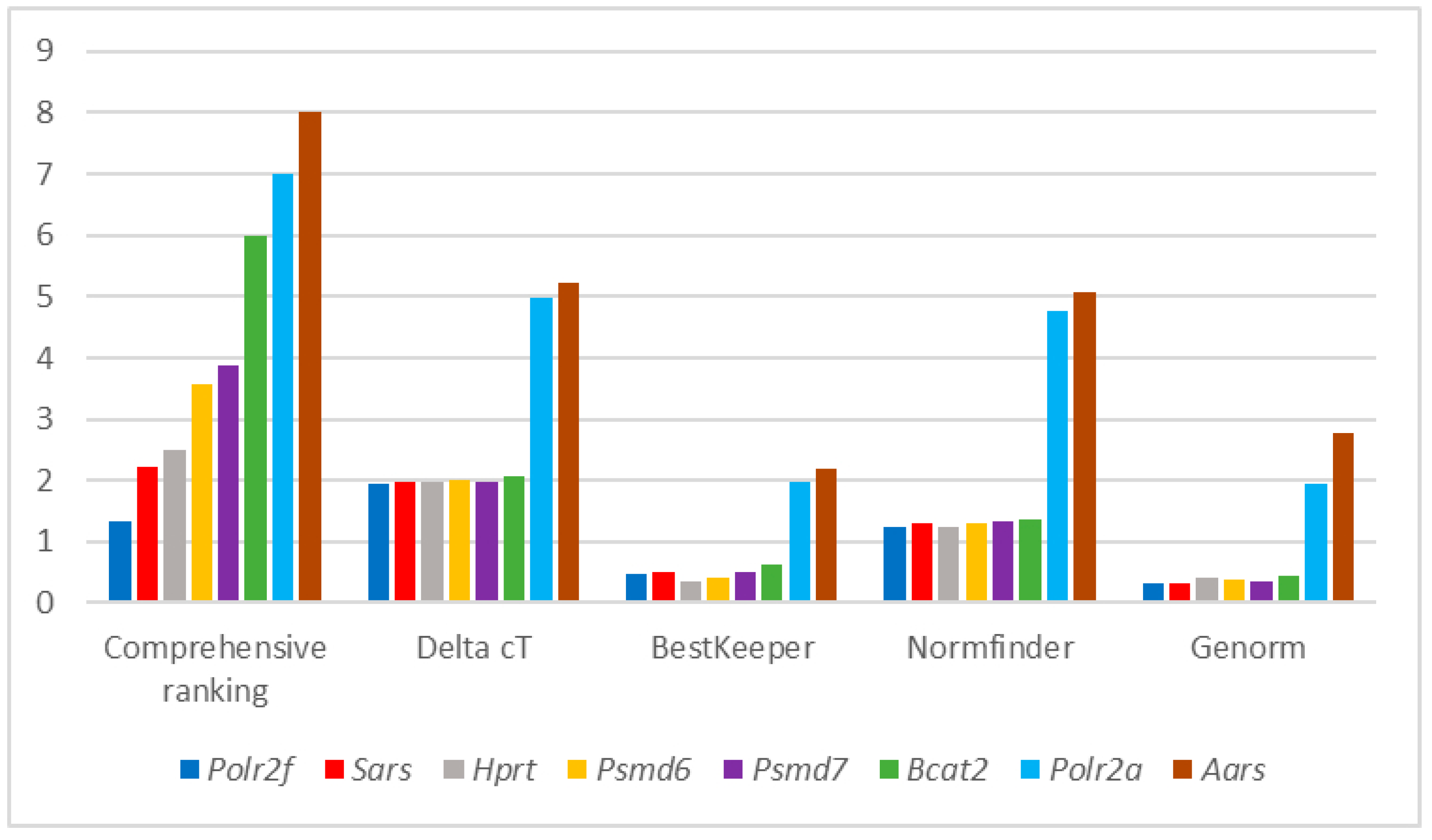
Primers and probes for analysis were selected using Beacon Designer 7.0 (Premier Biosoft International, San Francisco, CA, USA) and corresponding sequences from the NCBI database [16]. The specificities of primers and probes were checked using Primer3 [17] and Primer-BLAST [18]. Evaluation of expression of reference genes was performed using Cq values and the RefFinder tool [19].
The RefFinder program calculates the score of reference genes using the algorithms of geNorm [7], Normfinder [8], and BestKeeper [20] programs and the comparative ΔΔCt method [21].
Based on these scores, the program assigns an appropriate score to each gene and calculates the geometric mean of their weights for the overall final ranking. The program chooses the lowest score among the ranks assigned by each algorithm separately and then assigns the final comprehensive score. The closer the final score is to one, the more stably expressed the HKG is considered to be.

3. Results

For the selection of candidate genes in the HKGs database, screening was conducted in PubMed. A search for the key MeSH term “housekeeping gene” (filters applied: humans) yielded 26,399 publications since 2000. A search for the keyword “housekeeping PCR” (filters applied: humans) returned 880 results since 2000. Searching for the keyword “housekeeping real-time PCR” (filters applied: humans) returned 562 results since 2000. The search term “housekeeping real-time PCR brain” (filters applied: humans) returned only 29 results since 2001. Of these 29 studies, only 4 were directly related to the analysis of HKGs in nontumor samples [22,23,24,25].
In addition, data on the analysis of reference genes in brain tissues [26], a neuronal line differentiated from bone mesenchymal stem cells [27], and human tissues [28] were used to select potential HKGs. Using the data provided in the selected seven works, a list consisting of 542 potential HKGs was compiled for further analysis.
Next, we carried out an additional screening of publications on the query “gene expression Parkinson’s disease RNA” (filter: human) in the PubMed database, and 715 publications were identified. The Section 2 for 100 publications was randomly analyzed and a list of reference genes was compiled for use in gene expression studies.
Then, we compared the two generated lists and selected the genes included in both lists to study the stability of their expression in our work. As a result of the analysis, HKGs TBP [25,26], ACTB, RPL30 [25,29], GAPDH [23,29], YWHAZ [23], PPIA [27], B2M [26], HPRT [22,27], GUSB [25,27], SARS1, AARS1 [28], PSMD6, PSMD7 [26,28,29], POLR2F [30], POLR2A, and PSMA5 [28,31] were selected.
All potential HKGs must satisfy certain conditions. First, the genome should not contain pseudogenes of these genes. It is known that processed pseudogenes lacking introns and being almost complete generate copies of mature mRNA of the corresponding genes making up 70% of all pseudogenes [32]. In this regard, one cannot be sure that the selected primer systems will be absolutely specific to the target mRNA. Based on this criterion, HKGs ACTB [33], GUSB [34], PPIA [35], YWHAZ [36], RPL30 [37], and GAPDH [38] were excluded.
Second, a reference gene means a gene whose expression does not change under the conditions of the studied pathology and is also approximately the same in all studied tissues. In this regard, expression of all selected HKGs was analyzed using BioGPS, and HKG B2M [39] was excluded from further analysis (the difference in expression between whole brain and whole blood is more than 12-fold).
For the remaining HKGs POLR2F, POLR2A, PSMD6, PSMD7, HPRT1, PSMA5, SARS1, AARS1, TBP, and BCAT2 (the names are given in the nomenclature for humans), TaqMan primer systems were selected, and an analysis of changes in their relative mRNA levels was performed in brains of mice and mouse and human blood samples. It should be noted that at the stage of selection of primer systems and their validation for analysis, the TBP, POLR2F, and BCAT2 genes in humans were eliminated because their levels of transcripts were below the limit of detection. After obtaining the primary data, the Ct genes were analyzed using the RefFinder program.
An example of data processing for the analysis of relative mRNA levels in mouse peripheral blood is shown in general in Figure 1.
The cumulative results of the analysis are shown in Table 1 and Table 2 for mice and humans. To identify the most stably expressed HKGs under PD conditions, we analyzed the stability of expression of HKGs separately in the group of control mice and healthy individuals (Table 1 and separately in the group of mice with the MPTP-induced PD model and in patients with PD (Table 2). Stability of expression was assessed based on a comprehensive score for each gene in each tissue. In each specific case (tissue and presence/absence of pathology), the most stably expressed HKG is the one with a minimum comprehensive score. For example, Psmd7 is the most optimal and stably expressed HKG in the brain as a whole, as well as in three separate brain tissues (cerebellum, striatum, substantia nigra) in the control group of mice; however, this gene is the second in the cortex, where Aars1 is in the first place. At the same time, Psmd6 is the most stably expressed in the brain, as a whole, of experimental animals with the MPTP-induced PD model; however, it occupies only fourth place in the cortex and striatum and third place in the cerebellum and substantia nigra.
In general, as can be seen from the data presented, the stability of expression of the HKGs studied differs depending on the presence or absence of pathological processes.
Comprehensive scores and gene ranks were obtained for each gene in each tissue examined.
Based on the fact that the stability in different tissues and in the presence and absence of pathology is different, when carrying out work on expression analysis, it is necessary to use HKGs whose expression is most stable in a certain tissue both in the presence and in the absence of pathology. Accordingly, a table was compiled for the final ranking of HKGs, based on the calculation of the summarized comprehensive score (Table 3).
The summarized comprehensive score of each HKG in each tissue was summarized from the comprehensive score obtained from the control mice and healthy volunteers (Table 1) and mice with Parkinson-like phenotype and patients with PD (Table 2).
For example, in the brain as a whole, regardless of the presence or absence of pathology, Aars1 is the most stably expressed and takes the first place (gene rank = 1) with a total comprehensive score of 4.21 = 2.21 + 2.20, where 2.21 is a comprehensive score from the column “whole brain” of Table 1 and 2.20 is a comprehensive score from the “whole brain” column of Table 2. Thus, the gene with the lowest value is considered the most stably expressed and takes the highest place (gene rank = 1).
The final rank of each HKG in tissues of mice was summarized from the comprehensive score obtained from the control group (Table 1) and the experimental group (Table 2). Similarly, the final rank for HKGs in human tissues was obtained. At the same time, the gene with the lowest value is considered the most stably expressed and takes the highest place (final rank).
In general, based on the data obtained, the HKG Aars has the most stable expression among all HKGs studied in the brain. The same gene ranks second in terms of stability of expression in various parts of the brain. Ranking second in the whole brain, Psmd7 was most stably expressed in the striatum and substantia nigra. In the cortex, the most stably expressed HKG was Polr2f, and in the cerebellum, the most stably expressed HKG was Psmd6. It is noteworthy that the first of them was also the most stably expressed in the peripheral blood of mice, and the second was the most stably expressed in the peripheral blood of humans.
Summarized comprehensive scores and gene ranks were obtained for each gene in each tissue studied.
According to the widespread opinion of the scientific community that when analyzing changes in gene expression, it is necessary to take into account at least two HKGs, one should take into account the genes located at the first, second, and, at the most, at the third place in the stability rating of HKGs given in Table 3.
Furthermore, as the analysis shows, HKGs in similar tissues of the studied organisms are different. For example. Polr2f is the most stably expressed HKG in the peripheral blood of mice, whereas PSMD6 is the most stably expressed gene in humans (Table 3).

4. Discussion

Currently, TaqMan qPCR is one of the most widely used methods in molecular genetics. At the same time, one of the key factors in the success of this method is the correct choice of HKGs. Today, hundreds of reference genes are known both in the literature and in commercial panels. At the same time, data from one expression study often do not reproduce the results of other similar studies; differences in the results obtained may be partly explained by the choice and instability of expression of HKGs in varying experimental conditions. A striking example is the comparison of the results of two well-performed studies. In an analysis of the stability of HKGs during the reprogramming of the mouse neural stem cell line N31, Panina and coauthors in 2018 showed that GAPDH is one of the most stably expressed HKGs among those analyzed [40]. However, in 2020, the same group of scientists, when analyzing HKGs in induced pluripotent stem cells of the same lineage, concluded that GAPDH has a highly variable expression during reprogramming. This gene is not attractive as a reference gene for the analysis of expression in works related to the process of fibroblast reprogramming [41]. The authors point out that the way the primers were selected changed from the first publication to the second. With this in mind, they explain that in the first of these works, the Rps18 gene was considered as one of the variable genes while in the second work, the authors recognized Rps18 as a stably expressed HKG.
In this work, we analyzed the relative mRNA levels of reference genes in mouse brain tissues, as well as in the peripheral blood of mice and humans. The analysis was carried out in samples of brain tissues and blood of mice with modeling of Parkinson-like phenotype and in matching samples from a control group of mice. The work presented here was aimed, first of all, at studying the stability of expression of HKGs in the main tissues of the mouse brain, as one of the main objects of PD modeling. At the same time, a toxic model based on the introduction of MPTP (1-methyl-4-phenyl-1,2,3,6-tetrahydropyridine) was chosen. Models based on the introduction of this toxin are currently considered classical [42]. It has been shown that MPTP has an affinity for the DAT dopamine transporter, which causes a key feature of PD, degeneration of dopaminergic neurons and dopamine deficiency in the striatum [43,44]. For the analysis, we used a model of the early symptomatic stage of PD based on the administration of MPTP, similar to stages 1–2 on the Hoehn–Yahr scale in patients with PD [45]. In addition, peripheral blood samples from patients with PD (stages 1–2 on the Hoehn–Yahr scale), as well as healthy volunteers, were studied.
Based on a bioinformatic analysis searching for the presence of pseudogenes, the genes ACTB, GUSB, PPIA, YWHAZ, and GAPDH, widely used as HKGs, were removed from further study. For example, ACTB and GAPDH are the most frequently used housekeeping systems. ACTB has 22 pseudogenes and GAPDH has 47 [46]. Pseudogenes can be amplified together with cDNA obtained from the target RNA. On the one hand, treatment with DNases can serve as a solution to this problem, although this greatly decreases the yield of isolated RNA. This method is of little use considering that the purpose of the study was to analyze changes in the levels of poorly presented transcripts. On the other hand, amplification of genomic DNA can be excluded by selecting primers for exon–exon junctions, and thus only the target cDNA obtained from mature RNA devoid of introns will be expressed. As noted above, it is known that processed pseudogenes lack introns, are almost complete copies of mature mRNAs of the corresponding genes, and make up 70% of all pseudogenes [32,46]. In this regard, one cannot be sure that the selected primer system will be absolutely specific for the target mRNA, and it is appropriate not to select genes having pseudogenes for use as HKGs [47].
The rest of the HKGs selected based on the results of the literature analysis passed the bioinformatic analysis. Coincidentally, in silico pseudogenes were found for the PSMD7 (NM_002811.5) and HPRT1 (NM_000194.3) genes. However, there is currently no experimental confirmation of their existence, so we selected these genes for further analysis. In addition, primers were selected so that they did not anneal to the region of putative pseudogenes in the genome.
One of the fundamentally important conditions in the selection of HKGs is the stability of their expression level, regardless of the presence or absence of the development of a pathological process. In our work, it was shown that the presence of a pathological process affects the stability of the expression of HKGs. As can be seen from Table 1 and Table 2, none of the HKGs with the lowest score and, correspondingly, the highest rank in the control group of mice (Table 1) coincide with those in the group of mice with MPTP-induced PD (Table 2). A similar situation is observed when analyzing HKGs in human peripheral blood.
Overall, the most stably expressed HKG in the mouse brain (Table 3) is Aars, which encodes an alanyl-tRNA synthetase. This gene is not directly involved in the processes associated with the functioning of the nervous system and processes involved in the pathogenesis of PD. In addition, this gene is the second most stably expressed gene in some of the brain tissues we studied. Thus, this gene is the most stably expressed and attractive HKG for expression analysis.
The second most stably expressed gene in the mouse brain is the gene associated with the functioning of proteasome subunits. Even though the processes associated with the functioning of ubiquitin-dependent proteolysis are involved in the pathogenesis of PD, the Psmd7 gene in mice turned out to be one of the most stably expressed in the brain as a whole (Table 3). The same gene is the most stably expressed in the substantia nigra and striatum.
It should be noted that transcriptome studies in the human brain are possible only in postmortem tissues. Unfortunately, such studies do not reflect the processes involved in the pathogenesis of the disease, especially in the early stages. In this regard, the optimal approach to research in vivo is the analysis of peripheral blood that allows studies of the pathogenesis of the disease, including its early stages.
The gene encoding polymerase 2 subunits in mice, Polr2f, and gene encoding proteasome subunit 6 in humans, PSMD6, have the least variability in their expression when analyzing peripheral blood. These data are not surprising because the functioning of the polymerase in the cell is of decisive importance for its vital activity. In this regard, the stability of expression of Polr2f as HKGs is beyond doubt. Based on all the data obtained, it can be concluded that when analyzing changes in gene expression under pathological conditions, it is necessary to select HKGs for each specific tissue under study. As shown in Table 3 for the mouse brain, even if the stability of the relative mRNA levels of some reference gene, for example, Aars, is shown for the organ as a whole, this does not mean that it will be the most effective HKG for other tissues, which is shown, for example, for the cerebellum (Psmd6).

5. Conclusions

The data obtained indicate that there is no universal HKG both for different brain tissues of one organism and for similar tissues of different organisms. This conclusion is supported by other studies: researchers must select and validate a set of reference genes for each specific cell line [41].
In addition, we have shown that the presence of a pathological process may affect the expression of HKGs. This fact must be taken into account when choosing the optimal HKG for expression analysis in pathological conditions. In this case, it is necessary to choose those HKGs that are most stably expressed in both experimental and control groups, in other words, regardless of experimental conditions.
In conclusion, it should be noted that there are certain limitations of the use of the HKGs selected in this study as the most stably expressed. Thus, these HKGs can be most confidently used to study the pathological process caused by the pathogenesis of PD in young mice, as well as for PD patients at relatively early stages of the pathological process.

Supplementary Materials

The following are available online at https://www.mdpi.com/article/10.3390/cells10092252/s1, Table S1: Sequences of gene-specific primers and probes for qPCR (TaqMan).

Author Contributions

Conception, M.I.S., P.A.S.; organization, M.I.S., P.A.S., M.M.R.; execution, A.K.A., M.M.R., E.V.F. All authors have read and agreed to the published version of the manuscript.

Funding

This work was supported by Russian Science Foundation, grant No. 20-15-00262.

Institutional Review Board Statement

The study was conducted according to the guidelines of the Declaration of Helsinki and approved by the Ethics Committee of the Research Center of Neurology (protocol 18/4, 3 March 2021) and the Ethics Committee of the Institute of Molecular Genetics of National Research Centre “Kurchatov Institute” (protocol 3/21, 17 February 2021).

Informed Consent Statement

Informed consent was obtained from all subjects involved in the study.

Data Availability Statement

Not applicable.

Acknowledgments

We are deeply grateful to Sergey N. Illarioshkin and Alexey V. Karabanov (Research Center of Neurology) for the provided samples of peripheral blood of patients and healthy volunteers.

Conflicts of Interest

The authors declare no conflict of interest.

References

  1. Van Guilder, H.D.; Vrana, K.E.; Freeman, W.M. Twenty-five years of quantitative PCR for gene expression analysis. BioTechniques 2008, 44, 619–626. [Google Scholar] [CrossRef] [PubMed] [Green Version]
  2. Dheda, K.; Huggett, J.F.; Bustin, S.A.; Johnson, M.A.; Rook, G.; Zumla, A. Validation of housekeeping genes for normalizing RNA expression in real-time PCR. BioTechniques 2004, 37, 112–114, 116, 118–119. [Google Scholar] [CrossRef] [Green Version]
  3. Chervoneva, I.; Li, Y.; Schulz, S.; Croker, S.; Wilson, C.; Waldman, S.A.; Hyslop, T. Selection of optimal reference genes for normalization in quantitative RT-PCR. BMC Bioinform. 2010, 11, 253. [Google Scholar] [CrossRef] [PubMed] [Green Version]
  4. Thellin, O.; ElMoualij, B.; Heinen, E.; Zorzi, W. A decade of improvements in quantification of gene expression and internal standard selection. Biotechnol. Adv. 2009, 27, 323–333. [Google Scholar] [CrossRef]
  5. Takagi, S.; Ohashi, K.; Utoh, R.; Tatsumi, K.; Shima, M.; Okano, T. Suitable reference genes for the analysis of direct hyperplasia in mice. Biochem. Biophys. Res. Commun. 2008, 377, 1259–1264. [Google Scholar] [CrossRef]
  6. Tatsumi, K.; Ohashi, K.; Taminishi, S.; Okano, T.; Yoshioka, A.; Shima, M. Reference gene selection for real-time RT-PCR in regenerating mouse livers. Biochem. Biophys. Res. Commun. 2008, 374, 106–110. [Google Scholar] [CrossRef]
  7. Vandesompele, J.; De Preter, K.; Pattyn, F.; Poppe, B.; Van Roy, N.; De Paepe, A.; Speleman, F. Accurate normalization of real-time quantitative RT-PCR data by geometric averaging of multiple internal control genes. Genome Biol. 2002, 3, 1–12. [Google Scholar] [CrossRef] [PubMed] [Green Version]
  8. Andersen, C.L.; Jensen, J.L.; Orntoft, T.F. Normalization of real-time quantitative reverse transcription-PCR data: A model-based variance estimation approach to identify genes suited for normalization, applied to bladder and colon cancer data sets. Cancer Res. 2004, 64, 5245–5250. [Google Scholar] [CrossRef] [PubMed] [Green Version]
  9. Nicot, N.; Hausman, J.F.; Hoffmann, L.; Evers, D. Housekeeping gene selection for real-time RT-PCR normalization in potato during biotic and abiotic stress. J. Exp. Bot. 2005, 56, 2907–2914. [Google Scholar] [CrossRef]
  10. Kin, K.; Yasuhara, T.; Kameda, M.; Date, I. Animal Models for Parkinson’s Disease Research: Trends in the 2000s. Int. J. Mol. Sci. 2019, 20, 5402. [Google Scholar] [CrossRef] [Green Version]
  11. Goetz, C.G.; Tilley, B.C.; Shaftman, S.R.; Stebbins, G.T.; Fahn, S.; Martinez-Martin, P.; Poewe, W.; Sampaio, C.; Stern, M.B.; Dodel, R.; et al. Movement Disorder Society-sponsored revision of the Unified Parkinson’s Disease Rating Scale (MDS-UPDRS): Scale presentation and clinimetric testing results. Mov. Disord. 2008, 23, 2129–2170. [Google Scholar] [CrossRef]
  12. Goetz, C.G.; Poewe, W.; Rascol, O.; Sampaio, C.; Stebbins, G.T.; Counsell, C.; Giladi, N.; Holloway, R.G.; Moore, C.G.; Wenning, G.K.; et al. Movement Disorder Society Task Force report on the Hoehn and Yahr staging scale: Status and recommendations. Mov. Disord. 2004, 19, 1020–1028. [Google Scholar] [CrossRef]
  13. Hughes, A.J.; Daniel, S.E.; Kilford, L.; Lees, A.J. Accuracy of clinical diagnosis of idiopathic Parkinson’s disease: A clinico-pathological study of 100 cases. J. Neurol. Neurosurg. Psychiatry 1992, 55, 181–184. [Google Scholar] [CrossRef] [Green Version]
  14. The Guide for the Care and Use of Laboratory Animals, 8th ed.; National Academies Press: Washington, DC, USA, 2011.
  15. Suslov, O.; Steindler, D.A. PCR inhibition by reverse transcriptase leads to an overestimation of amplification efficiency. Nucleic Acids Res. 2005, 33, e181. [Google Scholar] [CrossRef] [PubMed]
  16. NCBI Database. Available online: https://www.ncbi.nlm.nih.gov/ (accessed on 15 July 2019).
  17. Primer3. Available online: http://bioinfo.ut.ee/primer3 (accessed on 15 July 2019).
  18. Primer-BLAST. Available online: https://www.ncbi.nlm.nih.gov/tools/primer-blast/ (accessed on 15 July 2019).
  19. Xie, F.; Xiao, P.; Chen, D.; Xu, L.; Zhang, B. miRDeepFinder: A miRNA analysis tool for deep sequencing of plant small RNAs. Plant Mol. Biol. 2012, 80, 75–84. [Google Scholar] [CrossRef] [PubMed]
  20. Pfaffl, M.W.; Tichopad, A.; Prgomet, C.; Neuvians, T.P. Determination of stable housekeeping genes, differentially regulated target genes and sample integrity: BestKeeper—Excel-based tool using pair-wise correlations. Biotechnol. Lett. 2004, 26, 509–515. [Google Scholar] [CrossRef] [PubMed]
  21. Silver, N.; Best, S.; Jiang, J.; Thein, S.L. Selection of housekeeping genes for gene expression studies in human reticulocytes using real-time PCR. BMC Mol. Biol. 2006, 7, 33. [Google Scholar] [CrossRef] [Green Version]
  22. Maurer-Morelli, C.V.; de Vasconcellos, J.F.; Reis-Pinto, F.C.; Rocha Cde, S.; Domingues, R.R.; Yasuda, C.L.; Tedeschi, H.; De Oliveira, E.; Cendes, F.; Lopes-Cendes, I. A comparison between different reference genes for expression studies in human hippocampal tissue. J. Neurosci. Methods 2012, 208, 44–47. [Google Scholar] [CrossRef]
  23. Penna, I.; Vella, S.; Gigoni, A.; Russo, C.; Cancedda, R.; Pagano, A. Selection of candidate housekeeping genes for normalization in human postmortem brain samples. Int. J. Mol. Sci. 2011, 12, 5461–5470. [Google Scholar] [CrossRef] [Green Version]
  24. Gebhardt, F.M.; Scott, H.A.; Dodd, P.R. Housekeepers for accurate transcript expression analysis in Alzheimer’s disease autopsy brain tissue. Alzheimer’s Dement. J. Alzheimer’s Assoc. 2010, 6, 465–474. [Google Scholar] [CrossRef]
  25. Swijsen, A.; Nelissen, K.; Janssen, D.; Rigo, J.M.; Hoogland, G. Validation of reference genes for quantitative real-time PCR studies in the dentate gyrus after experimental febrile seizures. BMC Res. Notes 2012, 5, 685. [Google Scholar] [CrossRef] [Green Version]
  26. Harrison, P.J.; Laatikainen, L.M.; Tunbridge, E.M.; Eastwood, S.L. Human brain weight is correlated with expression of the ‘housekeeping genes’ beta-2-microglobulin (beta2M) and TATA-binding protein (TBP). Neuropathol. Appl. Neurobiol. 2010, 36, 498–504. [Google Scholar] [CrossRef]
  27. He, Y.X.; Zhang, Y.; Yang, Q.; Wang, C.; Su, G. Selection of suitable reference genes for reverse transcription-quantitative polymerase chain reaction analysis of neuronal cells differentiated from bone mesenchymal stem cells. Mol. Med. Rep. 2015, 12, 2291–2300. [Google Scholar] [CrossRef]
  28. Warrington, J.A.; Nair, A.; Mahadevappa, M.; Tsyganskaya, M. Comparison of human adult and fetal expression and identification of 535 housekeeping/maintenance genes. Physiol. Genom. 2000, 2, 143–147. [Google Scholar] [CrossRef]
  29. Chapman, J.R.; Waldenstrom, J. With Reference to Reference Genes: A Systematic Review of Endogenous Controls in Gene Expression Studies. PLoS ONE 2015, 10, e0141853. [Google Scholar] [CrossRef]
  30. Hoerndli, F.J.; Toigo, M.; Schild, A.; Gotz, J.; Day, P.J. Reference genes identified in SH-SY5Y cells using custom-made gene arrays with validation by quantitative polymerase chain reaction. Anal. Biochem. 2004, 335, 30–41. [Google Scholar] [CrossRef]
  31. Alieva, A.K.; Filatova, E.V.; Karabanov, A.V.; Illarioshkin, S.N.; Slominsky, P.A.; Shadrina, M.I. Potential Biomarkers of the Earliest Clinical Stages of Parkinson’s Disease. Parkinson’s Dis. 2015, 2015, 294396. [Google Scholar] [CrossRef] [PubMed] [Green Version]
  32. Torrents, D.; Suyama, M.; Zdobnov, E.; Bork, P. A genome-wide survey of human pseudogenes. Genome Res. 2003, 13, 2559–2567. [Google Scholar] [CrossRef] [PubMed] [Green Version]
  33. NCBI-Gene/ACTB Pseudogenes. Available online: https://www.ncbi.nlm.nih.gov/gene/?Term=related_functional_gene_60%5Bgroup%5D (accessed on 8 June 2021).
  34. NCB-Gene/GUSB Pseudogenes. Available online: https://www.ncbi.nlm.nih.gov/gene/?Term=related_functional_gene_2990%5Bgroup%5D (accessed on 8 June 2021).
  35. NCBI-Gene/PPIA Pseudogenes. Available online: https://www.ncbi.nlm.nih.gov/gene/?Term=related_functional_gene_5478%5Bgroup%5D (accessed on 8 June 2021).
  36. NCBI-Gene/YWHAZ Pseudogenes. Available online: https://www.ncbi.nlm.nih.gov/gene/?Term=related_functional_gene_7534%5Bgroup%5D (accessed on 8 June 2021).
  37. NCBI-Gene/RPL30 Pseudogenes. Available online: https://www.ncbi.nlm.nih.gov/gene/?Term=related_functional_gene_6156%5Bgroup%5D (accessed on 8 June 2021).
  38. NCBI-Gene/GAPDH Pseudogenes. Available online: https://www.ncbi.nlm.nih.gov/gene/?Term=related_functional_gene_2597%5Bgroup%5D (accessed on 8 June 2021).
  39. BioGPS. Available online: http://biogps.org/#goto=genereport&id=567 (accessed on 8 June 2021).
  40. Panina, Y.; Germond, A.; Masui, S.; Watanabe, T.M. Validation of Common Housekeeping Genes as Reference for qPCR Gene Expression Analysis During iPS Reprogramming Process. Sci. Rep. 2018, 8, 8716. [Google Scholar] [CrossRef] [PubMed]
  41. Panina, Y.; Germond, A.; Watanabe, T.M. Analysis of the stability of 70 housekeeping genes during iPS reprogramming. Sci. Rep. 2020, 10, 21711. [Google Scholar] [CrossRef]
  42. Salari, S.; Bagheri, M. In vivo, in vitro and pharmacologic models of Parkinson’s disease. Physiol. Res. 2019, 68, 17–24. [Google Scholar] [CrossRef]
  43. German, D.C.; Nelson, E.L.; Liang, C.L.; Speciale, S.G.; Sinton, C.M.; Sonsalla, P.K. The neurotoxin MPTP causes degeneration of specific nucleus A8, A9 and A10 dopaminergic neurons in the mouse. Neurodegener. A J. Neurodegener. Disord. Neuroprot. Neuroregener. 1996, 5, 299–312. [Google Scholar] [CrossRef] [PubMed]
  44. Meredith, G.E.; Totterdell, S.; Potashkin, J.A.; Surmeier, D.J. Modeling PD pathogenesis in mice: Advantages of a chronic MPTP protocol. Parkinsonism Relat. Disord. 2008, 14 (Suppl. 2), S112–S115. [Google Scholar] [CrossRef] [Green Version]
  45. Ugrumov, M.V.; Khaindrava, V.G.; Kozina, E.A.; Kucheryanu, V.G.; Bocharov, E.V.; Kryzhanovsky, G.N.; Kudrin, V.S.; Narkevich, V.B.; Klodt, P.M.; Rayevsky, K.S.; et al. Modeling of presymptomatic and symptomatic stages of parkinsonism in mice. Neuroscience 2011, 181, 175–188. [Google Scholar] [CrossRef] [PubMed]
  46. Sasidharan, R.; Gerstein, M. Genomics: Protein fossils live on as RNA. Nature 2008, 453, 729–731. [Google Scholar] [CrossRef] [PubMed]
  47. Balaji, S.; Vanniarajan, A. Implication of Pseudo Reference Genes in Normalization of Data from Reverse Transcription-Quantitative PCR. Gene 2020, 757, 144948. [Google Scholar] [CrossRef] [PubMed]
Figure 1. Comprehensive ranking of HKGs in mouse peripheral blood by RefFinder.
Cells 10 02252 g001
Table 1. Table of the stability of expression of potential HKGs in the brain and peripheral blood of control group of mice and peripheral blood of control group of mice and healthy volunteers according to the RefFinder program.
Gene Rank 2MiceHumans
Whole BrainCortexCerebellumStriatumSubstantia NigraBloodPeripheral Blood
HKGScore 1HKGScoreHKGScoreHKGScoreHKGScoreHKGScoreHKGScore
1Psmd71.57Aars1.68Psmd71.86Psmd71.00Psmd71.32Sars1.57SARS11.57
2Aars2.21Psmd72.06Psmd62.63Aars2.21Aars1.68Polr2f2.38PSMD62.11
3Polr2a2.45Polr2f2.21Hprt3.13Hprt3.31Polr2a2.83Polr2a2.71HPRT13.16
4Bcat22.99Psmd63.83Aars3.31Polr2a3.31Bcat23.87Hprt3.98POLR2A3.22
5Psmd63.98Bcat24.53Polr2f4.30Sars4.56Sars4.61Psmd74.16PSMA53.94
6Hprt6.24Hprt4.95Bcat24.79Bcat25.92Polr2f6.09Psmd64.92PSMD74.56
7Sars7.24Polr2a6.44Polr2a5.66Psmd66.48Psmd66.24Bcat25.14AARS17.00
8Polr2f7.44Sars8.00Sars5.73Polr2f8.00Hprt8.00Aars8.00
1 Score—a comprehensive score calculated using the RefFinder program. 2 Gene rank (a place in the rating of stability of expression) is based on sorting HKGs by their comprehensive scores from minimum to maximum. The first place in the ranking (gene rank = 1) is assigned to the gene with the minimum comprehensive score, and this gene is considered the most stably expressed HKG in the tissue under study. The last place in the ranking (gene rank = 8) is assigned to the gene with the maximum comprehensive score, and this HKG is considered the least stably expressed HKG.
Table 2. Table of the stability of expression of potential HKGs in the brain and peripheral blood of experimental group of mice and peripheral blood of experimental group of mice and PD patients according to the RefFinder program.
Gene Rank 2MiceHumans
Whole BrainCortexCerebellumStriatumSubstantia NigraBloodPeripheral Blood
HKGScore 1HKGScoreHKGScoreHKGScoreHKGScoreHKGScoreHKGScore
1Psmd61.86Bcat21.32Aars1.78Sars1.50Hprt1.50Hprt1.78POLR2A1.41
2Aars2.00Polr2f2.06Polr2f1.86Psmd72.21Psmd72.00Polr2f1.86PSMD62.45
3Polr2f2.11Aars2.63Psmd62.45Aars3.25Bcat23.00Psmd62.45PSMA52.82
4Psmd73.08Psmd63.46Polr2a2.63Polr2a3.46Psmd63.83Psmd72.83PSMD73.98
5Sars4.16Polr2a5.00Bcat25.38Bcat24.60Aars4.43Sars4.61SARS4.23
6Bcat26.24Psmd75.09Psmd75.48Hprt4.61Polr2f5.12Bcat25.73HPRT14.30
7Polr2a7.24Sars7.24Sars6.96Psmd65.96Sars5.44Polr2a7.00AARS16.00
8Hprt7.44Hprt7.44Hprt7.74Polr2f7.20Polr2a8.00Aars8.00
1 Score—a comprehensive score calculated using the RefFinder program. 2 Gene rank (a place in the rating of stability of expression) is based on sorting HKGs by their comprehensive scores from minimum to maximum. The first place in the ranking (gene rank = 1) is assigned to the gene with the minimum comprehensive score, and this gene is considered the most stably expressed HKG in the tissue under study. The last place in the ranking (gene rank = 8) is assigned to the gene with the maximum comprehensive score, and this HKG is considered the least stably expressed HKG.
Table 3. Summary table of the stability of expression of potential HKGs based on total score in the brain and peripheral blood of mice and human peripheral blood according to the RefFinder program regardless of the presence or absence of pathology.
Gene Rank 2MiceHumans
Whole BrainCortexCerebellumStriatumSubstantia NigraPeripheral BloodPeripheral Blood
HKGS-Score 1HKGS-ScoreHKGS-ScoreHKGS-ScoreHKGS-ScoreHKGS-ScoreHKGS-Score
1Aars4.21Polr2f4.27Psmd65.08Psmd73.21Psmd73.32Polr2f4.24PSMD64.56
2Psmd74.65Aars4.31Aars5.09Aars5.46Aars6.11Hprt5.76POLR2A4.63
3Psmd65.84Bcat25.85Polr2f6.16Sars6.06Bcat26.87Sars6.18SARS15.80
4Bcat29.23Psmd77.15Psmd77.34Polr2a6.77Hprt9.50Psmd76.99PSMA56.76
5Polr2f9.55Psmd67.29Polr2a8.29Hprt7.92Sars10.05Psmd67.37HPRT17.46
6Polr2a9.69Polr2a11.44Bcat210.17Bcat210.52Psmd610.07Polr2a9.71PSMD78.54
7Sars11.40Hprt12.39Hprt10.87Psmd612.44Polr2a10.83Bcat210.87AARS113.00
8Hprt13.68Sars15.24Sars12.69Polr2f15.20Polr2f11.21Aars16.00
1 S-Score—the summarized comprehensive score for each HKG in each tissue was calculated as the sum of the scores from Table 1 and Table 2. 2 Gene rank is based on sorting HKGs by their summarized comprehensive score from the minimum value, where the corresponding gene is assigned gene rank = 1, to the maximum value, where the corresponding gene is assigned gene rank = 8. The 1st place in the ranking (gene rank = 1) belongs to the most stable housekeeping gene.
Publisher’s Note: MDPI stays neutral with regard to jurisdictional claims in published maps and institutional affiliations.

