Review

A Comprehensive Review of the Genomics of Multiple Myeloma: Evolutionary Trajectories, Gene Expression Profiling, and Emerging Therapeutics

1
Department of Translational Hematology and Oncology Research, Taussig Cancer Institute, Cleveland Clinic, Cleveland, OH 44106, USA
2
Division of Hematology and Oncology, Medical College of Wisconsin, Milwaukee, WI 53226, USA
*
Author to whom correspondence should be addressed.
These authors contributed equally and are co-first authors.
Cells 2021, 10(8), 1961; https://doi.org/10.3390/cells10081961
Submission received: 28 June 2021 / Revised: 21 July 2021 / Accepted: 30 July 2021 / Published: 2 August 2021
(This article belongs to the Special Issue Molecular Insights in Multiple Myeloma Pathogenesis)

Abstract

:
Multiple myeloma (MM) is a blood cancer characterized by the accumulation of malignant monoclonal plasma cells in the bone marrow. It develops through a series of premalignant plasma cell dyscrasia stages, most notable of which is the Monoclonal Gammopathy of Undetermined Significance (MGUS). Significant advances have been achieved in uncovering the genomic aberrancies underlying the pathogenesis of MGUS-MM. In this review, we discuss in-depth the genomic evolution of MM and focus on the prognostic implications of the accompanied molecular and cytogenetic aberrations. We also dive into the latest investigatory techniques used for the diagnoses and risk stratification of MM patients.

1. Introduction

Multiple myeloma (MM), is a malignant clonal proliferation of plasma cells compromising 1.8% of all new cancer cases in the U.S. based on the Surveillance, Epidemiology, and End Results Program (SEER) cancer database [1]. Data from randomized clinical trials revealed an overall five-year survival rate of about 54% and a median overall survival of approximately six years [2]. Most recently, a study on long-term outcomes in MM after autologous stem cell transplantation revealed better overall survival in patients treated in 2014 or after as compared to 1997 or before [3]. This significant difference in outcomes derives from the substantial progress made in the understanding of disease pathobiology and the introduction of novel therapeutics [4,5]; yet prognosis remains poor, especially in genetically defined high-risk subgroups [6]. MM is a heterogeneous disease characterized by the acquisition of complex genetic changes during disease evolution from the premalignant condition monoclonal gammopathy of undetermined significance (MGUS) [7] and smoldering MM (SMM) [8]. Seminal studies provided crucial information about the complex evolutionary process in MM patients and changes in genomics of clonal architecture as the disease progresses [9,10,11]. In clinical practice, conventional karyotyping and interphase fluorescence in situ hybridization (FISH) from bone marrow samples are utilized to classify genomic risk and identify patients with high-risk abnormalities [8]. Recently, extensive studies of MM genomics have led to an improved understanding of the molecular biology of MM. This resulted in new molecular classifications of MM subtypes, that have been proposed using gene expression profiling, and the identification of genes involved in the disease process [12,13]. Here, we summarize the current knowledge on the complex genomic landscape and pathophysiological mechanisms of MM.

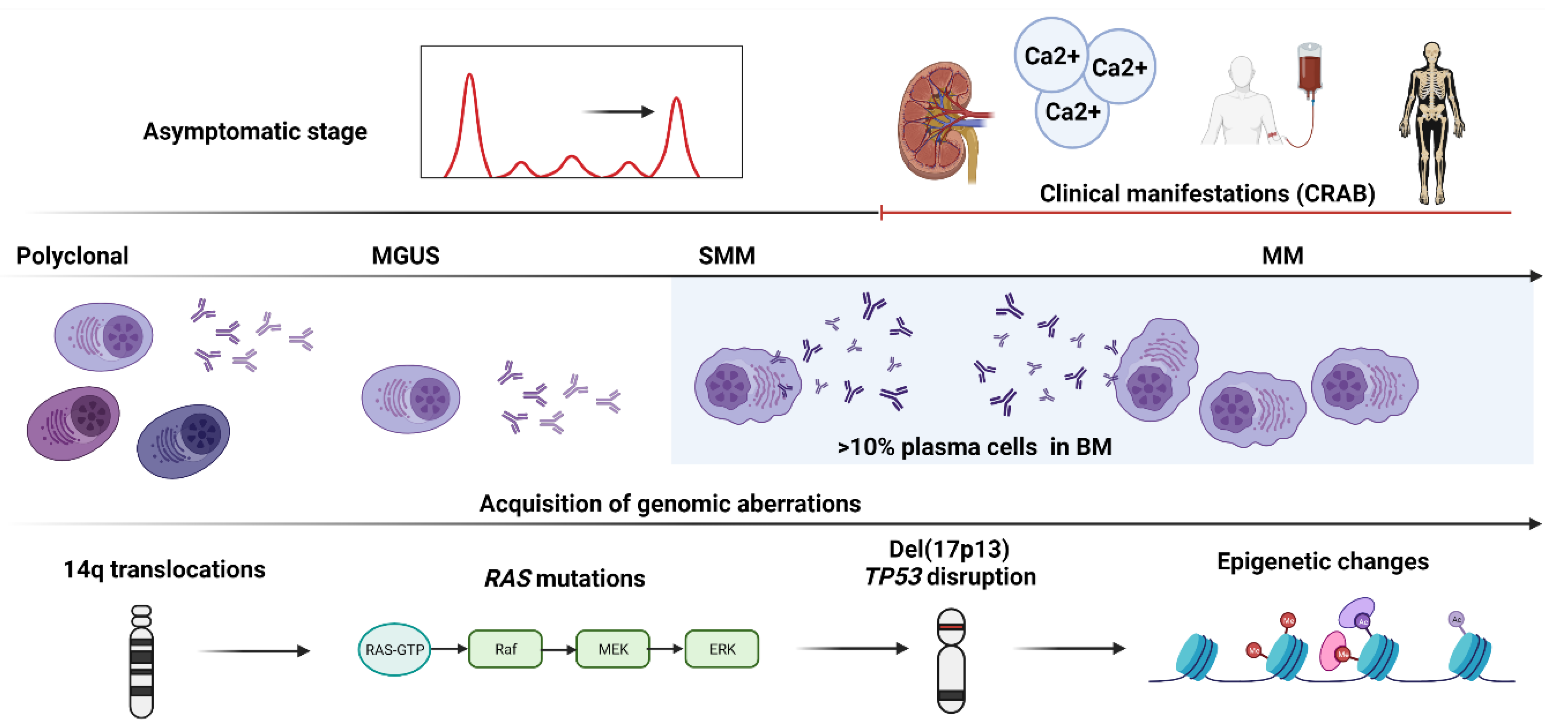
2. Genetic and Cytogenetic Abnormalities: A Long Way from MGUS to MM Progression

The genetic complexity of MM underlies a progressive, multistep process through which the continuous accumulation of genetic aberrancies drives monoclonal plasma cells towards malignancy [14]. These aberrancies are generally classified as either primary or secondary (Figure 1). Primary aberrancies initiate the process of plasma cell immortalization and its consequent commitment to the MM disease pathway [15]. Whether its progress eventually leads to MM or halts/remains at an earlier phase is dependent on the secondary aberrancies which further modulate disease progression [15]. It is now known that MM, in almost all patients, evolves from a premalignant precursor stage, MGUS, which is characterized by monoclonal plasma cells of limited malignant potential. Hence, to fully understand the genetic foundations of the pathophysiology of MM, we must also consider the pathogenesis of MGUS, its direct precursor. Several studies investigated the genomic changes occurring at the stage of MGUS [16,17].
Plasma cells are terminally differentiated B cells, which in physiologic conditions are incapable of undergoing cell division. The acquisition of genomic aberrations (cytogenetics primary events, left panel) may underpin the transformation from polyclonal to monoclonal plasma cells (stage of monoclonal gammopathy of undetermined significance, MGUS). Additional molecular events underlie the progression to smoldering multiple myeloma (SMM), multiple myeloma (MM) with clinical disease manifestations, and at the extreme pole, plasma cell leukemia when >2 × 10⁹ plasma cells/L are present in the peripheral blood (or >20% of nucleated blood cells are constituted by plasma cells) [18].
In fact, terminally differentiated plasma cells do not undergo cell division, however, epigenetic deregulation of gene expression has been proposed as a root cause of malignant transformation in MM [19]. Genome-wide association studies (GWAS) by Broderick and his team identified germline variants at 3p22.1, 7p15.3, and 2p23.3 as risk factors for developing MGUS [15,20]. Their associated gene pairs (DNMT3A and DTNB, ULK4 and TRAK1, DNAH11 and CDCA7L, respectively) were found to be implicated in the dysregulation of the transcription factor encoding the MYC proto-oncogene [21]. Beksac et al. investigated the association of different human leukocyte antigen (HLA) polymorphisms with the risk of developing MM [22]. While the authors confirmed DRB5*01, C*07:02 g and B*07:02 g as potential risk-alleles for MM, they also illustrated their high correlation by linkage disequilibrium in Whites. Moreover, the cross-population analysis suggested that C*07 represents the only independent risk-allele for MM while the other alleles occurring in the same haplotype rather than contributing to MM [22]. This analysis also demonstrated the protective associations of C*05:01 g and B*44:02 g when occurring on the same haplotype in Whites, and the predisposing associations of C*12:03 g~B*38:01 haplotype in Whites while B*58:01 g in Asians [22]. Nonetheless, somatic mutations and cytogenetic abnormalities are bigger contributors along the MGUS-MM clinical spectrum.

2.1. Primary Events Driving MGUS Progression to MM

Cytogenetic abnormalities split MM cases into two broad divisions: the hyperdiploid and nonhyperdiploid subtypes. These two subtypes differ in the type of the primary cytogenetic aberrancy that drives the plasma cells towards the MGUS-MM pathway. Hyperdiploidy in MGUS-MM involves the acquisition of one or more odd-numbered chromosomes in a clonal cell population, including chromosomes 3, 5, 7, 9, 11, 15, 17, and 19 [23,24,25,26]. The extra chromosomes carry genes whose overexpression may promote dysregulated replication and growth, transforming normal cells into an MGUS-MM clone.
On the contrary, the nonhyperdiploid subtype is mainly characterized by the translocation of the immunoglobulin heavy chain (IgH) locus on 14q32, which juxtaposes an oncogene on the affected recipient chromosome [24,27]. Consequently, the expression of the juxtaposed oncogene would now be under the influence of the active upstream IgH promoter [27]. The most commonly affected genes are that of cyclin D1 (11q13), cyclin D3 (6p21), FGFR3 and MMSET (4p16), c-maf (16q23), as well as mafB (22q11) [28,29]. Thus, a variety of transcription factors, growth factor receptors, and other cell cycle mediators may become overexpressed and dysregulate the plasma cell cycle, which may lead to cellular proliferation.
Although the triggers of these primary cytogenetic aberrancies remain unclear, some reports have suggested it is due to an abnormal response of plasma cells to antigenic stimulation [30,31]. Chronic antigenic stimulation predisposes to IgH class-switching recombination errors that could possibly lead to these translocations [15]. While the links remain to be fully clarified, several studies have demonstrated that the overexpression of Toll-like receptors (TLRs) and the interleukin 6 (IL-6) receptor on the surface of plasma cells promotes their proliferation, survival, and resistance to apoptosis [31,32,33]. IL-6, in particular, has been implicated in the expression of the anti-apoptotic proteins Mcl-1 and Bcl-Xl [34]. However, the sequence of events by which chronic stimulation of these receptors induces the cytogenetic aberrancies needs to be deciphered in order to fully elucidate the pathogenesis of MM.

2.2. Secondary Events Driving MGUS Progression to MM

Once the MGUS clone is established, secondary aberrancies dictate its transformation into MM or not [35]. These secondary events are generally thought to follow the random “second hit” model. In particular, this model provides the best explanation for the consistent—rather than the cumulative—annual rate (1%) of MGUS transformation into MM [7]. However, this long-standing belief has been contested recently by a study demonstrating that the individual risk of developing MM may increase over time, but in patients with MGUS diagnosed in the setting of immune-related disorders, the risk of progression could be lower [36,37]. Physicians usually determine the risk of transformation shortly after diagnosis with MGUS, but repeated reassessments may allow researchers to recognize that some of the “high risk” cases were “low risk” in previous tests. This study further emphasized the lack of certainty of models assessing MGUS progression into MM.
The majority of secondary events are also of a genetic/cytogenetic origin, which include translocations, deletions, mutations, and others. Secondary translocations are class-switching independent as opposed to primary translocations [38]. They mainly affect MYC (8q24), whose overexpression is associated with late disease progression and poor prognosis [29,39]. It is uncommonly seen in MGUS, yet witnessed in 15% of MM and 50% of advanced disease cases [15,40]. MYC is frequently dysregulated by t(8;14) involving the IgH locus at 14q32, but around 40% of the MYC translocations do not involve an immunoglobulin (Ig) locus [38]. Moreover, the pattern of its occurrence, with or without Ig loci involvement, is similar across both hyper and nonhyperdiploid MM, and its expression is alike regardless of whether an Ig or non-Ig enhancer is involved [39,41]. Interestingly, the presence of MYC rearrangements has been recently recognized as an independent adverse prognostic factor in newly diagnosed patients with MM [42].
The deletion of the tumor suppressor gene TP53 at 17p13 is present in 10% of newly diagnosed MM cases [43]. Its protein product, p53, functions as a transcriptional regulator that surveils for DNA damage, prompting cell cycle arrest and apoptosis if DNA repair fails. In addition, monoallelic 17p13 deletion increases the risk of mutation of the remaining TP53 allele by 37%, which explains why hemizygous patients are at a greater risk of rapid disease progression and may end up with plasma cell leukemia or central nervous system MM [44,45,46]. The prognostic effect of the 17p deletion cancer clonal fraction (CCF) (fraction of cancer cells carrying the deletion) has also been of research interest, as Thakurta and his team demonstrated in their work that a threshold CCF of 0.55 of this deletion is indicative of poor prognosis and shorter survival [47]. In contrast, patients with del17p CCF ≤0.55 have comparable clinical outcomes to those with wild-type TP53, while double-hit patients (biallelic deletions) have worse outcomes compared to monoallelic deletions regardless of the CCF category [47].
Ras mutations also promote the progression of MGUS into MM, and these oncogenes are mutated in up to 40% of newly diagnosed MM cases [48]. Indeed, KRAS mutations are significantly associated with TP53 mutation and cyclin D1 t(11;14) as opposed to other primary IgH translocations, while NRAS mutations significantly decrease disease sensitivity to bortezomib therapy [49,50]. Both RAS mutations are associated with poor prognosis, aggressive disease phenotype and lower survival rates [50].
The NF-kB pathway has also been implicated in the pathogenesis of MM, as its expression has been found to be constitutively active in at least 50% of MM cases [51]. Its role is evident in the pathogenesis of both MGUS and MM, and hence can be thought of as an aberrancy along the entire MGUS-MM pathway [52]. NF-Kb promotes the survival of plasma cells, and the gain of function mutations along its signaling pathways result in the malignant accumulation of these cells beyond physiological control [53].
Several other factors also increase the burden of the progression of MGUS into MM. Loss of function of cyclin-dependent kinase inhibitors (CdkI) through hypermethylation or deletions leads to the loss of negative regulation over the cell cycle [54]. Along with the aberrant expression of cyclins D1-3 from IgH translocation, these alterations lead to cell-cycle dysregulation, increased proliferation rate, and are associated with poor survival [54]. Finally, the overexpression of IL-6 and the anti-apoptotic protein Bcl-2, mutations in the Fas-FasL pathway, and hypermethylation of the apoptotic death-associated protein (DAP) kinase have been implicated in prolonged survival and the malignant accumulation of plasma cells [55,56,57,58].

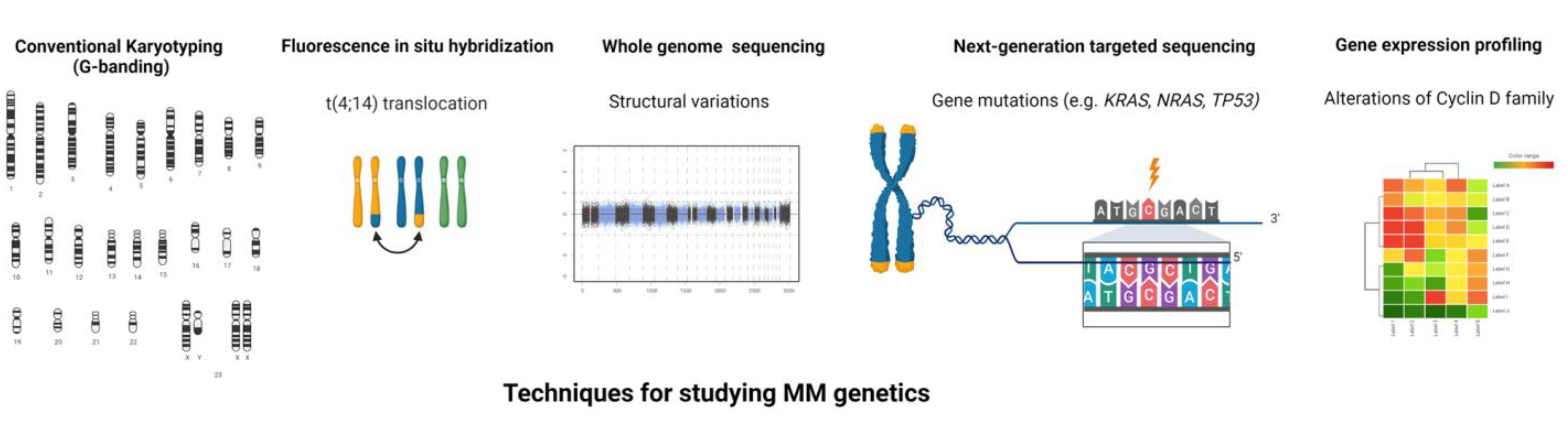
3. Fluorescent In-Situ Hybridization (FISH)

FISH is the current gold standard for identifying the genomic abnormalities in MM, which may predict the aggressiveness of the disease [59]. Prognostic abnormalities that can be detected by FISH include the trisomies of odd-numbered chromosomes, IgH translocations, 17p13 and 1p32 deletions, and 1q21 amplification.
FISH offers a far greater degree of sensitivity and specificity compared to traditional methods like G-banding karyotyping, which can detect only 20–30% of the cytogenetic abnormalities in aspirated bone marrow plasma cells and is hindered by the need of having the plasma cells in metaphase [59]. FISH, in contrast, requires the cell to be in interphase, the phase of the cell cycle in which the majority of cells are at in a given point of time [59]. FISH detection rate of cytogenetic abnormalities in plasma cell dyscrasia can further be enhanced with plasma cell enrichment, by increasing the number of the plasma cells collected in the bone marrow aspirate undergoing analysis [60,61,62].
FISH is also one of the main components of the Revised International Staging System (R-ISS) for MM [63] (Table 1). R-ISS is a simple and powerful prognostic staging system that is universally applicable since it takes into consideration clinical and laboratoristical information and FISH markers that are largely available in most treatment centers as opposed to the Mayo clinic risk stratification for MM (mSMART) [64]. It combines the elements of the original ISS of MM (serum beta-2 microglobulin (B2M) and albumin), chromosomal abnormalities detected by FISH, and serum LDH levels in order to create a tool that can effectively determine the relative risk of survival of newly diagnosed MM patients [63]. Depending on the measurements of these elements, patients are categorized as R-ISS I, II, or III, with R-ISS III having the worst overall survival (OS) and progression-free survival rates [63]. This system considers anyone with serum B2M ≥5.5 mg/L and elevated LDH to be of stage III, with or without del 17p13, t(4;14) or t(14;16) [63]. On the contrary, R-ISS I patients lack any of these cytogenetic anomalies and have serum B2M < 3.5 mg/L, serum albumin ≥3.5 g/dL and normal LDH levels [50]. Patients who do not meet the R-ISS I or III criteria are classified as stage II [50]. In the original study by Palumbo et al., median OS was not reached, 83 months and 43 months for R-ISS I, II, and III, respectively [50], while the progression free survival was 66 months, 42 months, and 29 months, respectively [50]. Furthermore, the R-ISS system can predict OS independent of age and therapy, and its reliability has been validated in the real-world setting [50].

4. Current Data on Co-Existing Abnormalities

The cytogenetic abnormalities underlying MM tend to occur in various combinations, each with distinct prognostic implications. Hence, to overcome the intrinsic limitations of R-ISS, which only accounted for three high-risk FISH abnormalities, Perrot et al. developed and validated a cytogenetic prognostic index (PI) that is based on the occurrence of cytogenetic abnormalities in a large cohort of 1635 MM patients enrolled in four trials [65]. They used FISH and SNP to investigate the effect of del(17p), t(4;14), del(1p32), 1q21 gain and trisomies 3, 5 and 21 on survival outcomes. Six of these abnormalities were statistically significant (all except trisomy 3) based on a multivariate cox proportional hazard regression model, from which a PI formula was derived: 0.4 × t(4;14) + 1.2 × del(17p) − 0.3 × trisomy 5 + 0.3 × trisomy 21 + 0.5 × 1q gain + 0.8 × del(1p32). Accordingly, low, intermediate and high-risk groups are identified by a PI score of ≤0, 0–1, and >1, respectively with an estimated 5-year survival of >75%, 50–75% and <50%. The most common association was reported between trisomies 5 and 21, followed by 1q21 gain with t(4;14) or del(1p32). Interestingly, the PI score had a higher C-index compared to the R-ISS, further demonstrating its discriminative and prognostic abilities.
The concept of double hit and triple hit MM further represents another approach to risk-stratify newly diagnosed MM patients based on their number of high-risk abnormalities such as t(4;14), t(14;16), t(14;20), del(17p), p53 mutation, gain (1q) and del(1p) [66]. Double and triple hit MM are defined by having two and three or more of these high-risk genetic abnormalities, respectively. As expected, patients with double hit MM have been shown to have a worse prognosis than patients with only one high-risk genetic abnormality, while triple hit MM patients tend to have the poorest outcome of all [67,68]. Walker et al. defined double hit myeloma as the subgroup harboring either bi-allelic TP53 inactivation or amplification (≥4 copies) of CKS1B (1q21) on the background of clinical International Staging System III disease.

5. Gene Expression Profiling

Despite its undoubtedly clinical utility, FISH is limited by the identification of only known cytogenetic abnormalities and the inability to decipher the molecular heterogeneity among patients. To this end, gene expression profiling (GEP) is a tool that helps us understand the biology of MM broadly by identifying genes involved in molecular pathogenesis and their clinical significance. Historically, GEP studies led to the identification of Cyclin D family deregulation in MM and MGUS [69,70,71]. In addition, several GEP studies have identified genes and pathways, which lead to the recognition of the molecular complexities involved in MM pathogenesis. Indeed, 11 different molecular subgroups of MM have been found based on transcriptomic studies [13]. Paralleling the differences in GEP, the different subgroups of MM also correlated with clinical outcomes. For instance, subclassifying MM patients according to the presence of IgH translocations and dysregulation of cyclin D genes (i.e., the translocation and cyclin D (TC) classification) allowed the identification of eight subgroups of MM (11q13, 6p21, 4p16, maf, D1, D1+D2, D2, and none) [69,70,71]. Another classification method from the University of Arkansas for Medical Sciences (UAMS) characterized seven different molecular subgroups of MM from 414 newly diagnosed MM based on activating translocations and hyperdiploidy [72]. These seven subgroups of myeloma are namely CD1 [(t(11;14)], CD2 [t(11;14) & t(11;16)], MS [t(4;14)], MF [t(14;16) & t(14;20)], Hyperdiploid cluster (HY), low bone disease (LB), and proliferation-associated genes (PR). The authors also identified myeloid gene expression signatures but were excluded from profiling analyses [72]. Most interestingly, CD1, CD2, LB, and HY subgroups were enriched in low-risk diseases with better overall survival outcomes while MS, MF, and PR subgroups were recognized as high-risk groups. Furthermore, the analyses of data from MM patients enrolled in the HOVON65/GMMG-HD4 trial were described in a European study that identified three additional subgroups of MM [73] including the nuclear factor kappa light chain-enhancer (NF-kB) subgroup, the Cancer testis antigen (CTA) subgroup characterized by high proliferation index, and the PRL3 subgroup characterized by up-regulation of protein tyrosine phosphatases PRL-3 and PTPRZ1.

Brief Summary of Genomic-Based Risk Stratification Studies

UAMS study: Initial study by the UAMS group identified a 70-gene high-risk signature from a training cohort of 351 newly diagnosed MM patients at times of diagnosis and relapse [74]. Investigators performed supervised clustering with 70 genes related to plasma cells from 14 MGUS patients, 22 healthy donors, 38 human MM cell lines, and 351 patients of the training cohort. Gene expression clustergram demonstrated high-risk groups with similar patterns as human MM cell lines, whereas low-risk MM groups exhibited patterns identical to MGUS and normal plasma cells. Further evaluation of the 70-gene risk model in relapse samples of 51 out of 351 of the training cohort revealed high-risk scores in 39 patients, which were associated with poor survival. Interestingly, 30% of the 70 high-risk genes were located in chromosome 1, shaping the high-risk profile of MM. Gene mapping studies revealed overexpressed genes at 1q21, 1q22, and 1q43-q44 in high-risk MM. Additionally, four other genes were identified in the 8q21-8q24 region in patients with high-risk features. Furthermore, the investigators identified a minimum of 17 genes out of the 70-gene model capable of distinguishing high-risk and low-risk MM. Of note, the 17-gene model predicted with 97.7% and 96.9% accuracy, the correct risk category assignment (high-risk vs. low-risk) in the training and the validation cohorts, respectively.
Skyline 92-HOVON study: Kuiper et al. identified 92-gene signatures (EMC-92) from newly diagnosed MM patients in the HOVON65/GMMG-HD4 trial that proved to be an independent prognostic factor for survival [75]. The authors only identified two overlapping genes, called BIRCS5 & LTBP1, compared to UAMS-17/70 gene signatures.
Intergroupe Francophone du Myelome identified 15 genes (IFM-15) from 250 newly diagnosed MM patients that were associated with poor prognosis [76]. The study found overexpression of genes involved in the cell cycle progression and its surveillance in high-risk MM patients. In IFM-15 model, only one gene (FAM49A) was common compared to EMC-92 model, albeit none with UAMS-70.
Other models for prognostication include six gene expression signatures by Dickens et al. [77], the millennium signature [78], and GEP based proliferation index [79]. Despite the identification of various gene models for prognostication as described above, there is no consensus so far in routine clinical practice to incorporate GEP in MM management. Considering its costs, GEP is mostly utilized for research purposes than in routine clinical practice. In addition, while most of the previous studies utilized micro-array-based GEP, recently RNA-sequencing-based methods were found to be highly sensitive and specific, providing new avenues for MM patients prognostications [13].

6. DNA Sequencing and Data on Mutations

As mentioned, given the remarkable clinical and biological heterogeneity of MM, FISH testing cannot capture the genomic complexity of the disease [80]. In addition, common primary events, such as hyperdiploidy and IgH translocations, are insufficient to drive overt MM [81]. Subsequently, secondary genetic events, such as translocations affecting MYC, copy number abnormalities, DNA hypomethylation and somatic mutations in oncogenic pathways (e.g., MAPK, NF-κB and DNA-repair), are required to drive tumor progression [8,82,83,84,85], often defining the true malignant potential but also serving as potential actionable therapeutic targets.
To this end, the use of new high throughput NGS techniques has markedly advanced our knowledge of MM biology (Figure 2). Since the first MM whole genome sequencing (WGS) study [81], an increasing number of recurrent mutations and structural variations (SVs) have been characterized in MM [86]. Although two-thirds of MM patients carry translocations in either IgH or MYC, a recent MM WGS study showed that there were more than 2000 SVs present in MM with IgH and MYC translations only accounted for 6.5% of all SVs [87], suggesting the importance of SVs as major drivers of MM development and progression. Among them, chromothripsis was the most frequent SVs, followed by chromoplexy [87]. Overall, an average of 1.6 mutations per Mb were observed in MM but no universal driver mutations were identified, with the most frequently mutated genes reported being KRAS and NRAS (in ~20% of patients each), followed by FAM46C and DIS3 (~11% each), TP53 (8%) and BRAF (6%) [8,82,83,84,85,87,88]. All other mutations (e.g., TRAF3, LTB and ATM) were observed in less than 5% of MM patients [8]. These mutations affect several signaling pathways with some patients carrying two or more mutations in genes pertaining to the same pathway (e.g., KRAS, NRAS and BRAF in the MAPK pathway) [83]. Beyond these well-defined myeloma genes, recent NGS-based studies have identified several other mutated genes, such as linker histones (HIST1H1B, HIST1H1D, HIST1H1E, and HIST1H2BK) [87], FUBP1 (MYC transcription regulator) and MAX (MYC DNA binding partner) [87], and non-coding mutations in the cis-regulatory elements (e.g., HOXB3, PAX5 and TPRG1) [85]. Chronological reconstruction of genetic events showed that MM development follows preferred evolutionary trajectories [87]. While the majority of somatic mutations occur later in MM, some mutations, such as in activation-induced cytidine deaminase (AID) target genes [89], consistently occur in the early stages of tumor evolution. Driver events accumulated over time, but the resulted clinical course is unpredictable given the clonal evolution and heterogeneity of MM. Efforts have been made to leverage genomic information for MM precision medicine. For example, studies that combine mutations in TP53, ATM or ATR, ZFH4 or CCND1, del(17p), t(4;14), amp(1q), and translocations involving MYC observed improved sensitivity for MM early detection and prognosis prediction, as compared to ISS [84]. The identification of these alterations also led to the development of targeted therapies to treat MM patients carrying specific driver mutations, such as Selumetinib (KRAS), Cobimetinib (NRAS), Palbociclib [del(1p), t(11;14) or t(6;14)], and Vemurafenib (BRAF) [90,91,92,93,94]. However, the genomic complexity of MM makes it challenging to find effective therapies and the “one size fits all” approach is inapplicable. More detailed assessment with genomic sequencing is needed for MM management, particularly because the majority of driver events can only be detected by WGS. Sequential samples (i.e., before and after progression or treatment) with both bone marrow and blood-based genetic profiling will be required in future studies to extend our knowledge of subclonal evolution and resistant clones.

7. Concept of Clonal and Subclonal Evolution

Clonal heterogeneity is a well-known phenomenon in MM contributing to the disease complexity. This puzzling characteristic is present both at the inter-patient and intra-patient levels, which adds to the intricacy in disease biology. In addition, the observed complexity in the clonal architecture of high-risk cytogenetic MM suggests a Darwinian-like somatic evolution rather than conventional linear evolution pattern [95,96,97,98]. Furthermore, the Darwinian model of tumor evolution explains the reason for the failure of therapeutic interventions [96]. Three landmark studies in 2012 provided insight into the complexity of the MM genomics landscape and its evolutionary process [9,10,11]. These studies demonstrated intraclonal heterogeneity early at the diagnosis and different stages of the disease after relapse. Egan et al. were the first to study the longitudinal evolution of myeloma to identify genomic changes during disease course by utilizing WGS data from longitudinal samples of a MM patient with t(4:14) [11]. The authors identified ten common single nucleotide variants (SNVs) that were shared at any point during the disease course. Interestingly, genomic variants were identified at alternating time points of the disease course suggesting the waxing and waning of different clones with treatment and disease status. The WGS study, in this case, demonstrated complex clonal dynamics with genomic heterogeneity at the different studied time points and clonal tiding during the disease course suggesting a Darwinian model of tumor evolution in MM.
Later on, Keats et al. studied genomic and clonal dynamics in 28 MM patients at different time points during the disease course [9]. The authors described three distinct patterns of genomic evolution: genetically stable, linearly evolving clones, or shifting predominant clones. In about one-third of patients with standard-risk (low-risk hyperdiploid disease) cytogenetics, they observed stable genomes with few changes over time and favorable clinical outcomes. Another two-thirds was comprised of high-risk cytogenetics and was characterized by genomic instability with an increased propensity to change over time. Some of the cases in the high-risk group acquired new copy number alterations (CNAs), suggesting a traditional model of linear evolution. However, most of the high-risk MM cases were found to have multiple unique clones at initial diagnosis with changes in relative frequency over time. In particular, MM with high-risk cytogenetics such as t(4;14), t(14;16), t(14;20), and del(17p13) was associated with increased CNAs over time and patients with del(17p13) had significantly more CNAs at diagnosis as compared to other high-risk cytogenetics. A further longitudinal investigation of patients carrying del(17p13) revealed loss of TP53 alleles in four out of five studied patients. Thus, biallelic inactivation over time potentially represents the molecular underpinning of the observed poor prognosis of del(17p13) cases. Like Egan et al., the authors also explored the tumor genomics at different time points in one patient with t(4:14), and they observed alternating clonal dominance [9,11]. Therapeutic interventions over the disease course were associated with clonal suppression and recurrence, which seemed to correlate with drug sensitivity and resistance.
In another seminal study by Walker et al., mutations in MM cases with t(4;14) and t(11;14) cytogenetics were compared to understand tumor evolution and treatment resistance [10]. Median acquired nonsynchronous exonic SNVs were found to be higher in cases with t(4;14) compared to t(11;14) (27 vs. 23.5), despite the absence of statistical significance, and the transition and transversion rates between both groups were comparable. Regardless of the driver status, RAS-MAPK pathway deregulation was common in both groups, and clones with this alteration were not always present in the dominant clone rather than in one or more subclonal populations. Furthermore, the persistent acquisition of mutations within subclones has led to the disease progression (Figure 3).

8. Evolving Therapeutic Implications

To date, MM is an incurable disease. Significant advancements have been made in translation and developmental therapeutic research to identify amenable genomic targets and develop novel agents that could potentially improve patients’ survival. However, these efforts have been severely challenged by the complex disease biology and molecular mechanisms of MM [99]. Detailed review on therapeutic implications in MM is beyond the scope of this article, but we will briefly describe some of the targeted agents that are currently explored [100]. BCL2 inhibitor Venetoclax as monotherapy and in combination with other anti-myeloma agents demonstrated improved outcome in early phases of clinical trials in patients with t(11;14) [101,102,103]. Molibresib (NCT01943851) and OTX015 (NCT01713582) are Bromodomain and Extra-Terminal motif (BET) inhibitors that have been actively investigated in patients with MYC translocations [104]. Poly (ADP-ribose) polymerase inhibitors (PARP) inhibitors such as Olaparib (NCT02693535, NCT03297606), veliparib (NCT01495351), and talazoparib (NCT02693535) targeting AMT and BRCA 1/2 mutations are in early phases of clinical trials. Vemurafenib (NCT03297606), encorafenib (NCT02834364), and dabrafenib (NCT03091257) are BRAF inhibitors which are also in early phase of drug development in combination with other agents.
Fc receptor-homolog 5 (FcRH5), whose gene is located on chromosome 1, is a type I membrane protein expressed in MM cells. Cevostamab is a T-cell engaging bispecific antibody that targets FcRH5, and early pharmacodynamics studies described its mechanism of action which encompasses T-cell activation, proliferation and cytokine production [105]. A phase-1 clinical trial (NCT03275103) is currently ongoing to evaluate the safety and pharmacokinetics of Cevostamab (BFCR4350A) in relapsed/refractory MM. Initial results from the phase-1 clinical trial revealed the favorable activity of Cevostamab in heavily pre-treated relapsed/refractory MM [106] and whether 1q amp/gain MM may respond better to FcRH5 targeted by Cevostamab needs to be investigated further. In addition, immune effector therapies (at this early stage of clinical development) appear to be mutation agnostic; yet they may improve chances to control high-risk MM [107].

9. Conclusions

Multiple myeloma is a heterogeneous disease that is driven by numerous genetic and epigenetic changes. Early events in MM progression include immunoglobulin translocation and hyperdiploidy; however, the recent advancements in high throughput technologies (i.e., WGS) have identified complex genomic variations in key myeloma-associated genes responsible for the development and progression of the disease. In the future, data obtained from large cohorts of patients studied by newer genomic tools and analyzed with more sophisticated machine learning approaches will better characterize the genomic complexity and evolutionary changes responsible for disease progression. This will not only help us develop better prognostic models, but also identify novel therapeutic targets to develop individualized therapies. Indeed, prospective advances in the understanding of disease development from its precursor stage at the molecular level may help create novel avenues of rational preventive strategies that aim to avert early malignant clones from progression.

Author Contributions

Conceptualization, H.A. (Hassan Awada), B.T., H.A. (Hussein Awada), J.D., P.H. and B.D.; formal analysis, H.A. (Hassan Awada), B.T., H.A. (Hussein Awada), J.D., C.G.; writing—original draft preparation, H.A. (Hassan Awada), B.T., H.A. (Hussein Awada), J.D., P.H. and B.D.; writing—review and editing, H.A. (Hassan Awada), B.T., H.A. (Hussein Awada), J.D., C.G., P.H. and B.D.; supervision, P.H. and B.D.; All authors have read and agreed to the published version of the manuscript.

Funding

This research received no external funding.

Conflicts of Interest

The authors declare no conflict of interest.

References

  1. SEER Cancer Statistics Review. 1975–2017. SEER. Available online: https://seer.cancer.gov/csr/1975_2017/index.html (accessed on 7 May 2021).
  2. Goldschmidt, H.; Lokhorst, H.M.; Mai, E.K.; Van Der Holt, B.; Blau, I.W.; Zweegman, S.; Weisel, K.C.; Vellenga, E.; Pfreundschuh, M.; Kersten, M.J.; et al. Bortezomib before and after high-dose therapy in myeloma: Long-term results from the phase III HOVON-65/GMMG-HD4 trial. Leukemia 2018, 32, 383–390. [Google Scholar] [CrossRef] [PubMed]
  3. Nishimura, K.K.; Barlogie, B.; Van Rhee, F.; Zangari, M.; Walker, B.A.; Rosenthal, A.; Schinke, C.; Thanendrarajan, S.; Davies, F.; Hoering, A.; et al. Long-term outcomes after autologous stem cell transplantation for multiple myeloma. Blood Adv. 2020, 4, 422–431. [Google Scholar] [CrossRef]
  4. Kumar, S.K.; Dispenzieri, A.; Lacy, M.Q.; Gertz, M.A.; Buadi, F.K.; Pandey, S.C.; Kapoor, P.; Dingli, D.; Hayman, S.R.; Leung, N.; et al. Continued improvement in survival in multiple myeloma: Changes in early mortality and outcomes in older patients. Leukemia 2014, 28, 1122–1128. [Google Scholar] [CrossRef] [Green Version]
  5. Fonseca, R.; Abouzaid, S.; Bonafede, M.; Cai, Q.; Parikh, K.; Cosler, L.; Richardson, P. Trends in overall survival and costs of multiple myeloma, 2000–2014. Leukemia 2017, 31, 1915–1921. [Google Scholar] [CrossRef] [PubMed] [Green Version]
  6. Szalat, R.; Munshi, N.C. Genomic heterogeneity in multiple myeloma. Curr. Opin. Genet. Dev. 2015, 30, 56–65. [Google Scholar] [CrossRef]
  7. Yle, R.O.A.K.; Therneau, T.M.; Rajkumar, S.V.; Fford, J.A.R.O.; Arson, D.I.R.L.; Plevak, M.F.; Melton, L.J. A Long-Term Study of Prognosis in Monoclonal Gammopathy of Undetermined Significance. N. Engl. J. Med. 2002, 346, 564–569. [Google Scholar] [CrossRef]
  8. Manier, S.; Salem, K.Z.; Park, J.; Landau, D.A.; Getz, G.; Ghobrial, S.M. Genomic complexity of multiple myeloma and its clinical implications. Nat. Rev. Clin. Oncol. 2017, 14, 100–113. [Google Scholar] [CrossRef]
  9. Keats, J.; Chesi, M.; Egan, J.B.; Garbitt, V.M.; Palmer, S.E.; Braggio, E.; Van Wier, S.; Blackburn, P.R.; Baker, A.S.; Dispenzieri, A.; et al. Clonal competition with alternating dominance in multiple myeloma. Blood 2012, 120, 1067–1076. [Google Scholar] [CrossRef]
  10. Walker, B.; Wardell, C.; Melchor, L.; Hulkki, S.; Potter, N.E.; Johnson, D.C.; Fenwick, K.; Kozarewa, I.; Gonzalez, D.; Lord, C.; et al. Intraclonal heterogeneity and distinct molecular mechanisms characterize the development of t(4;14) and t(11;14) myeloma. Blood 2012, 120, 1077–1086. [Google Scholar] [CrossRef]
  11. Egan, J.B.; Shi, C.-X.; Tembe, W.; Christoforides, A.; Kurdoglu, A.; Sinari, S.; Middha, S.; Asmann, Y.; Schmidt, J.; Braggio, E.; et al. Whole-genome sequencing of multiple myeloma from diagnosis to plasma cell leukemia reveals genomic initiating events, evolution, and clonal tides. Blood 2012, 120, 1060–1066. [Google Scholar] [CrossRef] [PubMed]
  12. Hultcrantz, M.; Yellapantula, V.; Rustad, E.H. Genomic profiling of multiple myeloma: New insights and modern technologies. Best Pr. Res. Clin. Haematol. 2020, 33, 101153. [Google Scholar] [CrossRef]
  13. Szalat, R.; Avet-Loiseau, H.; Munshi, N.C. Gene Expression Profiles in Myeloma: Ready for the Real World? Clin. Cancer Res. 2016, 22, 5434–5442. [Google Scholar] [CrossRef] [PubMed] [Green Version]
  14. Abdallah, N.; Rajkumar, S.V.; Greipp, P.; Kapoor, P.; Gertz, M.A.; Dispenzieri, A.; Baughn, L.B.; Lacy, M.Q.; Hayman, S.R.; Buadi, F.K.; et al. Cytogenetic abnormalities in multiple myeloma: Association with disease characteristics and treatment response. Blood Cancer J. 2020, 10, 1–9. [Google Scholar] [CrossRef]
  15. Prideaux, S.M.; O’Brien, E.C.; Chevassut, T.J. The Genetic Architecture of Multiple Myeloma. Adv. Hematol. 2014, 2014, 1–16. [Google Scholar] [CrossRef] [PubMed] [Green Version]
  16. Rasmussen, T.; Haaber, J.; Dahl, I.M.; Knudsen, L.M.; Kerndrup, G.B.; Lodahl, M.; Johnsen, H.E.; Kuehl, M. Identification of translocation products but not K-RAS mutations in memory B cells from patients with multiple myeloma. Haematologica 2010, 95, 1730–1737. [Google Scholar] [CrossRef] [PubMed] [Green Version]
  17. Landgren, O.; Kyle, R.A.; Pfeiffer, R.M.; Katzmann, J.A.; Caporaso, N.E.; Hayes, R.; Dispenzieri, A.; Kumar, S.; Clark, R.J.; Baris, D.; et al. Monoclonal gammopathy of undetermined significance (MGUS) consistently precedes multiple myeloma: A prospective study. Blood 2009, 113, 5412–5417. [Google Scholar] [CrossRef] [Green Version]
  18. Rajkumar, S.V.; Dimopoulos, M.A.; Palumbo, A.; Blade, J.; Merlini, G.; Mateos, M.-V.; Kumar, S.; Hillengass, J.; Kastritis, E.; Richardson, P.; et al. International Myeloma Working Group updated criteria for the diagnosis of multiple myeloma. Lancet Oncol. 2014, 15, e538–e548. [Google Scholar] [CrossRef]
  19. Ordoñez, R.; Kulis, M.; Russiñol, N.; Chapaprieta, V.; Carrasco-Leon, A.; García-Torre, B.; Charalampopoulou, S.; Clot, G.; Beekman, R.; Meydan, C.; et al. Chromatin activation as a unifying principle underlying pathogenic mechanisms in multiple myeloma. Genome Res. 2020, 30, 1217–1227. [Google Scholar] [CrossRef] [PubMed]
  20. Thomsen, H.; Campo, C.; Weinhold, N.; Filho, M.I.D.S.; Pour, L.; Gregora, E.; Vodička, P.; Vodickova, L.; Hoffmann, P.; Nöthen, M.M.; et al. Genomewide association study on monoclonal gammopathy of unknown significance (MGUS). Eur. J. Haematol. 2017, 99, 70–79. [Google Scholar] [CrossRef]
  21. Broderick, P.; Chubb, D.; Johnson, D.C.; Weinhold, N.; Försti, A.; Lloyd, A.; Olver, B.; Ma, Y.P.; Dobbins, S.E.; Walker, B.; et al. Common variation at 3p22.1 and 7p15.3 influences multiple myeloma risk. Nat. Genet. 2011, 44, 58–61. [Google Scholar] [CrossRef] [Green Version]
  22. Beksac, M.; Gragert, L.; Fingerson, S.; Maiers, M.; Zhang, M.-J.; Albrecht, M.; Zhong, X.; Cozen, W.; Dispenzieri, A.; Lonial, S.; et al. HLA polymorphism and risk of multiple myeloma. Leukemia 2016, 30, 2260–2264. [Google Scholar] [CrossRef] [PubMed] [Green Version]
  23. Brousseau, M.; Leleu, X.; Gerard, J.; Gastinne, T.; Godon, A.; Genevieve, F.; Dib, M.; Lai, J.-L.; Facon, T.; Zandecki, M. Hyperdiploidy Is a Common Finding in Monoclonal Gammopathy of Undetermined Significance and Monosomy 13 Is Restricted to These Hyperdiploid Patients. Clin. Cancer Res. 2007, 13, 6026–6031. [Google Scholar] [CrossRef] [PubMed] [Green Version]
  24. Chng, W.J. A validated FISH trisomy index demonstrates the hyperdiploid and nonhyperdiploid dichotomy in MGUS. Blood 2005, 106, 2156–2161. [Google Scholar] [CrossRef] [Green Version]
  25. Magrangeas, F.; Lodé, L.; Wuillème, S.; Minvielle, S.; Avet-Loiseau, H. Genetic heterogeneity in multiple myeloma. Leukemia 2004, 19, 191–194. [Google Scholar] [CrossRef] [PubMed] [Green Version]
  26. Kumar, S.; Fonseca, R.; Ketterling, R.; Dispenzieri, A.; Lacy, M.Q.; Gertz, M.A.; Hayman, S.R.; Buadi, F.K.; Dingli, D.; Knudson, R.A.; et al. Trisomies in multiple myeloma: Impact on survival in patients with high-risk cytogenetics. Blood 2012, 119, 2100–2105. [Google Scholar] [CrossRef] [Green Version]
  27. Chng, W.J.; Glebov, O.; Bergsagel, P.; Kuehl, W.M. Genetic events in the pathogenesis of multiple myeloma. Best Pr. Res. Clin. Haematol. 2007, 20, 571–596. [Google Scholar] [CrossRef] [PubMed] [Green Version]
  28. Bergsagel, P.L.; Kuehl, W.M. Chromosome translocations in multiple myeloma. Oncogene 2001, 20, 5611–5622. [Google Scholar] [CrossRef] [Green Version]
  29. Kuehl, W.M.; Bergsagel, P.L. Multiple myeloma: Evolving genetic events and host interactions. Nat. Rev. Cancer 2002, 2, 175–187. [Google Scholar] [CrossRef]
  30. Bourguet, C.C.; Logue, E.E. Antigenic stimulation and multiple myeloma. A prospective study. Cancer 1993, 72, 2148–2154. [Google Scholar] [CrossRef]
  31. Mantovani, A.; Garlanda, C. Inflammation and multiple myeloma: The Toll connection. Leukemia 2006, 20, 937–938. [Google Scholar] [CrossRef]
  32. Bagratuni, T.; Sklirou, A.D.; Kastritis, E.; Liacos, C.I.; Spilioti, C.; Eleutherakis-Papaiakovou, E.; Kanellias, N.; Gavriatopoulou, M.; Terpos, E.; Trougakos, I.P.; et al. Toll-Like Receptor 4 Activation Promotes Multiple Myeloma Cell Growth and Survival Via Suppression of The Endoplasmic Reticulum Stress Factor Chop. Sci. Rep. 2019, 9, 1–12. [Google Scholar] [CrossRef]
  33. Rawstron, A.C.; Fenton, J.A.; Ashcroft, J.; English, A.; Jones, R.A.; Richards, S.J.; Pratt, G.; Owen, R.; Davies, F.E.; Child, J.A.; et al. The interleukin-6 receptor alpha-chain (CD126) is expressed by neoplastic but not normal plasma cells. Blood 2000, 96, 3880–3886. [Google Scholar] [CrossRef]
  34. Puthier, D.; Derenne, S.; Barille-Nion, S.; Moreau, P.; Harousseau, J.-L.; Bataille, R.; Amiot, M. Mcl-1 and Bcl-xL are co-regulated by IL-6 in human myeloma cells. Br. J. Haematol. 1999, 107, 392–395. [Google Scholar] [CrossRef] [PubMed]
  35. Rajkumar, S.V. The screening imperative for multiple myeloma. Nat. Cell Biol. 2020, 587, S63. [Google Scholar] [CrossRef]
  36. Pang, L.; Rajkumar, S.V.; Kapoor, P.; Buadi, F.; Dispenzieri, A.; Gertz, M.; Lacy, M.; Kyle, R.; Kumar, S. Prognosis of young patients with monoclonal gammopathy of undetermined significance (MGUS). Blood Cancer J. 2021, 11, 1–8. [Google Scholar] [CrossRef]
  37. Landgren, O.; Hofmann, J.N.; McShane, C.M.; Santo, L.; Hultcrantz, M.; Korde, N.; Mailankody, S.; Kazandjian, D.; Murata, K.; Thoren, K.; et al. Association of Immune Marker Changes with Progression of Monoclonal Gammopathy of Undetermined Significance to Multiple Myeloma. JAMA Oncol. 2019, 5, 1293–1301. [Google Scholar] [CrossRef] [PubMed]
  38. Dib, A.; Gabrea, A.; Glebov, O.K.; Bergsagel, P.L.; Kuehl, W.M. Characterization of MYC Translocations in Multiple Myeloma Cell Lines. J. Natl. Cancer Inst. Monogr. 2008, 2008, 25–31. [Google Scholar] [CrossRef] [PubMed]
  39. Misund, K.; Network, M.C.; Keane, N.; Stein, C.K.; Asmann, Y.W.; Day, G.; Welsh, S.; Van Wier, S.A.; Riggs, D.L.; Ahmann, G.; et al. MYC dysregulation in the progression of multiple myeloma. Leukemia 2020, 34, 322–326. [Google Scholar] [CrossRef]
  40. Avet-Loiseau, H.; Gerson, F.; Magrangeas, F.; Minvielle, S.; Harousseau, J.-L.; Bataille, R. Rearrangements of the c-myc oncogene are present in 15% of primary human multiple myeloma tumors. Blood 2001, 98, 3082–3086. [Google Scholar] [CrossRef] [Green Version]
  41. Gabrea, A.; Martelli, M.L.; Qi, Y.; Roschke, A.; Barlogie, B.; Shaughnessy, J.D., Jr.; Sawyer, J.R.; Kuehl, W.M. Secondary genomic rearrangements involving immunoglobulin or MYC loci show similar prevalences in hyperdiploid and nonhyperdiploid myeloma tumors. Genes Chromosom. Cancer 2008, 47, 573–590. [Google Scholar] [CrossRef] [Green Version]
  42. Abdallah, N.; Baughn, L.B.; Rajkumar, S.V.; Kapoor, P.; Gertz, M.A.; Dispenzieri, A.; Lacy, M.Q.; Hayman, S.R.; Buadi, F.K.; Dingli, D.; et al. Implications of MYC Rearrangements in Newly Diagnosed Multiple Myeloma. Clin. Cancer Res. 2020, 26, 6581–6588. [Google Scholar] [CrossRef]
  43. Xiong, W.; Wu, X.; Starnes, S.; Johnson, S.K.; Haessler, J.; Wang, S.; Chen, L.; Barlogie, B.; Shaughnessy, J.J.D.; Zhan, F. An analysis of the clinical and biologic significance of TP53 loss and the identification of potential novel transcriptional targets of TP53 in multiple myeloma. Blood 2008, 112, 4235–4246. [Google Scholar] [CrossRef] [PubMed] [Green Version]
  44. Lodé, L.; Eveillard, M.; Trichet, V.; Soussi, T.; Wuillème, S.; Richebourg, S.; Magrangeas, F.; Ifrah, N.; Campion, L.; Traullé, C.; et al. Mutations in TP53 are exclusively associated with del(17p) in multiple myeloma. Haematologica 2010, 95, 1973–1976. [Google Scholar] [CrossRef] [PubMed] [Green Version]
  45. Fonseca, R.; Barlogie, B.; Bataille, R.; Bastard, C.; Bergsagel, P.L.; Chesi, M.; Davies, F.; Drach, J.; Greipp, P.R.; Kirsch, I.R.; et al. Genetics and Cytogenetics of Multiple Myeloma. Cancer Res. 2004, 64, 1546–1558. [Google Scholar] [CrossRef] [PubMed] [Green Version]
  46. Chang, H.; Sloan, S.; Li, D.; Stewart, A.K. Multiple myeloma involving central nervous system: High frequency of chromosome 17p13.1 (p53) deletions. Br. J. Haematol. 2004, 127, 280–284. [Google Scholar] [CrossRef] [PubMed]
  47. Thakurta, A.; Ortiz, M.; Blecua, P.; Towfic, F.; Corre, J.; Serbina, N.V.; Flynt, E.; Yu, Z.; Yang, Z.; Palumbo, A.; et al. High subclonal fraction of 17p deletion is associated with poor prognosis in multiple myeloma. Blood 2019, 133, 1217–1221. [Google Scholar] [CrossRef] [Green Version]
  48. Kalakonda, N.; Rothwell, D.; Scarffe, J.H.; Norton, J.D. Detection of N-Ras codon 61 mutations in subpopulations of tumor cells in multiple myeloma at presentation. Blood 2001, 98, 1555–1560. [Google Scholar] [CrossRef] [PubMed] [Green Version]
  49. Chng, W.J.; Gonzalez-Paz, N.; Price-Troska, T.; Jacobus, S.; Rajkumar, S.V.; Oken, M.M.; Kyle, R.A.; Henderson, K.J.; Van Wier, S.; Greipp, P.; et al. Clinical and biological significance of RAS mutations in multiple myeloma. Leukemia 2008, 22, 2280–2284. [Google Scholar] [CrossRef] [Green Version]
  50. Mulligan, G.; Lichter, D.I.; Di Bacco, A.; Blakemore, S.J.; Berger, A.; Koenig, E.; Bernard, H.; Trepicchio, W.; Li, B.; Neuwirth, R.; et al. Mutation of NRAS but not KRAS significantly reduces myeloma sensitivity to single-agent bortezomib therapy. Blood 2014, 123, 632–639. [Google Scholar] [CrossRef] [Green Version]
  51. Annunziata, C.M.; Davis, R.E.; Demchenko, Y.; Bellamy, W.; Gabrea, A.; Zhan, F.; Lenz, G.; Hanamura, I.; Wright, G.; Xiao, W.; et al. Frequent Engagement of the Classical and Alternative NF-κB Pathways by Diverse Genetic Abnormalities in Multiple Myeloma. Cancer Cell 2007, 12, 115–130. [Google Scholar] [CrossRef] [PubMed] [Green Version]
  52. Demchenko, Y.N.; Kuehl, W.M. A critical role for the NFkB pathway in multiple myeloma. Oncotarget 2010, 1, 59–68. [Google Scholar] [CrossRef]
  53. Keats, J.; Fonseca, R.; Chesi, M.; Schop, R.; Baker, A.; Chng, W.-J.; Van Wier, S.; Tiedemann, R.; Shi, C.-X.; Sebag, M.; et al. Promiscuous Mutations Activate the Noncanonical NF-κB Pathway in Multiple Myeloma. Cancer Cell 2007, 12, 131–144. [Google Scholar] [CrossRef] [PubMed] [Green Version]
  54. Kulkarni, M.; Daggett, J.; Bender, T.; Kuehl, W.; Bergsagel, P.; Williams, M. Frequent inactivation of the cyclin-dependent kinase inhibitor p18 by homozygous deletion in multiple myeloma cell lines: Ectopic p18 expression inhibits growth and induces apoptosis. Leukemia 2002, 16, 127–134. [Google Scholar] [CrossRef] [Green Version]
  55. Lichtenstein, A.; Tu, Y.; Fady, C.; Vescio, R.; Berenson, J. Interleukin-6 Inhibits Apoptosis of Malignant Plasma Cells. Cell. Immunol. 1995, 162, 248–255. [Google Scholar] [CrossRef] [PubMed]
  56. Ong, F.; Van Nieuwkoop, J.A.; De Groot-Swings, G.M.; Hermans, J.; Harvey, M.S.; Kluin, P.M.; Kluin-Nelemans, J.C. Bcl-2 protein expression is not related to short survival in multiple myeloma. Leukemia 1995, 9, 1282–1284. [Google Scholar] [PubMed]
  57. Landowski, T.H.; Qu, N.; Buyuksal, I.; Painter, J.S.; Dalton, W.S. Mutations in the Fas antigen in patients with multiple myeloma. Blood 1997, 90, 4266–4270. [Google Scholar] [CrossRef] [PubMed] [Green Version]
  58. Ng, M.; Chung, Y.; Lo, K.W.; Wickham, N.; Lee, J.; Huang, D. Frequent Hypermethylation of p16 and p15 Genes in Multiple Myeloma. Blood 1997, 89, 2500–2506. [Google Scholar] [CrossRef]
  59. Fonseca, R.; Bergsagel, P.L.; Drach, J.; Shaughnessy, J.; Gutierrez, N.; Stewart, A.K.; Morgan, G.; Van Ness, B.; Chesi, M.; Minvielle, S.; et al. International Myeloma Working Group molecular classification of multiple myeloma: Spotlight review. Leukemia 2009, 23, 2210–2221. [Google Scholar] [CrossRef] [Green Version]
  60. Lu, G.; Muddasani, R.; Orlowski, R.; Abruzzo, L.V.; Qazilbash, M.H.; You, M.J.; Wang, Y.; Zhao, M.; Chen, S.; Glitza, I.C.; et al. Plasma Cell Enrichment Enhances Detection of High-Risk Cytogenomic Abnormalities by Fluorescence In Situ Hybridization and Improves Risk Stratification of Patients with Plasma Cell Neoplasms. Arch. Pathol. Lab. Med. 2013, 137, 625–631. [Google Scholar] [CrossRef]
  61. Pozdnyakova, O.; Crowley-Larsen, P.; Zota, V.; Wang, S.A.; Miron, P.M. Interphase FISH in plasma cell dyscrasia: Increase in abnormality detection with plasma cell enrichment. Cancer Genet. Cytogenet. 2009, 189, 112–117. [Google Scholar] [CrossRef]
  62. Ma, E.S.K.; Wang, C.L.N.; Wong, A.T.C.; Choy, G.; Chan, T.L. Target fluorescence in-situ hybridization (Target FISH) for plasma cell enrichment in myeloma. Mol. Cytogenet. 2016, 9. [Google Scholar] [CrossRef] [PubMed] [Green Version]
  63. Palumbo, A.; Avet-Loiseau, H.; Oliva, S.; Lokhorst, H.M.; Goldschmidt, H.; Rosinol, L.; Richardson, P.; Caltagirone, S.; Lahuerta, J.J.; Facon, T.; et al. Revised International Staging System for Multiple Myeloma: A Report from International Myeloma Working Group. J. Clin. Oncol. 2015, 33, 2863–2869. [Google Scholar] [CrossRef] [PubMed]
  64. Rajkumar, S.V. Multiple myeloma: 2020 update on diagnosis, risk-stratification and management. Am. J. Hematol. 2020, 95, 548–567. [Google Scholar] [CrossRef] [Green Version]
  65. Perrot, A.; Lauwers-Cances, V.; Tournay, E.; Hulin, C.; Chretien, M.-L.; Royer, B.; Dib, M.; Decaux, O.; Jaccard, A.; Belhadj, K.; et al. Development and Validation of a Cytogenetic Prognostic Index Predicting Survival in Multiple Myeloma. J. Clin. Oncol. 2019, 37, 1657–1665. [Google Scholar] [CrossRef]
  66. Rajkumar, S.V.; Kumar, S. Multiple myeloma current treatment algorithms. Blood Cancer J. 2020, 10, 1–10. [Google Scholar] [CrossRef]
  67. Walker, B.A.; Mavrommatis, K.; Wardell, C.; Ashby, C.; Bauer, M.; Davies, F.; Rosenthal, A.; Wang, H.; Qu, P.; Hoering, A.; et al. A high-risk, Double-Hit, group of newly diagnosed myeloma identified by genomic analysis. Leukemia 2019, 33, 159–170. [Google Scholar] [CrossRef] [PubMed]
  68. Baysal, M.; Demirci, U.; Umit, E.; Kirkizlar, H.O.; Atli, E.I.; Gurkan, H.; Gulsaran, S.K.; Bas, V.; Mail, C.; Demir, A.M. Concepts of Double Hit and Triple Hit Disease in Multiple Myeloma, Entity and Prognostic Significance. Sci. Rep. 2020, 10, 1–6. [Google Scholar] [CrossRef] [Green Version]
  69. Bergsagel, P.L.; Kuehl, W.M.; Zhan, F.; Sawyer, J.; Barlogie, B.; Shaughnessy, J.J. Cyclin D dysregulation: An early and unifying pathogenic event in multiple myeloma. Blood 2005, 106, 296–303. [Google Scholar] [CrossRef] [PubMed] [Green Version]
  70. Miura, K.; Lida, S.; Hanamura, I.; Kato, M.; Banno, S.; Ishida, T.; Kusumoto, S.; Takeuchi, G.; Miwa, H.; Nitta, M.; et al. Frequent occurrence of CCND1 deregulation in patients with early stages of plasma cell dyscrasia. Cancer Sci. 2003, 94, 350–354. [Google Scholar] [CrossRef]
  71. Bergsagel, P.L.; Kuehl, W.M. Molecular Pathogenesis and a Consequent Classification of Multiple Myeloma. J. Clin. Oncol. 2005, 23, 6333–6338. [Google Scholar] [CrossRef]
  72. Zhan, F.; Huang, Y.; Colla, S.; Stewart, J.P.; Hanamura, I.; Gupta, S.; Epstein, J.; Yaccoby, S.; Sawyer, J.; Burington, B.; et al. The molecular classification of multiple myeloma. Blood 2006, 108, 2020–2028. [Google Scholar] [CrossRef] [PubMed] [Green Version]
  73. Broyl, A.; Hose, D.; Lokhorst, H.; De Knegt, Y.; Peeters, J.; Jauch, A.; Bertsch, U.; Buijs, A.; Stevens-Kroef, M.; Beverloo, H.B.; et al. Gene expression profiling for molecular classification of multiple myeloma in newly diagnosed patients. Blood 2010, 116, 2543–2553. [Google Scholar] [CrossRef] [Green Version]
  74. Shaughnessy, J.D.; Zhan, F.; Burington, B.E.; Huang, Y.; Colla, S.; Hanamura, I.; Stewart, J.P.; Kordsmeier, B.; Randolph, C.; Williams, D.R.; et al. A validated gene expression model of high-risk multiple myeloma is defined by deregulated expression of genes mapping to chromosome 1. Blood 2006, 109, 2276–2284. [Google Scholar] [CrossRef] [Green Version]
  75. Kuiper, R.; Broyl, A.; De Knegt, Y.; Van Vliet, M.H.; Van Beers, E.H.; Van Der Holt, B.; El Jarari, L.; Mulligan, G.; Gregory, W.; Morgan, G.; et al. A gene expression signature for high-risk multiple myeloma. Leukemia 2012, 26, 2406–2413. [Google Scholar] [CrossRef] [Green Version]
  76. Decaux, O.; Lodé, L.; Magrangeas, F.; Charbonnel, C.; Gouraud, W.; Jézéquel, P.; Attal, M.; Harousseau, J.-L.; Moreau, P.; Bataille, R.; et al. Prediction of Survival in Multiple Myeloma Based on Gene Expression Profiles Reveals Cell Cycle and Chromosomal Instability Signatures in High-Risk Patients and Hyperdiploid Signatures in Low-Risk Patients: A Study of the Intergroupe Francophone du Myélome. J. Clin. Oncol. 2008, 26, 4798–4805. [Google Scholar] [CrossRef] [PubMed]
  77. Dickens, N.; Walker, B.A.; Leone, P.E.; Johnson, D.C.; Brito, J.L.; Zeisig, A.; Jenner, M.; Boyd, K.; Gonzalez, D.; Gregory, W.M.; et al. Homozygous Deletion Mapping in Myeloma Samples Identifies Genes and an Expression Signature Relevant to Pathogenesis and Outcome. Clin. Cancer Res. 2010, 16, 1856–1864. [Google Scholar] [CrossRef] [PubMed] [Green Version]
  78. Mulligan, G.; Mitsiades, C.; Bryant, B.; Zhan, F.; Chng, W.J.; Roels, S.; Koenig, E.; Fergus, A.; Huang, Y.; Richardson, P.; et al. Gene expression profiling and correlation with outcome in clinical trials of the proteasome inhibitor bortezomib. Blood 2006, 109, 3177–3188. [Google Scholar] [CrossRef] [PubMed]
  79. Hose, D.; Rème, T.; Hielscher, T.; Moreaux, J.; Messner, T.; Seckinger, A.; Benner, A.; Shaughnessy, J.D.; Barlogie, B.; Zhou, Y.; et al. Proliferation is a central independent prognostic factor and target for personalized and risk-adapted treatment in multiple myeloma. Haematologica 2010, 96, 87–95. [Google Scholar] [CrossRef] [PubMed] [Green Version]
  80. Rajkumar, S.V. Sequencing of myeloma therapy: Finding the right path among many standards. Hematol. Oncol. 2021, 39, 68–72. [Google Scholar] [CrossRef]
  81. Chapman, M.A.; Lawrence, M.S.; Keats, J.; Cibulskis, K.; Sougnez, C.; Schinzel, A.C.; Harview, C.; Brunet, J.-P.; Ahmann, G.J.; Adli, M.; et al. Initial genome sequencing and analysis of multiple myeloma. Nat. Cell Biol. 2011, 471, 467–472. [Google Scholar] [CrossRef] [PubMed]
  82. Bolli, N.; Biancon, G.; Moarii, M.; Gimondi, S.; Li, Y.; De Philippis, C.; Maura, F.; Sathiaseelan, V.; Tai, Y.-T.; Mudie, L.; et al. Analysis of the genomic landscape of multiple myeloma highlights novel prognostic markers and disease subgroups. Leukemia 2018, 32, 2604–2616. [Google Scholar] [CrossRef]
  83. Lohr, J.G.; Stojanov, P.; Carter, S.L.; Cruz-Gordillo, P.; Lawrence, M.S.; Auclair, D.; Sougnez, C.; Knoechel, B.; Gould, J.; Saksena, G.; et al. Widespread Genetic Heterogeneity in Multiple Myeloma: Implications for Targeted Therapy. Cancer Cell 2014, 25, 91–101. [Google Scholar] [CrossRef] [PubMed] [Green Version]
  84. Walker, B.; Boyle, E.M.; Wardell, C.; Murison, A.; Begum, D.B.; Dahir, N.M.; Proszek, P.Z.; Johnson, D.C.; Kaiser, M.F.; Melchor, L.; et al. Mutational Spectrum, Copy Number Changes, and Outcome: Results of a Sequencing Study of Patients with Newly Diagnosed Myeloma. J. Clin. Oncol. 2015, 33, 3911–3920. [Google Scholar] [CrossRef]
  85. Hoang, P.H.; Dobbins, S.E.; Cornish, A.J.; Chubb, D.; Law, P.; Kaiser, M.; Houlston, R.S. Whole-genome sequencing of multiple myeloma reveals oncogenic pathways are targeted somatically through multiple mechanisms. Leukemia 2018, 32, 2459–2470. [Google Scholar] [CrossRef] [Green Version]
  86. Sharma, N.; Smadbeck, J.B.; Abdallah, N.; Zepeda-Mendoza, C.; Binder, M.; Pearce, K.E.; Asmann, Y.W.; Peterson, J.F.; Ketterling, R.P.; Greipp, P.T.; et al. The prognostic role of MYC structural variants identified by NGS and FISH in multiple myeloma. Clin. Cancer Res. 2021. [Google Scholar] [CrossRef] [PubMed]
  87. Maura, F.; Bolli, N.; Angelopoulos, N.; Dawson, K.J.; Leongamornlert, D.; Martincorena, I.; Mitchell, T.J.; Fullam, A.; Gonzalez, S.; Szalat, R.; et al. Genomic landscape and chronological reconstruction of driver events in multiple myeloma. Nat. Commun. 2019, 10, 1–12. [Google Scholar] [CrossRef] [PubMed] [Green Version]
  88. Bolli, N.; Avet-Loiseau, H.; Wedge, D.; Van Loo, P.; Alexandrov, L.; Martincorena, I.; Dawson, K.J.; Iorio, F.; Nik-Zainal, S.; Bignell, G.R.; et al. Heterogeneity of genomic evolution and mutational profiles in multiple myeloma. Nat. Commun. 2014, 5, 2997. [Google Scholar] [CrossRef] [Green Version]
  89. Maura, F.; Rustad, E.H.; Yellapantula, V.; Łuksza, M.; Hoyos, D.; MacLachlan, K.H.; Diamond, B.T.; Greenbaum, B.D.; Morgan, G.; Lesokhin, A.; et al. Role of AID in the temporal pattern of acquisition of driver mutations in multiple myeloma. Leukemia 2020, 34, 1476–1480. [Google Scholar] [CrossRef]
  90. Jänne, P.A.; Shaw, A.T.; Pereira, J.R.; Jeannin, G.; Vansteenkiste, J.; Barrios, C.; Franke, F.A.; Grinsted, L.; Zazulina, V.; Smith, P.; et al. Selumetinib plus docetaxel for KRAS-mutant advanced non-small-cell lung cancer: A randomised, multicentre, placebo-controlled, phase 2 study. Lancet Oncol. 2013, 14, 38–47. [Google Scholar] [CrossRef]
  91. Adjei, A.A.; Cohen, R.B.; Franklin, W.; Morris, C.; Wilson, D.; Molina, J.R.; Hanson, L.J.; Gore, L.; Chow, L.; Leong, S.; et al. Phase I Pharmacokinetic and Pharmacodynamic Study of the Oral, Small-Molecule Mitogen-Activated Protein Kinase Kinase 1/2 Inhibitor AZD6244 (ARRY-142886) in Patients with Advanced Cancers. J. Clin. Oncol. 2008, 26, 2139–2146. [Google Scholar] [CrossRef] [Green Version]
  92. Abdel-Wahab, O.; Klimek, V.M.; Gaskell, A.A.; Viale, A.; Cheng, D.; Kim, E.; Rampal, R.; Bluth, M.; Harding, J.J.; Callahan, M.K.; et al. Efficacy of Intermittent Combined RAF and MEK Inhibition in a Patient with Concurrent BRAF- and NRAS-Mutant Malignancies. Cancer Discov. 2014, 4, 538–545. [Google Scholar] [CrossRef] [PubMed] [Green Version]
  93. Flaherty, K.T.; Puzanov, I.; Kim, K.B.; Ribas, A.; McArthur, G.; Sosman, J.A.; O’Dwyer, P.J.; Lee, R.J.; Grippo, J.F.; Nolop, K.; et al. Inhibition of Mutated, Activated BRAF in Metastatic Melanoma. N. Engl. J. Med. 2010, 363, 809–819. [Google Scholar] [CrossRef] [Green Version]
  94. Baughn, L.B.; Di Liberto, M.; Wu, K.; Toogood, P.L.; Louie, T.; Gottschalk, R.; Niesvizky, R.; Cho, H.; Ely, S.; Moore, M.A.; et al. A Novel Orally Active Small Molecule Potently Induces G1 Arrest in Primary Myeloma Cells and Prevents Tumor Growth by Specific Inhibition of Cyclin-Dependent Kinase 4/6. Cancer Res. 2006, 66, 7661–7667. [Google Scholar] [CrossRef] [PubMed] [Green Version]
  95. Du Pont, S.R.; Cleynen, A.; Fontan, C.; Attal, M.; Munshi, N.; Corre, J.; Avet-Loiseau, H. Genomics of Multiple Myeloma. J. Clin. Oncol. 2017, 35, 963–967. [Google Scholar] [CrossRef] [PubMed]
  96. Greaves, M.; Maley, C.C. Clonal evolution in cancer. Nature 2012, 481, 306–313. [Google Scholar] [CrossRef]
  97. Bahlis, N.J. Darwinian evolution and tiding clones in multiple myeloma. Blood 2012, 120, 927–928. [Google Scholar] [CrossRef] [Green Version]
  98. Furukawa, Y.; Kikuchi, J. Molecular basis of clonal evolution in multiple myeloma. Int. J. Hematol. 2020, 111, 496–511. [Google Scholar] [CrossRef] [Green Version]
  99. Rajkumar, S.V. Multiple myeloma: Every year a new standard? Hematol. Oncol. 2019, 37, 62–65. [Google Scholar] [CrossRef] [Green Version]
  100. Cardona-Benavides, I.J.; de Ramón, C.; Gutiérrez, N.C. Genetic Abnormalities in Multiple Myeloma: Prognostic and Therapeutic Implications. Cells 2021, 10, 336. [Google Scholar] [CrossRef]
  101. Kumar, S.; Kaufman, J.L.; Gasparetto, C.; Mikhael, J.; Vij, R.; Pegourie, B.; Benboubker, L.; Facon, T.; Amiot, M.; Moreau, P.; et al. Efficacy of venetoclax as targeted therapy for relapsed/refractory t(11;14) multiple myeloma. Blood 2017, 130, 2401–2409. [Google Scholar] [CrossRef] [Green Version]
  102. Moreau, P.; Chanan-Khan, A.; Roberts, A.; Agarwal, A.B.; Facon, T.; Kumar, S.; Touzeau, C.; Punnoose, E.A.; Cordero, J.; Munasinghe, W.; et al. Promising efficacy and acceptable safety of venetoclax plus bortezomib and dexamethasone in relapsed/refractory MM. Blood 2017, 130, 2392–2400. [Google Scholar] [CrossRef] [PubMed] [Green Version]
  103. Kumar, S.K.; Harrison, S.J.; Cavo, M.; de la Rubia, J.; Popat, R.; Gasparetto, C.; Hungria, V.; Salwender, H.; Suzuki, K.; Kim, I.; et al. Venetoclax or placebo in combination with bortezomib and dexamethasone in patients with relapsed or refractory multiple myeloma (BELLINI): A randomised, double-blind, multicentre, phase 3 trial. Lancet Oncol. 2020, 21, 1630–1642. [Google Scholar] [CrossRef]
  104. Amorim, S.; Stathis, A.; Gleeson, M.; Iyengar, S.; Magarotto, V.; Leleu, X.; Morschhauser, F.; Karlin, L.; Broussais, F.; Rezai, K.; et al. Bromodomain inhibitor OTX015 in patients with lymphoma or multiple myeloma: A dose-escalation, open-label, pharmacokinetic, phase 1 study. Lancet Haematol. 2016, 3, e196–e204. [Google Scholar] [CrossRef]
  105. Nakamura, R.; Lear, S.; Wilson, D.; Koeppen, H.; Vaze, A.; Trudel, M.S.; Spencer, A.; Harrison, M.M.; Cohen, A.D.; Fine, B.M.; et al. Early Pharmacodynamic Changes in T-Cell Activation, Proliferation, and Cytokine Production Confirm the Mode of Action of BFCR4350A, a FcRH5/CD3 T-Cell-Engaging Bispecific Antibody, in Patients with Relapsed/Refractory Multiple Myeloma. Blood 2020, 136, 14–15. [Google Scholar] [CrossRef]
  106. Cohen, A.D.; Harrison, M.M.; Krishnan, A.; Fonseca, R.; Forsberg, P.A.; Spencer, A.; Berdeja, J.G.; Laubach, J.P.; Li, M.; Choeurng, V.; et al. Initial Clinical Activity and Safety of BFCR4350A, a FcRH5/CD3 T-Cell-Engaging Bispecific Antibody, in Relapsed/Refractory Multiple Myeloma. Blood 2020, 136, 42–43. [Google Scholar] [CrossRef]
  107. Kumar, S.; Rajkumar, S.V. BiTEing the Tumor. J. Clin. Oncol. 2020, 38, 2077–2079. [Google Scholar] [CrossRef] [PubMed]
Figure 1. The sequence of genomic events in the molecular pathogenesis of multiple myeloma.
Cells 10 01961 g001
Figure 2. Techniques used for studying the genomics of multiple myeloma. The figure illustrates the various techniques used for the genomic dissection of multiple myeloma.
Cells 10 01961 g002
Figure 3. The step-wise acquisition of genomic aberrations in a patient with multiple myeloma. The figure shows a schematic representation of the stepwise acquisition of genetic aberrations in an exemplary patient with multiple myeloma (MM) from the preclinical/asymptomatic stage (monoclonal gammopathy of undetermined significance, MGUS), to smoldering MM (SMM) and later to the clinical/symptomatic stage characterized by typical manifestations of calcium elevation, renal dysfunction, anemia, and bone disease (CRAB).
Cells 10 01961 g003
Table 1. Revised International Staging System (R-ISS) stages according to Palumbo et al. [63].
ISerum β2 microglobulin < 3.5 mg/L
Serum albumin ≥ 3.5 g/dL
Normal serum lactate dehydrogenase
No high-risk cytogenetics *
IINot meeting criteria for Stages I or III
IIISerum β2 microglobulin > 5.5 mg/L and at least one of the following:
-Elevated serum lactate dehydrogenase
-High-risk cytogenetics *
* High risk defined as: presence of del(17p) and/or translocation t(4;14) and/or translocation t(14;16) according to fluorescent in-situ hybridization (FISH) studies.
Publisher’s Note: MDPI stays neutral with regard to jurisdictional claims in published maps and institutional affiliations.

