Article

Exogenous Vitamins K Exert Anti-Inflammatory Effects Dissociated from Their Role as Substrates for Synthesis of Endogenous MK-4 in Murine Macrophages Cell Line

1
Jagiellonian Centre for Experimental Therapeutics (JCET), Jagiellonian University, Bobrzynskiego 14, 30-348 Krakow, Poland
2
Department of Pharmacology, Medical College, Jagiellonian University, Grzegorzecka 16, 31-531 Krakow, Poland
3
Chemistry Department, Pharmaceutical Research Institute, Rydygiera 8, 01-793 Warszawa, Poland
*
Author to whom correspondence should be addressed.
Cells 2021, 10(7), 1571; https://doi.org/10.3390/cells10071571
Submission received: 31 May 2021 / Revised: 16 June 2021 / Accepted: 17 June 2021 / Published: 22 June 2021
(This article belongs to the Section Cellular Immunology)

Abstract

:
Vitamins K exert a range of activities that extend far beyond coagulation and include anti-inflammatory effects, but the mechanisms involved in anti-inflammatory action remain unclear. In the present study, we showed that various forms of exogenous vitamins—K1, K3, K2 (MK-4, MK-5, MK-6 and MK-7)—regulated a wide scope of inflammatory pathways in murine macrophages in vitro, including NOS-2, COX-2, cytokines and MMPs. Moreover, we demonstrated for the first time that macrophages are able to synthesise endogenous MK-4 on their own. Vitamins with shorter isoprenoid chains—K1, K3 and MK-5—exhibited stronger anti-inflammatory potential than vitamins with longer isoprenoid chains (MK-6 and MK-7) and simultaneously were preferably used as a substrate for MK-4 endogenous production. Most interesting, atorvastatin pretreatment inhibited endogenous MK-4 production but had no impact on the anti-inflammatory activity of vitamins K. In summary, our results demonstrate that macrophages are able to synthesise endogenous MK-4 using exogenous vitamins K, and statin inhibits this process. However, the anti-inflammatory effect of exogenous vitamins K was independent of endogenous MK-4 synthesis.

Graphical Abstract

1. Introduction

Vitamins K was discovered in 1929 as a substance essential for blood coagulation and was named the “coagulation vitamin.” Today, it is well known that “vitamins K” encompasses a group of fat-soluble 2-methyl-1,4 naphthoquinone derivatives with isopropyl side chains, including phylloquinone (vitamin K1), menaquinones (vitamin K2) and menadione (vitamin K3) [1]. The mechanism of activity of vitamins K is attributed to the reduced form of vitamins K (KH2) that activates blood coagulation factors (prothrombin, factor VII, IX, X) through post-translational modification of vitamins K-dependent proteins (VKDPs) by γ-glutamyl carboxylase (GGCX), which converts glutamate (Glu) residues to γ–carboxyglutamate (Gla). Hence, vitamins K antagonists inhibit coagulation by targeting vitamins K reduction and the warfarin-sensitive enzyme VKOR (vitamins K epoxide reductase) during the “vitamins K cycle” [2]. Vitamins K-dependent γ–carboxylation was described also for anticoagulant proteins (protein C and protein S) and a variety of other VKDPs identified in extrahepatic tissues. At present, there are at least 17 types of VKPDs reported with various functions. For example, osteocalcin (OC) regulates bone metabolism and insulin sensitivity, matrix Gla protein (MGP) is involved in vascular calcification, growth arrest-specific protein-6 (Gas6) is linked to inflammation and platelet aggregation, and periostin is critical to heart remodelling [3]. In fact, evidence has accumulated that vitamins K exerts a range of activities that extend far beyond coagulation. Vitamins K-dependent mechanisms might play a role not only in the regulation of coagulation factor synthesis in the liver but also in a number of other processes and diseases, such as osteoporosis, osteoarthritis, cancer, diabetes, cognition, vascular calcification [4,5,6,7,8] and inflammation [9,10,11,12].
Indeed, the anti-inflammatory activity of vitamin K1 was demonstrated in vivo in rats with endotoxemia, as evidenced by a decreased mRNA level of macrophage migration inhibitory factor in vitamin K1-treated animals [12]. The anti-inflammatory effect of vitamin K1 was also reported in humans, as evidenced by the inverse correlation between plasma phylloquinone/phylloquinone intake and circulating IL-6 [9]. Nevertheless, evidence supporting the anti-inflammatory activity of vitamins K in immune cells is limited, and the mechanisms involved are not entirely clear. The in vitro studies demonstrated that vitamins MK-3, MK-4 and MK-7 decreased cytokine production (IL-1, IL-6 and TNFα) in macrophages and macrophage-like cells [10,13]. Moreover, Cavaco et al. reported on the anti-inflammatory activity of exogenous Gla-rich protein (GRP) that resulted in the reduction of PGE2 production and matrix metalloproteinase 13 gene expression in chondrocytes and synoviocytes stimulated with IL-1β [14].
Accordingly, these studies seem to suggest that apart from the canonical mechanism of VKD carboxylation of proteins [8], other non-canonical mechanisms of activity of vitamins K could be involved in the anti-inflammatory effects of vitamins K. Non-canonical mechanisms include regulation of NFκB activity suggested for macrophages [13] or the regulation of mitochondrial function suggested for other types of cells [15,16,17].
The aim of this work was to characterise more comprehensively than in previous studies [10,13,14] the anti-inflammatory effect of a wide range of forms of vitamins: K1, K3, MK-4, MK-5, MK-6 and MK-7 in murine macrophages RAW 264.7 cell line. We studied the ability of these vitamins to inhibit the induction of nitric oxide synthase 2 (NOS-2) and NO production, induction of cyclooxygenase 2 (COX-2) and eicosanoid production, cytokine production and MMP activation in macrophages stimulated by LPS. In particular, we examined the possibility that the mechanism of anti-inflammatory action of vitamins K in the macrophages RAW 264.7 cell line would be mediated by endogenous vitamin MK-4 derived from exogenous vitamins K analogues. Altogether, our results showed that the anti-inflammatory effects of vitamins K in the murine macrophages cell line, RAW 264.7 cells could not be attributed to regulation of mitochondrial function, and endogenous MK-4. The anti-inflammatory effect of exogenous vitamins K are most likely linked to the inhibition of nuclear translocation of NFκB [12,13] but not to VKD-carboxylation-dependent mechanisms [13,14]. At the same time, we demonstrated that macrophages displayed active MK-4 synthesis from some, but not all, forms of exogenous vitamins K and that statin inhibited endogenous MK-4 synthesis.

2. Materials and Methods

2.1. Cell Lines and Cell Culture

The murine macrophage cell line—RAW 264.7—was purchased from the European Collection of Authenticated Cell Cultures (ECCAC, Salisbury, England, UK). Cells were cultured in Dulbecco’s Modified Eagle Medium DMEM (Gibco, Paisley, Scotland, UK) and supplemented with 10% fetal bovine serum (Gibco, Paisley, Scotland, UK) at 37 °C in a humidified atmosphere containing 5% CO2. In all experiments, the cells between the first and third passage were used.

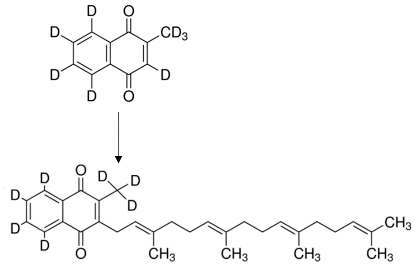
2.2. Synthesis of Various Forms of Vitamins K

The menadione (K3) was purchased from Sigma-Aldrich; vitamin K1 was bought from Hefei TNJ Chemical Industry Co., Ltd. (Hefei, China).; and MK-4, MK-5, MK-6 and MK-7 were synthesised based on an approach described in patent No. WO/2014/058330. In brief, synthesis consisted of the following key steps: conversion of menadione into a sulfone-activated analogue of vitamin MK-1, the formation of a polyisoprenoid chain of the desired length, joint of both building blocks and completion of the structure by removing sulfones and protecting groups. Vitamin MK-7 was synthesised directly via the method referred to in the patent as “variant I.” Vitamin MK-4 synthesis, simplified by omitting the polyisoprenoid-chain-elongation procedure, involved the same techniques, where the side chain building block consisted directly of E,E-farnesyl bromide. Interchangeable employment of geraniol and farnesol in the proceedings resulted in a variety of new building blocks, allowing for customisation of the side-chain length with ease. Geranylgeranyl and geranylfarnesyl intermediates were obtained, yielding vitamins MK-5 and MK-6, respectively. The purity assessment of the vitamins was based on the previously described method [18].

2.3. Anti-Inflammatory Effect of Vitamins K in RAW 264.7 Cells

To determine the anti-inflammatory effect of various forms of vitamins K, cells were seeded (5 × 10⁵ cells/well) in 24-well plates and incubated 48 h in presence or absence of vitamins K (0, 0.1, 1 and 10 μM, including 24 h pre-incubation with vitamins before pro-inflammatory stimulation with LPS (from Salmonella enterica serotype Abortusequi 1 ng/mL) adopting an experimental approach described previously [10]. Vitamins K at a concentration of 10 μM did not influence the viability of RAW264.7 cells as confirmed using the MTT test. The concentration of LPS (1 ng/mL) and time of pro-inflammatory stimulation (24-h) were chosen based on a preliminary experiment. All experiments were performed in dark conditions to protect the loss of physiological activity of the vitamins K analogues. The medium was changed every 24 h to ensure the presence of fresh vitamins K.

2.4. Measurements of Cytokines Production by RAW 264.7 Cells

The levels of TNFα and IL-6 in the cell culture supernatant were determined via the enzyme-linked immunosorbent assay (ELISA) with a commercially available kit (R&D system, Abingdon, UK) according to the manufacturer’s instructions.

2.5. Measurements of NO Production in RAW 264.7 Cells

The NO production was assessed based on the measurements of the concentration of nitrite (NO2) and nitrate (NO3) in cell supernatants by the ENO-20 NOx Analyzer (Amuza Inc., San Diego, CA, USA), as described previously [19]. In brief, the ENO-20 applies the liquid chromatography method with post-column derivatisation using a Griess reagent. In this method, nitrite and nitrate were separated on the NO-PAK column (4.6 × 50 mm, Amuza Inc., San Diego, CA, USA). Next, nitrate was reduced to nitrite by cadmium-cooper column (NO- RED, Amuza, Inc., San Diego, CA, USA), mixed with a Griess reagent and the absorbance of formed purple azo derivatives was measured at 540 nm.

2.6. UHPLC-MS/MS Measurements of Eicosanoid Production by RAW 264.7 Cells

The concentrations of PGE2, PGD2, PGF, 11β-PGF, 15-deoxy-PGJ2 and 8-iso-PGF were measured using a UFLC Nexera liquid chromatograph system (Shimadzu, Kyoto, Japan) coupled to a QTrap 5500 triple quadrupole mass spectrometer (Sciex, Framingham, MA, USA). Briefly, samples were spiked with the internal standard (IS) mixture and extracted using ethyl acetate (EtOAc) acidified with acetic acid. After vigorous shaking, samples were centrifuged, and the upper organic layer was evaporated to dryness under a nitrogen stream (37 °C). The dry residue was reconstituted in nitrogen-purged ethanol (EtOH) and injected onto an Acquity UHPLC BEH C18 (3.0 × 100 mm, 1.7 µm, Waters, Milford) analytical column. Analytes were separated under the gradient elution mode. The MS detection of studied eicosanoid and their deuterated internal standards was carried out in negative ion electrospray ionisation by applying the multiple reaction monitoring mode (MRM); this method and the instrument operation parameters were described previously [20].
The quantification of studied eicosanoid was performed based on the calibration curves plotted for each eicosanoid as the relationship between the peak area ratios of analyte/IS to the nominal concentration of the analyte. Eicosanoid levels in cell medium samples were calculated based on the regression equations estimated for each analyte.

2.7. Measurements of Expression of NOS-2, COX-2 and MMPs in RAW 264.7 Cells

The expression of NOS-2, COX-2, MMP-2 and MMP-9 was assessed by the Western blot method. Cells were washed three times with Dulbecco’s phosphate-buffered saline (DPBS) (Gibco, Paisley, Scotland, UK) and lysed in a mammalian lysis buffer with protease and phosphatase inhibitors (Thermo Fisher Scientific, Waltham, MA, USA). Protein concentration was measured with the use of a BCA assay. The samples were reduced in a loading buffer and desaturated by heating at 95 °C for 5 min. An equal amount of protein from samples of 3 independent biological experiments was loaded and run on the gel and then transferred to a PVDF membrane, activated in methanol, blocked with 5% dry milk and incubated with the primary antibodies directed against the following antigens: COX-2 (16012; Cayman Chemicals, Ann Arbor, MI, USA), NOS-2 (PA3-030A; Thermo Fisher Scientific, Waltham, MA, USA), MMP-2 (ab19167; Abcam, Cambridge, UK) and MMP-9 (ab19016; Abcam, Cambridge, UK) and the appropriate horseradish peroxidase (HRP)-conjugated secondary antibodies (Santa Cruz Biotechnology, Texas, DA, USA). Equal protein loading was confirmed after transfer onto membranes, as measured by a stain-free technique provided by Bio-Rad [21]. Densitometric assessment of band intensity was performed using Image Lab software (Bio-Rad, Warsaw, Poland). The results are presented as the fold change of control corresponding to LPS-stimulated RAW 264.7 cells, without vitamins K. Total protein was used as a loading control.

2.8. Measurements of Mitochondrial Function in RAW 264.7 Cells

To assess effects of vitamins K on mitochondrial function, RAW 264.7 cells were seeded (6.5 × 10³ cells/well) into 96-well Seahorse plates and incubated 48 h with 1 μM of vitamin K3 or 10 μM of other vitamins K. The Seahorse XFe 96 Analyzer was used to measure the oxygen consumption rate (OCR; reflecting mitochondrial respiration) and extracellular acidification rate (ECAR; reflecting glycolysis). Measurements of OCR and ECAR were performed simultaneously according to protocols used previously [22]. Sequential injection of modulators of respiration that target components of the electron transport chain (ETC)—oligomycin (inhibitor of ATP synthase), FCCP (uncoupler), rotenone with antimycin A (respectively, inhibitors of complex I and complex III in the ETC)—enabled determination of the following parameters of mitochondrial function: basal respiration, proton leak, ATP-linked respiration, maximal respiration, reserve capacity and non-mitochondrial oxygen-consumption, as described previously [22].

2.9. Measurements of Endogenous Content of Vitamins K in RAW 264.7 Cells

RAW 264.7 cells were seeded (1 × 10⁶) in 24-well plates and incubated with exogenous vitamins K (K1, K3, MK-4, MK-5, MK-6 and MK-7) 24, 48 or 72 h. To assess the involvement of UBIAD-1 in endogenous vitamins K synthesis, the cells were incubated in the presence of atorvastatin (1 μM) shown to effectively inhibit UBIAD-1/MK-4 production, accordingly to literature data [23].
After 24, 48 or 72 h incubation, cells were washed with DPBS. Then, the cells were scraped off in 500 μL deionised distilled water and lysed by 10 min sonification. Cell lysates, as well as the post-culture medium, were kept at −80 °C until further analysis, i.e., LC-MS/MS quantification of vitamins K.

2.10. UHPLC-MS/MS Quantification of Vitamins K

The concentration of vitamins K (K1, MK-4 and MK-7) and CoQ10 was determined in the cell sample and post-culture medium using ultra pressure liquid chromatography coupled to tandem mass spectrometry with an atmospheric pressure chemical ionisation (UHPLC-APCI-MS/MS) technique, as described previously [24]. After the sample thawing, the solution of an internal standard containing K1-d7 (10 µL) was added, obtaining its final concentration of 100 ng/mL. Next, the ethanol (500 µL) was added after gentle mixing (10 min). The analytes were extracted using hexane (3 mL). After 10 min of vigorous shaking, the samples were centrifuged (4500× g, 10 min, 4 °C), and the upper organic layer was transferred to a fresh glass tube. The extraction step was repeated, and the whole collected organic layer was evaporated to dryness under a nitrogen stream (37 °C). The sample residue was reconstituted in 50 μL of 2-propanol, and 5 μL was injected into the UPLC-MS system. The chromatographic analysis was carried out using an Ultimate 3000 HPLC system (Dionex, Sunnyvale, CA, USA) equipped with a reversed-phase PFP analytical column (Kinetex 2.6 μm PFP, 100 Å, 100.0 × 3.3 mm; Phenomenex, Torrance, CA, USA). The mobile phase consisted of 0.1% formic acid in 2-propanol, and 0.1% formic acid in 5 mM ammonium formate was delivered in gradient elution mode. The signal from analytes and their internal standards was registered by applying a TSQ Quantum Ultra triple quadrupole mass spectrometer (Thermo Fisher Scientific, Waltham, MA, USA) in positive ionisation mode. The following most abundant ion transitions were monitored for quantification in selected reaction monitoring mode (SRM): K1: 451.4→187.2; K1-d7: 458.5→194.2; MK-4: 445.4→187.2; MK-4-d7: 452.4→194, MK-7: 649.6→187.2 and CoQ10: 863.6→197.1. The working parameters of the mass spectrometer were as follows: corona discharge needle voltage: 4 kV; vaporiser temperature: 325 °C; sheath gas pressure: 50 arb; ion sweep gas pressure: 10 arb; auxiliary gas pressure: 30 arb; capillary temperature: 325 °C; and collision pressure 1.5 mTorr, using argon as the collision gas.
The calibration curves were plotted as the relationship between the peak area ratios of the analyte/internal standard to the nominal concentration of the analyte. Vitamins K and CoQ10 levels in the studied samples were calculated based on the regression equations estimated for each analyte. The quantification of MK-4-d7 was performed based on the regression equations calculated for MK-4.

2.11. Statistical Analysis

Statistical analysis was performed using GraphPad Prism 9 (San Diego, CA, USA). Depending on data distribution (tested with the Shapiro–Wilk or D’Agostino–Pearson omnibus normality test), data were presented as mean ± SD or median ± IQR, respectively, of at least three biological experiments, with a minimum of three technical replicates without outliers. Only post hoc p-values < 0.05 were considered significant.

3. Results

3.1. Effects of Vitamins K on LPS-Induced NO Production and Induction of NOS-2 in RAW 264.7 Cells

RAW 264.7 cells were stimulated with LPS (1 ng/mL, 24 h) in the presence or absence of various forms of vitamins K (K1, K3, MK-4, MK-5, MK-6 and MK-7) at a concentration of 0, 0.1, 1 and 10 μM added 24 h before LPS stimulation. As shown in Figure 1, all forms of vitamins K inhibited nitrite (Figure 1A) and nitrate (Figure 1B) accumulation in a concentration-dependent manner, with the most pronounced effect at a concentration of 10 μM. The strongest inhibitory effect on NO2 production was noted for K1 (54.4% inhibition), MK-4 (51.2% inhibition), K3 (48.3% inhibition) and MK-5 (35.9% inhibition) forms, as compared with MK-6 (24.5% inhibition) and MK-7 (12.4% inhibition), which displayed weaker effects. To confirm that vitamins K inhibit NO production by the regulation of NOS-2 expression, a Western blot analysis was performed (Figure S1A,E). The inhibition of an LPS-induced increase in NOS-2 expression was visible for all forms of vitamins K.

3.2. Effects of Vitamins K on LPS-Induced Cytokines Production in RAW 264.7 Cells

All forms of vitamins K (K1, K3, MK-4, MK-5, MK-6 and MK-7) reduced the LPS-induced production of IL-6 (Figure 1C) and TNFα (Figure 1D) in RAW 264.7 cells. The strongest inhibitory effect on cytokine production was observed for K1, K3 and MK-4, as compared with vitamins K forms with longer side chains, such as MK-6 and MK-7, which induced weaker effects.

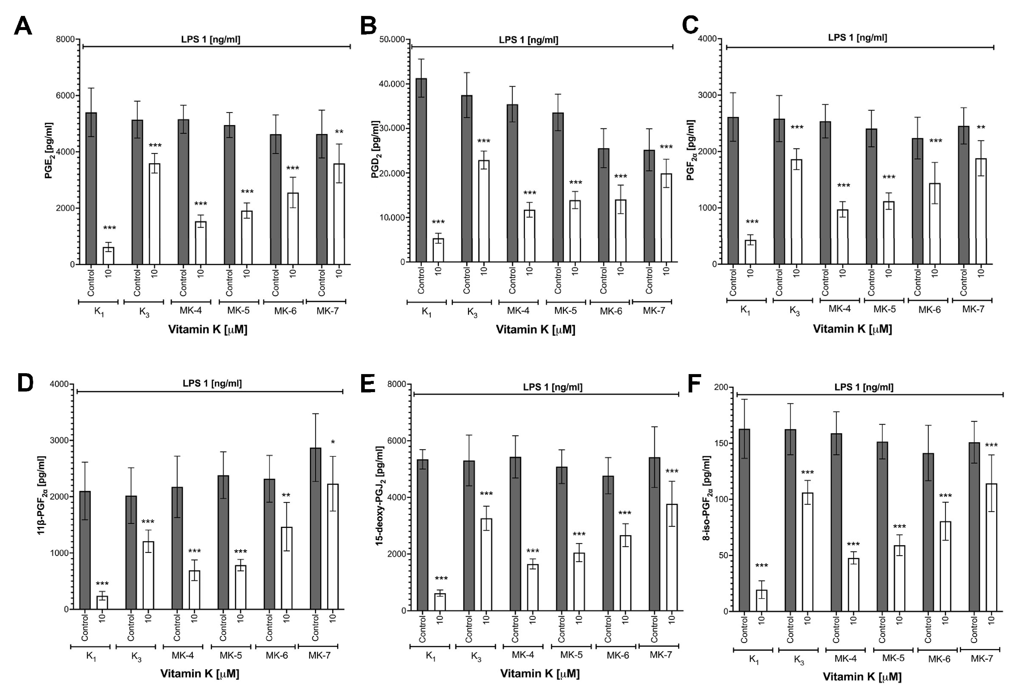
3.3. Effects of Vitamins K on LPS-Induced Eicosanoid Production and Induction of COX-2 in RAW 264.7 Cells

As shown in Figure 2, all forms of vitamins K inhibited eicosanoid (PGE2, PGD2, PGF, 11β-PGF, 15-deoxy-PGJ2 and 8-iso-PGF) production in a concentration-dependent manner. The strongest inhibitory effect on eicosanoid production was observed for vitamin K1: PGE2 (88.5% inhibition), PGD2 (87.1% inhibition), PGF (83.5% inhibition), 11β-PGF (88.5% inhibition), 15-deoxy-PGJ2 (88.4% inhibition) and 8-iso-PGF (88.1% inhibition); MK-7, in contrast, afforded weaker effects: PGE2 (22.6% inhibition), PGD2 (21.0% inhibition), PGF (23.4% inhibition), 11β-PGF (22.3% inhibition), 15-deoxy-PGJ2 (30.4% inhibition) and 8-iso-PGF (24.2% inhibition). To confirm that vitamins K affected eicosanoid production by the regulation of COX-2 expression, a Western blot analysis was performed. All forms of vitamins K inhibited LPS-induced increase in COX-2 (Figure S1B) without an effect on COX-1 expression (data not shown).

3.4. Effects of Vitamins K on LPS-Induced MMP-2 and MMP-9 Expression in RAW 264.7 Cells

All examined form of vitamins K (K1, K3, MK-4, MK-5, MK-6 and MK-7) inhibited LPS-induced MMP-2 as well as MMP-9 protein expression (Figure S1C,D,G,H). However, the inhibition of the expression of MMP-2 protein seemed to be more pronounced than the inhibition of MMP-9 appeared.

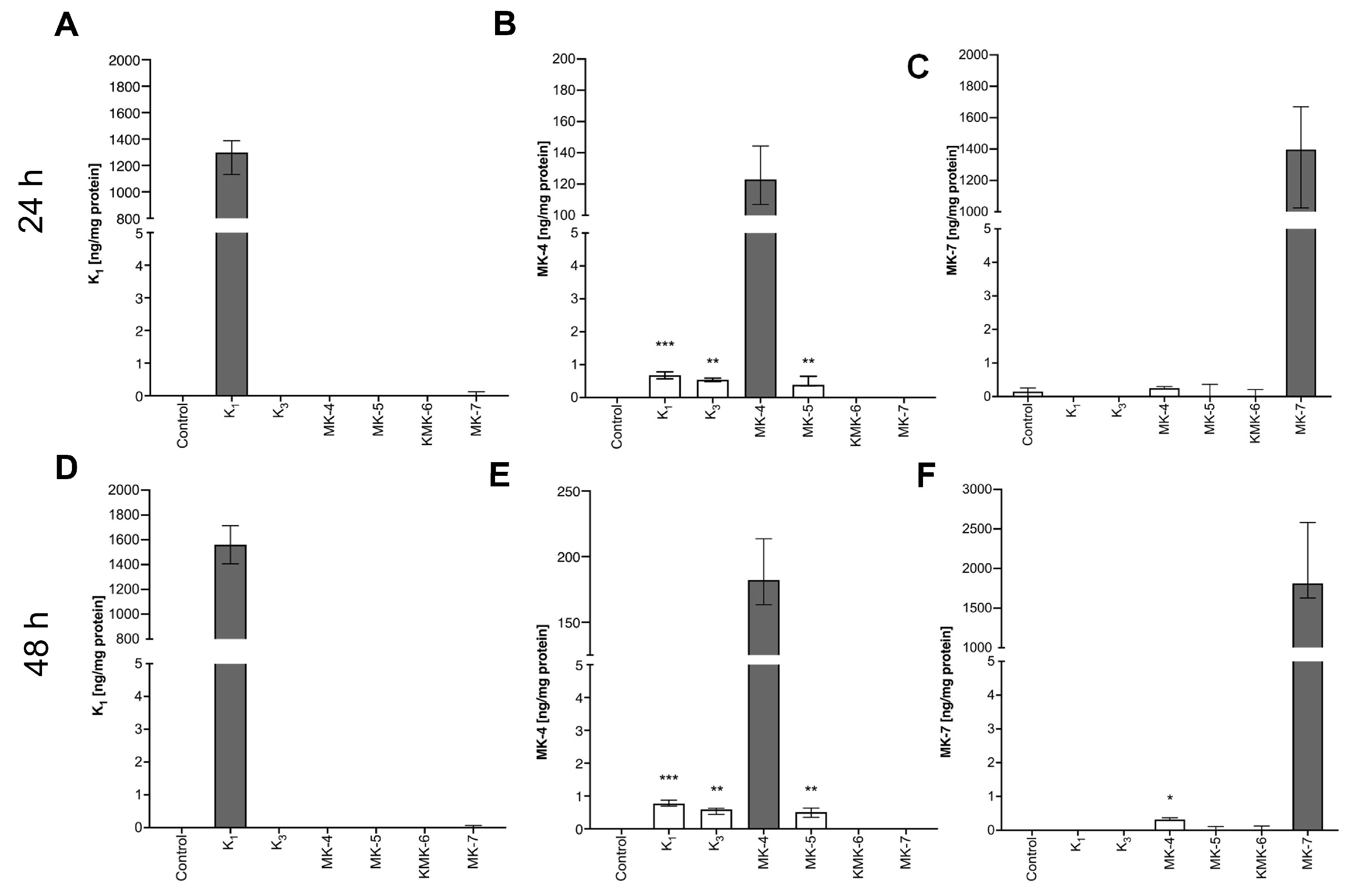
3.5. Synthesis of Endogenous MK-4 from Exogenous Vitamins K

Exogenous vitamins K1, K3 and MK-5, but not MK-6 or MK-7, resulted in an increase in endogenous MK-4 content in RAW 264.7 cells after 24 h (Figure 3B), 48 h (Figure 3E) and 72 h (data not shown) of incubation.
To confirm that endogenous MK-4 may be produced from exogenous K1 and K3, deuterium-labelled K1 (K1-d7) and K3 (K3-d8) vitamins were used. Twenty-four hours’ incubation of RAW 264.7 cells with K1-d7 and K3-d8 resulted in an increased intracellular concentration of vitamin MK-4-d7 analysed in cell lysate (Table 1).

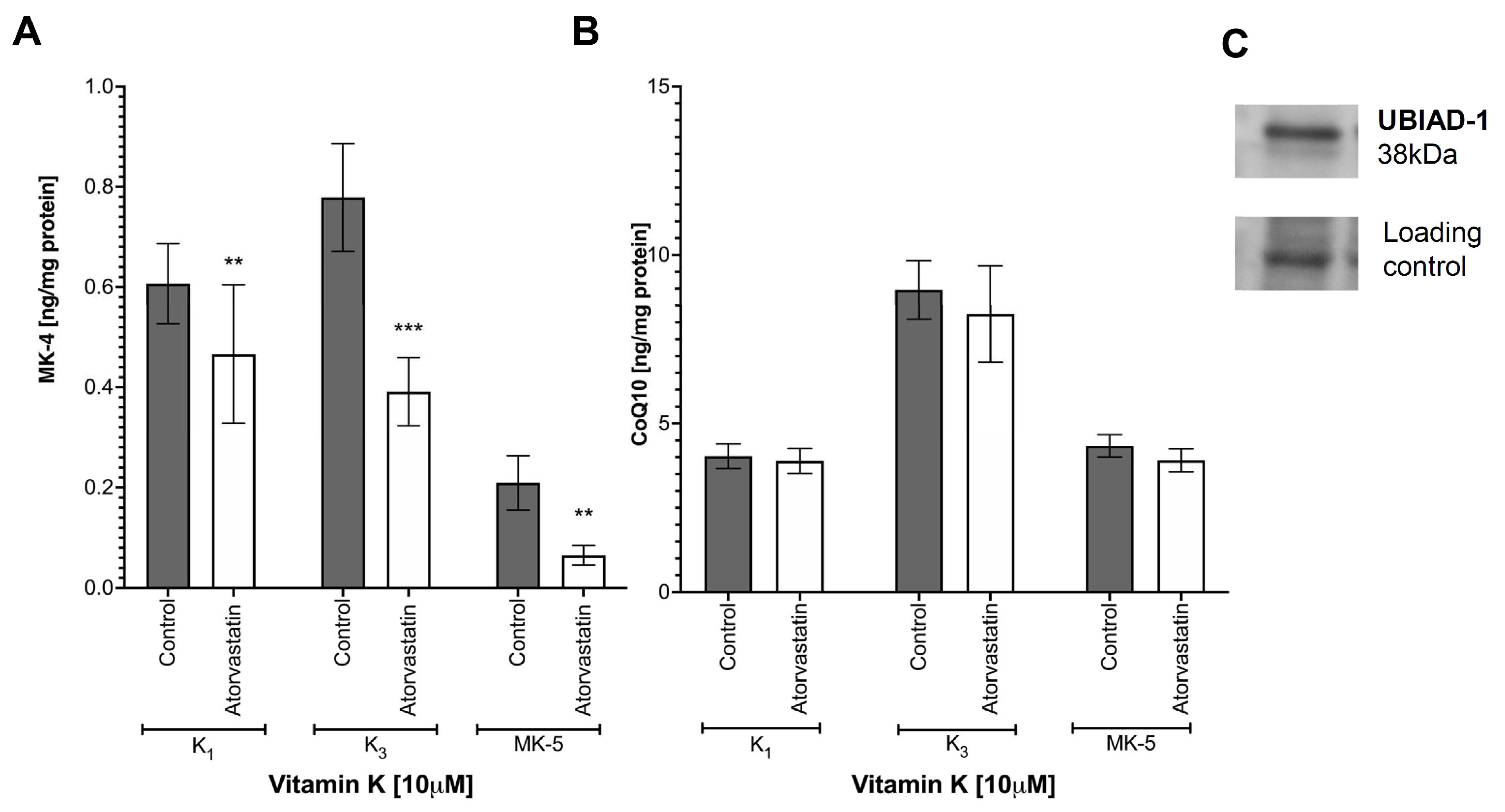
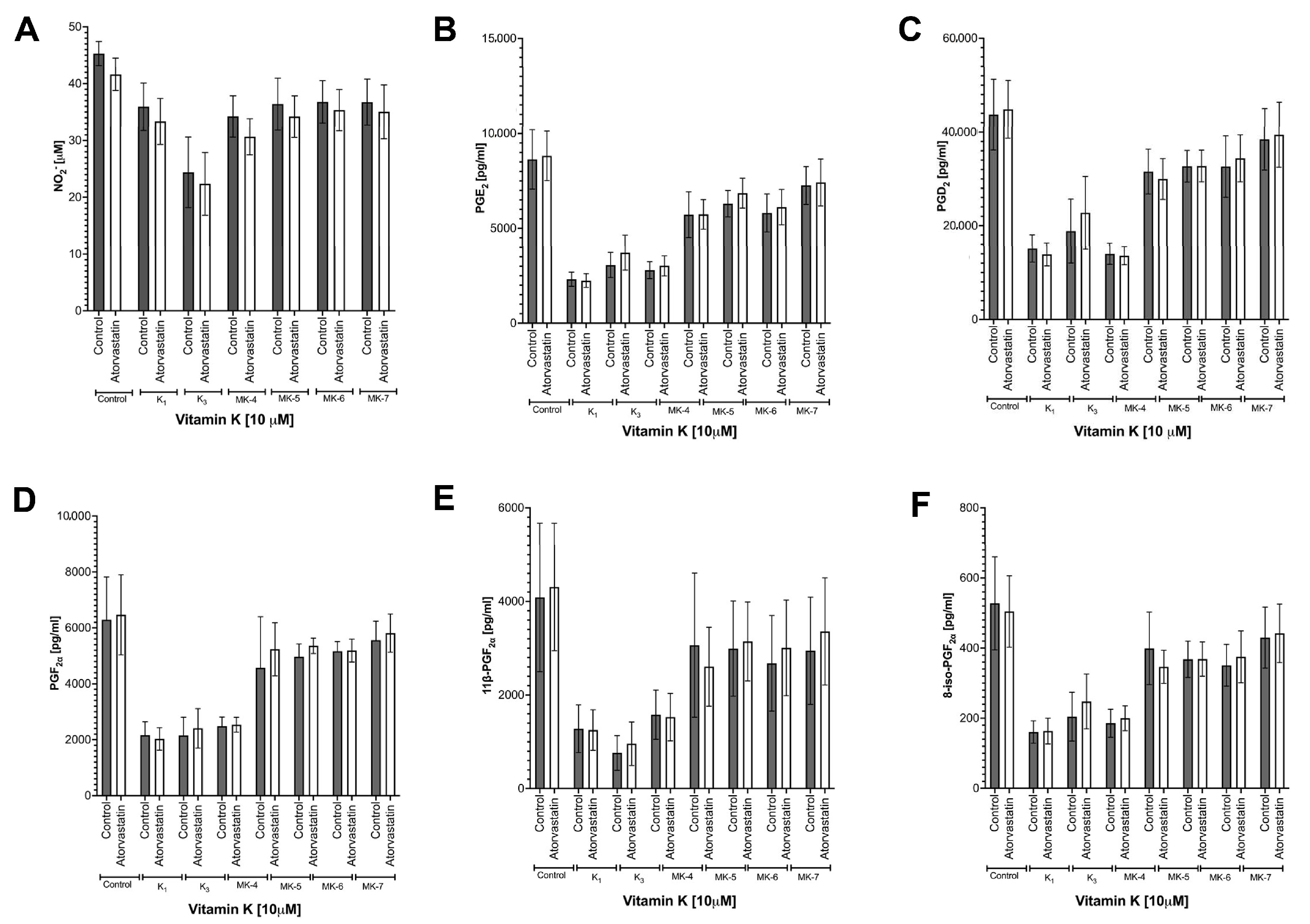
3.6. Effects of Atorvastatin on Endogenous Production of MK-4 from Exogenous Vitamins K and on Anti-Inflammatory Effect of Vitamins K

To test whether endogenous production of MK-4 from vitamin K1, K3 or MK-5 involves an UBIAD-1-dependent pathway, exogenous vitamin K1, K3 or MK-5 (Figure 4A) were incubated in the absence or the presence of atorvastatin (1 μM). As shown in Figure 4 atorvastatin (1 μM) significantly decreased endogenous production of MK-4 from exogenous vitamins K1, K3 and MK-5. However, in the presence of atorvastatin, CoQ10 content in macrophages was not altered (Figure 4B). The presence of UBIAD-1 protein in the murine macrophages cell line, RAW 264.7, was confirmed by Western blot (Figure 4C).
Despite the inhibitory effects of endogenous production of MK-4 by atorvastatin, the anti-inflammatory effect of exogenous K1, K3 and MK-5 remained unchanged by atorvastatin, as evidenced by the unmodified LPS-induced accumulation of nitrite and eicosanoid: PGE2, PGD2, PGF, 11β-PGF, 15-deoxy-PGJ2 and 8-isoPGF (Figure 5).

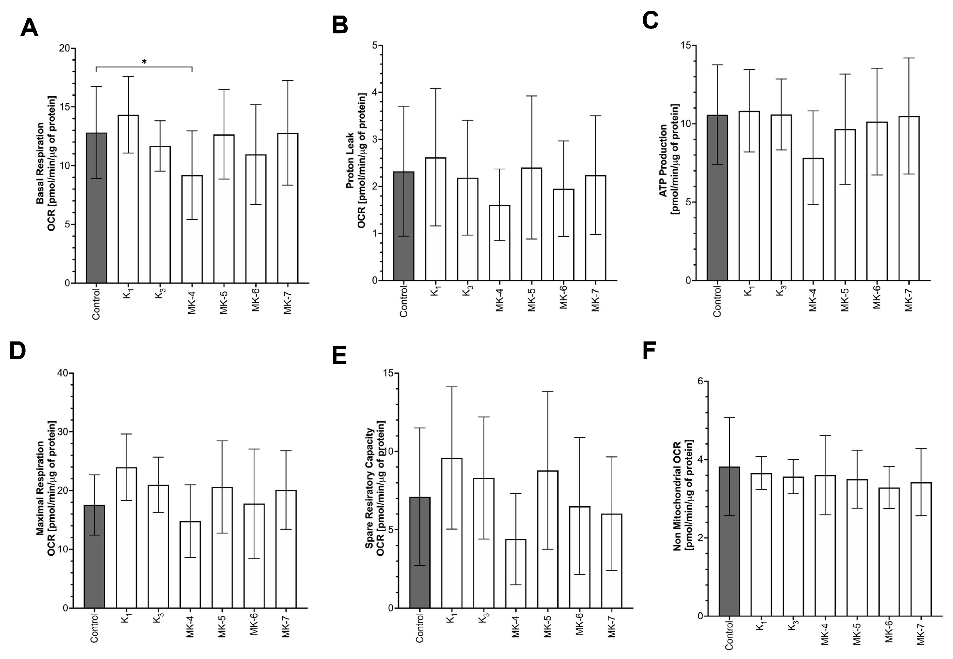
3.7. Effects of Vitamins K on Mitochondrial Respiration
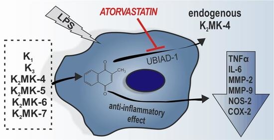
The influence of vitamins K on mitochondrial function was comprehensively analysed using the Seahorse method. None form of vitamins K (K1, K3, MK-4, MK-5, MK-6 or MK-7) improved mitochondrial function in non-stimulated macrophages, as evidenced by unchanged basal respiration, maximal respiration, ATP production, spare respiratory capacity, proton leak and non-mitochondrial oxygen consumption in the presence and absence of vitamins K (Figure 6).

4. Discussion

In the present work, we characterise the pharmacological effects of a wide range of forms of vitamins K (K1, K3, MK-4, MK-5, MK-6 and MK-7) added exogenously to LPS-stimulated RAW 264.7 cells and demonstrated their anti-inflammatory actions, which included the following: (1) the downregulation of the induction of NOS-2 and NO production, (2) the downregulation of COX-2 induction and various eicosanoid production (PGE2, PGD2, PGF, 11β-PGF, 15-deoxy-PGJ2 and 8-isoPGF), (3) the inhibition of cytokine production (IL-6 and TNFα) and (4) the inhibition of MMP-2 and MMP-9 expression. Interestingly, the vitamins K forms with shorter isoprenoid chains, such as K1, K3, MK-4 and MK-5, displayed stronger anti-inflammatory activity than the forms with longer isoprenoid chains (MK-6 and MK-7). The nexus of our results is our demonstration, which is to our knowledge for the first time, that endogenous MK-4 synthesis is activated in RAW 264.7 cells when some forms of exogenous vitamins K, including K1, K3 and MK-5 as substrates, are available. Finally, our results showed that endogenous MK-4 synthesis was inhibited by atorvastatin, while the anti-inflammatory effects of exogenous vitamins K were not. Altogether, our results suggest that macrophages display an active synthesis of endogenous MK-4 from some forms of exogenous vitamins K that could be inhibited by statin, but anti-inflammatory effects of exogenous vitamins K could not be attributed to endogenous MK-4, but could be ascribed to the inhibition of NFκB-dependent pathway [12].
Our work extends previous reports on the anti-inflammatory effects of vitamins K. Previously, the anti-inflammatory effects of vitamin K1 [13], MK-4 [9] MK-3 and MK-7 [10] on cytokine production (e.g., IL-1, IL-6 and TNFα) were demonstrated, but prior research did not investigate any such effects on other inflammatory pathways, such as COX-2, NOS-2 or MMP. This study also incorporated a broader spectrum of forms of vitamins K than previous studies; the anti-inflammatory effects of K1 [13], K3 [25], MK-4 [9] and MK-7 [10], but not MK-5 and MK-6 were previously characterised. We confirmed anti-inflammatory effects in the murine macrophages cell line, RAW 264.7, for all forms of vitamins K (K1, K3, MK-4, MK-5, MK-6 and MK-7), but striking differences in their potency were observed. Specifically, there were stronger anti-inflammatory effects of K3 and forms of vitamins K with shorter isoprenoid chains (K1, MK-4 and MK-5) than among MK-6 and MK-7, which contain longer isoprenoid chains. This pattern diverged, for example, from endothelial cells, where MK-7 displayed more potent effects than MK-4 [26], as well as in the case of in vivo model, where MK-7 was shown to be more efficient to achieve carboxylation of extrahepatic (e.g., osteocalcin) and hepatic (e.g., prothrombin) proteins [27]. The latter could be due to the advantageous pharmacokinetic profile of MK-7, but the order of potency of binding of K1, MK-4, MK-7 and K3 to VCORC1 and the efficacy of various forms of vitamins K to serve as substrates for post-translational γ-carboxylation pointed to the weakest effects for K3 [28]; in contrast, in our experiments, K3 was among the more potent forms of vitamins K in inducing anti-inflammatory effects in RAW 264.7 cells.
To understand these findings more effectively, we analysed in detail the intracellular metabolism of vitamins K in macrophages by using deuterium-labelled K1 (K1-d7) and K3 (K3-d8) vitamins and our recently established UHPLC-MS/MS-based method of vitamins K quantification [24]. Using this approach, we provided unequivocal evidence that the macrophages produced endogenous vitamin MK-4 from exogenous vitamin K1, K2 or K3.
The production of endogenous MK-4 from exogenous precursors has been described in intestinal bacteria, the kidneys, the brain, the intestines and the endothelium [29,30,31,32] but not in macrophages.
It has been previously reported that intestinal bacteria or epithelial cells in the intestines have specific enzymes producing menadione from dietary exogenous vitamin K1 [32]. However, prenylation of menadione moiety could be executed in a number of cells via UBIAD-1, the enzyme involved in the intracellular de novo cholesterol synthesis [33]. In the present study, we observed the unique pattern of interconversion of exogenous vitamins K in RAW 264.7 cells to endogenous MK-4 synthesis. Exogenous K1, K3 and MK-5, but not MK-6 and MK-7, were used as exogenous substrates for endogenous MK-4 synthesis in macrophages, suggesting that shorter, unsaturated isoprenoid chains of vitamin K2; monounsaturated phytol sidechains (vitamin K1); or ready-to-use menadione (K3) [34] represent preferable substrates for endogenous MK-4 synthesis as compared with the longer unsaturated isoprenoid chain, which is harder to cleave [35]. Surprisingly, the inhibitory effect of atorvastatin was considerably weaker for endogenous production of MK-4 from vitamin K1 as compared with the effects of atorvastatin on the endogenous production of MK-4 from K3. Again, the explanation of this phenomenon may be the fact that menadione is ready to use substrate to prenylation, while in the case of K1, firstly, the isoprenoid side chain has to be cleaved. It could well be that the longer time of incubation with atorvastatin could result in a stronger inhibition of the conversion of K1 into MK-4. Additionally, we noted MK-7 synthesis after exogenous MK4 administration, but to explain this phenomenon, further research is needed.
Our results clearly indicate that macrophages have full machinery for the deprenylation of exogenous vitamins and, subsequently, prenylation of menadione for the synthesis of endogenous vitamin MK-4. In contrast to endothelial cells [34], coenzyme Q10 (CoQ10) content in macrophages was not inhibited by atorvastatin, but in our hands, the endogenous CoQ10 content was several folds higher than MK-4 content and higher than CoQ10 content in endothelial cells.
To summarise, macrophages extend the list of cells, including the bacteria, intestinal epithelium, kidneys, the brain, and the endothelium [30,31,32,36,37], that are able to synthesise MK-4. Macrophages displayed the ability to synthesise endogenous vitamin MK-4 from various exogenous sources of vitamins K that could originate from the diet or bacteria. Whether this pattern represents a cell-specific feature is not known; previous work reporting turnover into endogenous MK-4 in various cells examined only vitamin K1, K3 or MK-7, not the whole spectrum of exogenous forms of vitamins K, as used in the present study [32,38,39,40]. Generally, it is believed that MK-4 is produced in the body after K1 or MK-7 administration in vivo [41]. At the same time, it is accepted that menaquinone and phylloquinone have different functions: vitamin K2 mainly exerts extrahepatic activity, including vasoprotective action, while vitamin K1 primarily engenders hepatic activity and regulates coagulation. In contrast to this notion, our data demonstrate that various forms of vitamins K, including exogenous K1 and MK-4, exhibit anti-inflammatory activity.
In the present work, we did not identify the enzyme or enzymes involved in the cleavage of side chains of exogenous vitamins K, but we indicated that the prenylation activity was related to UBIAD-1 activity and was inhibited by statins. In fact, atorvastatin inhibited endogenous MK-4 production, but without an impact on the anti-inflammatory effects of vitamins K. Consequently, our results demonstrated that the anti-inflammatory effects of exogenous vitamins K in murine macrophages could not be explained by endogenous MK-4 synthesis despite active MK-4 synthesis in the cells. On the other hand, it should be noted that treatment with exogenous vitamins at micromolar concentration resulted in the nanomolar concentration of endogenous MK-4. Therefore, we are not able to exclude that endogenous MK-4—if achieving micromolar concentration—would inhibit the inflammatory response.
Obviously, endogenous MK-4 might be involved in the regulation of macrophage function, for example, via the regulation of senescence-related genes, regulation of differentiation, autophagy and apoptosis [42,43] or the regulation of proliferation [44,45]. Further studies are needed to define the functional role of endogenous MK-4 in macrophages.
Previous studies indicate the role of reduced forms of vitamins K in vitamins K-dependent carboxylation in various processes, and the number of VKDPs has been identified [8,46]. Although we confirmed the presence of GGCX protein in RAW 264.7 cells via Western blot, the VCOR blockade using two different antagonists of vitamins K (acenocoumarol and warfarin) had no impact on the anti-inflammatory effects of vitamin K1, K3 and MK-4, as assessed on the basis of NO2 production (data now shown) thereby confirming the previous reports indicating that regulation of inflammation by vitamins K was independent of VKD carboxylation in macrophage-like cells [13,14]. Indeed, in THP-1 cells stimulated with LPS, the inhibition of VKOR by warfarin had no impact on the inhibitory activity of vitamin MK-4 on IL-6 mRNA expression [13]. Furthermore, the anti-inflammatory activity of the Gla-rich protein was observed for carboxylated as well as non-carboxylated forms of protein despite the changes of expression of GGCX protein in response to vitamins K treatment [11,14]. Altogether, the presence and changes of GGCX expression in response to vitamins K suggest that VKD carboxylation can be somehow involved in the regulation of macrophage function but that the anti-inflammatory effects of various forms of vitamins K reported here were independent of γ-carboxylation.
Finally, in the present work, we also excluded the mitochondrial-targeted mechanisms of the anti-inflammatory action of vitamins K. Even though mitochondria play an important role in inflammation [47] and vitamin K2 can act as a membrane-bound electron carrier in bacteria [16,17], our results demonstrated that vitamins K had no impact on mitochondrial function in the murine macrophages cell line, RAW 264.7, as evidenced by unchanged parameters of mitochondrial respiration such as basal respiration, ATP-linked respiration, maximal respiration and reserve capacity. It is likely that we were unable to observe the effects of vitamins K on mitochondrial function because of a large reserve of ubiquinone in RAW 264.7 cells. Indeed, the concentration of CoQ10 was approximately four times higher than the concentration of endogenous MK-4 in macrophages, as measured here by UHPLC-MS/MS. In fibroblasts, even in the state of CoQ10 deficiency, vitamin K2 did not act as an electron carrier [17].
Furthermore, the role of vitamins K as a ligand of nuclear steroid receptor (SXR) seems an unlikely mechanism of anti-inflammatory effects of vitamins K. Although previous studies reported that vitamin K1 or K2 [48,49,50,51] were ligands of steroid and xenobiotic receptor (SXR), the vitamin K1 was a weaker ligand of SXR than MK-4, as compared with the more potent or similar anti-inflammatory activity of vitamin K1 vs. MK-4 reported here.
Previous work demonstrated that K3 and their analogues inhibited NFκB activation and the phosphorylation of IKKα/β in macrophages stimulated with LPS [13]. Accordingly, the pattern of the anti-inflammatory profile of vitamins K described in the present work, which involves effects on NOS-2, COX-2, cytokines and MMPs known to be regulated by NFκB [52,53,54], suggest that the anti-inflammatory activity of vitamins K was mediated by menadione, the intermediate product of the endogenous conversion of vitamins K in macrophages. This notion is in line with the stronger anti-inflammatory potential of the shorter forms of vitamins to convert more easily into menadione [55].
In fact, our results confirmed the inhibitory function of vitamins K on NFκB nuclear translocation in LPS-stimulated RAW 264.7 cells (Figure S2). Thus, the anti-inflammatory effects of vitamins K could be ascribed to the regulation of the NFκB pathway [12,13]. However, the details of this mechanisms need to be further explored [56,57,58].
The anti-inflammatory activity of vitamins K in macrophages elaborated upon here and reported previously [10,13] represents an important asset of vitamins K action that may have a therapeutic significance in various inflammatory conditions [5,9,24,59,60,61,62]. In fact, high vitamin K1 status was associated with lower concentrations of pro-inflammatory markers in post-menopausal women [9]. In contrast, statins blocking endogenous MK-4 synthesis in macrophages, and presumably in many other cells, may have detrimental effects. Indeed, possible side effects of statins, which include myalgia [63], neuropathy [64] or diabetes [65], may be related to the inhibition of endogenous synthesis of MK-4. To conclude, despite the vast literature on vitamins K pharmacology, in the present work, we provided a novel insight into the profile of anti-inflammatory action of exogenous vitamins K. We demonstrated that anti-inflammatory effects of vitamins K1, K2 and K3 in LPS-induced macrophages encompass a wide spectrum of activities, including the inhibition of NOS-2/NO, COX-2/eicosanoid, cytokine and MMP-dependent pathways. Furthermore, our study provided evidence, to our knowledge for the first time, that besides the intestinal bacteria, kidneys, brain, intestines and the endothelium also macrophages are able to synthesise endogenous MK-4 vitamin. Finally, our results uncovered that various forms of exogenous vitamins K (K1, K3 and MK-5 but not MK-6, MK-7) provide a substrate for endogenous MK-4 synthesis in macrophages. However, the anti-inflammatory effects of various forms of vitamins K were independent of the UBIAD-1-dependent synthesis of endogenous MK-4 but ascribed to the inhibition of the NFκB pathway as suggested also previously [12,13]. The role of endogenous MK-4 in the regulation of the function of macrophages remains to be established.

Supplementary Materials

The following are available online at https://www.mdpi.com/article/10.3390/cells10071571/s1. Figure S1 Inhibition of NOS-2, COX-2, MMP-2 and MMP-9 expression by various forms of vitamins K in RAW 264.7 cells stimulated with LPS. Figure S2 Role of NFκB in the anti-inflammatory action of Vitamins K.

Author Contributions

A.K.-R. and S.C. conceived and designed the experiments; A.K.-R. performed all experiments, measured cytokine production, performed Western blots and immunocytochemical analysis of NFκB nuclear translocation; A.K. measured eicosanoid production and vitamins K content by UHPLC-MS/MS; P.K. measured mitochondrial respiration via the Seahorse technique; and A.T. measured nitrite and nitrate using ENO-20-NOx Analyzer. M.N. and K.S. synthesised vitamins K analogues. A.K.-R. performed data analysis and prepared figures, A.K.-R. drafted the manuscript, and S.C. revised the manuscript. A.K.-R. and S.C. wrote the final version of the manuscript. All authors have read and agreed to the published version of the manuscript.

Funding

This work was supported by Polish National Science Centre (NCN) grants: PRELUDIUM No.2018/31/N/NZ7/03900 and OPUS No. 2018/29/B/NZ7/01684 and partially by National Centre for Research and Development the InterDokMed project No. POWR.03.02.00-00-I013/16. The open-access publication of this article was funded by the Priority Research Area BioS under the program “Excellence Initiative—Research University” at the Jagiellonian University in Krakow.

Institutional Review Board Statement

Not applicable.

Informed Consent Statement

Not applicable.

Data Availability Statement

The data presented in this study are available on reasonable request from the corresponding author.

Acknowledgments

The authors would like to thank Renata Budzynska for technical assistance with cell culture and Agnieszka Burzynska-Prajzner and Przemyslaw Dorozynski for their support to initiate this study.

Conflicts of Interest

The authors declare no competing interests.

References

  1. Ferland, G. Vitamins K, an emerging nutrient in brain function. BioFactors 2012, 38, 151–157. [Google Scholar] [CrossRef]
  2. Tie, J.-K.; Jin, D.-Y.; Straight, D.L.; Stafford, D.W. Functional study of the vitamins K cycle in mammalian cells. Blood 2011, 117, 2967–2974. [Google Scholar] [CrossRef] [PubMed] [Green Version]
  3. Wen, L.; Feil, S.; Wolters, M.; Thunemann, M.; Regler, F.; Schmidt, K.; Friebe, A.; Olbrich, M.; Langer, H.; Gawaz, M.; et al. A shear-dependent NO-cGMP-cGKI cascade in platelets acts as an auto-regulatory brake of thrombosis. Nat. Commun. 2018, 1–11. [Google Scholar] [CrossRef]
  4. Booth, S.L. Roles for vitamins K beyond coagulation. Annu. Rev. Nutr. 2009, 29, 89–110. [Google Scholar] [CrossRef] [PubMed]
  5. Schwalfenberg, G.K. Vitamins K and K2: The Emerging Group of Vitamins Required for Human Health. J. Nutr. Metab. 2017, 2017, 1–6. [Google Scholar] [CrossRef] [PubMed]
  6. Shearer, M.J.; Fu, X.; Booth, S.L. Vitamins K nutrition, metabolism, and requirements: Current concepts and future research. Adv. Nutr. 2012, 3, 182–195. [Google Scholar] [CrossRef] [PubMed] [Green Version]
  7. Shearer, M.J.; Newman, P. Metabolism and cell biology of vitamins K. Thromb. Haemost. 2008. [Google Scholar] [CrossRef]
  8. Siltari, A.; Vapaatalo, H. Vascular Calcification, Vitamins K and Warfarin Therapy-Possible or Plausible Connection? Basic Clin. Pharmacol. Toxicol. 2018, 122, 19–24. [Google Scholar] [CrossRef] [Green Version]
  9. Shea, M.K.; Booth, S.L.; Massaro, J.M.; Jacques, P.F.; D’Agostino, R.B.; Dawson-Hughes, B.; Ordovas, J.M.; O’Donnell, C.J.; Kathiresan, S.; Keaney, J.F.; et al. Vitamins K and vitamin D status: Associations with inflammatory markers in the Framingham offspring study. Am. J. Epidemiol. 2008. [Google Scholar] [CrossRef] [Green Version]
  10. Pan, M.H.; Maresz, K.; Lee, P.S.; Wu, J.C.; Ho, C.T.; Popko, J.; Mehta, D.S.; Stohs, S.J.; Badmaev, V. Inhibition of TNF-α, IL-1α, and IL-1β by Pretreatment of Human Monocyte-Derived Macrophages with Menaquinone-7 and Cell Activation with TLR Agonists in Vitro. J. Med. Food 2016, 19, 663–669. [Google Scholar] [CrossRef]
  11. Cavaco, S.; Viegas, C.S.B.; Rafael, M.S.; Ramos, A.; Magalhães, J.; Blanco, F.J.; Vermeer, C.; Simes, D.C. Gla-rich protein is involved in the cross-talk between calcification and inflammation in osteoarthritis. Cell. Mol. Life Sci. 2016, 73, 1051–1065. [Google Scholar] [CrossRef] [PubMed]
  12. Ohsaki, Y.; Shirakawa, H.; Hiwatashi, K.; Furukawa, Y.; Mizutani, T.; Komai, M. Vitamins K suppresses lipopolysaccharide-induced inflammation in the rat. Biosci. Biotechnol. Biochem. 2006. [Google Scholar] [CrossRef] [PubMed] [Green Version]
  13. Ohsaki, Y.; Shirakawa, H.; Miura, A.; Giriwono, P.E.; Sato, S.; Ohashi, A.; Iribe, M.; Goto, T.; Komai, M. Vitamins K suppresses the lipopolysaccharide-induced expression of inflammatory cytokines in cultured macrophage-like cells via the inhibition of the activation of nuclear factor αB through the repression of IKKα/β phosphorylation. J. Nutr. Biochem. 2010, 21, 1120–1126. [Google Scholar] [CrossRef]
  14. Viegas, C.S.B.; Costa, R.M.; Santos, L.; Videira, P.A.; Silva, Z.; Araújo, N.; Macedo, A.L.; Matos, A.P.; Vermeer, C.; Simes, D.C. Gla-rich protein function as an anti-inflammatory agent in monocytes/macrophages: Implications for calcification-related chronic inflammatory diseases. PLoS ONE 2017, 12, e0177829. [Google Scholar] [CrossRef] [PubMed]
  15. Suvarna, K.; Stevenson, D.; Meganathan, R.; Hudspeth, M.E.S. Menaquinone (vitamins K2) biosynthesis: Localization and characterization of the menA gene from Escherichia coli. J. Bacteriol. 1998, 180, 2782–2787. [Google Scholar] [CrossRef] [Green Version]
  16. Vos, M.; Esposito, G.; Edirisinghe, J.N.; Vilain, S.; Haddad, D.M.; Slabbaert, J.R.; Van Meensel, S.; Schaap, O.; De Strooper, B.; Meganathan, R.; et al. Vitamins K is a mitochondrial electron carrier that rescues pink1 deficiency. Science 2012. [Google Scholar] [CrossRef] [Green Version]
  17. Cerqua, C.; Casar, A.; Pierrel, F.; Fonseca, L.V.; Viola, G.; Salviati, L.; Trevisson, E. Vitamins K cannot substitute Coenzyme Q 10 as electron carrier in the mitochondrial respiratory chain of mammalian cells. Sci. Rep. 2019, 1–7. [Google Scholar] [CrossRef] [Green Version]
  18. Jedynak, Ł.; Jedynak, M.; Kossykowska, M.; Zagrodzka, J. A novel method for the determination of chemical purity and assay of menaquinone-7. Comparison with the methods from the official USP monograph. J. Pharm. Biomed. Anal. 2017. [Google Scholar] [CrossRef]
  19. Przyborowski, K.; Proniewski, B.; Czarny, J.; Smeda, M.; Sitek, B.; Zakrzewska, A.; Zoladz, J.A.; Chlopicki, S. Vascular Nitric Oxide-Superoxide Balance and Thrombus Formation after Acute Exercise. Med. Sci. Sports Exerc. 2018. [Google Scholar] [CrossRef]
  20. Kij, A.; Kus, K.; Czyzynska-Cichon, I.; Chlopicki, S.; Walczak, M. Development and validation of a rapid, specific and sensitive LC-MS/MS bioanalytical method for eicosanoid quantification-assessment of arachidonic acid metabolic pathway activity in hypertensive rats. Biochimie 2020. [Google Scholar] [CrossRef]
  21. Rivero-Gutiérrez, B.; Anzola, A.; Martínez-Augustin, O.; De Medina, F.S. Stain-free detection as loading control alternative to Ponceau and housekeeping protein immunodetection in Western blotting. Anal. Biochem. 2014. [Google Scholar] [CrossRef] [Green Version]
  22. Kaczara, P.; Motterlini, R.; Rosen, G.M.; Augustynek, B.; Bednarczyk, P.; Szewczyk, A.; Foresti, R.; Chlopicki, S. Carbon monoxide released by CORM-401 uncouples mitochondrial respiration and inhibits glycolysis in endothelial cells: A role for mitoBKCa channels. Biochim. Biophys. Acta-Bioenerg. 2015. [Google Scholar] [CrossRef] [Green Version]
  23. Jiang, S.Y.; Li, H.; Tang, J.J.; Wang, J.; Luo, J.; Liu, B.; Wang, J.K.; Shi, X.J.; Cui, H.W.; Tang, J.; et al. Discovery of a potent HMG-CoA reductase degrader that eliminates statin-induced reductase accumulation and lowers cholesterol. Nat. Commun. 2018. [Google Scholar] [CrossRef] [Green Version]
  24. Bar, A.; Kus, K.; Manterys, A.; Proniewski, B.; Sternak, M.; Przyborowski, K.; Moorlag, M.; Sitek, B.; Marczyk, B.; Jasztal, A.; et al. Vitamins K-MK-7 improves nitric oxide-dependent endothelial function in ApoE/LDLR−/− mice. Vasc. Pharmacol. 2019. [Google Scholar] [CrossRef]
  25. Checker, R.; Sharma, D.; Sandur, S.K.; Khan, N.M.; Patwardhan, R.S.; Kohli, V.; Sainis, K.B. Vitamins K suppressed inflammatory and immune responses in a redox-dependent manner. Free Radic. Res. 2011, 45, 975–985. [Google Scholar] [CrossRef]
  26. Cirilli, I.; Orlando, P.; Marcheggiani, F.; Dludla, P.V.; Silvestri, S.; Damiani, E.; Tiano, L. The protective role of bioactive quinones in stress-induced senescence phenotype of endothelial cells exposed to cigarette smoke extract. Antioxidants 2020, 9, 1008. [Google Scholar] [CrossRef] [PubMed]
  27. Gröber, U.; Reichrath, J.; Holick, M.F.; Kisters, K. Vitamins K: An old vitamin in a new perspective. Dermato-Endocrinology 2014. [Google Scholar] [CrossRef] [PubMed] [Green Version]
  28. Chatron, N.; Hammed, A.; Benoît, E.; Lattard, V. Structural insights into phylloquinone (Vitamins K), menaquinone (MK4, MK7), and menadione (vitamins K3) binding to VKORC1. Nutrients 2019, 11, 67. [Google Scholar] [CrossRef] [Green Version]
  29. Conly, J.M.; Stein, K. The production of menaquinones (vitamins K2) by intestinal bacteria and their role in maintaining coagulation homeostasis. Prog. Food Nutr. Sci. 1992, 16, 307–343. [Google Scholar] [PubMed]
  30. Harshman, S.G.; Kyla Shea, M.; Fu, X.; Grusak, M.A.; Smith, D.; Lamon-Fava, S.; Kuliopulos, A.; Greenberg, A.; Booth, S.L. Atorvastatin decreases renal menaquinone-4 formation in C57BL/6 Male mice. J. Nutr. 2019. [Google Scholar] [CrossRef] [PubMed] [Green Version]
  31. Okano, T.; Shimomura, Y.; Yamane, M.; Suhara, Y.; Kamao, M.; Sugiura, M.; Nakagawa, K. Conversion of phylloquinone (vitamins K1) into menaquinone-4 (vitamins K2) in mice: Two possible routes for menaquinone-4 accumulation in cerebra of mice. J. Biol. Chem. 2008. [Google Scholar] [CrossRef] [PubMed] [Green Version]
  32. Hirota, Y.; Tsugawa, N.; Nakagawa, K.; Suhara, Y.; Tanaka, K.; Uchino, Y.; Takeuchi, A.; Sawada, N.; Kamao, M.; Wada, A.; et al. Menadione (vitamins K3) is a catabolic product of oral phylloquinone (vitamins K1) in the intestine and a circulating precursor of tissue menaquinone-4 (vitamins K2) in rats. J. Biol. Chem. 2013. [Google Scholar] [CrossRef] [PubMed] [Green Version]
  33. Hegarty, J.M.; Yang, H.; Chi, N.C. UBIAD1-mediated vitamins K2 synthesis is required for vascular endothelial cell survival and development. Development 2013. [Google Scholar] [CrossRef] [PubMed] [Green Version]
  34. Broniarek, I.; Dominiak, K.; Galganski, L.; Jarmuszkiewicz, W. The influence of statins on the aerobic metabolism of endothelial cells. Int. J. Mol. Sci. 2020, 21, 1485. [Google Scholar] [CrossRef] [Green Version]
  35. Schurgers, L.J.; Teunissen, K.J.F.; Hamulyák, K.; Knapen, M.H.J.; Vik, H.; Vermeer, C. Vitamins K-containing dietary supplements: Comparison of synthetic vitamins K1 and natto-derived menaquinone-7. Blood 2007. [Google Scholar] [CrossRef] [Green Version]
  36. Conly, J.M.; Stein, K.; Worobetz, L.; Rutledge-Harding, S. The Contribution of Vitamins K (Menaquinones) Produced by the Intestinal Microflora to Human Nutritional Requirements for Vitamins K. Am. J. Gastroenterol. 1994. [Google Scholar] [CrossRef]
  37. Presse, N.; Belleville, S.; Gaudreau, P.; Greenwood, C.E.; Kergoat, M.J.; Morais, J.A.; Payette, H.; Shatenstein, B.; Ferland, G. Vitamins K status and cognitive function in healthy older adults. Neurobiol. Aging 2013. [Google Scholar] [CrossRef]
  38. Kimura, S.; Satoh, H.K.M. The roles of intestinal flora and intestinal function on vitamins K metabolism. J. Nutr. Sci. Vitaminol. 1992, 38, 425–428. [Google Scholar] [CrossRef]
  39. Nakagawa, K.; Hirota, Y.; Sawada, N.; Yuge, N.; Watanabe, M.; Uchino, Y.; Okuda, N.; Shimomura, Y.; Suhara, Y.; Okano, T. Identification of UBIAD1 as a novel human menaquinone-4 biosynthetic enzyme. Nature 2010. [Google Scholar] [CrossRef]
  40. Thijssen, H.H.W.; Vervoort, L.M.T.; Schurgers, L.J.; Shearer, M.J. Menadione is a metabolite of oral vitamins K. Br. J. Nutr. 2006. [Google Scholar] [CrossRef] [PubMed] [Green Version]
  41. Ikeda, S.; Hanzawa, F.; Takahashi, S.; Suzuki, N.; Sano, K.; Oda, H.; Uchida, T. Tissue distribution of menaquinone-7 and the effect of a-tocopherol intake on menaquinone-7 concentration in rats. J. Nutr. Sci. Vitaminol. 2018. [Google Scholar] [CrossRef]
  42. Miyazawa, K.; Yaguchi, M.; Funato, K.; Gotoh, A.; Kawanishi, Y.; Nishizawa, Y.; Yuo, A.; Ohyashiki, K. Apoptosis/differentiation-inducing effects of vitamins K2 on HL-60 cells: Dichotomous nature of vitamins K2 in leukemia cells. Leukemia 2001. [Google Scholar] [CrossRef] [Green Version]
  43. Yokoyama, T.; Miyazawa, K.; Naito, M.; Toyotake, J.; Tauchi, T.; Itoh, M.; Yuo, A.; Hayashi, Y.; Georgescu, M.M.; Kondo, Y.; et al. Vitamins K induces autophagy and apoptosis simultaneously in leukemia cells. Autophagy 2008. [Google Scholar] [CrossRef]
  44. Iguchi, T.; Miyazawa, K.; Asada, M.; Gotoh, A.; Mizutani, S.; Ohyashiki, K. Combined treatment of leukemia cells with vitamins K2 and 1α,25-dihydroxy vitamin D3 enhances monocytic differentiation along with becoming resistant to apoptosis by induction of cytoplasmic p21 CIP1. Int. J. Oncol. 2005. [Google Scholar] [CrossRef]
  45. Mandatori, D.; Penolazzi, L.; Pipino, C.; Di Tomo, P.; Di Silvestre, S.; Di Pietro, N.; Trevisani, S.; Angelozzi, M.; Ucci, M.; Piva, R.; et al. Menaquinone-4 enhances osteogenic potential of human amniotic fluid mesenchymal stem cells cultured in 2D and 3D dynamic culture systems. J. Tissue Eng. Regen. Med. 2018. [Google Scholar] [CrossRef]
  46. Willems, B.A.G.; Vermeer, C.; Reutelingsperger, C.P.M.; Schurgers, L.J. The realm of vitamins K dependent proteins: Shifting from coagulation toward calcification. Mol. Nutr. Food Res. 2014. [Google Scholar] [CrossRef]
  47. Hoffmann, R.F.; Jonker, M.R.; Brandenburg, S.M.; de Bruin, H.G.; ten Hacken, N.H.T.; van Oosterhout, A.J.M.; Heijink, I.H. Mitochondrial dysfunction increases pro-inflammatory cytokine production and impairs repair and corticosteroid responsiveness in lung epithelium. Sci. Rep. 2019. [Google Scholar] [CrossRef] [PubMed]
  48. Tabb, M.M.; Sun, A.; Zhou, C.; Grün, F.; Errandi, J.; Romero, K.; Pham, H.; Inoue, S.; Mallick, S.; Lin, M.; et al. Vitamins K Regulation of Bone Homeostasis Is Mediated by the Steroid and Xenobiotic Receptor SXR. J. Biol. Chem. 2003. [Google Scholar] [CrossRef] [Green Version]
  49. Ichikawa, T.; Horie-Inoue, K.; Ikeda, K.; Blumberg, B.; Inoue, S. Steroid and xenobiotic receptor SXR mediates vitamins K2- activated transcription of extracellular matrix-related genes and collagen accumulation in osteoblastic cells. J. Biol. Chem. 2006. [Google Scholar] [CrossRef] [PubMed] [Green Version]
  50. Horie-Inoue, K.; Inoue, S. Steroid and xenobiotic receptor mediates a novel vitamins K2 signaling pathway in osteoblastic cells. J. Bone Miner. Metab. 2008. [Google Scholar] [CrossRef]
  51. Sultana, H.; Kato, A.; Ohashi, A.; Takashima, R.; Katsurai, T.; Sato, S.; Monma, M.; Ohsaki, Y.; Goto, T.; Komai, M.; et al. Effect of Vitamins K-Mediated PXR Activation on Drug-Metabolizing Gene Expression in Human Intestinal Carcinoma LS180 Cell Line. Nutrients 2021, 13, 1709. [Google Scholar] [CrossRef] [PubMed]
  52. Tsai, S.H.; Lin-Shiau, S.Y.; Lin, J.K. Suppression of nitric oxide synthase and the down-regulation of the activation of NFκB in macrophages by resveratrol. Br. J. Pharmacol. 1999. [Google Scholar] [CrossRef] [Green Version]
  53. Kim, J.B.; Han, A.R.; Park, E.Y.; Kim, J.Y.; Cho, W.; Lee, J.; Seo, E.K.; Lee, K.T. Inhibition of LPS-induced iNOS, COX-2 and cytokines expression by poncirin through the NF-κB inactivation in RAW 264.7 macrophage cells. Biol. Pharm. Bull. 2007. [Google Scholar] [CrossRef] [PubMed] [Green Version]
  54. Jae, W.R.; Lee, K.W.; Kim, D.; Lee, Y.; Jeon, O.H.; Kwon, H.J.; Kim, D.S. NF-κB-dependent regulation of matrix metalloproteinase-9 gene expression by lipopolysaccharide in a macrophage cell line RAW 264.7. J. Biochem. Mol. Biol. 2007. [Google Scholar] [CrossRef] [Green Version]
  55. Halder, M.; Petsophonsakul, P.; Akbulut, A.C.; Pavlic, A.; Bohan, F.; Anderson, E.; Maresz, K.; Kramann, R.; Schurgers, L. Vitamins K: Double bonds beyond coagulation insights into differences between vitamins K1 and K2 in health and disease. Int. J. Mol. Sci. 2019, 20, 896. [Google Scholar] [CrossRef] [Green Version]
  56. Rasmi, R.R.; Sakthivel, K.M.; Guruvayoorappan, C. NF-κB inhibitors in treatment and prevention of lung cancer. Biomed. Pharmacother. 2020. [Google Scholar] [CrossRef] [PubMed]
  57. Baeuerle, P.A.; Baichwal, V.R. NF-κB as a frequent target for immunosuppressive and anti-inflammatory molecules. Adv. Immunol. 1997, 65, 111–137. [Google Scholar]
  58. Biswas, R.; Bagchi, A. NFkB pathway and inhibition: An overview. Comput. Mol. Biol. 2016. [Google Scholar] [CrossRef]
  59. Dofferhoff, A.S.M.; Piscaer, I.; Schurgers, L.J.; Visser, M.P.J.; van den Ouweland, J.M.W.; de Jong, P.A.; Gosens, R.; Hackeng, T.M.; van Daal, H.; Lux, P.; et al. Reduced Vitamins K Status as a Potentially Modifiable Risk Factor of Severe Coronavirus Disease 2019. Clin. Infect. Dis. 2020. [Google Scholar] [CrossRef]
  60. Varsha, M.K.N.S.; Thiagarajan, R.; Manikandan, R.; Dhanasekaran, G. Vitamins K alleviates streptozotocin-induced type 1 diabetes by mitigating free radical stress, as well as inhibiting NF-κB activation and iNOS expression in rat pancreas. Nutrition 2015. [Google Scholar] [CrossRef]
  61. Van Den Heuvel, E.G.H.M.; Van Schoor, N.M.; Lips, P.; Magdeleyns, E.J.P.; Deeg, D.J.H.; Vermeer, C.; Heijer, M. Den Circulating uncarboxylated matrix Gla protein, a marker of vitamins K status, as a risk factor of cardiovascular disease. Maturitas 2014. [Google Scholar] [CrossRef]
  62. Shea, M.K.; Kritchevsky, S.B.; Hsu, F.C.; Nevitt, M.; Booth, S.L.; Kwoh, C.K.; McAlindon, T.E.; Vermeer, C.; Drummen, N.; Harris, T.B.; et al. The association between vitamins K status and knee osteoarthritis features in older adults: The Health, Aging and Body Composition Study. Osteoarthr. Cartil. 2015. [Google Scholar] [CrossRef] [PubMed] [Green Version]
  63. Taylor, B.A.; Thompson, P.D. Muscle-related side-effects of statins: From mechanisms to evidence-based solutions. Curr. Opin. Pediatr. 2015. [Google Scholar] [CrossRef]
  64. Golomb, B.A.; McGraw, J.J.; Evans, M.A.; Dimsdale, J.E. Physician response to patient reports of adverse drug effects: Implications for patient-targeted adverse effect surveillance. Drug Saf. 2007. [Google Scholar] [CrossRef] [PubMed]
  65. Aiman, U.; Najmi, A.; Khan, R.A. Statin induced diabetes and its clinical implications. J. Pharmacol. Pharmacother. 2014. [Google Scholar] [CrossRef] [Green Version]
Figure 1. The inhibition of NO (NO2, NO3) and cytokine (IL-6, TNFα) production by various forms of vitamins K in RAW 264.7 cells stimulated with LPS. Nitrite (A) and nitrate (B) production was measured via the ENO-20-NOx Analyzer. The cytokine concentration, IL-6 (C) and TNFα (D), was measured using ELISA after 48 h incubation with various forms of 10 μM vitamins K in the presence of 1 ng/mL LPS. Data are shown as means ± SD from three independent experiments and technical replicates without outliers (n = 6–9). Statistical significance was evaluated by two-way ANOVA followed by Dunnett’s post hoc multiple comparisons using GraphPad Prism 9 software. The symbols *, ** and *** indicate statistical significance at p < 0.05, 0.01 and 0.001, respectively.
Cells 10 01571 g001
Figure 2. The inhibition of eicosanoid production by various forms of vitamins K in RAW 264.7 cells stimulated with LPS. PGE2 (A), PGD2 (B), PGF (C), 11β-PGF (D), 15-deoxy-PGJ2 (E) and 8-iso-PGF (F) were measured in medium using the UHPLC-MS/MS technique in post-culture after 48 h incubation with various forms of 10 μM vitamins K in the presence of 1 ng/mL LPS (24 h). Data are shown as means ± SD from three independent experiments and technical replicates without outliers (n = 8–9). Statistical significance was evaluated by two-way ANOVA followed by Sidak’s post hoc multiple comparisons using GraphPad Prism 9 software. The symbols *, ** and *** indicate statistical significance at p < 0.05, 0.01 and 0.001, respectively.
Cells 10 01571 g002
Figure 3. The intracellular concentration of vitamins K1, MK-4 and MK-7 in RAW 264.7 cells treated with 10 μM exogenous vitamins K (K1, K3, MK-4, MK-5, MK-6 and MK-7). To assess the interconversion of exogenous vitamins K to endogenous MK-4, RAW 264.7 cells were incubated 24 and 48 h with various forms of exogenous vitamins K. Then, the concentrations of K1 (A,D), MK-4 (B,E) and MK-7 (C,F) were measured in cell lysates using the UHPLC-MS/MS method. Data are shown as median and IQR of three independent experiments and technical replicates (n = 9). Statistical significance was evaluated by the Kruskal Wallis test followed by Dunn’s post hoc multiple comparisons using GraphPad Prism 9 software. The symbols *, ** and *** indicate statistical significance at p < 0.05, 0.01 and 0.001, respectively. The measured vitamins K, which was added exogenously to the medium, were excluded from the statistical analysis (indicated by the black in the graph).
Cells 10 01571 g003
Figure 4. The inhibition of endogenous MK-4 synthesis by atorvastatin in RAW 264.7 cells. To measure MK-4 (A) and CoQ10 (B) levels, cells were incubated 48 h with 10 μM vitamins K1, K3 and MK-5 in the presence or the absence of 1 μM atorvastatin. Then, the concentration of endogenous MK-4 and CoQ10 were measured in cell lysates using UHPLC-MS/MS. Data are shown as means of three independent experiments and technical replicates (n = 9). Statistical significance was evaluated by two-way ANOVA followed by Sidak’s post hoc multiple comparisons using GraphPad Prism 9 software. The symbols ** and *** indicate statistical significance at p < 0.01 and 0.001, respectively. To confirm the presence of UBIAD-1 protein in RAW 264.7 cells, a Western blot was performed (C).
Cells 10 01571 g004
Figure 5. The lack of involvement of endogenous MK-4 in anti-inflammatory activity of exogenous vitamins K in RAW 264.7 cells stimulated with LPS. To determine the influence of vitamins K interconversion on anti-inflammatory activity of various forms of vitamins K, the production of endogenous MK-4 was blocked using 1 μM atorvastatin. Then, NO2 (A) and eicosanoid (BF) production were measured. Data are shown as means ± SD of three independent experiments with technical replicates excluding outliers (n = 6–9). Statistical significance was evaluated by two-way ANOVA followed by Sidak’s post hoc multiple comparisons using GraphPad Prism 9 software.
Cells 10 01571 g005
Figure 6. Effects of various forms of vitamins K on mitochondrial functions of RAW 264.7 cells. The mitochondrial function, in particular: basal respiration (A), proton leak (B), ATP production (C), maximal respiration (D), spare respiratory capacity (E) and non-mitochondrial oxygen-consumption (F) were analysed using the Seahorse method after 24 h incubation with various forms of vitamins K: K1, MK-4, MK-5, MK-6, MK-7 (10 μM) and K3 (1 μM). Data are shown as means ± SD of four independent experiments with technical replicates excluding outliers (n = 22–24). Statistical significance was evaluated by two-way ANOVA followed by Sidak’s post hoc multiple comparisons using GraphPad Prism 9 software. The symbol * indicates statistical significance at p < 0.05.
Cells 10 01571 g006
Table 1. A comparison of endogenous production of MK-4/MK4-d7 in response to K1/K1-d7 or K3/K3-d8 treatment. RAW 264.7 cells were incubated 48 h with vitamin K1/K1-d7 or K3/K3- d8. Then, the concentration of MK-4/MK-4-d7 was measured in cell lysates using the UHPLC-MS/MS technique. Data are shown as median ± SD of three independent experiments with technical replicates (n = 9).
Cells 10 01571 i001
Exogenous
Vitamins K
Endogenous
MK-4
[ng/mg Protein]
Endogenous
MK-4-d7
[ng/mg Protein]
K10.78 ± 0.13-
K1-d7-0.65 ± 0.11
K30.53 ± 0.13-
K3-d8-1.14 ± 0.33
Publisher’s Note: MDPI stays neutral with regard to jurisdictional claims in published maps and institutional affiliations.

