Article

Misshapen Disruption Cooperates with RasV12 to Drive Tumorigenesis

1
School of Medicine, Zhejiang University, Hangzhou 310058, China
2
Key Laboratory of Growth Regulation and Translational Research of Zhejiang Province, School of Life Sciences, Westlake University, Hangzhou 310024, China
3
Westlake Laboratory of Life Sciences and Biomedicine, Hangzhou 310024, China
4
Institute of Biology, Westlake Institute for Advanced Study, Hangzhou 310024, China
5
Baylor College of Medicine, Hematology & Oncology, Houston, TX 77054, USA
6
College of Resources and Environmental Sciences, China Agricultural University, Beijing 100193, China
7
Beijing Key Laboratory of Biodiversity and Organic Farming, Beijing 100193, China
8
Key Laboratory of Structural Biology of Zhejiang Province, School of Life Sciences, Westlake University, Hangzhou 310024, China
*
Authors to whom correspondence should be addressed.
Cells 2021, 10(4), 894; https://doi.org/10.3390/cells10040894
Submission received: 24 February 2021 / Revised: 9 April 2021 / Accepted: 12 April 2021 / Published: 14 April 2021

Abstract

:
Although RAS family genes play essential roles in tumorigenesis, effective treatments targeting RAS-related tumors are lacking, partly because of an incomplete understanding of the complex signaling crosstalk within RAS-related tumors. Here, we performed a large-scale genetic screen in Drosophila eye imaginal discs and identified Misshapen (Msn) as a tumor suppressor that synergizes with oncogenic Ras (RasV12) to induce c-Jun N-terminal kinase (JNK) activation and Hippo inactivation, then subsequently leads to tumor overgrowth and invasion. Moreover, ectopic Msn expression activates Hippo signaling pathway and suppresses Hippo signaling disruption-induced overgrowth. Importantly, we further found that Msn acts downstream of protocadherin Fat (Ft) to regulate Hippo signaling. Finally, we identified msn as a Yki/Sd target gene that regulates Hippo pathway in a negative feedback manner. Together, our findings identified Msn as a tumor suppressor and provide a novel insight into RAS-related tumorigenesis that may be relevant to human cancer biology.

1. Introduction

The RAS genes (HRAS, NRAS, and KRAS) identified in 1982 are the most frequently mutated oncogenes in various human cancers [1,2]. There are substantial experiments showing that mutated RAS is a critical cancer driver and anti-RAS therapy is expected to be a promising direction for cancer treatment [3,4]. However, despite decades of efforts and breakthroughs, effective therapies are still underdeveloped for RAS-related tumors [1,2,3,4], largely because of the complexity of signaling crosstalk and synergetic effects within RAS-related tumors.
A large-scale genetic screen is an effective unbiased method to systematically dissect the genetic bases in RAS-related tumors. Over the last few decades, Drosophila has been proven to be an excellent model organism for cancer research [5,6,7,8] and a variety of tumor models have been established in Drosophila [9,10,11,12]. Importantly, the genetic screens in Drosophila have identified that the disruption of cell polarity genes (scrib, dlg, lgl) collaborate with oncogenic Ras (RasV12) or Notch to promote tumor overgrowth and invasion [12,13]. Thus, these established tumor models and genetic tools make it feasible to conduct large-scale genetic screens in Drosophila to dissect the mechanisms of RAS-related cooperative oncogenesis.
Both c-Jun N-terminal kinase (JNK) and Hippo signaling pathways have been established in tumorigenesis. JNK pathway activation cooperates with Ras signaling and promotes tumor overgrowth and invasion [14]. Simultaneously, blocked JNK activity could dramatically reduce tumor overgrowth and invasion in various Drosophila tumor models [15,16,17]. Hippo signaling pathway has an essential role in organ size control and cell proliferation; its dysregulation causes a lot of human disease, including cancer [18,19,20,21].
Recently, we performed a large-scale ethyl methanesulphonate (EMS)-induced genetic screen, aiming to uncover novel tumor suppressors that could synergistically promote RasV12-induced tumor overgrowth [16,22]. Here, we identified that misshapen (msn, CG16973) acts as a tumor suppressor that cooperates with oncogenic RasV12 to significantly promote tumor overgrowth and invasion by simultaneously activating JNK pathway and inactivating Hippo pathway. We found that Msn overexpression impeded Hippo-disruption-induced overgrowth by genetically acting downstream of protocadherin Fat (Ft). Additionally, we revealed that msn is a potential Yki/Sd target gene and it regulates Hippo pathway in a negative feedback manner. Together, these findings not only uncovered Msn as a novel regulator of RAS-related cooperative oncogenesis, but also provided a potential therapeutic target for RAS-related tumors.

2. Results

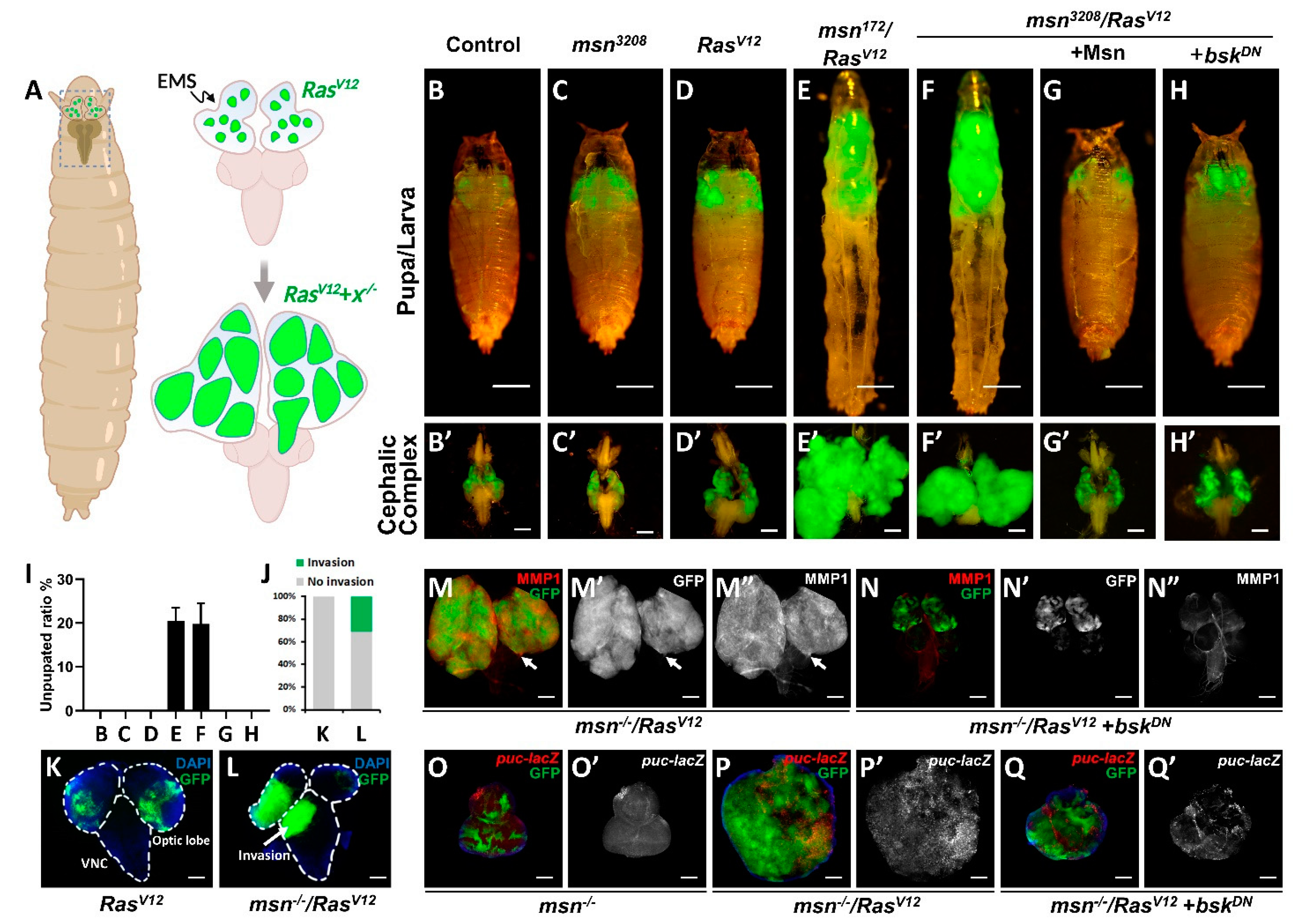
2.1. msn Mutant Synergizes with Oncogenic RasV12 to Drive Tumorigenesis and Invasion

In order to identify novel tumor suppressors that can cooperate with RasV12 to promote tumor growth and invasion, we performed a large-scale EMS-induced genetic screen on Drosophila eye-antennal imaginal discs utilizing the ey-FLP-based mosaic analysis with repressible cell marker (MARCM) technique [16,22] (Figure 1A). Here, we identified a recessive-lethal allele (#3208) that exhibited invasive tumor overgrowth phenotype (Figure 1B,D,F). Subsequent deficiency mapping and complementation test revealed that #3208 allele disrupted the misshapen (CG16973) gene (labeled as msn3208 thereafter). Consistent with this, we found that null allele of msn (msn172) [23] also synergized with RasV12 and exhibited similar degree of tumor overgrowth (Figure 1E). More importantly, the overgrowth phenotype and reduced pupation caused by msn3208/RasV12 were both completely rescued by co-expression of wild type Msn (Figure 1G,I and Supplementary Materials Figure S1L). We further validated the phenotype of msn mutant clones, and found that compared with wild type clones, loss of msn alone did not show significant growth advantage or changes in apoptosis, as indicated by PH3 and Caspase 3 staining, respectively (Figure 1B,C, Figure S1A,B,E,F,I–K,M). In accordance with previous reports [16,22], RasV12 expression alone only showed mild tumor overgrowth and did not invade into the ventral nerve cord (VNC) (Figure 1D,K), a commonly used tissue for tumor invasion observation in Drosophila [12,14,16,22], whereas larvae bearing msn3208/RasV12 tumors exhibited dramatic tumor overgrowth (Figure 1F,F’), autonomous cell proliferation (Figure S1G,H,M), reduced pupation ratio (Figure 1I), and increased VNC invasion behavior (Figure 1J,L). In contrast, we did not observe a significant change in apoptosis (Figure S1C,D). Consistent with increased tumor invasion, we also observed intensive MMP1 activation, a protein essential for basement membrane degradation and epithelial–mesenchymal transition (EMT) [24,25], in both primary tumor and invasive leading edges (Figure 1M). Taken together, these findings suggest that msn is a tumor suppressor that cooperatively induces tumor growth and invasion with RASV12.

2.2. msn−/− Collaborates with RasV12 to Activate JNK Signaling

Given that MMP1 also acts as a transcriptional target of JNK signaling pathway [24,25], the increase of MMP1 in msn3208/RasV12 tumors implies that JNK activation might be essential for tumorigenesis. Inconsequently, previous studies indicate that Msn acts as an MKKKK to activate the c-Jun N-terminal kinase (JNK) signaling pathway [23,26,27]. Our research also shows that ectopic Msn overexpression indeed induced mild JNK activation (Figure 3A,B), whereas loss of msn alone had no significant change on puc transcription (Figure 1O), another canonical JNK pathway target [25]. Intriguingly, we found loss of msn synergized with RasV12 to induce intensive MMP1 and puc activation (Figure 1M,P), both of which were significantly suppressed by expression of a dominant-negative form of Drosophila JNK homologue basket (bskDN) (Figure 1H,N,Q and Figure S1L). It is also worth noting that although inhibition of JNK activity completely blocked msn−/−/RasV12-induced puc upregulation in a cell-autonomous manner, we detected strong non-autonomous JNK activation in the surrounding region (Figure 1Q), which is probably caused by JNK propagation [28]. Together, these data indicate that loss of msn synergizes with RasV12 to promote tumor overgrowth via activating JNK signaling in vivo.

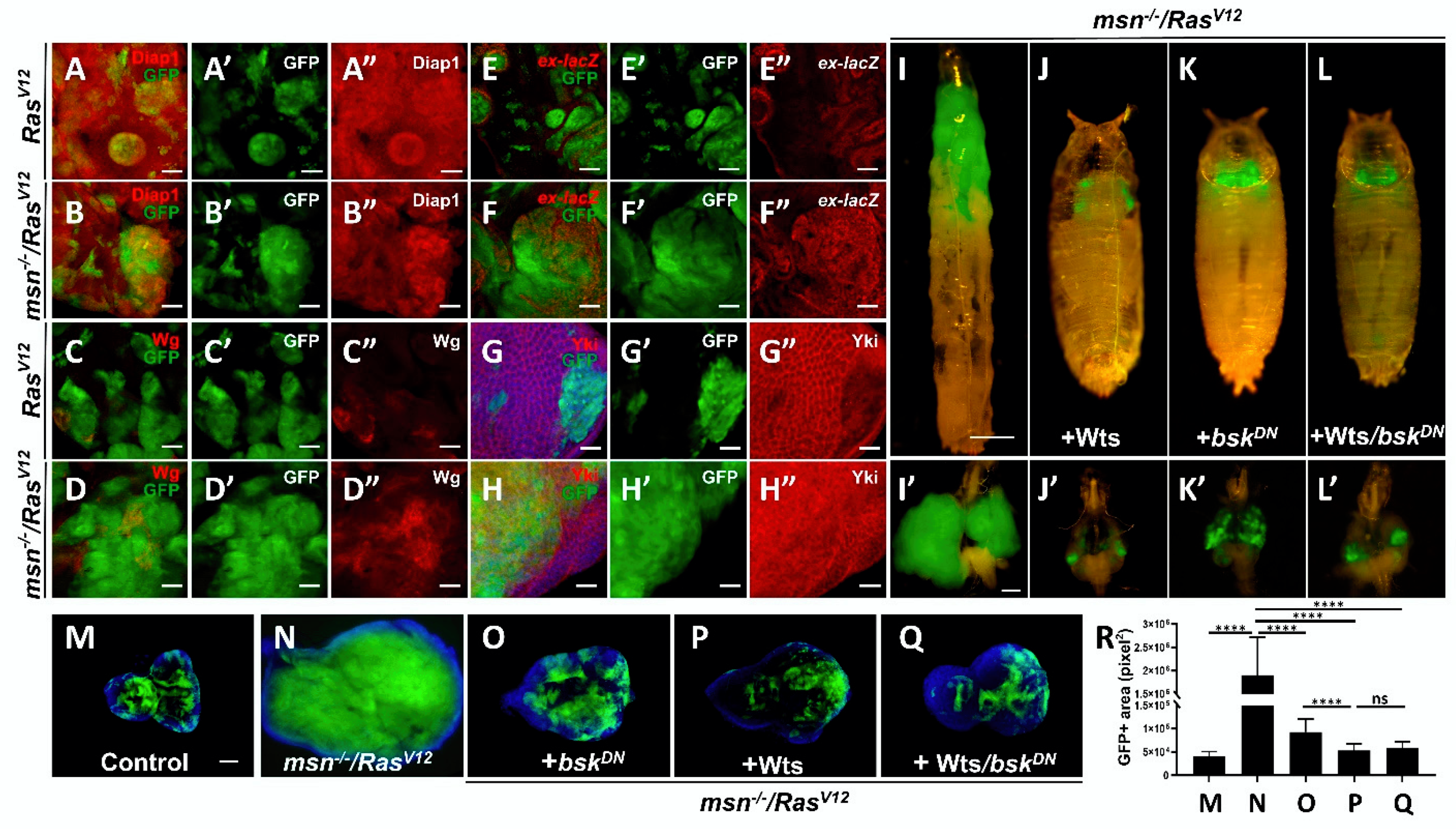
2.3. Loss of msn Synergizes with RasV12 to Inactivate Hippo Pathway

Recent studies in Drosophila gut have uncovered a genetic link between the STE20 kinase Msn and the Hippo pathway, indicating that Msn may act in parallel with mammalian homologue MST 1/2 (Hpo), the key component of Hippo pathway, to regulate mammalian homologue Lats 1/2 (Wts) and mammalian homologue YAP/TAZ (Yki) activity [29,30,31,32,33,34]. Given that Hippo pathway plays a key role in regulating tumorigenesis [18,20,21], we hypothesized that the synergistic effect in msn−/−/RasV12 tumors might be caused by inactivation of Hippo signaling. We found that loss of msn alone in the developing eye disc showed neither changes in the endogenous expression of Hippo pathway target gene including Diap1 (Figure S2A,B), ex (Figure S2C,D), and Wg (Figure S2E,F), nor Yki localization (Figure S2G–H), while overexpression of RasV12 alone mildly upregulated Hippo target genes (Figure 2A,C,E) and Yki nucleus localization (Figure 2G); it is also worth noting that overall Yki levels were decreased in RasV12 clones (Figure 2G and Figure S2I). In msn−/−/RasV12 tumors, we observed intensive upregulation of Diap1, Wg, and ex (Figure 2B,D,F), as well as strong Yki nucleus localization (Figure 2H). Interestingly, we found that Wts overexpression not only dramatically blocked msn−/−/RasV12-induced tumor growth (Figure 2I–J), but also further enhanced the tumor suppression phenotype caused by JNK inhibition (Figure 2K,L,P–R). Together, these data indicate that msn−/−/RasV12 drives tumor growth via Hippo signaling inactivation in vivo.

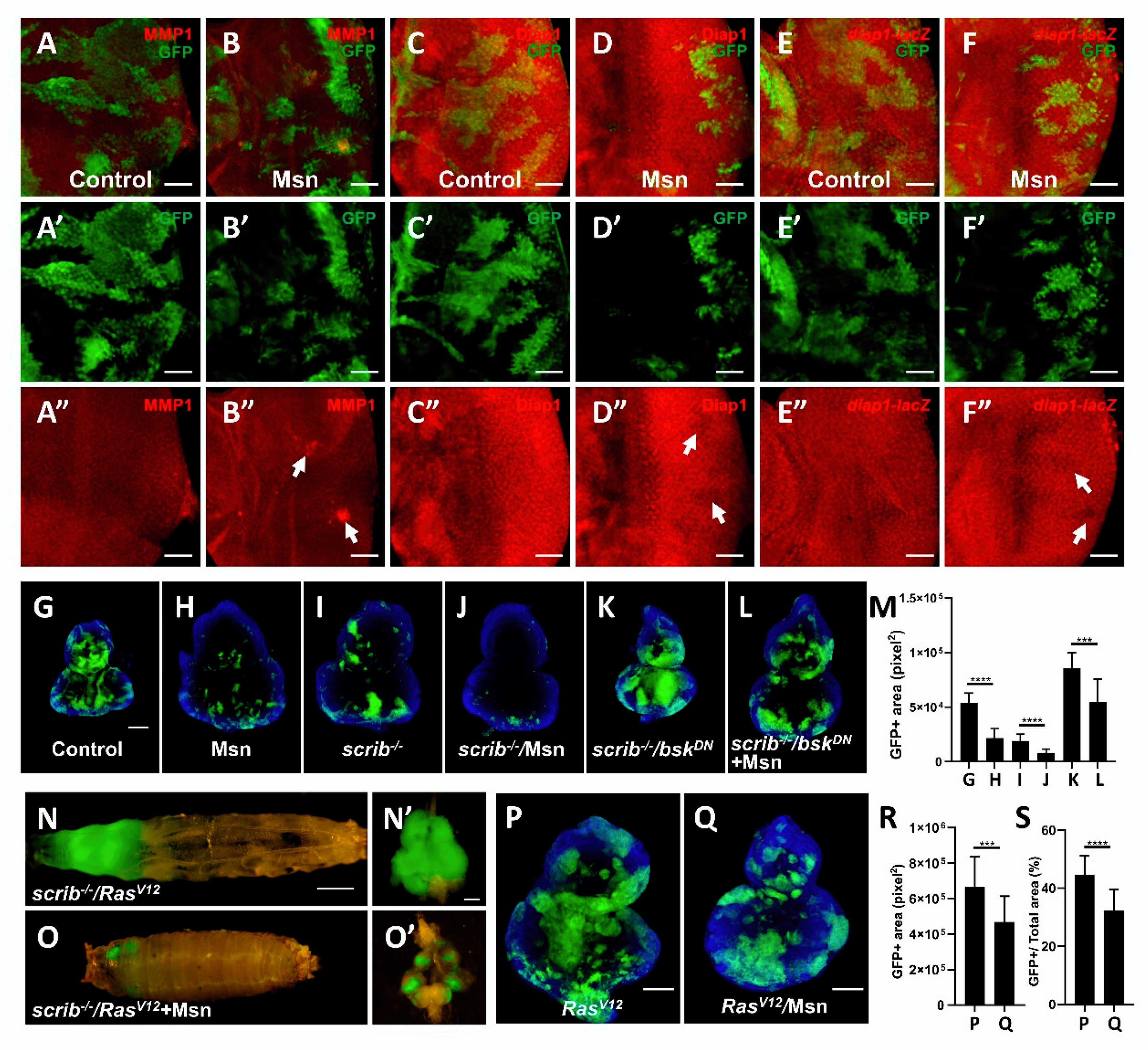
2.4. Msn Positively Regulates Hippo Signaling in Tumorigenesis

Previous studies indicated that Msn can functionally substitute for Hippo to restrict Yki activity in intestine [29,30,31]. Consistent with this notion that Msn is a positive regulator of Hippo pathway, we found Msn overexpression alone caused clone undergrowth (Figure 3G,H,M) and significantly reduced endogenous expression of Yki reporter Diap1 (Figure 3C,D and Figure S3G,H) and diap1-lacZ (Figure 3E,F and Figure S3I,J) in Drosophila imaginal discs, whereas it had no obvious changes on apoptosis and mitosis (Figure S3A–D). In Drosophila, cell polarity gene scribble (scrib) acts as a neoplastic tumor suppressor and would be eliminated by Hippo-mediated cell competition [35,36]. We found that Msn overexpression caused a further reduction of scrib−/− clone sizes (Figure 3I,J,M). It has been proven that the elimination of scrib−/− clone depends on JNK-activation-induced Yki inhibition, and given that Msn overexpression can activate both JNK and Hippo signaling (Figure 3A,B), we blocked JNK activity and simultaneously overexpressed Msn in scrib−/− clones. We found that the overgrowth phenotype of scrib−/−/bskDN clone (Figure 3K) was significantly reduced by co-expression of Msn (Figure 3L,M), indicating that Msn promotes scrib−/− elimination in a JNK-independent manner. In accordance with this, we also found that Msn overexpression significantly suppressed RasV12/scrib−/−-induced tumor overgrowth (Figure 3N,O and Figure S3E,F), which has been proven to be mediated by Hippo signaling [37,38,39]. Moreover, we found that Msn overexpression reduced both the relative and absolute clone sizes of RasV12 (Figure 3P–S). Together, these results indicate that ectopic Msn overexpression activates Hippo signaling to suppress tumorigenesis.

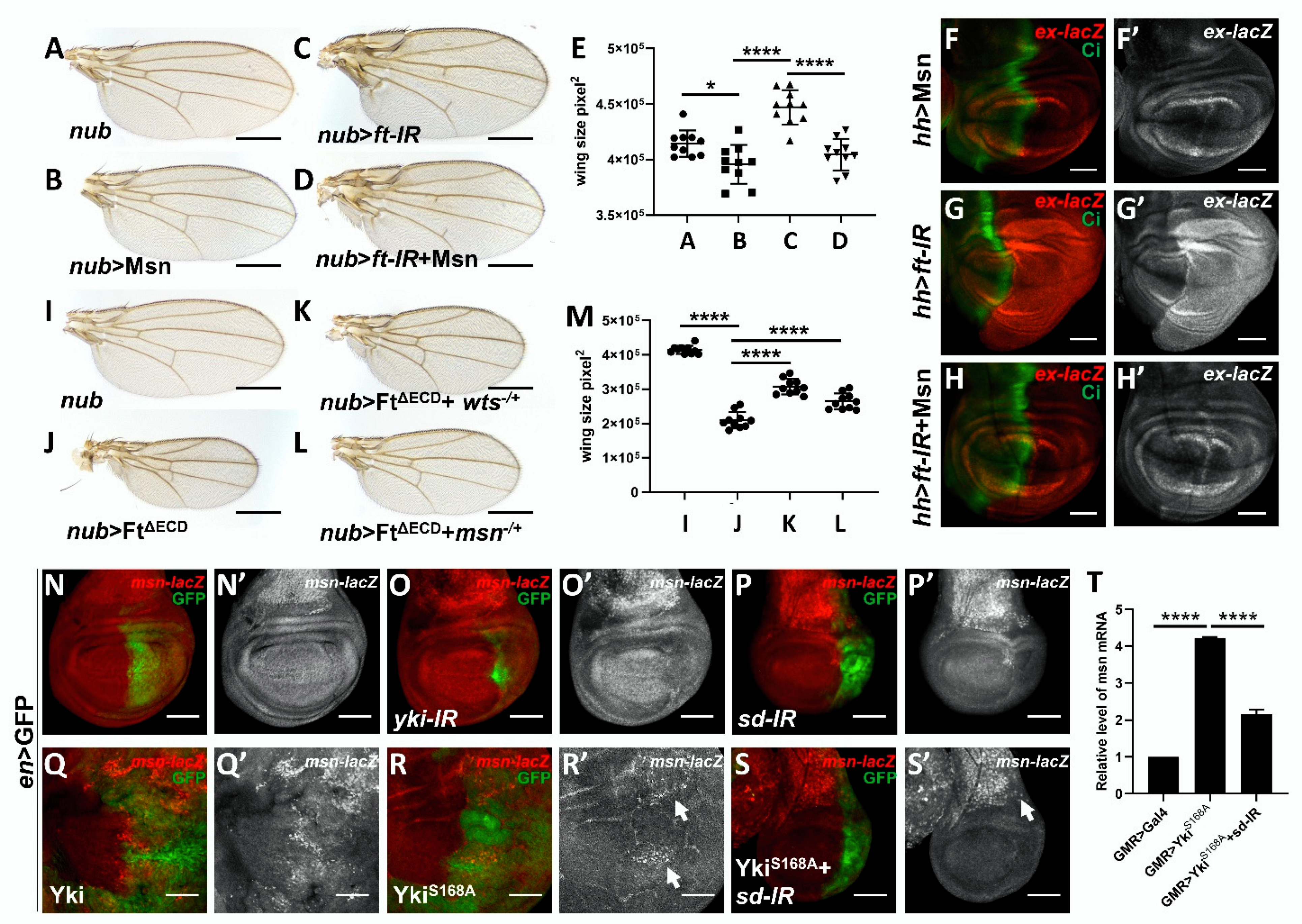
2.5. Msn Acts Downstream of Ft in Modulating Hippo Signaling

Although we and others proved that Msn positively activates Hippo signaling in various fly organs, the upstream regulator, especially the membrane anchor of Msn, remains unclear [31]. Fat signaling has previously been shown to be involved in both the establishment of planar cell polarity and growth control, Ft is a transmembrane protein that negatively regulates organ size and functions as a well-established, essential regulator of Hippo signaling in both eye and wing disc [19,40,41,42,43]. Because the quantification of wing size is relatively easy and standard deviation is lower, we dissected the genetic interactions between Msn and Ft in developing wing, with specific focus on the overall size of the adult wing [43]. As previously shown, knockdown of ft expression in the wing pouch region using nub-Gal4 results in a significant increase in wing size [44] (Figure 4A,C,E), while overexpression of Msn alone results in a significant decrease in wing size (Figure 4B,E), consistent with its role as a positive regulator of Hippo signaling. When we knocked down ft and simultaneously overexpressed Msn, the wing size phenocopies that of Msn overexpression alone (Figure 4D–E). Similarly, we found that ft knockdown-induced ex upregulation was also suppressed when Msn was co-expressed (Figure 4F–H and Figure S4B). To further dissect the physiological function of msn in regulating Ft-mediated growth, we removed one copy of msn, and found that ectopic expression of truncated form of Ft that removed most of the extracellular domains (FtΔECD) caused small wing phenotype (Figure 4J); this reduction in wing size was suppressed by heterozygosity of the msn allele (Figure 4L), phenocopying that of wts deletion (Figure 4K). Taken together, these genetic data indicate that Msn acts downstream of Ft in modulating Hippo signaling.
Interestingly, a protein–protein interaction screen in fly uncovered a potential physical link between Ft and Msn [45]; therefore, we further examined the physical interaction between Ft and Msn in S2 cells. Unfortunately, our co-immunoprecipitation experiment did not detect a physical binding between FtΔECD and Msn, suggesting that Ft might act through other unknown protein(s) to regulate Msn-mediated Hippo activation (Figure S4C,D).

2.6. Msn Acts as a Hippo Target Gene in a Negative Feedback Manner

Given that human ortholog of Msn (MAP4K4) is overexpressed in various human cancers [46,47,48,49,50] and a number of Hippo pathway components regulate Hippo signaling in a negative feedback manner, including kibra, ex, wts [18,20,21,51], we utilized the Drosophila wing imaginal discs to explore whether Msn also functions as a Hippo pathway target gene. Compared with control, knockdown Hippo signaling effector Yki or the transcription factor Scalloped (Sd) under en-Gal4 significantly reduced the Msn transcription level, as demonstrated by a lacZ enhancer trap insertion within the msn locus (Figure 4N–P). Conversely, Yki and active form of Yki (YkiS168A) overexpression significantly upregulated msn transcription level (Figure 4Q,R) and co-expression of an sd RNAi significantly impeded YkiS168A-induced msn-lacZ upregulation (Figure 4S). Interestingly, we also found that Msn overexpression suppressed transcription of itself (Figure S4A). To further assess the role of Yki and Sd in regulating msn expression, we performed qRT-PCR in vivo using adult fly heads, which are relatively easy to collect and extract mRNA from for qPCR analysis. Consistent with wing disc results, Yki overexpression in the developing eye disc upregulated msn transcription, which was significantly impaired by knocking down sd (Figure 4T). Taken together, these data indicate that Msn also acts as a Yki/Sd target to form a negative feedback loop.

3. Discussion

Drosophila has been widely used as a cancer model for the past few decades to dissect various human cancer biology questions, including cell–cell communication, tumor heterogeneity, clonal evolution, cancer cachexia, and antitumor drug resistance [5,6,7]. The powerful genetic tools established in Drosophila, especially the mosaic analysis with a repressible cell marker (MARCM) system [52,53], make it possible to perform large-scale genetic screens aimed at identifying novel tumor suppressor genes in vivo [12,16,22]. In this study, we conducted an EMS-induced unbiased modified genetic screen and identified misshapen (CG16973) as a tumor suppressor that synergizes with RasV12 to drive tumor overgrowth by inactivating Hippo signaling in Drosophila (Figure S5).
Misshapen (Msn) is a member of the STE20 kinase family; it was initially identified as a MAP kinase kinase kinase kinase (MKKKK) that activates the c-Jun N-terminal kinase (JNK) pathway [23,26,27]. Msn also regulates diverse physiological functions including dorsal closure, photoreceptor axon targeting, germline ring canal stabilization, and intestine homeostasis [23,29,31,54,55]. Moreover, recent studies have shown that Drosophila Msn and its mammalian homologue MAP4K4/6/7 act in parallel and partially redundantly with canonical Hippo signaling component mammalian homologue MST 1/2 (Hpo) in regulating mammalian homologue Lats 1/2 (Wts) and mammalian homologue YAP/TAZ (Yki) [29,30,31,32,33,34,56,57]. Given that Hippo signaling pathway is an evolutionarily conserved pathway that regulates various essential physiological processes and its disruption contributes to a number of human diseases including cancer [18,20,21,58], it is possible that Msn and its mammalian homologue MAP4K4/6/7 may also function as a tumor suppressor in a context-dependent manner.
RAS family genes are considered to be one of the most highly mutated genes in various cancers [1,2,3,4]; however, treatment targeting RAS-related tumors remains unsatisfactory, and the in vivo molecular mechanisms of RAS-related tumorigenesis are still not completely understood. Here, we identified msn as a tumor suppressor that cooperates with oncogenic RasV12 to promote tumor overgrowth and invasion by simultaneously activating JNK pathway and inactivating Hippo pathway. We found that Msn overexpression dramatically suppressed scrib1/RasV12-induced tumor overgrowth and invasion, a well-established Drosophila cancer model. Additionally, we revealed that msn acts downstream of Ft and regulates Hippo pathway in a negative feedback manner. It is worth noting that we could not exclude the possibility that msn−/−/RasV12 tumors could also regulate other growth regulating pathways, including JAK-STAT signaling, which requires further investigation (Figure S5).
Given the high conservation of signaling pathways and cancer-related genes between Drosophila and human, we assume that similar mechanisms could be involved in human cancer progression. Our study here identified Msn as a tumor suppressor and further investigation in mammal and human may provide potential therapeutic targets for cancer treatment, especially for Hippo-related tumors.

4. Experimental Procedures

4.1. Drosophila Stocks and Genetics

All crosses were raised on standard Drosophila media at 25 °C unless otherwise indicated. Fluorescently labeled clones were produced in the eye discs as previously described [12,16,22] using the following strains: ey-Flp1; Act > y+ > Gal4, UAS-GFP; tub-Gal80, FRT79E (79E tester); ey-Flp1; Act> y+ > Gal4, UAS-GFP; tub-Gal80, FRT80B (80B tester); ey-Flp5, Act > y+ > Gal4, UAS-GFP; FRT82B, tub-Gal80 (82B tester). Additional strains, including GMR-Gal4, en-Gal4, hh-Gal4, nub-Gal4, UAS-GFP, UAS-ft-IR, wtsX1 (#44251), msn172 (#5947), msn-LacZ (#11707), and UAS-Msn (#5946), were obtained from Bloomington Drosophila Stock Center; pucE69, UAS-RasV12 [15], UAS-bskDN [17], and scrib1 [59] were previously described. UAS-Wts, UAS-Yki, UAS-YkiS168A, UAS-yki-IR, and UAS-sd-IR were previously described [60]. UAS-FtΔECD is a kind gift from Kenneth Irvine.

4.2. EMS Mutagenesis and Genetic Screen

We focused on Drosophila chromosome 3 L in this screen. Male flies carrying FRT79E (Sp/CyO-GFP; FRT79E) were starved for 8 h and subsequently fed with 25 mM EMS solution overnight at room temperature. The mutagenized males were then mated to females of the genotype UAS-RasV12; sb/TM6B. Single F1 males of the genotype UAS-RasV12/CyO-GFP; ∗FRT79E/TM6B were crossed to Sp/CyO; sb/TM6B first and then crossed with the 79E tester line for validation of GFP-labeled tumor overgrowth phenotype under fluorescent microscope.

4.3. Immunostaining

Third-instar larvae eye-antennal discs were dissected in 1 × PBS, fixed in freshly made 4% paraformaldehyde, and stained as described previously [61] using the following primary antibodies: mouse anti-MMP1 (1:200, Developmental Studies Hybridoma Bank, DSHB, Iowa City, IA, USA), mouse anti-β-Gal (1:1000, Promoga, Madison, WI, USA), rabbit anti-phospho-histone 3 (PH3) (1:200, Cell Signaling Technology, CST, Danvers, MA, USA), rabbit anti-active caspase-3 (1:400, Cell Signaling Technology, CST, Danvers, MA, USA), mouse anti-Diap1 (1:200, a gift from Bruce Hay, California Institute of Technology, Pasadena, CA, USA), rabbit anti-Yki (1:500, gift from Duojia Pan, University of Texas Southwestern Medical Center, Dallas, TX, USA), and mouse anti-Wg (1:200, Developmental Studies Hybridoma Bank, DSHB, Iowa City, IA, USA). Secondary antibodies were anti-rabbit-Cy3 (1:400, Thermo Fisher Scientific, Waltham, MA, USA) and anti-mouse-Cy3 (1:400, Thermo Fisher Scientific, Waltham, MA, USA).

4.4. Cell Culture

S2 cells were maintained in Schneider’s Drosophila Medium (Gibco #21720024) supplemented with 10% fetal bovine serum (Cellmax #SA112) and 1% penicillin–streptomycin (Gibco #15140122) at 28 °C.

4.5. Immunoprecipitation and Western Blotting

Transfection, Co-IP, and Western blot analysis were performed as previously described with some modification [62]. paw-Gal4, UAS-attB-msn-HA, and UAS-attB-FatΔECD-FLAG plasmids were co-transfected to S2 cells, performed using Effectene transfection reagent (Qiagen #301427) following the manufacturer’s instructions and harvested 72 h after transfection. Harvested S2 cells were lysed in 500 µL RIPA lysis buffer (50 mM Tris pH 7.5, 150 mM NaCl, 1 mM EDTA, 1% NP40, 5% glycerol, protease inhibitor cocktail, DTT). After the samples were rotated for 1 h at 4 °C, lysates were centrifuged at 13000 rpm for 13 min at 4 °C. The lysate supernatant was incubated at 4 °C with 20 µL cleaned anti-FLAG M2 Affinity Gel (Sigma #A2220) for 3 h. The beads were washed with PBST four times, then mixed with SDS-PAGE Loading Buffer (reducing, 5×) (CWBIO #CW0027S) and heated to 95 °C for 7 min. After separation of proteins by SDS-PAGE, proteins were transferred to PVDF membrane (Merck Millipore #IPVH00010) which was blocked with 5% milk–PBST for 1 h, incubated in primary antibody overnight at 4 °C, washed with PBST, incubated with secondary antibody at room temperature for 1 h, and washed with PBST four times. Protein was detected using a Viber Fusion FX6 Spectra imaging system (Vilber, Collégien, France). The following antibodies for Western blotting were used: primary antibodies, rabbit-anti-FLAG (1:10,000) (EASYBIO #BE2005); rabbit-anti–HA (1:9000) (EASYBIO #BE2006); and secondary antibody anti-rabbit-HRP (1:8000) (Promega #W4011).

4.6. Quantitative Real-Time PCR

Drosophila adult head of indicated genotypes was removed and total RNA was then extracted using TRIzol (Ambion). Total RNA was reverse-transcribed into cDNA with the HiScript II 1st Strand cDNA Synthesis Kit (Vazyme); and quantitative PCR was performed with KAPA SYBR® FAST (KAPA BIOSYSTEMS) and quantified by the QuantStudio™ 5 Real-Time PCR System (ThermoFisher). RP49 was used as an internal control. The following primer sequences were used for real-time PCR. msn: F: 5′-TCCCTTGGACAGCAGCGATT-3′, R: 5′-AGTTCCATCGTTCCTAGCC-3′; rp49: F: 5′-TCCTACCAGCTTCAAGATGACC-3′, R: 5′-CACGTTGTGCACCAGGAACT-3′.

4.7. Statistical Analysis

Clone and wing size were measured with ImageJ and Photoshop, respectively. Quantification of the data was presented in bar graphs created with GraphPad Prism 8. Data represent mean values + SD. We used Mann–Whitney U test for multiple comparisons to calculate statistical significance (* p < 0.05; ** p < 0.01; *** p < 0.001; **** p < 0.0001).

Supplementary Materials

The following are available online at https://www.mdpi.com/article/10.3390/cells10040894/s1, Figure S1: Loss of msn collaborates with RasV12 to induce tumorigenesis. Figure S2: Loss of msn alone does not affect Hippo signaling activation. Figure S3: Msn overexpression suppresses tumorigenesis. Figure S4: Msn regulates Hippo signaling in a feedback manner. Figure S5: A schematic model depicting the role of Msn in regulating Hippo signaling and tumorigenesis.

Author Contributions

D.K., X.W. and X.M. conceived the study. D.K., J.-Y.L. and X.M. performed most experiments and analyzed data. X.L. cultured S2 cells and performed co-IP. S.Z. and J.F. assisted in immunofluorescence staining. W.X. performed qRT-PCR experiment. D.K. and X.M. wrote the manuscript. All authors have read and agreed to the published version of the manuscript.

Funding

This work was partially supported by National Natural Science Foundation of China (31801190), and the Chinese Universities Scientific Fund (2018QC155, 2018ZH003, and 2019TC068) to X.W.

Acknowledgments

We thank Duojia Pan, Bruce Hay, Kenneth Irvine, Bloomington stock center for providing fly stocks and reagents. This work was supported by startup funds from Westlake University to X.M, Westlake Laboratory of Life Sciences and Biomedicine (Grant 10128A092001), and “Team for Growth Control and Size Innovative Research” (Grant 201804016).

Conflicts of Interest

The authors declare no conflict of interest.

References

  1. Ryan, M.B.; Der, C.J.; Wang-Gillam, A.; Cox, A.D. Targeting RAS-mutant cancers: Is ERK the key? Trends Cancer 2015, 1, 183–198. [Google Scholar] [CrossRef] [PubMed] [Green Version]
  2. Stephen, A.G.; Esposito, D.; Bagni, R.K.; McCormick, F. Dragging ras back in the ring. Cancer Cell 2014, 25, 272–281. [Google Scholar] [CrossRef] [PubMed] [Green Version]
  3. Cox, A.D.; Fesik, S.W.; Kimmelman, A.C.; Luo, J.; Der, C.J. Drugging the undruggable RAS: Mission possible? Nat. Rev. Drug Discov. 2014, 13, 828–851. [Google Scholar] [CrossRef] [PubMed] [Green Version]
  4. Samatar, A.A.; Poulikakos, P.I. Targeting RAS-ERK signalling in cancer: Promises and challenges. Nat. Rev. Drug Discov. 2014, 13, 928–942. [Google Scholar] [CrossRef] [PubMed]
  5. Saavedra, P.; Perrimon, N. Drosophila as a Model for Tumor-Induced Organ Wasting. Adv. Exp. Med. Biol. 2019, 1167, 191–205. [Google Scholar] [CrossRef] [PubMed]
  6. Enomoto, M.; Siow, C.; Igaki, T. Drosophila As a Cancer Model. Adv. Exp. Med. Biol. 2018, 1076, 173–194. [Google Scholar] [CrossRef]
  7. Mirzoyan, Z.; Sollazzo, M.; Allocca, M.; Valenza, A.M.; Grifoni, D.; Bellosta, P. Drosophila melanogaster: A Model Organism to Study Cancer. Front. Genet. 2019, 10, 51. [Google Scholar] [CrossRef] [PubMed] [Green Version]
  8. Richardson, H.E.; Cordero, J.B.; Grifoni, D. Basic and Translational Models of Cooperative Oncogenesis. Int. J. Mol. Sci. 2020, 21, 5919. [Google Scholar] [CrossRef] [PubMed]
  9. Pastor-Pareja, J.C.; Xu, T. Dissecting social cell biology and tumors using Drosophila genetics. Annu. Rev. Genet. 2013, 47, 51–74. [Google Scholar] [CrossRef] [Green Version]
  10. Gonzalez, C. Drosophila melanogaster: A model and a tool to investigate malignancy and identify new therapeutics. Nat. Rev. Cancer 2013, 13, 172–183. [Google Scholar] [CrossRef]
  11. Figueroa-Clarevega, A.; Bilder, D. Malignant Drosophila tumors interrupt insulin signaling to induce cachexia-like wasting. Dev. Cell 2015, 33, 47–55. [Google Scholar] [CrossRef] [PubMed] [Green Version]
  12. Pagliarini, R.A.; Xu, T. A genetic screen in Drosophila for metastatic behavior. Science 2003, 302, 1227–1231. [Google Scholar] [CrossRef]
  13. Brumby, A.M.; Richardson, H.E. scribble mutants cooperate with oncogenic Ras or Notch to cause neoplastic overgrowth in Drosophila. EMBO J. 2003, 22, 5769–5779. [Google Scholar] [CrossRef] [PubMed]
  14. Igaki, T.; Pagliarini, R.A.; Xu, T. Loss of cell polarity drives tumor growth and invasion through JNK activation in Drosophila. Curr. Biol. 2006, 16, 1139–1146. [Google Scholar] [CrossRef] [Green Version]
  15. Ma, X.; Li, W.; Yu, H.; Yang, Y.; Li, M.; Xue, L.; Xu, T. Bendless modulates JNK-mediated cell death and migration in Drosophila. Cell Death Differ. 2014, 21, 407–415. [Google Scholar] [CrossRef] [Green Version]
  16. Ma, X.; Lu, J.Y.; Dong, Y.; Li, D.; Malagon, J.N.; Xu, T. PP6 Disruption Synergizes with Oncogenic Ras to Promote JNK-Dependent Tumor Growth and Invasion. Cell Rep. 2017, 19, 2657–2664. [Google Scholar] [CrossRef] [PubMed] [Green Version]
  17. Ma, X.; Xu, W.; Zhang, D.; Yang, Y.; Li, W.; Xue, L. Wallenda regulates JNK-mediated cell death in Drosophila. Cell Death Dis. 2015, 6, e1737. [Google Scholar] [CrossRef] [Green Version]
  18. Pan, D. The hippo signaling pathway in development and cancer. Dev. Cell 2010, 19, 491–505. [Google Scholar] [CrossRef] [Green Version]
  19. Staley, B.K.; Irvine, K.D. Hippo signaling in Drosophila: Recent advances and insights. Dev. Dyn. 2012, 241, 3–15. [Google Scholar] [CrossRef] [Green Version]
  20. Zeng, R.; Dong, J. The Hippo Signaling Pathway in Drug Resistance in Cancer. Cancers 2021, 13, 318. [Google Scholar] [CrossRef] [PubMed]
  21. Zheng, Y.; Pan, D. The Hippo Signaling Pathway in Development and Disease. Dev. Cell 2019, 50, 264–282. [Google Scholar] [CrossRef]
  22. Ma, X.; Lu, J.Y.; Moraru, A.; Teleman, A.A.; Fang, J.; Qiu, Y.; Liu, P.; Xu, T. A novel regulator of ER Ca(2+) drives Hippo-mediated tumorigenesis. Oncogene 2020, 39, 1378–1387. [Google Scholar] [CrossRef]
  23. Su, Y.C.; Treisman, J.E.; Skolnik, E.Y. The Drosophila Ste20-related kinase misshapen is required for embryonic dorsal closure and acts through a JNK MAPK module on an evolutionarily conserved signaling pathway. Genes Dev. 1998, 12, 2371–2380. [Google Scholar] [CrossRef] [Green Version]
  24. Srivastava, A.; Pastor-Pareja, J.C.; Igaki, T.; Pagliarini, R.; Xu, T. Basement membrane remodeling is essential for Drosophila disc eversion and tumor invasion. Proc. Natl. Acad. Sci. USA 2007, 104, 2721–2726. [Google Scholar] [CrossRef] [PubMed] [Green Version]
  25. Uhlirova, M.; Bohmann, D. JNK- and Fos-regulated Mmp1 expression cooperates with Ras to induce invasive tumors in Drosophila. EMBO J. 2006, 25, 5294–5304. [Google Scholar] [CrossRef] [PubMed]
  26. Liu, H.; Su, Y.C.; Becker, E.; Treisman, J.; Skolnik, E.Y. A Drosophila TNF-receptor-associated factor (TRAF) binds the ste20 kinase Misshapen and activates Jun kinase. Curr. Biol. 1999, 9, 101–104. [Google Scholar] [CrossRef] [Green Version]
  27. Paricio, N.; Feiguin, F.; Boutros, M.; Eaton, S.; Mlodzik, M. The Drosophila STE20-like kinase misshapen is required downstream of the Frizzled receptor in planar polarity signaling. EMBO J. 1999, 18, 4669–4678. [Google Scholar] [CrossRef] [Green Version]
  28. Wu, M.; Pastor-Pareja, J.C.; Xu, T. Interaction between Ras(V12) and scribbled clones induces tumour growth and invasion. Nature 2010, 463, 545–548. [Google Scholar] [CrossRef] [Green Version]
  29. Li, Q.; Li, S.; Mana-Capelli, S.; Roth Flach, R.J.; Danai, L.V.; Amcheslavsky, A.; Nie, Y.; Kaneko, S.; Yao, X.; Chen, X.; et al. The conserved misshapen-warts-Yorkie pathway acts in enteroblasts to regulate intestinal stem cells in Drosophila. Dev. Cell 2014, 31, 291–304. [Google Scholar] [CrossRef] [Green Version]
  30. Li, Q.; Nirala, N.K.; Chen, H.J.; Nie, Y.; Wang, W.; Zhang, B.; Czech, M.P.; Wang, Q.; Xu, L.; Mao, J.; et al. The Misshapen subfamily of Ste20 kinases regulate proliferation in the aging mammalian intestinal epithelium. J. Cell. Physiol. 2019, 234, 21925–21936. [Google Scholar] [CrossRef]
  31. Li, Q.; Nirala, N.K.; Nie, Y.; Chen, H.J.; Ostroff, G.; Mao, J.; Wang, Q.; Xu, L.; Ip, Y.T. Ingestion of Food Particles Regulates the Mechanosensing Misshapen-Yorkie Pathway in Drosophila Intestinal Growth. Dev. Cell 2018, 45, 433–449.e436. [Google Scholar] [CrossRef] [PubMed] [Green Version]
  32. Li, S.; Cho, Y.S.; Yue, T.; Ip, Y.T.; Jiang, J. Overlapping functions of the MAP4K family kinases Hppy and Msn in Hippo signaling. Cell Discov. 2015, 1, 15038. [Google Scholar] [CrossRef] [PubMed] [Green Version]
  33. Meng, Z.; Moroishi, T.; Mottier-Pavie, V.; Plouffe, S.W.; Hansen, C.G.; Hong, A.W.; Park, H.W.; Mo, J.S.; Lu, W.; Lu, S.; et al. MAP4K family kinases act in parallel to MST1/2 to activate LATS1/2 in the Hippo pathway. Nat. Commun. 2015, 6, 8357. [Google Scholar] [CrossRef]
  34. Meng, Z.; Qiu, Y.; Lin, K.C.; Kumar, A.; Placone, J.K.; Fang, C.; Wang, K.C.; Lu, S.; Pan, M.; Hong, A.W.; et al. RAP2 mediates mechanoresponses of the Hippo pathway. Nature 2018, 560, 655–660. [Google Scholar] [CrossRef] [Green Version]
  35. Chen, C.L.; Schroeder, M.C.; Kango-Singh, M.; Tao, C.; Halder, G. Tumor suppression by cell competition through regulation of the Hippo pathway. Proc. Natl. Acad. Sci. USA 2012, 109, 484–489. [Google Scholar] [CrossRef] [Green Version]
  36. Nagata, R.; Igaki, T. Cell competition: Emerging mechanisms to eliminate neighbors. Dev. Growth Differ. 2018, 60, 522–530. [Google Scholar] [CrossRef] [PubMed] [Green Version]
  37. Doggett, K.; Grusche, F.A.; Richardson, H.E.; Brumby, A.M. Loss of the Drosophila cell polarity regulator Scribbled promotes epithelial tissue overgrowth and cooperation with oncogenic Ras-Raf through impaired Hippo pathway signaling. BMC Dev. Biol. 2011, 11, 57. [Google Scholar] [CrossRef] [Green Version]
  38. Enomoto, M.; Igaki, T. Deciphering tumor-suppressor signaling in flies: Genetic link between Scribble/Dlg/Lgl and the Hippo pathways. J. Genet. Genomics 2011, 38, 461–470. [Google Scholar] [CrossRef] [PubMed]
  39. Fahey-Lozano, N.; La Marca, J.E.; Portela, M.; Richardson, H.E. Drosophila Models of Cell Polarity and Cell Competition in Tumourigenesis. Adv. Exp. Med. Biol. 2019, 1167, 37–64. [Google Scholar] [CrossRef] [PubMed]
  40. Thomas, C.; Strutt, D. The roles of the cadherins Fat and Dachsous in planar polarity specification in Drosophila. Dev. Dyn. 2012, 241, 27–39. [Google Scholar] [CrossRef]
  41. Montes, A.J.; Morata, G. Homeostatic response to blocking cell division in Drosophila imaginal discs: Role of the Fat/Dachsous (Ft/Ds) pathway. Dev. Biol. 2017, 424, 113–123. [Google Scholar] [CrossRef]
  42. Cho, E.; Feng, Y.; Rauskolb, C.; Maitra, S.; Fehon, R.; Irvine, K.D. Delineation of a Fat tumor suppressor pathway. Nat. Genet. 2006, 38, 1142–1150. [Google Scholar] [CrossRef]
  43. Willecke, M.; Hamaratoglu, F.; Kango-Singh, M.; Udan, R.; Chen, C.L.; Tao, C.; Zhang, X.; Halder, G. The fat cadherin acts through the hippo tumor-suppressor pathway to regulate tissue size. Curr. Biol. 2006, 16, 2090–2100. [Google Scholar] [CrossRef] [Green Version]
  44. Verghese, S.; Waghmare, I.; Kwon, H.; Hanes, K.; Kango-Singh, M. Scribble acts in the Drosophila fat-hippo pathway to regulate warts activity. PLoS ONE 2012, 7, e47173. [Google Scholar] [CrossRef] [Green Version]
  45. Kwon, Y.; Vinayagam, A.; Sun, X.; Dephoure, N.; Gygi, S.P.; Hong, P.; Perrimon, N. The Hippo signaling pathway interactome. Science 2013, 342, 737–740. [Google Scholar] [CrossRef] [Green Version]
  46. Hao, J.M.; Chen, J.Z.; Sui, H.M.; Si-Ma, X.Q.; Li, G.Q.; Liu, C.; Li, J.L.; Ding, Y.Q.; Li, J.M. A five-gene signature as a potential predictor of metastasis and survival in colorectal cancer. J. Pathol. 2010, 220, 475–489. [Google Scholar] [CrossRef] [Green Version]
  47. Liang, J.J.; Wang, H.; Rashid, A.; Tan, T.H.; Hwang, R.F.; Hamilton, S.R.; Abbruzzese, J.L.; Evans, D.B.; Wang, H. Expression of MAP4K4 is associated with worse prognosis in patients with stage II pancreatic ductal adenocarcinoma. Clin. Cancer Res. 2008, 14, 7043–7049. [Google Scholar] [CrossRef] [PubMed] [Green Version]
  48. Qiu, M.H.; Qian, Y.M.; Zhao, X.L.; Wang, S.M.; Feng, X.J.; Chen, X.F.; Zhang, S.H. Expression and prognostic significance of MAP4K4 in lung adenocarcinoma. Pathol. Res. Pract. 2012, 208, 541–548. [Google Scholar] [CrossRef] [PubMed]
  49. Rizzardi, A.E.; Rosener, N.K.; Koopmeiners, J.S.; Isaksson Vogel, R.; Metzger, G.J.; Forster, C.L.; Marston, L.O.; Tiffany, J.R.; McCarthy, J.B.; Turley, E.A.; et al. Evaluation of protein biomarkers of prostate cancer aggressiveness. BMC Cancer 2014, 14, 244. [Google Scholar] [CrossRef] [PubMed] [Green Version]
  50. Liu, A.W.; Cai, J.; Zhao, X.L.; Jiang, T.H.; He, T.F.; Fu, H.Q.; Zhu, M.H.; Zhang, S.H. ShRNA-targeted MAP4K4 inhibits hepatocellular carcinoma growth. Clin. Cancer Res. 2011, 17, 710–720. [Google Scholar] [CrossRef] [Green Version]
  51. Park, G.S.; Oh, H.; Kim, M.; Kim, T.; Johnson, R.L.; Irvine, K.D.; Lim, D.S. An evolutionarily conserved negative feedback mechanism in the Hippo pathway reflects functional difference between LATS1 and LATS2. Oncotarget 2016, 7, 24063–24075. [Google Scholar] [CrossRef] [Green Version]
  52. Lee, T.; Luo, L. Mosaic analysis with a repressible cell marker (MARCM) for Drosophila neural development. Trends Neurosci. 2001, 24, 251–254. [Google Scholar] [CrossRef]
  53. Lee, T.; Luo, L. Mosaic analysis with a repressible cell marker for studies of gene function in neuronal morphogenesis. Neuron 1999, 22, 451–461. [Google Scholar] [CrossRef] [Green Version]
  54. Kline, A.; Curry, T.; Lewellyn, L. The Misshapen kinase regulates the size and stability of the germline ring canals in the Drosophila egg chamber. Dev. Biol. 2018, 440, 99–112. [Google Scholar] [CrossRef] [PubMed]
  55. Su, Y.C.; Maurel-Zaffran, C.; Treisman, J.E.; Skolnik, E.Y. The Ste20 kinase misshapen regulates both photoreceptor axon targeting and dorsal closure, acting downstream of distinct signals. Mol. Cell Biol. 2000, 20, 4736–4744. [Google Scholar] [CrossRef] [Green Version]
  56. Zheng, Y.; Wang, W.; Liu, B.; Deng, H.; Uster, E.; Pan, D. Identification of Happyhour/MAP4K as Alternative Hpo/Mst-like Kinases in the Hippo Kinase Cascade. Dev. Cell 2015, 34, 642–655. [Google Scholar] [CrossRef] [Green Version]
  57. Jiang, J. Misshapen Connects Food, Mechanosensing, and Intestinal Growth. Dev. Cell 2018, 45, 417–418. [Google Scholar] [CrossRef] [Green Version]
  58. Dey, A.; Varelas, X.; Guan, K.L. Targeting the Hippo pathway in cancer, fibrosis, wound healing and regenerative medicine. Nat. Rev. Drug Discov. 2020, 19, 480–494. [Google Scholar] [CrossRef] [PubMed]
  59. Igaki, T.; Pastor-Pareja, J.C.; Aonuma, H.; Miura, M.; Xu, T. Intrinsic tumor suppression and epithelial maintenance by endocytic activation of Eiger/TNF signaling in Drosophila. Dev. Cell 2009, 16, 458–465. [Google Scholar] [CrossRef] [Green Version]
  60. Ma, X.; Chen, Y.; Xu, W.; Wu, N.; Li, M.; Cao, Y.; Wu, S.; Li, Q.; Xue, L. Impaired Hippo signaling promotes Rho1-JNK-dependent growth. Proc. Natl. Acad. Sci. USA 2015, 112, 1065–1070. [Google Scholar] [CrossRef] [PubMed] [Green Version]
  61. Ma, X.; Yang, L.; Yang, Y.; Li, M.; Li, W.; Xue, L. dUev1a modulates TNF-JNK mediated tumor progression and cell death in Drosophila. Dev. Biol. 2013, 380, 211–221. [Google Scholar] [CrossRef] [PubMed] [Green Version]
  62. Zhang, Y.; Wang, X.; Matakatsu, H.; Fehon, R.; Blair, S.S. The novel SH3 domain protein Dlish/CG10933 mediates fat signaling in Drosophila by binding and regulating Dachs. Elife 2016, 5, e16624. [Google Scholar] [CrossRef] [PubMed] [Green Version]
Figure 1. Loss of Misshapen (msn) synergizes with oncogenic Ras (RasV12) to induce tumorigenesis and invasion. (A) Strategy of an ethyl methanesulphonate (EMS)-induced forward genetic screen in developing Drosophila eye to identify novel RasV12 collaborating tumor suppressors. (BH) Fluorescence micrographs of GFP-labeled pupa/larva are shown in the top panels, cephalic complexes dissected from third-instar larvae are shown in the bottom. Compared with control (B,B’), loss of msn alone has no obvious overgrowth (C,C’), ectopic expression of RasV12 alone only induces mild tumor growth (D,D’). msn−/−/RasV12 tumors display significant overgrowth (E,F and E’,F’), which can be rescued by co-expression of Msn (G,G’) or co-expression of a dominant negative form of basket (H,H’). (I) Quantification of unpupated ratio for indicated genotypes in BH. (JL) Fluorescence micrographs of GFP-labeled Optic lope and ventral nerve cord (VNC) dissected from third-instar larvae are shown, arrow indicates invasion sites (K,L). (J) Quantification of invasion ratio in (K) and (L). (MQ) Loss of msn collaborates with RasV12 to induce tumorigenesis through activating c-Jun N-terminal kinase (JNK) signaling pathway. Expression of basket (bskDN ) suppressed msn−/−/RasV12-induced MMP1 expression, tumor invasion (MN”), and upregulation of puc transcription (OQ’); note that loss of msn alone does not significantly induce MMP1 activation (O,O’). Scale bars represent 500 μm (BH), 200 μm (B’–H’, MM”, and NN”), or 100 μm (KL, OQ, and O’–Q’).
Cells 10 00894 g001
Figure 2. Loss of msn collaborates with RasV12 to inactivate Hippo pathway. (AH) Fluorescence micrographs of GFP-labeled clones of eye discs are shown. Compared with RasV12 clones, msn−/−/RasV12 tumors show dramatic upregulation of Diap1 (BB”), ex-lacZ (DD”), Wg (FF”), and mammalian homologue YAP/TAZ (Yki) localization (HH”). (IL) Fluorescence micrographs of GFP-labeled pupa/larva and cephalic complexes are shown. msn−//RasV12-induced tumor growth phenotype is completely suppressed by mammalian homologue Lats 1/2 (Wts) overexpression with or without JNK blocking. (MR) Ectopic Wts overexpression causes stronger suppression of msn−//RasV12 tumor growth phenotype than that of JNK inhibition alone (MQ). Quantification of GFP-positive area of indicated genotypes (R). **** p < 0.0001(mean + S.D.). Scale bars represent 50 μm (AF), 20 μm (G,H), 500 μm (IL), 200 μm (I’–L’), 100 μm (MQ).
Cells 10 00894 g002
Figure 3. Msn positively regulates Hippo signaling. (AF, A’F’ and A’’F’’) Fluorescence micrographs of eye discs are shown. Compared with wild type (AA’’, CC’’ and EE’’), Msn overexpression induces mild JNK activation (BB’’) and Hippo target gene downregulation (DD’’ and FF’’). (GL) Fluorescence micrographs of GFP-labeled clones of eye discs are shown. Msn overexpression decreases wild type clone size (G and H), scribble (scrib)−/− clone size (I and J), and significantly impedes scrib−/−/bskDN-induced clone overgrowth (K and L). (M) Quantification of GFP-positive area of indicated genotypes. (N,O) Msn overexpression suppresses RasV12/scrib−/− tumor growth. (PS) Msn overexpression suppresses RasV12 absolute and relative clone size (P and Q). Quantification of GFP-positive absolute (R) and relative area (S) of indicated genotypes. *** p < 0.001 (mean + S.D.); **** p < 0.0001 (mean + S.D.). Scale bars represent 50 μm (AF), 100 μm (GL and P,Q), 500 μm (N,O), 200 μm (N’,O’).
Cells 10 00894 g003
Figure 4. Msn acts downstream of protocadherin Fat (Ft) in modulating Hippo signaling. (AE) Light micrographs of Drosophila adult wings. Compared with control (A), Msn overexpression significantly impedes ft knockdown-induced overgrowth phenotype (C and D); note that overexpression of Msn alone mildly reduces wing size (B). (E) Quantification of wing size in (AD). (FH and F’H’) Fluorescence micrographs of wing discs are shown. Msn overexpression does not significantly affect ex transcription under hh-Gal4 (F and F’), but significantly suppresses ft knockdown-induced ex upregulation (GG’ and HH’). (IL) Light micrographs of Drosophila adult wings are shown. Removing one copy of wts (K) or msn−/+ (L) significantly impedes nub > FtΔECD-induced wing undergrowth phenotype (J). (M) Quantification of wing size in (IL). (NS) Fluorescence micrographs of wing discs are shown. Compared with control (N and N’), knockdown yki or sd reduce endogenous msn transcription (O,P and O’,P’); Yki or YkiS168A overexpression upregulates msn transcription (QR’), which is suppressed by knocking down sd (S). (T) Quantification of relative level of msn mRNA of indicated genotypes. * p < 0.05 (mean + S.D.); **** p < 0.0001 (mean + S.D.). Scale bars represent 200 μm (AD and IL), 100 μm (FH, NS, and N’–S’).
Cells 10 00894 g004
Publisher’s Note: MDPI stays neutral with regard to jurisdictional claims in published maps and institutional affiliations.

