Article

The Inhibitory Receptor GPR56 (Adgrg1) Is Specifically Expressed by Tissue-Resident Memory T Cells in Mice But Dispensable for Their Differentiation and Function In Vivo

by
Cheng-Chih Hsiao
1,
Natasja A. M. Kragten
2,
Xianhua Piao
3,
Jörg Hamann
1,* and
Klaas P. J. M. van Gisbergen
2,*
1
Department of Experimental Immunology, Amsterdam Institute for Infection and Immunity, Amsterdam University Medical Centers, University of Amsterdam, 1105 AZ Amsterdam, The Netherlands
2
Department of Hematopoiesis, Sanquin Research and Landsteiner Laboratory, Amsterdam University Medical Centers, University of Amsterdam, 1066 CX Amsterdam, The Netherlands
3
Department of Pediatrics, Newborn Brain Research Institute, Weill Institute for Neuroscience, University of California, San Francisco, CA 94158, USA
*
Authors to whom correspondence should be addressed.
Cells 2021, 10(10), 2675; https://doi.org/10.3390/cells10102675
Submission received: 17 September 2021 / Accepted: 29 September 2021 / Published: 6 October 2021
(This article belongs to the Special Issue Tissue-Resident Memory T Cells)

Abstract

:
Tissue-resident memory T (TRM) cells with potent antiviral and antibacterial functions protect the epithelial and mucosal surfaces of our bodies against infection with pathogens. The strong proinflammatory activities of TRM cells suggest requirement for expression of inhibitory molecules to restrain these memory T cells under steady state conditions. We previously identified the adhesion G protein-coupled receptor GPR56 as an inhibitory receptor of human cytotoxic lymphocytes that regulates their cytotoxic effector functions. Here, we explored the expression pattern, expression regulation, and function of GPR56 on pathogen-specific CD8+ T cells using two infection models. We observed that GPR56 is expressed on TRM cells during acute infection and is upregulated by the TRM cell-inducing cytokine TGF-β and the TRM cell-associated transcription factor Hobit. However, GPR56 appeared dispensable for CD8+ T-cell differentiation and function upon acute infection with LCMV as well as Listeria monocytogenes. Thus, TRM cells specifically acquire the inhibitory receptor GPR56, but the impact of this receptor on TRM cells after acute infection does not appear essential to regulate effector functions of TRM cells.

1. Introduction

Cytotoxic CD8+ T cells protect the body against pathogenic viruses and intracellular bacteria through the targeted production of cytolytic enzymes and inflammatory cytokines to infected cells and other immune cells, respectively. Cytotoxic CD8+ T cells are activated upon recognition of peptide antigens derived from viruses or bacteria in the context of MHC class I molecules through their T-cell receptors. These CD8+ T cells are also equipped with a comprehensive repertoire of inhibitory receptors that maintain quiescence at steady state but still allow rapid activation upon pathogen encounter [1]. Given that cytotoxic CD8+ T cells are also important for the clearance of tumor cells, current immunotherapies aim to exploit these cells for the benefit of cancer patients. In particular, tuning the balance between inhibition and activation of CD8+ T cells through blockade of immune checkpoints, such as PD-1, appears to be a promising strategy for the treatment of cancer patients [2].
We and others previously identified GPR56 as a surrogate surface marker indicating cytotoxic capacity across human lymphocyte subsets, including CD8+ and CD4+ T cells as well as NK cells [3,4,5,6]. GPR56 is an adhesion G protein-coupled receptor (GPCR) with established roles in brain development, hematopoiesis, male fertility, muscle hypertrophy, and tumor growth and progression [7,8,9]. We obtained evidence that GPR56 negatively regulates effector functions, including production of inflammatory cytokines and cytolytic enzymes, degranulation, and target cell killing in NK cells [10]. Expression of GPR56 in NK cells is driven by Hobit (homolog of Blimp in T cells; ZNF683), which is expressed in circulating cytotoxic CD4+ and CD8+ T cells [11,12]. This suggests that Hobit may drive expression of GPR56 in T cells and that GPR56 limits the potentially harmful cytotoxic effects of granzyme B and proinflammatory cytokines under homeostatic conditions when activity of these cytotoxic T cells requires restraints.
Circulating memory CD8+ T cells can be separated into central memory T (TCM) and effector memory T (TEM) cells. Both of these populations continually circulate through the blood, but TCM cells migrate to lymph nodes and spleen, whereas TEM cells migrate to the peripheral tissues. Recently, a novel population of memory CD8+ T cells has been described that, in contrast to TCM and TEM cells, does not circulate, and these memory T cells are therefore named tissue-resident memory T (TRM) cells [13]. TRM cells are found predominantly in epithelial and mucosal tissues, such as the lungs, the small intestine, and the skin, where they are ideally positioned as a first-line defense against invading pathogens. TRM cells have an important sentinel function, as they are able to alarm surrounding parenchymal cells and recruit circulating effector cells, such as neutrophils and monocytes through the immediate release of proinflammatory IFN-γ upon encounter of pathogens [14,15]. The strategic location at pathogen entry sites and their capacity to immediately initiate pathogen clearance make TRM cells important immune weapons for protection against reinfection [15,16,17]. However, the long-term persistence of these proinflammatory memory T cells in peripheral tissues during the steady state also appears to require control mechanisms to prevent inadvertent immune activation. Indeed, TRM cells highly express inhibitory receptors such as PD-1, which protect the peripheral tissues against proinflammatory actions of TRM cells under homeostatic conditions [18,19,20].
Here, we addressed the contribution of GPR56, which we have previously identified as an inhibitory receptor suppressing the proinflammatory activity of cytotoxic lymphocytes [10], to the regulation of pathogen-specific TRM cells. Exploring experimental models of acute viral and bacterial infection, we found that murine TRM cells, in contrast to other memory T cells, specifically upregulated expression of GPR56. The upregulation of GPR56 was driven by the TRM cell-inducing cytokine TGF-β and partially depended on the transcription factor Hobit. However, analysis of GPR56 gene-deficient mice infected with lymphocytic choriomeningitis virus (LCMV) or Listeria monocytogenes did not reveal an essential role for GPR56 in CD8 T-cell differentiation and function during the effector and memory phase.

2. Material and Methods

2.1. Mice

Adgrg1flox/flox × Lck-Cre (G56KO) [21], Zfp683−/− [22] or Zfp683−/CRE (HKO) [23], Zfp683+/CRE × Prdm1flox/flox (BKO) mice [24], and Zpf683−/CRE × Prdm1flox/flox (DKO) were maintained on a C57BL/6JRj background under specific pathogen-free conditions in the animal facility of the Netherlands Cancer Institute (Amsterdam, The Netherlands). Animal experiments were conducted according to institutional and national guidelines.

2.2. Assessment of CRE Recombinase Activity at Adgrg1 Locus

CD8+ T cells were isolated from spleen by mouse CD8α MicroBeads (Miltenyi Biotec, Bergisch Gladbach, Germany). Cell lysates of toes and CD8+ T cells were obtained using lysis buffer (100 mM Tris-HCl, 5 mM EDTA pH 8.0, 0.2% (v/v) SDS, 200 mM NaCl, 200 μg/mL proteinase K (all reagents from Sigma-Aldrich, St. Louis, MO, USA)). Genomic DNA (gDNA) was isolated from cell lysates using isopropanol extraction. The following forward and reverse primers were used for amplification of Adgrg1 locus (forward: 5′-GCAGATTCCCCAGAACACCA-3′, reverse: 5′-ACCCAAGACCTTCTCACCCA-3′). Amplification of gDNA was performed on a Veriti 96-Well Fast Thermal Cycler (Applied Biosystems, Waltham, MA, USA) using the DreamTaq Hot Start Green PCR Master Mix (Thermo Fisher Scientific, Waltham, MA, USA).

2.3. Lymphocytic Choriomeningitis Virus (LCMV) and Listeria Monocytogenes Bacterial Infection

Mice were infected intraperitoneally with 30 plaque-forming units (PFU) of the LCMV strain LCMV-WE or 1 × 10⁵ PFU of the LCMV strain LCMV-Armstrong or were infected by oral administration with 2.5 × 10⁹ colony-forming unit (CFU) of recombinant Listeria monocytogenes expressing OVA (Listeria-OVA) InlAM (kindly provided by B. Sheridan, Stony Brook University). For rechallenge responses, mice that had been orally infected with 2.5 × 10⁹ Listeria-OVA InlAM were reinfected 30 days later with a second dose of 2.5 × 10¹⁰ Listeria-OVA InlAM. At the indicated time points after infection, mice were sacrificed and organs were collected for analysis of CD8+ T-cell responses.

2.4. Tissue Preparation and Flow-Cytometric Analysis

Mononuclear cells from blood, spleen, liver, and gut, including small intestinal intraepithelial lymphocytes (SI-IELs) and lamina propria lymphocytes (LPLs), were isolated as described previously [25]. Cells were incubated with fluorochrome-conjugated antibodies and tetramers for 30 min at 4 °C and washed with PBS supplemented with 0.5% (v/v) fetal calf serum. Exclusion of dead cells was performed with live/dead fixable near-IR dead cell stain kit (Thermo Fisher Scientific, Waltham, MA, USA). Flow cytometry was performed using an LSRFortessa cell analyzer (BD Biosciences, San Jose, CA, USA) and FlowJo software (version 10; Tree Star, Ashland, OR, USA) using standard procedures and antibodies directed against CD3 (clone 17A2), CD4 (clone GK1.5 or RM4-5), CD8α (clone 53–6.7), CD8β (clone YTS156.7.7), CD62L (clone MEL-14), CD69 (clone H1.2F3), CD103 (clone M290), CD107a (clone 1D4B), CD127 (clone A7R34), granzyme B (clone GB-11), IFN-γ (clone XMG1.2), Ki-67 (clone 16A8), KLRG1 (clone 2F1), TCRβ (clone H57-597), and TCRγ/δ (clone UC7-13D5), purchased from BioLegend (San Diego, CA, USA), eBiosciences (San Diego, CA, USA), and BD Biosciences (San Jose, CA, USA). To detect LCMV-specific CD8+ T cells, MHC class I Db-restricted tetramers for the viral epitopes GP33-41 and NP396−404 were produced as described [26] (kindly provided by R. Arens, Leiden University Medical Center). To detect OVA-specific CD8+ T cells, MHC class I Kb-restricted tetramers for the ovalbumin epitope OVA257-264 were used [27] (also kindly provided by R. Arens, Leiden University Medical Center). For the staining of intracellular molecules, the Foxp3/Transcription Factor Staining Buffer Set (eBioscience) was used according to the manufacturer’s specifications.

2.5. In Vitro CD8 T Cell Stimulation

Murine CD8+ T cells isolated from spleen and small intestine were activated in 96-well plates (Corning, Corning, NY, USA), coated with 10 μg/mL anti-CD3 (clone 145 2C11; BD Bioscience) and 2 μg/mL anti-CD28 (Clone 37.51; BD Bioscience), in the presence of 10 ng/mL IL-2, 10 ng/mL IL-7, and 10 ng/mL IL-15 with or without 10 ng/mL TGF-β (all from PeproTech, London, UK). After 3 days of culture, CD8+ T cells were replated and cultured for 6 additional days with only IL-2, IL-7, and IL-15. For the peptide stimulation assay, GP33-41-specific CD8+ T cells were activated in 96-wells plates in the presence of 5 μg/mL of the GP33-41 peptide KAVYNFATC for 5 hr. The anti-CD107a (clone 1D4B) antibody was added to assess degranulation. Brefeldin A and Monensin (both from eBioscience) were added to enable intracellular capture of IFN-γ and Cytofix Fixation Buffer (BD Bioscience) was used for staining of intracellular cytokines.

2.6. Quantitative RT-PCR

Total RNA was isolated with Trizol reagent (Thermo Fisher Scientific) or RNeasy Micro Kit (Qiagen, Hilden, Germany), and cDNA synthesis was performed on a Veriti 96-Well Fast Thermal Cycler (Applied Biosystems) using the iScript RT PCR kit (Bio-Rad, Hercules, CA, USA). Relative gene expression levels were measured via quantitative reverse transcription-polymerase chain reaction (RT-PCR) using Fast SYBR Green Master mix (Applied Biosystems) on a StepOnePlus system (Applied Biosystems) with the cycle threshold method. Primers for the following genes were used: Adgrg1 (forward: 5′-CTGCGGCAGATGGTCTACTTC-3′, reverse: 5′-CCACACAAAGATGTGAGGCTC-3′) and Hprt (forward: 5′-TGAAGAGCTACTGTAATGATCAGTCAAC-3′, reverse: 5′-AGCAAGCTTGCAACCTTAACCA-3′). Expression values are represented relative to that of Hprt and calibrated relative to naive CD8+ T cells from spleen.

2.7. Statistical Analysis

All analysis was performed in GraphPad Prism 9 (GraphPad Software, San Diego, CA, USA) using one-way or two-way ANOVA test and Tukey’s multiple comparisons test.

3. Results

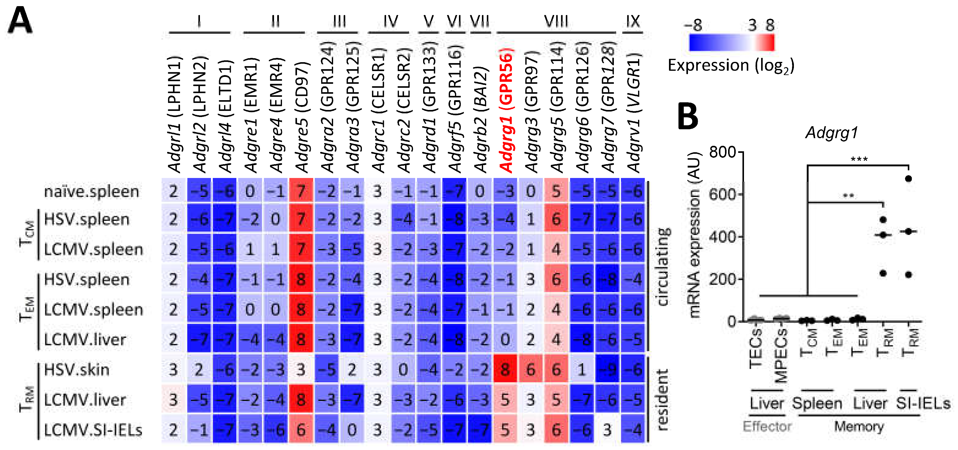
3.1. CD8+ TRM Cells Specifically Upregulate Adgrg1/GPR56

To establish the expression profile of adhesion GPCRs in pathogen-specific T cells, we analyzed RNA sequencing data of memory CD8+ T cells that arose after herpes simplex virus (HSV) or lymphocytic choriomeningitis virus (LCMV) infection in mice. We observed that T cells only expressed 4 out of a panel of 31 adhesion GPCRs, including Adgre5/CD97 and the gene cluster Adgrg1/GPR56, Adgrg3/GPR97, and Adgrg5/GPR114. Adgre5 and Adgrg5 expression was upregulated in naïve CD8+ T cells and was retained in all analyzed memory CD8+ T-cell lineages (Figure 1A). In contrast, Adgrg1 and Adgrg3 tran-scripts were not expressed in naïve CD8+ T cells but were upregulated in virus-specific CD8+ T cells after HSV or LCMV infection (Figure 1A). Upregulation of Adgrg3 was most pronounced in HSV-specific TRM cells in skin, whereas expression of Adgrg1 was strongly upregulated in HSV-specific TRM cells of the skin and in LCMV-specific TRM cells of the liver and the small intestine (Figure 1A). We did not find expression of Adgrg1 in circulating memory CD8+ T cells, including TCM- and TEM-cell populations in the spleen and the liver (Figure 1A), indicating that this adhesion GPCR was specifically induced in pathogen-specific TRM cells. The quantitative RT-PCR analysis validated the specific upregulation of Adgrg1 in virus-specific TRM cells after acute infection with LCMV (Figure 1B). The TRM cell-specific upregulation of Adgrg1 appeared to occur in the memory phase, given that we were unable to detect expression of Adgrg1 in effector populations, including terminal effector cells (TECs) and memory precursor effector cells (MPECs) at day 8 after infection with LCMV (Figure 1B). Interestingly, TRM cells also specifically express high levels of the transcription factor Hobit and the cytolytic enzyme granzyme B, in contrast to circulating TCM and TEM cells [25,28]. Therefore, Adgrg1/GPR56 expression appears to correlate with the expression of Zfp683/Hobit and the presence of granzyme B protein in murine as well as human CD8+ T cells.

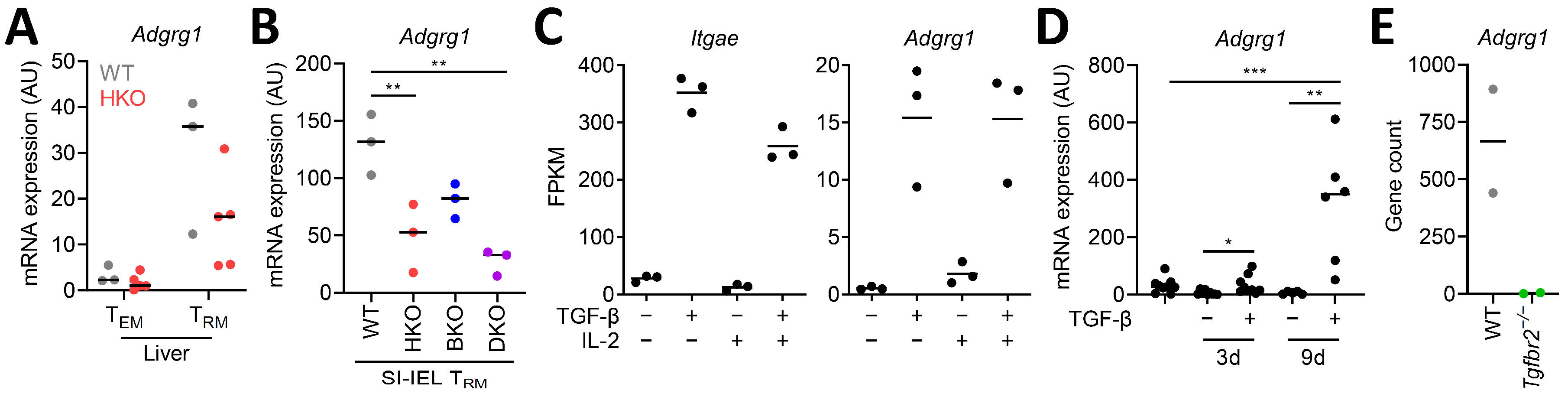
3.2. CD8+ TRM Cell-Inducing Factors Regulate Adgrg1/GPR56 Expression

We previously showed that Hobit and Blimp-1 strongly collaborate in the transcriptional regulation of TRM cells [28] and that Hobit is the driving transcription factor for the expression of granzyme B in TRM cells [25]. The analysis of Adgrg1 expression in TRM cells of Zfp683-deficient and Prdm1-deficient mice showed strong reduction of Adgrg1 expression in mouse TRM cells lacking Hobit and, to a lesser extent, in those lacking Blimp-1 (Figure 2A,B), in support of a role for Hobit in the transcriptional regulation of GPR56. The expression of Adgrg1 was further reduced in Zfp683 and Prdm1 double-deficient TRM cells (Figure 2B), indicating coregulation of Hobit and Blimp-1 in the maintenance of Adgrg1 expression in TRM cells. It is important to note that the reduced numbers of TRM cells in Hobit and Blimp-1 double-deficient mice may also impact Adgrg1 expression. However, the partial defects in GPR56 expression in single Hobit and Blimp-1 deficient mice that have normal numbers of TRM in small intestine suggest a direct impact of these transcription factors on the regulation of GPR56 expression.
TGF-β has been shown to specifically promote the development or maintenance of TRM cells in vivo [29] in the skin and small intestine [30]. TGF-β can also establish part of the residency-specific transcriptional profile of TRM cells, including the induction of Itgae/CD103 [31]. To determine the impact of TGF-β on Adgrg1 expression, we analyzed RNA sequencing data of spleen CD8+ T cells cultured in the presence of IL-2 and/or TGF-β. We observed that TGF-β not only induced expression of Itgae under these conditions but also strongly upregulated expression of Adgrg1 (Figure 2C). The presence of IL-2 did not impact Adgrg1 expression (Figure 2C). Analysis using the quantitative RT-PCR validated the upregulation of Adgrg1 transcripts in splenic CD8+ T cells after culture with TGF-β (Figure 2D). Furthermore, the expression of Adgrg1 is substantially reduced in skin TRM cells of mice with a deficiency in the TGF-β receptor II (Tgfbr2−/−) (Figure 2E). Our results suggest that the TRM cell-inducing transcription factor Hobit and the TRM cell-inducing cytokine TGF-β contribute to the expression of Adgrg1 in TRM cells.

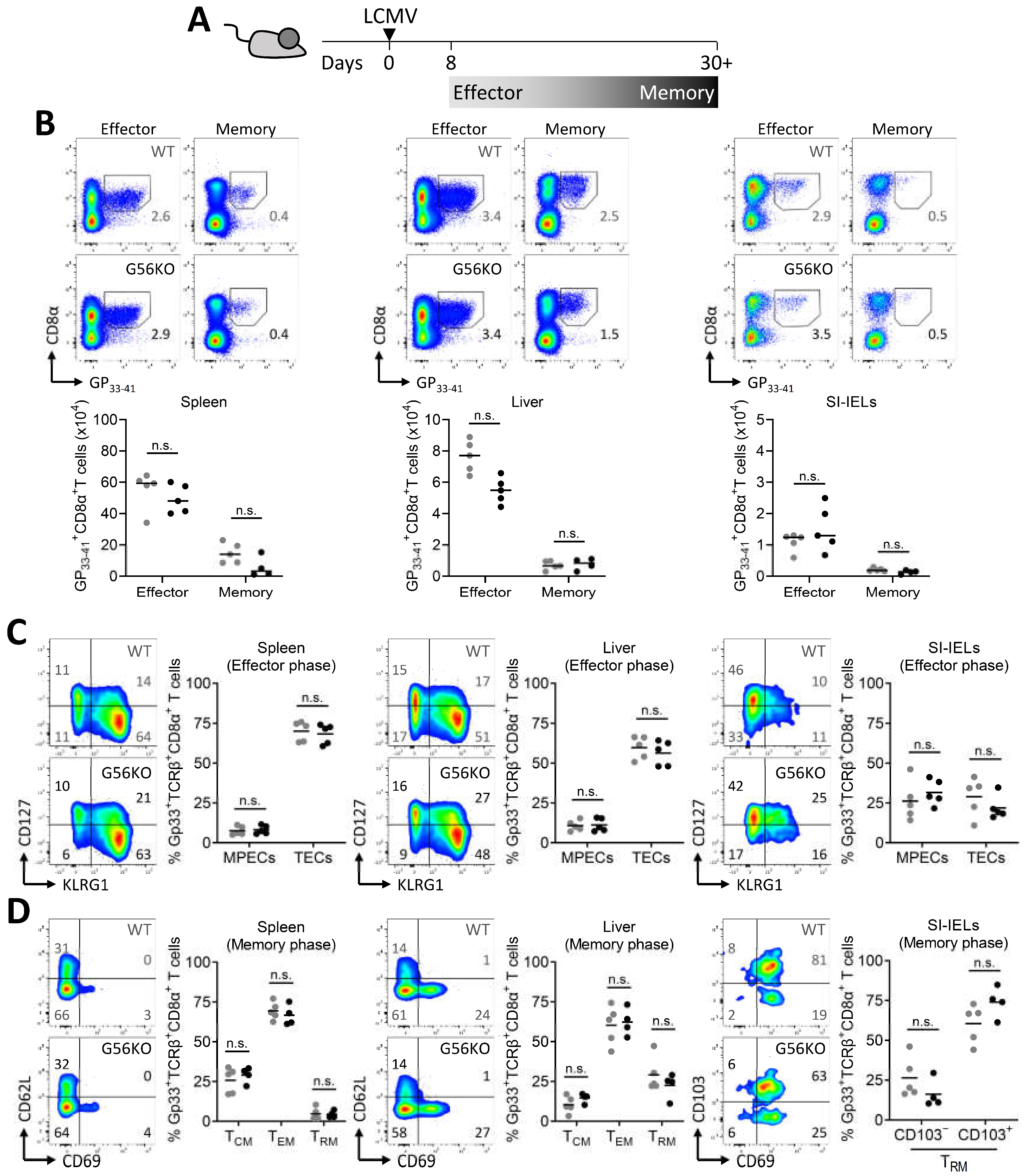
3.3. Adgrg1/GPR56 Is Dispensable for the Development of CD8+ TRM Cells

To study the involvement of GPR56 in CD8+ T-cell differentiation, we crossed floxed Adgrg1 mice [21] with Lck-Cre mice [24], thereby generating mice with deficient GPR56 expression in T cells. Analysis of the activity of Lck-driven CRE at the Adgrg1 locus in T cells indicated the specific deletion of this gene (Supplementary Figure S2). The impact of GPR56 was analyzed on virus-specific CD8+ T cells arising after an acute viral infection with LCMV (Figure 3A). Virus-specific CD8+ T cells recognizing the dominant epitope GP33-41 were detected using tetramers at different locations and time points after infection. We found that GPR56 was not essential for the formation of GP33-41+ CD8+ T cells in spleen, liver, and small intestine at effector (day 8) and memory time points (day 30+) after LCMV infection (Figure 3B). Expression of IL-7Rα chain (CD127) in conjunction with KLRG1 has been used to distinguish memory precursor effector cells (MPECs; KLRG1CD127+) from terminal effector cells (TECs; KLRG1+CD127) in mice [32,33]. Our analysis showed that these two effector populations appear to develop normally in the absence of GPR56 during the effector phase of an LCMV infection (Figure 3C). Expression of CD69 and CD62L divides memory CD8+ T cells into CD69CD62L+ TCM, CD69CD62L TEM, and CD69+CD62L TRM cells. In contrast to TRM cells in liver, a large fraction of TRM cells in the intraepithelial compartment of the small intestine expressed CD103 [34]. Analysis of the memory subsets in WT and Adgrg1-deficient mice showed that the formation of TCM-, TEM-, and TRM-cell populations in the spleen and liver and the formation of CD103 and CD103+ TRM cells in the small intestine was not dependent on GPR56 (Figure 3D). Thus, GPR56 does not appear to impact the differentiation of virus-specific CD8+ T cells into memory precursors and the development of downstream TCM-, TEM-, and TRM-cell populations.

3.4. Adgrg1/GPR56 Does Not Regulate Cytokine Production and Release of CD8+ TRM Cells

Virus-specific memory CD8+ T cells that develop after LCMV infection can effectively respond with the production and release of proinflammatory cytokines, such as IFN-γ. To examine whether GPR56 is involved in the regulation of IFN-γ production upon reactivation, we briefly cultured spleen and liver cells of LCMV-infected mice in the presence of the LCMV peptide GP33-41 (Figure 4A,B). Taking advantage of the TRM cell-associated molecule CD69 to distinguish circulating memory T cells from TRM cells, we observed that restimulated TRM cells from liver produced more IFN-γ than circulating memory T cells from the spleen and liver (Figure 4A,B). Moreover, TRM cells upregulated expression of CD107a, indicating the ability to degranulate and release cytokines, to a higher extent than circulating memory T cells (Figure 4A,B). However, IFN-γ production and degranulation was independent of GPR56 (Figure 4A,B). Taken together, we did not find evidence that GPR56 has an effect on the production and release of the proinflammatory cytokine IFN-γ by CD8+ T cells.

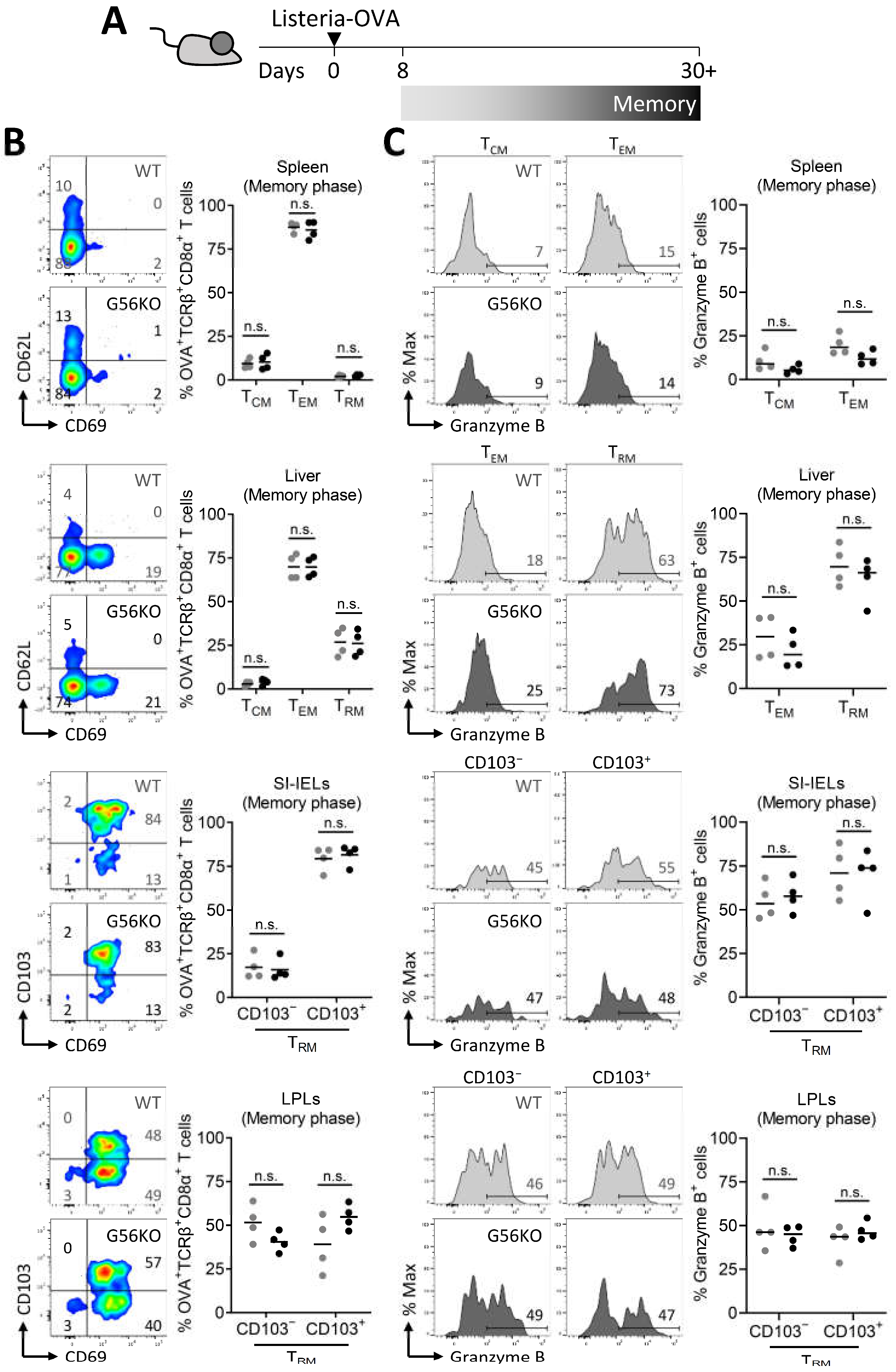
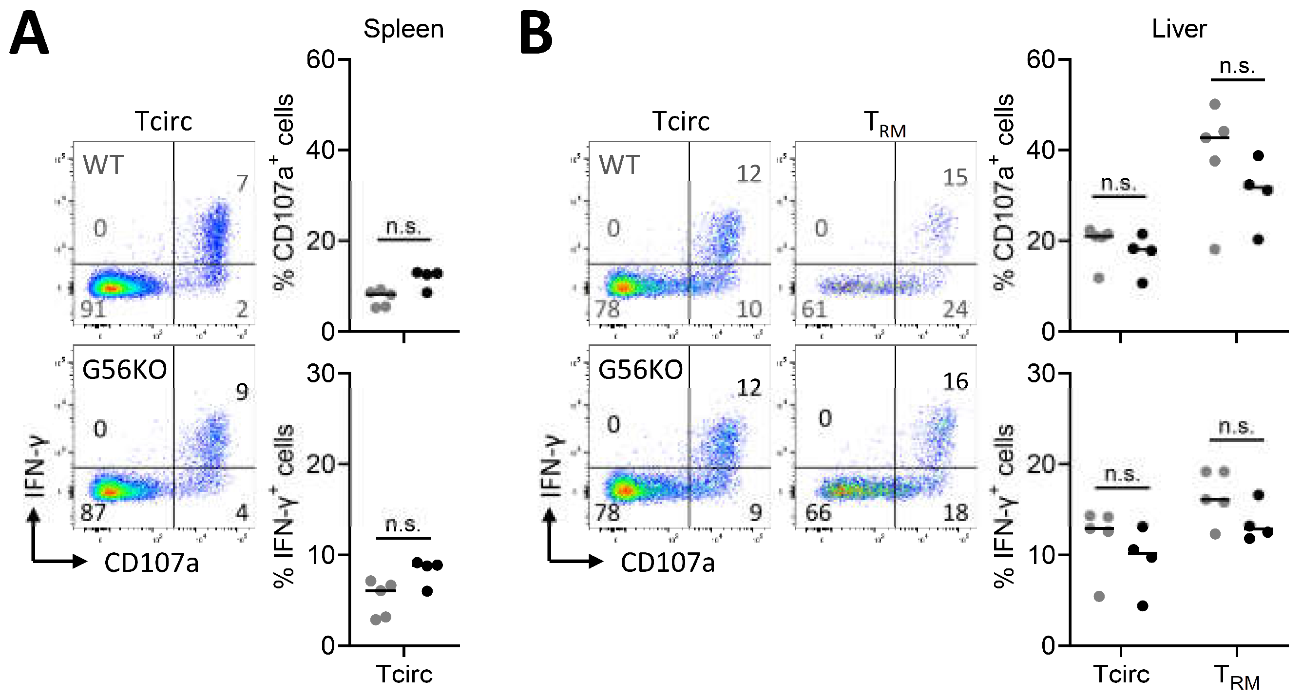
3.5. Adgrg1/GPR56 Does Not Regulate Cytotoxic Function of CD8+ TRM Cells

We exploited the Listeria-ovalbumin (OVA) infection model to validate our findings on the role of GPR56 in TRM cells in the LCMV infection model. Listeria monocytogenes is an intracellular bacterium that, upon oral administration, establishes acute infection in the small intestine and after systemic spread in other organs such as the liver. Similar as in the LCMV infection model, TRM-cell populations develop in liver and small intestine after Listeria-OVA infection [35]. We used tetramers recognizing OVA-specific CD8+ T cells to determine the effect of GPR56 on the differentiation of OVA-specific T cells after primary infection with Listeria-OVA (Figure 5A). Corroborating our results in the LCMV infection model, GPR56 did not appear to impact the differentiation of pathogen-specific CD8+ T cells into TCM, TEM, and TRM cells in the spleen and liver or into CD103 and CD103+ TRM-cell populations in the intraepithelial and lamina propria compartment of the small intestine after Listeria-OVA infection (Figure 5B). Effector CD8+ T cells upregulate protein expression of the cytolytic enzyme granzyme B, which is retained in TRM cells after pathogen clearance, but not in circulating memory CD8+ T cells [25]. Consistent with these findings, we observed that granzyme B protein expression is elevated in TRM cells in liver and small intestine compared to TCM and TEM cells in liver and spleen (Figure 5C). However, comparison between WT and Adgrgr1-deficient CD8+ T cells did not reveal differences in the expression of granzyme B (Figure 5C). Thus, GPR56 does not appear to essentially contribute to TRM-cell differentiation in the Listeria-OVA infection model and the regulation of granzyme B expression in Listeria-OVA specific TRM cells.

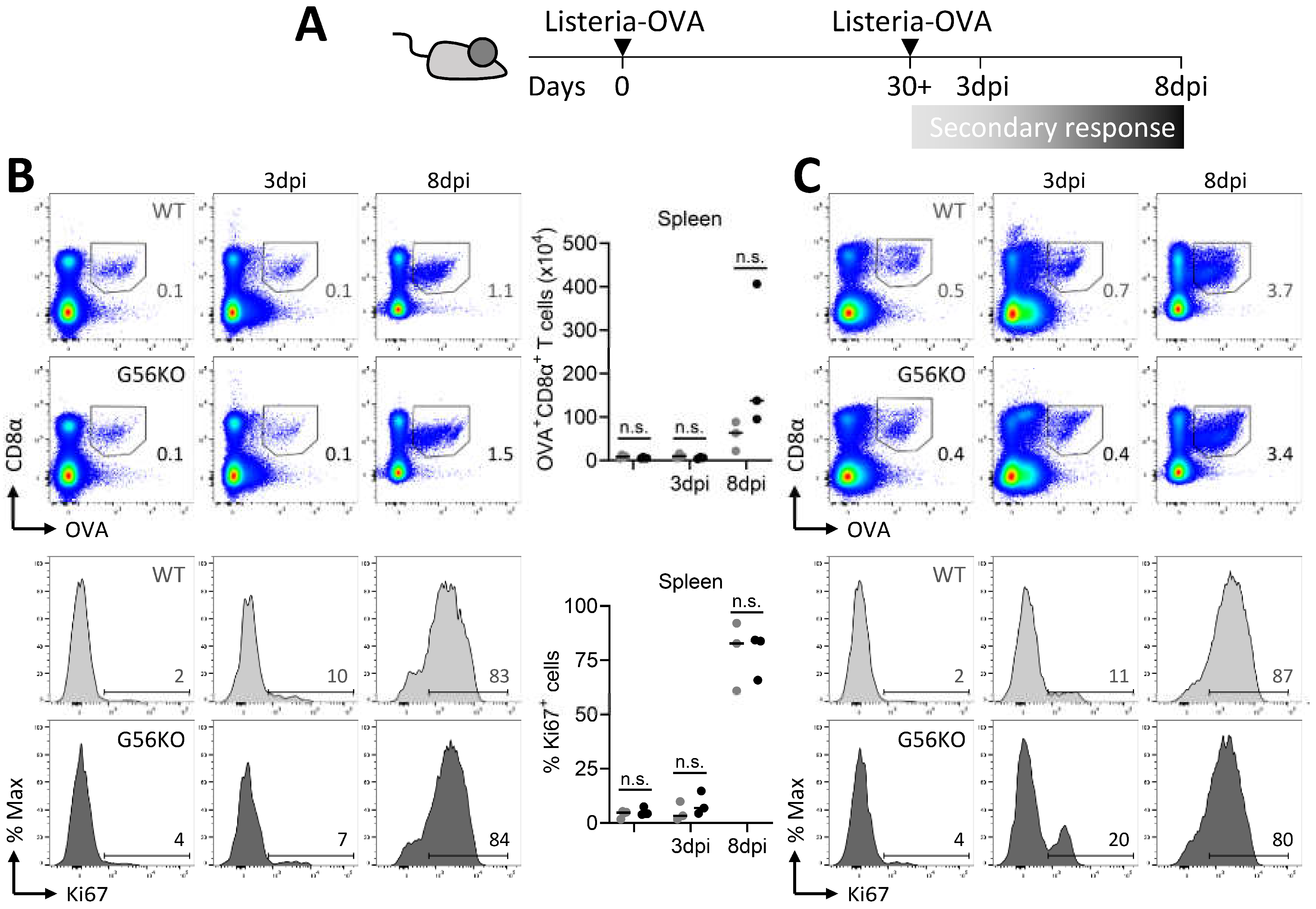
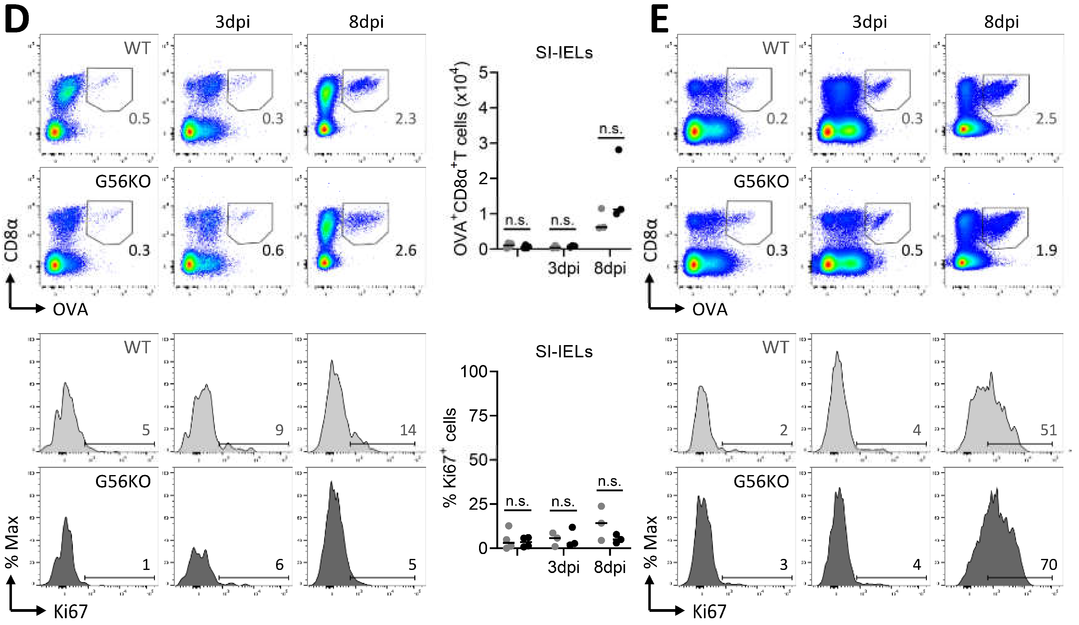
3.6. Secondary CD8+ T Cell Responses Are Not Regulated by Adgrg1/GPR56

TRM cells have the ability to expand and differentiate into effector cells that combat re-encountered pathogens upon reinfection. In fact, TRM cells substantially contribute to the establishment of secondary CD8+ T-cell responses [23]. We therefore employed the Listeria-OVA infection model to study the role of GPR56 in T-cell responses in the context of prime boost infection (Figure 6A). We observed that secondary infection of Listeria-OVA resulted in a substantial increase in the percentage and number of pathogen-specific CD8+ T cells in the spleen, liver, and intraepithelial and lamina propria compartment of the small intestine (Figure 6B–E). This increase in the number of pathogen-specific CD8+ T cells was notable as early as day 8 after reinfection (Figure 6B–E). However, GPR56 did not appear to have an effect on the magnitude of the secondary CD8+ T cell response in spleen, liver, and small intestine (Figure 6B–E). We further analyzed the secondary response of pathogen-specific CD8+ T cells using expression analysis of the proliferation-associated marker Ki-67. In line with their re-expansion, we detected increased expression of Ki-67 in pathogen-specific CD8+ T cells at day 8 after reinfection with Listeria-OVA (Figure 6B–E). However, the expression of Ki-67 was not different between pathogen-specific CD8+ T cells of WT and Adgrg1-deficient mice (Figure 6B–E). In sum, GPR56 expression did not appear to have an effect on the proliferative responses of pathogen-specific CD8+ T cells upon secondary infection with Listeria-OVA. Thus, we conclude that GPR56, despite its highly specific expression on TRM cells, does not essentially contribute to the regulation of the differentiation of TRM cells after primary infection and their reactivation and re-expansion after secondary infection.

4. Discussion

In this report, we have studied the role of the inhibitory receptor GPR56 on pathogen-specific CD8+ T cells. We found that Adgrg1 was specifically upregulated in CD8+ TRM cells in the memory phase after infection with HSV or LCMV. Moreover, we established evidence that implicates the TRM cell-associated transcriptional regulators Hobit and Blimp-1 and the TRM cell-inducing cytokine TGF-β and in the upregulation of Adgrg1 in CD8+ TRM cells. However, under the setting of acute infection with LCMV and Listeria monocytogenes, we did not observe an essential inhibitory role of GPR56 in the regulation of proliferative, cytokine, or cytotoxic responses of CD8+ TRM cells. We conclude that GPR56 is specifically upregulated on CD8+ TRM cells but does not provide an essential contribution in the regulation of the proinflammatory activity of TRM cells after acute infection.
Of note, the balance of GPR56 and GPR97 in hematopoietic stem cells (HSCs) is crucial for the development and differentiation of HSCs [36]. Therefore, it is possible that compensatory effects of other adhesion GPCRs such as GPR97 negate the impact of Adgrg1-deficiency on TRM cells. In line with potential redundant functions between adhesion GPCRs in TRM, we found pronounced expression of Adgrg3 in skin TRM and ubiquitous expression of Adgrg1 throughout the analyzed TRM-cell populations. The study of the combined role of these two adhesion GPCRs in TRM cells is an important future research direction.
TRM cells have been established as a separate lineage of memory CD8+ T cells with a unique transcriptional profile [33]. Here, we have identified GPR56 as a TRM cell-associated receptor. Adgrg1 was expressed in TRM cells in skin, liver, and small intestine, but not in circulating TCM- and TEM-cell populations in spleen and liver. TRM-cell populations in different tissues are exposed to distinct environmental conditions and therefore may have tissue-specific expression profiles. The expression of Adgrg1 in TRM cells in skin, liver, and small intestine suggests that GPR56 is upregulated on TRM cells in diverse microenvironments and may be part of the universal gene signature of TRM cells. However, further analysis of TRM cells in other tissues is required to determine whether GPR56 is ubiquitously expressed in TRM-cell populations throughout tissues.
In line with conserved expression of GPR56 between TRM-cell populations of humans and mice, we recently reported GPR56 expression on human brain TRM cells [37]. Although GPR56 has a wider expression pattern in humans than in mice, the retained expression of GPR56 in human TRM-cell populations suggests that our findings in mice have relevance for humans. Importantly, we identified expression of GPR56 protein in human brain TRM cells, indicating that this adhesion GPCR, at least in humans, is not regulated at protein level [37]. Unfortunately, antibodies are not yet available for murine GPR56, preventing us from analyzing protein expression in mice. Therefore, the possibility that lack of protein expression underlies the absence of functional defects in TRM cells of GPR56-deficient mice exists. However, we consider this option unlikely, given that we can detect both GPR56 mRNA and protein in humans.
The expression of Adgrg1 appears to be induced in the TRM-cell lineage at a late time point during T-cell differentiation after acute infection. We did not observe expression of Adgrg1 in naïve T cells or in effector CD8+ T cells including memory precursors that are upstream of TRM cells. We cannot exclude the possibility that a minor subset of these memory precursors upregulates Adgrg1 expression. Memory precursors with exclusive potential to form TRM cells appear to separate early from other effector cells [38,39,40]. These TRM precursor cells may be present in the bloodstream [40] and locally within peripheral tissues [38], where they eventually settle and form TRM cells. With current tools, it remains difficult to unequivocally identify precursor stages of TRM cells; therefore, analysis of the expression of GPR56 by these subsets awaits further characterization of TRM precursor cells. The repertoire of surface markers to identify TRM cells includes CD69, CD103, CD49a, and P2XR7 and other surface molecules [33,41,42]. Our identification of the specific expression of GPR56 on TRM cells suggests that this surface receptor may also be employed as a surrogate marker of TRM cells upon the establishment of antibodies recognizing mouse GPR56. As mentioned above, the expression pattern of GPR56 in humans appears broader than in mice. In humans, GPR56 is also expressed on cytotoxic lymphocytes, including NK cells and effector-type CD8+ and CD4+ T cells in peripheral blood [4] besides TRM cells in peripheral tissues [37]. The specific expression of GPR56 on TRM cells, rather than circulating memory T cells, in mice is consistent with their immediate cytotoxic capacity, given that TRM cells retain cytotoxic molecules, such as granzymes, at protein level in contrast to other memory T cells [25]. Thus, although GPR56 in humans may be differentially expressed compared to GPR56 in mice, upregulation of this adhesion GPCR on the cytotoxic fraction of memory T cells appears conserved between both species.
The differentiation of TRM cells is instructed by cytokines in their local environment. In particular, TGF-β has been found instrumental in the differentiation of TRM cells in the epithelial compartment of the skin and the small intestine [29,33]. We have identified TGF-β as a potent inducer of Adgrg1 expression in CD8+ T cells in in vitro cultures. Notably, developing TRM cells in skin require TGF-β signaling to acquire Adgrg1 expression. These findings suggest that TRM-cell populations in the skin and small intestine upregulate GPR56 expression in response to TGF-β signaling. Expression of CD103, the αE component of the αEβ7 integrin, also strongly depends on TGF-β signaling [29]. In contrast to CD103, we have observed that GPR56 is also expressed on liver TRM cells outside of CD103-expressing TRM-cell populations in skin and intestine. Therefore, the expression regulation of GPR56 in TRM cells appears more complex and may include environmental cues other than TGF-β. We have previously observed in human NK cells that the expression of GPR56 is downregulated in the presence of the homeostatic cytokine IL-15 and upon activation with inflammatory cytokines, including IL-2 and IL-18 [10]. Therefore, it is unlikely that IL-15, which can contribute to TRM-cell maintenance in mice [43,44,45], and these other cytokines play a role in the upregulation of GPR56 on TRM cells. We have previously also observed that GPR56 was induced by the transcription factor Hobit in human NK cells [10]. Hobit and the related transcription factor Blimp-1 are master regulators of TRM-cell differentiation, which, through suppression of tissue exit molecules, such as S1PR1 and CCR7, can permanently lock these memory T cells into the peripheral tissues [28]. Consistent with these findings, we observed here that Hobit together with Blimp-1 contributed to the transcriptional regulation of GPR56 expression in TRM cells. Hobit and Blimp-1 are broadly expressed in TRM cells throughout tissues including those of the skin, liver, and small intestine [28], suggesting that the transcription factor may drive GPR56 expression on CD103 and CD103+ TRM-cell populations in these tissues. The expression of Hobit and Blimp-1 appears independent of TGF-β signaling, but currently, it remains unknown how the expression of these transcription factors is upregulated during TRM-cell differentiation. Thus, GPR56 expression may be regulated through separate pathways, involving TGF-β and yet unresolved cytokines that trigger Hobit and Blimp-1 expression.
The strong proinflammatory activities of TRM cells after reinfection may have harmful impact on the surrounding healthy tissues. Pathogen-specific TRM cells express a multitude of inhibitory receptors including PD-1, TIM-3, LAG-3 and CD39 that can restrain TRM cell-driven immune responses [33]. Interestingly, CD103+ TRM cells appear to be under stronger regulation by inhibitory receptors than CD103 TRM cells [30]. In line with these findings, we also found that the inhibitory receptor GPR56 is responsive to TGF-β suggesting elevated expression of this receptor on CD103+ TRM cells compared to CD103 TRM cells. We have previously found that GPR56 is an inhibitory receptor, which can suppress the cytotoxic and cytokine responses of NK cells [10]. In this paper, we addressed the suppressive function of GPR56 on TRM cells after infection with LCMV and Listeria monocytogenes. In contrast to its essential role in regulating proinflammatory responses of NK cells, we did not observe that GPR56 substantially contributed to the control of the magnitude of TRM-cell responses or the cytokine production of TRM cells. We also did not find evidence that GPR56 contributed to the regulation of the cytolytic enzyme granzyme B in TRM cells, suggesting that the regulatory role of GPR56 is not essential under these conditions. Evidence is accumulating that expression of inhibitory receptors, such as PD-1, on TRM cells is highly relevant to protect against TRM cell-driven inflammatory responses that otherwise may develop in the intestine, pancreas, and the lungs [18,19,20]. Possibly, the inhibitory impact of GPR56 in the regulation of TRM cells is masked by the presence of other inhibitory receptors on TRM cells. Given that proinflammatory cytokines suppress GPR56 expression [10], unleashing the inhibitory potential of GPR56 may also require an anti-inflammatory environment, such as occurs in a tumor setting. More recently, TRM cells have been detected in tumor tissue of mouse models of melanoma [46], and in patients with melanoma, lung carcinoma, and ovarian carcinoma [47,48,49]. These tumor-residing TRM cells highly express PD-1, and inhibition of the PD-1-driven checkpoint blockade pathway appears to reinvigorate their antitumor activity [49]. Therefore, inhibitory receptors appear relevant on TRM cells to restrain antitumor immune responses. Our findings show that TRM cells also express dedicated inhibitory receptors such as GPR56 that suggest the presence of unique regulatory mechanisms in TRM compared to circulating memory T cells. However, resolving the potential role of GPR56 on tumor-resident TRM cells awaits future research.

Supplementary Materials

The following are available online at https://www.mdpi.com/article/10.3390/cells10102675/s1, Supplementary Figure S1: The sorting strategy for the quantitative RT-PCR analysis of Adgrg1 expression in T cells. Supplementary Figure S2: T cell-specific targeting of Adgrg1.

Author Contributions

C.-C.H. and N.A.M.K. performed the experiments; X.P. generated floxed Adgrg1 mice; C.-C.H., J.H. and K.P.J.M.v.G. designed the experiments and wrote the manuscript. All authors have read and agreed to the published version of the manuscript.

Funding

This work has been supported by the Thyssen Foundation (2015-00387, J.H. and K.P.J.M.v.G.) and the German Research Foundation (FOR 2149, J.H.). K.P.J.M.v.G. is a fellow of the Landsteiner Foundation for Blood Transfusion Research (LSBR).

Institutional Review Board Statement

The study was approved by the national Animal Ethics Committee of The Netherlands (protocol code: AVD3010020172205 and date of approval: 18 July 2017).

Informed Consent Statement

Not applicable.

Data Availability Statement

The data that support the findings of this study are available from the corresponding author upon reasonable request.

Acknowledgments

Listeria-OVA InlAM kindly provided by B. Sheridan, Stony Brook University. GP33-41, NP396-404 and OVA257-264 kindly provided by R. Arens, Leiden University Medical Center.

Conflicts of Interest

All authors declare no conflict of interest.

References

  1. Golstein, P.; Griffiths, G.M. An early history of T cell-mediated cytotoxicity. Nat. Rev. Immunol. 2018, 18, 527–535. [Google Scholar] [CrossRef]
  2. Waldman, A.D.; Fritz, J.M.; Lenardo, M.J. A guide to cancer immunotherapy: From T cell basic science to clinical practice. Nat. Rev. Immunol. 2020, 20, 651–668. [Google Scholar] [CrossRef]
  3. Della Chiesa, M.; Falco, M.; Parolini, S.; Bellora, F.; Petretto, A.; Romeo, E.; Balsamo, M.; Gambarotti, M.; Scordamaglia, F.; Tabellini, G.; et al. GPR56 as a novel marker identifying the CD56dull CD16+ NK cell subset both in blood stream and in inflamed peripheral tissues. Int. Immunol. 2009, 22, 91–100. [Google Scholar] [CrossRef] [PubMed] [Green Version]
  4. Peng, Y.-M.; van de Garde, M.D.B.; Cheng, K.-F.; Baars, P.A.; Remmerswaal, E.B.M.; van Lier, R.A.W.; Mackay, C.R.; Lin, H.-H.; Hamann, J. Specific expression of GPR56 by human cytotoxic lymphocytes. J. Leukoc. Biol. 2011, 90, 735–740. [Google Scholar] [CrossRef] [PubMed] [Green Version]
  5. Tian, Y.; Babor, M.; Lane, J.; Schulten, V.; Patil, V.S.; Seumois, G.; Rosales, S.L.; Fu, Z.; Picarda, G.; Burel, J.; et al. Unique phenotypes and clonal expansions of human CD4 effector memory T cells re-expressing CD45RA. Nat. Commun. 2017, 8, 1473. [Google Scholar] [CrossRef] [PubMed]
  6. Truong, K.L.; Schlickeiser, S.; Vogt, K.; Boës, D.; Stanko, K.; Appelt, C.; Streitz, M.; Grütz, G.; Stobutzki, N.; Meisel, C.; et al. Killer-like receptors and GPR56 progressive expression defines cytokine production of human CD4+ memory T cells. Nat. Commun. 2019, 10, 2263. [Google Scholar] [CrossRef] [Green Version]
  7. Piao, X.; Hill, S.S.; Bodell, A.; Chang, B.S.; Basel-Vanagaite, L.; Straussberg, R.; Dobyns, W.B.; Qasrawi, B.; Winter, R.M.; Innes, A.M.; et al. G Protein-Coupled Receptor-Dependent Development of Human Frontal Cortex. Science 2004, 303, 2033–2036. [Google Scholar] [CrossRef] [Green Version]
  8. Hamann, J.; Aust, G.; Araç, D.; Engel, F.B.; Formstone, C.; Fredriksson, R.; Hall, R.A.; Harty, B.L.; Kirchhoff, C.; Knapp, B.; et al. International union of basic and clinical pharmacology. XCIV. adhesion G protein-coupled receptors. Pharmacol. Rev. 2015, 67, 338–367. [Google Scholar] [CrossRef]
  9. Lin, H.H.; Hsiao, C.C.; Pabst, C.; Hébert, J.; Schöneberg, T.; Hamann, J. Adhesion GPCRs in Regulating Immune Responses and Inflammation. Adv. Immunol. 2017, 136, 163–201. [Google Scholar] [CrossRef]
  10. Chang, G.W.; Hsiao, C.C.; Peng, Y.M.; Vieira Braga, F.A.; Kragten, N.A.M.; Remmerswaal, E.B.M.; van de Garde, M.D.B.; Straussberg, R.; König, G.M.; Kostenis, E.; et al. The Adhesion G Protein-Coupled Receptor GPR56/ADGRG1 Is an Inhibitory Receptor on Human NK Cells. Cell Rep. 2016, 15, 1757–1770. [Google Scholar] [CrossRef] [Green Version]
  11. Vieira Braga, F.A.; Hertoghs, K.M.L.; Kragten, N.A.M.; Doody, G.M.; Barnes, N.A.; Remmerswaal, E.B.M.; Hsiao, C.C.; Moerland, P.D.; Wouters, D.; Derks, I.A.M.; et al. Blimp-1 homolog Hobit identifies effector-type lymphocytes in humans. Eur. J. Immunol. 2015, 45, 2945–2958. [Google Scholar] [CrossRef] [Green Version]
  12. Oja, A.E.; Vieira Braga, F.A.; Remmerswaal, E.B.M.; Kragten, N.A.M.; Hertoghs, K.M.L.; Zuo, J.; Moss, P.A.; van Lier, R.A.W.; van Gisbergen, K.P.J.M.; Hombrink, P. The transcription factor hobit identifies human cytotoxic CD4+ T cells. Front. Immunol. 2017, 8, 325. [Google Scholar] [CrossRef] [PubMed] [Green Version]
  13. Gisbergen, K.P.J.M.; Zens, K.D.; Münz, C. T cell memory in tissues. Eur. J. Immunol. 2021, 51, 1310–1324. [Google Scholar] [CrossRef] [PubMed]
  14. Schenkel, J.M.; Fraser, K.A.; Vezys, V.; Masopust, D. Sensing and alarm function of resident memory CD8+ T cells. Nat. Immunol. 2013, 14, 509–513. [Google Scholar] [CrossRef] [PubMed] [Green Version]
  15. Ariotti, S.; Hogenbirk, M.A.; Dijkgraaf, F.E.; Visser, L.L.; Hoekstra, M.E.; Song, J.Y.; Jacobs, H.; Haanen, J.B.; Schumacher, T.N. Skin-resident memory CD8+ T cells trigger a state of tissue-wide pathogen alert. Science 2014, 346, 101–105. [Google Scholar] [CrossRef]
  16. Gebhardt, T.; Whitney, P.G.; Zaid, A.; MacKay, L.K.; Brooks, A.G.; Heath, W.R.; Carbone, F.R.; Mueller, S.N. Different patterns of peripheral migration by memory CD4+ and CD8+ T cells. Nature 2011, 477, 216–219. [Google Scholar] [CrossRef]
  17. Jiang, X.; Clark, R.A.; Liu, L.; Wagers, A.J.; Fuhlbrigge, R.C.; Kupper, T.S. Skin infection generates non-migratory memory CD8 + T RM cells providing global skin immunity. Nature 2012, 483, 227–231. [Google Scholar] [CrossRef]
  18. Wang, Z.; Wang, S.; Goplen, N.P.; Li, C.; Cheon, I.S.; Dai, Q.; Huang, S.; Shan, J.; Ma, C.; Ye, Z.; et al. PD-1hi CD8+ resident memory T cells balance immunity and fibrotic sequelae. Sci. Immunol. 2019, 4, eaaw1217. [Google Scholar] [CrossRef]
  19. Weisberg, S.P.; Carpenter, D.J.; Chait, M.; Dogra, P.; Gartrell-Corrado, R.D.; Chen, A.X.; Campbell, S.; Liu, W.; Saraf, P.; Snyder, M.E.; et al. Tissue-Resident Memory T Cells Mediate Immune Homeostasis in the Human Pancreas through the PD-1/PD-L1 Pathway. Cell Rep. 2019, 29, 3916–3932.e5. [Google Scholar] [CrossRef] [Green Version]
  20. Sasson, S.C.; Slevin, S.M.; Cheung, V.T.; Nassiri, I.; Olsson-Brown, A.; Fryer, E.; Ferreira, R.C.; Trzupek, D.; Gupta, T.; Al-Hillawi, L.; et al. IFNγ-producing CD8+ tissue resident memory T cells are a targetable hallmark of immune checkpoint inhibitor-colitis. Gastroenterology 2021, 161, 1229–1244.e9. [Google Scholar] [CrossRef]
  21. Giera, S.; Deng, Y.; Luo, R.; Ackerman, S.D.; Mogha, A.; Monk, K.R.; Ying, Y.; Jeong, S.J.; Makinodan, M.; Bialas, A.R.; et al. The adhesion G protein-coupled receptor GPR56 is a cell-autonomous regulator of oligodendrocyte development. Nat. Commun. 2015, 6, 6121. [Google Scholar] [CrossRef] [Green Version]
  22. Van Gisbergen, K.P.J.M.; Kragten, N.A.M.; Hertoghs, K.M.L.; Wensveen, F.M.; Jonjic, S.; Hamann, J.; Nolte, M.A.; Van Lier, R.A.W. Mouse Hobit is a homolog of the transcriptional repressor Blimp-1 that regulates NKT cell effector differentiation. Nat. Immunol. 2012, 13, 864–871. [Google Scholar] [CrossRef]
  23. Behr, F.M.; Parga-Vidal, L.; Kragten, N.A.M.; van Dam, T.J.P.; Wesselink, T.H.; Sheridan, B.S.; Arens, R.; van Lier, R.A.W.; Stark, R.; van Gisbergen, K.P.J.M. Tissue-resident memory CD8+ T cells shape local and systemic secondary T cell responses. Nat. Immunol. 2020, 21, 1070–1081. [Google Scholar] [CrossRef]
  24. Kallies, A.; Xin, A.; Belz, G.T.; Nutt, S.L. Blimp-1 Transcription Factor Is Required for the Differentiation of Effector CD8+ T Cells and Memory Responses. Immunity 2009, 31, 283–295. [Google Scholar] [CrossRef] [Green Version]
  25. Kragten, N.A.M.; Behr, F.M.; Vieira Braga, F.A.; Remmerswaal, E.B.M.; Wesselink, T.H.; Oja, A.E.; Hombrink, P.; Kallies, A.; van Lier, R.A.W.; Stark, R.; et al. Blimp-1 induces and Hobit maintains the cytotoxic mediator granzyme B in CD8 T cells. Eur. J. Immunol. 2018, 48, 1644–1662. [Google Scholar] [CrossRef] [PubMed] [Green Version]
  26. Altman, J.D.; Moss, P.A.H.; Goulder, P.J.R.; Barouch, D.H.; McHeyzer-Williams, M.G.; Bell, J.I.; McMichael, A.J.; Davis, M.M. Phenotypic analysis of antigen-specific T lymphocytes. Science 1996, 274, 94–96. [Google Scholar] [CrossRef] [PubMed]
  27. Rötzschke, O.; Falk, K.; Stevanovic, S.; Jung, G.; Walden, P.; Rammensee, H.-G. Exact prediction of a natural T cell epitope. Eur. J. Immunol. 1991, 21, 2891–2894. [Google Scholar] [CrossRef] [PubMed]
  28. Mackay, L.K.; Minnich, M.; Kragten, N.A.M.; Liao, Y.; Nota, B.; Seillet, C.; Zaid, A.; Man, K.; Preston, S.; Freestone, D.; et al. Hobit and Blimp1 instruct a universal transcriptional program of tissue residency in lymphocytes. Science 2016, 352, 459–463. [Google Scholar] [CrossRef] [PubMed] [Green Version]
  29. Zhang, N.; Bevan, M.J. Transforming growth factor-β signaling controls the formation and maintenance of gut-resident memory T cells by regulating migration and retention. Immunity 2013, 39, 687–696. [Google Scholar] [CrossRef] [Green Version]
  30. Christo, S.N.; Evrard, M.; Park, S.L.; Gandolfo, L.C.; Burn, T.N.; Fonseca, R.; Newman, D.M.; Alexandre, Y.O.; Collins, N.; Zamudio, N.M.; et al. Discrete tissue microenvironments instruct diversity in resident memory T cell function and plasticity. Nat. Immunol. 2021, 22, 1140–1151. [Google Scholar] [CrossRef]
  31. Nath, A.P.; Braun, A.; Ritchie, S.C.; Carbone, F.R.; Mackay, L.K.; Gebhardt, T.; Inouye, M. Comparative analysis reveals a role for TGF-β in shaping the residency-related transcriptional signature in tissue-resident memory CD8+ T cells. PLoS ONE 2019, 14, e0210495. [Google Scholar] [CrossRef]
  32. Joshi, N.S.; Cui, W.; Chandele, A.; Lee, H.K.; Urso, D.R.; Hagman, J.; Gapin, L.; Kaech, S.M. Inflammation Directs Memory Precursor and Short-Lived Effector CD8+ T Cell Fates via the Graded Expression of T-bet Transcription Factor. Immunity 2007, 27, 281–295. [Google Scholar] [CrossRef] [Green Version]
  33. MacKay, L.K.; Rahimpour, A.; Ma, J.Z.; Collins, N.; Stock, A.T.; Hafon, M.L.; Vega-Ramos, J.; Lauzurica, P.; Mueller, S.N.; Stefanovic, T.; et al. The developmental pathway for CD103+ CD8+ tissue-resident memory T cells of skin. Nat. Immunol. 2013, 14, 1294–1301. [Google Scholar] [CrossRef]
  34. Schenkel, J.M.; Masopust, D. Tissue-resident memory T cells. Immunity 2014, 41, 886–897. [Google Scholar] [CrossRef] [Green Version]
  35. Sheridan, B.S.; Pham, Q.M.; Lee, Y.T.; Cauley, L.S.; Puddington, L.; Lefrançois, L. Oral infection drives a distinct population of intestinal resident memory cd8+ t cells with enhanced protective function. Immunity 2014, 40, 747–757. [Google Scholar] [CrossRef] [PubMed] [Green Version]
  36. Maglitto, A.; Mariani, S.A.; de Pater, E.; Rodriguez-Seoane, C.; Vink, C.S.; Piao, X.; Lukke, M.L.; Dzierzak, E. Unexpected redundancy of Gpr56 and Gpr97 during hematopoietic cell development and differentiation. Blood Adv. 2021, 5, 829–842. [Google Scholar] [CrossRef] [PubMed]
  37. Fransen, N.L.; Hsiao, C.C.; Van Der Poel, M.; Engelenburg, H.J.; Verdaasdonk, K.; Vincenten, M.C.J.; Remmerswaal, E.B.M.; Kuhlmann, T.; Mason, M.R.J.; Hamann, J.; et al. Tissue-resident memory T cells invade the brain parenchyma in multiple sclerosis white matter lesions. Brain 2020, 143, 1714–1730. [Google Scholar] [CrossRef] [PubMed]
  38. Milner, J.J.; Toma, C.; He, Z.; Kurd, N.S.; Nguyen, Q.P.; McDonald, B.; Quezada, L.; Widjaja, C.E.; Witherden, D.A.; Crowl, J.T.; et al. Heterogenous Populations of Tissue-Resident CD8+ T Cells Are Generated in Response to Infection and Malignancy. Immunity 2020, 52, 808–824.e7. [Google Scholar] [CrossRef] [PubMed]
  39. Kurd, N.S.; He, Z.; Louis, T.L.; Milner, J.J.; Omilusik, K.D.; Jin, W.; Tsai, M.S.; Widjaja, C.E.; Kanbar, J.N.; Olvera, J.G.; et al. Early precursors and molecular determinants of tissue-resident memory CD8+ T lymphocytes revealed by single-cell RNA sequencing. Sci. Immunol. 2020, 5, eaaz6894. [Google Scholar] [CrossRef] [PubMed]
  40. Kok, L.; Dijkgraaf, F.E.; Urbanus, J.; Bresser, K.; Vredevoogd, D.W.; Cardoso, R.F.; Perié, L.; Beltman, J.B.; Schumacher, T.N. A committed tissue-resident memory T cell precursor within the circulating CD8+ effector T cell pool. J. Exp. Med. 2020, 217, e20191711. [Google Scholar] [CrossRef]
  41. Beura, L.K.; Fares-Frederickson, N.J.; Steinert, E.M.; Scott, M.C.; Thompson, E.A.; Fraser, K.A.; Schenkel, J.M.; Vezys, V.; Masopust, D. CD4+ resident memory T cells dominate immunosurveillance and orchestrate local recall responses. J. Exp. Med. 2019, 216, 1214–1229. [Google Scholar] [CrossRef] [PubMed]
  42. Stark, R.; Wesselink, T.H.; Behr, F.M.; Kragten, N.A.M.; Arens, R.; Koch-Nolte, F.; van Gisbergen, K.P.J.M.; van Lier, R.A.W. TRM maintenance is regulated by tissue damage via P2RX7. Sci. Immunol. 2018, 3, eaau1022. [Google Scholar] [CrossRef] [Green Version]
  43. Mackay, L.K.; Wynne-Jones, E.; Freestone, D.; Pellicci, D.G.; Mielke, L.A.; Newman, D.M.; Braun, A.; Masson, F.; Kallies, A.; Belz, G.T.; et al. T-box Transcription Factors Combine with the Cytokines TGF-β and IL-15 to Control Tissue-Resident Memory T Cell Fate. Immunity 2015, 43, 1101–1111. [Google Scholar] [CrossRef] [Green Version]
  44. Adachi, T.; Kobayashi, T.; Sugihara, E.; Yamada, T.; Ikuta, K.; Pittaluga, S.; Saya, H.; Amagai, M.; Nagao, K. Hair follicle-derived IL-7 and IL-15 mediate skin-resident memory T cell homeostasis and lymphoma. Nat. Med. 2015, 21, 1272–1279. [Google Scholar] [CrossRef] [PubMed]
  45. Schenkel, J.M.; Fraser, K.A.; Casey, K.A.; Beura, L.K.; Pauken, K.E.; Vezys, V.; Masopust, D. IL-15–Independent Maintenance of Tissue-Resident and Boosted Effector Memory CD8 T Cells. J. Immunol. 2016, 196, 3920–3926. [Google Scholar] [CrossRef] [PubMed] [Green Version]
  46. Park, S.L.; Buzzai, A.; Rautela, J.; Hor, J.L.; Hochheiser, K.; Effern, M.; McBain, N.; Wagner, T.; Edwards, J.; McConville, R.; et al. Tissue-resident memory CD8+ T cells promote melanoma–immune equilibrium in skin. Nature 2019, 565, 366–371. [Google Scholar] [CrossRef] [PubMed]
  47. Djenidi, F.; Adam, J.; Goubar, A.; Durgeau, A.; Meurice, G.; de Montpréville, V.; Validire, P.; Besse, B.; Mami-Chouaib, F. CD8+ CD103+ Tumor–Infiltrating Lymphocytes Are Tumor-Specific Tissue-Resident Memory T Cells and a Prognostic Factor for Survival in Lung Cancer Patients. J. Immunol. 2015, 194, 3475–3486. [Google Scholar] [CrossRef] [PubMed] [Green Version]
  48. Ganesan, A.P.; Clarke, J.; Wood, O.; Garrido-Martin, E.M.; Chee, S.J.; Mellows, T.; Samaniego-Castruita, D.; Singh, D.; Seumois, G.; Alzetani, A.; et al. Tissue-resident memory features are linked to the magnitude of cytotoxic T cell responses in human lung cancer. Nat. Immunol. 2017, 18, 940–950. [Google Scholar] [CrossRef] [PubMed]
  49. Edwards, J.; Wilmott, J.S.; Madore, J.; Gide, T.N.; Quek, C.; Tasker, A.; Ferguson, A.; Chen, J.; Hewavisenti, R.; Hersey, P.; et al. CD103+ tumor-resident CD8+ T cells are associated with improved survival in immunotherapy-naïve melanoma patients and expand significantly during anti-PD-1 treatment. Clin. Cancer Res. 2018, 24, 3036–3045. [Google Scholar] [CrossRef] [Green Version]
Figure 1. CD8+ TRM cells express Adgrg1/GPR56. (A) Naïve CD8+ T cells were sorted from uninfected mice, and virus-specific memory CD8+ T-cell populations were sorted from indicated tissues of mice, infected with herpes simplex virus (HSV) or lymphocytic choriomeningitis virus (LCMV), at day 40+ postinfection (memory phase). A panel of 31 adhesion GPCRs was analyzed, of which 19 adhesion GPCRs were present, and 12 adhesion GPCRs were absent in the RNA sequencing data. Data are derived from GSE70813. (B) Adgrg1 transcripts were determined by quantitative RT-PCR in indicated CD8+ T-cell populations from liver, spleen, and small intestinal intraepithelial lymphocytes (SI-IELs). LCMV-specific CD8+ T cells were collected at different time points of infection. Terminal effector cells (TECs) and memory precursor cells (MPECs) were isolated at day 8 (effector phase), and memory CD8+ T cells were obtained at day 30+ postinfection (memory phase). The sorting strategy is shown in Supplementary Figure S1. One-way ANOVA with Tukey’s multiple comparisons test; ** p < 0.01, *** p < 0.005.
Cells 10 02675 g001
Figure 2. Adgrg1/GPR56 expression in CD8+ TRM cells requires Hobit and TGF-β. (A) Adgrg1 transcripts were determined using quantitative RT-PCR in LCMV-specific CD8+ TEM and TRM cells from liver of wild type (WT, ) and Zfp683-deficient (HKO, ●) mice at day 40+ after LCMV-WE infection. The gating strategy is shown in Supplementary Figure S1. (B) Adgrg1 transcripts were determined using quantitative RT-PCR in LCMV-specific CD8+ TRM cells from small intestine intraepithelial lymphocytes (SI-IELs) of WT, Zfp683−/CRE (HKO), Zfp683+/CRE × Prdm1flox/flox (BKO), and Zfp683−/CRE × Prdm1flox/flox (DKO) mice at day 30+ after LCMV-Armstrong infection. The sorting strategy is similar, as shown in Supplementary Figure S1. (C) Itgae and Adgrg1 transcript counts are shown upon different condition treatments as indicated. Data are derived from GSE125471. FPKM, fragments per kilobase of transcript per million mapped reads. (D) Adgrg1 transcripts were determined using quantitative RT-PCR in sorted spleen CD8+ T cells after culture with or without TGF-β for 3 and 9 days from two independent experiments (n = 6). (E) Adgrg1 transcript counts are shown for OTI cells from WT or Tgfbr2-deficient mice in skin of HSV-OVA-infected recipients. Data are derived from GSE178769. One-way or two-way ANOVA with Tukey’s multiple comparisons test; * p < 0.05, ** p < 0.01, *** p < 0.005.
Cells 10 02675 g002
Figure 3. Adgrg1/GPR56 is dispensable for the formation of virus-specific CD8+ TRM cells. (A) Experimental scheme shows the generation of LCMV-specific CD8+ T cells and indicates the effector and memory time points for analysis of virus-specific CD8+ T cells after acute LCMV-Armstrong infection. (B) Representative plots and absolute counts of virus-specific (GP33-41+) TCRβ+CD8+ T cells in wild-type (WT, ) and Adgrg1-deficient (G56KO, ●) spleen, liver, and small intestinal intraepithelial lymphocytes (SI-IELs) at day 8 (effector phase) and 30+ (memory phase) after LCMV-Armstrong infection. (C) Representative flow cytometry plots and frequencies of virus-specific GP33-41+TCRβ+CD8+ memory precursor effector cells (MPECs, KLRG1CD127+) and terminal effector cells (TECs, KLRG1+CD127) in WT () and G56KO (●) spleen, liver, and SI-IELs at day 8 post-infection. (D) Representative flow cytometry plots and frequencies of virus-specific GP33-41+TCRβ+CD8+ CD69CD62L+ TCM, CD69CD62L TEM, CD69+CD62L TRM, and CD103−/+CD69+ TRM cells in WT () and G56KO (●) spleen, liver, and SI-IELs at day 30+ post-infection. Two-way ANOVA with Tukey’s multiple comparisons test; n.s., no significant difference.
Cells 10 02675 g003
Figure 4. Adgrg1/GPR56 is dispensable for the function of virus-specific CD8+ TRM cells upon in vitro stimulation with peptide antigen. (A, B) Representative flow cytometry plots and graphs displaying CD107a and IFN-γ expression of CD8+ CD69 circulating T (Tcirc) and CD69+ TRM cells in wild-type (WT, ) and Adgrg1-deficient (G56KO, ●) spleen (A) and liver (B) upon in vitro restimulation with GP33-41 peptide. Two-way ANOVA with Tukey’s multiple comparisons test; n.s., no significant difference.
Cells 10 02675 g004
Figure 5. Adgrg1/GPR56 is dispensable for the formation and function of Listeria-specific CD8+ TRM cells upon rechallenge. (A) Experimental scheme shows the generation of Listeria-OVA-specific memory T cells. (B) Representative flow cytometry plots and frequencies of Listeria-specific (OVA+) TCRβ+CD8+ CD69CD62L+ TCM, CD69CD62L TEM, CD69+CD62L TRM, and CD103−/+ CD69+ TRM cells in wild-type (WT, ) and Adgrg1-deficient (G56KO, ●) spleen, liver, and small intestinal intraepithelial lymphocytes (SI-IELs) at day 30+ postinfection. (C) Representative histograms and frequencies of granzyme B in indicated subsets of Listeria-specific (OVA+) TCRβ+CD8+ TCM, TEM, and TRM cells in WT () and G56KO (●) spleen, liver, SI-IELs, and lamina propria lymphocytes (LPLs). Two-way ANOVA with Tukey’s multiple comparisons test; n.s., no significant difference.
Cells 10 02675 g005
Figure 6. Adgrg1/GPR56 is dispensable for the expansion of Listeria-specific CD8+ TRM cells upon rechallenge. (A) Experimental scheme shows the time points of analysis of Listeria-OVA-specific T cells after primary challenge and after-challenge with Listeria-OVA infection. (BE) Representative flow cytometry plots and absolute counts of Listeria-specific (OVA+) CD8+ T cells and representative histograms of Ki-67 expression in Listeria-specific (OVA+) CD8+ T cells in wild-type (WT, ) and Adgrg1-deficient (G56KO, ●) spleen (B), liver (C), small intestinal intraepithelial lymphocytes (SI-IELs) (D), and lamina propria lymphocytes (LPLs) (E) ex vivo and upon 3 and 8 days after rechallenge infection (3 and 8 dpi). Two-way ANOVA with Tukey’s multiple comparisons test; n.s., no significant difference.
Cells 10 02675 g006aCells 10 02675 g006b
Publisher’s Note: MDPI stays neutral with regard to jurisdictional claims in published maps and institutional affiliations.

