Review

Autophagy in Tumor Immunity and Viral-Based Immunotherapeutic Approaches in Cancer

1
Apoptosis Research Centre, Children’s Hospital of Eastern Ontario Research Institute, Ottawa, ON K1H 8L1, Canada
2
Department of Biochemistry Microbiology and Immunology, Faculty of Medicine, University of Ottawa, Ottawa, ON K1H 8L1, Canada
3
Centre for Infection, Immunity and Inflammation (CI3), University of Ottawa, Ottawa, ON K1H 8L1, Canada
4
Department of Cellular and Molecular Medicine, University of Ottawa, Ottawa, ON K1H 8M5, Canada
*
Author to whom correspondence should be addressed.
Cells 2021, 10(10), 2672; https://doi.org/10.3390/cells10102672
Submission received: 4 September 2021 / Revised: 22 September 2021 / Accepted: 27 September 2021 / Published: 6 October 2021

Abstract

:
Autophagy is a fundamental catabolic process essential for the maintenance of cellular and tissue homeostasis, as well as directly contributing to the control of invading pathogens. Unsurprisingly, this process becomes critical in supporting cellular dysregulation that occurs in cancer, particularly the tumor microenvironments and their immune cell infiltration, ultimately playing a role in responses to cancer therapies. Therefore, understanding “cancer autophagy” could help turn this cellular waste-management service into a powerful ally for specific therapeutics. For instance, numerous regulatory mechanisms of the autophagic machinery can contribute to the anti-tumor properties of oncolytic viruses (OVs), which comprise a diverse class of replication-competent viruses with potential as cancer immunotherapeutics. In that context, autophagy can either: promote OV anti-tumor effects by enhancing infectivity and replication, mediating oncolysis, and inducing autophagic and immunogenic cell death; or reduce OV cytotoxicity by providing survival cues to tumor cells. These properties make the catabolic process of autophagy an attractive target for therapeutic combinations looking to enhance the efficacy of OVs. In this article, we review the complicated role of autophagy in cancer initiation and development, its effect on modulating OVs and immunity, and we discuss recent progress and opportunities/challenges in targeting autophagy to enhance oncolytic viral immunotherapy.

1. Introduction

Oncolytic viruses (OVs), either occurring naturally or through genetic engineering, are promising candidate therapies against cancer since they can selectively amplify in and kill tumor cells without affecting normal cells. Moreover, OVs can boost systemic anti-tumor immunity by lysing tumor cells; the consequent release of tumor-associated antigens (TAAs), damage-associated molecular patterns (DAMPs), and pathogen-associated molecular patterns (PAMPs) to the tumor microenvironment (TME) promote activation of antigen-presenting cells (APCs), in turn stimulating anti-tumor adaptive immune responses [1,2]. With their unique capacity to also encode for a vast array of therapeutic transgenes to enhance specific responses, OVs, therefore, represent promising candidate therapies against cancer.
Macro-autophagy (hereafter, autophagy) is a conserved and tightly regulated catabolic process in eukaryotic cells. Its main role is to supply energy during development, and under conditions of cellular stress, wherein selective or non-selective formation of double-membrane vesicles called autophagosomes engulf entire regions of the cytoplasm, superfluous or damaged organelles, protein aggregates, and invading pathogens (Figure 1). Whereas non-selective (bulk) autophagy is a cellular response to starvation and typically involves random uptake of cytoplasm into phagophores (the precursors to autophagosomes), selective autophagy is mainly responsible for removing certain components such as superfluous or damaged organelles, protein aggregates, and is driven by receptors with an affinity for both the cargo and LC3/ATG8 [3,4]. Autophagosomes are sequentially fused with lysosomes to form autolysosomes, whose contents are subsequently digested by lysosomal hydrolases and released for metabolic recycling [4]. This process is also used to eliminate foreign particles or infectious agents; however, like many other invading pathogens, many viruses, including OVs, have been known to interfere with the autophagic machinery either in cancer cells or cells of the TME to enhance their replication and spread [5,6,7,8,9,10]. This is important because as a component of the host defense system in cell-autonomous elimination of invading pathogens, autophagy can promote tumorigenesis or act as a tumor suppressor in different cancer models [11,12]. Further, autophagy regulates several immunological functions, such as inflammation activation, cytokine production, antigen, antigen processing, and T- and B-lymphocyte immune responses [13]. Thus, viral infection-induced autophagy may also contribute to anti-tumor immunity via enhancing processing and cross-presentation of tumor antigens by dendritic cells (DCs) [14,15], signifying that OV-mediated anti-tumor effects can be mediated by crosstalk between virus and host tumor and normal cells. Thus, a better understanding of the “host-virus-environment” in the context of autophagic regulation could help to efficiently manipulate their interconnections for more effective therapies.
We herein review autophagic mechanisms, their roles in cancer initiation and development, OV efficacy, and anti-tumor immune responses. Finally, the prospect and challenges of combinatorial OV therapy with autophagy modulators in cancer immunotherapy are explored.

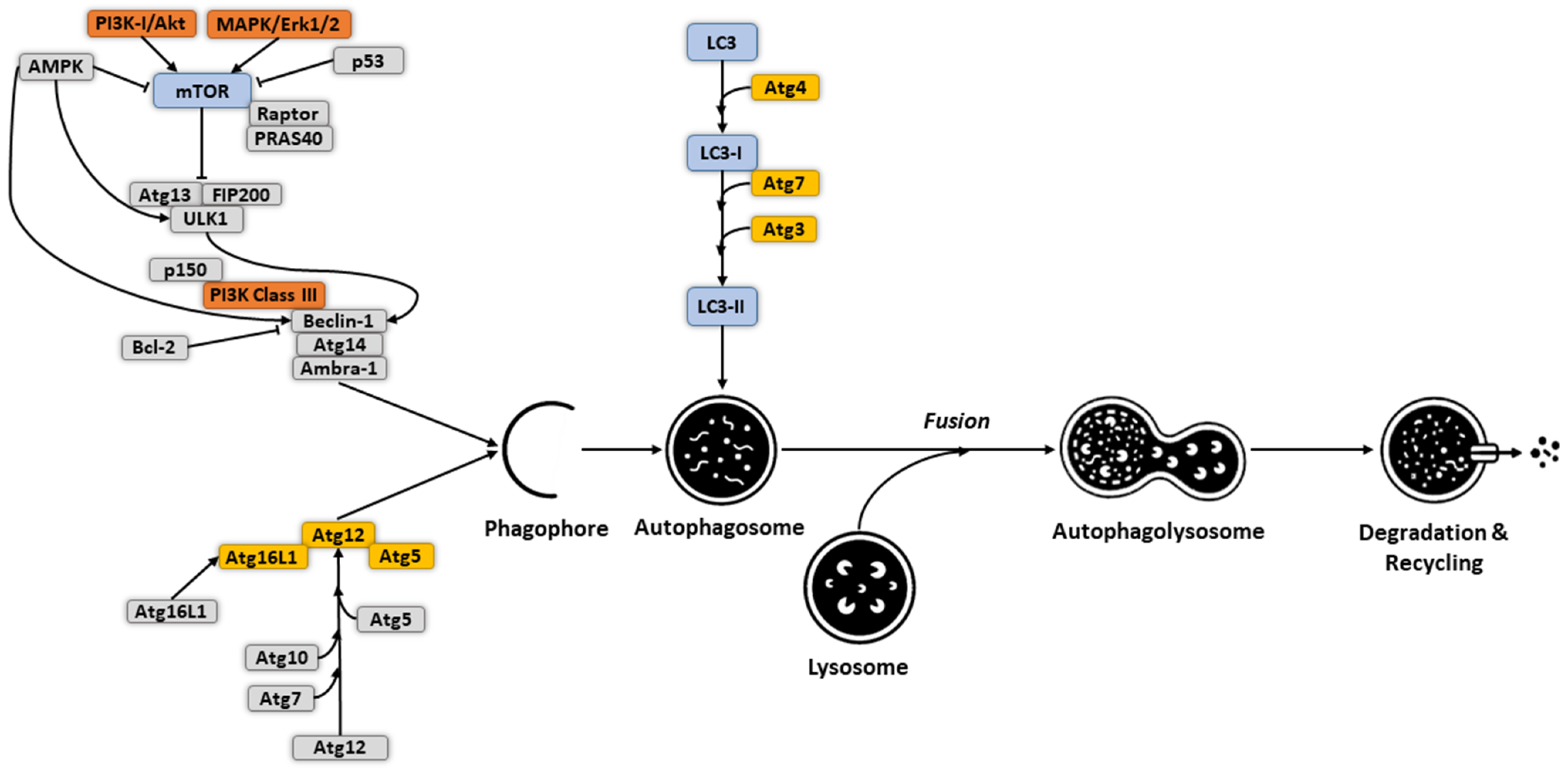
2. Molecular Mechanism and Regulation of Autophagy

Although different signals are involved in initiating “selective” versus “non-selective” autophagy, both converge at the formation of the autophagosome upon membrane remodeling. Several factors come into play to initiate and sustain the system. Here we will describe the main known activation mechanisms that modulate the autophagic machinery; for visual representation, please refer to Figure 1. Activation of the Unc-51-like autophagy activating kinase 1 (ULK1) complex (ATG1 complex in yeast), which includes ULK1 and the subunits FIP200, ATG13, and ATG101, represents the initial hallmark of mammalian autophagy [16,17]. This occurs through autophosphorylation of ULK1 at the Thr180 residue, facilitated via kinase dimerization or oligomerization [18]. Upon sensing changes in cellular energy, the ULK1 complex acts as a scaffold to initiate the phagophore assembly site (PAS), a process triggered by binding of ATG13 to the early autophagy targeting (EAT) domain of ULK1 [19]. Localization of the ULK1 complex to the PAS can also be directed through interactions between members of the LC3 protein family and LIR/AIM short linear motifs of ULK1, which also have roles in later stages of autophagosome formation [20,21]. The binding of RAB1A, a small GTPase regulating vesicular trafficking, to the DENN domain (differentially expressed in normal and neoplastic cells) of C9orf72 is another essential factor contributing to ULK1 translocation to the PAS [22]. ULK1 phosphorylation of the phosphatidylinositol 3 kinase class III (PI3KC3) complex creates sub-complexes I (C1) and II (C2) and initiates autophagy. Common catalytic subunits of these complexes include ATG34, ATG15, and Beclin-1 (BECN1). PI3KC3-C1 is involved in membrane elongation and encompasses ATG14L that transports this complex to PAS. Interaction of PI3KC3-C2 with UVRAG then contributes to endosomal and autophagosome biogenesis [23].
Several protein-protein interactions and post-translational modifications regulate the kinase activity within PI3KC3-C1. For instance, NRBF2 promotes kinase activity and dimerization of this complex via interaction with ATG14L and BECN1 [24]. The binding of BCL2, an anti-apoptotic factor, to the BH3 domain of BECN1 represses the kinase activity of ATG34, resulting in inhibition of autophagy [25]. AMPBRA1, another PI3KC3-C1 regulator, can be phosphorylated by ULK1 to activate PI3KC3-C1 [26]. Following autophagy induction, the formation of the ATG12-ATG5 ubiquitination complex is catalyzed by ATG7 and ATG10 [27]. Once formed, this complex acts as a ubiquitin ligase to increase lipidation of LC3, a light chain of the microtubule-associated protein whose lipidation is essential for autophagosome formation and cargo recognition. The ATG12-ATG5 conjugate further interacts with ATG16L1 to establish the ATG12-ATG5-ATG16L1 complex [28], which is needed for activation of ATG3. This in turn facilitates LC3-phosphatidylethanolamine (PE) formation [29]. Although the exact role of the LC3-PE complex in autophagosome biogenesis is under investigation, in vitro findings suggest this complex is involved in membrane expansion and elongation of phagocytic vesicles for controlling the size and volume of autophagosomes, as well as in closure of these double-layer structures for completion of autophagy [21,29].
Numerous signal transduction pathways contribute to the regulation of autophagy. The main example is the mammalian target of rapamycin (mTOR) pathway, which functions as a serine/threonine protein kinase signaling regulating autophagy. Amino acid-triggered activation of mTOR complex 1 (mTORC1) on lysosomes can be mediated by several regulatory factors and complexes, such as RAG, SLC38A9, RAGULATOR-Rag, and KICSTOR [30]. AKT1, another protein kinase whose activation and phosphorylation could be stimulated by growth factors boosting PI3KC1, is also known to activate mTORC1 [31,32]. mTORC1 modulates the ULK1 complex, transcription factors, and phosphorylation of histone acetyltransferases. The second mTOR complex (mTORC2) can upregulate AKT1, which suppresses the activity of the FOXO3 transcription factor [33]. The transcription of autophagy-related genes such as BNIP3 and LC3 are in turn promoted by high levels of FOXO3, ultimately inducing autophagosome formation. AMP-activated protein kinase (AMPK) is another regulator of autophagy, of which activation of this kinase leads to increased autophagy via direct or indirect regulatory effects [34]. Direct regulation occurs through phosphorylation of components of autophagic complexes, including ULK1, PIK3C3, and mTORC1 [34,35]. Indirect effects involve alterations in autophagy-related gene expression.
Autophagic homeostasis can also be controlled by interactions between BECN1 and BCL-2. The downregulation of BECN1 reduces autophagy and promotes cell death under conditions of cell stress and nutrient deficiency [36]. In contrast, decreased BCL-2 levels substantially increase autophagy, also leading to cell death [37]. Moreover, upon disassociation from BCL2, BECN1 establishes various links with PI3KC3, resulting in the formation of inductive and activator complexes composed of BECN1, VPS15, and VPS34. The inductive complex includes ATG14L and can induce autophagy, whereas the activator complex activates both autophagy and endocytosis and contains UVRAG instead of ATG14L [36]. Among transcription factors, tumor protein 53 (TP53) plays a bidirectional and multilevel role in controlling autophagic activity. TP53-mediated increases in autophagy occur following upregulation of IGF-BP3, REDD1, and the AMPK pathway [38]. TP53-mediated autophagy initiation can also be triggered through targeting MAP1B, phosphorylated BECN1, and regulation of DAPK-1 [39]. Conversely, TP53 is also capable of inhibiting autophagy under certain circumstances via AMPK regulation. PTEN, DRAM, BNIP3, PUMA, BAX, BAD, and TIGAR represent other TP53 targets with autophagy-regulating capacity.
Several cellular responses mediate activation and modulation of the above-described factors to initiate the autophagic machinery. For instance, autophagy is regulated by endoplasmic reticulum (ER) stress, a cell-protective event that occurs in response to different intracellular and extracellular stimulating factors. ER stress promotes degradation of accumulated unfolded or misfolded proteins within the ER, triggering the unfolded protein response (UPR) and the ubiquitin-proteasome system (UPS) [40], with the goal of restoring ER homeostasis. On the other hand, high levels of ER stress-mediated autophagy can cause cell death [41,42]. Following the ER stress signal, autophagic vesicles partially engulf the damaged ER, leading to the reassembly of degraded fragments into new ER components [40]. Restoration of misfolded and unfolded proteins by UPR occurs via elevating protein expression, as well as activating ATF6, pancreatic endoplasmic reticulum kinase (PERK), and inositol-requiring enzyme 1α (IRE1 α) [43]. Restoration of ER homeostasis via UPR- and UPS-mediated autophagy requires persistent, strong stimulating signals.
Another potent cellular autophagic-inducing element is reactive oxygen species (ROS) generated during oxidative stress. Following exposure to oxidative stressors that overwhelm antioxidant capacity, an excessive redox state takes hold, with proteolysis, cellular damage, and DNA hydroxylation soon following. Lysosomal degradation pathways and autophagy can avert or delay cell death processes. Most ROS originate from the respiratory chain within the inner mitochondrial membrane. These act as autophagy inducers during oxidative stress, with high levels of ROS blocking the PI3K-Akt-mTOR pathway [44]. In vivo, PI3K-Akt-mTOR inhibition following ROS overproduction occurs through activation of AMPK [45]. ROS-triggered ATG4 deactivation subsequently increases LC3-II accumulation, promoting autophagosome formation. Elevated ROS, especially H2O2, has been found to block LC3-II following ATG4 oxidation, extending autophagic bodies [46]. ROS increases the degradation of ubiquitinated components by combining them with P62 and LC3-II for getting targeted and degraded by elongated autophagic bodies. ROS-mediated induction of autophagy can also occur via MAPK, in turn, governed by JNK, ERK, and p38 [47]. In murine mesenchymal stem cells, ROS have been found to induce autophagy via JNK signaling [48]. These regulate the expression of several autophagy-related genes by influencing AP-1, FoxO, and NF-κB. The p38 pathway also has roles in ROS-mediated autophagy, stimulating autophagosome-lysosomal fusion by increasing ROS levels and regulating ATG7 and E3 expression via ubiquitination [49].
Mounting evidence has revealed that several noncoding RNAs regulate autophagy. For example, microRNAs (miRNAs), which are short noncoding RNAs controlling gene expression, were shown to regulate different steps of autophagy, from induction to fusion. During the initiation stage, different miRNAs act on ULK1/2, mTOR, and FIP200, thereby controlling the induction of autophagy [50]. Entirely different varieties of miRNAs were found to control the nucleation step of the autophagy pathway via altering the expression of BECN1, AMBRA1, UVRAG, and ATG14 [51]. miRNA-related regulation of the autophagosome elongation stage involves ATG12, ATG5, and ATG7, while the autolysosome fusion step can be affected by miRNAs regulating RAB7, LAMP2, RAB27A [52,53]. In addition to miRNAs, various steps of autophagy pathways can be altered by long noncoding RNAs (lncRNAs), as well as non-protein-coding RNA transcripts [54].
Other mechanisms that regulate autophagy pathways, among others, can include DNA methylation, histone modifications, and the NF-κB pathway, although research into these regulatory pathways is still in its early stages [55,56]. In the next sections, we address cancer immunity, immunotherapy, and alterations in autophagy and its regulatory mechanisms, particularly mediated by different types of OVs.

3. Autophagy in Tumor Initiation and Development

Depending on the cancer stage, type, and components of the TME, autophagy acts in a double-edged manner during tumor initiation and growth. Table 1 provides a summary of the dual role of autophagy in tumor promotion and inhibition in different cancers. As a degradation system for recycling damaged organelles and dysfunctional proteins, autophagy prevents chronic tissue damage and the aggregation of oncogenic p62 protein under stress conditions, ultimately inhibiting not only the formation of tumors but also their proliferation, invasion, and metastasis during the early stages of tumorigenesis [57]. Conversely, in late-stage tumors, autophagy protects cancer cells against factors such as DNA damaging agents, nutrient and growth factor starvation, and hypoxia, thereby enhancing tumor growth and development [12]. Therefore, defining ways to activate or reverse autophagic mechanisms could provide avenues to control cancer progression and is currently an active field of research.
First and foremost, mutations in autophagy regulator genes have long been known to be associated with various malignancies. For instance, tumorigenesis might occur following abnormal expression of proteins controlling autophagy such as BECN1, considered a tumor suppressor monoallelically found in several types of tumors while downregulated in others, such as hepatocellular carcinoma [58]. In vivo studies have reported that BECN1+/− mouse models more readily develop tumors [59,60]. Other autophagy-related genes with identified roles in tumorigenesis include ATG2B, ATG5, ATG9B, and ATG12; frameshift mutations in each have been detected in patients with colorectal and gastric cancers [61]. ATG5 point mutations were detected in biopsies of patients with hepatocellular carcinoma, gastric and colorectal cancers, suggesting the potential involvement of ATG dysregulations in abnormal autophagy and tumor development [62]. Accumulation of p62 has also been demonstrated in various types of cancer, including breast, prostate, lung adenocarcinoma, and gastrointestinal cancer [63]. Elevated p62 is considered a marker of tumor progression. During the normal process of autophagy, p62 is naturally degraded after interaction with LC3, a process that inhibits tumorigenesis [64]. In tumors progressing to late stages, autophagy can maintain cancer cell survival and growth, and one such mechanism through which this is achieved is overexpression of BECN1 rather than reduced expression, then contributing to tumor growth and survival [65]. Degradation of dysfunctional mitochondria represents another way in which autophagy promotes cancer cell resistance to cell stress and cytotoxic stimuli [66]. Similarly, autophagy-triggered suppression of necrosis and inflammatory cell infiltration inhibit the initial stages of epithelial-to-mesenchymal transition and can participate in promoting metastasis [67]. In contrast, later stages of metastasis are enhanced by autophagic processes, facilitating the spread of cancer cells into the circulation, colonization of targeted organs, and activating the latency of metastatic cells [68]. After colonization of destination organs, autophagic flux is activated to protect against hypoxia, nutrient deficiencies, and segregation from extracellular matrix [69,70]. Several studies have reported upregulation of LC3B in metastases of different cancers, including hepatocellular carcinoma, melanoma, glioblastoma, and breast cancer, illustrating a key role for autophagy in promoting metastasis [71,72,73,74].

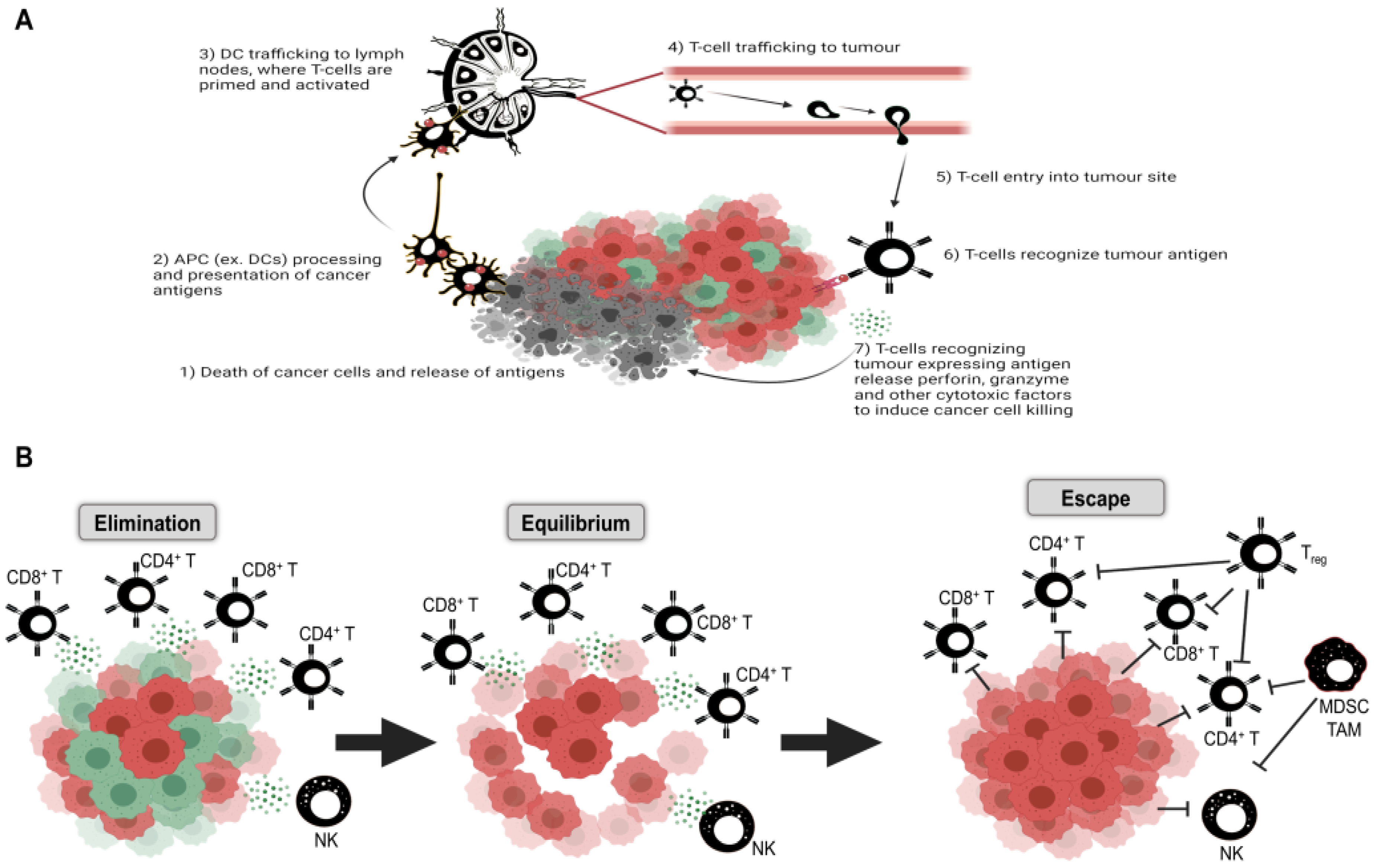
4. Autophagy in Tumor Immunity

Despite oncogenic mutations and their consequent effects on normal cellular processes generating unique, novel antigens (neoantigens) that differentiate them from normal cells, effective immune responses against cancer are rare. A tenuous equilibrium develops between the tumor and immune system wherein susceptible cancer cells expressing neoantigens are cleared, survived by those lacking neoantigen expression or possessing other mechanisms of immune evasion, such as increased immune checkpoints or loss of MHC expression. This process is referred to as immune editing [85]; the immune system becomes a vital component of cancer progression [86,87]. Disrupting this equilibrium is the goal of targeted cancer immunotherapies, and with its critical role in mediating homeostasis, it is clear that autophagic mechanisms directly participate in regulating responses to anti-tumor immune responses. Active immunotherapies look to directly stimulate long-term anti-cancer immunity (for example, cancer vaccines), whereas passive immunotherapies produce transient increases in the anti-cancer immune response, requiring multiple doses (for example, monoclonal antibody treatments) [88]. Below we will discuss these approaches taking into consideration the role of autophagy within cancer cells, the tumor microenvironment, and its immune profile.
There are three broad phases of immunoediting: (1) elimination, (2) equilibrium, and (3) escape [89]. First, cells with defective DNA repair mechanisms either die or are killed by the innate and adaptive immune system through normal immunosurveillance, of which autophagy mechanisms have been known to contribute (reviewed in the work of [90]). Tumor cells evading immune destruction establish equilibrium with immune cells, which edit tumor growth and immunogenicity. Finally, these edited tumors escape via upregulation of multiple immunosuppressive mechanisms and continue to proliferate unchecked [88,89]. Cancer stem cells (CSCs) represent a subpopulation within the tumor bulk that are thought to be responsible for tumor initiation, immune evasion, resistance to therapy, and relapse and are capable of adopting “dormant” and proliferative states [91,92]; this concept of CSC dormancy is reviewed in [93]. Recent findings have elucidated the role of autophagy in maintaining the CSC population [94]; among the survival mechanisms employed by “dormant” cells, autophagy and senescence have emerged as key resistance mechanisms to apoptotic triggers [95]. For instance, autophagy can be initiated in prostate cancer cells exposed to IL-6 and IL-2, while anti-inflammatory IL-10 blunted autophagic responses [96,97]. IL-24 signaling likewise induces autophagy in melanoma cells via PI3K-mediated BECN1 induction [98]. Intratumoral cell-cell contacts can induce autophagy in infiltrating lymphocytes [96]; co-therapies with chloroquine, an inhibitor of autophagy, enhance immunotherapies against melanoma and renal cell carcinoma [99].
As the initial, non-specific responders to viral and bacterial pathogens and cancer and a vital component of an effective adaptive immune response, the innate immune system is an important target for enhancing adaptive, T cell-based cancer immunity; the role of B cells is less well defined [87]. In addition, given the role of OVs in tumor cell death, the initial response and potential resistance mechanisms mediated by the innate immune system are of particular concern for oncolytic virotherapy. The first three steps of the cancer immunity cycle are performed primarily by DCs. Autophagy plays a key role in DC antigen presentation on MHCs, seeming to enhance MHC-II presentation [14] and, through inhibition of MHC-I externalization, reduce MHC-I presentation and subsequent CD8+ T-cell stimulation [100]. The role of autophagy in all processes of the innate immune system is reviewed in [101,102]. While experimental work has shown that plasmacytoid DCs (pDCs), activated by Toll-like receptors (TLRs), are able to secrete type 1 IFNs, prime T cells and coordinate anti-tumor immune responses [103,104,105], their infiltration into the tumor is accompanied by the adoption of an immunosuppressive phenotype; high pDC levels are associated with a worse prognosis [106]. Autophagy has been found to play a role in all aspects of DC function (reviewed in [107]). Briefly, autophagy seems to play an inhibitory role in DC maturation [108] and migration [109] while acting upstream to enhance TLR signaling [110]. Autophagy was found to be required for TLR9-induced IFN-α production by pDCs [111], and similar roles for autophagy have been found for DC production of TNF-α, IL-12, and IL-6 and subsequent T-cell responses [112]; DC ATG5 activity is required for CD4+ T-cell production of IFN-γ and IL-2 [113], and DC autophagy is required for T-cell activation [114], including regulatory T cells (Tregs) [115]. Interestingly, conflicting findings in regards to DC autophagy and Tregs have been found; Clement et al. noted in atherosclerosis models that disrupted DC autophagy results in Treg expansion [116]. Contrasting roles have also been found for cytokine production; IL-10 production requires functional DC autophagy [117], while Tregs inhibit DC autophagy, contributing to the resolution of inflammation [118]. The other major DC subtype, conventional DCs (cDCs), display more anti-tumor activity. The CD8α+ (mouse) and CD141+ (human) type 1 cDC (cDC1) subtype efficiently presents major histocompatibility complex (MHC) class 1 antigens to CD8+ T cells, initializing potent anti-tumor immune responses in “hot” tumors [119,120]. Contrary to pDCs, increased cDC1 infiltration is associated with an improved prognosis and more effective anti-tumor immune responses [121,122].
Tumor-associated macrophages (TAMs) represent another early innate immune cell with key roles in tumor development, recruited by chemokines released by tumor cells and associated stroma in a colony-stimulating factor 1 (CSF-1)-CSF-1R-dependent manner [123,124]. A crucial role for autophagy in the differentiation of monocytes into macrophages has been described [125]. TAMs play vital roles in every aspect of tumor progression [87]. Initially, their cytokine secretion profile induces low-level inflammation mediated by IL-1β and IL-6. Later, they play crucial roles in tumor cell migration and angiogenesis via growth factor and matrix metalloproteinase secretion [126,127]. Autophagy has been found to modulate phagocytosis [128,129] and reduce inflammasome activation and IL-1β expression [130,131]. TAMs can assume a spectrum of differentiation phenotypes, from the classically activated pro-inflammatory M1 to the alternatively activated, anti-inflammatory M2, each with subtypes characterized by unique transcriptional and secretory features [132]. The majority of TAMs display an M2-esque, anti-inflammatory phenotype, induced through IL-4, IL-13, and IL-10 signaling and, through the secretion of anti-inflammatory factors such as TGF-β and CCL22 (which recruits Tregs) and upregulation of immune checkpoints, inhibit anti-tumor T-cell responses [133,134] and promote many aspects of tumor biology, including migration and angiogenesis [135,136]. As a major mechanism of tumor resistance to immune attack, conversion of M2 TAMs into a pro-inflammatory M1-like phenotype (or reduction in the overall TAM population) has become a feature of many combination immunotherapies, aiming to enhance T-cell attack. This may act to reduce Treg immunosuppressive functions, and targeting this population also represents a feature of many immunotherapies. Tregs (CD4+CD25+FoxP3+) are the most potent anti-inflammatory cell in the body and act to prevent autoimmunity, which, in the context of cancer, significantly limits anti-tumor immunity. M1 macrophages are characterized by lower levels of autophagy [137], and autophagy is generally considered anti-inflammatory [101]. The mechanisms through which these cells suppress inflammation include secretion of anti-inflammatory cytokines (IL-10, IL-35, TGF-β), induction of naïve and effector T-cell apoptosis, inhibition of DC function and maturation, immune checkpoint expression, inhibition of T-cell co-stimulation, and T-cell receptor signaling through connexin channels, and metabolic disruption, and inhibition of naïve T-cell proliferation through high CD25 expression that sequesters IL-2 [138,139,140].
Another key member of the early immune response to tumors is natural killer (NK) cells. NK cells possess natural cytotoxicity receptors that respond to ligands associated with DNA damage and cellular stress, such as NKp30, NKp44, and NKp46, NKG2D, -2C, and DNAM-1 [141]. Discrimination of normal vs. tumor cells is achieved through sensors of MHC class I (MHC1), with NK cells preferentially killing MHC1- cells via the release of perforin and granzymes. Cells lacking MHC1 are sensed primarily through killer-cell immunoglobulin-like receptors (KIRs) and NKG2D [141,142]. Autophagy is required for NK cell development, differentiation, survival, cytolytic functions, and memory responses [143,144]. Communication between NK cells and DCs increases the frequency of cDC1s while further promoting tumor cell death, antigen capture, presentation, and T-cell activation; high NK cell infiltration is associated with improved prognosis and immunotherapy responses [145,146]. Autophagy blockade in melanoma models resulted in increased expression of CCL5, increasing intratumoral NK cell infiltration [147]. As with both TAMs and DCs, however, factors within the TME restrict NK cell activity and can lead to NK cell exhaustion, such as that seen in T cells (reviewed in [148,149]). While certain tumors limit NK cell entry through restrictions to effective homing and entry, within the TME immunosuppressive stroma, Tregs and TAMs (including myeloid-derived suppressor cells, MDSCs) limit their cytotoxic capacity. High levels of immune checkpoints [150,151] and upregulation of enzymes such as indoleamine 2,3-Dioxygenase (IDO1) and inducible nitric oxide synthase (iNOS) within a tumor restrict the available pool of amino acids required for NK cell (and T-cell) effector functions [141,152,153]. Hypoxia, a key feature of many tumors, is sensed through hypoxia-inducible factor-1 and -2 (HIF-1 and -2). Increased HIF signaling limits DC antigen presentation and effector T-cell migration, proliferation, and cytotoxic capacity while in turn enhancing TAM retention, Treg development, and the immunosuppressive actions of both [154,155]. Natural killer (NK) cell cytotoxic effects are reduced, while MDSC numbers and actions are enhanced. Hypoxia is able to trigger tumor cell autophagy through HIF1-BNIP3-BECN1 signaling, clearing damaged mitochondria and reducing ROS production [156], contributing to enhanced survival. Tumor cells secrete increased levels of anti-inflammatory cytokines and exhibit reduced major histocompatibility complex and increased immune checkpoint expression [157], contributing to the reduced NK cell effector function and efficacy of checkpoint blockade therapies. Therapies looking to enhance the anti-tumor effect of NK cells are currently receiving significant attention, looking to remove inhibitory signals and enhance NK cell activation (reviewed in [141]) as a potential alternative, or in combination with T cell-based therapies, depending on the tumor. Given the seeming ubiquitous role of autophagy in NK cell processes, the potential of broad autophagy blockade in vivo would produce similar results is unlikely, suggesting the necessity for tumor cell-targeted therapies.
While the diversity of immunosuppressive mechanisms within a tumor may limit the efficacy of monotherapies, targeted combinations against multiple inflammatory and cell death pathways, including the autophagic machinery, as well as against both arms of the immune system, are likely to represent the most effective way forward in cancer immunotherapies. Among these pathways, as mentioned previously, autophagy can represent a double-edged sword, regulating a plethora of inflammatory functions and immune phenotypes while also promoting tumor cell survival [158,159,160]. The exact role of autophagy in each context, especially in a site as metabolically complex and genetically unstable as a TME, will be crucial to elucidate prior to its effective use in targeted immunotherapies.

5. Targeting Autophagy for Immunotherapy

The cancer immunity cycle, outlined by Chen and Mellman [161], describes seven key steps in the anti-tumor immune response, which in turn can be therapeutically targeted (Figure 2). Briefly, these steps are: (1) neoantigen capture and processing by antigen-presenting cells (APCs), most notably dendritic cells (DCs); (2) trafficking of DCs to lymph nodes for antigen presentation to lymphocytes; (3) lymphocyte priming and activation upon antigen presentation; (4) trafficking of activated T cells through the blood to the tumor location; and (5) out of circulation into the tumor itself, wherein; (6) T cells recognize cancer cells expressing neoantigens and; (7) effect tumor cell killing, with consequent antigen capture and processing by APCs, starting the cycle anew [161]. Each of these steps can be modulated by the autophagic response.
Given the importance of the immune system and the autophagic response in cancer progression, repurposing the established inflammatory milieu to permit enhanced T-cell attack remains the major goal of immunotherapy. Two broad categories of tumors have been defined based on relative immune infiltration. Type 1, T cell-inflamed (“hot”) tumors, with high tumor mutation burden, spontaneous T-cell activation, and infiltration, along with general expression of chemokines, activated APC markers, and type 1 IFN responses. These escape immune elimination via increases in immune checkpoint expression, reductions in neoantigen expression, resistance to cytotoxic molecules, and a metabolic microenvironment that hinders effective immune responses [89,126]. The second type 2 category is the non-T cell-inflamed (“cold”) tumors, lacking any such markers, with low mutation burden and characterized by little to no T-cell infiltration upon immunohistochemical analysis [89,126]. Other types of tumor microenvironments (TME) have also been described, defined by intermediate levels of inflammation and tumor mutation burden [89]. These subtypes point to unique immune defects between tumor types, wherein “hot” tumors fail at the effector phase and “cold” tumors display immune exclusion [126], generally corresponding to late and early stages of the cancer immunity cycle, respectively. Autophagy has key roles in both stages (reviewed in [162]). Relating to the initial steps, the release of DAMPs, recognized by innate immune cells that consequently initiate immune responses, occurs following cancer cell death. The pro-survival effects of autophagy would reduce, while autophagic cell death would increase, DAMP release [163]; thus, autophagic regulation of cancer cell survival is key to mediating early stages. DAMPs, in turn, regulate autophagic signaling [162]. Inhibition of autophagy via ATG5 deletion has been found to increase intratumoral anti-inflammatory Tregs [164], contributing to the suppression of cytotoxic T-cell responses and inhibiting effector functions, corresponding to control of late stages of the cycle. Interestingly, inhibition of autophagy improves the efficacy of anti-PD-1 and anti-CTLA-4 checkpoint therapy in normally resistant pancreatic ductal adenocarcinoma models [165], as well as anti-PD-1 treatments of murine prostate cancer [166]. Conversely, activation of autophagy can lead to degradation of PD-L1 and CTLA-4, allowing for enhanced immune-mediated tumor destruction [167]. Therefore, determining the most effective means of regulating autophagy is likely to provide significant benefit to immunotherapies targeting any stage of the cancer immunity cycle.
Aside from direct effects on tumor cells, autophagic modulators also affect systemic immunity, likely a result of the multifunctionality of many pathways crucial to autophagy. As with the double-edged role in cancer treatment, autophagic modulators have multifaceted effects on the immune system; in general, however, they are immunosuppressive. Rapamycin, an mTOR inhibitor, has been found to enhance γδ T-cell cytotoxicity and anti-cancer effects [168]. In models of influenza infection, rapamycin has been found to reduce B-cell class switching, proliferation, formation of germinal centers, and antibody specificities. Further, populations of memory CD8+ and CD4+ T cells, including Tregs, are increased by rapamycin treatment [169]. Chronic, long-term mTOR inhibition using rapamycin has been found to extend mouse lifespan; total T-cell populations were reduced in the spleen, and a preference for naïve phenotypes was observed [170]. In models of bone marrow failure, similar reductions in effector and memory T-cell populations and a preference for naïve phenotypes upon rapamycin treatment have been observed, with consequent improvements in mouse outcomes [171]. Following chronic rapamycin treatment, T-cell trafficking markers (and trafficking capacity) were increased, while checkpoints and exhaustion were decreased. Conversely, Treg populations were increased, as were myeloid cell populations, with alterations to DC-T-cell interactions [170]. mTORC1 inhibition increased myeloid cell stimulation of TH1 or TH17 inflammatory responses, including increased IFN-γ and IL-17 production [172]. Rapamycin expands natural and induced Treg populations and enhances DC migration while inhibiting their maturation while also reducing differentiation of MDSCs [173].
Chloroquine, another modulator of autophagy, is often used to treat autoimmune diseases given well-characterized immunosuppressive activity (reviewed in [174]). Briefly, hydroxychloroquine and chloroquine dampen TLR signaling and interfere with antigen processing, MHCII expression, and presentation with a consequent reduction in T-cell activation, cytokine production, and effector functions [174,175]. In mouse models of malaria, chloroquine significantly suppresses macrophage, B-cell, and helper T-cell activation while dampening DC maturation [176]. Inhibition of many aspects of innate immunity leads to reduced type I IFN and other pro-inflammatory cytokine responses, including TNF-α, IL-6, and IL-1β, which result in dampened adaptive immune responses [177]. Similar immunosuppressive effects have been noted for vorinostat, a histone deacetylase inhibitor. Reduced NK cell cytotoxic effects [178], enhanced Treg immunosuppression [179,180], and increased DC expression of IDO1 [181]. Interestingly, levels of anti-inflammatory IL-10 are reduced following treatment with vorinostat [182].
In summary, while autophagy blockade may induce immunogenic cell death, and its anti-cancer effects are at least partially dependent on a functional immune system, much of the immunomodulatory effects of current pharmacological agents are immunosuppressive, illustrating a need for improved understanding of autophagic processes and more targeted therapies.

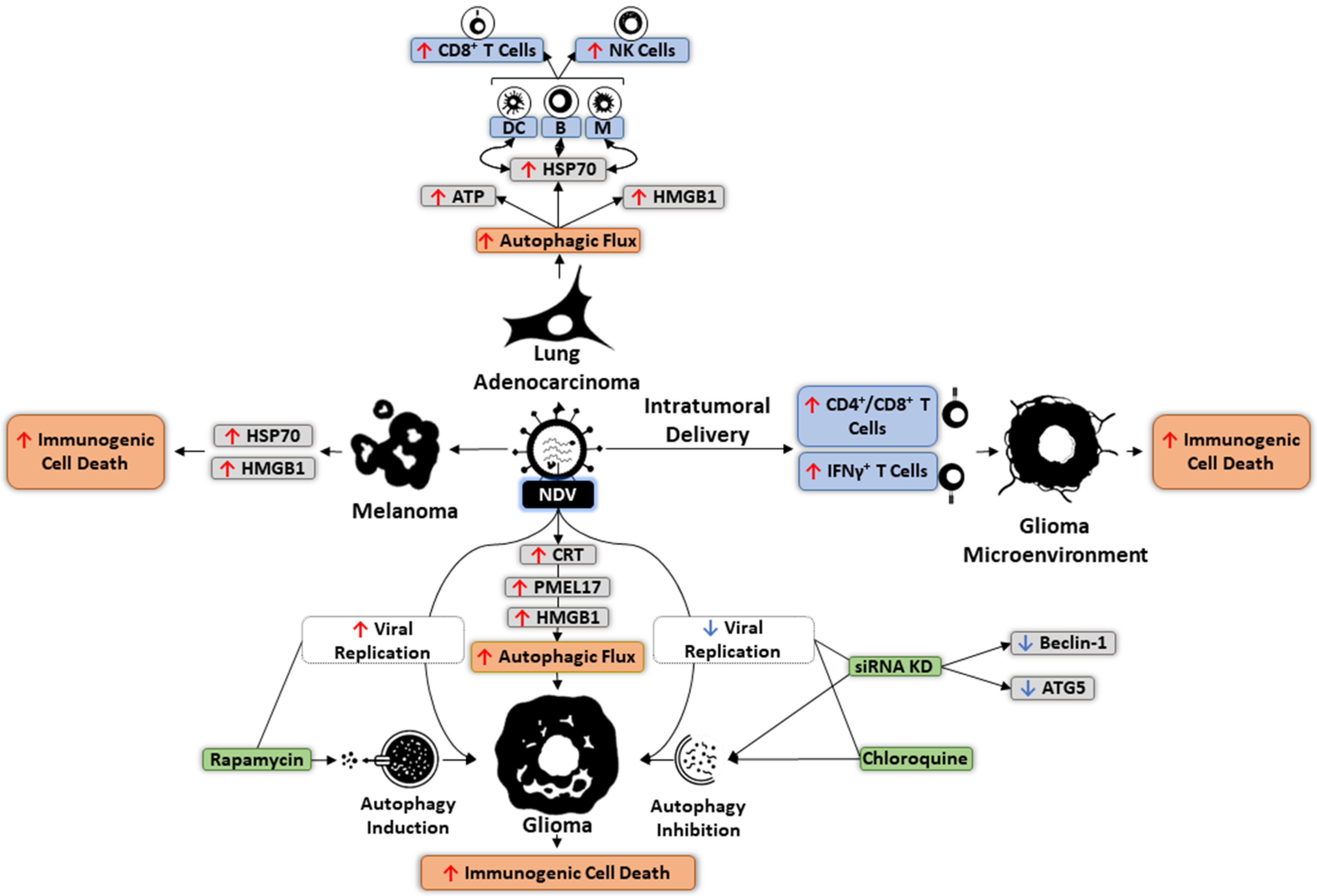
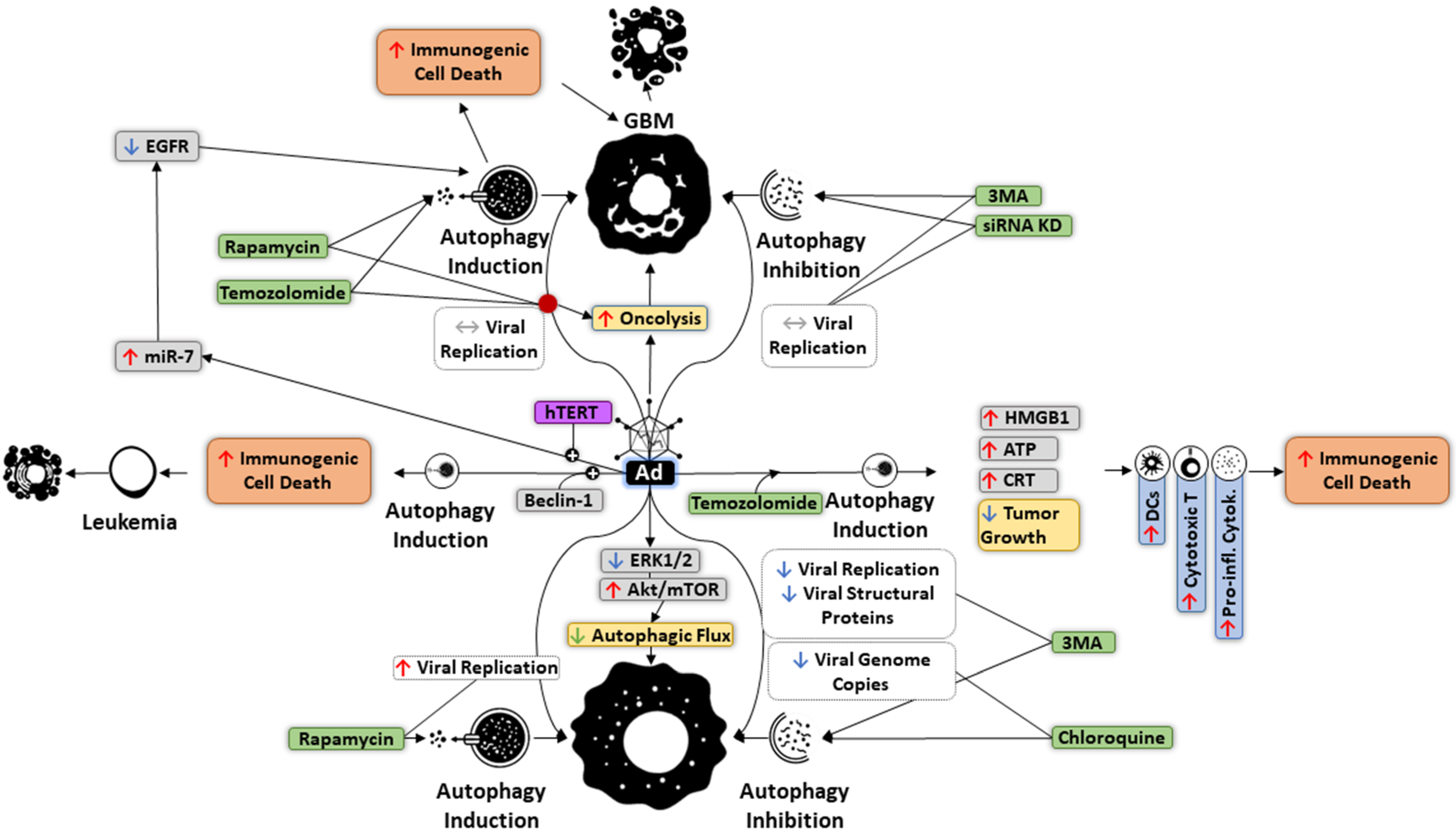
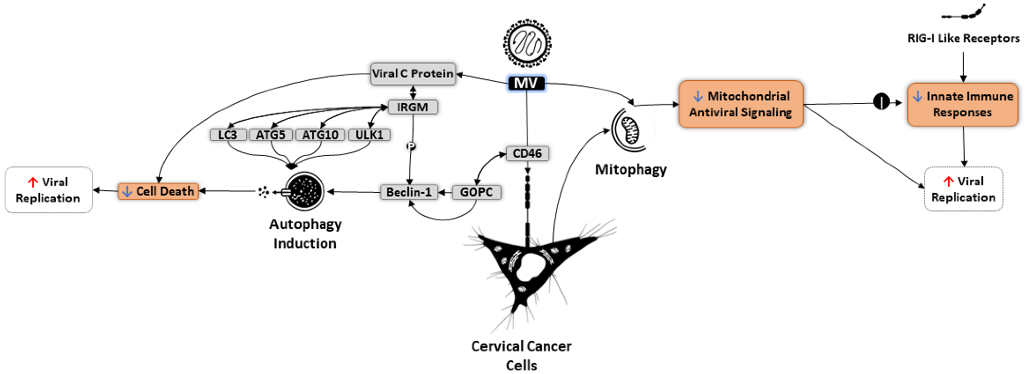
6. Autophagy and OVs

OVs are defined as modified or natural viruses that preferentially replicate within cancer cells to induce cell lysis. OVs trigger immunogenic cancer cell death with the consequent release of TAAs and DAMPs. Ultimately, these stimulate APCs and T cell-based adaptive immune responses toward tumor antigens [1,2]. Given this, OVs represent a promising avenue of cancer immunotherapeutic, with herpes simplex virus (HSV), reovirus (REO), measles virus (MV), retroviruses, myxoma virus (MYXV), coxsackievirus, parvoviruses, adenovirus (Ad), vesicular stomatitis virus (VSV), vaccinia virus, polioviruses, Newcastle disease virus (NDV), and Seneca Valley virus (SVV) translated into clinical trials, alone or in combination therapies [183]. Autophagy plays a dual role in oncolytic virotherapy. Although the effects of viruses on normal autophagy appear to be both virus- and cell-specific, replication of OVs can be promoted by autophagy, enhancing tumor cell death. Paradoxically, autophagy can also metabolically nourish cancer cells, protecting them from OV-mediated oncolysis and cytotoxicity. Irrespective of autophagy modulation toward oncolysis, OVs themselves can also induce different types of cell death in cancer cells through various mechanisms. For example, Ad infection was reported to mediate necroptosis via upregulating RIP3 and promoting the phosphorylation of mixed-lineage kinase domain-like (MLKL) in lung cancer cells [184]. However, in malignant pleural mesothelioma cells, Ad triggers immunogenic cell death by overexpressing HMGB1 and CRT [185]. These cell-specific differences in response to cell death have also been discovered to occur following infection with other OVs, such as vaccinia, HSV, and NDV. Several studies have demonstrated that certain OVs are capable of altering autophagic signaling to enhance their replicative and lytic capacity while simultaneously suppressing antiviral innate immune responses. Below we will describe reports of specific OVs and their interaction with the autophagy response in modulating therapeutic efficacy.
Ad uses autophagy to improve its spread among cancer cells (Figure 3). Rapamycin, an autophagy promoter, and immunosuppressant, has been shown to increase replication of chimeric Ad5/F35, while autophagy inhibition using 3-methyladenine (3MA) restricts Ad replication, as well as production of viral structural proteins [7,186]. Conversely, treatment of glioma cells with the oncolytic Ad d1922–947 was characterized by Akt/mTOR activation, Erk1/2 inhibition, and reductions in autophagic flux. Furthermore, chloroquine-mediated inhibition of autophagy in these cells has been found to reduce Ad genome copies [187]. Autophagy induction by Ad may enhance cell lysis without concurrent increases in replication. An example of this phenomenon has been observed in glioblastoma cells after infection with the OBP-405 Ad virus, where neither autophagy activation by temozolomide and rapamycin nor targeted siRNA and 3-MA-mediated inhibition altered viral replication. The level of oncolysis was counter-intuitively increased through autophagic activity [9]. OBP-301, another Ad, is capable of causing autophagic cell death through miR-7-induced EGFR inhibition [188]. Similarly, in leukemia cells, infection with BECN1-equipped chimeric oncolytic Ad was noted to cause robust autophagic cell death in vitro and in vivo [189]. In addition to autophagic cell death, it has been shown that combining oncolytic Ad with temozolomide not only decreases tumor growth but evokes immunogenic cell death via upregulation of high mobility group box protein 1 (HMGB1), CRT, and ATP, which collectively stimulate various anti-tumor immune responses [190]. These include phagocytosis by DCs, elevated cytotoxic T lymphocytes, and increased expression of pro-inflammatory cytokines [191]. As discussed earlier, due to the paradoxical mechanism of action in respect to various tumors, autophagy could restrict the oncolytic capacity via protecting and nourishing cancer cells. For instance, in ovarian cancer cells, Ad-triggered autophagy was reported to inhibit the oncolytic activity of this OV, while autophagy suppression by 3-MA and chloroquine appears to increase the cytotoxicity mediated by Ad [192]. Similar results were obtained in U373MG and U87MG glioma cell lines following inhibition of autophagy and infection with Ad [187].
Attenuated MV has been found to induce autophagy to enhance viral replication (Figure 4). In HeLa cells, MV binds CD46, which contains a cytoplasmic domain linked to the BECN1 complex through the scaffold protein GOPC. During autophagy induction, the C protein of MV plays a fundamental role in increasing viral replication and production of viral progeny particles by delaying cell death [5]. One mechanism through which MV-C protein triggers autophagy is via interacting with immunity-related GTPase family M (IRGM), a protein that influences autophagy master regulators such as LC3, ULK1, ATG5, and ATG10, in addition to phosphorylating BECN1 [193,194,195]. Oncolytic MV can also use mitophagy to downregulate mitochondrial antiviral signaling (MAVS), consequently suppressing innate immune responses activated by RIG-I-like receptors and elevating MV replication in tumors [8].
Replication of oncolytic NDV can similarly be boosted by autophagy (Figure 5). In NDV-treated glioma cells, chloroquine treatment or siRNA-mediated downregulation of BECN1 and ATG5 significantly reduce viral replication, whereas rapamycin-induced activation of autophagy negatively affects NDV dissemination [196]. Unlike Ad, NDV leads to glioma cell autophagy activation, subsequently mediating immunogenic cell death through increased production of CRT, PMEL17, and HMGB1 [183]. Moreover, in mouse glioma models, intratumoral NDV administration enhanced infiltration of IFNγ+ T cells into the TME, suggesting increased immunogenic cell death [197]. In lung cancer cells, autophagy induction by NDV has been demonstrated to increase levels of HSP70, which interacts with its receptor on APCs to stimulate CD8+ T cell responses and activate NK cells [10] (Figure 5). NDV-infected melanoma cells were also found to express higher levels of immunogenic cell death regulators such as HSP70 and HMGB1. Inhibiting autophagy in these cells can result in reduced expression of these markers, highlighting the role of NDV-mediated autophagy in enhancing immunogenic cell death of tumor cells [198].
VSV also takes advantage of autophagy for intratumoral expansion (Figure 6). NRF2-induced autophagy, mediated through overexpression of HO-1, has been found to increase the replication of oncolytic VSV while suppressing VSV-triggered immune responses, achieved by dysregulating the pathway controlling RIG-I-MAVS interactions [199]. A combination of VSV with different autophagy regulators was also revealed to boost VSV replication and lytic capacity. For instance, in pancreatic cancer cells, co-treatment of oncolytic VSV and vorinostat upregulated the expression of some ATGs via hyperacetylation of NF-κB. In contrast, blocking autophagy using 3-MA increases the activity of IFN stimulated genes, resulting in decreased oncolytic capacity and restricted VSV replication [200]. Further, BECN1 knockdown appears to results in HeLa cell sensitivity to VSV-induced oncolysis, illustrating the role of autophagy in altering cancer cell resistance to oncolytic virotherapy [201].
Among other OVs mediating or exploiting autophagy, HSV-1 enhances cytoplasmic aggregation of LC3 and autophagosome formation, causing autophagic cell death in squamous cell carcinoma [6]. HSV-1 replication does not seem to be influenced by autophagy inhibition, but its lytic capacity and cytotoxicity are reduced when autophagy is inhibited [183]. Conversely, disrupted or abnormal autophagy has been found to increase both the replication and anti-tumor activity of oncolytic SVV in orthotopic xenograft models [202]. Autophagic cell death can be seen in leukemia and myeloma after infection with engineered oncolytic vaccinia expressing BECN1, which elevates viral replication [203]. Oncolytic reovirus and some of its derived formulations have also been shown to use autophagy. Pelareorep is one example that was recently demonstrated to increase the expression of autophagy-related proteins, such as LC3 and ATG5, in colorectal cancer cells, a phenomenon that boosts not only the propagation of reovirus but also cell death [204]. Avian reovirus p17 non-structural protein upregulates AMPK and PTEN while suppressing mTORC1, eventually activating autophagy [205]. However, the exact mechanism through which mammalian reovirus induces autophagy remains largely undiscovered. Taken together, the results of different studies suggest virus-specific and cancer-specific alterations in autophagy pathway and cell death mechanism after infection of various cancer cells or tumors with OVs.

7. Targeting Autophagy in Oncolytic Immunotherapy

Aside from the aforementioned pharmacological modulations, autophagy is regulated by numerous upstream molecules that represent potential targets for altering autophagic signaling in order to enhance oncolytic immunotherapy. PERK and IRE1α, which are involved in ER stress responses, are promising targets [206]. Li et al. have demonstrated that STF083010, a nuclease inhibitor of IRE1α, promotes accumulation of oncolytic M1 alphavirus in glioma cells in vitro and in vivo via suppression of autophagic degradation [207]. Interestingly, the M1 virus itself is able to induce autophagy in glioma cells, a phenomenon that subverts effective oncolysis. Reduced IRE1α had no influence on M1-triggered STAT1 phosphorylation, with no effect on type I IFN signaling and innate antiviral response following infection [207,208]. IRE1α inhibition also had no effect on M1 viral replication. In addition to glioma, siRNA-mediated knockdown of IRE1Α has been found to increase cell death of leukemia cells through PERK-dependent autophagy [209]. These results suggest that targeting IRE1A may sensitize cancer cells to OVs through either autophagy-dependent or -independent mechanisms, depending on tumor stage and type. The detailed effects of IRE1A manipulation on autophagy require further investigation.
UPR is another candidate target in modulating autophagy to enhance virotherapy. UPR activation occurs after severe ER stress (e.g., following a persistent infection) that cannot be neutralized by tumor cells, inducing cell death; given this, UPR has the capacity to kill OV-infected cancer cells. This UPR-triggered cell death occurs upon failure of UPR to restore cellular homeostasis; inefficient UPR function can be compensated for by activating autophagic degradation to remove the ER and other damaged organelles [210]. The reciprocal communication between autophagy and UPR might play a fundamental role in directing cell fate decisions in tumors after exposure to OVs. For instance, UPR inhibition via IRE1α silencing can increase maraba virus-induced cytotoxicity in glioblastoma cells [211]. In contrast, oncolysis of melanoma and lung adenocarcinoma cells following oncolytic adenovirus infection was enhanced following downregulation of Golgi-specific brefeldin A-resistance guanine nucleotide exchange factor 1 (GBF-1), which activates UPR [212]. Although not investigated, UPR suppression is thought to inhibit autophagy, while its activation is inducing autophagy. Some OVs can naturally induce UPR and consequently autophagy; VSV was discovered to increase UPR expression markers such as ATF4, BIP, and eIF2α, in fibrosarcoma cells [213], as has M1 virus similar to maraba virus, siRNA-mediated suppression of UPR regulators promotes M1 virus-induced oncolysis in U87 glioma cells, a condition accompanied by inhibition of autophagy [207]. Different OVs and tumor cells need to be tested to fully elucidate mechanisms of oncolysis and autophagy upon UPR-targeted therapies.
In summary, whether targeting the ER stress or UPR pathways produce the ideal effect on autophagy for enhancing OV therapies will depend on both the type of tumor and OV used. Preferably, such alterations would not only lead to increased replication and viral protein synthesis but also enhanced immunogenic cell death and oncolysis. Considering OV-specific differences in using ER and UPR, more screenings should be implemented to characterize the vulnerability of various OV-infected cancer cells in response to dual aspects of autophagy regulation by these pathways. Additionally, several other master regulators and interacting molecules governing ER stress and UPR might have the capacity to modulate autophagy through other mechanisms, which could reveal new intracellular targets for improving oncolysis and anti-tumor immune responses.

8. Concluding Remarks and Future Perspectives

The efficacy of targeting autophagy in oncolytic virotherapy relies on several factors, including OV variants, cancer cell types, and effective induction of anti-tumor immune responses. In cell-specific and OV-specific manners, both activation and inhibition of autophagy appear to boost oncolysis via the mediation of immunogenic cell death. Such dual functions make it difficult to target autophagy unilaterally for enhancing OV propagation in a variety of tumors. Consistently, autophagy also appears to have pro- and anti-inflammatory effects within the TME, depending on context and immune cell type. Accordingly, a precise understanding of the molecular interaction between autophagy, the immune system, and OV biology could open new avenues for optimizing oncolytic viral and other immunotherapy. Nevertheless, many obstacles remain, including off-target effects of pharmacological agents within the TME and the instability of autophagic processes at various stages of tumor development (e.g., acting as both tumor suppressor and tumor survival factor), altering the optimal timing for targeting autophagy and affecting the efficacy of oncolytic virotherapy. Various recombinant systems and inducible alleles allow for in vitro and in vivo modeling of OV-infected cancer cell responses to agents targeting autophagy during different stages of tumor development and progression. Recent advances in metabolomics allow for accurate determination of the role of autophagy in tumor initiation, progression, and relationship with the immune system. Overall, research will determine the safety and efficacy of using autophagy modulators in future oncolytic immunotherapy.

Author Contributions

Conceptualization, A.Z.-A., K.M., S.T.B., T.A., and B.Y.; writing, A.Z.-A., K.M., S.T.B., T.A., and B.Y. All authors have read and agreed to the published version of the manuscript.

Funding

This work was funded by the Terry Fox Foundation, The Natural Sciences and Engineering Research Council of Canada (NSERC), The Canadian Institutes of Health Research (CIHR) and Cancer Research Society.

Conflicts of Interest

The authors declare no conflict of interest.

Abbreviations

AdAdenovirus
AMPK AMP-activated protein kinase
APCsAntigen-presenting cells
CRTCalreticulin
cDCsConventional DCs
CSCs Cancer stem cells
CSF-1Colony-stimulating factor 1
DAMPsDamage-associated molecular patterns
DCsDendritic cells
EATEarly autophagy targeting
EREndoplasmic reticulum
GBMGlioblastoma multiform
HIFHypoxia-inducible factor
HMGB1High mobility group box protein 1
HSVHerpes simplex virus
IFNInterferon
iNOSInducible nitric oxide synthase
IRE1α Inositol-requiring enzyme 1α
IRGMImmunity-related GTPase family M
KIRsKiller-cell immunoglobulin-like receptors
lncRNAsLong noncoding RNAs
MAVSMitochondrial antiviral signaling
MDSCs Myeloid-derived suppressor cells
miRNAsMicroRNAs
mTORMamalian target of rapamycin
MVMeasles virus
MYXVMyxoma virus
NDVNewcastle disease virus
NKNatural killer
OVsOncolytic viruses
PAMPsPathogen-associated molecular patterns
pDCsPlasmacytoid DCs
PEPhosphatidylethanolamine
PERK Pancreatic endoplasmic reticulum kinase
PAS Phagophore assembly site
ROSReactive oxygen species
SVVSeneca Valley virus
TAAsTumor-associated antigens
TAMsTumor-associated macrophages
TLRsToll-like receptors
TMETumor microenvironment
Treg Regulatory T cell
UPRUnfolded protein response
UPSUbiquitin-proteasome system
VSVVesicular stomatitis virus

References

  1. Aurelian, L. Oncolytic viruses as immunotherapy: Progress and remaining challenges. OncoTargets Ther. 2016, 9, 2627–2637. [Google Scholar] [CrossRef] [PubMed] [Green Version]
  2. Hu, L.; Jiang, K.; Ding, C.; Meng, S. Targeting Autophagy for Oncolytic Immunotherapy. Biomedicines 2017, 5, 5. [Google Scholar] [CrossRef] [PubMed]
  3. Lin, M.G.; Hurley, J.H. Structure and function of the ULK1 complex in autophagy. Curr. Opin. Cell Biol. 2016, 39, 61–68. [Google Scholar] [CrossRef] [PubMed] [Green Version]
  4. Mizushima, N. A brief history of autophagy from cell biology to physiology and disease. Nature 2018, 20, 521–527. [Google Scholar] [CrossRef] [PubMed]
  5. Richetta, C.; Grégoire, I.P.; Verlhac, P.; Azocar, O.; Baguet, J.; Flacher, M.; Tangy, F.; Rabourdin-Combe, C.; Faure, M. Sustained Autophagy Contributes to Measles Virus Infectivity. PLOS Pathog. 2013, 9, e1003599. [Google Scholar] [CrossRef]
  6. Furukawa, Y.; Takasu, A.; Yura, Y. Role of autophagy in oncolytic herpes simplex virus type 1-induced cell death in squamous cell carcinoma cells. Cancer Gene Ther. 2017, 24, 393–400. [Google Scholar] [CrossRef]
  7. Rodriguez-Rocha, H.; Gomez-Gutierrez, J.G.; Garcia-Garcia, A.; Rao, X.-M.; Chen, L.; McMasters, K.M.; Zhou, H.S. Adenoviruses induce autophagy to promote virus replication and oncolysis. Virology 2011, 416, 9–15. [Google Scholar] [CrossRef] [PubMed] [Green Version]
  8. Xia, M.; Gonzalez, P.; Li, C.; Meng, G.; Jiang, A.; Wang, H.; Gao, Q.; Debatin, K.-M.; Beltinger, C.; Wei, J. Mitophagy Enhances Oncolytic Measles Virus Replication by Mitigating DDX58/RIG-I-Like Receptor Signaling. J. Virol. 2014, 88, 5152–5164. [Google Scholar] [CrossRef] [PubMed] [Green Version]
  9. Yokoyama, T.; Iwado, E.; Kondo, Y.; Aoki, H.; Hayashi, Y.; Georgescu, M.M.; Sawaya, R.; Hess, K.R.; Mills, G.B.; Kawamura, H.; et al. Autophagy-inducing agents augment the antitumor effect of telerase-selve oncolytic adenovirus OBP-405 on glioblastoma cells. Gene Ther. 2008, 15, 1233–1239. [Google Scholar] [CrossRef] [Green Version]
  10. Ye, T.; Jiang, K.; Wei, L.; Barr, M.; Xu, Q.; Zhang, G.; Ding, C.; Meng, S.; Piao, H. Oncolytic Newcastle disease virus induces autophagy-dependent immunogenic cell death in lung cancer cells. Am. J. Cancer Res. 2018, 8. [Google Scholar]
  11. Jin, K.-T.; Lu, Z.-B.; Lv, J.-Q.; Zhang, J.-G. The role of long non-coding RNAs in mediating chemoresistance by modulating autophagy in cancer. RNA Biol. 2020, 17, 1727–1740. [Google Scholar] [CrossRef]
  12. Guo, J.Y.; Xia, B.; White, E. Autophagy-Mediated Tumor Promotion. Cell 2013, 155, 1216–1219. [Google Scholar] [CrossRef] [PubMed] [Green Version]
  13. Kuballa, P.; Nolte, W.M.; Castoreno, A.B.; Xavier, R.J. Autophagy and the Immune System. Annu. Rev. Immunol. 2012, 30, 611–646. [Google Scholar] [CrossRef] [PubMed]
  14. Dengjel, J.; Schoor, O.; Fischer, R.; Reich, M.; Kraus, M.; Muller, M.; Kreymborg, K.; Altenberend, F.; Brandenburg, J.; Kalbacher, H.; et al. Autophagy promotes MHC class II presentation of peptides from intracellular source proteins. Proc. Natl. Acad. Sci. USA 2005, 102, 7922–7927. [Google Scholar] [CrossRef] [PubMed] [Green Version]
  15. Gauvrit, A.; Brandler, S.; Sapede-Peroz, C.; Boisgerault, N.; Tangy, F.; Gregoire, M. Measles Virus Induces Oncolysis of Mesothelioma Cells and Allows Dendritic Cells to Cross-Prime Tumor-Specific CD8 Response. Cancer Res. 2008, 68, 4882–4892. [Google Scholar] [CrossRef] [PubMed] [Green Version]
  16. Mercer, C.; Kaliappan, A.; Dennis, P.B. A novel, human Atg13 binding protein, Atg101, interacts with ULK1 and is essential for macroautophagy. Autophagy 2009, 5, 649–662. [Google Scholar] [CrossRef] [Green Version]
  17. Hara, T.; Takamura, A.; Kishi, C.; Iemura, S.-I.; Natsume, T.; Guan, J.-L.; Mizushima, N. FIP200, a ULK-interacting protein, is required for autophagosome formation in mammalian cells. J. Cell Biol. 2008, 181, 497–510. [Google Scholar] [CrossRef] [Green Version]
  18. Lazarus, M.B.; Novotny, C.J.; Shokat, K.M. Structure of the Human Autophagy Initiating Kinase ULK1 in Complex with Potent Inhibitors. ACS Chem. Biol. 2014, 10, 257–261. [Google Scholar] [CrossRef] [Green Version]
  19. Fujioka, Y.; Suzuki, S.W.; Yamamoto, H.; Kondo-Kakuta, C.; Kimura, Y.; Hirano, H.; Akada, R.; Inagaki, F.; Ohsumi, Y.; Noda, N. Structural basis of starvation-induced assembly of the autophagy initiation complex. Nat. Struct. Mol. Biol. 2014, 21, 513–521. [Google Scholar] [CrossRef]
  20. Weidberg, H.; Shvets, E.; Shpilka, T.; Shimron, F.; Shinder, V.; Elazar, Z. LC3 and GATE-16/GABARAP subfamilies are both essential yet act differently in autophagosome biogenesis. EMBO J. 2010, 29, 1792–1802. [Google Scholar] [CrossRef]
  21. Kabeya, Y.; Mizushima, N.; Ueno, T.; Yamamoto, A.; Kirisako, T.; Noda, T.; Kominami, E.; Ohsumi, Y.; Yoshimori, T. LC3, a mammalian homologue of yeast Apg8p, is localized in autophagosome membranes after processing. EMBO J. 2000, 19, 5720–5728. [Google Scholar] [CrossRef] [PubMed]
  22. Webster, C.P.; Smith, E.F.; Bauer, C.S.; Moller, A.; Hautbergue, G.M.; Ferraiuolo, L.; Myszczynska, M.; Higginbottom, A.; Walsh, M.J.; Whitworth, A.J.; et al. The C9orf72 protein interacts with Rab1a and the ULK 1 complex to regulate initiation of autophagy. EMBO J. 2016, 35, 1656–1676. [Google Scholar] [CrossRef] [PubMed]
  23. Ma, M.; Liu, J.-J.; Li, Y.; Huang, Y.; Ta, N.; Chen, Y.; Fu, H.; Ye, M.-D.; Ding, Y.; Huang, W.; et al. Cryo-EM structure and biochemical analysis reveal the basis of the functional difference between human PI3KC3-C1 and -C2. Cell Res. 2017, 27, 989–1001. [Google Scholar] [CrossRef] [PubMed] [Green Version]
  24. Young, L.N.; Cho, K.; Lawrence, R.; Zoncu, R.; Hurley, J.H. Dynamics and architecture of the NRBF2-containing phosphatidylinositol 3-kinase complex I of autophagy. Proc. Natl. Acad. Sci. USA 2016, 113, 8224–8229. [Google Scholar] [CrossRef] [Green Version]
  25. Ohashi, Y.; Soler, N.; García Ortegón, M.; Zhang, L.; Kirsten, M.L.; Perisic, O.; Masson, G.R.; Burke, J.E.; Jakobi, A.J.; Apostolakis, A.A.; et al. Characterization of Atg38 and NRBF2, a fifth subunit of the autophagic Vps34/PIK3C3 complex. Autophagy 2016, 12, 2129–2144. [Google Scholar] [CrossRef]
  26. Di Bartolomeo, S.; Corazzari, M.; Nazio, F.; Oliverio, S.; Lisi, G.; Antonioli, M.; Pagliarini, V.; Matteoni, S.; Fuoco, C.; Giunta, L.; et al. The dynamic interaction of AMBRA1 with the dynein motor complex regulates mammalian autophagy. J. Cell Biol. 2010, 191, 155–168. [Google Scholar] [CrossRef] [Green Version]
  27. Mizushima, N.; Noda, T.; Yoshimori, T.; Tanaka, Y.; Ishii, T.; George, M.D.; Klionsky, D.J.; Ohsumi, M.; Ohsumi, Y. A protein conjugation system essential for autophagy. Nature 1998, 395, 395–398. [Google Scholar] [CrossRef]
  28. Mizushima, N.; Noda, T.; Ohsumi, Y. Apg16p is required for the function of the Apg12p–Apg5p conjugate in the yeast autophagy pathway. EMBO J. 1999, 18, 3888–3896. [Google Scholar] [CrossRef] [Green Version]
  29. Ichimura, Y.; Kirisako, T.; Takao, T.; Satomi, Y.; Shimonishi, Y.; Ishihara, N.; Mizushima, N.; Tanida, I.; Kominami, E.; Ohsumi, M.; et al. A ubiquitin-like system mediates protein lipidation. Nature 2000, 408, 488–492. [Google Scholar] [CrossRef]
  30. Shen, K.; Choe, A.; Sabatini, D.M. Intersubunit crosstalk in the Rag GTPase heterodimer enables mTORC1 to respond rapidly to amino acid availability. Mol. Cell 2017, 68, 552–565. [Google Scholar] [CrossRef]
  31. Vanhaesebroeck, B.; Stephens, L.; Hawkins, P. PI3K signalling: The path to discovery and understanding. Nat. Rev. Mol. Cell Biol. 2012, 13, 195–203. [Google Scholar] [CrossRef] [PubMed]
  32. Sciarretta, S.; Forte, M.; Frati, G.; Sadoshima, J. New Insights Into the Role of mTOR Signaling in the Cardiovascular System. Circ. Res. 2018, 122, 489–505. [Google Scholar] [CrossRef]
  33. Chen, S.; Han, Q.; Wang, X.; Yang, M.; Zhang, Z.; Li, P.; Chen, A.; Hu, C.; Li, S. IBP-mediated suppression of autophagy promotes growth and metastasis of breast cancer cells via activating mTORC2/Akt/FOXO3a signaling pathway. Cell Death Dis. 2013, 4, e842. [Google Scholar] [CrossRef]
  34. Kim, J.; Kundu, M.; Viollet, B.; Guan, K.-L. AMPK and mTOR regulate autophagy through direct phosphorylation of Ulk1. Nature 2011, 13, 132–141. [Google Scholar] [CrossRef] [Green Version]
  35. Tamargo-Gómez, I.; Mariño, G. AMPK: Regulation of Metabolic Dynamics in the Context of Autophagy. Int. J. Mol. Sci. 2018, 19, 3812. [Google Scholar] [CrossRef] [Green Version]
  36. Pattingre, S.; Tassa, A.; Qu, X.; Garuti, R.; Liang, X.H.; Mizushima, N.; Packer, M.; Schneider, M.; Levine, B. Bcl-2 Antiapoptotic Proteins Inhibit Beclin 1-Dependent Autophagy. Cell 2005, 122, 927–939. [Google Scholar] [CrossRef] [Green Version]
  37. Saeki, K.; Yuo, A.; Okuma, E.; Yazaki, Y.; Susin, S.A.; Kroemer, G.; Takaku, F. Bcl-2 down-regulation causes autophagy in a caspase-independent manner in human leukemic HL60 cells. Cell Death Differ. 2000, 7, 1263–1269. [Google Scholar] [CrossRef]
  38. Feng, Z.; Hu, W.; De Stanchina, E.; Teresky, A.K.; Jin, S.; Lowe, S.; Levine, A.J. The regulation of AMPK β1, TSC2, and PTEN expression by p53: Stress, cell and tissue specificity, and the role of these gene products in modulating the IGF-1-AKT-mTOR pathways. Cancer Res. 2007, 67, 3043–3053. [Google Scholar] [CrossRef] [Green Version]
  39. Harrison, B.; Kraus, M.; Burch, L.; Stevens, C.; Craig, A.; Gordon-Weeks, P.; Hupp, T.R. DAPK-1 Binding to a Linear Peptide Motif in MAP1B Stimulates Autophagy and Membrane Blebbing. J. Biol. Chem. 2008, 283, 9999–10014. [Google Scholar] [CrossRef] [PubMed] [Green Version]
  40. Yorimitsu, T.; Nair, U.; Yang, Z.; Klionsky, D.J. Endoplasmic Reticulum Stress Triggers Autophagy. J. Biol. Chem. 2006, 281, 30299–30304. [Google Scholar] [CrossRef] [PubMed] [Green Version]
  41. Cheng, Y.-C.; Chang, J.-M.; Chen, C.-A.; Chen, H.-C. Autophagy modulates endoplasmic reticulum stress-induced cell death in podocytes: A protective role. Exp. Biol. Med. 2014, 240, 467–476. [Google Scholar] [CrossRef] [PubMed] [Green Version]
  42. Sano, R.; Reed, J.C. ER stress-induced cell death mechanisms. Biochim. Et Biophys. Acta (BBA)-Mol. Cell Res. 2013, 1833, 3460–3470. [Google Scholar] [CrossRef] [PubMed] [Green Version]
  43. Kouroku, Y.; Fujita, E.; Tanida, I.; Ueno, T.; Isoai, A.; Kumagai, H.; Ogawa, S.; Kaufman, R.J.; Kominami, E.; Momoi, T. ER stress (PERK/eIF2 α phosphorylation) mediates the polyglutamine-induced LC3 conversion, an essential step for autophagy formation. Cell Death Differ. 2007, 14, 230–239. [Google Scholar] [CrossRef] [Green Version]
  44. Portal-Núñez, S.; Esbrit, P.; Alcaraz, M.J.; Largo, R. Oxidative stress, autophagy, epigenetic changes and regulation by miRNAs as potential therapeutic targets in osteoarthritis. Biochem. Pharmacol. 2015, 108, 1–10. [Google Scholar] [CrossRef]
  45. Shiomi, M.; Miyamae, M.; Takemura, G.; Kaneda, K.; Inamura, Y.; Onishi, A.; Koshinuma, S.; Momota, Y.; Minami, T.; Figueredo, V.M. Sevoflurane induces cardioprotection through reactive oxygen species-mediated upregulation of autophagy in isolated guinea pig hearts. J. Anesth. 2013, 28, 593–600. [Google Scholar] [CrossRef] [Green Version]
  46. Scherz-Shouval, R.; Shvets, E.; Fass, E.; Shorer, H.; Gil, L.; Elazar, Z. Reactive oxygen species are essential for autophagy and specifically regulate the activity of Atg4. EMBO J. 2007, 26, 1749–1760. [Google Scholar] [CrossRef]
  47. Sui, X.; Kong, N.; Ye, L.; Han, W.; Zhou, J.; Zhang, Q.; He, C.; Pan, H. p38 and JNK MAPK pathways control the balance of apoptosis and autophagy in response to chemotherapeutic agents. Cancer Lett. 2013, 344, 174–179. [Google Scholar] [CrossRef] [PubMed]
  48. Liu, G.-Y.; Jiang, X.-X.; Zhu, X.; He, W.-Y.; Kuang, Y.-L.; Ren, K.; Lin, Y.; Gou, X. ROS activates JNK-mediated autophagy to counteract apoptosis in mouse mesenchymal stem cells in vitro. Acta Pharmacol. Sin. 2015, 36, 1473–1479. [Google Scholar] [CrossRef] [PubMed] [Green Version]
  49. McClung, J.M.; Judge, A.R.; Powers, S.K.; Yan, Z. p38 MAPK links oxidative stress to autophagy-related gene expression in cachectic muscle wasting. Am. J. Physiol. Physiol. 2010, 298, C542–C549. [Google Scholar] [CrossRef] [Green Version]
  50. Wu, H.; Wang, F.; Hu, S.; Yin, C.; Li, X.; Zhao, S.; Wang, J.; Yan, X. MiR-20a and miR-106b negatively regulate autophagy induced by leucine deprivation via suppression of ULK1 expression in C2C12 myoblasts. Cell. Signal. 2012, 24, 2179–2186. [Google Scholar] [CrossRef]
  51. Zhang, J.-A.; Zhou, B.-R.; Xu, Y.; Chen, X.; Liu, J.; Gozali, M.; Wu, D.; Yin, Z.-Q.; Luo, D. MiR-23a-depressed autophagy is a participant in PUVA- and UVB-induced premature senescence. Oncotarget 2016, 7, 37420–37435. [Google Scholar] [CrossRef] [PubMed] [Green Version]
  52. Ang, L.; Han, B.; Zhang, Y.; Bai, Y.; Chao, J.; Hu, G.; Yao, H. Engagement of circular RNA HECW2 in the nonautophagic role of ATG5 implicated in the endothelial-mesenchymal transition. Autophagy 2018, 14, 404–418. [Google Scholar] [CrossRef] [PubMed] [Green Version]
  53. Ouimet, M.; Koster, S.; Sakowski, E.; Ramkhelawon, B.; Van Solingen, C.; Oldebeken, S.; Karunakaran, D.; Portal-Celhay, C.; Sheedy, F.; Ray, T.D.; et al. Mycobacterium tuberculosis induces the miR-33 locus to reprogram autophagy and host lipid metabolism. Nat. Immunol. 2016, 17, 677–686. [Google Scholar] [CrossRef] [PubMed]
  54. Wei, D.-M.; Jiang, M.-T.; Lin, P.; Yang, H.; Dang, Y.-W.; Yu, Q.; Liao, D.-Y.; Luo, D.-Z.; Chen, G. Potential ceRNA networks involved in autophagy suppression of pancreatic cancer caused by chloroquine diphosphate: A study based on differentially-expressed circRNAs, lncRNAs, miRNAs and mRNAs. Int. J. Oncol. 2018, 54, 600–626. [Google Scholar]
  55. Baek, S.H.; Kim, K.I. Epigenetic Control of Autophagy: Nuclear Events Gain More Attention. Mol. Cell 2017, 65, 781–785. [Google Scholar] [CrossRef] [Green Version]
  56. Trocoli, A.; Djavaheri-Mergny, M. The complex interplay between autophagy and NF-κB signaling pathways in cancer cells. Am. J. Cancer Res. 2011, 1, 629. [Google Scholar]
  57. Barnard, R.A.; Regan, D.P.; Hansen, R.J.; Maycotte, P.; Thorburn, A.; Gustafson, D.L. Autophagy Inhibition Delays Early but Not Late-Stage Metastatic Disease. J. Pharmacol. Exp. Ther. 2016, 358, 282–293. [Google Scholar] [CrossRef] [Green Version]
  58. Ding, Z.-B.; Shi, Y.-H.; Zhou, J.; Qiu, S.-J.; Xu, Y.; Dai, Z.; Shi, G.-M.; Wang, X.-Y.; Ke, A.-W.; Wu, B.; et al. Association of Autophagy Defect with a Malignant Phenotype and Poor Prognosis of Hepatocellular Carcinoma. Cancer Res. 2008, 68, 9167–9175. [Google Scholar] [CrossRef] [Green Version]
  59. Yue, Z.; Jin, S.; Yang, C.; Levine, A.J.; Heintz, N. Beclin 1, an autophagy gene essential for early embryonic development, is a haploinsufficient tumor suppressor. Proc. Natl. Acad. Sci. USA 2003, 100, 15077–15082. [Google Scholar] [CrossRef] [Green Version]
  60. Qu, X.; Yu, J.; Bhagat, G.; Furuya, N.; Hibshoosh, H.; Troxel, A.; Rosen, J.; Eskelinen, E.-L.; Mizushima, N.; Ohsumi, Y.; et al. Promotion of tumorigenesis by heterozygous disruption of the beclin 1 autophagy gene. J. Clin. Investig. 2003, 112, 1809–1820. [Google Scholar] [CrossRef] [Green Version]
  61. Kang, M.R.; Kim, M.S.; Oh, J.E.; Kim, Y.R.; Song, S.Y.; Kim, S.S.; Ahn, C.H.; Yoo, N.J.; Lee, S.H. Frameshift mutations of autophagy-related genes ATG2B, ATG5, ATG9B and ATG12 in gastric and colorectal cancers with microsatellite instability. J. Pathol. 2009, 217, 702–706. [Google Scholar] [CrossRef]
  62. An, C.H.; Kim, M.S.; Yoo, N.J.; Park, S.W.; Lee, S.H. Mutational and expressional analyses of ATG5, an autophagy-related gene, in gastrointestinal cancers. Pathol.-Res. Pract. 2011, 207, 433–437. [Google Scholar] [CrossRef]
  63. Mathew, R.; Karp, C.M.; Beaudoin, B.; Vuong, N.; Chen, G.; Chen, H.-Y.; Bray, K.; Reddy, A.; Bhanot, G.; Gelinas, C.; et al. Autophagy Suppresses Tumorigenesis through Elimination of p62. Cell 2009, 137, 1062–1075. [Google Scholar] [CrossRef] [Green Version]
  64. Pankiv, S.; Clausen, T.H.; Lamark, T.; Brech, A.; Bruun, J.A.; Outzen, H.; Øvervatn, A.; Bjørkøy, G.; Johansen, T. p62/SQSTM1 binds directly to Atg8/LC3 to facilitate degradation of ubiquitinated protein aggregates by autophagy. J. Biol. Chem. 2007, 282, 24131–24145. [Google Scholar] [CrossRef] [Green Version]
  65. Ahn, C.H.; Jeong, E.G.; Lee, J.W.; Kim, M.S.; Kim, S.H.; Kim, S.S.; Yoo, N.J.; Lee, S.H. Expression of beclin-1, an autophagy-related protein, in gastric and colorectal cancers. APMIS 2007, 115, 1344–1349. [Google Scholar] [CrossRef]
  66. Weinberg, F.; Hamanaka, R.; Wheaton, W.W.; Weinberg, S.; Joseph, J.; Lopez, M.; Kalyanaraman, B.; Mutlu, G.M.; Budinger, G.R.S.; Chandel, N.S. Mitochondrial metabolism and ROS generation are essential for Kras-mediated tumorigenicity. Proc. Natl. Acad. Sci. USA 2010, 107, 8788–8793. [Google Scholar] [CrossRef] [PubMed] [Green Version]
  67. Kenific, C.M.; Thorburn, A.; Debnath, J. Autophagy and metastasis: Another double-edged sword. Curr. Opin. Cell Biol. 2010, 22, 241–245. [Google Scholar] [CrossRef] [PubMed] [Green Version]
  68. Li, X.; He, S.; Ma, B. Autophagy and autophagy-related proteins in cancer. Mol. Cancer 2020, 19, 1–16. [Google Scholar] [CrossRef] [PubMed]
  69. Fung, C.; Lock, R.; Gao, S.; Salas, E.; Debnath, J. Induction of Autophagy during Extracellular Matrix Detachment Promotes Cell Survival. Mol. Biol. Cell 2008, 19, 797–806. [Google Scholar] [CrossRef] [Green Version]
  70. Chaffer, C.L.; Weinberg, R.A. A Perspective on Cancer Cell Metastasis. Science 2011, 331, 1559–1564. [Google Scholar] [CrossRef]
  71. Lazova, R.; Camp, R.L.; Klump, V.; Siddiqui, S.F.; Amaravadi, R.K.; Pawelek, J.M. Punctate LC3B Expression Is a Common Feature of Solid Tumors and Associated with Proliferation, Metastasis, and Poor Outcome. Clin. Cancer Res. 2011, 18, 370–379. [Google Scholar] [CrossRef] [Green Version]
  72. Zhao, H.; Yang, M.; Zhao, J.; Wang, J.; Zhang, Y.; Zhang, Q. High expression of LC3B is associated with progression and poor outcome in triple-negative breast cancer. Med. Oncol. 2013, 30, 1–8. [Google Scholar] [CrossRef]
  73. Lazova, R.; Klump, V.; Pawelek, J. Autophagy in cutaneous malignant melanoma. J. Cutan. Pathol. 2009, 37, 256–268. [Google Scholar] [CrossRef]
  74. Galavotti, S.; Bartesaghi, S.; Faccenda, D.; Shaked-Rabi, M.; Sanzone, S.; McEvoy, A.L.; Dinsdale, D.; Condorelli, F.; Brandner, S.; Campanella, M.; et al. The autophagy-associated factors DRAM1 and p62 regulate cell migration and invasion in glioblastoma stem cells. Oncogene 2012, 32, 699–712. [Google Scholar] [CrossRef] [Green Version]
  75. Kim, K.W.; Paul, P.; Qiao, J.; Lee, S.; Chung, D.H. Enhanced autophagy blocks angiogenesis via degradation of gastrin-releasing peptide in neuroblastoma cells. Autophagy 2013, 9, 1579–1590. [Google Scholar] [CrossRef] [PubMed]
  76. Peng, X.; Gong, F.; Chen, Y.; Jiang, Y.; Liu, J.; Yu, M.; Zhang, S.; Wang, M.; Xiao, G.; Liao, H. Autophagy promotes paclitaxel resistance of cervical cancer cells: Involvement of Warburg effect activated hypoxia-induced factor 1-α-mediated signaling. Cell Death Dis. 2014, 5, e1367. [Google Scholar] [CrossRef] [Green Version]
  77. Qin, W.; Li, C.; Zheng, W.; Guo, Q.; Zhang, Y.; Kang, M.; Zhang, B.; Yang, B.; Li, B.; Yang, H.; et al. Inhibition of autophagy promotes metastasis and glycolysis by inducing ROS in gastric cancer cells. Oncotarget 2015, 6, 39839–39854. [Google Scholar] [CrossRef] [PubMed] [Green Version]
  78. Kong, Q.; Liang, Y.; He, Q.; You, Y.; Wu, L.; Liang, L.; Liang, J. Autophagy inhibits TLR4-mediated invasiveness of oral cancer cells via the NF-κB pathway. Oral Dis. 2020, 26, 1165–1174. [Google Scholar] [CrossRef] [PubMed]
  79. Yang, X.; Yin, H.; Zhang, Y.; Li, X.; Tong, H.; Zeng, Y.; Wang, Q.; He, W. Hypoxia-induced autophagy promotes gemcitabine resistance in human bladder cancer cells through hypoxia-inducible factor 1α activation. Int. J. Oncol. 2018, 53, 215–224. [Google Scholar] [CrossRef] [Green Version]
  80. Yamamoto, K.; Venida, A.; Yano, J.; Biancur, D.E.; Kakiuchi, M.; Gupta, S.; Sohn, A.S.W.; Mukhopadhyay, S.; Lin, E.Y.; Parker, S.; et al. Autophagy promotes immune evasion of pancreatic cancer by degrading MHC-I. Nature 2020, 581, 100–105. [Google Scholar] [CrossRef] [PubMed]
  81. Yao, G.; Chen, H.; Chen, L.; Ge, M.; Yang, J.; Liu, W.; Xia, M.; Hayashi, T.; Guo, C.; Ikejima, T. Autophagy promotes apoptosis induction through repressed nitric oxide generation in the treatment of human breast cancer MCF-7 cells with L-A03, a dihydroartemisinin derivative. Med. Chem. Res. 2017, 26, 1427–1436. [Google Scholar] [CrossRef]
  82. Ramirez, L.V.; Vodnala, S.K.; Nini, R.; Hunter, K.W.; Green, J.E. Autophagy promotes the survival of dormant breast cancer cells and metastatic tumour recurrence. Nat. Commun. 2018, 9, 1–12. [Google Scholar]
  83. Li, J.; Yang, B.; Zhou, Q.; Wu, Y.; Shang, D.; Guo, Y.; Song, Z.; Zheng, Q.; Xiong, J. Autophagy promotes hepatocellular carcinoma cell invasion through activation of epithelial–mesenchymal transition. Carcinogenesis 2013, 34, 1343–1351. [Google Scholar] [CrossRef] [PubMed] [Green Version]
  84. Fan, Q.; Yang, L.; Zhang, X.; Ma, Y.; Li, Y.; Dong, L.; Zong, Z.; Hua, X.; Su, D.; Li, H.; et al. Autophagy promotes metastasis and glycolysis by upregulating MCT1 expression and Wnt/β-catenin signaling pathway activation in hepatocellular carcinoma cells. J. Exp. Clin. Cancer Res. 2018, 37, 9. [Google Scholar] [CrossRef] [Green Version]
  85. Dunn, G.P.; Bruce, A.T.; Ikeda, H.; Old, L.J.; Schreiber, R.D. Cancer immunoediting: From immunosurveillance to tumor escape. Nat. Immunol. 2002, 3, 991–998. [Google Scholar] [CrossRef]
  86. Garner, H.; de Visser, K.E. Immune crosstalk in cancer progression and metastatic spread: A complex conversation. Nat. Rev. Immunol. 2020, 20, 483–497. [Google Scholar] [CrossRef] [PubMed]
  87. Gonzalez, H.; Hagerling, C.; Werb, Z. Roles of the immune system in cancer: From tumor initiation to metastatic progression. Genes Dev. 2018, 32, 1267–1284. [Google Scholar] [CrossRef] [Green Version]
  88. Abbott, M.; Ustoyev, Y. Cancer and the Immune System: The History and Background of Immunotherapy. Semin. Oncol. Nurs. 2019, 35, 150923. [Google Scholar] [CrossRef] [PubMed]
  89. O’Donnell, J.S.; Teng, M.W.L.; Smyth, M.J. Cancer immunoediting and resistance to T cell-based immunotherapy. Nat. Rev. Clin. Oncol. 2018, 16, 151–167. [Google Scholar] [CrossRef]
  90. Pietrocola, F.; Pedro, J.M.B.-S.; Galluzzi, L.; Kroemer, G. Autophagy in natural and therapy-driven anticancer immunosurveillance. Autophagy 2017, 13, 2163–2170. [Google Scholar] [CrossRef] [Green Version]
  91. Nguyen, L.V.; Vanner, R.; Dirks, P.; Eaves, C.J. Cancer stem cells: An evolving concept. Nat. Rev. Cancer 2012, 12, 133–143. [Google Scholar] [CrossRef]
  92. Kreso, A.; O’Brien, C.A.; van Galen, P.; Gan, O.I.; Notta, F.; Brown, A.M.K.; Ng, K.; Ma, J.; Wienholds, E.; Dunant, C.; et al. Variable Clonal Repopulation Dynamics Influence Chemotherapy Response in Colorectal Cancer. Science 2012, 339, 543–548. [Google Scholar] [CrossRef] [PubMed] [Green Version]
  93. Talukdar, S.; Bhoopathi, P.; Emdad, L.; Das, S.; Sarkar, D.; Fisher, P.B. Dormancy and cancer stem cells: An enigma for cancer therapeutic targeting. Adv. Cancer Res. 2019, 141, 43–84. [Google Scholar]
  94. Talukdar, S.; Pradhan, A.K.; Bhoopathi, P.; Shen, X.-N.; August, L.A.; Windle, J.J.; Sarkar, D.; Furnari, F.B.; Cavenee, W.K.; Das, S.K.; et al. MDA-9/Syntenin regulates protective autophagy in anoikis-resistant glioma stem cells. Proc. Natl. Acad. Sci. USA 2018, 115, 5768–5773. [Google Scholar] [CrossRef] [Green Version]
  95. Gewirtz, D.A. Autophagy, senescence and tumor dormancy in cancer therapy. Autophagy 2009, 5, 1232–1234. [Google Scholar] [CrossRef] [Green Version]
  96. Buchser, W.; Laskow, T.; Pavlik, P.J.; Lin, H.-M.; Lotze, M.T. Cell-Mediated Autophagy Promotes Cancer Cell Survival. Cancer Res. 2012, 72, 2970–2979. [Google Scholar] [CrossRef] [Green Version]
  97. Chang, P.-C.; Wang, T.-Y.; Chang, Y.-T.; Chu, C.-Y.; Lee, C.-L.; Hsu, H.-W.; Zhou, T.-A.; Wu, Z.; Kim, R.; Desai, S.J.; et al. Autophagy Pathway Is Required for IL-6 Induced Neuroendocrine Differentiation and Chemoresistance of Prostate Cancer LNCaP Cells. PLoS ONE 2014, 9, e88556. [Google Scholar] [CrossRef] [Green Version]
  98. Yang, C.; Tong, Y.; Ni, W.; Liu, J.; Xu, W.; Li, L.; Liu, X.; Meng, H.; Qian, W. Inhibition of autophagy induced by overexpression of mda-7/interleukin-24 strongly augments the antileukemia activity in vitro and in vivo. Cancer Gene Ther. 2009, 17, 109–119. [Google Scholar] [CrossRef] [Green Version]
  99. Liang, X.; De Vera, M.E.; Buchser, W.; Chavez, A.R.D.V.; Loughran, P.; Stolz, D.B.; Basse, P.; Wang, T.; Van Houten, B.; Zeh, H.J.; et al. Inhibiting Systemic Autophagy during Interleukin 2 Immunotherapy Promotes Long-term Tumor Regression. Cancer Res. 2012, 72, 2791–2801. [Google Scholar] [CrossRef] [PubMed] [Green Version]
  100. Loi, M.; Müller, A.; Steinbach, K.; Niven, J.; da Silva, R.B.; Paul, P.; Ligeon, L.A.; Caruso, A.; Albrecht, R.A.; Becker, A.C.; et al. Macroautophagy Proteins Control. MHC Class. I Levels on Dendritic Cells and Shape Anti-viral CD8(+) T Cell Responses. Cell Rep. 2016, 15, 1076–1087. [Google Scholar] [CrossRef] [Green Version]
  101. Germic, N.; Frangež, Z.; Yousefi, S.; Simon, H.-U. Regulation of the innate immune system by autophagy: Monocytes, macrophages, dendritic cells and antigen presentation. Cell Death Differ. 2019, 26, 715–727. [Google Scholar] [CrossRef] [PubMed]
  102. Germic, N.; Frangež, Z.; Yousefi, S.; Simon, H.-U. Regulation of the innate immune system by autophagy: Neutrophils, eosinophils, mast cells, NK cells. Cell Death Differ. 2019, 26, 703–714. [Google Scholar] [CrossRef] [PubMed]
  103. Liu, C.; Lou, Y.; Lizée, G.; Qin, H.; Liu, S.; Rabinovich, B.; Kim, G.J.; Wang, Y.H.; Ye, Y.; Sikora, A.G.; et al. Plasmacytoid dendritic cells induce NK cell-dependent, tumor antigen-specific T cell cross-priming and tumor regression in mice. J. Clin. Investig. 2008, 118, 1165–1175. [Google Scholar] [CrossRef] [PubMed] [Green Version]
  104. Villadangos, J.A.; Schnorrer, P. Intrinsic and cooperative antigen-presenting functions of dendritic-cell subsets in vivo. Nat. Rev. Immunol. 2007, 7, 543–555. [Google Scholar] [CrossRef] [PubMed]
  105. Lou, Y.; Liu, C.; Kim, G.J.; Liu, Y.-J.; Hwu, P.; Wang, G. Plasmacytoid Dendritic Cells Synergize with Myeloid Dendritic Cells in the Induction of Antigen-Specific Antitumor Immune Responses. J. Immunol. 2007, 178, 1534–1541. [Google Scholar] [CrossRef] [Green Version]
  106. Labidi-Galy, S.I.; Treilleux, I.; Goddard-Leon, S.; Combes, J.-D.; Blay, J.-Y.; Ray-Coquard, I.; Caux, C.; Bendriss-Vermare, N. Plasmacytoid dendritic cells infiltrating ovarian cancer are associated with poor prognosis. OncoImmunology 2012, 1, 380–382. [Google Scholar] [CrossRef] [Green Version]
  107. Ghislat, G.; Lawrence, T. Autophagy in dendritic cells. Cell. Mol. Immunol. 2018, 15, 944–952. [Google Scholar] [CrossRef]
  108. Hubbard-Lucey, V.M.; Shono, Y.; Maurer, K.; West, M.L.; Singer, N.V.; Ziegler, C.; Lezcano, C.; Motta, A.C.; Schmid, K.; Levi, S.; et al. Autophagy Gene Atg16l1 Prevents Lethal T Cell Alloreactivity Mediated by Dendritic Cells. Immunity 2014, 41, 579–591. [Google Scholar] [CrossRef] [Green Version]
  109. E Wildenberg, M.; Koelink, P.J.; Diederen, K.; Velde, A.A.T.; Wolfkamp, S.C.S.; Nuij, V.J.; Peppelenbosch, M.; Nobis, M.; Sansom, O.J.; I Anderson, K.; et al. The ATG16L1 risk allele associated with Crohn’s disease results in a Rac1-dependent defect in dendritic cell migration that is corrected by thiopurines. Mucosal Immunol. 2016, 10, 352–360. [Google Scholar] [CrossRef] [PubMed]
  110. Weindel, C.G.; Richey, L.J.; Mehta, A.J.; Shah, M.; Huber, B.T. Autophagy in Dendritic Cells and B Cells Is Critical for the Inflammatory State of TLR7-Mediated Autoimmunity. J. Immunol. 2016, 198, 1081–1092. [Google Scholar] [CrossRef] [PubMed] [Green Version]
  111. Henault, J.; Martinez, J.; Riggs, J.M.; Tian, J.; Mehta, P.; Clarke, L.; Sasai, M.; Latz, E.; Brinkmann, M.M.; Iwasaki, A.; et al. Noncanonical Autophagy Is Required for Type I Interferon Secretion in Response to DNA-Immune Complexes. Immunity 2012, 37, 986–997. [Google Scholar] [CrossRef] [PubMed] [Green Version]
  112. Morris, S.; Swanson, M.; Lieberman, A.; Reed, M.; Yue, Z.; Lindell, D.M.; Lukacs, N.W. Autophagy-Mediated Dendritic Cell Activation Is Essential for Innate Cytokine Production and APC Function with Respiratory Syncytial Virus Responses. J. Immunol. 2011, 187, 3953–3961. [Google Scholar] [CrossRef] [PubMed] [Green Version]
  113. Liu, E.; Van Grol, J.; Subauste, C.S. Atg5 but not Atg7 in dendritic cells enhances IL-2 and IFN-gamma production by Toxoplasma gondii-reactive CD4+ T cells. Microbes Infect 2015, 17, 275–284. [Google Scholar] [CrossRef] [PubMed] [Green Version]
  114. Khan, N.; Vidyarthi, A.; Pahari, S.; Negi, S.; Aqdas, M.; Nadeem, S.; Agnihotri, T.; Agrewala, J.N. Signaling through NOD-2 and TLR-4 Bolsters the T cell Priming Capability of Dendritic cells by Inducing Autophagy. Sci. Rep. 2016, 6, 19084. [Google Scholar] [CrossRef] [PubMed] [Green Version]
  115. Xiong, A.; Duan, L.; Chen, J.; Fan, Z.; Zheng, F.; Tan, Z.; Gong, F.; Fang, M. Flt3L Combined with Rapamycin Promotes Cardiac Allograft Tolerance by Inducing Regulatory Dendritic Cells and Allograft Autophagy in Mice. PLoS ONE 2012, 7, e46230. [Google Scholar] [CrossRef] [PubMed] [Green Version]
  116. Clement, M.; Raffort, J.; Lareyre, F.; Tsiantoulas, D.; Newland, S.; Lu, Y.; Masters, L.; Harrison, J.; Saveljeva, S.; Ma, M.K.; et al. Impaired Autophagy in CD11b(+) Dendritic Cells Expands CD4(+) Regulatory T Cells and Limits Atherosclerosis in Mice. Circ. Res. 2019, 125, 1019–1034. [Google Scholar] [CrossRef]
  117. Strisciuglio, C.; Duijvestein, M.; Verhaar, A.P.; Vos, A.C.W.; Brink, G.R.V.D.; Hommes, D.W.; E Wildenberg, M. Impaired autophagy leads to abnormal dendritic cell-epithelial cell interactions. J. Crohn’s Coliti 2013, 7, 534–541. [Google Scholar] [CrossRef] [Green Version]
  118. Alissafi, T.; Banos, A.; Boon, L.; Sparwasser, T.; Ghigo, A.; Wing, K.; Vassilopoulos, D.; Boumpas, D.; Chavakis, T.; Cadwell, K.; et al. Tregs restrain dendritic cell autophagy to ameliorate autoimmunity. J. Clin. Investig. 2017, 127, 2789–2804. [Google Scholar] [CrossRef] [Green Version]
  119. Hildner, K.; Edelson, B.T.; Purtha, W.E.; Diamond, M.; Matsushita, H.; Kohyama, M.; Calderon, B.; Schraml, B.; Unanue, E.R.; Schreiber, R.D.; et al. Batf3 Deficiency Reveals a Critical Role for CD8 + Dendritic Cells in Cytotoxic T Cell Immunity. Science 2008, 322, 1097–1100. [Google Scholar] [CrossRef] [Green Version]
  120. Fuertes, M.B.; Kacha, A.K.; Kline, J.; Woo, S.R.; Kranz, D.M.; Murphy, K.M.; Gajewski, T.F. Host type I IFN signals are required for antitumor CD8+ T cell responses through CD8{alpha}+ dendritic cells. J. Exp. Med. 2011, 208, 2005–2016. [Google Scholar] [CrossRef] [Green Version]
  121. Broz, M.L.; Binnewies, M.; Boldajipour, B.; Nelson, A.E.; Pollack, J.L.; Erle, D.J.; Barczak, A.; Rosenblum, M.D.; Daud, A.; Barber, D.L.; et al. Dissecting the Tumor Myeloid Compartment Reveals Rare Activating Antigen-Presenting Cells Critical for T Cell Immunity. Cancer Cell 2014, 26, 938. [Google Scholar] [CrossRef]
  122. Ruffell, B.; Chang-Strachan, D.; Chan, V.; Rosenbusch, A.; Ho, C.M.; Pryer, N.; Daniel, D.; Hwang, E.S.; Rugo, H.S.; Coussens, L.M. Macrophage IL-10 Blocks CD8+ T Cell-Dependent Responses to Chemotherapy by Suppressing IL-12 Expression in Intratumoral Dendritic Cells. Cancer Cell 2014, 26, 623–637. [Google Scholar] [CrossRef] [Green Version]
  123. Lin, E.Y.; Nguyen, A.V.; Russell, R.G.; Pollard, J.W. Colony-Stimulating Factor 1 Promotes Progression of Mammary Tumors to Malignancy. J. Exp. Med. 2001, 193, 727–740. [Google Scholar] [CrossRef] [Green Version]
  124. Wyckoff, J.; Wang, W.; Lin, E.Y.; Wang, Y.; Pixley, F.; Stanley, E.R.; Graf, T.; Pollard, J.W.; Segall, J.; Condeelis, J. A Paracrine Loop between Tumor Cells and Macrophages Is Required for Tumor Cell Migration in Mammary Tumors. Cancer Res. 2004, 64, 7022–7029. [Google Scholar] [CrossRef] [Green Version]
  125. Zhang, Y.; Morgan, M.J.; Chen, K.; Choksi, S.; Liu, Z.-G. Induction of autophagy is essential for monocyte-macrophage differentiation. Blood 2012, 119, 2895–2905. [Google Scholar] [CrossRef] [PubMed] [Green Version]
  126. Woo, S.-R.; Corrales, L.; Gajewski, T.F. Innate Immune Recognition of Cancer. Annu. Rev. Immunol. 2015, 33, 445–474. [Google Scholar] [CrossRef] [PubMed]
  127. Kessenbrock, K.; Plaks, V.; Werb, Z. Matrix metalloproteinases: Regulators of the tumor microenvironment. Cell 2010, 141, 52–67. [Google Scholar] [CrossRef] [PubMed] [Green Version]
  128. Zhao, Z.; Fux, B.; Goodwin, M.; Dunay, I.R.; Strong, D.; Miller, B.; Cadwell, K.; Delgado, M.A.; Ponpuak, M.; Green, K.G.; et al. Autophagosome-Independent Essential Function for the Autophagy Protein Atg5 in Cellular Immunity to Intracellular Pathogens. Cell Host Microbe 2008, 4, 458–469. [Google Scholar] [CrossRef] [Green Version]
  129. Bonilla, D.L.; Bhattacharya, A.; Sha, Y.; Xu, Y.; Xiang, Q.; Khan, A.; Jagannath, C.; Komatsu, M.; Eissa, N.T. Autophagy Regulates Phagocytosis by Modulating the Expression of Scavenger Receptors. Immunity 2013, 39, 537–547. [Google Scholar] [CrossRef] [PubMed] [Green Version]
  130. Saitoh, T.; Fujita, N.; Jang, M.H.; Uematsu, S.; Yang, B.G.; Satoh, T.; Omori, H.; Noda, T.; Yamamoto, N.; Komatsu, M.; et al. Loss of the autophagy protein Atg16L1 enhances endotoxin-induced IL-1beta production. Nature 2008, 456, 264–268. [Google Scholar] [CrossRef]
  131. Pu, Q.; Gan, C.; Li, R.; Li, Y.; Tan, S.; Li, X.; Wei, Y.; Lan, L.; Deng, X.; Liang, H.; et al. Atg7 Deficiency Intensifies Inflammasome Activation and Pyroptosis inPseudomonasSepsis. J. Immunol. 2017, 198, 3205–3213. [Google Scholar] [CrossRef] [Green Version]
  132. Murray, P.J.; Allen, J.; Biswas, S.K.; Fisher, E.; Gilroy, D.; Goerdt, S.; Gordon, S.; A Hamilton, J.; Ivashkiv, L.B.; Lawrence, T.; et al. Macrophage Activation and Polarization: Nomenclature and Experimental Guidelines. Immunity 2014, 41, 14–20. [Google Scholar] [CrossRef] [Green Version]
  133. Kuang, D.-M.; Zhao, Q.; Peng, C.; Xu, J.; Zhang, J.-P.; Wu, C.; Zheng, L. Activated monocytes in peritumoral stroma of hepatocellular carcinoma foster immune privilege and disease progression through PD-L1. J. Exp. Med. 2009, 206, 1327–1337. [Google Scholar] [CrossRef]
  134. Curiel, T.J.; Coukos, G.; Zou, L.; Alvarez, X.; Cheng, P.; Mottram, P.; Evdemon-Hogan, M.; Conejo-Garcia, J.; Zhang, L.; Burow, M.; et al. Specific recruitment of regulatory T cells in ovarian carcinoma fosters immune privilege and predicts reduced survival. Nat. Med. 2004, 10, 942–949. [Google Scholar] [CrossRef]
  135. Lin, E.Y.; Li, J.-F.; Gnatovskiy, L.; Deng, Y.; Zhu, L.; Grzesik, D.A.; Qian, H.; Xue, X.-N.; Pollard, J.W. Macrophages Regulate the Angiogenic Switch in a Mouse Model of Breast Cancer. Cancer Res. 2006, 66, 11238–11246. [Google Scholar] [CrossRef] [Green Version]
  136. Qian, B.-Z.; Pollard, J.W. Macrophage Diversity Enhances Tumor Progression and Metastasis. Cell 2010, 141, 39–51. [Google Scholar] [CrossRef] [PubMed] [Green Version]
  137. Matta, S.K.; Kumar, D. AKT mediated glycolytic shift regulates autophagy in classically activated macrophages. Int. J. Biochem. Cell Biol. 2015, 66, 121–133. [Google Scholar] [CrossRef] [PubMed]
  138. Kawai, K.; Uchiyama, M.; Hester, J.; Wood, K.; Issa, F. Regulatory T cells for tolerance. Hum. Immunol. 2018, 79, 294–303. [Google Scholar] [CrossRef]
  139. Akkaya, B.; Shevach, E.M. Regulatory T cells: Master thieves of the immune system. Cell. Immunol. 2020, 355, 104160. [Google Scholar] [CrossRef] [PubMed]
  140. Dadey, R.E.; Workman, C.J.; Vignali, D.A.A. Regulatory T Cells in the Tumor Microenvironment. Adv. Cancer Res. 2020, 1273, 105–134. [Google Scholar]
  141. Ben-Shmuel, A.; Biber, G.; Barda-Saad, M. Unleashing Natural Killer Cells in the Tumor Microenvironment-The Next Generation of Immunotherapy? Front. Immunol. 2020, 11, 275. [Google Scholar] [CrossRef] [Green Version]
  142. Wensveen, F.; Jelenčić, V.; Polić, B. NKG2D: A Master Regulator of Immune Cell Responsiveness. Front. Immunol. 2018, 9, 441. [Google Scholar] [CrossRef]
  143. Marçais, A.; Cherfils-Vicini, J.; Viant, C.; Degouve, S.; Viel, S.; Fenis, A.; Rabilloud, J.; Mayol, K.; Tavares, A.; Bienvenu, J.; et al. The metabolic checkpoint kinase mTOR is essential for IL-15 signaling during the development and activation of NK cells. Nat. Immunol. 2014, 15, 749–757. [Google Scholar] [CrossRef]
  144. O’Sullivan, T.E.; Johnson, L.R.; Kang, H.H.; Sun, J.C. BNIP3- and BNIP3L-Mediated Mitophagy Promotes the Generation of Natural Killer Cell Memory. Immunity 2015, 43, 331–342. [Google Scholar] [CrossRef] [Green Version]
  145. Böttcher, J.P.; Bonavita, E.; Chakravarty, P.; Blees, H.; Cabeza-Cabrerizo, M.; Sammicheli, S.; Rogers, N.; Sahai, E.; Zelenay, S.; E Sousa, C.R. NK Cells Stimulate Recruitment of cDC1 into the Tumor Microenvironment Promoting Cancer Immune Control. Cell 2018, 172, 1022–1037. [Google Scholar] [CrossRef] [Green Version]
  146. Barry, K.C.; Hsu, J.; Broz, M.L.; Cueto, F.J.; Binnewies, M.; Combes, A.J.; Nelson, A.E.; Loo, K.; Kumar, R.; Rosenblum, M.D.; et al. A natural killer–dendritic cell axis defines checkpoint therapy–responsive tumor microenvironments. Nat. Med. 2018, 24, 1178–1191. [Google Scholar] [CrossRef]
  147. Mgrditchian, T.; Arakelian, T.; Paggetti, J.; Noman, M.Z.; Viry, E.; Moussay, E.; Van Moer, K.; Kreis, S.; Guerin, C.; Buart, S.; et al. Targeting autophagy inhibits melanoma growth by enhancing NK cells infiltration in a CCL5-dependent manner. Proc. Natl. Acad. Sci. USA 2017, 114, E9271–E9279. [Google Scholar] [CrossRef] [Green Version]
  148. Ando, M.; Ito, M.; Srirat, T.; Kondo, T.; Yoshimura, A. Memory T cell, exhaustion, and tumor immunity. Immunol. Med. 2020, 43, 1–9. [Google Scholar] [CrossRef] [PubMed] [Green Version]
  149. Wherry, E.J. T cell exhaustion. Nat. Immunol. 2011, 12, 492–499. [Google Scholar] [CrossRef] [PubMed]
  150. Whiteside, T.L. The tumor microenvironment and its role in promoting tumor growth. Oncogene 2008, 27, 5904–5912. [Google Scholar] [CrossRef] [PubMed] [Green Version]
  151. Jiang, Y.; Li, Y.; Zhu, B. T-cell exhaustion in the tumor microenvironment. Cell Death Dis. 2015, 6, e1792. [Google Scholar] [CrossRef] [Green Version]
  152. Guerrouahen, B.S.; Maccalli, C.; Cugno, C.; Rutella, S.; Akporiaye, E.T. Reverting Immune Suppression to Enhance Cancer Immunotherapy. Front. Oncol. 2020, 9, 1554. [Google Scholar] [CrossRef] [Green Version]
  153. Muenst, S.; Läubli, H.; Soysal, S.; Zippelius, A.; Tzankov, A.; Hoeller, S. The immune system and cancer evasion strategies: Therapeutic concepts. J. Intern. Med. 2016, 279, 541–562. [Google Scholar] [CrossRef]
  154. Lee, C.T.; Mace, T.; Repasky, E.A. Hypoxia-driven immunosuppression: A new reason to use thermal therapy in the treatment of cancer? Int. J. Hyperth. 2010, 26, 232–246. [Google Scholar] [CrossRef]
  155. Pietrobon, V.; Marincola, F.M. Hypoxia and the phenomenon of immune exclusion. J. Transl. Med. 2021, 19, 1–26. [Google Scholar] [CrossRef] [PubMed]
  156. Barsoum, I.B.; Koti, M.; Siemens, D.R.; Graham, C.H. Mechanisms of Hypoxia-Mediated Immune Escape in Cancer. Cancer Res. 2014, 74, 7185–7190. [Google Scholar] [CrossRef] [PubMed] [Green Version]
  157. Vito, A.; El-Sayes, N.; Mossman, K. Hypoxia-Driven Immune Escape in the Tumor Microenvironment. Cells 2020, 9, 992. [Google Scholar] [CrossRef] [PubMed]
  158. Jiang, G.-M.; Tan, Y.; Wang, H.; Peng, L.; Chen, H.-T.; Meng, X.-J.; Li, L.-L.; Liu, Y.; Li, W.-F.; Shan, H. The relationship between autophagy and the immune system and its applications for tumor immunotherapy. Mol. Cancer 2019, 18, 1–22. [Google Scholar] [CrossRef] [PubMed] [Green Version]
  159. Gerada, C.; Ryan, K.M. Autophagy, the innate immune response and cancer. Mol. Oncol. 2020, 14, 1913–1929. [Google Scholar] [CrossRef]
  160. Xu, Y.; Eissa, N.T. Autophagy in Innate and Adaptive Immunity. Proc. Am. Thorac. Soc. 2010, 7, 22–28. [Google Scholar] [CrossRef]
  161. Chen, D.; Mellman, I. Oncology Meets Immunology: The Cancer-Immunity Cycle. Immunity 2013, 39, 1–10. [Google Scholar] [CrossRef] [Green Version]
  162. Deretic, V.; Saitoh, T.; Akira, S. Autophagy in infection, inflammation and immunity. Nat. Rev. Immunol. 2013, 13, 722–737. [Google Scholar] [CrossRef] [PubMed]
  163. Das, G.; Shravage, B.; Baehrecke, E.H. Regulation and Function of Autophagy during Cell Survival and Cell Death. Cold Spring Harb. Perspect. Biol. 2012, 4, a008813. [Google Scholar] [CrossRef] [PubMed] [Green Version]
  164. Rao, S.; Tortola, L.; Perlot, T.; Wirnsberger, G.H.; Novatchkova, M.; Nitsch, R.; Sykacek, P.; Frank, L.W.; Schramek, D.; Komnenovic, V.; et al. A dual role for autophagy in a murine model of lung cancer. Nat. Commun. 2014, 5, 3056. [Google Scholar] [CrossRef] [PubMed] [Green Version]
  165. Autophagy Inhibition Synergizes with Immunotherapy in Pancreatic Cancer. Cancer Discov. 2020, 10, 760.
  166. Qiao, Y.; Choi, J.E.; Tien, J.C.; Simko, S.A.; Rajendiran, T.; Vo, J.N.; Delekta, A.D.; Wang, L.; Xiao, L.; Hodge, N.B.; et al. Autophagy inhibition by targeting PIKfyve potentiates response to immune checkpoint blockade in prostate cancer. Nat. Rev. Cancer 2021, 1–16. [Google Scholar]
  167. Duan, Y.; Tian, X.; Liu, Q.; Jin, J.; Shi, J.; Hou, Y. Role of autophagy on cancer immune escape. Cell Commun. Signal. 2021, 19, 1–12. [Google Scholar] [CrossRef]
  168. Dao, V.; Liu, Y.; Pandeswara, S.; Svatek, R.S.; Gelfond, J.A.; Liu, A.; Hurez, V.; Curiel, T.J. Immune-Stimulatory Effects of Rapamycin Are Mediated by Stimulation of Antitumor gammadelta T Cells. Cancer Res. 2016, 76, 5970–5982. [Google Scholar] [CrossRef] [Green Version]
  169. Keating, R.; Hertz, T.; Wehenkel, M.; Harris, T.L.; A Edwards, B.; McClaren, J.L.; A Brown, S.; Surman, S.; Wilson, Z.S.; Bradley, P.; et al. The kinase mTOR modulates the antibody response to provide cross-protective immunity to lethal infection with influenza virus. Nat. Immunol. 2013, 14, 1266–1276. [Google Scholar] [CrossRef] [Green Version]
  170. Hurez, V.; Dao, V.; Liu, A.; Pandeswara, S.; Gelfond, J.; Sun, L.; Bergman, M.; Orihuela, C.J.; Galvan, V.; Padrón, Á.; et al. Chronic mTOR inhibition in mice with rapamycin alters T, B, myeloid, and innate lymphoid cells and gut flora and prolongs life of immune-deficient mice. Aging Cell 2015, 14, 945–956. [Google Scholar] [CrossRef] [PubMed]
  171. Feng, X.; Lin, Z.; Sun, W.; Hollinger, M.K.; Desierto, M.J.; Keyvanfar, K.; Malide, D.; Muranski, P.; Chen, J.; Young, N.S. Rapamycin is highly effective in murine models of immune-mediated bone marrow failure. Haematologica 2017, 102, 1691–1703. [Google Scholar] [CrossRef] [PubMed] [Green Version]
  172. Janes, M.R.; Fruman, D.A. Immune Regulation by Rapamycin: Moving Beyond T Cells. Sci. Signal. 2009, 2, pe25. [Google Scholar] [CrossRef] [PubMed]
  173. Stallone, G.; Infante, B.; Di Lorenzo, A.; Rascio, F.; Zaza, G.; Grandaliano, G. mTOR inhibitors effects on regulatory T cells and on dendritic cells. J. Transl. Med. 2016, 14, 1–9. [Google Scholar] [CrossRef] [PubMed] [Green Version]
  174. Schrezenmeier, E.; Dörner, T. Mechanisms of action of hydroxychloroquine and chloroquine: Implications for rheumatology. Nat. Rev. Rheumatol. 2020, 16, 155–166. [Google Scholar] [CrossRef]
  175. Gies, V.; Bekaddour, N.; Dieudonné, Y.; Guffroy, A.; Frenger, Q.; Gros, F.; Rodero, M.P.; Herbeuval, J.-P.; Korganow, A.-S. Beyond Anti-viral Effects of Chloroquine/Hydroxychloroquine. Front. Immunol. 2020, 11, 1409. [Google Scholar] [CrossRef]
  176. Qin, X.; Chen, G.; Feng, Y.; Zhu, X.; Du, Y.; Pang, W.; Qi, Z.; Cao, Y. Early Treatment with Chloroquine Inhibits the Immune Response against Plasmodium yoelii Infection in Mice. Tohoku J. Exp. Med. 2014, 234, 271–280. [Google Scholar] [CrossRef] [Green Version]
  177. Chandler, L.C.; Yusuf, I.H.; McClements, M.E.; Barnard, A.R.; MacLaren, R.E.; Xue, K. Immunomodulatory Effects of Hydroxychloroquine and Chloroquine in Viral Infections and Their Potential Application in Retinal Gene Therapy. Int. J. Mol. Sci. 2020, 21, 4972. [Google Scholar] [CrossRef]
  178. Fiegler, N.; Textor, S.; Arnold, A.; Rölle, A.; Oehme, I.; Breuhahn, K.; Moldenhauer, G.; Witzens-Harig, M.; Cerwenka, A. Downregulation of the activating NKp30 ligand B7-H6 by HDAC inhibitors impairs tumor cell recognition by NK cells. Blood 2013, 122, 684–693. [Google Scholar] [CrossRef]
  179. Shen, L.; Pili, R. Class I histone deacetylase inhibition is a novel mechanism to target regulatory T cells in immunotherapy. OncoImmunology 2012, 1, 948–950. [Google Scholar] [CrossRef] [Green Version]
  180. Lucas, J.L.; Mirshahpanah, P.; Haas-Stapleton, E.; Asadullah, K.; Zollner, T.M.; Numerof, R.P. Induction of Foxp3+ regulatory T cells with histone deacetylase inhibitors. Cell. Immunol. 2009, 257, 97–104. [Google Scholar] [CrossRef]
  181. Reddy, P.; Sun, Y.; Toubai, T.; Duran-Struuck, R.; Clouthier, S.G.; Weisiger, E.; Maeda, Y.; Tawara, I.; Krijanovski, O.; Gatza, E.; et al. Histone deacetylase inhibition modulates indoleamine 2,3-dioxygenase-dependent DC functions and regulates experimental graft-versus-host disease in mice. J. Clin. Investig. 2008, 118, 2562–2573. [Google Scholar] [CrossRef]
  182. Laengle, J.; Kabiljo, J.; Hunter, L.; Homola, J.; Prodinger, S.; Egger, G.; Bergmann, M. Histone deacetylase inhibitors valproic acid and vorinostat enhance trastuzumab-mediated antibody-dependent cell-mediated phagocytosis. J. Immunother. Cancer 2020, 8. [Google Scholar] [CrossRef] [PubMed] [Green Version]
  183. Jin, K.-T.; Tao, X.-H.; Fan, Y.-B.; Wang, S.-B. Crosstalk between oncolytic viruses and autophagy in cancer therapy. Biomed. Pharmacother. 2020, 134, 110932. [Google Scholar] [CrossRef] [PubMed]
  184. Ma, J.; Ramachandran, M.; Jin, C.; Quijano-Rubio, C.; Martikainen, M.; Yu, D.; Essand, M. Characterization of virus-mediated immunogenic cancer cell death and the consequences for oncolytic virus-based immunotherapy of cancer. Cell Death Dis. 2020, 11, 1–15. [Google Scholar] [CrossRef] [PubMed] [Green Version]
  185. Di Somma, S.; Iannuzzi, C.A.; Passaro, C.; Forte, I.M.; Iannone, R.; Gigantino, V.; Indovina, P.; Botti, G.; Giordano, A.; Formisano, P.; et al. The Oncolytic Virus dl922-947 Triggers Immunogenic Cell Death in Mesothelioma and Reduces Xenograft Growth. Front. Oncol. 2019, 9, 564. [Google Scholar] [CrossRef] [PubMed] [Green Version]
  186. Kim, S.Y.; Kang, S.; Song, J.; Kim, J.-H. The effectiveness of the oncolytic activity induced by Ad5/F35 adenoviral vector is dependent on the cumulative cellular conditions of survival and autophagy. Int. J. Oncol. 2013, 42, 1337–1348. [Google Scholar] [CrossRef] [Green Version]
  187. Botta, G.; Passaro, C.; Libertini, S.; Abagnale, A.; Barbato, S.; Maione, A.S.; Hallden, G.; Beguinot, F.; Formisano, P.; Portella, G. Inhibition of Autophagy Enhances the Effects of E1A-Defective Oncolytic Adenovirus dl922–947 Against Glioma Cells In Vitro and In Vivo. Hum. Gene Ther. 2012, 23, 623–634. [Google Scholar] [CrossRef]
  188. Tazawa, H.; Yano, S.; Yoshida, R.; Yamasaki, Y.; Sasaki, T.; Hashimoto, Y.; Kuroda, S.; Ouchi, M.; Onishi, T.; Uno, F.; et al. Genetically engineered oncolytic adenovirus induces autophagic cell death through an E2F1-microRNA-7-epidermal growth factor receptor axis. Int. J. Cancer 2012, 131, 2939–2950. [Google Scholar] [CrossRef]
  189. Tong, Y.; You, L.; Liu, H.; Li, L.; Meng, H.; Qian, Q.; Qian, W. Potent antitumor activity of oncolytic adenovirus expressing Beclin-1 via induction of autophagic cell death in leukemia. Oncotarget 2013, 4, 860–874. [Google Scholar] [CrossRef]
  190. Liikanen, I.; Ahtiainen, L.; Hirvinen, M.L.; Bramante, S.; Cerullo, V.; Nokisalmi, P.; Hemminki, O.; Diaconu, I.; Pesonen, S.; Koski-Palkén, A.; et al. Oncolytic Adenovirus With Temozolomide Induces Autophagy and Antitumor Immune Responses in Cancer Patients. Mol. Ther. 2013, 21, 1212–1223. [Google Scholar] [CrossRef] [Green Version]
  191. Kroemer, G.; Galluzzi, L.; Kepp, O.; Zitvogel, L. Immunogenic Cell Death in Cancer Therapy. Annu. Rev. Immunol. 2013, 31, 51–72. [Google Scholar] [CrossRef]
  192. Baird, S.K.; Aerts, J.L.; Eddaoudi, A.; Lockley, M.; Lemoine, N.R.; A McNeish, I. Oncolytic adenoviral mutants induce a novel mode of programmed cell death in ovarian cancer. Oncogene 2007, 27, 3081–3090. [Google Scholar] [CrossRef] [Green Version]
  193. Petkova, D.S.; Viret, C.; Faure, M. IRGM in autophagy and viral infections. Front. Immunol. 2013, 3, 426. [Google Scholar] [CrossRef] [PubMed] [Green Version]
  194. Keshavarz, M.; Solaymani-Mohammadi, F.; Miri, S.M.; Ghaemi, A. Oncolytic paramyxoviruses-induced autophagy; a prudent weapon for cancer therapy. J. Biomed. Sci. 2019, 26, 1–11. [Google Scholar]
  195. Hansen, M.D.; Johnsen, I.B.; Stiberg, K.A.; Sherstova, T.; Wakita, T.; Richard, G.M.; Kandasamy, R.K.; Meurs, E.F.; Anthonsen, M.W. Hepatitis C virus triggers Golgi fragmentation and autophagy through the immunity-related GTPase M. Proc. Natl. Acad. Sci. USA 2017, 114, E3462–E3471. [Google Scholar] [CrossRef] [Green Version]
  196. Meng, C.; Zhou, Z.; Jiang, K.; Yu, S.; Jia, L.; Wu, Y.; Liu, Y.; Meng, S.; Ding, C. Newcastle disease virus triggers autophagy in U251 glioma cells to enhance virus replication. Arch. Virol. 2012, 157, 1011–1018. [Google Scholar] [CrossRef] [PubMed]
  197. Koks, C.A.; Garg, A.; Ehrhardt, M.; Riva, M.; Vandenberk, L.; Boon, L.; De Vleeschouwer, S.; Agostinis, P.; Graf, N.; Van Gool, S.W. Newcastle disease virotherapy induces long-term survival and tumor-specific immune memory in orthotopic glioma through the induction of immunogenic cell death. Int. J. Cancer 2014, 136, E313–E325. [Google Scholar] [CrossRef]
  198. Shao, X.; Wang, X.; Guo, X.; Jiang, K.; Ye, T.; Chen, J.; Fang, J.; Gu, L.; Wang, S.; Zhang, G.; et al. STAT3 Contributes To Oncolytic Newcastle Disease Virus-Induced Immunogenic Cell Death in Melanoma Cells. Front. Oncol. 2019, 9, 436. [Google Scholar] [CrossRef] [Green Version]
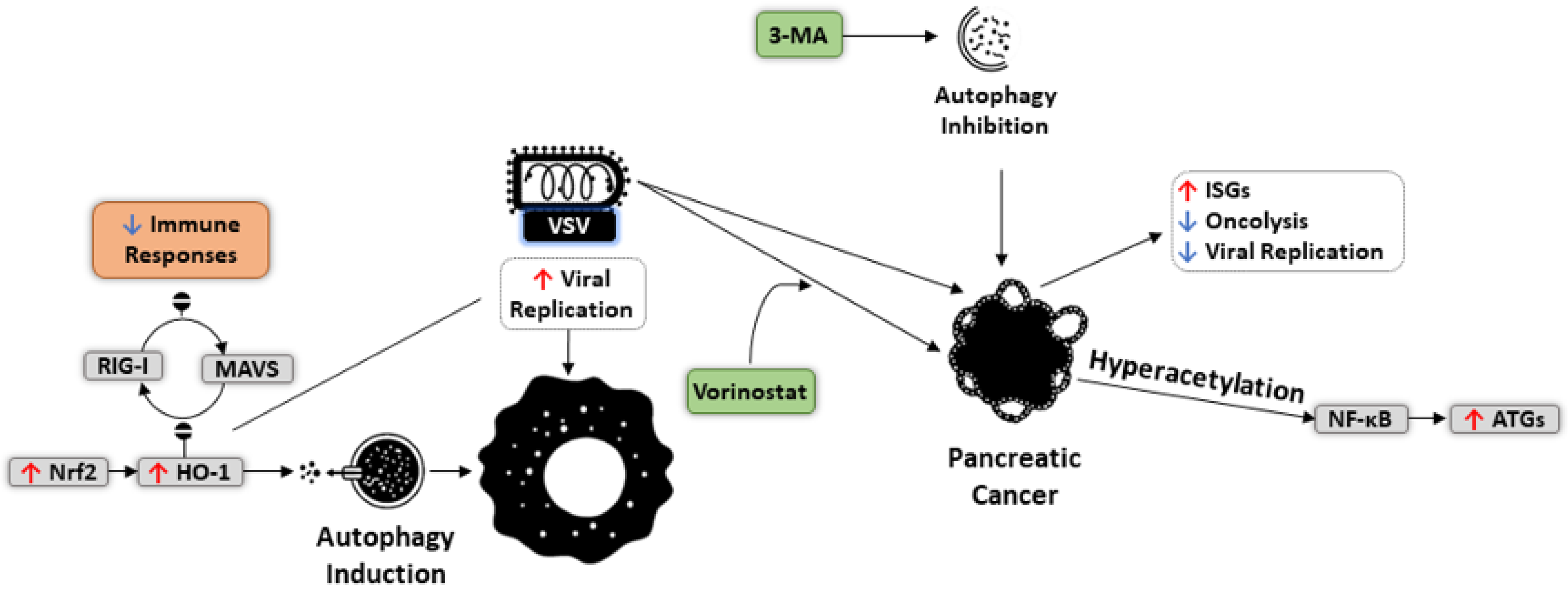
  199. Olagnier, D.; Lababidi, R.R.; Hadj, S.B.; Sze, A.; Liu, Y.; Naidu, S.D.; Ferrari, M.; Jiang, Y.; Chiang, C.; Beljanski, V.; et al. Activation of Nrf2 Signaling Augments Vesicular Stomatitis Virus Oncolysis via Autophagy-Driven Suppression of Antiviral Immunity. Mol. Ther. 2017, 25, 1900–1916. [Google Scholar] [CrossRef] [Green Version]
  200. Shulak, L. Histone deacetylase inhibitors potentiate vesicular stomatitis virus oncolysis in prostate cancer cells by modulating NF-κB-dependent autophagy. J. Virol. 2014, 88, 2927–2940. [Google Scholar] [CrossRef] [PubMed] [Green Version]
  201. Nabizadeh, A.; Bamdad, T.; Arefian, E.; Nikoo, S.H.R. Autophagy Gene Activity May Act As a Key Factor for Sensitivity of Tumor Cells to Oncolytic Vesicular Stomatitis Virus. Iran. J. Cancer Prev. 2016, 9, e3919. [Google Scholar] [CrossRef]
  202. Yu, L.; Baxter, P.A.; Zhao, X.; Liu, Z.; Wadhwa, L.; Zhang, Y.; Su, J.M.F.; Tan, X.; Yang, J.; Adesina, A.; et al. A single intravenous injection of oncolytic picornavirus SVV-001 eliminates medulloblastomas in primary tumor-based orthotopic xenograft mouse models. Neuro-Oncol. 2010, 13, 14–27. [Google Scholar] [CrossRef] [PubMed]
  203. Lei, W.; Wang, S.; Xu, N.; Chen, Y.; Wu, G.; Zhang, A.; Chen, X.; Tong, Y.; Qian, W. Enhancing therapeutic efficacy of oncolytic vaccinia virus armed with Beclin-1, an autophagic Gene in leukemia and myeloma. Biomed. Pharmacother. 2020, 125, 110030. [Google Scholar] [CrossRef] [PubMed]
  204. Jiffry, J.; Thavornwatanayong, T.; Rao, D.; Fogel, E.J.; Saytoo, D.; Nahata, R.; Guzik, H.; Chaudhary, I.; Augustine, T.; Goel, S.; et al. Oncolytic Reovirus (pelareorep) Induces Autophagy in KRAS-mutated Colorectal Cancer. Clin. Cancer Res. 2020, 27, 865–876. [Google Scholar] [CrossRef] [PubMed]
  205. I Chi, P.; Huang, W.-R.; Lai, I.H.; Cheng, C.Y.; Liu, H.J. The p17 Nonstructural Protein of Avian Reovirus Triggers Autophagy Enhancing Virus Replication via Activation of Phosphatase and Tensin Deleted on Chromosome 10 (PTEN) and AMP-activated Protein Kinase (AMPK), as well as dsRNA-dependent Protein Kinase (PKR)/eIF2α Signaling Pathways. J. Biol. Chem. 2013, 288, 3571–3584. [Google Scholar] [PubMed] [Green Version]
  206. Lhomond, S.; Avril, T.; Dejeans, N.; Voutetakis, K.; Doultsinos, D.; McMahon, M.; Pineau, R.; Obacz, J.; Papadodima, O.; Jouan, F.; et al. Dual IRE 1 RN ase functions dictate glioblastoma development. EMBO Mol. Med. 2018, 10, e7929. [Google Scholar] [CrossRef] [PubMed]
  207. Li, K.; Hu, C.; Xing, F.; Gao, M.; Liang, J.; Xiao, X.; Cai, J.; Tan, Y.; Hu, J.; Zhu, W.; et al. Deficiency of the IRE1α-Autophagy Axis Enhances the Antitumor Effects of the Oncolytic Virus M1. J. Virol. 2018, 92, e01331-e17. [Google Scholar] [CrossRef] [PubMed] [Green Version]
  208. Zhang, J.; Liu, Y.; Tan, J.; Zhang, Y.; Wong, C.W.; Lin, Z.; Liu, X.; Sander, M.; Yang, X.; Liang, L.; et al. Necroptotic virotherapy of oncolytic alphavirus M1 cooperated with Doxorubicin displays promising therapeutic efficacy in TNBC. Oncogene 2021, 1–13. [Google Scholar]
  209. Storniolo, A.; Alfano, V.; Carbotta, S.; Ferretti, E.; Di Renzo, L. IRE1α deficiency promotes tumor cell death and eIF2α degradation through PERK dipendent autophagy. Cell Death Discov. 2018, 4, 3. [Google Scholar] [CrossRef] [PubMed] [Green Version]
  210. Tameire, F.; Verginadis, I.I.; Koumenis, C. Cell intrinsic and extrinsic activators of the unfolded protein response in cancer: Mechanisms and targets for therapy. Semin. Cancer Biol. 2015, 33, 3–15. [Google Scholar] [CrossRef] [Green Version]
  211. Mahoney, D.J.; Lefebvre, C.; Allan, K.; Brun, J.; Sanaei, C.A.; Baird, S.; Pearce, N.; Grönberg, S.; Wilson, B.; Prakesh, M.; et al. Virus-Tumor Interactome Screen Reveals ER Stress Response Can Reprogram Resistant Cancers for Oncolytic Virus-Triggered Caspase-2 Cell Death. Cancer Cell 2011, 20, 443–456. [Google Scholar] [CrossRef] [PubMed] [Green Version]
  212. Prasad, V.; Suomalainen, M.; Pennauer, M.; Yakimovich, A.; Andriasyan, V.; Hemmi, S.; Greber, U.F. Chemical Induction of Unfolded Protein Response Enhances Cancer Cell Killing through Lytic Virus Infection. J. Virol. 2014, 88, 13086–13098. [Google Scholar] [CrossRef] [PubMed] [Green Version]
  213. Liu, J.; HuangFu, W.-C.; Kumar, K.S.; Qian, J.; Casey, J.P.; Hamanaka, R.B.; Grigoriadou, C.; Aldabe, R.; Diehl, J.A.; Fuchs, S.Y. Virus-Induced Unfolded Protein Response Attenuates Antiviral Defenses via Phosphorylation-Dependent Degradation of the Type I Interferon Receptor. Cell Host Microbe 2009, 5, 72–83. [Google Scholar] [CrossRef] [PubMed] [Green Version]
Figure 1. Molecular mechanism of autophagy activation. The kinase mTOR is known as the master regulator of autophagy initiation. In nutrient-rich conditions, the mTOR kinase associates with the ULK1 complex to inhibit the initiation of autophagy. However, under growth factor deprivation, nutrient starvation, or other stimuli, activation of energy sensor AMPK or TP53 (p53) leads to activation of the ULK1 complex and inhibition of mTOR activity through phosphorylation. Phosphorylated ULK1 also promotes phosphorylation of ATG13 and FIP200 subunits and dissociate from mTOR. PI3K class III (PI3KC3), BECN1, ATG14, p150, and AMBRA1, form a protein complex (PI3KC3 complex) and initiates phagophore formation in the proximity of the ER or other membrane sources. ATG5-ATG12 complex and LC3 are involved in double-membrane vesicle (autophagosome) formation, elongation, and closure. A ubiquitin-like reaction involving ATG7 and ATG10 contributes to the conjugation of ATG12 to ATG5, which eventually interacts with ATG16 for generating a “large complex”. While ATG4 cleaves LC3 (a light chain of the microtubule-associated protein) to form LC3-I, the “large complex” acts as a ubiquitin ligase to conjugate LC3-I with the polar head of phosphatidylethanolamine (PE) to produce LC3-II, a process essential for autophagosome formation and cargo recognition. The ATG5-ATG12 complex further interacts with ATG16L1 to establish the ATG12-ATG5-ATG16L1 complex. Autophagosomes are sequentially fused with lysosomes to form autolysosomes, whose contents are subsequently digested by lysosomal hydrolases and released for metabolic recycling.
Cells 10 02672 g001
Figure 2. Cancer immunity cycle and immunoediting. (A) Cancer immunity cycle, as outlined by Chen and Mellman [161]. (B) Steps in the cancer immunoediting concept, with immune cell elimination of cancer cells; cancer-immune equilibrium; and escape through a multitude of immunosuppressive mechanisms (image created with BioRender.com, accessed on 21 September 2021).
Cells 10 02672 g002
Figure 3. Effect of autophagy on the replication of oncolytic adenovirus (Ad). Although the entry of Ad decreases autophagic flux by affecting Erk1/2 and Akt/mTOR pathways, induction of autophagy with rapamycin can promote viral replication in cancer cells. Arming Ad with BECN1 and hTERT promoters results in autophagic cell death through different mechanisms in glioblastoma (GBM) and leukemia, respectively. Moreover, Ad-mediated oncolysis in GBM can be boosted by autophagy activation without significant impact on viral replication. Combination of Ad and temozolomide induces autophagy, which mediates immunogenic cell death via eliciting dendritic cells, cytotoxic T lymphocytes, and pro-inflammatory cytokines.
Cells 10 02672 g003
Figure 4. Mechanisms of autophagy induction and mitophagy modulation by oncolytic measles virus (MV). MV binds CD46 on the surface of HeLa cells. Interactions between CD46 and GOPC stimulate BECN1, contributing to autophagy induction. On the other hand, the C protein of MV interacts with IRGM, which subsequently influences several autophagy regulators such as LC3, ATG5, ATG10, and ULK1, to activate autophagy. During this process, C viral protein postpones cell death to improve MV replication and production of progeny viruses. In addition, MV uses the intact mitophagy process to inhibit mitochondrial antiviral signaling (MAVS), a mechanism that not only promotes viral replication but also limits innate immune responses against MV.
Cells 10 02672 g004
Figure 5. Induction of immunogenic cell death by Newcastle disease virus (NDV)-modulated autophagy. In lung adenocarcinoma cells, increased level of autophagy following NDV entry upregulates HSP70, which mediates interactions with the receptors on dendritic cells (DC), B cells, and macrophages (M). Such interactions activate natural killer cells (NK) and CD8+ T cell responses. In melanoma cells, NDV-induced immunogenic cell death involves upregulation of HSP70 and HMGB1. This type of cell death can also be elicited in NDV-infected glioma that occurs by overexpression of CRT, PMEL17, and HMHB1, which elevates autophagy. Rapamycin-induced promotion of autophagy in these cells increases NDV replication. In mice models, intratumoral delivery of NDV boosts the infiltration of IFNγ+ T cells toward the glioma microenvironment.
Cells 10 02672 g005
Figure 6. Impact of autophagy induction on the replication of oncolytic vesicular stomatitis Indiana virus (VSV). Activation of autophagy by Nrf2 pathways not only suppresses immune responses by blocking RIG-1-MAVS interactions but also elevates VSV replication in cancer cells. Combining VSV with vorinostat in pancreatic cancer cells can increase the hyperacetylation of NF-κB, ultimately promoting the expression of ATG genes.
Cells 10 02672 g006
Table 1. Dual role of autophagy in tumor promotion and inhibition.
ModelAutophagy RoleMechanismReference
Neuroblastoma
(BE(2)-C and BE(2)-M17)
InhibitionDegradation of gastrin-releasing peptide and inhibition of angiogenesis[75]
Cervical Cancer
(HeLa)
PromotionActivating HIF-1α and increasing drug resistance (paclitaxel)[76]
Gastric Cancer
(MGC-803 and SGC-7901)
InhibitionDownregulating HIF-1α and decreasing metastasis and glycolysis[77]
Oral Cancer
(SCC-9)
InhibitionSuppressing the NF-κB pathway and inhibiting invasiveness[78]
Bladder Cancer
(T24)
PromotionIncreasing HIF-1α expression and counteracting gemcitabine-induced apoptosis[79]
Pancreatic Cancer
(PDAC)
PromotionDegrading MHC-1 and boosting immune evasion[80]
Breast Cancer
(MCF-7)
InhibitionBlocking nitric oxide generation and inducing apoptosis[81]
Breast Cancer
(D2A1 and MCF-7)
PromotionImproving survival and metastasis[82]
Hepatocellular Carcinoma
(BEL7402 and HepG2)
PromotionElevating invasiveness via upregulating EMT markers (E-cadherin, CK18, and fibronectin)[83]
Hepatocellular Carcinoma
(SMMC-7721 and HepG2)
PromotionEnhancing glycolysis and metastasis via upregulating MCT1 and activating Wnt/β-catenin [84]
Publisher’s Note: MDPI stays neutral with regard to jurisdictional claims in published maps and institutional affiliations.

