Article

Comparative Analysis of Morphological, Histological, and Metabolic Differences of In Vitro- and Ex Vitro-Grown Panax ginseng

1
Department of Herbal Crop Research, National Institution of Horticultural and Herbal Science, Rural Develipment Administration, Eumseong 27709, Republic of Korea
2
Department of Food Science and Biotechnology, Gyeongkuk National University, Andong 36729, Republic of Korea
*
Authors to whom correspondence should be addressed.
Agronomy 2025, 15(9), 2222; https://doi.org/10.3390/agronomy15092222
Submission received: 21 August 2025 / Revised: 10 September 2025 / Accepted: 18 September 2025 / Published: 20 September 2025
(This article belongs to the Special Issue Application of In Vitro Culture for Horticultural Crops)

Abstract

Ginseng (Panax ginseng) is highly sensitive to heat stress caused by climate change; thus, the introduction of heat-tolerant cultivars is essential. However, the stable dissemination of heat-tolerant cultivars remains limited due to low propagation efficiency. Plant tissue culture has been introduced as an alternative approach, yet in vitro-grown ginseng often exhibit low survival rates during acclimatization, thereby restricting their practical application. This study was conducted as a fundamental investigation to address this limitation by comparing the morphological, histological, physiological, and metabolic differences between ginseng plants grown in vitro and ex vitro. The results demonstrated that in vitro-grown ginseng had stems and roots that were approximately 30% shorter, less prominent taproot development, and more than 30% lower root fresh weight. These plants also contained about 50% lower chlorophyll content and 52% higher stomatal density compared with ex vitro-grown ginseng. Histologically, in vitro plants exhibited narrow intercellular spaces, underdeveloped root cambium, and lignin deposition in cell walls. Metabolically, in vitro-grown ginseng was clearly distinguishable based on ginsenoside content and volatile compound profiles. The comprehensive findings of this study provide baseline information for future research and can be utilized to enhance the practicality of tissue culture-based micropropagation of ginseng.

1. Introduction

Ginseng (Panax ginseng), a herbaceous perennial crop cultivated in temperate regions, is highly valued for its medicinal properties. Its major secondary metabolites, ginsenosides, exhibit diverse bioactivities—including antioxidant, anticancer, and anti-inflammatory effects—which contribute to its widespread use in East Asia, particularly in South Korea [1]. However, ginseng is highly susceptible to heat stress, and the rising incidence of extreme heat events due to global warming has threatened its stable production [2]. According to the Ginseng Statistics Yearbook published by the Ministry of Agriculture, Food and Rural Affairs (MAFRA, Korea) [3], the cultivation area of ginseng decreased from 15,160 ha in 2020 to 11,745 ha in 2023, representing a reduction of approximately 22.5%. To mitigate this issue, heat-tolerant ginseng cultivars have been developed. Nevertheless, their adoption among farmers remains limited [4,5]. This constraint is largely attributed to ginseng’s intrinsically low reproductive capacity, which significantly hampers the dissemination of elite cultivars. Ginseng primarily propagates through seeds, but it takes more than three years from sowing to reach the flowering stage [6]. Although seed yield per plant varies among cultivars, it typically does not exceed 40 seeds [7]. These challenges underscore the need for alternative propagation strategies to overcome current limitations and ensure the sustainable production of elite ginseng cultivars.
Plants possess cellular totipotency, the remarkable ability of a single cell to regenerate an entire organism—a property that is actively harnessed in tissue culture for clonal propagation. Plant tissue culture has emerged as a powerful technique for cultivating plants under sterile conditions and enabling large-scale propagation [8]. Given the intrinsic limitations of seed propagation, tissue culture currently represents the only realistic strategy for the large-scale dissemination of elite ginseng cultivars. Furthermore, it is widely applied in various areas such as the production of aseptic plants, genetic transformation, and genome editing [9].
Research on ginseng tissue culture, initiated by the pioneering work of Butenko [10], has since advanced considerably in key areas including micropropagation [11], anther culture [12], and gene editing [13]. Micropropagation of ginseng is primarily achieved through somatic embryogenesis, which facilitates the production of large numbers of tissue-cultured plantlets [14]. However, a major challenge in ginseng micropropagation lies in the low acclimatization rate, with only 30–40% of plantlets reported to survive upon transfer to ex vitro conditions [15]. Thus, low acclimatization success remains the primary barrier preventing the practical application of tissue culture in ginseng. Although recent studies have reported improved survival rates exceeding 70% through the induction of in vitro-grown taproots [16], acclimatization remains a critical bottleneck. This limitation is likely due to environmental differences between in vitro and ex vitro conditions, highlighting the need for further understanding of this transition. Understanding these limitations is essential not only for improving acclimatization success but also for ensuring the practicality of tissue culture as a propagation strategy for ginseng.
In vitro environments differ markedly from natural growing conditions, characterized by high humidity, limited light, and restricted gas exchange. As a result, in vitro-cultivated plants often exhibit substantial differences compared to their ex vitro counterparts [17,18].
Previous studies have reported morphological differences in various traits, including stem length, number of stems, leaf size, root weight, root diameter, and root hair development [19,20] Histological analyses have revealed variations in cell organization and wall thickness, presumably attributable to the enclosed culture environment and limited gas exchange [21]. The limited light intensity and artificial light spectrum in in vitro environments are known to disrupt normal photosynthetic responses [22,23]. Additionally, differences in stomatal structure, shape, and density have been observed compared to ex vitro-grown plants [24]. Such conditions also influence the biosynthesis of physiologically active compounds in plants. While some studies have examined differences in the content and composition of ginsenosides in in vitro-cultured ginseng [4], limited attention has been given to antioxidant compounds, such as total phenolics, and to volatile metabolites.
To support the practical application and dissemination of ginseng micropropagation techniques via tissue culture and to establish precise in vitro cultivation methods, it is essential to gain a comprehensive understanding of the differences between plants grown in vitro and ex vitro. In ginseng, only a few studies have sporadically reported differences in morphology, histology, physiology, or metabolites, and to our knowledge, no study has systematically compared these traits together in relation to the acclimatization problem. Therefore, this study investigates the morphological, physiological, histological, and compositional differences between ginseng cultivated under in vitro and ex vitro conditions, with the aim of elucidating how in vitro environments affect plant survival and quality. The findings offer insights into the underlying causes of low survival rates in in vitro-cultured seedlings and provide a basis for overcoming this limitation. Ultimately, this work may contribute to the advancement of propagation technologies, promoting the stable and efficient distribution of elite ginseng cultivars.

2. Materials and Methods

2.1. Plant Materials

The ginseng cultivar ‘Geumpoong’ was used in this study. Seeds were collected in July 2023 from the experimental field of the Department of Herbal Crop Research, National Institute of Horticultural and Herbal Science, affiliated with the Rural Development Administration (RDA), South Korea. The seeds were subjected to dehiscence and cold stratification following the method described by Lee et al. [25].

2.2. In Vitro Cultivation of Ginseng

To initiate in vitro culture, approximately 1000 ginseng seeds were used. Seed coats were removed, and the seeds were immersed in 2% (v/v) sodium hypochlorite for 20 min, followed by treatment with 70% (v/v) ethanol for 1 min and two rinses with sterile distilled water. To complete the sterilization process, the seeds were again treated with 2% (v/v) sodium hypochlorite for 20 min and rinsed four to five times with sterile distilled water. The surface-sterilized seeds were cultured in vitro following the method described by Lee et al. [26]. Briefly, zygotic embryos were excised and cultured on germination medium consisting of half-strength Murashige and Skoog (½ MS) medium containing 2% sucrose and 0.8% agar (pH 5.7). All zygotic embryos germinated except for contaminated ones. After germination, seedlings were cultured on a growth medium composed of one-third strength Schenk and Hildebrandt (⅓ SH) medium containing 2% sucrose and 0.5% gelrite (pH 5.7), and maintained for approximately five months in a tissue culture room. Each seedling was grown on 50 mL of medium dispensed in Incu Tissue (65.40 × 65.40 × 98.20 mm) (inCu Tissue, SPL Life Sciences, Pocheon, Korea). Cultures were maintained at 23 °C ± 2 °C under a 16/8 h light/dark cycle with white fluorescent light at an intensity of 24 μmol m−2 s−1.

2.3. Ex Vitro Cultivation of Ginseng

For ex vitro cultivation, 1000 intact ginseng seeds were sown in plastic boxes (0.5 × 0.3 × 0.3 m) filled with artificial soil (Golden Root Ginseng Soil, Nongkyung Co, Jincheon, Korea) composed of peat moss and perlite at a 3:1 (v/v) ratio. The average soil pH was 6.17, as determined by soil analysis. The planting density for ex vitro conditions was adjusted to match the same unit area. Irrigation was performed once per week by applying 2 L of water per box to maintain field capacity, and the boxes were placed in a greenhouse. Germination rate was 92.38%. No additional nutrient supplementation was applied to the soil. Plants were cultivated for five months.

2.4. Experimental Design and Replication

All experiments were conducted under a completely randomized design (CRD). For each cultivation condition (in vitro and ex vitro), 1000 seeds were used. For growth measurements, 100 plants per treatment were randomly sampled. Chlorophyll content and stomatal analyses were conducted using ten randomly selected plants per treatment. Metabolite and histological analyses were performed with three biological replicates per treatment. No subplots were included in this study.

2.5. Histological Analysis of Aerial and Underground Parts

Histological analysis was performed on the leaves and roots of ginseng plants cultivated in vitro and ex vitro for 60 days. For root samples, transverse sections were obtained from the proximal 0.5–1.5 cm region of the primary root directly connected to the shoot. For leaf samples, the central leaflet of each plant was selected, and rectangular segments were excised to include the widest region of the lamina, spanning from the midrib at the midpoint toward the leaf apex. Paraffin sectioning was conducted following the procedure reported by Hong et al. [2] to obtain cross-sectional images of the tissues. Fresh leaf and root tissue segments were fixed overnight at 4 °C in phosphate-buffered saline (PBS, pH 7.0) containing 3% glutaraldehyde and 3% formaldehyde. The fixed tissues were dehydrated through a graded ethanol series (30%, 50%, 70%, 90%, and 100%), with each step repeated once for 1 h. Samples were embedded in paraffin and sectioned at 5–10 μm thickness using a microtome. The sections were dewaxed, stained with 1% Safranin-O (Sigma, St. Louis, MO, USA) and 0.5% Astra Blue (Santa Cruz Biochem, Dallas, TX, USA), and then dehydrated in a reverse ethanol series. Bright-field and polarized light images were acquired using a Slideview scanner and a BX53 microscope (Olympus, Tokyo, Japan).

2.6. Chlorophyll Content Analysis

Chlorophyll a and b contents in leaves harvested from in vitro- and ex vitro-grown ginseng were measured according to the method described by Lichtenthaler [27]. Ten leaves from each treatment were cut into 5 mm × 5 mm pieces, and each sample was extracted with 95% ethanol at 80 °C for 20 min using a solvent volume 50 times the fresh weight. To prevent photodegradation, samples were kept in the dark after extraction. Absorbance was measured at 648 nm (A648) and 664 nm (A664) using a Multiskan GO Microplate Spectrophotometer (Thermo Scientific, Waltham, MA, USA), and chlorophyll concentrations were calculated using the following equations:
Chl a = 13.36 × A664 − 5.19 × A648
Chl b = 27.43 × A648 − 812 × A664
Total Chl = 5.24 × A664 + 22.24 × A648

2.7. Stomatal Traits Analysis

To investigate changes in stomatal characteristics under different cultivation environments, stomatal size and density were analyzed. The central leaflet of each plant was selected, and the stomata on the abaxial leaf surface were exposed by carefully scraping the central lamina with a scalpel, avoiding the midrib. For each treatment, ten leaves were collected, and three impressions per leaf were prepared. Ten stomata were measured per impression to determine stomatal length and width, pore length, and aperture (μm). The stomatal length-to-width ratio was also calculated. Stomatal density was calculated as the average of the three regions per leaf. Observations were conducted using a Leica DM3000 microscope (Leica Microsystems, Wetzlar, Germany).

2.8. Growth Measurements

After 60 days of cultivation, in vitro- and ex vitro-grown ginseng plants reached the developmental stage shown in Figure S1. A total of 100 plants from each treatment were harvested, and their aerial parts were separated for growth measurements. Stem length and diameter, and leaf length and width were measured using a ruler and caliper. Fresh weights were recorded, and dry weights were measured after lyophilization using a freeze-dryer (FDU-2200, Eyela, Tokyo, Japan).
After five months of cultivation, once the aerial parts had senesced, roots were harvested from both in vitro- and ex vitro-grown plants. For each treatment, 100 roots were collected, and root traits including length, diameter, fresh weight, and dry weight were assessed.

2.9. Freeze-Drying

For the analysis of ginsenosides and volatile compounds, ginseng plants cultivated for five months under in vitro and ex vitro conditions were subjected to freeze-drying. In vitro-grown plants were carefully separated from the culture medium, while ex vitro-grown plants were washed to remove soil residues and gently blotted dry with tissue paper. The aerial parts were removed, and only roots were used. The samples were freeze-dried for 48 h using a freeze dryer (FDU-2200, Eyela, Tokyo, Japan) and subsequently ground into a fine powder for analysis.

2.10. Ginsenoside Profiling

Freeze-dried ginseng powder (0.2 g) was extracted with 2 mL of 70% methanol–water (v/v) by vortexing and ultrasonication at 50 °C for 30 min. Samples were centrifuged using an Eppendorf Centrifuge 5427 R (Eppendorf SE, Hamburg, Germany) at 15,000 rpm for 3 min. The supernatant was purified using a solid-phase extraction (SPE) cartridge (Agilent Technologies, Santa Clara, CA, USA), then filtered through a 0.45 µm PTFE membrane.
Ginsenosides were quantified using HPLC (Agilent Technologies, Santa Clara, CA, USA) equipped with a Halo® RP-Amide column (4.6 × 150 mm, 2.7 µm; Wilmington, DE, USA) at 50° C. The mobile phases were (A) 10% acetonitrile and (B) 90% acetonitrile, with a gradient flow (0.6 mL/min). The gradient program was set as follows: 0.0 min (27% B), 0–6.0 min (28% B), 6.0–10.0 min (28% B), 10.0–30.0 min (34% B), 30.0–33.0 min (70% B), 33.0–35.0 min (80% B), 35.0–45.0 min (100% B), 45.0–50.0 min (100% B), 50.0–51.0 min (27% B), and 51.0–60.1 min (27% B). Peaks were detected at 203 nm using a photodiode array (PDA) detector. Standards were obtained from ChromaDex (Irvine, CA, USA) and Biosynth (Staad, Switzerland).

2.11. Volatile Compound Analysis

Volatile profiles were analyzed using HS-SPME–GC–MS (Agilent 8890 GC/5977B MSD with Gerstel MPS 2XL). Samples (2 g) were ground in liquid nitrogen and mixed with 3 mL distilled water and 0.025 µg/mL of 2-methylbutyl 2-methylbutyrate as an internal standard in a 20 mL vial.
A DVB/CAR/PDMS SPME fiber (50/30 µm; Supelco) was preconditioned at 250 °C for 10 min. Vials were incubated at 50 °C for 40 min, and volatiles were adsorbed at 50 °C for 20 min while agitating at 250 rpm. Desorption was performed at 240 °C for 10 min in splitless mode.
Separation was carried out on a DB-WAX column (30 m × 0.25 mm, 0.25 µm film; Agilent) with helium as the carrier gas. Oven temperature was programmed from 35 °C (2 min) to 225 °C. Mass spectra were scanned over 35–550 m/z. Compound identification was based on NIST 2014 library matches and retention indices using a C7–C30 alkane standard. Volatile levels were quantified as peak area ratios relative to the internal standard. Analyses were performed in triplicate.

2.12. Statistical Analysis

The experimental layout followed a randomized complete block design. Growth data were analyzed using R software (v4.3.3; The R Foundation). Depending on variance equality, either a two-sample t-test or Welch’s t-test was applied. One-way ANOVA followed by Tukey’s HSD test was used for post hoc comparisons. Principal component analysis (PCA) was performed using SIMCA-P 17.0 (Umetrics, Umeå, Sweden), and heatmaps were generated via MetaboAnalyst 6.0 (https://www.metaboanalyst.ca/ accessed on 4 March 2025 ).

3. Results

3.1. Phenotypic Differences in Ginseng Under Different Cultivation Environments

After two months of cultivation, ginseng plants cultivated under in vitro and ex vitro conditions exhibited distinct phenotypic differences (Figure S1). In the aerial parts, ex vitro-grown ginseng had uniformly shaped, symmetrical leaves with a bright green coloration (Figure 1a). In contrast, the leaves of in vitro-grown ginseng varied in color and size and often exhibited slightly curled margins with an oblong shape.
After five months of cultivation, clear phenotypic differences were also observed in the underground parts (Figure 1b). Ex vitro-grown ginseng formed a distinct taproot with abundant lateral roots, whereas in vitro-grown ginseng exhibited atypical root morphology as an adaptive response to in vitro culture conditions, characterized by the absence of a clearly defined taproot and limited lateral root development. Additionally, ex vitro-grown ginseng typically formed a single rhizome, in contrast to in vitro-grown ginseng, which often developed two to four rhizomes per plant.

3.2. Histological Differences in Ginseng Under Different Cultivation Environments

Histological differences were evident between leaves of ginseng cultivated under in vitro and ex vitro conditions (Figure 2). In vitro-grown ginseng showed irregularly shaped epidermal cells, while those of ex vitro-grown plants were symmetrically arranged (Figure 2a). The leaf cross-sectional thickness averaged 185.6 µm in vitro and 155.3 µm ex vitro, indicating that in vitro-grown leaves tended to be thicker. Mesophyll cells in in vitro-grown leaves were evenly distributed and uniform in size. By contrast, ex vitro-grown leaves exhibited an irregular mesophyll cell size and arrangement, larger intercellular spaces, and a looser tissue structure (Figure 2b). The cell walls of in vitro-grown leaves appeared to contain more lignin, as indicated by more intense red staining.
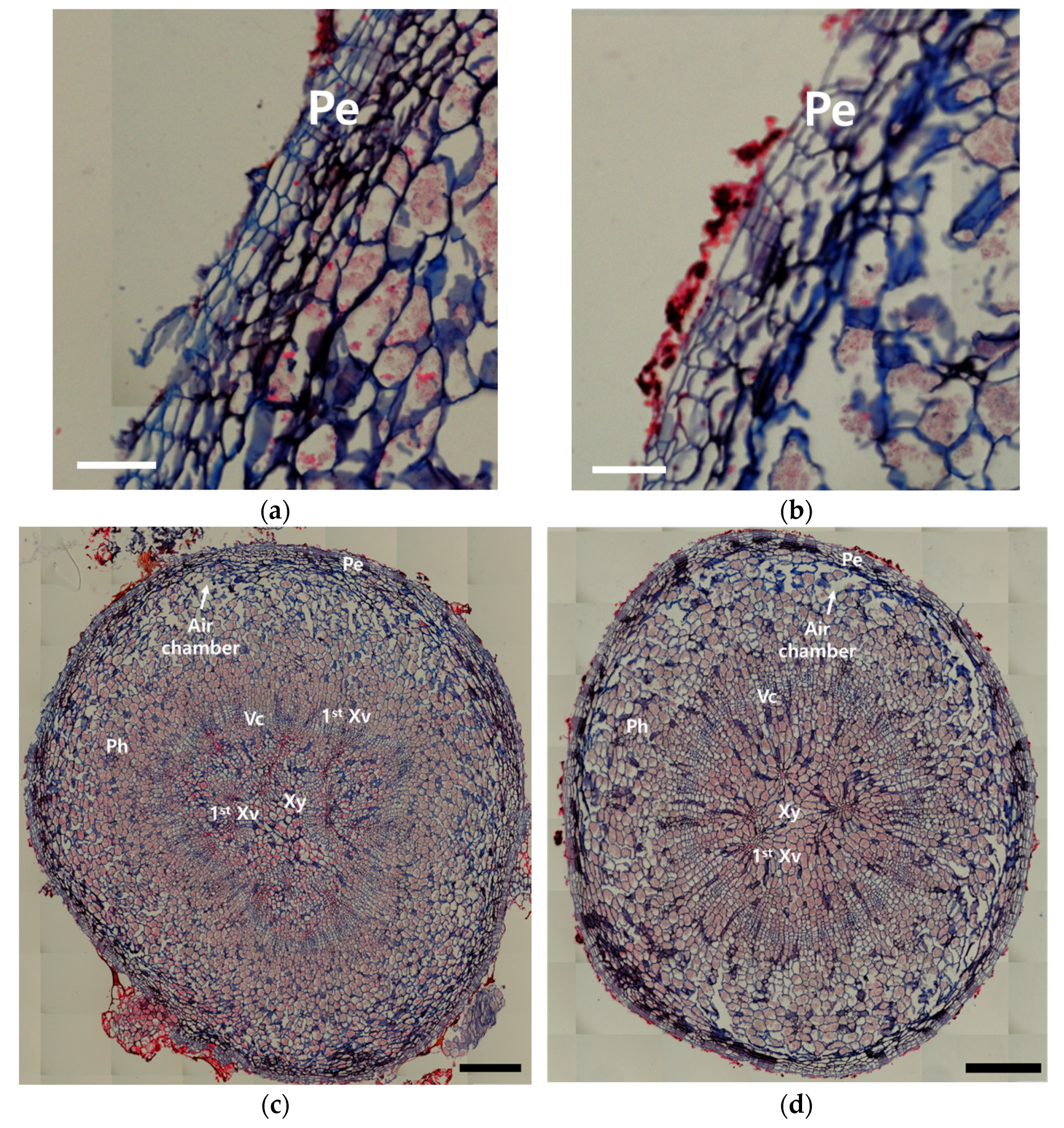
Substantial histological differences were also observed in the roots (Figure 3). In vitro-grown ginseng had a relatively thin epidermis with indistinct boundaries and irregular cell arrangement (Figure 3a). In contrast, ex vitro-grown ginseng exhibited a thicker epidermis with clearly defined boundaries and uniform cell organization (Figure 3b). The epidermal thickness averaged 49.20 µm in vitro and 66.37 µm ex vitro, while the mean width per epidermal cell was 15.67 µm and 16.86 µm, respectively. In the vascular tissues, in vitro-grown roots showed a disorganized structure with poorly differentiated xylem and phloem and irregular xylem vessel size and arrangement (Figure 3c). Ex vitro-grown roots, on the other hand, exhibited a well-organized vascular structure with a clear triarch xylem pattern and distinct xylem-phloem boundaries (Figure 3d).
The cambium in in vitro-grown roots was underdeveloped, with blurred margins and minimal signs of secondary growth. In contrast, the cambium in ex vitro-grown roots was clearly located between the xylem and phloem and exhibited active secondary development. Red staining, indicative of lignin accumulation, was more intense and irregularly distributed in in vitro-grown roots, while in ex vitro-grown roots, lignin was concentrated in the xylem and surrounding cell walls. Additionally, in vitro-grown roots had a denser tissue structure with smaller and uniformly distributed intercellular spaces, and the proportion of intercellular area relative to the total root cross-sectional area was 1.20%. In contrast, ex vitro-grown roots had larger and more irregular intercellular spaces that formed broader air cavities, with the proportion measured at 5.34%.

3.3. Chlorophyll Content Under Different Cultivation Environments

Significant differences in photosynthetic pigment content were observed between in vitro- and ex vitro-grown ginseng leaves. The chlorophyll a (Chl a) content in in vitro-grown leaves was 0.54 ± 0.28 mg/g, which was significantly lower than the 1.07 ± 0.22 mg/g measured in ex vitro-grown leaves (Figure 4a). Similarly, the chlorophyll b (Chl b) content was lower in in vitro-grown leaves than in ex vitro-grown leaves (Figure 4b). As a result, the total chlorophyll content was significantly reduced in in vitro-grown ginseng compared to its ex vitro counterpart (Figure 4c).

3.4. Stomatal Characteristics Under Different Cultivation Environments

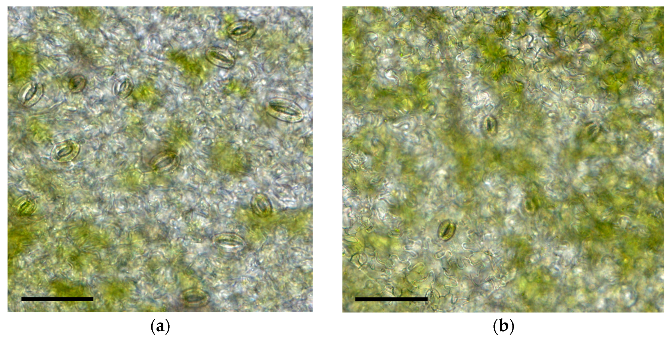
A comparison of stomatal characteristics between in vitro- and ex vitro-grown ginseng leaves revealed notable differences (Figure 5). Stomatal density was approximately 1.5 times higher in in vitro-grown leaves compared to ex vitro-grown leaves (Table 1). In vitro-grown stomata were also larger, showing increased stomatal length, width, pore length, and aperture.
Notably, stomata in in vitro-grown leaves remained open, while those in ex vitro-grown leaves were mostly closed or partially open. However, there was no statistically significant difference in the stomatal length-to-width ratio between the two groups.

3.5. Growth Differences Under Different Cultivation Environments

Significant differences in aerial growth were observed between in vitro- and ex vitro-grown ginseng, except for in fresh weight (Table 2). The dry weight of aerial parts was significantly higher in ex vitro-grown plants compared to those grown in vitro. Stem length was greater in ex vitro-grown ginseng, while stem diameter was thicker in in vitro-grown plants. Leaf length and width were likewise greater in ex vitro-grown ginseng. Similarly, root growth following aerial senescence differed significantly between the two cultivation environments (Table 3). Both fresh and dry root weights were higher in ex vitro-grown ginseng. Root length was also greater in ex vitro-grown plants, whereas root diameter was thicker in those grown in vitro.

3.6. Ginsenoside Analysis

Ginsenoside content and composition were compared between in vitro- and ex vitro-grown ginseng based on underground parts harvested after five months of cultivation. A total of seven ginsenosides (Rb2, Rc, Rd, Re, Rg1, Rf, and Rh1) were identified in ex vitro-grown ginseng. In contrast, only six (Rb2, Rc, Rd, Re, Rg1, and Rf) were detected in in vitro-grown samples, with Rh1 exclusively present in the ex vitro group (Table 4).
Among individual ginsenosides, protopanaxadiol (PPD)-type compounds (Rb2, Rc, and Rd) accumulated at higher levels in ex vitro-grown ginseng. In contrast, Rg1—a protopanaxatriol (PPT)-type ginsenoside—was more abundant in in vitro-grown ginseng, while Re was significantly higher in the ex vitro group. Rf was detected at similar levels in both groups, and Rh1 was exclusively found in ex vitro-grown plants. Ginsenosides Rg3, Rg5, Rh2, and quinquenoside R1 were not detected in either condition.
The total content of PPD-type ginsenosides was significantly higher in ex vitro-grown ginseng compared to in vitro-grown plants (Table 5). In contrast, PPT-type ginsenoside levels did not differ significantly between the two environments. Consequently, the PPD/PPT ratio was markedly higher under ex vitro conditions (2.55 ± 0.08) than under in vitro conditions (0.55 ± 0.03). Furthermore, the total ginsenoside content was significantly greater in ex vitro-grown ginseng.

3.7. Volatile Compound Profiles

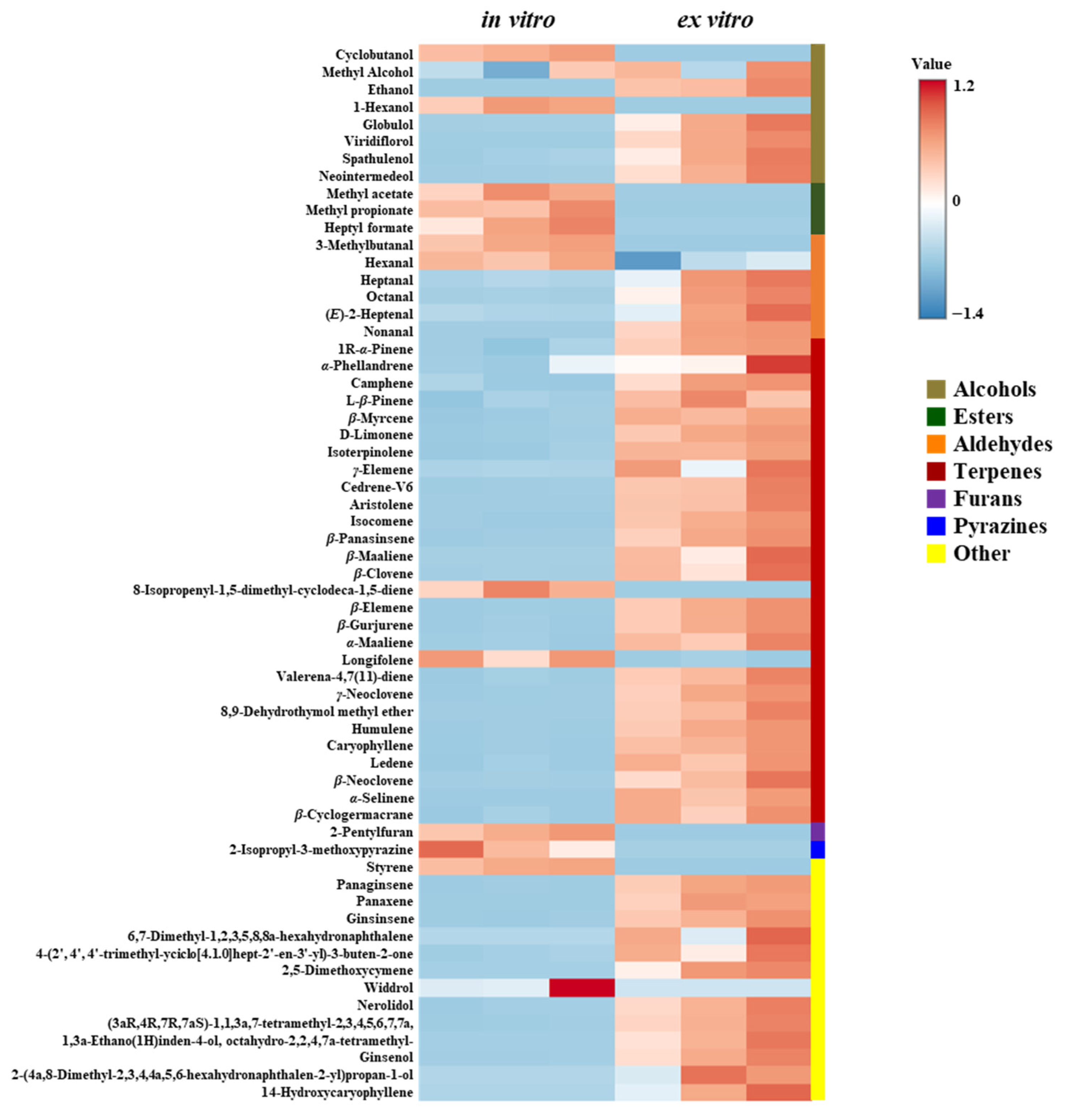
A total of 61 volatile compounds were identified from the underground parts of ginseng cultivated under in vitro and ex vitro conditions (Figure 6, Table 6). Overall, a greater number of compounds were detected in ex vitro-grown ginseng. Among the volatiles common to both conditions, most exhibited higher concentrations in the ex vitro samples (Figure 6 and Figure S2).
Terpenes represented the largest chemical class, comprising 28 compounds, followed by alcohols (8), aldehydes (6), esters (3), furans (1), pyrazines (1), and other miscellaneous compounds (14). In ex vitro-grown ginseng, major alcohols included globulol, spathulenol, and neointermedeol, while γ-neoclovene and β-panasinsene were the dominant terpenes. In contrast, in vitro-grown ginseng showed higher levels of methyl alcohol and 1-hexanol among alcohols, and caryophyllene and β-panasinsene among terpenes.
Among aldehydes, octanal and heptanal were predominant in ex vitro-grown samples, whereas hexanal and octanal were abundant in in vitro-grown ginseng. Interestingly, ester compounds such as methyl acetate and methyl propionate were detected only in in vitro-grown roots. Similarly, 2-pentylfuran (a furan) and 2-isopropyl-3-methoxypyrazine (a pyrazine) were uniquely found in the in vitro group. Among the miscellaneous compounds, ginsinsene and ginsenol were present in both groups but were notably more concentrated in ex vitro-grown ginseng.

4. Discussion

Ginseng is inherently difficult to propagate via conventional seed-based methods due to its low reproductive efficiency [7]. Micropropagation via tissue culture offers a potential solution, although widespread use is restricted by acclimatization difficulties [13,14]. This stress is induced by artificial in vitro conditions, including high humidity, limited gas exchange, and low light intensity [28,29]. Therefore, a deeper understanding of the physiological, histological, and metabolic characteristics of in vitro-grown ginseng is essential for overcoming current bottlenecks in ginseng micropropagation.
Morphological differences were evident between ginseng grown in vitro and ex vitro, particularly in their root structures. Compared to ex vitro-grown ginseng, in vitro-grown ginseng exhibited a degree of leaf asymmetry (Figure 1a), which may be attributable to low-intensity, unidirectional lighting conditions. According to Shilpha et al. [30], when ginseng was exposed to lighting from either the side or the top alone, leaf morphology was distorted, whereas balanced development was promoted under simultaneous lateral and overhead lighting. Root morphology exhibited a more marked difference than the aerial parts: in vitro-grown ginseng developed shortened taproots and excessive lateral roots (Figure 1b). These atypical root structures are consistent with previous findings [14,26] and are generally attributed to in vitro conditions such as high sucrose concentrations, hormonal imbalances, and limited gas exchange factors known to contribute to the low survival rate during acclimatization [16,31].
Histological comparisons revealed marked histological differences in ginseng tissues under in vitro and ex vitro conditions. In vitro-grown leaves had densely packed mesophyll cells with minimal intercellular spaces, whereas ex vitro leaves exhibited more developed intercellular spaces (Figure 2). These tissue traits are presumed to represent adaptive responses to in vitro culture conditions—such as excessive humidity, low light intensity, and restricted gas exchange [32]. Similar tissue responses were reported in Ilex paraguariensis under poor gas exchange conditions [33]. The intense red staining in in vitro leaves suggests greater lignin accumulation, likely as a stress response under sealed and artificial culture conditions [34].
In root tissues, in vitro-grown ginseng exhibited a thinner epidermis, irregular vascular organization, and underdeveloped cambium with minimal secondary growth (Figure 3). Comparable traits have been observed in Forsythia and Malus domestica, which showed reduced vascular development under in vitro conditions [35], and in Eucalyptus grandis, which exhibited poorly differentiated xylem [36]. These abnormalities may result from suppressed transpiration and altered mineral availability in the in vitro environment [21]. Lee et al. [26] also reported that ex vitro-grown ginseng roots exhibited a triarch primary xylem pattern, whereas in vitro-grown roots showed a polyarch arrangement. Our findings confirmed increased lignification in in vitro roots, which may reduce tissue elasticity and hinder taproot development [34,37]. Inhibiting lignin biosynthesis has been shown to enhance storage root development in Ipomoea batatas [38], Rehmannia glutinosa [39], and Panax notoginseng [40]. To the best of our knowledge, no previous studies have directly demonstrated a link between root anatomical structures and ginsenoside or other metabolite profiles; therefore, further integrated studies are required to clarify the potential relationships between anatomy and metabolic regulation in ginseng.
Chlorophyll a and b contents were significantly higher in ex vitro-grown ginseng than in in vitro ginseng (Figure 4), likely due to natural light conditions promoting chlorophyll synthesis. In contrast, artificial lighting and limited gas exchange in vitro may suppress chlorophyll biosynthesis [17,41,42]. This inhibition under high humidity and low light has also been reported in crops such as apple and cherry [41].
In vitro plants often exhibit photomixotrophic growth due to limited photosynthesis and dependence on external carbon sources, which can suppress chlorophyll synthesis and induce morphological abnormalities [32]. Ševčíková et al. [22] reported species-specific responses under such conditions, with decreased chlorophyll levels in potato and strawberry, no change in tobacco, and increases in rapeseed. This variation suggests that Panax ginseng responds in a manner similar to plants that show chlorophyll reduction under such conditions.
In addition to chlorophyll differences, stomatal traits also differed notably. In vitro-grown ginseng had 52.4% higher stomatal density than ex vitro ginseng (Table 1), likely due to elevated humidity promoting stomatal development [33]. For instance, in walnuts, reducing humidity via KCl treatment led to a 23% decrease in stomatal density after 2 weeks and a fourfold reduction after 4 weeks [43]. Limited CO2 in vitro may also stimulate increases in stomatal number and size as adaptive mechanisms [17]. Stomatal size—including length, width, and aperture—was significantly larger in vitro, while the length/width ratio remained stable, indicating uniform expansion. Abnormally large and fully open stomata were frequently observed, reflecting adaptations to humidity and CO2 limitations [42,43].
Environmental conditions strongly influenced overall ginseng growth and biomass accumulation. Ex vitro-grown ginseng exhibited longer stems and larger leaves, whereas in vitro-grown ginseng showed limited shoot growth (Table 2). These growth patterns are consistent with the effects of directional lighting and restricted gas exchange under in vitro conditions, which are known to reduce photosynthetic efficiency and resource allocation [32,44]. Although the fresh weight of aerial parts was comparable between treatments, dry weight was lower in in vitro plants, suggesting lower accumulation of structural biomass under high humidity and limited gas exchange [42,45].
Significant differences in root biomass were also observed. Ex vitro plants exhibited 1.5- and 1.7-fold greater fresh and dry root weights, respectively (Table 3), while in vitro roots showed reduced elongation and dry mass accumulation. These patterns may reflect suppressed metabolic activity and nutrient assimilation under low-oxygen and high-humidity conditions. Under such environments, fresh weight may be retained through excess water absorption, while dry matter production is reduced [45]. In vitro-grown ginseng may rely more heavily on external carbon sources, showing traits of photomixotrophic growth [22]. While this mode supports development in sealed systems, it may shape root development trajectories differently than in ex vitro conditions [46,47]. For example, Paulownia fortunei grown under photomixotrophic conditions exhibited reduced photosynthetic performance and shorter root systems [48]. These findings illustrate how altered physiological responses under in vitro conditions can lead to growth patterns distinct from those observed under ex vitro cultivation.
Ginsenoside profiling revealed significantly higher total contents in ex vitro-grown ginseng, particularly for PPD-type compounds such as Rb2, Rc, and Rd. This resulted in a 4.64-fold increase in the PPD/PPT ratio compared to in vitro plants (Table 4 and Table 5). Multivariate analyses further confirmed this trend, showing elevated levels of Rh1 in ex vitro plants and higher Rg1 levels in vitro samples. These findings suggest that environmental fluctuations in light and temperature stimulate PPD-type ginsenoside biosynthesis, possibly as a stress-responsive adaptation [49]. This difference may be due to the stable light and temperature conditions in vitro, whereas ex vitro conditions involve natural fluctuations that potentially enhance ginsenoside biosynthesis [50]. In contrast, constant high humidity in in vitro systems may suppress PPD accumulation, as reported in both P. ginseng and P. quinquefolius under water-saturated conditions [51,52].
PPT-type ginsenosides exhibited more variable accumulation patterns. Re was higher in ex vitro plants, Rg1 was more abundant under in vitro conditions, Rf remained unchanged, and Rh1 appeared only in ex vitro roots. These differences may reflect compound-specific regulation via environmental factors such as light, genotype, and developmental stage [53,54]. For example, Rg1 content in 5-year-old ginseng roots showed a significant correlation with soil water potential, unlike Re or Rf [45,52], highlighting intra-class variability in environmental responsiveness. These findings underscore the complexity of PPT-type ginsenoside regulation and support the need for further studies to clarify how cultivation conditions shape their biosynthetic pathways. In particular, no previous studies have directly explained the contrasting accumulation patterns observed here, such as higher Rg1 in vitro and higher Rb2 ex vitro. Therefore, further investigations are required to elucidate the metabolic basis of these differences.
Volatile compound composition in ginseng roots varied significantly between cultivation conditions. Ex vitro-grown ginseng exhibited greater diversity and higher concentrations of volatiles, particularly terpenes, compared to in vitro plants (Table 6, Figure 6). This may reflect the stimulatory effects of environmental variability—such as fluctuating temperature, adequate oxygen, and biotic interactions—on volatile biosynthesis [55]. Consistent with previous studies, some volatiles were detected exclusively in ex vitro plants, while several ester compounds appeared either solely or in greater abundance under in vitro conditions. Similar patterns have also been observed in other species, such as Petiveria alliacea, where terpenes were more abundant under ex vitro conditions, while esters increased under in vitro cultivation [56]. Such patterns may result from unique features of the in vitro environment, including high humidity, limited gas exchange, and elevated carbon availability, which are known to influence volatile metabolic pathways [57]. For example, Talaverano et al. [58] reported that esters such as ethyl butyrate and isoamyl acetate decreased under drought stress in grapevine, suggesting that their presence in in vitro ginseng may be favored by moisture-rich conditions.
While prior studies have primarily examined volatile differences by cultivar and plant age [59,60], this study provides the first comparative analysis in in vitro and ex vitro environments. These findings offer foundational insight into how cultivation systems shape volatile composition in ginseng, and support future efforts to optimize environmental conditions for desired metabolite production. The results of this study reveal significant differences and suggest the possibility of a sequential causal linkage among these traits. For example, under in vitro conditions, high humidity and limited gas exchange may reduce chlorophyll content and increase stomatal density and size, thereby lowering photosynthetic efficiency and affecting assimilate accumulation. These physiological changes may in turn influence root development and structure, ultimately leading to differences in metabolite accumulation, particularly ginsenoside composition. Overall, these observations suggest that morphological, histological, and metabolic characteristics may be interconnected, providing important insights into the acclimatization and metabolic regulation of ginseng, and indicating the need for further studies to clarify the underlying mechanisms.

5. Conclusions

This study comprehensively compared in vitro- and ex vitro-grown ginseng in terms of morphological, histological, physiological, and compositional characteristics. Clear differences were observed in the morphology and tissue structure of ginseng depending on the cultivation environment. In particular, in vitro conditions markedly influenced chlorophyll content and stomatal characteristics. Ex vitro-grown samples had greater content and diversity of ginsenosides and volatile compounds. These findings provide valuable insights into the effects of in vitro culture environments on the growth, development, and metabolite profiles of ginseng, and serve as a useful baseline for future studies aimed at refining in vitro culture systems to improve acclimatization and propagation stability.

Supplementary Materials

The following supporting information can be downloaded at: https://www.mdpi.com/article/10.3390/agronomy15092222/s1, Figure S1: Phenotypic characteristics of in vitro and ex vitro ginseng 60 days after sowing. Scale bar = 1 cm; Figure S2: Principal component analysis (PCA) biplot showing the differentiation of volatile compound profiles between in vitro- and ex vitro-grown ginseng roots. IN1~3; in vitro-grown ginseng, EX1~3; ex vitro-grown ginseng.

Author Contributions

Designed the experiments, J.-W.L. and K.H.M.; performed methodology and investigation (in vitro culture, chlorophyll content, stomatal analysis), and wrote the original draft, S.-J.K.; carried out ginsenoside analysis and metabolite profiling, J.S. and Y.J.; conducted histological and stomatal analyses, J.H.; analyzed data and contributed to ex vitro culture, J.-U.K. and J.Y.H.; contributed to data analysis and interpretation, S.C.K.; provided methodology, supervision, and writing—review and editing, J.S. and J.-W.L.; contributed to conceptualization, project administration, reviewing and approving the manuscript, and funding acquisition, K.H.M. All authors have read and agreed to the published version of the manuscript.

Funding

This work was carried out with the support of the Cooperative Research Program for Agriculture Science and Technology Development (Project No. PJ01476002) of the Rural Development Administration, Republic of Korea.

Data Availability Statement

All data is available in the article.

Acknowledgments

This study was supported by 2025 the RDA Fellowship Program of National Institute of Horticultural & Herbal Science, Rural Development Administration, Republic of Korea. Additionally, ChatGPT (OpenAI, GPT-4, 2025 version) was used solely for English translation and minor editing assistance. The authors have reviewed and modified the manuscript and take full responsibility for content of the publication.

Conflicts of Interest

The authors declare that they have no conflicts of interest.

Abbreviations

The following abbreviations are used in this manuscript:
PPDProtopanaxadiol
PPTProtopanaxatriol
PCAPrincipal Component Analysis

References

  1. Ratan, Z.A.; Haidere, M.F.; Hong, Y.H.; Park, S.H.; Lee, J.O.; Lee, J.; Cho, J.Y. Pharmacological potential of ginseng and its major component ginsenosides. J. Ginseng Res. 2020, 45, 199–210. [Google Scholar] [CrossRef] [PubMed]
  2. Hong, J.; Geem, K.R.; Kim, J.; Jo, I.H.; Yang, T.J.; Shim, D.; Ryu, H. Prolonged exposure to high temperature inhibits shoot primary and root secondary growth in Panax ginseng. Int. J. Mol. Sci. 2022, 23, 11647. [Google Scholar] [CrossRef]
  3. Ministry of Agriculture, Food and Rural Affairs (MAFRA). Ginseng Statistics Yearbook 2023; MAFRA: Sejong, Korea, 2024; Available online: https://lib.mafra.go.kr/skyblueimage/32579.pdf (accessed on 8 September 2025). (In Korean)
  4. Lee, J.S.; Kim, Y.S.; Shim, S.L.; Park, C.S. ‘Sunmyoung’: A variety with high production stability due to its resistance against high temperature and lodging stresses. Korean J. Breed. Sci. 2021, 53, 475–481. [Google Scholar] [CrossRef]
  5. Kim, J.U.; Kim, Y.C.; Kim, D.H.; Bang, K.H.; Kwon, N.; Jung, S.M.; Lee, S.W.; Lee, J.W.; Kim, K.M. New Korean ginseng variety ‘Jinwon’ with high-yielding and high-temperature stress tolerance. Korean J. Med. Crop Sci. 2024, 32, 73–79. [Google Scholar] [CrossRef]
  6. Kim, D.H.; Kim, Y.C.; Bang, K.H.; Kim, J.U.; Lee, J.W.; Cho, I.H.; Kim, Y.B.; Lim, J.Y.; Kim, K.H. Flowering and fruits formation characteristics in major varieties of Panax ginseng. Korean J. Med. Crop Sci. 2016, 24, 207–213. [Google Scholar] [CrossRef]
  7. Lee, J.W.; Kim, J.U.; Bang, K.H.; Kim, D.H.; Jo, I.H.; Kim, Y.C. Somatic embryogenesis and acclimatization from anther culture of Panax ginseng C.A. Meyer. Korean J. Med. Crop Sci. 2023, 31, 73–80. [Google Scholar]
  8. Mahato, A.; Chaudhari, G.; Banjade, G.; Uprety, S. Tissue culture and its application in modern agriculture. INWASCON Technol. Mag. (i-TECH MAG) 2023, 5, 9–11. [Google Scholar] [CrossRef]
  9. Atkins, P.A.; Voytas, D.F. Overcoming bottlenecks in plant gene editing. Curr. Opin. Plant Biol. 2020, 54, 79–84. [Google Scholar] [CrossRef]
  10. Butenko, R.G.; Popov, A.S.; Volkova, L.A.; Chernyak, N.D.; Nosov, A.M. Recovery of cell cultures and their biosynthetic capacity after storage of Dioscorea deltoidea and Panax ginseng cells in liquid nitrogen. Plant Sci. Lett. 1984, 33, 285–292. [Google Scholar] [CrossRef]
  11. Lee, J.W.; Kim, J.U.; Bang, K.H.; Kwon, N.; Kim, Y.C.; Jo, I.H.; Park, Y.D. Efficient somatic embryogenesis, regeneration and acclimatization of Panax ginseng Meyer: True-to-type conformity of plantlets as confirmed by ISSR analysis. Plants 2023, 12, 1270. [Google Scholar] [CrossRef] [PubMed]
  12. Lee, J.W.; Kim, J.U.; Bang, K.H.; Kwon, N.; Kim, Y.C.; Jo, I.H.; Park, Y.D. Efficient micropropagation of genetically stable Panax ginseng Meyer by somatic embryogenesis. Agronomy 2023, 13, 1139. [Google Scholar] [CrossRef]
  13. Choi, H.S.; Koo, H.B.; Jeon, S.W.; Han, J.Y.; Kim, J.S.; Jun, K.M.; Choi, Y.E. Modification of ginsenoside saponin composition via the CRISPR/Cas9-mediated knockout of protopanaxadiol 6-hydroxylase gene in Panax ginseng. J. Ginseng Res. 2022, 46, 505–514. [Google Scholar] [CrossRef]
  14. Lee, J.W.; Kwon, N.; Kim, J.U.; Bang, K.H.; Jung, S.M.; Lee, S.W.; Park, Y.D. In vitro micropropagation of commercial ginseng cultivars (Panax ginseng Meyer) via somatic embryogenesis compared to traditional seed production. Horticulturae 2023, 9, 435. [Google Scholar] [CrossRef]
  15. Choi, Y.E.; Yang, D.C.; Choi, K.T. Induction of somatic embryos by macrosalt stress from mature zygotic embryos of Panax ginseng. Plant Cell Tissue Organ. Cult. 1998, 52, 177–181. [Google Scholar] [CrossRef]
  16. Kim, J.Y.; Kim, D.H.; Kim, Y.C.; Kim, K.H.; Han, J.Y.; Choi, Y.E. In vitro grown thickened taproots, a new type of soil transplanting source in Panax ginseng. J. Ginseng Res. 2016, 40, 409–414. [Google Scholar] [CrossRef]
  17. Hazarika, B.N. Morpho-physiological disorders in in vitro culture of plants. Sci. Hortic. 2006, 108, 105–120. [Google Scholar] [CrossRef]
  18. Nicolova, R.L. The derivation of diminutives from abstract noun bases in Bulgarian. Juznoslov. Filolog 2013, 69, 149–168. [Google Scholar] [CrossRef]
  19. Barry-Etienne, D.; Bertrand, B.; Vasquez, N.; Etienne, H. Comparison of somatic embryogenesis-derived coffee (Coffea arabica L.) plantlets regenerated in vitro or ex vitro: Morphological, mineral and water characteristics. Ann. Bot. 2002, 90, 77–85. [Google Scholar] [CrossRef]
  20. Debnath, S.C.; Arigundam, U. In vitro propagation strategies of medicinally important berry crop, lingonberry (Vaccinium vitis-idaea L.). Agronomy 2020, 10, 744. [Google Scholar] [CrossRef]
  21. Moyo, M.; Finnie, J.F.; Van Staden, J. Microculture effects on leaf epidermis and root structure in Sclerocarya birrea subsp. caffra. S. Afr. J. Bot. 2012, 78, 170–177. [Google Scholar] [CrossRef]
  22. Ševčíková, H.; Lhotáková, Z.; Hamet, J.; Lipavská, H. Mixotrophic in vitro cultivations: The way to go astray in plant physiology. Physiol. Plant 2019, 167, 365–377. [Google Scholar] [CrossRef]
  23. Firmin, A.; Selosse, M.A.; Dunand, C.; Elger, A. Mixotrophy in aquatic plants, an overlooked ability. Trends Plant Sci. 2022, 27, 147–157. [Google Scholar] [CrossRef]
  24. Wang, F.; Xin, X.; Wei, H.; Qiu, X.; Liu, B. In vitro regeneration, ex vitro rooting and foliar stoma studies of Pseudostellaria heterophylla (Miq.) Pax. Agronomy 2020, 10, 949. [Google Scholar] [CrossRef]
  25. Lee, J.W.; Jo, I.H.; Kim, J.U.; Hong, C.E.; Kim, Y.C.; Kim, D.H.; Park, Y.D. Improvement of seed dehiscence and germination in ginseng by stratification, gibberellin, and/or kinetin treatments. Hortic. Environ. Biotechnol. 2018, 59, 293–301. [Google Scholar] [CrossRef]
  26. Lee, J.W.; Do, G.R.; Jo, I.H.; Hong, C.E.; Bang, K.H.; Kim, J.U.; Park, Y.D. Zygotic embryo culture is an efficient way to optimize in vitro growth in Panax ginseng. Ind. Crops Prod. 2021, 167, 113497. [Google Scholar] [CrossRef]
  27. Lichtenthaler, H.K. Chlorophylls and carotenoids: Pigments of photosynthetic biomembranes. In Methods in Enzymology; Academic Press: San Diego, CA, USA, 1987; Volume 148, pp. 350–382. [Google Scholar]
  28. Kumar, K.; Rao, I.U. Morphophysiological problems in acclimatization of micropropagated plants in ex vitro conditions—A review. J. Appl. Hortic. 2012, 14, 271–283. [Google Scholar]
  29. Gautam, N.; Faroda, P.; Agarwal, T.; Harish; Gupta, A.K. Effect of microclimatic physical factors on in vitro morphogenesis of plants: A systematic review. Discover Agric. 2024, 2, 13. [Google Scholar] [CrossRef]
  30. Shilpha, J.; Noh, K.; Yang, J.; Yeom, S.I.; Jeong, B.R. Lighting direction in controlled setting impacts the growth and quality of Panax ginseng C.A. Meyer sprouts. Hortic. Environ. Biotechnol. 2024, 66, 297–317. [Google Scholar] [CrossRef]
  31. Kim, J.H.; Kim, M.Y.; Lee, J.H.; Kim, J.U.; Lee, O.R. Establishment of a qualified ex vitro-acclimatized whole plant reproduction system using secondary somatic embryos in Panax ginseng. Plant Biotechnol. Rep. 2023, 17, 331–339. [Google Scholar] [CrossRef]
  32. Desjardins, Y.; Dubuc, J.F.; Badr, A. In vitro culture of plants: A stressful activity. In Proceedings of the III International Symposium on Acclimatization and Establishment of Micropropagated Plants, Faro, Portugal, 12–15 September 2007. [Google Scholar]
  33. Luna, C.V.; Gonzalez, A.M.; Mroginski, L.A.; Sansberro, P.A. Anatomical and histological features of Ilex paraguariensis leaves under different in vitro shoot culture systems. Plant Cell Tissue Organ. Cult. 2017, 129, 457–467. [Google Scholar] [CrossRef]
  34. Han, X.; Zhao, Y.; Chen, Y.; Xu, J.; Jiang, C.; Wang, X.; Zhuo, R.; Lu, M.Z.; Zhang, J. Lignin biosynthesis and accumulation in response to abiotic stresses in woody plants. For. Res. 2022, 2, 9. [Google Scholar] [CrossRef]
  35. Smith, M.A.L.; McClelland, M.T.; Timmermann, R. Anomalous root structure on woody plants in vitro. J. Environ. Hortic. 1991, 9, 61–64. [Google Scholar] [CrossRef]
  36. Gonçalves, J.E.L. A necessidade de reinventar as empresas. Rev. Adm. Empresas 1998, 38, 6–17. [Google Scholar] [CrossRef]
  37. Zhang, M.; Sun, Y.; Lan, Y.; Cheng, L.; Lv, Z.; Han, M.; Yang, L. Multiomics joint analysis reveals the potential mechanism of differences in the taproot thickening between cultivated ginseng and mountain-cultivated ginseng. BMC Genom. 2024, 25, 1228. [Google Scholar] [CrossRef]
  38. Singh, V.; Zemach, H.; Shabtai, S.; Aloni, R.; Yang, J.; Zhang, P.; Sergeeva, L.; Ligterink, W.; Firon, N. Proximal and distal parts of sweetpotato adventitious roots display differences in root architecture, lignin, and starch metabolism and their developmental fates. Front. Plant Sci. 2021, 11, 609923. [Google Scholar] [CrossRef]
  39. Gu, L.; Wu, Y.; Li, M.; Wang, F.; Li, Z.; Yuan, F.; Zhang, Z. Over-immunity mediated abnormal deposition of lignin arrests the normal enlargement of the root tubers of Rehmannia glutinosa under consecutive monoculture stress. Plant Physiol. Biochem. 2021, 165, 36–46. [Google Scholar] [CrossRef]
  40. Li, X.J.; Yang, J.L.; Hao, B.; Lu, Y.C.; Qian, Z.L.; Li, Y.; Ye, S.; Tang, J.R.; Chen, M.; Long, G.Q.; et al. Comparative transcriptome and metabolome analyses provide new insights into the molecular mechanisms underlying taproot thickening in Panax notoginseng. BMC Plant Biol. 2019, 19, 451. [Google Scholar] [CrossRef]
  41. Kovalikova, Z.; Jiroutova, P.; Toman, J.; Dobrovolna, D.; Drbohlavova, L. Physiological responses of apple and cherry in vitro culture under different levels of drought stress. Agronomy 2020, 10, 1689. [Google Scholar] [CrossRef]
  42. Isah, T. Adjustments to in vitro culture conditions and associated anomalies in plants. Acta Biol. Cracov. Bot. 2015, 57, 9–28. [Google Scholar] [CrossRef]
  43. Asayesh, Z.M.; Vahdati, K.; Aliniaeifard, S.; Askari, N. Enhancement of ex vitro acclimation of walnut plantlets through modification of stomatal characteristics in vitro. Sci. Hortic. 2017, 220, 114–121. [Google Scholar] [CrossRef]
  44. Yang, J.; Song, J.; Shilpha, J.; Jeong, B.R. Top and side lighting induce morphophysiological improvements in Korean ginseng sprouts (Panax ginseng C.A. Meyer) grown from one-year-old roots. Plants 2023, 12, 2849. [Google Scholar] [CrossRef] [PubMed]
  45. Lei, H.; Sun, H.; Zhang, Z.; Li, M.; Shao, C.; Jin, Q.; Liang, H.; Wu, H.; Ruan, Y.; Zhang, Y. Effects of soil moisture on plant growth and ginsenoside production of Panax ginseng C.A. Meyer. Eur. J. Hortic. Sci. 2022, 87, 055. [Google Scholar]
  46. Silva, T.D.; Chagas, K.; Batista, D.S.; Felipe, S.H.S.; Louback, E.; Machado, L.T.; Fernandes, A.M.; Buttrós, V.H.T.; Koehler, A.D.; Farias, L.M.; et al. Morphophysiological in vitro performance of Brazilian ginseng (Pfaffia glomerata (Spreng.) Pedersen) based on culture medium formulations. In Vitro Cell Dev. Biol. Plant 2019, 55, 454–467. [Google Scholar] [CrossRef]
  47. Souza, D.M.S.C.; Fernandes, S.B.; Duarte, V.P.; Molinari, L.V.; Teixeira, G.L.; Brondani, G. Effect of light intensity and seal type on the in vitro elongation and adventitious rooting of Eucalyptus grandis × E. urophylla. N. Z. J. For. Sci. 2024, 54, 2. [Google Scholar]
  48. Sha Valli Khan, P.S.; Kozai, T.; Nguyen, Q.T.; Kubota, C.; Dhawan, V. Growth and water relations of Paulownia fortunei under photomixotrophic and photoautotrophic conditions. Biol. Plant 2003, 46, 161–166. [Google Scholar] [CrossRef]
  49. Yang, J.L.; Hu, Z.F.; Zhang, T.T.; Gu, A.D.; Gong, T.; Zhu, P. Progress on the studies of the key enzymes of ginsenoside biosynthesis. Molecules 2018, 23, 589. [Google Scholar] [CrossRef]
  50. Xu, F.; Valappil, A.K.; Mathiyalagan, R.; Tran, T.N.A.; Ramadhania, Z.M.; Awais, M.; Yang, D.C. In vitro cultivation and ginsenosides accumulation in Panax ginseng: A review. Plants 2023, 12, 3165. [Google Scholar] [CrossRef]
  51. Jiang, M.; Liu, J.; Quan, X.; Quan, L.; Wu, S. Different chilling stresses stimulated the accumulation of different types of ginsenosides in Panax ginseng cells. Acta Physiol. Plant 2016, 38, 210. [Google Scholar] [CrossRef]
  52. Zhang, T.; Han, M.; Yang, L.; Han, Z.; Cheng, L.; Sun, Z.; Yang, L. The effects of environmental factors on ginsenoside biosynthetic enzyme gene expression and saponin abundance. Molecules 2018, 24, 14. [Google Scholar] [CrossRef]
  53. Kang, O.J.; Kim, J.S. Comparison of ginsenoside contents in different parts of Korean ginseng (Panax ginseng C.A. Meyer). Prev. Nutr. Food Sci. 2016, 21, 389. [Google Scholar] [CrossRef]
  54. Hou, M.; Wang, R.; Zhao, S.; Wang, Z. Ginsenosides in Panax genus and their biosynthesis. Acta Pharm. Sin. B 2021, 11, 1813–1834. [Google Scholar] [CrossRef] [PubMed]
  55. Murali-Baskaran, R.; Mooventhan, P.; Das, D.; Dixit, A.; Sharama, K.C.; Senthil-Nathan, S.; Kaushal, P.; Ghosh, P.K. The future of plant volatile organic compounds (pVOCs) research: Advances and applications for sustainable agriculture. Environ. Exp. Bot. 2022, 200, 104912. [Google Scholar] [CrossRef]
  56. Castellar, A.; Gagliardi, R.F.; Mansur, E.; Bizzo, H.R.; Souza, A.M.; Leitão, S.G. Volatile constituents from in vitro and ex vitro plants of Petiveria alliacea L. J. Essent. Oil Res. 2014, 26, 19–23. [Google Scholar] [CrossRef]
  57. Niinemets, Ü.; Kännaste, A.; Copolovici, L. Quantitative patterns between plant volatile emissions induced by biotic stresses and the degree of damage. Front. Plant Sci. 2013, 4, 262. [Google Scholar] [CrossRef]
  58. Talaverano, I.; Ubeda, C.; Cáceres-Mella, A.; Valdés, M.E.; Pastenes, C.; Peña-Neira, Á. Water stress and ripeness effects on the volatile composition of Cabernet Sauvignon wines. J. Sci. Food Agric. 2018, 98, 1140–1152. [Google Scholar] [CrossRef]
  59. Cui, S.; Wang, J.; Yang, L.; Wu, J.; Wang, X. Qualitative and quantitative analysis on aroma characteristics of ginseng at different ages using E-nose and GC–MS combined with chemometrics. J. Pharm. Biomed. Anal. 2015, 102, 64–77. [Google Scholar] [CrossRef] [PubMed]
  60. Kim, Y.; Lee, J.W.; Jo, I.H.; Kwon, N.; Kim, D.; Chung, J.W.; Bang, K.H.; Sung, J. Volatile compositions of Panax ginseng and Panax quinquefolium grown for different cultivation years. Foods 2022, 12, 136. [Google Scholar] [CrossRef] [PubMed]
Figure 1. Phenotypic characteristics of ginseng grown under in vitro and ex vitro conditions. (a) Shoot morphology at 60 days after sowing. (b) Root and rhizome morphology at 5 months after sowing. Scale bar = 1 cm.
Agronomy 15 02222 g001
Figure 2. Transverse sections of ginseng leaves grown under (a) in vitro and (b) ex vitro conditions. Scale bar = 200 μm.
Agronomy 15 02222 g002
Figure 3. Histological characteristics of ginseng roots grown under in vitro and ex vitro conditions. (a) Periderm structure of in vitro roots; (b) periderm structure of ex vitro roots; (c) transverse section of in vitro roots; (d) transverse section of ex vitro roots. Scale bars: (a,b) = 100 μm; (c,d) = 500 μm. Pe, periderm; Ph, phloem; Vc, vascular cambium; Xy, xylem, 1st Xv, primary xylem vessel.
Agronomy 15 02222 g003
Figure 4. Comparison of chlorophyll content in ginseng leaves grown under in vitro and ex vitro conditions. (a) chlorophyll a, (b) chlorophyll b, and (c) total chlorophyll contents (mg/g) (***, p < 0.001). Data are presented as mean ± standard deviation (n = 10). Statistical significance was determined using a two-sample t-test or Welch’s t-test. *** p < 0.001.
Agronomy 15 02222 g004
Figure 5. Stomatal morphology on the abaxial surface of ginseng leaves grown under (a) in vitro and (b) ex vitro conditions. Scale bar = 100 μm.
Agronomy 15 02222 g005
Figure 6. Heatmap visualization of volatile compound distribution in the underground parts of ginseng grown under in vitro and ex vitro conditions. Each row represents a compound, and columns represent biological replicates from each treatment group. Color intensity indicates relative abundance after normalization.
Agronomy 15 02222 g006
Table 1. Stomatal characteristics of ginseng leaves grown under in vitro and ex vitro conditions.
TreatStomata Density (cm−2)Stomata Length (μm)Stomata Width (μm)Pore Length (μm)Pore Aperture (μm)Stomata Length/Width
in vitro3918 ± 93655.3 ± 7.335.6 ± 6.029.3 ± 6.38.8 ± 4.41.6 ± 0.3
ex vitro2571 ± 59534.4 ± 4.522.3 ± 2.617.0 ± 3.44.5 ± 1.61.6 ± 0.2
***************ns
Values are expressed as mean ± standard deviation (n = 10). Statistical significance was determined using a two-sample t-test or Welch’s t-test. *** p < 0.001; ns, not significant (p ≥ 0.05).
Table 2. Growth characteristics of the aerial parts of ginseng plants grown under in vitro and ex vitro conditions at 60 days after sowing.
TreatFresh Weight (mg)Dry Weight (mg)Stem Length (cm)Stem Diameter (mm)Leaf Length (cm)Leaf Width (cm)
in vitro187.8 ± 27.229.1 ± 11.04.7 ± 1.71.08 ± 0.262.9 ± 0.61.3 ± 0.3
ex vitro181.3 ± 68.540.6 ± 5.97.0 ± 0.70.94 ± 0.163.9 ± 0.41.9 ± 0.2
ns***************
Values are expressed as mean ± standard deviation (n = 100). Statistical significance was assessed using a two-sample t-test or Welch’s t-test. *** p < 0.001; ns, not significant (p ≥ 0.05).
Table 3. Growth characteristics of the underground parts of ginseng plants grown under in vitro and ex vitro conditions at 5 months after sowing.
TreatFresh Weight
(mg)
Dry Weight
(mg)
Root Length
(cm)
Root Diameter
(mm)
in vitro396.5 ± 115.7109.4 ± 31.910.4 ± 3.64.68 ± 1.10
ex vitro602.7 ± 73.6192.9 ± 23.616.5 ± 3.53.66 ± 0.64
************
Values are expressed as mean ± standard deviation (n = 100). Statistical significance was assessed using a two-sample t-test or Welch’s t-test. *** p < 0.001.
Table 4. Contents of individual ginsenosides (μg/g dry weight) in the underground parts of ginseng grown under in vitro and ex vitro conditions.
Retention TimeIn VitroEx Vitro
PPD (μg/g)Ginsenoside Rb225.69162.97 ± 22.88872.97 ± 9.96***
Ginsenoside Rc24.13195.91 ± 3.88694.72 ± 30.62***
Ginsenoside Rd31.31110.07 ± 3.98529.26 ± 7.90***
Ginsenoside Rg335.59ndnd-
Ginsenoside Rg536.62ndnd-
Ginsenoside Rh237.15ndnd-
PPT (μg/g)Ginsenoside Re4.62279.47 ± 9.18359.31 ± 6.92***
Ginsenoside Rf14.60162.96 ± 6.37163.55 ± 9.27ns
Ginsenoside Rg14.82414.72 ± 33.62256.14 ± 17.93**
Ginsenoside Rh122.76nd44.02 ± 4.40-
Quinquenoside R129.01ndnd-
Values are expressed as mean ± standard deviation (SD) from three independent replicates. nd, not detected. Statistical significance was determined using a two-sample t-test or Welch’s t-test. *** p < 0.001; ** p < 0.01; ns, not significant (p ≥ 0.05).
Table 5. Summary of ginsenoside classes and total content in underground parts of ginseng grown under in vitro and ex vitro conditions.
TreatPPDPPTPPD/PPTTotal Ginsenoside Content (mg/g)
in vitro468.95 ± 20.6857.2 ± 33.70.55 ± 0.031326.1 ± 42.2
ex vitro2097.0 ± 23.9823.0 ± 33.52.55 ± 0.082920.0 ± 56.2
***ns******
Values are expressed as mean ± standard deviation (SD) from three independent replicates. Statistical significance was determined using a two-sample t-test or Welch’s t-test. *** p < 0.001; ns, not significant (p ≥ 0.05).
Table 6. Volatile compound profiles in the underground parts of ginseng cultivated under in vitro and ex vitro conditions.
RT ARI BIn VitroEx VitroIdentification C
Alcohols
1Cyclobutanol5.34-0.09 ± 0.01Nd DMS, TI
2Methyl Alcohol8.788990.53 ± 0.060.59 ± 0.07MS, TI
3Ethanol9.91936nd0.24 ± 0.03MS, TI
41-Hexanol22.6613550.11 ± 0.02ndMS, TI
5Globulol33.9920870.17 ± 0.012.85 ± 0.96MS, TI
6Viridiflorol34.232111nd0.24 ± 0.05MS, TI
7Spathulenol34.6921530.12 ± 0.010.54 ± 0.14MS, TI
8Neointermedeol34.8621690.14 ± 0.011.24 ± 0.30MS, TI
Esters
9Methyl acetate7.078230.06 ± 0.01ndMS, TI
10Methyl propionate8.959050.08 ± 0.01ndMS, TI
11Heptyl formate25.2214590.06 ± 0.02ndMS, TI
Aldehydes
123-Methylbutanal9.249140.07 ± 0.01ndMS, TI
13Hexanal14.4910790.52 ± 0.030.24 ± 0.10MS, TI
14Heptanal17.8111840.17 ± 0.010.56 ± 0.22MS, TI
15Octanal20.9112900.43 ± 0.022.28 ± 0.67MS, TI
16(E)-2-Heptenal21.9413280.06 ± 0.000.13 ± 0.04MS, TI
17Nonanal23.761391nd0.10 ± 0.02MS, TI
Terpenes
181R-α-Pinene12.5210190.94 ± 0.102.47 ± 0.26MS, TI
19α-Phellandrene12.6810240.10 ± 0.050.19 ± 0.01MS, TI
20Camphene13.9610630.31 ± 0.030.81 ± 0.12MS, TI
21L-β-Pinene15.1711000.26 ± 0.030.81 ± 0.10MS, TI
22β-Myrcene17.0211590.47 ± 0.031.62 ± 0.08MS, TI
23D-Limonene18.1811960.31 ± 0.021.50 ± 0.17MS, TI
24Isoterpinolene20.7512850.09 ± 0.010.34 ± 0.02MS, TI
25γ-Elemene25.9314900.04 ± 0.000.39 ± 0.04MS, TI
26Cedrene-V626.3515100.13 ± 0.011.75 ± 0.35MS, TI
27Aristolene26.811535nd0.97 ± 0.16MS, TI
28Isocomene26.8715380.25 ± 0.023.53 ± 0.44MS, TI
29β-Panasinsene26.9715441.30 ± 0.0715.32 ± 2.65MS, TI
30β-Maaliene27.311562nd0.62 ± 0.21MS, TI
31β-Clovene27.5015730.06 ± 0.010.95 ± 0.30MS, TI
328-Isopropenyl-1,5-dimethyl-cyclodeca-1,5-diene27.8115900.05 ± 0.01ndMS, TI
33β-Elemene28.0116010.72 ± 0.0711.26 ± 1.77MS, TI
34β-Gurjurene28.1816120.59 ± 0.056.97 ± 1.05MS, TI
35α-Maaliene28.3616230.08 ± 0.010.80 ± 0.15MS, TI
36Longifolene28.4316280.23 ± 0.031.45 ± 0.29MS, TI
37Valerena-4,7(11)-diene28.5916380.52 ± 0.073.79 ± 0.67MS, TI
38γ-Neoclovene29.0616692.66 ± 0.2028.83 ± 4.77MS, TI
398,9-Dehydrothymol methyl ether29.321686nd0.98 ± 0.18MS, TI
40Humulene29.4116920.95 ± 0.0810.99 ± 1.55MS, TI
41Caryophyllene29.5116980.64 ± 0.067.63 ± 0.85MS, TI
42Ledene29.7417160.18 ± 0.031.40 ± 0.18MS, TI
43β-Neoclovene29.8817260.28 ± 0.033.62 ± 0.98MS, TI
44α-Selinene30.201751nd5.24 ± 0.53MS, TI
45β-Cyclogermacrane30.3117590.35 ± 0.082.82 ± 0.47MS, TI
Furans
462-Pentylfuran19.1712300.11 ± 0.01ndMS, TI
Pyrazines
472-Isopropyl-3-methoxypyrazine24.8014410.11 ± 0.04ndMS, TI
Others
48Styrene20.0012590.05 ± 0.00ndMS, TI
49Panaginsene24.4414250.12 ± 0.011.95 ± 0.29MS, TI
50Panaxene24.881444nd1.27 ± 0.19MS, TI
51Ginsinsene25.1114540.27 ± 0.034.98 ± 0.76MS, TI
526,7-Dimethyl-1,2,3,5,8,8a-hexahydronaphthalene25.431468nd0.50 ± 0.09MS, TI
534-(2′, 4′, 4′-trimethyl-yciclo [4.1.0]hept-2′-en-3′-yl)-3-buten-2-one25.6014750.07 ± 0.021.03 ± 0.34MS, TI
542,5-Dimethoxycymene31.851885nd0.25 ± 0.08MS, TI
55Widdrol32.8219740.02 ± 0.00ndMS, TI
56Nerolidol33.5120410.02 ± 0.000.31 ± 0.07MS, TI
57(3aR,4R,7R,7aS)-1,1,3a,7-tetramethyl-2,3,4,5,6,7,7a,7b-octahydro-1aH-cyclopropa [a]naphthalen-4-ol33.8920780.02 ± 0.000.34 ± 0.08MS, TI
581,3a-Ethano(1H)inden-4-ol, octahydro-2,2,4,7a-tetramethyl-35.072188nd0.33 ± 0.09MS, TI
59Ginsenol35.1721980.13 ± 0.012.53 ± 0.65MS, TI
602-(4a,8-Dimethyl-2,3,4,4a,5,6-hexahydronaphthalen-2-yl)propan-1-ol35.542229nd0.13 ± 0.08MS, TI
6114-Hydroxycaryophyllene38.072414nd0.27 ± 0.15MS, TI
A Retention time on the DB-WAX capillary column; B Retention indices were determined on DB-WAX capillary column using n-alkanes (C7–C30) as external reference; C Compound identification based on MS (mass spectra matched with NIST library) and TI (tentatively assigned using spectral data and literature reports); nd, not detected.
Disclaimer/Publisher’s Note: The statements, opinions and data contained in all publications are solely those of the individual author(s) and contributor(s) and not of MDPI and/or the editor(s). MDPI and/or the editor(s) disclaim responsibility for any injury to people or property resulting from any ideas, methods, instructions or products referred to in the content.