Article

Transcriptome-Based Analysis of Mitochondrial Influence on Key Agronomic Traits and Nutritional Components in Auricularia heimuer

1
Laboratory of the Genetic Breeding of Edible Mushroom, College of Horticulture, Jilin Agricultural University, Changchun 130118, China
2
Engineering Research Centre of Chinese Ministry of Education for Edible and Medicinal Fungi, Jilin Agricultural University, Changchun 130118, China
*
Author to whom correspondence should be addressed.
Agronomy 2025, 15(9), 2188; https://doi.org/10.3390/agronomy15092188
Submission received: 28 July 2025 / Revised: 8 September 2025 / Accepted: 12 September 2025 / Published: 13 September 2025
(This article belongs to the Section Agricultural Biosystem and Biological Engineering)

Abstract

Mitochondria play a pivotal role in fungal growth, development, and metabolic regulation, yet their significance has often been overlooked in traditional breeding programs. Auricularia heimuer, the second most widely cultivated edible fungus in China, has attracted increasing attention due to its nutritional and health-promoting properties, underscoring the urgent need for the development of functional cultivars and the elucidation of mitochondrial regulatory mechanisms. In this study, we constructed isonuclear alloplasmic strains with identical nuclear genotypes but distinct mitochondrial backgrounds. Comparative analyses of mycelial growth, fruiting body morphology, and nutritional composition were conducted, alongside transcriptomic profiling. The results showed no significant morphological differences on sawdust-based medium; however, on PDA medium, the strains exhibited notable differences in growth rate, melanin content, β-glucan levels, iron ion concentration, and amino acid content. Transcriptomic analysis identified 3385 differentially expressed genes (DEGs), which were enriched in pathways related to lysine biosynthesis, purine metabolism, DNA replication, and repair. Key DEGs involved in lysine biosynthesis were found to encode aminoadipate reductase (AAR) and saccharine dehydrogenase (SDH), with AAR localized in the cytoplasm and potentially regulating lysine synthesis through its enzymatic activity. This study highlights the critical influence of mitochondrial genes on the metabolic composition and transcriptional landscape of A. heimuer, providing a theoretical foundation for genetic improvement and the development of functional fungal cultivars.

1. Introduction

Auricularia heimuer (F. Wu, B. K. Cui, Y. C. Dai) is an important edible and medicinal fungus belonging to the phylum Basidiomycota, class Agaricomycetes, order Auriculariales, family Auriculariaceae, and genus Auricularia [1]. A. heimuer is a gelatinous fungus that grows either solitarily or in clusters, and is widely collected or cultivated in many countries, including China, Russia, and South Korea. It ranks among the top three most widely produced edible fungi in China and has been listed as one of the four major cultivated mushrooms globally [2,3]. In addition to being rich in essential nutrients such as proteins, carbohydrates, and amino acids, the fruiting bodies of A. heimuer also contain a variety of bioactive compounds, including polysaccharides, melanin, and polyphenols [4,5,6,7]. Among these, carbohydrates are the most abundant components, with polysaccharides—particularly β-glucans—serving as the primary functional constituents. These polysaccharides have been reported to exhibit a range of beneficial effects, including antioxidant [8,9,10], antitumor [11], lipid-lowering [12], and hypoglycemic activities [13]. A. heimuer fruiting bodies are also rich in melanin, which, compared with synthetic pigments, can be used as a safer and healthier natural colorant in the food and pharmaceutical industries [14,15]. Furthermore, polyphenols present in A. heimuer have shown potential for cholesterol-lowering and anti-neuroinflammatory effects [16]. Therefore, enhancing the content of bioactive components and developing functional varieties of A. heimuer have become important research focuses on recent years.
Mushroom breeding is a long and labor-intensive process that involves selecting desirable traits from different parental strains and performing hybridization to develop cultivars with improved characteristics. Among the various approaches, hybrid breeding remains the most practical and widely adopted method in current mushroom production, serving as a fundamental strategy for varietal improvement. In Auricularia heimuer, mono-mono hybridization is commonly used, during which reciprocal nuclear migration between compatible monokaryotic strains occurs. However, in the process of hybrid breeding, the potential influence of mitochondria is often overlooked, despite their crucial role in cellular energy metabolism, development, and the expression of important phenotypic traits.
As a cytoplasmic genetic element, mitochondrion is a vital organelle in eukaryotic cells. It generates ATP through oxidative phosphorylation, thereby supplying energy for various cellular processes [17,18]. In addition to ATP synthesis, mitochondria are also involved in oxidative metabolism, ion homeostasis, signal transduction, and the regulation of programmed cell death [19,20]. Although most genetic information in eukaryotic cells is stored in the nucleus, mitochondria possess their own genetic material and have the capacity for independent replication and inheritance [21]. Mitochondria not only directly influences fungal cell physiology but also interact with nuclear genes to exert regulatory effects. Recent studies have demonstrated that mitochondria can affect fungal adaptive evolution either independently or in coordination with the nuclear genome [22]. While the majority of mitochondrial proteins are encoded by the nuclear genome [23], the mitochondrial genome can activate transcription factors, which are subsequently translocated to the nucleus where they regulate specific nuclear genes and modulate the transcription of mitochondrial proteins [24,25]. Research on nuclear–mitochondrial interactions has been conducted in certain medicinal and edible fungi such as Ganoderma lucidum [26] and Flammulina velutipes [27], but no such studies have yet been reported in Auricularia heimuer.

2. Materials and Methods

2.1. Test Strain

Protoplast monokaryotization was performed on three parental dikaryotic strains of Auricularia heimuer—A30, A40, and A356—resulting in the isolation of monokaryotic strains A30-4, A40-1, and A356-12, respectively. All strains were preserved at the College of Horticulture, Jilin Agricultural University.

2.2. Construction of Isonuclear Alloplasmic Strains

As shown in Figure 1, monokaryotic strain A356-12 was co-inoculated with monokaryotic strain A40-1 onto a PDA plate at a spacing of 2.5 cm. The plate was incubated at 25 °C in the dark for 10 days. Mycelial plugs were then taken from both sides of the interaction zone and transferred to fresh PDA plates for subculturing. Microscopic observation revealed the presence of clamp connections in both resulting dikaryons, designated D1 and D2, indicating successful dikaryotization. These two strains share identical nuclear genotypes but possess distinct mitochondrial backgrounds, thus forming a pair of isonuclear alloplasmic strains. Similarly, strain A356-12 was crossed with strain A30-4, and a mycelial plug was taken from the side near A30-4 after dikaryotization. The resulting dikaryotic strain was designated D3. All three dikaryons (D1, D2, and D3) were subsequently subjected to protoplast monokaryotization. The resulting monokaryons were backcrossed with their respective parental monokaryons, and those capable of forming clamp connections were selected as the target monokaryotic strains. Finally, monokaryon M2 was crossed with M1 and M3, respectively, resulting in three pairs of isonuclear alloplasmic strains: IA1, IA2, and IA3.

2.3. Verification of Isonuclear Alloplasmic Strains

Whole-genome resequencing of the three parental strains was performed using an Illumina sequencing platform (LingEn Co., Ltd., Shanghai, China). Mitochondrial genomes were assembled from the sequencing data, and comparative genomic analysis was conducted to identify sequence polymorphisms among the strains. Based on the nuclear and mitochondrial sequence differences, specific primers were designed to verify the nuclear and cytoplasmic (mitochondrial) origins of the isonuclear alloplasmic strains.

2.4. Measurement of Phenotypic Traits

Mycelial growth was evaluated under dark conditions at 25 °C in a constant-temperature incubator. After 7 days of incubation, the radial growth rate was measured using the “crossline method.” The fruiting substrate consisted of 78% sawdust, 20% wheat bran, 1% lime, and 1% gypsum, with a moisture content adjusted to 60%. Agronomic traits of the fruiting bodies were measured after mushroom emergence.

2.5. Determination of Bioactive Components

Melanin content was determined using a liquid chromatography-based method [28]. The β-glucan content was measured by the aniline blue fluorescence assay [29]. Iron ion concentration was quantified using the dry ashing method followed by UV-Vis spectrophotometry [30].

2.6. RNA Extraction and Transcriptome Sequencing Analysis

Total RNA was extracted from A. heimuer mycelia and fruiting bodies using a modified Trizol method [31]. RNA integrity was assessed by 0.2% agarose gel electrophoresis, and RNA purity was evaluated using a Nanodrop spectrophotometer (Thermo Fisher Scientific, Waltham, MA, USA) by analyzing the OD260/280 and OD260/230 ratios. High-quality RNA samples were selected for cDNA synthesis and subsequently subjected to transcriptome sequencing using the Illumina high-throughput sequencing platform.
The raw reads generated from sequencing were quality-controlled using fastp [32] to remove low-quality reads, resulting in clean reads. These clean reads were aligned to the A. heimuer reference genome using HISAT2. Based on the alignment results, transcripts were reconstructed with String Tie, and gene expression levels were quantified using RSEM [33,34]. Differentially expressed genes (DEGs) were identified using DESeq2 with a threshold of |log2FoldChange| ≥ 1 and a false discovery rate (FDR) < 0.05 [35]. Functional annotation and pathway enrichment analyses of DEGs were performed using the KEGG and Gene Ontology (GO) databases.

3. Results

3.1. Verification of Nuclear and Cytoplasmic Backgrounds of Isonuclear Alloplasmic Strains

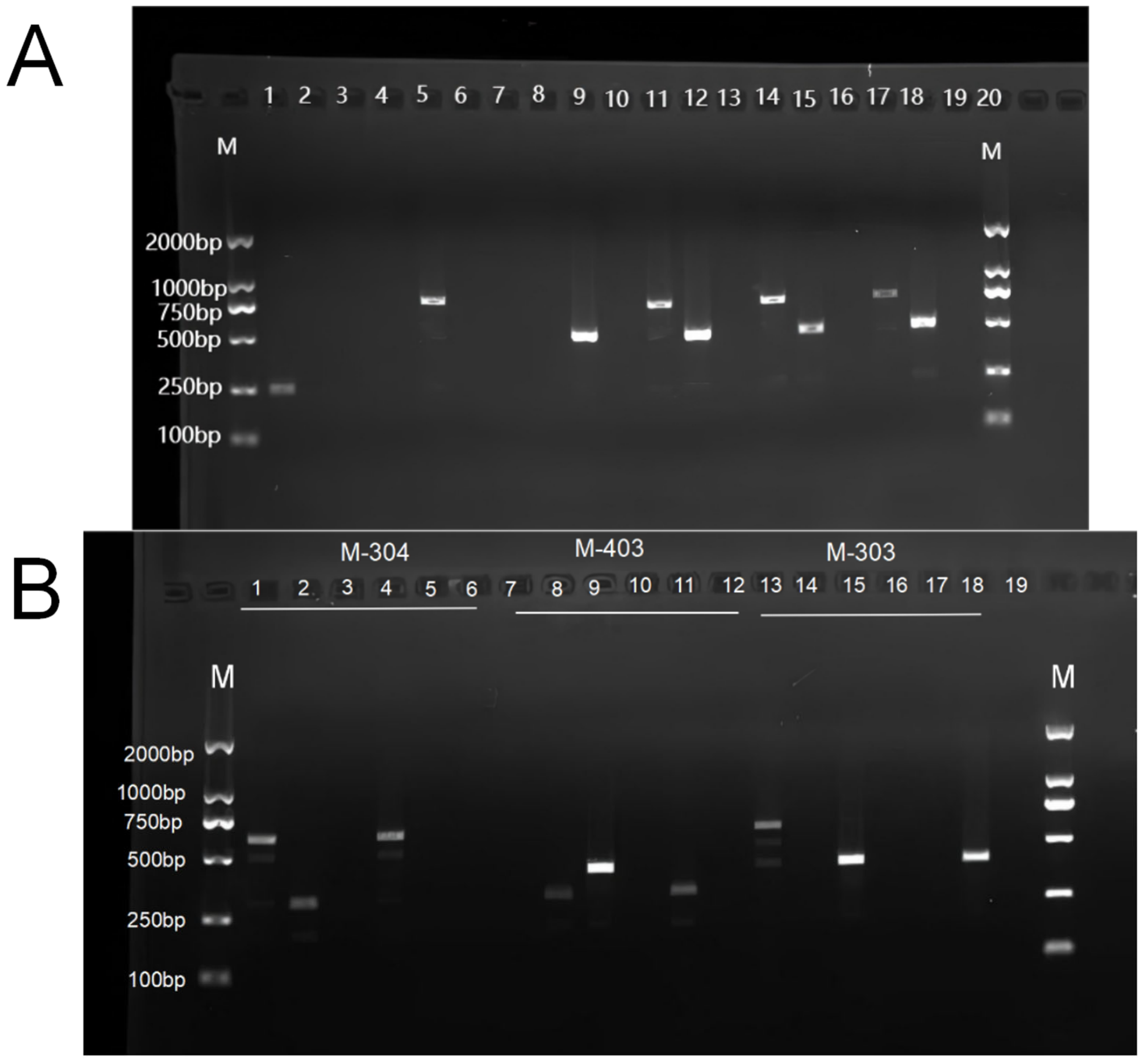
As shown in Figure 2A, three pairs of primers (primer sequences listed in Table A1) were designed based on polymorphic regions in the nuclear genomes of the parental strains. PCR amplification results revealed that all isonuclear alloplasmic strains exhibited two distinct bands, corresponding to those observed in the parental strains A40 and A356. This indicates that the nuclear genomes of all isonuclear alloplasmic strains were derived from both A40 and A356, confirming their nuclear identity. As shown in Figure 2B, mitochondrial genotypes of the isonuclear alloplasmic strains were verified using specific primers (primer sequences listed in Appendix A, Table A1). Electrophoretic analysis indicated that the mitochondrial genome of IA1 was derived from A40-1, IA2 from A356-12, and IA3 from A30-4. These results confirm that IA1, IA2, and IA3 possess identical nuclear genomes but distinct mitochondrial backgrounds, thereby validating their identity as isonuclear alloplasmic strains.

3.2. Morphological Observation of Mycelia and Fruiting Bodies of Isonuclear Alloplasmic Strains

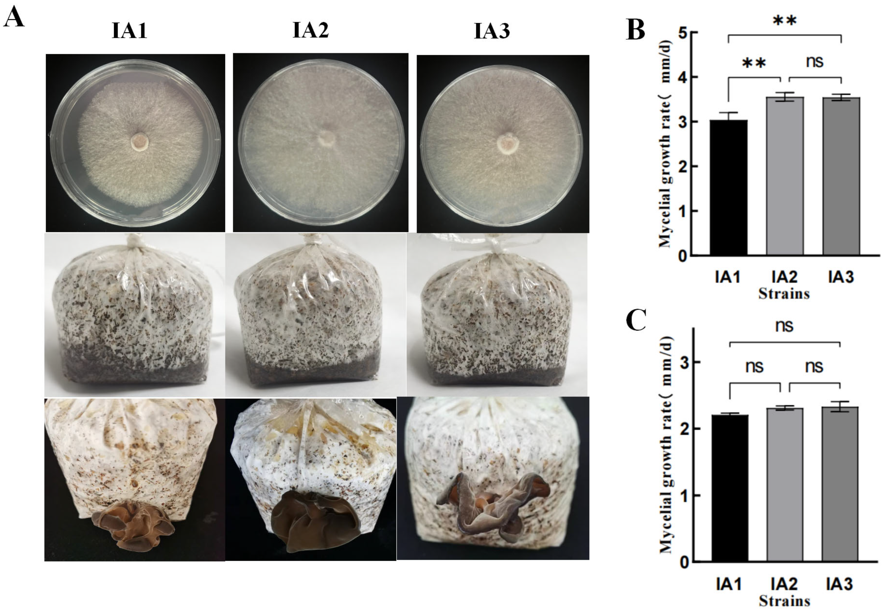
The three isonuclear alloplasmic strains exhibited no major differences in colony morphology on PDA medium (Figure 3A). However, significant differences in growth rate were observed between IA1 and IA2/IA3, whereas no significant difference was detected between IA2 and IA3 (Figure 3B). On sawdust medium, no significant differences in growth rate were observed among the three strains (Figure 3C). The fruiting body morphology also showed no obvious variation: all three strains formed clustered, multi-fold fruiting bodies without wavy margins, consistent with the typical characteristics of A. heimuer. Nonetheless, color differences were evident, with IA1 exhibiting a lighter color compared with IA2 and IA3 (Figure 3A). These results indicate that different mitochondria influence mycelial growth rate of A. heimuer on PDA medium but have no significant effect on mycelial growth rate on sawdust medium.

3.3. Comparative Analysis of Bioactive Component Contents in Isonuclear Alloplasmic Strains

The melanin content, β-glucan content, iron content, and amino acid content of the isonuclear heterokaryotic strains IA1, IA2, and IA3 were determined and comparatively analyzed. As shown in the Table 1, a significant difference in melanin content was observed between strains IA1 and IA3. In terms of β-glucan content, strains IA1 and IA2 exhibited a significant difference, whereas no significant difference was detected between IA1 and IA3. For both iron and amino acid contents, significant differences were identified between strain IA1 and the other two strains (IA2 and IA3), while no significant difference was observed between IA2 and IA3. These results indicate that mitochondrial background influences the accumulation of melanin, β-glucan, iron, and ergosterol in A. heimuer.

3.4. Transcriptome Analysis of Isonuclear Alloplasmic Strains

Compared with the other strains, IA1 and IA2 exhibited significant differences in several traits, including mycelial growth rate on PDA medium, fruiting body color, as well as the contents of melanin, β-glucan, iron, and amino acids. No significant differences were observed between IA2 and IA3. To further explore the effects of mitochondrial variation on A. heimuer, whole-transcriptome sequencing was conducted for IA1 and IA2, which share identical nuclear DNA but differ in mitochondrial DNA, in order to investigate the underlying mechanisms responsible for these phenotypic and metabolic differences.
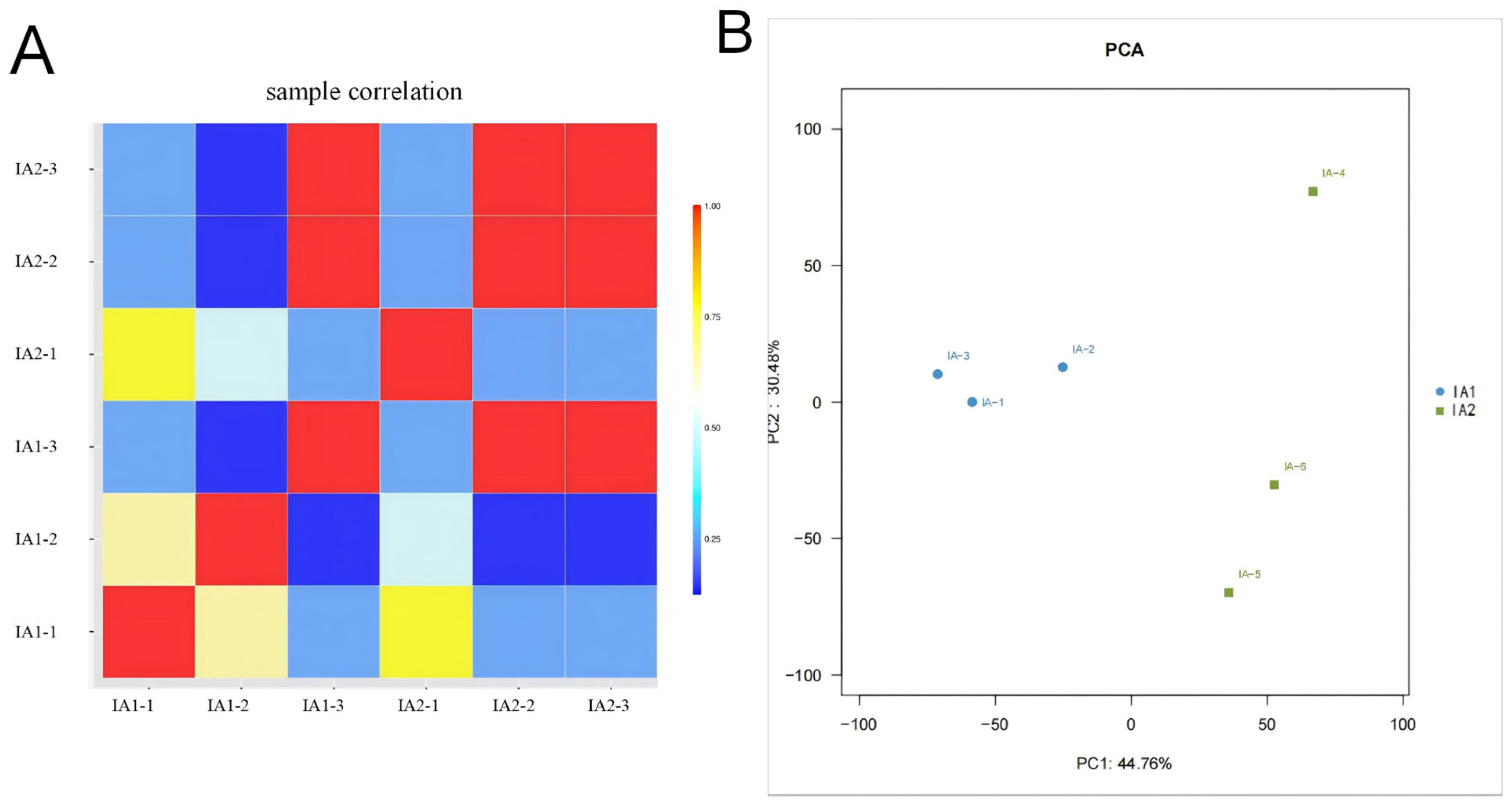
Pearson correlation analysis (Figure 4A) and principal component analysis (PCA, Figure 4B) were performed on the transcriptome data. The results showed high correlations among biological replicates within the same strain (correlation coefficients close to 1), indicating good reproducibility and data reliability. The samples were clearly separated along with the first principal component (PC1), which accounted for 44.76% of the total variance between IA1 and IA2, demonstrating the robustness and suitability of the transcriptome assembly for further analysis.

3.5. Differential Expression Analysis Between Isonuclear Alloplasmic Strains

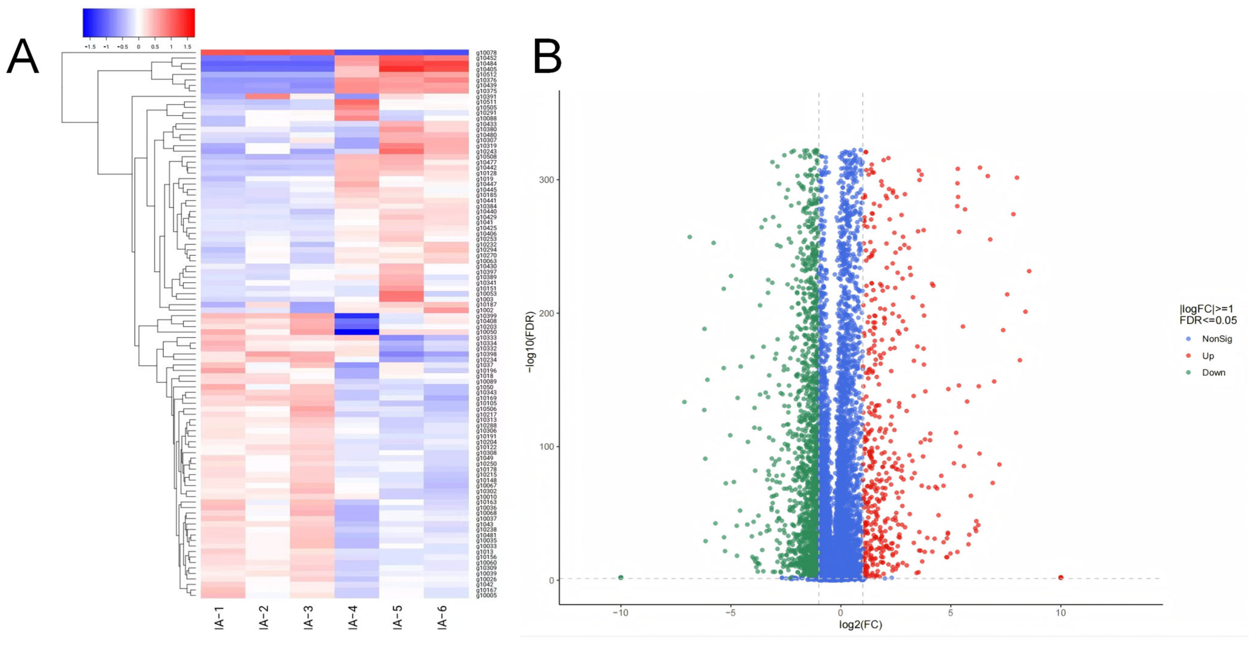
A hierarchical clustering heatmap based on gene expression levels was generated to visualize the transcriptional differences between the two strains (Figure 5A). The samples were clearly separated, indicating distinct gene expression profiles between IA1 and IA2. Volcano plot analysis revealed a total of 3385 differentially expressed genes (DEGs) between IA1 and IA2 (Figure 5B). Compared with IA1, 1295 genes were upregulated, and 2090 genes were downregulated in IA2.

3.6. Enrichment Analysis of DEGs Between Isonuclear Alloplasmic Strains

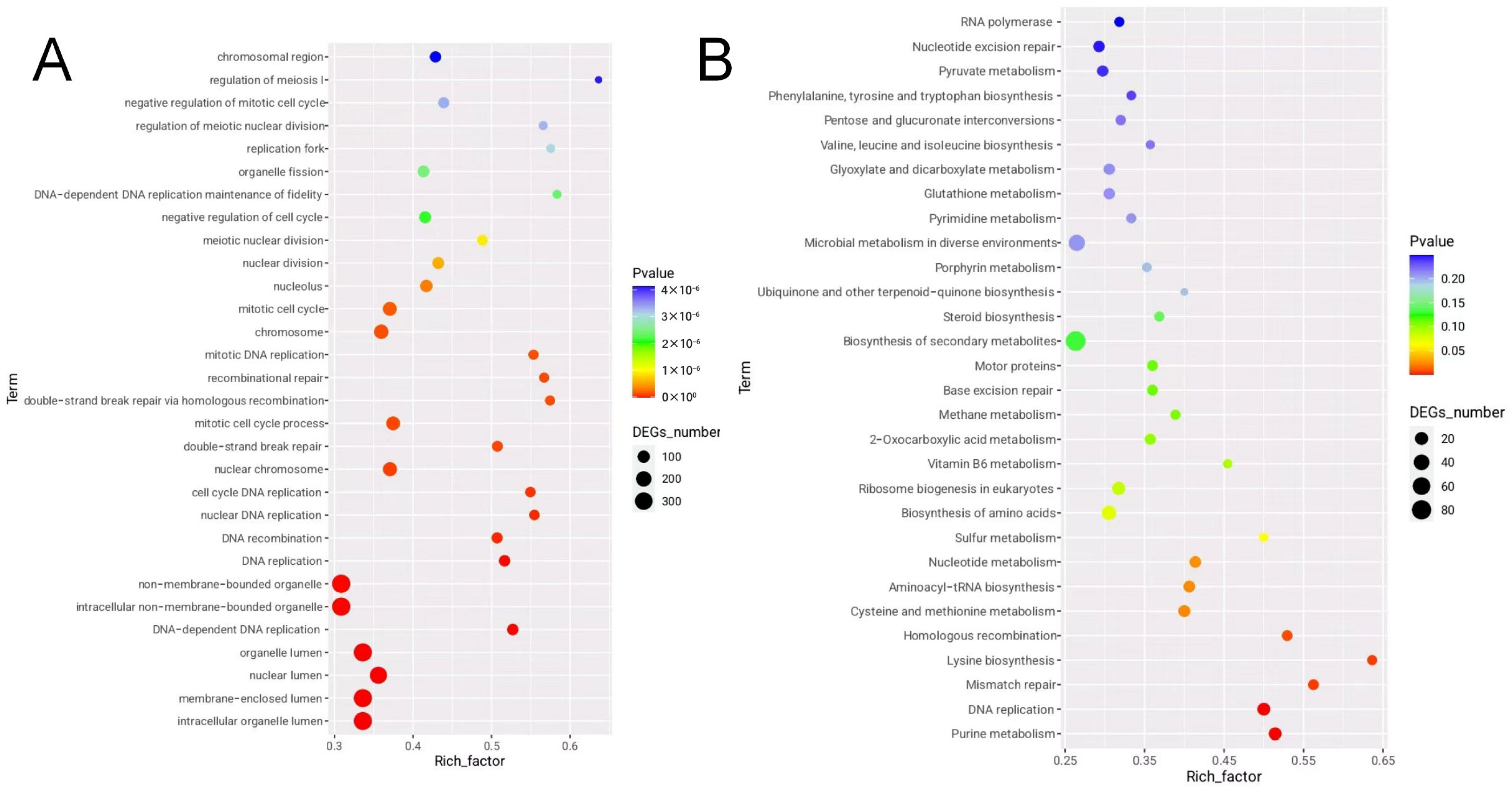
Gene Ontology (GO) enrichment analysis was performed on the differentially expressed genes (DEGs) between IA1 and IA2. The DEGs were categorized into three main GO domains: biological process (BP), cellular component (CC), and molecular function (MF). Terms were filtered based on a p-value < 0.05, and the top categories were ranked by the number of enriched DEGs. The results are shown in (Figure 6A).
In the cellular component category, DEGs were primarily enriched in membrane-related organelles, including the non-membrane-bounded organelle (GO:0043228), intracellular non-membrane-bounded organelle (GO:0043232), organelle lumen (GO:0043233), nuclear lumen (GO:0031981), and membrane-enclosed lumen (GO:0031974). In the biological process category, DEGs were significantly enriched in pathways related to the mitotic cell cycle process (GO:1903047), DNA-dependent DNA replication (GO:0006261), and nuclear DNA replication (GO:0033260). In the molecular function category, the most enriched terms included DNA-dependent ATPase activity (GO:0008094), DNA binding (GO:0003677), and heterocyclic compound binding (GO:1901363).
For KEGG pathway analysis, DEGs were mapped to five major functional categories: metabolism, genetic information processing, environmental information processing, cellular processes, and organismal systems. A total of 108 pathways were identified, including 79 involved in metabolism, 21 in genetic information processing, 2 in environmental information processing, 5 in cellular processes, and 1 in organismal systems. As shown in (Figure 6B), the top 30 significantly enriched KEGG pathways included purine metabolism (ko00230), DNA replication (ko03030), mismatch repair (ko03430), lysine biosynthesis (ko00300), and homologous recombination (ko03440). These results suggest that mitochondrial variation may influence multiple critical cellular functions through transcriptional regulation of nuclear genes.

3.7. Lysine Biosynthesis Pathway Analysis

Differential gene enrichment analysis between the isonuclear alloplasmic strains IA1 and IA2 indicated that mitochondrial variation affects amino acid content, potentially through the lysine biosynthesis pathway (Figure 7). In fungi, lysine is primarily synthesized via the α-aminoadipate (AAA) pathway, which is distinct from the diaminopimelate (DAP) pathway found in plants, bacteria, and lower fungi. A total of seven differentially expressed genes were enriched in the lysine biosynthesis pathway, including g2149, g2508, g7604, g10717, g10755, g12312, and g12494. Notably, g12494 encodes alpha-aminoadipate reductase (AAR), also known as Lys1p, which is a key enzyme in the fungal lysine biosynthesis pathway. AAR catalyzes the conversion of α-aminoadipate to α-aminoadipate semialdehyde, a reaction that requires ATP and NADPH. In addition, g2149 and g2508 encode saccharopine dehydrogenase (SDH), an enzyme that plays a critical role in lysine metabolism. All of these key genes were significantly downregulated in IA2 compared with IA1.

4. Discussion

To investigate the effects of different cytoplasmic backgrounds on key phenotypic and metabolic traits in A. heimuer, three pairs of isonuclear alloplasmic strains (IA1, IA2, and IA3) were constructed and verified. The results demonstrated that variation in cytoplasmic (mitochondrial) background significantly affected mycelial growth rate as well as the contents of melanin, β-glucan, iron ions, and amino acids.
Melanin, β-glucan, and amino acids are among the most important bioactive compounds in A. heimuer and are considered key indicators in the development of functional cultivars. With advances in society and improvements in living standards, increasing attention has been paid to breeding A. heimuer strains with enhanced functional properties. However, the influence of cytoplasmic inheritance on bioactive compound accumulation is often overlooked during conventional breeding. In this study, we found that differences in cytoplasmic background significantly impacted amino acid accumulation, particularly lysine content.
A. heimuer is rich in a variety of amino acids, among which lysine is of particular importance. Transcriptomic analysis revealed that the differentially expressed genes (DEGs) between IA1 and IA2 were mainly enriched in the lysine biosynthesis pathway, suggesting a potential mechanism through which mitochondrial differences influence amino acid accumulation. Notably, gene g12494 encodes alpha-aminoadipate reductase (AAR), a key enzyme in the fungal lysine biosynthesis pathway. AAR catalyzes the reduction in α-aminoadipate to α-aminoadipate semialdehyde, a critical ATP- and NADPH-dependent step in lysine synthesis. This enzyme has also been reported to localize in the cytoplasm of Saccharomyces cerevisiae [36] and Flammulina velutipes [37], implying that variations in cytoplasmic environment may affect its catalytic efficiency, thereby altering lysine accumulation.
In the lysine biosynthesis pathway, the differentially expressed genes g2149 and g2508 encode saccharopine dehydrogenase (SDH), which may influence the terminal step of lysine metabolism and thereby affect glutamate levels. Since glutamate is abundant in A. heimuer, variation in SDH activity could be an important factor contributing to differences in amino acid composition.
Mitochondrial selection could be integrated into breeding programs to accelerate the improvement of A. heimuer. Mitochondrial haplotypes can serve as molecular markers to distinguish maternal lineages and to track cytoplasmic inheritance. Furthermore, identifying mitochondrial genes or sequence variants associated with desirable traits such as faster mycelial growth, higher fruiting body yield, or enhanced stress resistance could provide useful selection criteria. In practice, breeders could combine mitochondrial genotyping with traditional crossing schemes to ensure compatibility between nuclear and mitochondrial genomes, thereby avoiding deleterious nuclear–mitochondrial interactions. Such approaches would not only broaden the genetic basis of breeding but also increase the efficiency and stability of trait improvement.
Although the current transcriptomic data does not fully explain how mitochondrial variation affects mycelial growth rate and other biochemical traits, this study provides valuable insights into the role of mitochondria in fungal development and metabolism. These findings lay the groundwork for future efforts to improve functional traits and enhance bioactive compound content through cytoplasmic selection and mitochondrial engineering.

5. Conclusions

In this study, three pairs of isonuclear alloplasmic strains were successfully constructed and effectively validated using molecular markers. Transcriptomic analysis revealed that different mitochondrial backgrounds had a significant impact on the lysine biosynthesis pathway. The key differentially expressed genes were mainly associated with lysine biosynthetic enzymes, including alpha-aminoadipate reductase (AAR) and saccharopine dehydrogenase (SDH). These findings demonstrate that mitochondria can influence amino acid metabolism by regulating nuclear gene expression, thereby enriching our understanding of the molecular mechanisms underlying nuclear–cytoplasmic interactions. This study provides a theoretical basis and technical support for the development of functional strains and the targeted biosynthesis of lysine and other metabolic products.

Author Contributions

F.Y. designed the experiments; L.L., W.W., J.M. and M.F. revised the manuscript; X.M. guided the experiment; X.S., Y.C. and J.S. formal analysis; K.S. prepared the materials for the experiments, analyzed the data and wrote the manuscript. All authors have read and agreed to the published version of the manuscript.

Funding

This research was funded by the Strategic Priority Research Program of the Chinese Academy of Sciences, grant number XDA28110400 and the earmarked fund for China Agriculture Research System, grant number CARS-20-3.

Data Availability Statement

All experimental data in this study will be made available upon reasonable request from readers.

Acknowledgments

The authors thank the reviewers for their valuable suggestions.

Conflicts of Interest

The authors declare no conflicts of interest.

Appendix A

Table A1. Primer information for nuclear and cytoplasm identification.
Primer NameDifferential ParentingPrimer SequenceProduction Size (bp)
Primer1A30 F: GTATCCTACTTTGTTAGGAC
R: GATATTGTCCAACTCTACTC
254
Primer2A40 F: GTATCCTACTTTGTTAGGAC
R: GATATTGTCCAACTCTACTC
749
Primer3A356 F: GTATCCTACTTTGTTAGGAC
R: GATATTGTCCAACTCTACTC
520
PrimerM-304A356-12, A40-1 F: GTATCCTACTTTGTTAGGAC
R: GATATTGTCCAACTCTACTC
600
PrimerM-403A30-4, A356-12 F: GTATCCTACTTTGTTAGGAC
R: GATATTGTCCAACTCTACTC
302
PrimerM-303A30-4, A40-1 F: GTATCCTACTTTGTTAGGAC
R: GATATTGTCCAACTCTACTC
414

References

  1. Wu, F.; Tohtirjap, A.; Fan, L.-F.; Zhou, L.-W.; Alvarenga, R.L.M.; Gibertoni, T.B.; Dai, Y.-C. Global Diversity and Updated Phylogeny of Auricularia (Auriculariales, Basidiomycota). J. Fungi 2021, 7, 933. [Google Scholar] [CrossRef]
  2. Hermawan, N.; Romulo, A.; Wardana, A.A. Development and texture profile of wood-ear mushroom (Auricularia auricula) sausage formulated with carrageenan. IOP Conf. Ser. Earth Environ. Sci. 2020, 426, 012182. [Google Scholar] [CrossRef]
  3. Khan, A.A.; Lu, L.-X.; Yao, F.-J.; Fang, M.; Wang, P.; Zhang, Y.-M.; Meng, J.-J.; Ma, X.-X.; He, Q.; Shao, K.-S.; et al. Characterization, antioxidant activity, and mineral profiling of Auricularia cornea mushroom strains. Front. Nutr. 2023, 10, 1167805. [Google Scholar] [CrossRef] [PubMed]
  4. Islam, T.; Yao, F.; Kang, W.; Lu, L.; Xu, B. A systematic study on mycochemical profiles, antioxidant, and anti-inflammatory activities of 30 varieties of Jew’s ear (Auricularia auricula-judae). Food Sci. Hum. Wellness 2022, 11, 781–794. [Google Scholar] [CrossRef]
  5. Pak, S.; Chen, F.; Ma, L.; Hu, X.; Ji, J. Functional perspective of black fungi (Auricularia auricula): Major bioactive components, health benefits and potential mechanisms. Trends Food Sci. Technol. 2021, 114, 245–261. [Google Scholar] [CrossRef]
  6. Sun, S.; Zhang, X.; Chen, W.; Zhang, L.; Zhu, H. Production of natural edible melanin by Auricularia auricula and its physicochemical properties. Food Chem. 2016, 196, 486–492. [Google Scholar] [CrossRef]
  7. Yao, H.; Liu, Y.; Ma, Z.F.; Zhang, H.; Fu, T.; Li, Z.; Li, Y.; Hu, W.; Han, S.; Zhao, F. Analysis of nutritional quality of black fungus cultivated with corn stalks. J. Food Qual. 2019, 2019, 9590251. [Google Scholar] [CrossRef]
  8. Xu, S.; Zhang, Y.; Jiang, K. Antioxidant activity in vitro and in vivo of the polysaccharides from different varieties of Auricularia auricula. Food Funct. 2016, 7, 3868–3879. [Google Scholar] [CrossRef]
  9. Islam, T.; Yu, X.; Xu, B. Phenolic profiles, antioxidant capacities and metal chelating ability of edible mushrooms commonly consumed in China. LWT-Food Sci. Technol. 2016, 72, 423–431. [Google Scholar] [CrossRef]
  10. Lu, A.; Shen, M.; Fang, Z.; Xu, Y.; Yu, M.; Wang, S.; Zhang, Y.; Wang, W. Antidiabetic effects of the Auricularia auricular polysaccharides simulated hydrolysates in experimental type-2 diabetic rats. Nat. Prod. Commun. 2018, 13, 195–200. [Google Scholar] [CrossRef]
  11. Yoon, S.-J.; Yu, M.-A.; Pyun, Y.-R.; Hwang, J.-K.; Chu, D.-C.; Juneja, L.R.; Mourão, P.A.S. The nontoxic mushroom Auricularia auricula contains a polysaccharide with anticoagulant activity mediated by antithrombin. Thromb. Res. 2003, 112, 151–158. [Google Scholar] [CrossRef]
  12. Huang, D.; Liu, Z.; Zhang, Y. Review on anti-tumor mechanism of Auricularia auricular Polysaccharide. J. Plateau Agric. 2019, 3, 694–699. [Google Scholar]
  13. Hu, X.; Liu, C.; Wang, X.; Jia, D.; Lu, W.; Sun, X.; Liu, Y.; Yuan, L. Hpyerglycemic and anti-diabetic nephritis activities of polysaccharides separated from Auricularia auricular in diet-streptozotocin-induced diabetic rats. Exp. Ther. Med. 2017, 13, 352–358. [Google Scholar] [CrossRef]
  14. Zou, Y.; Xie, C.; Fan, G.; Gu, Z.; Han, Y. Optimization of ultrasound-assisted extraction of melanin from Auricularia auricula fruit bodies. Innov. Food Sci. Emerg. Technol. 2010, 11, 611–615. [Google Scholar] [CrossRef]
  15. Ma, Y.; Zhang, P.; Dai, X.; Yao, X.; Zhou, S.; Ma, Q.; Liu, J.; Tian, S.; Zhu, J.; Zhang, J.; et al. Extraction, physicochemical properties, and antioxidant activity of natural melanin from Auricularia heimuer fermentation. Front. Nutr. 2023, 10, 1131542. [Google Scholar] [CrossRef]
  16. Wang, J.; Zhang, T.; Liu, X.; Fan, H.; Wei, C. Aqueous extracts of se-enriched Auricularia auricular attenuates D-galactose-induced cognitive deficits, oxidative stress and neuroinflammation via suppressing RAGE/MAPK/NF-κB pathway. Neurosci. Lett. 2019, 704, 106–111. [Google Scholar] [CrossRef]
  17. Joardar, V.; Abrams, N.F.; Hostetler, J.; Paukstelis, P.J.; Pakala, S.; Pakala, S.B.; Zafar, N.; Abolude, O.O.; Payne, G.; Andrianopoulos, A.; et al. Sequencing of mitochondrial genomes of nine Aspergillus and Penicillium species identifies mobile introns and accessory genes as main sources of genome size variability. BMC Genom. 2012, 13, 698. [Google Scholar] [CrossRef]
  18. Ballard, J.W.O.; Whitlock, M.C. The incomplete natural history of mitochondria. Mol. Ecol. 2004, 13, 729–744. [Google Scholar] [CrossRef] [PubMed]
  19. Kouvelis, V.N.; Hausner, G. Editorial: Mitochondrial Genomes and Mitochondrion Related Gene Insights to Fungal Evolution. Front. Microbiol. 2022, 13, 897981. [Google Scholar] [CrossRef]
  20. Chatre, L.; Ricchetti, M. Are mitochondria the Achilles’ heel of the Kingdom Fungi? Curr. Opin. Microbiol. 2014, 20, 49–54. [Google Scholar] [CrossRef] [PubMed]
  21. Bernt, M.; Braband, A.; Schierwater, B.; Stadler, P.F. Genetic aspects of mitochondrial genome evolution. Mol. Phylogenetics Evol. 2013, 69, 328–338. [Google Scholar] [CrossRef] [PubMed]
  22. Steensels, J.; Gallone, B.; Verstrepen, K.J. Interspecific hybridization as a driver of fungal evolution and adaptation. Nat. Rev. Microbiol. 2021, 19, 485–500. [Google Scholar] [CrossRef]
  23. Burger, G.; Gray, M.W.; Lang, B.F. Mitochondrial genomes: Anything goes. Trends Genet. 2003, 19, 709–716. [Google Scholar] [CrossRef]
  24. Soledad, R.B.; Charles, S.; Samarjit, D. The secret messages between mitochondria and nucleus in muscle cell biology. Arch. Biochem. Biophys. 2019, 666, 52–62. [Google Scholar] [CrossRef]
  25. Ng, S.; De Clercq, I.; Van Aken, O.; Law, S.R.; Ivanova, A.; Willems, P.; Giraud, E.; Van Breusegem, F.; Whelan, J. Anterograde and Retrograde Regulation of Nuclear Genes Encoding Mitochondrial Proteins during Growth, Development, and Stress. Mol. Plant 2014, 7, 1075–1093. [Google Scholar] [CrossRef]
  26. Ye, L.; He, X.; Su, C.; Feng, H.; Meng, G.; Chen, B.; Wu, X. The Effect of Mitochondria on Ganoderma lucidum Growth and Bioactive Components Based on Transcriptomics. J. Fungi 2022, 8, 1182. [Google Scholar] [CrossRef] [PubMed]
  27. Liu, J.; Li, T.; Song, L.; Kong, J.; Niu, Q.; Wang, Y.; Wu, C.; Deng, B.; Wang, H.; Gai, Y. Comparative Transcriptome of Isonuclear Alloplasmic Strain Revealed the Important Role of Mitochondrial Genome in Regulating Flammulina filiformis. Agronomy 2023, 13, 998. [Google Scholar] [CrossRef]
  28. Ito, S.; Del Bino, S.; Hirobe, T.; Wakamatsu, K. Improved HPLC Conditions to Determine Eumelanin and Pheomelanin Contents in Biological Samples Using an Ion Pair Reagent. Int. J. Mol. Sci. 2020, 21, 5134. [Google Scholar] [CrossRef]
  29. Koenig, S.; Rühmann, B.; Sieber, V.; Schmid, J. Quantitative assay of β-(1,3)–β-(1,6)–glucans from fermentation broth using aniline blue. Carbohydr. Polym. 2017, 174, 57–64. [Google Scholar] [CrossRef]
  30. Peng, B.; Shen, Y.; Gao, Z.; Zhou, M.; Ma, Y.; Zhao, S. Determination of total iron in water and foods by dispersive liquid–liquid microextraction coupled with microvolume UV–vis spectrophotometry. Food Chem. 2015, 176, 288–293. [Google Scholar] [CrossRef] [PubMed]
  31. Huarachi-Olivera, R.; Teresa Mata, M.; Ardiles-Candia, A.; Escobar-Méndez, V.; Gatica-Cortes, C.; Ahumada, M.; Orrego, J.; Vidal-Veuthey, B.; Cárdenas, J.P.; González, L.; et al. Modification of the Trizol Method for the Extraction of RNA from Prorocentrum triestinum ACIZ_LEM2. Int. J. Mol. Sci. 2024, 25, 9642. [Google Scholar] [CrossRef] [PubMed]
  32. Chen, S.; Zhou, Y.; Chen, Y.; Gu, J. fastp: An ultra-fast all-in-one FASTQ preprocessor. Bioinformatics 2018, 34, i884–i890. [Google Scholar] [CrossRef]
  33. Pertea, M.; Pertea, G.M.; Antonescu, C.M.; Chang, T.-C.; Mendell, J.T.; Salzberg, S.L. StringTie enables improved reconstruction of a transcriptome from RNA-seq reads. Nat. Biotechnol. 2015, 33, 290–295. [Google Scholar] [CrossRef] [PubMed]
  34. Li, B.; Dewey, C.N. RSEM: Accurate transcript quantification from RNA-Seq data with or without a reference genome. BMC Bioinform. 2011, 12, 323. [Google Scholar] [CrossRef]
  35. Love, M.I.; Huber, W.; Anders, S. Moderated estimation of fold change and dispersion for RNA-seq data with DESeq2. Genome Biol. 2014, 15, 550. [Google Scholar] [CrossRef]
  36. Bhattacharjee, J.K. α-Aminoadipate Pathway for the Biosynthesis of Lysine in Lower Eukaryotes. CRC Crit. Rev. Microbiol. 1985, 12, 131–151. [Google Scholar] [CrossRef] [PubMed]
  37. Shaba, C.; Urban, P.L. Metabolic remodeling during fructification of enoki mushroom. Food Chem. 2025, 486, 144613. [Google Scholar] [CrossRef]
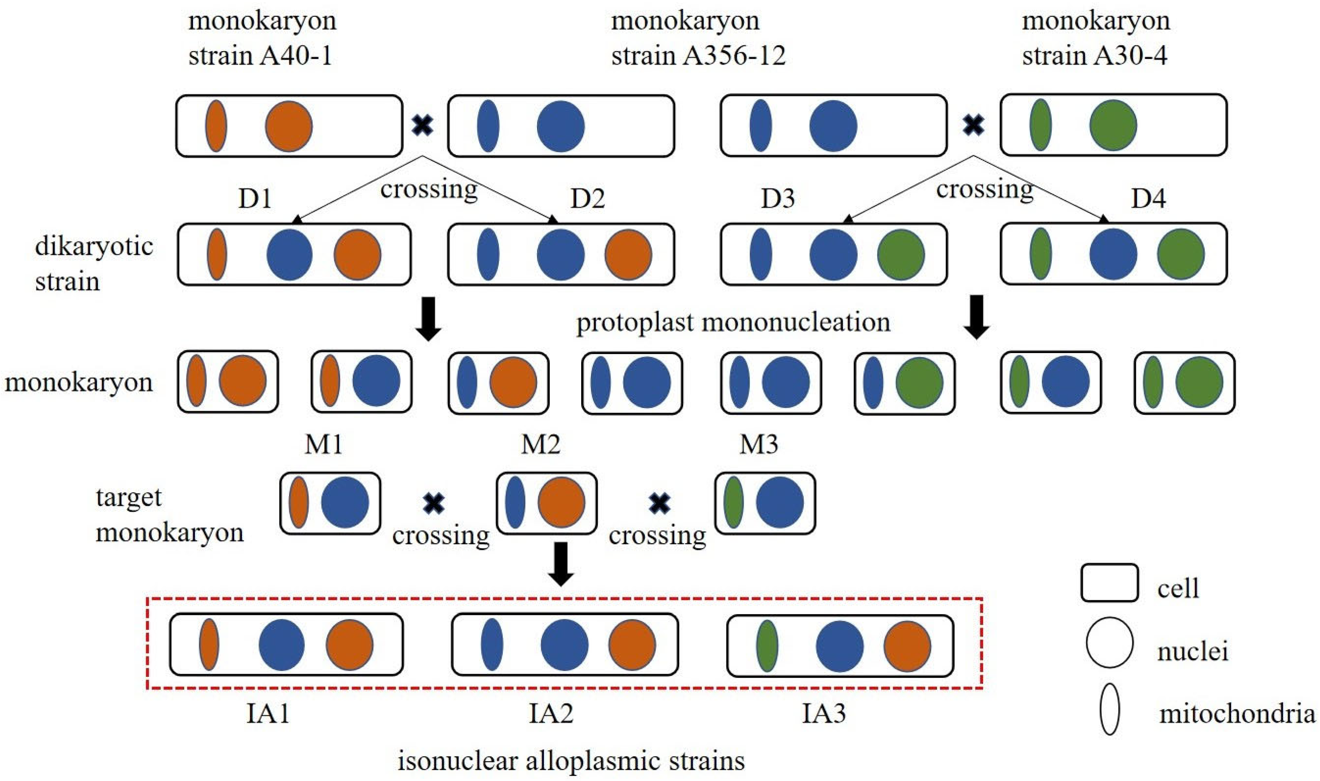
Figure 1. Schematic diagram of isonuclear alloplasmic strains construction. Note: The different colors in the figure represent different nuclei and mitochondria.
Agronomy 15 02188 g001
Figure 2. (A) Verification of nuclear origin; (B) Verification of mitochondrial origin. Note: M is 2000 DNA molecular markers; primer 1 was designed with a genome-specific fragment of parental A30 in lanes 1, 4, 7, 10, 13, and 16; primer 2 was designed with a genome-specific fragment of parental A40 in lanes 2, 5, 8, 11, 14, and 17; and primer 3 was designed with a genome-specific fragment of parental A356 in lanes 3, 6, 9, 12, 15, and 18. where the template DNA of lanes 1–3 was A30, the template DNA of lanes 7–9 was A356, and the template DNA of lanes 13–15 was IA2. A40 for template DNA in lanes 4–6, A356 for template DNA in lanes 7–9, IA1 for template DNA in lanes 10–12, IA2 for template DNA in lanes 13–15, and IA3 for template DNA in lanes 16–18, and lane 19 was used as a control with sterile water. Primer M-304 was designed with the mitochondrial difference between mononuclear parent A356-12 and mononuclear parent A40-1, primer M-403 was designed with the mitochondrial difference between mononuclear parent A30-4 and mononuclear parent A356-12, and primer M-303 was designed with the mitochondrial difference between mononuclear parent A30-4 and mononuclear parent A40-1. They were distributed in lanes 1–6, 7–12, and 13–18, respectively. template DNA in lanes 1, 7, and 13 was A40-1, template DNA in lanes 2, 8, and 14 was A356-12, template DNA in lanes 3, 9, and 15 was A30-4, template DNA in lanes 4, 10, and 16 was IA1, template DNA in lanes 5, 11, and 17 was IA2, and template DNA in lanes IA3 for template DNA in lanes 6, 12, and 18, and a sterile water control in lane 19.
Agronomy 15 02188 g002
Figure 3. Biological characteristics of isonuclear alloplasmic strains. (A) Mycelial growth morphology and ascospore morphology. (B) Growth rate of mycelium on PDA medium. (C) Growth rate of mycelium in wood chip medium. Note: ns indicates no significant difference; ** p ≤ 0.01.
Agronomy 15 02188 g003
Figure 4. Sample relationship analysis. (A) Pearson correlation analysis. (B) Principal component analysis.
Agronomy 15 02188 g004
Figure 5. Differential genes between isonuclear alloplasmic strains. (A) Differential gene clustering heat map. (B) Differential gene volcano map.
Agronomy 15 02188 g005
Figure 6. Enrichment analysis of DEGs. (A) GO enrichment of DEGs. (B) Bubble plot of KEGG enrichment of DEGs.
Agronomy 15 02188 g006
Figure 7. The lysine synthesis pathway.
Agronomy 15 02188 g007
Table 1. Comparison of different component contents between isonuclear alloplasmic strains.
Strain IDMelanin Content (mg/g)β-Glucan Content (%of Dry Weight)Iron Content (mg/kg)Amino Acid Content (g/100 g)
IA11.25 ± 0.08 b10.35 ± 1.69 b56.84 ± 5.09 b10.64 ± 0.06 b
IA21.68 ± 0.18 a15.68 ± 3.11 a65.49 ± 3.28 a9.45 ± 0.08 a
IA31.54 ± 0.17 ab14.33 ± 0.41 ab64.13 ± 1.30 a9.42 ± 0.09 a
Note: Different superscript letters (a, b) within the same column indicate significant differences (p < 0.05). The same letters indicate no significant difference.
Disclaimer/Publisher’s Note: The statements, opinions and data contained in all publications are solely those of the individual author(s) and contributor(s) and not of MDPI and/or the editor(s). MDPI and/or the editor(s) disclaim responsibility for any injury to people or property resulting from any ideas, methods, instructions or products referred to in the content.