Article

Expression and Functional Analysis of the ABORTED MICROSPORES (AMS) Gene in Marigold (Tagetes erecta L.)

1
Plateau Flower Research Centre, Qinghai University, Xining 810016, China
2
Qinghai Key Laboratory of Garden Plants and Ornamental Horticulture, Xining 810016, China
*
Author to whom correspondence should be addressed.
Agronomy 2025, 15(9), 2058; https://doi.org/10.3390/agronomy15092058
Submission received: 30 July 2025 / Revised: 25 August 2025 / Accepted: 25 August 2025 / Published: 26 August 2025

Abstract

Male sterility is an important trait in heterosis utilization and marigold (Tagetes erecta L.) breeding. Currently, most male-sterile lines used in production are derived from natural mutations. ABORTED MICROSPORES (AMS) is an important gene that regulates tapetum and microspore development. Therefore, the effect of AMS on fertility was studied. TeAMS was located in the nucleus and exhibited self-activation activity. TeAMS was highly expressed in the flower buds of T. erecta. The expression of this gene in fertile plants was higher than that in sterile plants, and the expression level gradually increased with the development of flower buds. The expression level of TeAMS was highest in the flower buds with a diameter of 1.2 cm at the floret differentiation stage, while the expression level was extremely low in the flower buds with a diameter of 1.6 cm. The expression trend of TeAMS in sterile plants was opposite to that in fertile plants. At the inflorescence primordium differentiation stage, flower buds with a diameter of 0.2 cm had the highest expression level, and the stem tip had the lowest expression level. In tobacco (Nicotiana tabacum L.), overexpression of the TeAMS gene resulted in shortened floral tubes, increased thousand-seed weight, a reduced flowering period, and decreased flower numbers. The pollen viability of transgenic tobacco was significantly lower than that of the wild type, and the pollen grains were smaller and showed irregular shapes. The pollen wall was dry and shrunk. Some pollen germinal furrows were distorted, and a few were almost invisible. Silencing TeAMS resulted in a longer flowering period in tobacco, reduced thousand-seed weight, and high pollen viability. Pollen morphology in silenced lines showed no significant differences compared to the wild-type and empty vector controls. Only a few pollen grains were smaller, shriveled, and shrunken. Therefore, the TeAMS gene plays an important role in regulating the fertility of marigolds. This study provides a theoretical foundation for breeding marigold male-sterile lines.

1. Introduction

Marigold (Tagetes erecta L.), an annual herbaceous plant of the genus Tagetes in the Asteraceae family, has large, vibrantly colored flowers, superior stress resistance, easy cultivation, high ornamental value, a long flowering period, and extensive cultivar diversity. These attributes make it widely applicable to ornamental landscaping projects [1]. In addition, marigold flowers are rich in lutein, which is widely used in food, medical, biopharmaceutical, and other industries [2,3]. Emasculation in marigold crossbreeding is very difficult to achieve because of its capitulum. Male-sterile dual-purpose lines are usually used as parents to breed heterosis varieties. Therefore, male sterility is an important trait for marigold crossbreeding [4]. In order to ensure the purity of the F1 generation, it is necessary to manually remove fertile plants, build a net room, and pollinate marigolds during hybrid seed production, which is time-consuming and laborious [5]. Pollination should be arranged according to the time of pollen release, number of flowers, temperature, and humidity; otherwise, incomplete pollination of some sterile plants will occur. Therefore, the breeding and utilization of stable genetically male-sterile lines are of great significance for marigold seed production.
Male sterility (MS) is a phenomenon in which male reproductive organs (such as anthers and pollen) are dysfunctional and unable to produce normal fertile pollen due to genetic or environmental factors during the reproductive development of plants. Male sterility in marigolds is controlled by a pair of recessive nuclear genes (msms) [6], and the male sterile system is known as the ‘dual-purpose line’ [7]. In recent years, with the development of molecular biotechnology, scholars worldwide have studied marigold male sterility-related genes and molecular regulation mechanisms at the molecular level. He et al. [6] employed Amplified Fragment Length Polymorphism (AFLP) molecular markers to conduct fine-scale mapping of the Tems gene encoding male sterility traits in an F2 segregating population. Sumalatha [8] identified a marker, CPSSR-39, closely linked to the male sterility locus, which is consistent with the gene that controls male sterility of the petal type. Wang et al. [9] developed RAPD markers to identify male and fertile plants. Cholin [10] verified the SCAR marker associated with male sterility in marigold and confirmed the relationship between the marker and the sterile site. Tang et al. [11] identified candidate genes related to marigold fertility through transcriptome sequencing and proteome quantification, in which the anther development-related gene TeAMS showed significantly low expression in male sterile plants.
Pollen development begins with an anther primordium, which includes three layers of cells: L1, L2, and L3. The L1 layer differentiates into epidermal cells, the L2 layer differentiates into pollen grains and some anther wall structures, and the L3 layer finally differentiates into anther vascular bundles and supporting tissues [12]. The pollen mother cells in the L2 layer release mononuclear microspores, the tapetum begins to degrade, and the pollen outer wall begins to form, which then develops into mature pollen grains through the binucleate stage [13,14]. In the anther development process, the tapetum-specific gene (aborted microspores, AMS) is a key factor in tapetum and microspore development and in pollen wall synthesis. AMS belongs to the MYC subfamily of bHLH transcription factors [15]. Mutations in this gene lead to premature tapetum degradation and pollen abortion, which leads to male plant sterility [16]. The AMS gene plays an important role in regulating fertility in different species. Guo et al. [17] verified the biological function of CaAMS in pepper (Capsicum annuum L.) using VIGS technology. Their results showed that the filaments of the silenced C. annuum were shortened and shrunken. The stamens did not crack, and the pollen was aborted. Several genes involved in the formation of pollen exine in the anthers of the silenced plants were downregulated. Bao et al. [18] carried out bioinformatic analysis of SlAMS and verified the biological function of SlAMS in tomato (Lycopersicon esculentum (Mill.)) anther development using VIGS, CRISPR/Cas9, and overexpression technology. The results showed that the pollen viability of silencing, gene knockout, and overexpression SlAMS plants was significantly lower than that of wild-type plants, which proved that the downregulation or upregulation of the SlAMS gene in tomato may lead to abnormal pollen development, thereby reducing pollen viability and producing male sterile lines. In summary, the AMS gene plays a crucial role in the regulatory network of tapetum and pollen wall development in different species.
In our previous study, we used transcriptome sequencing combined with proteome quantification to screen out candidate genes related to marigold fertility. Among these genes, TeAMS, a gene related to anther development with significant differential expression in male-sterile and fertile marigold plants, was identified [11]. Based on this, the basic characteristics of TeAMS were clarified using subcellular localization, transcriptional self-activation, and expression analyses. The biological function of TeAMS was preliminarily verified using stable transformation of tobacco and VIGS to explore the molecular mechanism of TeAMS in marigold fertility formation. These results potentially deepen our understanding of the AMS function in plant fertility.

2. Materials and Methods

2.1. Plant Material

The male sterile line ‘2-2’, wild type 95 tobacco, and N. benthamiana, provided by Plateau Flower Research Center of Qinghai University, were selected as test materials. In September 2023, seedlings of line ‘2-2’ were transplanted at the four-leaf stage. A total of 40 plants were used in this study. Four plants were planted in each pot and cultured in an intelligent light incubator (RTOP-268Y, Top Cloud Agricultural Technology Co., Ltd., Hangzhou, China). The environmental parameters were set as follows: temperature, 25 °C; relative humidity, 60%; light intensity, 160 μmol m−2 s−1; and light duration, 14 h. Seedlings of N. benthamiana were raised in plug trays in September 2024.

2.2. RNA Preparation

A total RNA extraction kit (TaKaRa, Beijing, China) was used to extract RNA from the buds of the ‘2-2’ fertile plants and sterile plants at different developmental stages. The quality and concentration of RNA were determined using 1% agarose gel electrophoresis and an ultramicro-spectrophotometer (TIANGEN, Beijing, China). The first strand of cDNA was synthesized using a FastKing one-step genomic cDNA premix kit (TIANGEN, Beijing, China).

2.3. Expression Analysis of TeAMS

The morphological differentiation of marigold inflorescences and florets was observed using a stereomicroscope. Roots (young roots), stems (upper stems), leaves (young leaves), florets, and buds of fertile and sterile plants were collected when the first flower opened, and the relative expression of the TeAMS gene in different tissues was determined. Flower buds at different developmental stages (inflorescence primordium differentiation stage/IP, inflorescence formation stage/IF, and floret differentiation stage/F) were collected to determine the relative expression of TeAMS genes at different developmental stages. Primers YAM-F and YAM-R (Table S1) were designed based on the TeAMS gene sequence. The expression pattern of the TeAMS gene in the marigold male-sterile dual-purpose line was determined using SYBR Green (FP209, TIANGEN, Beijing, China), with 18S as the internal reference gene. Pre-denaturation was performed in a fluorescence quantitative PCR instrument (LongGene Q2000, LongGene Scientific Instruments Co., Hangzhou, China) at 95 °C for 5 min, followed by 40 cycles at 95 °C for 10 s, 60 °C for 20 s, and 72 °C for 30 s. The 2−ΔΔct method was used to calculate relative gene expression [19]. qRT-PCR reactions were biologically repeated 3 times. OriginPro 2025 was used for single-factor analysis of variance, and GraphPad Prism 9.5 software was used for plotting.

2.4. Subcellular Localization of TeAMS

Primer AMS-2300-F/R (Table S1) was designed based on the TeAMS CDS sequence and the PCAMBIA2300-GFP vector sequence. PCR amplification was performed using the cDNA of fertile flower buds as a template, and the target fragment was recovered using a glue recovery kit (TaKaRa). The vector PCAMBIA2300-GFP was double-digested with Sac I and Xba I restriction endonucleases (TaKaRa). After the enzyme digestion reaction was completed, the PCR product was detected by electrophoresis, and the target fragment was purified using a gel recovery kit (the recovered product was recorded as 2300-eGFP). The recovered AMS product and the vector digestion product 2300-eGFP were used to construct the PCAMBIA2300-GFP-TeAMS vector by homologous recombination. Competent E. coli cells were transformed via heat shock and plated onto LB agar plates. Bacterial liquid PCR detection was performed using gene-specific primers (AMS-2300-F/R), followed by Sanger sequencing. A plasmid extraction kit (TaKaRa) was used to extract the positive bacterial plasmids, which were then transferred into Agrobacterium GV3101 using the freeze–thaw method [20]. Tobacco plants with good growth were selected, and the bacterial solution was injected into the lower epidermis of the tobacco leaves using a 1 mL syringe without a needle. Tobacco leaves were cultured in the dark for 2 days, and then observed and photographed under a laser confocal microscope.

2.5. Verification of Transcription Self-Activation of TeAMS

According to the TeAMS gene sequence and BD vector map, the restriction endonuclease EcoR I and BamH I linearized vector BD were selected and purified. The target gene was ligated to the linearized vector using gene synthesis technology to obtain the recombinant vector BD-AMS. The recombinant product was transformed into E. coli DH5α competent cells and verified by sequencing to obtain the recombinant vector plasmid BD-AMS. The recombinant vector BD-AMS plasmid was transformed into yeast competent Y2H Gold cells using the PEG/LiAc method [21]. BD-Lam and AD-T empty plasmids were simultaneously transformed as negative controls, and BD-53 and AD-T were co-transformed as positive controls. Each component was added according to Table S2 and mixed slowly. Then, a 50 mL suspension of each was drawn and coated on SD/-Trp/-Leu/X-α-Gal (DDO/X) and SD/-Trp/-Leu/-His/-Ade/X-α-Gal/AbA (QDO/X/A) plates, and the plates were inverted at 30 °C until colonies appeared (3–5 d). Yeast color change was observed to determine the self-activation activity of TeAMS.

2.6. Construction of TeAMS-sGFP-1300 Overexpression Vector

Based on the TeAMS gene sequence, primers T-AMS-F/R (Table S1) were designed using Primer Premier 5.0 software. The homologous recombination arm sequence of the linear vector was added to the 5’ end of the primer, and the TeAMS fragment was amplified by pfu polymerase DNA (TaKaRa) using the cDNA of marigold ‘2-2’ fertile flower buds as the template. The amplification product was verified using 1% agarose gel electrophoresis, and the target fragment (the product was recorded as DNA-AMS1) was purified and recovered using a gel-cutting recovery kit. The overexpression vector sGFP-1300 was subjected to a single enzyme digestion reaction using Sal I restriction endonuclease (TaKaRa). The digested product was denoted as AMS-1300. A homologous recombination cloning kit (TransGen Biotech, Beijing, China) was used to recombine the target fragment recovery product DNA-AMS1 and the vector linearization product AMS-1300. The product was immediately transformed into Trans1-T1 competent cells and cultured at 37 °C for 12–16 h. Through colony PCR screening, positive clones were selected for sequencing verification, and the recombinant plasmid sGFP-1300-TeAMS was extracted.

2.7. Transformation of Tobacco Using the Leaf Disc Method

The overexpression vector sGFP-1300-TeAMS was transferred into Agrobacterium GV3101. The transformed bacterial solution was placed in a cradle at 28 °C (200 rpm) for 3 h and then centrifuged at 4000 rpm for 10 min to collect the bacteria. The precipitate was evenly coated on an LB solid medium containing 50 μg/mL kanamycin and cultured for 48 h at 28 °C in the dark. A single colony was selected for PCR verification. The positive bacterial solution was inoculated into 50 mL LB liquid medium containing 50 μg/mL kanamycin and cultured for 14 h at 28 °C in the dark. The bacteria were collected by centrifugation at 4000 rpm, and the supernatant was discarded and resuspended in Murashige and Skoog (MS) basic medium (containing 100 μM acetosyringone). The bacterial suspension OD600 was 0.6.
The leaves of aseptic tobacco seedlings were cut into 0.5 cm × 0.5 cm pieces on an ultra-clean bench, pre-cultured on MS medium + 2 mg/L 6-BA + 0.2 mg/L NAA (pH 5.8) for 3 d, and then transferred to Agrobacterium infection solution for 15 min. After infection, the leaves were clipped with tweezers, placed on sterile filter paper to dry the bacterial solution from the leaf surface, and then quickly transferred to the co-culture medium (MS + 2 mg/L 6-BA + 0.2 mg/L NAA, pH 5.8). After 3 days of culture in the dark, the surface of each leaf was rinsed with sterile water 3 times, soaked in 400 mg/L for 10 min, and rinsed with sterile water 3 times. After cleaning, the surfaces were dried using sterile filter paper. The medium was transferred to the resistance screening medium containing Timentin and hygromycin (MS + 2 mg/L 6-BA + 0.2 mg/L NAA + 200 mg/L Timentin + 20 mg/L hygromycin, pH 5.8) for screening culture. The medium was changed every 7 days for 30–40 days. When the resistant buds grew to 2–3 cm, they were transferred to a rooting medium (1/2MS + 2 mg/L 6-BA + 0.2 mg/L NAA + 200 mg/L Timentin + 20 mg/L hygromycin, pH 5.8). Finally, the aseptic tobacco seedlings were transplanted into a 6 cm × 6 cm plug tray and cultured in a growth chamber for 2 weeks. The culture conditions were the same as those listed in Section 2.1. Leaf genomic DNA was extracted using a plant tissue PCR rapid extraction kit (TransGen Biotech, Beijing, China). Positive plants (Table S1) were detected using TeAMS gene-specific primers and sGFP-1300 vector primers. Wild-type tobacco was used as the control.

2.8. Expression Analysis and Phenotypic Characterization of TeAMS-Transgenic Tobacco

Flower buds of transgenic tobacco and wild-type plants were collected to determine the relative expression of TeAMS. The fluorescence quantitative method was performed as described in Section 2.3. At the full bloom stage, flower diameter and flower tube length were determined, and the number of flowers was counted. Thousand-seed weight was measured using a ten-thousandth balance. Single-factor analysis of variance was performed using OriginPro 2025, and graphing was performed using GraphPad Prism 9.5 software.

2.9. Pollen Viability and Morphology of Transgenic Tobacco

Pollen viability was determined using the TTC staining method [22]. A 0.5% TTC staining solution was prepared and stored at 4 °C in the dark. Anthers at the full-bloom stage were collected and placed in sunlight to collect pollen after anther dehiscence. The appropriate amount of pollen was transferred to the bottom of a clean PCR tube using sterilized toothpicks, and 100 μL of 0.5% TTC staining solution was added. After being fully mixed at a low speed using a vortex oscillator, the pollen was stained in the dark at 37 °C for 15 min, and 2–3 drops were placed on a clean slide. After uniform coating, microscopic examination was performed. Viable pollen was red, while inviable pollen was not colored. Three microscopic fields of view were selected for each sample for observation and statistical analysis.
Pollen viability was determined using an in vitro pollen germination method [23]. Pollen samples from both transgenic and CK plants were grown in vitro on a pollen germination medium with 0.8% agar. After incubation at 28 °C for 2 h in the dark, the germination status of the pollen samples was observed and photographed using an EX30 biological microscope (SOPTOP, Ningbo, China). Pollen germination was considered when the length of the pollen tube was greater than the diameter of the pollen grains. For each sample, three microscopic fields of view were selected for observation and statistical analysis. The mean germination rate was calculated as an activity evaluation index.
Pollen morphology was observed using scanning electron microscopy [24]. Fresh pollen samples from wild-type and transgenic tobacco were evenly distributed on a conductive adhesive sample table. After vacuum metal coating treatment, field emission scanning electron microscopy (JSM-7900F, Japan Electronics Corporation, Tokyo, Japan) was used for microscopic imaging analysis.

2.10. Construction of the pTRV2-TeAMS VIGS Vector and Transformation of N. benthamiana

Primers were designed using the SGN-VIGS (https://vigs.solgenomics.net/ (accessed on 18 July 2024)) online program according to the sequence of TeAMS and the TRV2 vector. A 300 bp fragment of the non-conservative domain was selected as the target fragment. Restriction enzyme sites, EcoR I and BamH I, were added. The primer AMS-TRV2-F/R (Table S1) was synthesized by Shanghai Shenggong Biological Company (China). The TeAMS fragment was amplified with high-fidelity PCR using cDNA of marigold ‘2-2’ fertile flower bud as template. The PCR amplification product was detected using 1% agarose gel electrophoresis, and the target fragment was purified and recovered (the recovered fragment was recorded as DNA-AMS2). The recovered product was stored in a refrigerator at −20 °C. The plasmid DNA of the pTRV2 vector was extracted using a plasmid extraction kit, and the plasmid was double-digested with EcoR I/BamH I (TaKaRa). The linearized vector was named pTRV2-AMS. The target gene fragment DNA-AMS2 was mixed with the linearized vector pTRV2-AMS according to the recombination reaction system. The reaction solution was placed in a PCR instrument at 50 °C for 20 min, and the product was transferred to DH5α competent cells after a brief ice bath. It was spread evenly on LB solid medium containing kanamycin and cultured at 37 °C for 12–16 h. Positive clones were screened using colony PCR (Table S1: AMS-TRV2-F/R) and verified by sequencing. Finally, Agrobacterium GV3101 was transformed using the freeze–thaw method (TaKaRa, Beijing, China).
The bacterial solution was activated with pTRV1 (auxiliary vector), pTRV2 (empty vector), and pTRV2-TeAMS (experimental group). pTRV1 was mixed with pTRV2 and pTRV2-TeAMS in a 1:1 ratio and placed in the dark at room temperature for 2–4 h. Healthy N. benthamiana plants with uniform growth were selected for injection. Before infection, the plants were placed under a white fluorescent lamp for 1 h to open stomata. The 3rd and 4th leaves from the top of the plants were selected as infection targets. Two functional leaves were treated for each plant.
A needleless syringe was used to gently rub the target injection area (0.5 cm2) on the back of selected leaves. Then, 1 mL of Agrobacterium solution was obtained using a needleless syringe and injected at a uniform speed into the marked area. After the leaf tissue was soaked in the bacterial solution, the infection area was delineated using a marker pen. The leaves were sprayed with water and covered with fresh-keeping bags. After 24 h of dark culture, the leaves were transferred to an artificial climate chamber for normal culture. Wild-type plants were co-cultured without any treatment as negative controls.

2.11. Molecular Detection of TRV2

To verify successful TRV2 infection and whether it was expressed in N. benthamiana leaves, the new inner and middle leaves of WT, TRV::00 empty vector, and pTRV-TeAMS-inoculated plants were sampled. RNA was extracted and reverse-transcribed into cDNA (TIANGEN, Beijing, China). Primers for the TRV coat protein (Table S1) were used for PCR amplification. The amplified products were detected by 1% agarose gel electrophoresis.

2.12. Determination of TeAMS VIGS Expression Efficiency

The expression level of TeAMS in the silenced plants was detected using qRT-PCR with cDNA from the young leaves of the silenced plants as a template. Empty TRV::00 and WT plants were used as controls. The ACTIN gene was used as the internal reference gene (Table S1), and YAM was used as the fluorescence quantitative detection primer (Table S1). The fluorescence quantitative method was the same as that described in Section 2.3.

2.13. Plant Phenotype, Pollen Viability, and Pollen Morphology of the Silenced N. benthamiana Plants

Phenotypic observations, pollen viability determination, and scanning electron microscopy observations of pollen morphology were performed using the same methods described in Section 2.8 and Section 2.9.

3. Results

3.1. Morphological Differentiation Process and Expression Analysis of Marigold Inflorescence and Florets

In this study, marigold flower bud differentiation began in the eighteen-leaf stage. The growing point of the stem tip began to swell gradually and presented a hemispherical shape. Inflorescence primordium (IP/K1-K3) was formed at this stage. At the inflorescence formation stage (IF/K4-K6), the floret primordia differentiated orderly from the outside inward, forming a circular arrangement of spherical protrusions at the edge of the inflorescence. In the floret differentiation stage (F/K7-K10), the ligulate flower showed a single spirally curled corolla structure, with the central protrusion as the pistil primordium. Tubular flowers formed a cylindrical corolla, and the central protrusion was a pistil and stamen structure (Figure 1A). The flower bud size at the inflorescence primordium differentiation stage was as follows: stem tip of the eighteen-leaf stage, 0.2 and 0.4 cm (K1/B1, K2/B2, K3/B3). The flower bud sizes during inflorescence formation were 0.6, 0.7, and 0.8 cm (K4/B4, K5/B5, and K6/B6). The flower bud sizes at the floret differentiation stage were 1.0, 1.2, 1.4, and 1.6 cm (K7/B7, K8/B8, K9/B9, and K10/B10) (Figure 1B). The inflorescence and floret morphology development processes were distinguished according to flower bud size.
Expression analysis revealed that TeAMS was expressed in all tissues of the dual-purpose line. The expression levels in different tissues were significantly different, indicating that TeAMS has obvious tissue specificity (Figure 1C,D). The expression level of TeAMS was highest in flower buds, followed by florets, roots, and stems. The expression of TeAMS at different floral development stages (Figure 1E,G) showed that the expression level of TeAMS in fertile plants was significantly higher than that in sterile plants. The expression level of this gene increased with the development of floral organs, and was highest at the floret differentiation stage (F). The expression level of TeAMS in the sterile plants decreased gradually with flower bud development, reaching its highest level in the floret differentiation stage (IP) and being extremely low in the inflorescence formation (IF) and floret differentiation (F) stages (Figure 1G). The overall expression trend of TeAMS at the different stages in sterile plants was the opposite of that in fertile plants.
The expression level of TeAMS in the floret differentiation stage (F) in fertile plants peaked in flower buds with a diameter of 1.2 cm (F-K8). The expression of TeAMS was significantly downregulated at 1.4 cm (F-K9) and maintained a low expression level. In flower buds with a diameter of 1.6 cm (F-K10), TeAMS was almost not expressed (Figure 1F). The expression level of TeAMS in the inflorescence primordium differentiation stage (IP) in sterile plants was high in buds with a diameter of 0.2 cm (IP-B2) and low in the stem tip (IP-B1) (Figure 1H).

3.2. TeAMS Is Localized in the Nucleus

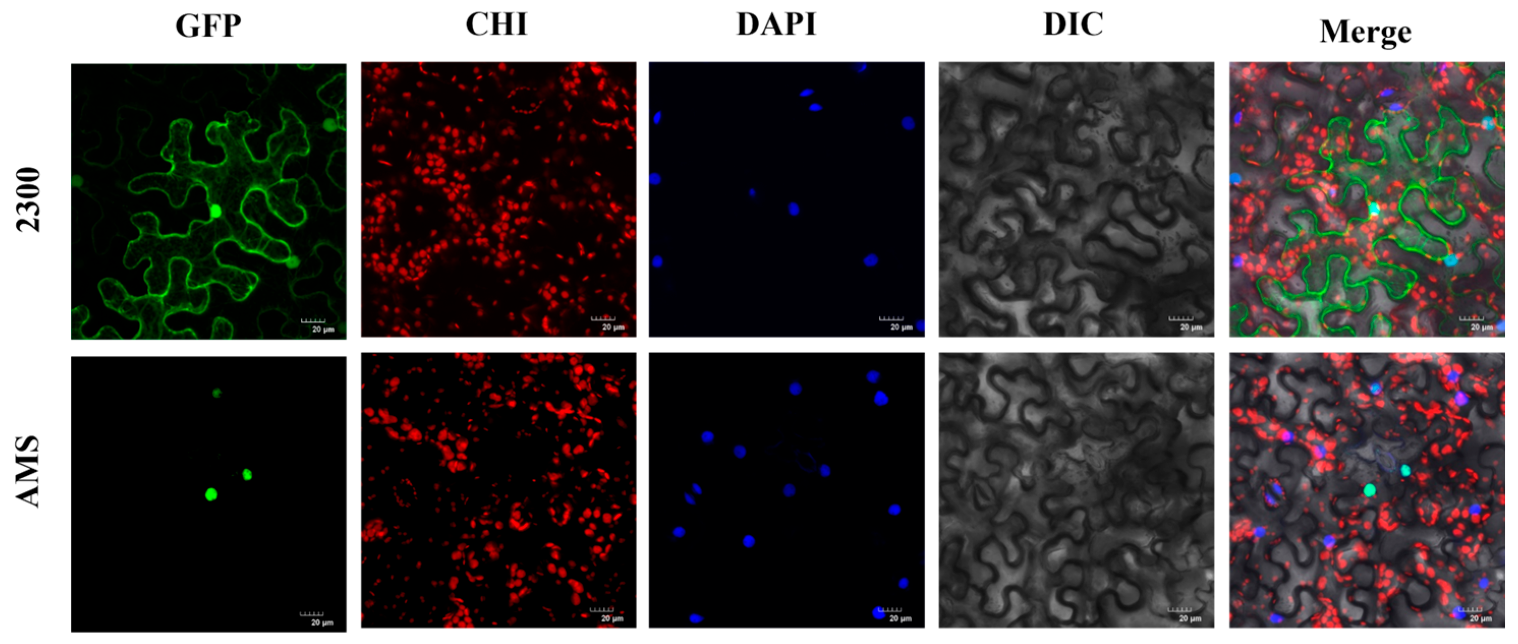
The CAMBIA2300-GFP-TeAMS vector was successfully constructed, introduced into the Agrobacterium solution, and transiently transformed into tobacco epidermal cells. The subcellular localization of TeAMS was observed using laser confocal microscopy, and it was found that TeAMS was localized in the nucleus (Figure 2).

3.3. TeAMS Possesses Self-Activation Property

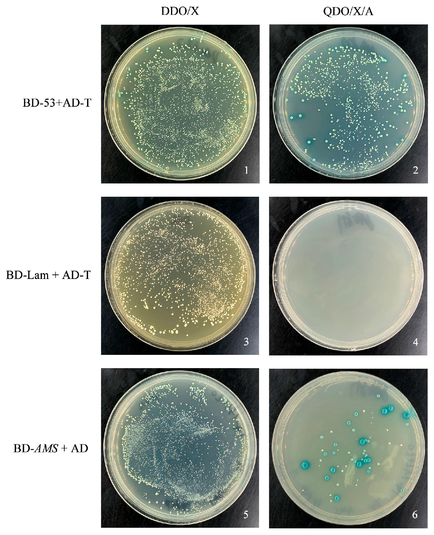
The recombinant BD-AMS plasmid was transformed into yeast-competent Y2H Gold using the PEG/LiAc method. The empty vector was transformed into Y2H Gold as a negative control, and BD-53 and AD-T were co-transformed as positive controls. The activity of the TeAMS transcription factor was determined by observing the growth status of yeast in DDO/X and QDO/X/A solid media and the color change of the QDO/X/A yeast. The results showed that the positive control group (BD-53 + AD-T) appeared as blue clones in DDO/X and QDO/X/A media, indicating the success of the positive control experiment. The negative control group (BD-Lam + AD-T) only grew in DDO/X medium and had no color, indicating that the negative control experiment was successful. The experimental group (BD-AMS + AD) was successfully cloned in both media and appeared as blue clones on the QDO/X/A medium (Figure 3), indicating that the BD-AMS plasmid was successfully transferred into the yeast and self-activation occurred.

3.4. Construction of the sGFP-1300-TeAMS Overexpression Vector and Transformation of Tobacco

The sGFP-1300 overexpression vector was single-digested with Sal I restriction enzyme, and electrophoresis showed a single linearized band (Figure 4A). After confirming complete digestion, the vector was used for subsequent ligation reactions. The recombinant plasmid sGFP-1300-TeAMS was transformed into Trans1-T1 competent cells using the freeze–thaw method. Monoclonal colonies were selected for colony PCR amplification, and the universal primer pEGFP-N3 was used for sequencing analysis. The sGFP-1300-TeAMS recombinant plasmid was transformed into Agrobacterium competent GV3101, and an Agrobacterium infection solution was prepared. A total of 142 leaf explants were infected, and calluses were successfully obtained. A total of 65 adventitious buds were cut for subculturing. The adventitious buds grew well, and 20 regenerated plants were transplanted into the plug after acclimatization (Figure 4B). Transgenic tobacco plants were screened using PCR (Figure 4C). Among the 20 transgenic plants, three were identified as positive, resulting in a conversion rate of 15%.

3.5. Determination of Relative Expression and Phenotypic Observation of TeAMS in Transgenic Tobacco

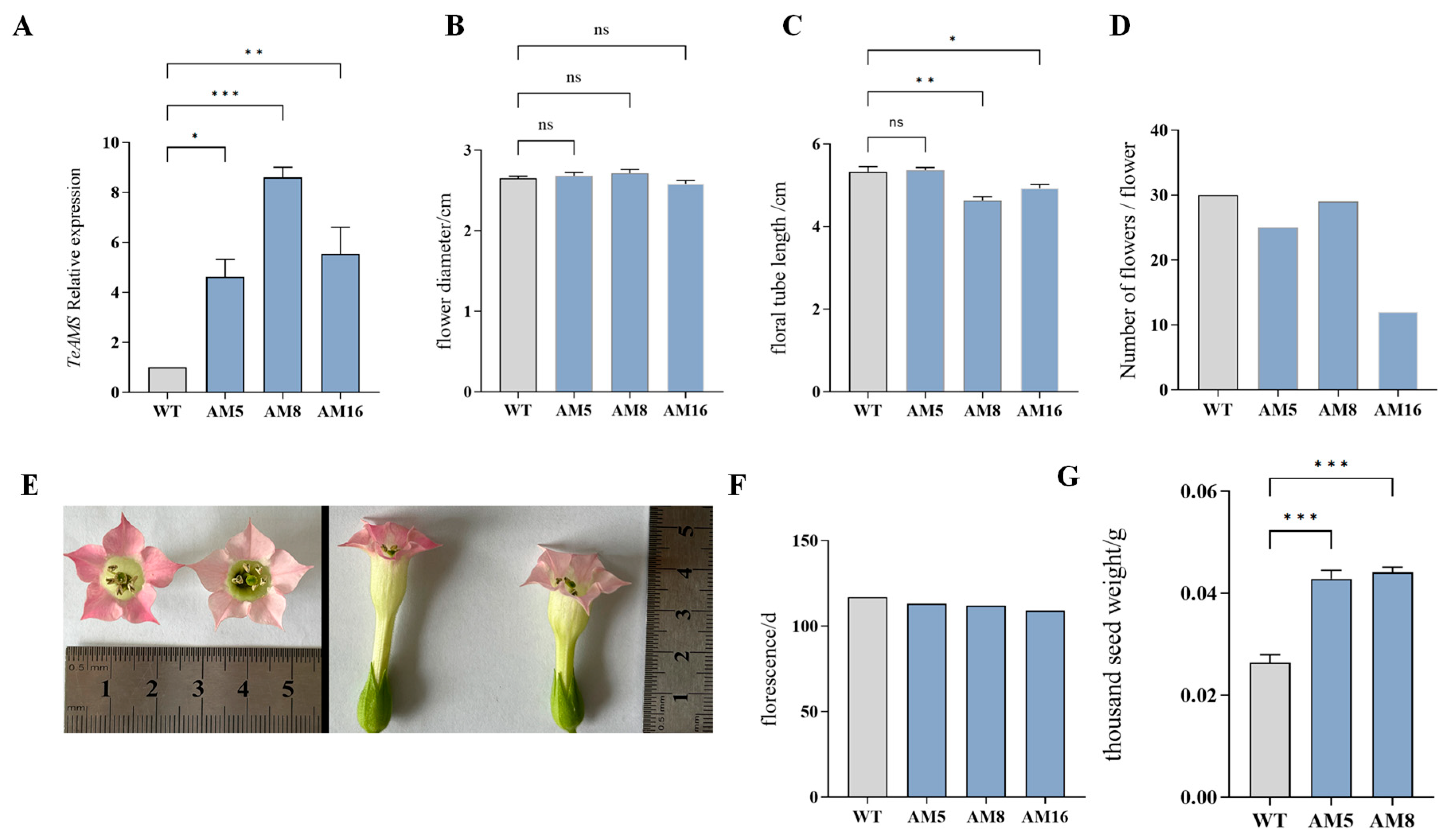
Fluorescence quantitative analysis showed that the expression level of TeAMS in the transgenic plants was significantly higher than that in the control plants (Figure 5A). Phenotypic analysis revealed a significant morphological difference between the transgenic-positive and WT plants (Figure 5E). No significant differences were observed in flower diameter between WT and AM5, AM8, and AM16 (Figure 5B). The flower tube lengths of AM8 (4.63 cm) and AM16 (4.93 cm) were significantly shorter than that of the WT (5.33 cm) (Figure 5C). The thousand-seed weights of AM5 (0.0428 g) and AM8 (0.0441 g) were significantly higher than that of the WT (0.0264 g) (Figure 5G). The florescence of the transgenic-positive plants was shorter, and the number was less than that of the WT (Figure 5D,F). In summary, compared with wild-type tobacco, the transgenic-positive plants had shortened floral tubes, increased thousand-seed weight, shortened flowering period, and decreased flower numbers.

3.6. Pollen Viability of Transgenic Tobacco

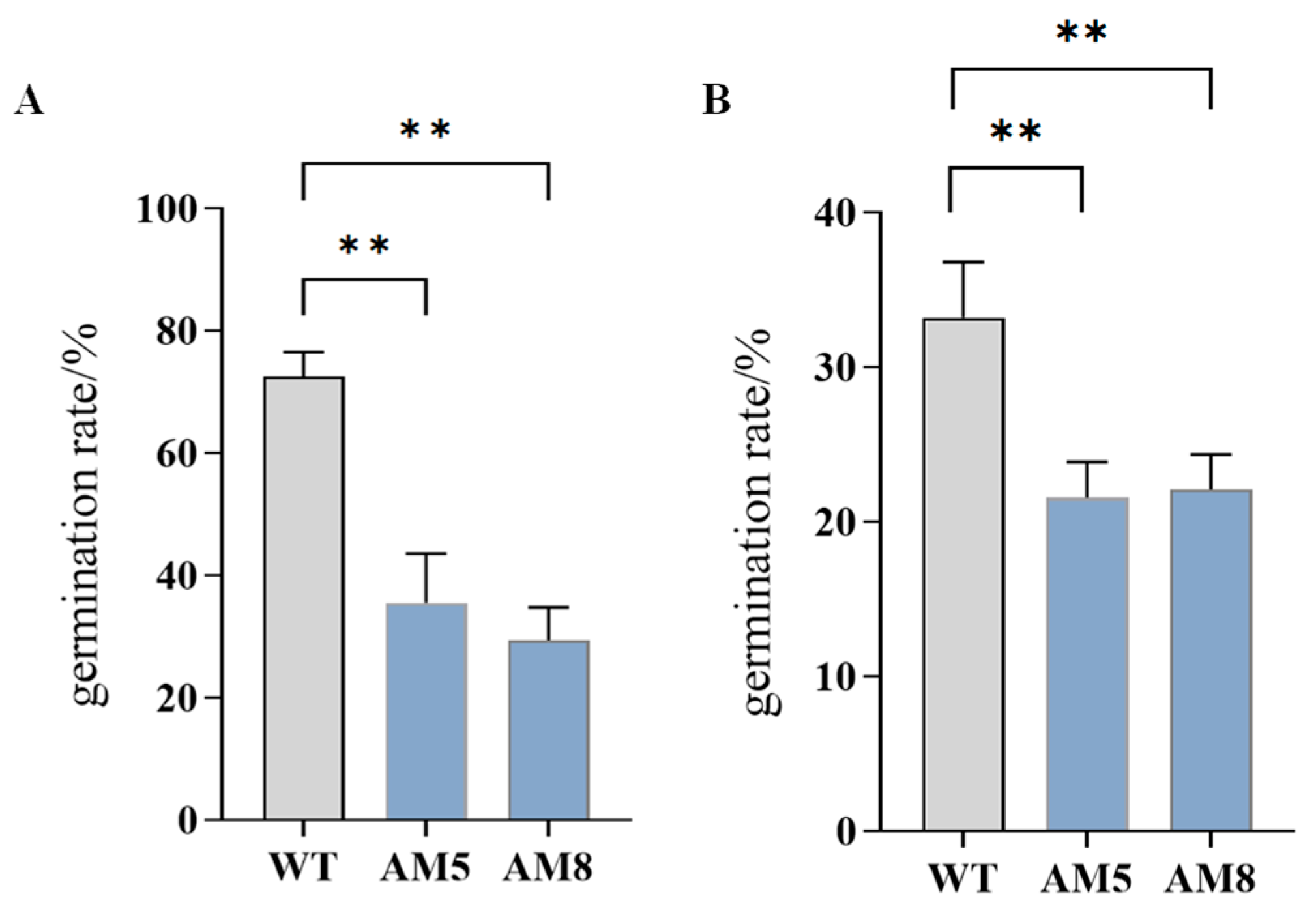
The pollen viability of transgenic tobacco and WT plants was determined using TTC staining and the in vitro germination method. The pollen viability of transgenic plants AM5 (TTC: 36%, isolated germination method: 22%) and AM8 (TTC: 29%, isolated germination method: 22%) was significantly lower than that of WT plants (TTC: 73%, isolated germination method: 33%) (Figure 6).

3.7. Pollen Morphology of Transgenic Tobacco

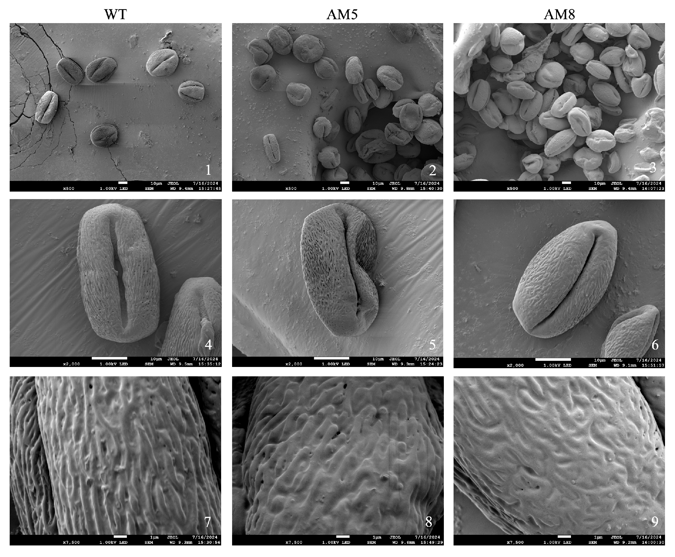
The pollen type of WT was determined to be tricolporate, and boat-shaped, and striae-reticular pollen ornamentation was observed. The morphology of most pollen grains changed in the transgenic plants (AM5 and AM8). The pollen grains became smaller, and their shape became irregular. The pollen wall was shrunken to varying degrees. The germination grooves of some pollen grains were distorted, and their shapes were irregular. A few germination grooves were almost undetectable. The pollen grain surface had reticular ornamentation similar to that of WT; however, most of them showed irregular infoldings and pores (Figure 7).

3.8. Construction of the pTRV2-TeAMS VIGS Vector

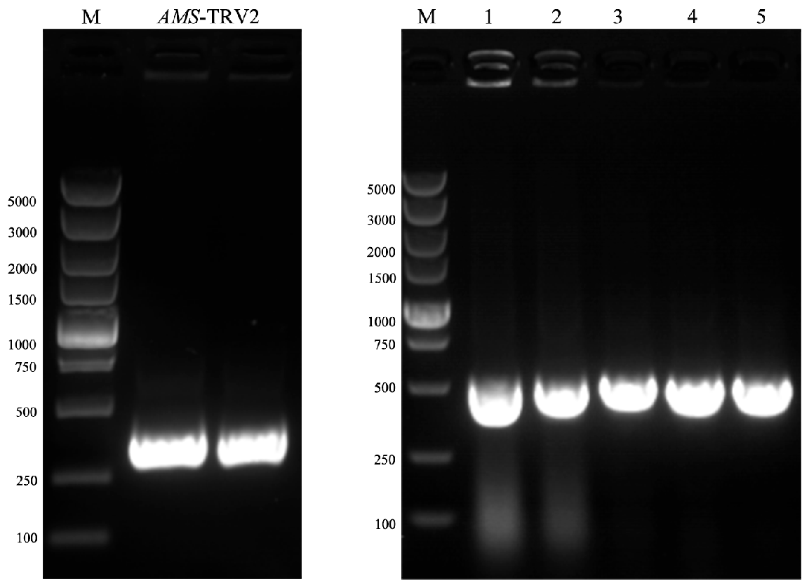
The silencing vector pTRV2 was double-digested with EcoRI and BamHI restriction enzymes, and the purified vector fragment was recovered after digestion. The purified product of the target fragment was homologously recombined with the digested pTRV2 vector and then transferred into E. coli DH5α competent cells. Positive colonies were screened using PCR and sequencing (Figure 8). The pTRV2-TeAMS recombinant plasmid DNA was transformed into Agrobacterium GV3101 to prepare a VIGS infection solution.

3.9. Successful Viral Infection Mediated by TRV2 Vectors

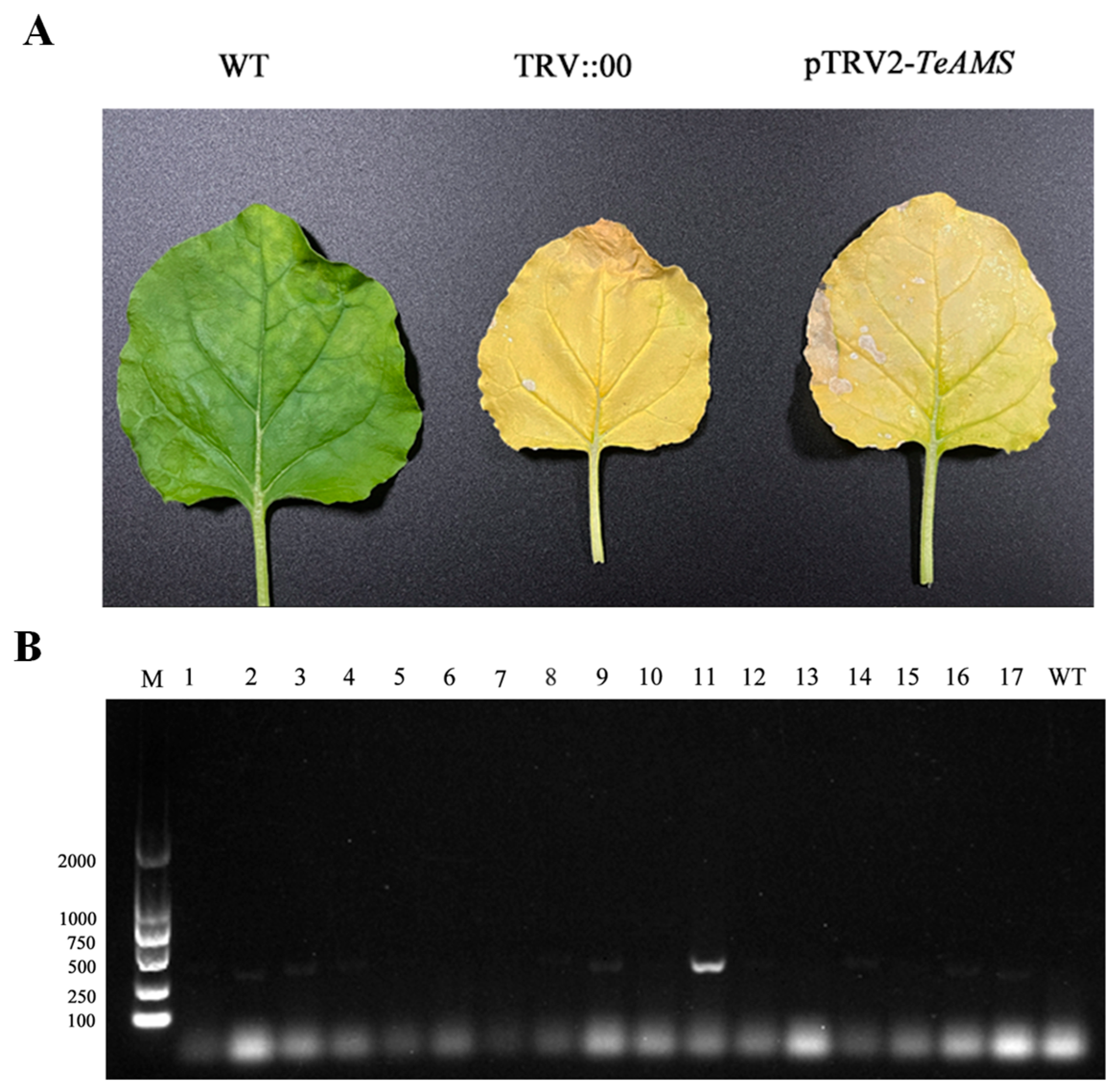
Ten days after injection, new leaves were sampled when the injected leaves were bleached (Figure 9A). cDNA samples from the new leaves of the negative control group, TRV::00 empty vector transformed plants, and pTRV2-TeAMS transformed plants were used as templates for PCR amplification. Electrophoresis analysis showed that both the TRV::00 and pTRV2-TeAMS treatment groups specifically amplified a 300 bp band, while the CK group had no target band (Figure 9B). This indicated that both the empty and recombinant vectors successfully mediated virus infection.

3.10. Determination of Relative Expression and Phenotypic Observation of TeAMS in Silenced Plants

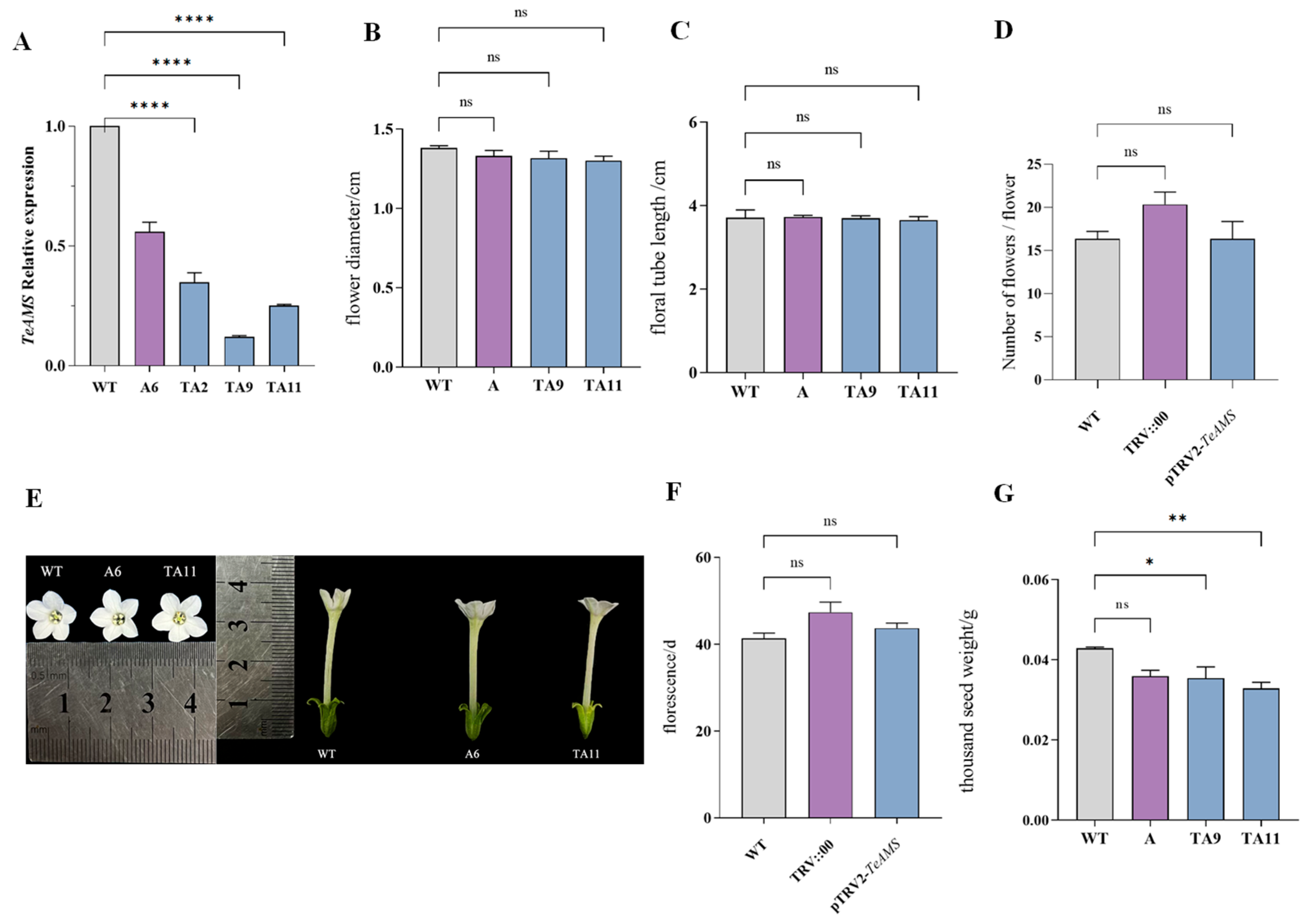
The TeAMS expression levels in tobacco plants in the different treatment groups were analyzed using qRT-PCR. The results showed no significant difference in the expression levels of TeAMS between the negative control WT and empty control TRV::00 (Figure 10A). Compared with WT and TRV::00, the expression level of TeAMS in the pTRV2-TeAMS test group was significantly lower than that in the negative control groups, indicating that the expression of TeAMS in pTRV2-TeAMS-silenced plants was successfully inhibited.
Differences in phenotypes were observed between the silenced and WT plants (Figure 10E). No significant difference was observed in flower diameter (1.35 cm) or tube length (3.70 cm) between the silenced (TA9 and TA11) and WT plants (Figure 10B,C). There was no significant difference in the number of flowers between the WT and silenced plants, both of which were about 16. However, the flowering period of the silenced plants (44 d) was significantly longer than that of the WT plants (41 d) (Figure 10D,F). The thousand-seed weights of TA9 (0.0354 g) and TA11 (0.0329 g) were significantly lower than that of WT (0.0429 g) (Figure 10G). In summary, silenced pTRV-TeAMS plants had a longer flowering period and reduced thousand-seed weight.

3.11. Pollen Viability of pTRV-TeAMS N. benthamiana Silenced Plants

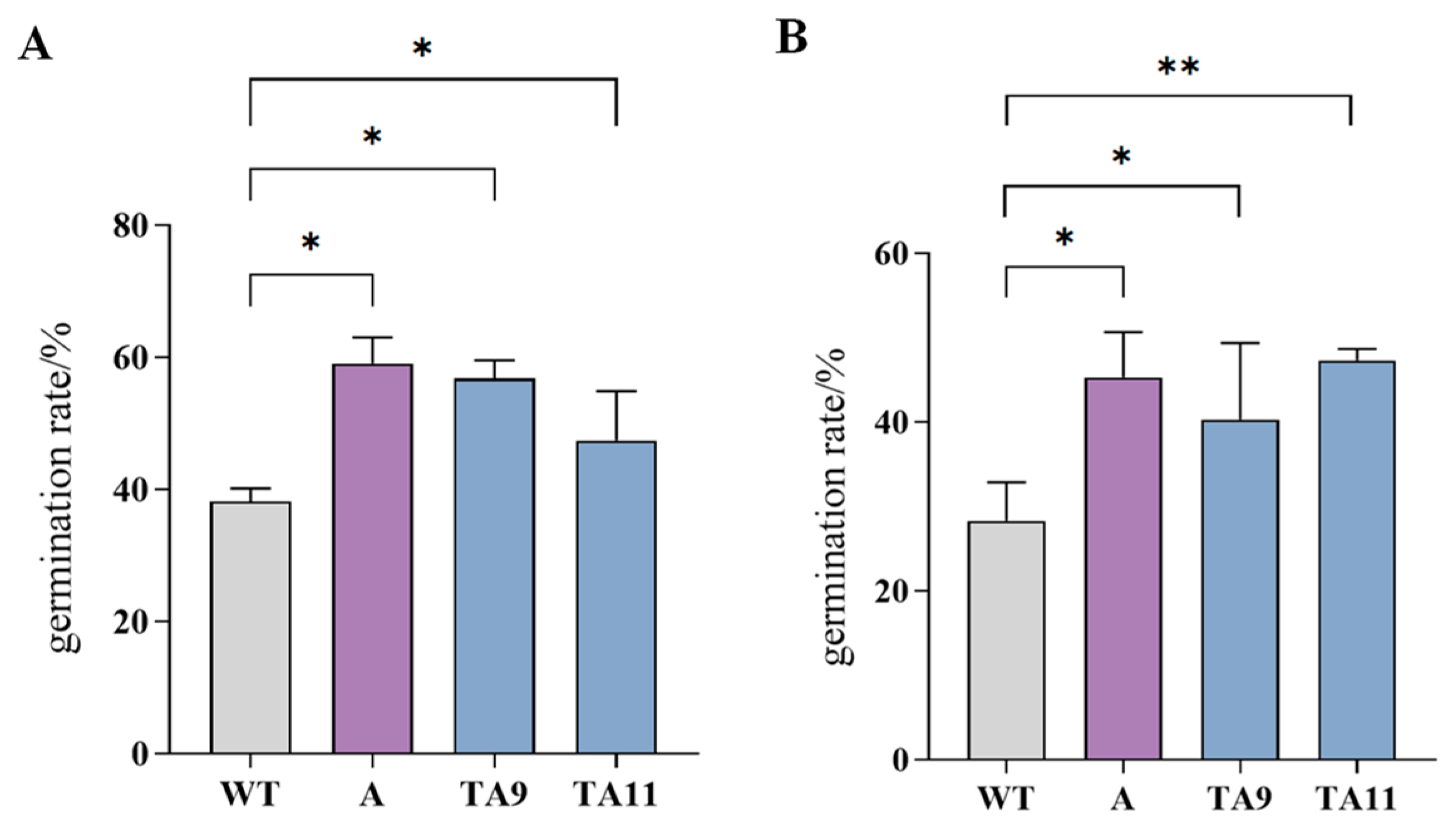
The pollen viability of wild-type tobacco and pTRV-TeAMS-silenced plants was determined using TTC staining and an in vitro germination method. The results showed that the pollen viability of TA9 (TTC: 57%, isolated germination method: 40%) and TA11 (TTC: 47%, isolated germination method: 47%) was significantly higher than that of WT (TTC: 38%, isolated germination method: 28%) (Figure 11).

3.12. Pollen Morphology of pTRV-TeAMS N. benthamiana

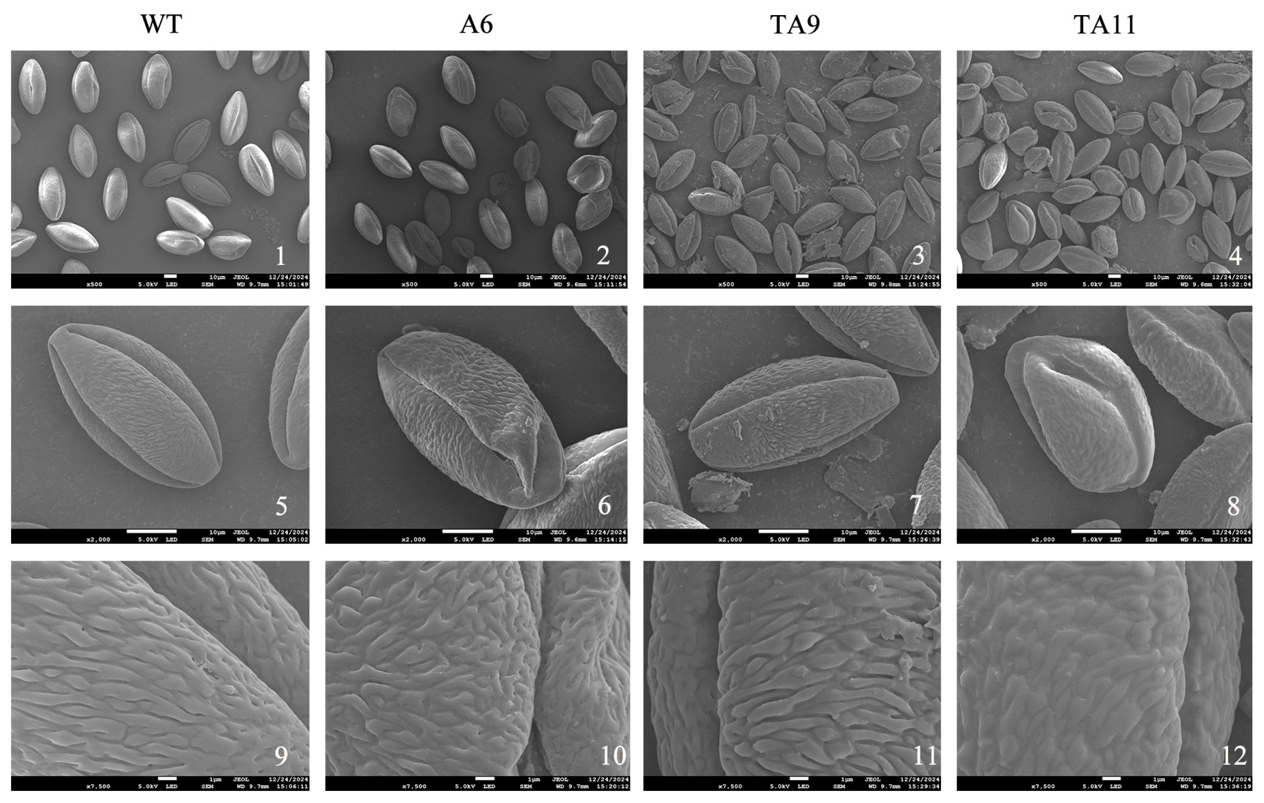
Fresh pollen grains from the silenced and control plants were collected for morphology observation. The results showed that most of the pollen grains of the WT and TRV::00 empty-vector control plants were normal in shape, including boat-shaped and tricolporate with striae reticular pollen ornamentation. Very few pollen grains were small, and the pollen walls were shrunken.
The three germinal furrows were clearly observed. The surfaces had smooth reticular ornamentation, most of which were striped; a few had punctate depressions, and some pollen surfaces had spherical protrusions. Only a small number of TA9 and TA11 pollen grains were irregular in shape, and the pollen wall was shrunken to varying degrees. The pollen grains were smaller, and the germination grooves of some pollen grains were distorted, with irregular shapes. The surface of the pollen grains of these plants had reticular ornamentation similar to that of WT pollen grains; however, most of them had punctate infoldings and pores (Figure 12).

4. Discussion

4.1. TeAMS Is a Key Factor in Regulating Marigold Male Fertility

Male sterility is an important trait for heterosis utilization and marigold breeding. In this study, the expression of TeAMS in fertile marigold plants was significantly higher than that in sterile plants, and it was highly expressed in flower buds, whereas previous studies have not focused on the expression period of the AMS gene.
Sheng et al. [25] identified AMS as a candidate sterility gene in melon (Cucumis melo L.), which was highly expressed in fertile plants, and the expression levels in 2 mm buds were lower than those in 5 mm buds in ms-5 plants (sterile plants). Guo et al. [17] isolated the AMS gene in pepper flower buds. Their qRT-PCR results showed that CaAMS is preferentially expressed in the stamens of flower buds at the tetrad stage and that its transcription level gradually decreases as flower buds develop. Using in situ hybridization, Lou et al. [26] found that the largest hybridization signal of Arabidopsis (Arabidopsis thaliana (L.) Heynh.) appeared in the tapetum and tetrad, but neither signal was observed in the late-stage anthers. These results show that the expression patterns of AMS genes in different crops are similar, and the expression level decreases in the late stage of flower development. The same results were obtained in the present study. In fertile marigold plants, the expression level was upregulated as the buds became larger. This expression pattern is highly consistent with the development process of floral organs, suggesting that AMS is essential for the sequential regulation of pollen development. In sterile marigold plants, the expression level of TeAMS was low at all time points. The abnormal expression of AMS in sterile plants also explains the molecular cause of pollen abortion. A similar expression pattern was observed for the tomato AMS gene. The expression of AMS began when the tomato bud length was 3 mm (microsporocyte meiosis stage), and the gene expression was the highest when the bud length was 6 mm (free microspore stage). The gene is not expressed when bud length reaches 8 mm-OF (anther tapetum completely degraded) [18]. In conclusion, TeAMS was highly expressed in the late stage of flower bud development in fertile marigold plants, whereas it was highly expressed in the early stage of flower bud development in sterile plants. This indicates that TeAMS is related to the formation of sterility in marigolds. This finding provides a molecular marker for the early identification of male sterility in marigold breeding at the seedling or early bud stage. Breeders can efficiently identify and maintain male-sterile lines, significantly reducing the labor and cost of traditional phenotypic screening.
Previous research has shown that the AMS transcription factor is related to male sterility, especially regarding stamen development and function [27]. AMS is a major regulator of pollen wall development [16,28]. In Arabidopsis, the AMS gene encodes a bHLH protein that plays a crucial role in the differentiation of tapetal cells and microspores within the developing anther [15]. In this study, the TeAMS protein was localized in the nucleus and exhibited transcriptional activation activity. It is speculated that AMS plays a role in regulating the expression of downstream target genes. The AMS gene has also been identified as a transcription factor that acts in the nucleus of tomato [18]. Subcellular localization results of the AMS gene in pepper also showed that it was localized in the nucleus.

4.2. Overexpression of TeAMS Influenced the Development of Pollen and Fertility of Tobacco

Pollen viability and morphology are important indicators for evaluating plant fertility. In this study, we observed that the phenotype of transgenic tobacco plants was significantly different from that of wild-type plants, and the pollen viability of transgenic tobacco plants was significantly lower than that of wild-type plants. This indicated that overexpression of TeAMS interfered with the pollen development process, resulting in abnormal pollen and decreased pollen viability. Similar results were found in tomatoes, in which the SlAMS gene was overexpressed and led to a decrease in pollen viability [18]. In soybean (Glycine max (L.) Merr.), CRISPR/Cas9-induced mutant Gmams1-1 displayed complete male sterility with small pods but normal vegetative growth [29]. In this study, interestingly, TeAMS not only regulated pollen development but also had pleiotropic effects, which also influenced the morphogenesis and flowering of floral organs.
To better understand the role of TeAMS in pollen development, morphology variation was observed in the pollen grains of transgenic plants. The pollen grains were irregular in shape and shrunken to different degrees, which indicated that overexpression of the TeAMS gene led to abnormal pollen development. Similar pollen variation results were also found in tomato SlAMS-overexpressing plants, where the pollen was diamond-shaped, shrunken, shriveled, or had other abnormal shapes [18]. Unfortunately, due to the serious inhibition of floral organ development, the overexpression line AM16 in this experiment failed to obtain sufficient pollen for viability statistics and electron microscopy observations. However, existing data have shown that AMS overexpression has a destructive effect on pollen development.

4.3. Accurate Regulation of Silent TeAMS Is Essential for Fertility

There are few reports on the establishment of the marigold VIGS system, and because the marigold male sterile line has no anthers, it is impossible to detect pollen viability after silencing a gene. Therefore, this study used Nicotiana benthamiana to preliminarily explore the biological function of TeAMS. The results showed that pTRV2-TeAMS-silenced plants had a longer flowering period, reduced seed weight, and high pollen viability. It was inferred that there was functional redundancy and gene compensation in N. benthamiana, or the homologous genes of AMS [15]. The silencing experiment of heterologous plants may differ from that of marigolds; for example, it may be related to gene regulatory networks or the number and functions of redundant genes. Further clarification of the precise regulation of TeAMS expression and expression time is crucial for fertility. Lou et al. [26] also proposed the same conclusion in Arabidopsis, where the temporal regulation of AMS expression is important for functioning. More importantly, this finding is contrary to the changes in pollen morphology caused by overexpression. According to previous studies, the formation of male sterility in plants is a complex regulatory network. AMS may affect plant fertility by regulating a series of downstream genes directly related to tapetal PCD. Researchers [16,30,31] found 549 anther-specific genes related to tapetum function and pollen wall formation in the flower buds of Arabidopsis ams mutants. They proposed a regulatory network of AMS during anther and pollen development and identified 23 AMS target genes in Arabidopsis ams mutants. We can further verify the function of TeAMS in marigolds and use yeast two-hybrid, chromosome immunoprecipitation, and other techniques to determine the genes that interact with TeAMS to clarify the regulatory mechanism of TeAMS in the formation of marigold fertility. These findings suggest that the expression of TeAMS will promote the formation of male sterility, and it has been preliminarily verified that TeAMS is involved in plant fertility and male sterility.
Other studies have also confirmed the regulatory function of AMS in plant fertility. Guo et al. [17] verified the biological function of CaAMS in pepper using VIGS technology. Bao et al. [18] verified the function of SlAMS in tomato anther development using VIGS. In soybean, targeted editing of AMS homologs using CRISPR/Cas9 technology generates stable male-sterile lines. Targeted editing of GmAMS1 leads to a male-sterile phenotype and plays a role in controlling the degradation of the soybean tapetum [29]. By knocking out AMS, Seol et al. [32] observed an AMS-like edited organism with a distorted shape due to aberrant sporoderm. These studies have shown that the downregulated expression of the AMS gene also affects pollen development to a certain extent. However, in this study, the effects on pollen development were not obvious. It is necessary to introduce the TeAMS gene into marigolds to observe its mechanism and effect. The TeAMS gene appears to play an important regulatory role in marigold pollen development by regulating complex networks, and this regulation is biphasic [31]. However, the upstream and downstream target genes of TeAMS and their regulatory networks remain unclear and require further exploration.

5. Conclusions

To reduce the cost of hybrid seed production and ensure high purity, it is important to utilize male sterility in marigold crossbreeding. In this study, we identified a relationship between the TeAMS gene and marigold fertility using expression analysis. TeAMS is a transcriptional activator that localizes to the nucleus. Overexpression and VIGS of TeAMS in tobacco caused two completely different variations in seed weight, flowering period, and pollen viability. Only the overexpressed tobacco plants presented abnormal pollen grains. These preliminary results suggest that the upregulated expression of the TeAMS gene will affect pollen development and thus plant fertility, while the downregulated expression level accurately regulates the expression level and time. In general, these insights lay the foundation for the development of molecular tools for male sterility or selection in marigolds. TeAMS can be used as a target for CRISPR/Cas9-mediated knockout in the later stages, which provides a direct method for creating stable male-sterile lines. These methods are of great significance for reducing seed production costs and ensuring the genetic purity of marigolds.

Supplementary Materials

The following supporting information can be downloaded at: https://www.mdpi.com/article/10.3390/agronomy15092058/s1, Table S1: Primer Information Table; Table S2: PEG/LiAc method was transferred into the yeast competent reaction system; Figure 6-S1: The difference in pollen viability between transgenic N. tabacum and WT. WT, wild type; AM5 and AM8 are transgenic N. tabacum. (A) Pollen viability determined by TTC method. WT, wild type; AM5 and AM8 was the transgenic N. tabacum plants. (B) Pollen viability determined by in in vitro germination method; Figure 11-S1: Pollen viability of wild-type N. tabacum and pTRV2-TeAMS-silenced N. tabacum. (A) Pollen viability was detected by TTC. WT, wild type; A6 empty control, AM5, TA9 and TA11 were silent N. tabacum. (B) Pollen viability was detected by in vitro germination.

Author Contributions

Conceptualization, X.M. and N.T.; methodology, X.M., J.T. and D.T.; software, J.T. and Q.L.; validation, X.M., D.T. and N.T.; formal analysis, X.M., J.T. and D.T.; investigation, X.M., J.T. and Q.L.; resources, X.M., D.T. and N.T.; data curation, X.M. and N.T.; writing—original draft preparation, X.M., J.T. and N.T.; writing—review and editing, X.M. and N.T.; visualization, X.M., J.T. and D.T.; supervision, D.T. and N.T.; project administration, N.T.; funding acquisition, N.T. All authors have read and agreed to the published version of the manuscript.

Funding

This research was funded by the Science and Technology Department of Qinghai Province (2024-ZJ-920) and the Graduate School of Qinghai University (2025-GPKY-18), China.

Data Availability Statement

The original data underlying this study are available within this article and its Supplementary Materials. For additional inquiries, please contact the corresponding author.

Conflicts of Interest

The authors declare no conflicts of interest.

References

  1. Burlec, A.F.; Pecio, Ł.; Kozachok, S.; Mircea, C.; Corciovă, A.; Vereștiuc, L.; Cioancă, O.; Oleszek, W.; Hăncianu, M. Phytochemical Profile, Antioxidant Activity, and Cytotoxicity Assessment of Tagetes erecta L. Flowers. Molecules 2021, 26, 1201. [Google Scholar] [CrossRef] [PubMed]
  2. Zafar, J.; Aqeel, A.; Shah, F.I.; Ehsan, N.; Gohar, U.F.; Moga, M.A.; Festila, D.; Ciurea, C.; Irimie, M.; Chicea, R. Biochemical and Immunological Implications of Lutein and Zeaxanthin. Int. J. Mol. Sci. 2021, 22, 10910. [Google Scholar] [CrossRef]
  3. Mrowicka, M.; Mrowicki, J.; Kucharska, E.; Majsterek, I. Lutein and Zeaxanthin and Their Roles in Age-Related Macular Degeneration-Neurodegenerative Disease. Nutrients 2022, 14, 827. [Google Scholar] [CrossRef]
  4. Du, M.; Zhou, K.; Liu, Y.; Deng, L.; Zhang, X.; Lin, L.; Zhou, M.; Zhao, W.; Wen, C.; Xing, J.; et al. A Biotechnology-Based Male-Sterility System for Hybrid Seed Production in Tomato. Plant J. 2020, 102, 1090–1100. [Google Scholar] [CrossRef]
  5. Luo, H.; Liu, Z.; Zhu, J.; Li, Z.; Qiu, X.; Wang, W.; Gao, C.; Qi, J.; Bao, M.; Liu, Y. Identification and Expression of Laccase Gene Family in Potato (Solanum tuberosum). Agronomy 2025, 15, 585. [Google Scholar] [CrossRef]
  6. He, Y.-H.; Ning, G.-G.; Sun, Y.-L.; Hu, Y.; Zhao, X.-Y.; Bao, M.-Z. Cytological and Mapping Analysis of a Novel Male Sterile Type Resulting from Spontaneous Floral Organ Homeotic Conversion in Marigold (Tagetes erecta L.). Mol. Breed. 2010, 26, 19–29. [Google Scholar] [CrossRef]
  7. Liu, X.-Q.; Xu, X.; Tan, Y.-P.; Li, S.-Q.; Hu, J.; Huang, J.-Y.; Yang, D.-C.; Li, Y.-S.; Zhu, Y.-G. Inheritance and Molecular Mapping of Two Fertility-Restoring Loci for Honglian Gametophytic Cytoplasmic Male Sterility in Rice (Oryza sativa L.). Mol. Genet. Genom. 2004, 271, 586–594. [Google Scholar] [CrossRef]
  8. Sumalatha, A. Genetic Inheritance and Identification of Molecular Markers Linked to Male Sterility in African Marigold (Tagetes erecta L.). arXiv 2025. [Google Scholar] [CrossRef]
  9. Wang, Z.; Yin, D.; Wang, P.; Zhao, J.; Pan, B.; Zhang, H. Rapd Markers Developed for Identifying Male Sterility in Tagetes erecta. Acta Hortic. 2010, 88, 289–294. [Google Scholar] [CrossRef]
  10. Cholin, S. Validation of SCAR Marker Linked to Genic Male Sterility in Marigold: As a Forward Step towards Marker Assisted Breeding Programme. Int. J. Curr. Microbiol. Appl. Sci. 2019, 8, 3373–3383. [Google Scholar] [CrossRef]
  11. Tang, N.; Liu, W.; Zhang, W.; Tang, D. Integrative Analysis of Transcriptomic and Proteomic Changes Related to Male Sterility in Tagetes erecta. Physiol. Mol. Biol. Plants 2020, 26, 2061–2074. [Google Scholar] [CrossRef]
  12. Sanders, P.M.; Bui, A.Q.; Weterings, K.; McIntire, K.N.; Hsu, Y.-C.; Lee, P.Y.; Truong, M.T.; Beals, T.P.; Goldberg, R.B. Anther Developmental Defects in Arabidopsis thaliana Male-Sterile Mutants. Sex. Plant Reprod. 1999, 11, 297–322. [Google Scholar] [CrossRef]
  13. Szymkowiak, E.J.; Sussex, I.M. What chimeras can tell us about plant development. Annu. Rev. Plant Physiol. Plant Mol. Biol. 1996, 47, 351–376. [Google Scholar] [CrossRef]
  14. Berger, F.; Haseloff, J.; Schiefelbein, J.; Dolan, L. Positional Information in Root Epidermis Is Defined during Embryogenesis and Acts in Domains with Strict Boundaries. Curr. Biol. 1998, 8, 421–430. [Google Scholar] [CrossRef] [PubMed]
  15. Sorensen, A.; Kröber, S.; Unte, U.S.; Huijser, P.; Dekker, K.; Saedler, H. The Arabidopsis ABORTED MICROSPORES (AMS) Gene Encodes a MYC Class Transcription Factor. Plant J. 2003, 33, 413–423. [Google Scholar] [CrossRef]
  16. Xu, J.; Ding, Z.; Vizcay-Barrena, G.; Shi, J.; Liang, W.; Yuan, Z.; Werck-Reichhart, D.; Schreiber, L.; Wilson, Z.A.; Zhang, D. ABORTED MICROSPORES Acts as a Master Regulator of Pollen Wall Formation in Arabidopsis. Plant Cell 2014, 26, 1544–1556. [Google Scholar] [CrossRef]
  17. Guo, J.; Liu, C.; Wang, P.; Cheng, Q.; Sun, L.; Yang, W.; Shen, H. The Aborted Microspores (AMS)-like Gene Is Required for Anther and Microspore Development in Pepper (Capsicum annuum L.). Int. J. Mol. Sci. 2018, 19, 1341. [Google Scholar] [CrossRef] [PubMed]
  18. Bao, H.; Ding, Y.; Yang, F.; Zhang, J.; Xie, J.; Zhao, C.; Du, K.; Zeng, Y.; Zhao, K.; Li, Z.; et al. Gene Silencing, Knockout and over-Expression of a Transcription Factor ABORTED MICROSPORES (SlAMS) Strongly Affects Pollen Viability in Tomato (Solanum lycopersicum). BMC Genom. 2022, 23, 346. [Google Scholar] [CrossRef]
  19. Tang, N.; Zhang, W.; Chen, L.; Wang, Y.; Tang, D. Reference Gene Selection for Real-Time Quantitative Reverse-Transcription Polymerase Chain Reaction in Flower Buds of Marigold. J. Am. Soc. Hortic. Sci. 2021, 146, 363–373. [Google Scholar] [CrossRef]
  20. Weigel, D.; Glazebrook, J. Transformation of Agrobacterium Using the Freeze-Thaw Method. CSH Protoc. 2006, 2006, pdb.prot4666. [Google Scholar] [CrossRef]
  21. Gietz, R.D.; Woods, R.A. Yeast Transformation by the LiAc/SS Carrier DNA/PEG Method. In Yeast Protocol; Xiao, W., Ed.; Humana Press: Totowa, NJ, USA, 2006; pp. 107–120. ISBN 978-1-59259-958-5. [Google Scholar]
  22. Sevilleno, S.S.; An, H.R.; Cabahug-Braza, R.A.M.; Ahn, Y.-J.; Hwang, Y.-J. Cytogenetic Study and Pollen Viability of Phalaenopsis Queen Beer ‘Mantefon’. Plants 2023, 12, 2828. [Google Scholar] [CrossRef]
  23. Bouatta, A.M.; Anzenberger, F.; Riederauer, L.; Lepper, A.; Denninger, P. Polarized Subcellular Activation of Rho Proteins by Specific ROPGEFs Drives Pollen Germination in Arabidopsis thaliana. PLoS Biol. 2025, 23, e3003139. [Google Scholar] [CrossRef]
  24. Chen, X.; Zhang, H.; Sun, H.; Luo, H.; Zhao, L.; Dong, Z.; Yan, S.; Zhao, C.; Liu, R.; Xu, C.; et al. IRREGULAR POLLEN EXINE1 Is a Novel Factor in Anther Cuticle and Pollen Exine Formation. Plant Physiol. 2016, 173, 307–325. [Google Scholar] [CrossRef]
  25. Sheng, Y.; Wang, Y.; Jiao, S.; Jin, Y.; Ji, P.; Luan, F. Mapping and Preliminary Analysis of ABORTED MICROSPORES (AMS) as the Candidate Gene Underlying the Male Sterility (MS-5) Mutant in Melon (Cucumis melo L.). Front. Plant Sci. 2017, 8, 902. [Google Scholar] [CrossRef] [PubMed]
  26. Lou, Y.; Zhou, H.-S.; Han, Y.; Zeng, Q.-Y.; Zhu, J.; Yang, Z.-N. Positive Regulation of AMS by TDF1 and the Formation of a TDF1–AMS Complex Are Required for Anther Development in Arabidopsis thaliana. New Phytol. 2018, 217, 378–391. [Google Scholar] [CrossRef]
  27. Thorstensen, T.; Grini, P.E.; Mercy, I.S.; Alm, V.; Erdal, S.; Aasland, R.; Aalen, R.B. The Arabidopsis SET-Domain Protein ASHR3 Is Involved in Stamen Development and Interacts with the bHLH Transcription Factor ABORTED MICROSPORES (AMS). Plant Mol. Biol. 2008, 66, 47–59. [Google Scholar] [CrossRef]
  28. Lou, Y.; Xu, X.-F.; Zhu, J.; Gu, J.-N.; Blackmore, S.; Yang, Z.-N. The Tapetal AHL Family Protein TEK Determines Nexine Formation in the Pollen Wall. Nat. Commun. 2014, 5, 3855. [Google Scholar] [CrossRef]
  29. Chen, X.; Yang, S.; Zhang, Y.; Zhu, X.; Yang, X.; Zhang, C.; Li, H.; Feng, X. Generation of Male-Sterile Soybean Lines with the CRISPR/Cas9 System. Crop J. 2021, 9, 1270–1277. [Google Scholar] [CrossRef]
  30. Xu, J.; Yang, C.; Yuan, Z.; Zhang, D.; Gondwe, M.Y.; Ding, Z.; Liang, W.; Zhang, D.; Wilson, Z.A. The ABORTED MICROSPORES Regulatory Network Is Required for Postmeiotic Male Reproductive Development in Arabidopsis thaliana. Plant Cell 2010, 22, 91–107. [Google Scholar] [CrossRef] [PubMed]
  31. Ferguson, A.C.; Pearce, S.; Band, L.R.; Yang, C.; Ferjentsikova, I.; King, J.; Yuan, Z.; Zhang, D.; Wilson, Z.A. Biphasic Regulation of the Transcription Factor ABORTED MICROSPORES (AMS) Is Essential for Tapetum and Pollen Development in Arabidopsis. New Phytol. 2017, 213, 778–790. [Google Scholar] [CrossRef] [PubMed]
  32. Seol, Y.-J.; Oh, J.-H.; Bae, S.-H.; Lee, J.; Jeong, H.-W.; Ahn, I.P.; Kim, E.; Kim, S.M.; Park, J. CRISPR/Cas9-Mediated Knockout of Transcription Factor MS1-like and AMS-like Genes Causes Male Sterility in Tomatoes. Plant Biotechnol. Rep. 2025, 19, 301–310. [Google Scholar] [CrossRef]
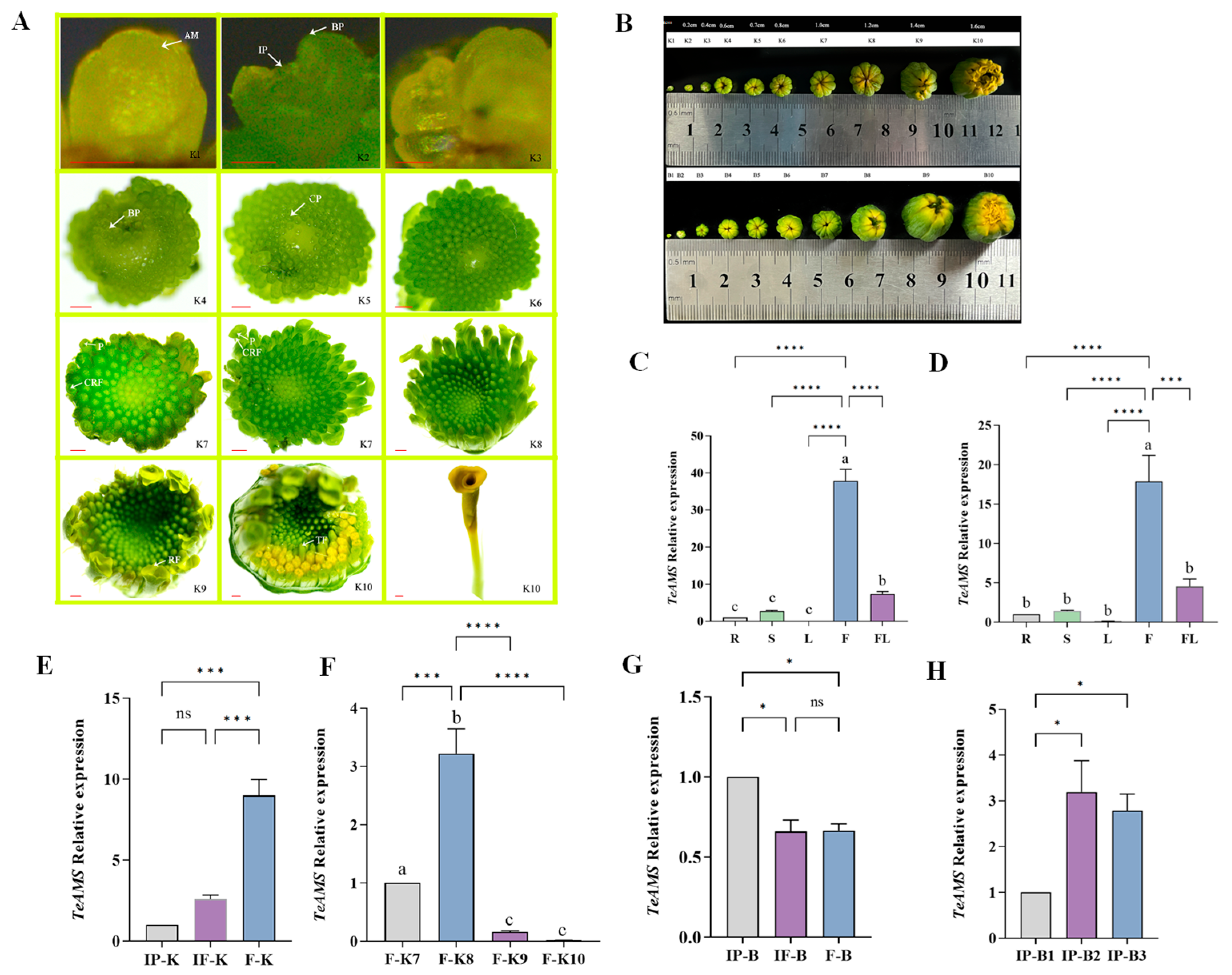
Figure 1. Flower morphological development process and expression analysis of TeAMS in Marigold. (A) Inflorescence and floret morphological development process: inflorescence primordium differentiation stage (K1–K3), inflorescence formation stage (K4–K6), and floret differentiation stage (K7–K10). AM, stem tip growth point; IP, inflorescence primordium; BP, bract primordium; CP, corolla primordium; CRF, ligulate corolla; P, pistil; TF, tubular flower; RF, ligulate flower. (B) Size of flower buds at different developmental stages. Stem tip (K1), 0.2 cm (K2), 0.4 cm (K3), 0.6 cm (K4), 0.7 cm (K5), 0.8 cm (K6), 1.0 cm (K7), 1.2 cm (K8), 1.4 cm (K9), 1.6 cm (K10). (C) Relative expression of TeAMS in different tissues of marigold ‘2-2’ fertile plants. (D) Relative expression of TeAMS in different tissues of marigold ‘2-2’ sterile plants. R, root; S, stem; L, leaf; F, flower bud; FL, floret. (E) Relative expression of TeAMS in different flower bed developmental stages of marigold ‘2-2’ fertile plants. IP, inflorescence primordium differentiation stage. IF, inflorescence formation stage; F, floret differentiation stage. (F) Relative expression of TeAMS in flower buds of ‘2-2’ fertile plants at the floret differentiation stage. (G) Relative expression of TeAMS in different flower bed developmental stages of marigold ‘2-2’ sterile plants. (H) Relative expression levels of TeAMS in the flower buds of ‘2-2’ sterile plants at the inflorescence primordium differentiation stage. Asterisks indicate a significant difference from the control value: * p  <  0.05, *** p  <  0.001, and **** p  <  0.0001.’ns’ represents not significant. The bar is 1 mm. Lowercase letters represent statistically significant differences between groups.
Agronomy 15 02058 g001
Figure 2. Subcellular localization of TeAMS protein. GFP represents the green fluorescence field, CHI represents the chloroplast autofluorescence field, DAPI represents nuclear staining, DIC represents the bright field, Merge represents the superposition field, 2300 represents the empty control, and AMS represents the experimental group. The bar is 20 μm.
Agronomy 15 02058 g002
Figure 3. Transcriptional self-activation analysis of TeAMS: (1,2), positive control (BD-53 + AD-T); (3,4), negative control (DB-Lam + AD-T); (5,6), BD-AMS + AD. DDO/X, single-lack medium; QDO/X/A, three-def.
Agronomy 15 02058 g003
Figure 4. Construction of the sGFP-1300-TeAMS overexpression vector and transformation of tobacco (A) TeAMS amplification and overexpression vector sGFP-1300 single-enzyme digestion. (B) Leaf disc transformation of tobacco: 1, Tobacco leaves were pre-cultured for 3 days; 2, after 15 days of transfer into the screening medium, the callus expanded and the leaves curled; 3, screening the growth of adventitious buds in the medium; 4, adventitious buds; 5, seedling acclimatization of wild-type tobacco; 6, transplanted transgenic tobacco. (C) Vector-specific primer, GFP fluorescent protein primer, and TeAMS gene-specific primer amplification results. WT, negative control; a single band at 1754 bp indicates a positive clone, and no band indicates a negative clone. The bar is 1 cm.
Agronomy 15 02058 g004
Figure 5. Differences in TeAMS gene expression and phenotypic characteristics of transgenic tobacco plants and wild-type plants. (A) Relative expression of TeAMS in transgenic tobacco and WT. WT, wild type; AM5, AM8, and AM16 are transgenic tobacco (B) Differences in flower diameter between transgenic tobacco and WT. (C) Differences in floral tube length between transgenic tobacco and WT. (D) Differences in flower numbers between transgenic tobacco and WT. (E) Variation in petal diameter and floral tube length. (F) Variation in flowering period. (G) Variation in thousand-seed weight. Asterisks indicate a significant difference from the control value: * p  <  0.05, ** p  <  0.01, *** p  <  0.001. ’ns’ represents not significant.
Agronomy 15 02058 g005
Figure 6. Difference in pollen viability between transgenic tobacco and WT. WT, wild type; AM5 and AM8 are transgenic tobacco (A) Germination rate of pollen detected using the TTC method. (B) Germination rate of pollen detected using the in vitro germination method. Asterisks indicate a significant difference from the control value: ** p  <  0.01.
Agronomy 15 02058 g006
Figure 7. Differences in pollen morphology of transgenic tobacco and WT under a scanning electron microscope: (13): size and structure of pollen grains; (46): shape of pollen and germination grooves; (79): ornamentation of pollen grains. In sub-figure (16), the bar is 10 μm, and in sub-figure (79), the bar is 1 μm.
Agronomy 15 02058 g007
Figure 8. Cloning of the target gene fragment and PCR detection of AMS-TRV2.
Agronomy 15 02058 g008
Figure 9. Phenotypic results of VIGS and virus detection in N. benthamiana leaves. (A) After 10 days of silencing, the leaves of N. benthamiana exhibited a bleaching phenotype. (B) Molecular detection of TRV2 virus in silenced plants.
Agronomy 15 02058 g009
Figure 10. Differences in TeAMS expression and phenotypic characteristics between pTRV2-TeAMS-silenced and WT plants. (A) Relative expression of TeAMS in silenced tobacco and WT. WT: wild type; A6: no-load control; TA9 and TA11: silenced plants of tobacco (B) Differences in flower diameter between silenced tobacco and WT. (C) Differences in floral tube length between silenced tobacco plants and WT. (D) Variation in flower number. (E) Variation in petal diameter and floral tube length. (F) Variation in the flowering period. (G) Variation in thousand-seed weight. Asterisks indicate a significant difference from the control value: * p  <  0.05, ** p  <  0.01 and **** p  <  0.0001. ‘ns’ represents not significant.
Agronomy 15 02058 g010
Figure 11. Pollen viability of wild-type tobacco and pTRV2-TeAMS-silenced tobacco (A) Variation in the germination rate in the silenced and WT plants, detected using the TTC method. (B) Variation in the germination rate of the silenced and WT plants, determined using the in vitro germination method. Asterisks indicate a significant difference from the control value: * p  <  0.05, ** p  <  0.01.
Agronomy 15 02058 g011
Figure 12. External pollen grain morphology of pTRV2-TeAMS-silenced and control plants identified using scanning electron microscopy: (14), size and structure of pollen grains; (58), shape of the pollen and germination grooves; (912), ornamentation of the pollen grains. In sub-figure (18), the bar is 10 μm, and in sub-figure (912), the bar is 1 μm.
Agronomy 15 02058 g012
Disclaimer/Publisher’s Note: The statements, opinions and data contained in all publications are solely those of the individual author(s) and contributor(s) and not of MDPI and/or the editor(s). MDPI and/or the editor(s) disclaim responsibility for any injury to people or property resulting from any ideas, methods, instructions or products referred to in the content.