Article

Pecan Shell Extract Effectively Inhibits Listeria monocytogenes, E. coli O157:H7, and Pseudomonas spp. on Contaminated Lettuce Seeds

by
Ivannova Lituma
1,
Francisco Valle
2,
Jong Hyun Ham
2 and
Achyut Adhikari
1,*
1
School of Nutrition and Food Sciences, Louisiana State University Agricultural Center, 261 Knapp Hall, Baton Rouge, LA 70803, USA
2
Department of Plant Pathology and Crop Physiology, Louisiana State University Agricultural Center, Baton Rouge, LA 70803, USA
*
Author to whom correspondence should be addressed.
Agronomy 2025, 15(8), 1865; https://doi.org/10.3390/agronomy15081865
Submission received: 25 June 2025 / Revised: 25 July 2025 / Accepted: 27 July 2025 / Published: 31 July 2025
(This article belongs to the Section Pest and Disease Management)

Abstract

Contaminated seeds pose a major risk in hydroponic systems, as a single contaminated seed can compromise the entire setup. Effective decontamination strategies are essential to control seed-borne pathogens. Pecan shells, a byproduct comprising nearly 50% of the nut’s weight, have demonstrated antimicrobial properties against key pathogens. This study evaluated pecan shell extract (PSE) as a treatment to inactivate Listeria monocytogenes, Escherichia coli O157:H7, and Pseudomonas spp. on lettuce seeds and its effect on germination. Lettuce seeds were inoculated with L. monocytogenes strains (101 M, V7, LCDC, and Scott A) and treated with PSE (1:10 w/v) either by coating in sodium alginate or priming for 6 h (4 °C or room temperature). Hydropriming was used as a control. Additional trials with E. coli and Pseudomonas spp. tested PSE at 1:10, 1:20, and 1:30 w/v ratios. Priming at refrigeration significantly reduced Listeria levels. E. coli priming treatments showed significant reductions at 1:20 and 1:30 w/v. For Pseudomonas, priming at 1:20 showed the highest reduction. PSE priming also enhanced germination (88.3%), outperforming other treatments. These findings suggest PSE is a sustainable and effective seed treatment to reduce microbial contamination and enhance seed germination in hydroponic systems.

1. Introduction

Hydroponics systems are an agricultural practice that enables farming to cultivate crops in water-based, nutrient-rich environments without needing soil [1]. This method offers numerous advantages, such as efficient resource management, increased crop yields due to controlled settings, and lower pesticide use [2]. Lettuce (Lactuca sativa), among its different varieties such as romaine, iceberg, and leaf lettuce, is the most widely consumed leafy green in the US, serving as the main ingredient in salads [3]. Globally, lettuce production significantly contributes to the fresh vegetable market valued at USD 3.7 billion in 2023 [4]. Lettuce is one of the most widely cultivated crops in hydroponic systems due to its fast growth, high market demand, and adaptability to soilless environments [5,6]. Its production in hydroponics, particularly through methods like deep-water culture and nutrient film technique (NFT), has been associated with increased yields and efficient resource use compared to soil-based methods [7,8,9]. One of the most critical challenges in hydroponics is the risk of microbial contamination [10], particularly from the seeds used to initiate crop growth. Unlike soil-based farming systems, where pathogens may be localized, hydroponic systems recirculate water and nutrients, which can rapidly spread contaminants throughout the entire system [10]. This means that even a single contaminated seed can pose a substantial risk, compromising the health of the entire crop in a hydroponic cultivation unit by contaminating the water circulating in the system [11].
Seed-borne pathogens are a key vector for contamination in hydroponic and traditional agricultural systems alike [12]. These pathogens, which include several bacterial genera, can survive on the surface or within the tissues of seeds and initiate infection during germination [13]. Hydroponic systems provide controlled environments that optimize crop productivity, but the high humidity and constant nutrient availability also create favorable conditions for microbial growth. Lettuce can be affected by plant pathogens like Pseudomonas, which are known to cause diseases such as leaf spot and soft rot [14,15], as well as postharvest infections caused by L. monocytogenes and E. coli O157:H7. Recent outbreaks highlight the importance of this issue; a 2023 multistate Listeria outbreak linked to leafy greens resulted in 19 reported cases and 18 hospitalizations across 16 U.S. states [16], while a 2019 E. coli outbreak associated with romaine lettuce caused 167 cases and 85 hospitalizations across 27 states, leading to a national recall [17]. These events emphasize the critical need for effective pathogen control strategies in hydroponic lettuce production.
Traditional seed decontamination techniques often rely on chemical agents such as chlorine, hydrogen peroxide (H2O2), and sodium hypochlorite [18,19,20]. Chemical agents can be effective in reducing the presence of microorganisms. However, they have some limitations. Many of these compounds require specific training to be handled, have negative environmental consequences, and, when used incorrectly, can have an adverse effect on seed viability or germination rates [21]. Thus, there is a need to develop safer and more sustainable agriculture practices, leading to a search for natural or plant-based alternatives to synthetic seed disinfectants.
One promising avenue of research lies in exploring agricultural byproducts for antimicrobial applications [22]. Pecan shells, a significant byproduct of the pecan industry, offer such potential. Nearly 50% of the pecan’s weight comprises its shell, which is typically discarded or used for low-value purposes such as mulch or biomass fuel. However, recent studies have identified pecan shell extracts as a rich source of bioactive compounds with antimicrobial properties. Pecan shell extracts are a rich source of bioactive phenolic compounds, including phenolic acids, flavonoids, condensed tannins, and lignin-derived oligomers, which contribute to their strong antioxidant and antimicrobial properties [23,24,25]. Comparative analysis of different pecan cultivars using aqueous and ethanolic extraction methods revealed that ethanolic extracts generally yielded higher total phenolic content, with values up to 304.2 mg gallic acid equivalents per gram (GAE/g) of dry extract, and these phenolic constituents, particularly condensed tannins and lignols, have been associated with antimicrobial activity against foodborne pathogens [26]. Additionally, pecan shell extract red coloration, associated with anthocyanidins and phlobaphenes, may indicate the presence of oxidized tannin derivatives, which have also been linked to antimicrobial mechanisms [27]. Pecan shell extract has demonstrated notable antimicrobial activity against L. monocytogenes, Salmonella, and E. coli O157:H7, pathogens frequently associated with produce-related outbreaks [23,28,29].
Applying pecan shell extract as a seed treatment in hydroponic systems remains underexplored. Seed treatment techniques such as coating and priming represent viable delivery methods for antimicrobial agents. Coating is the process of applying a thin layer of a beneficial substance directly onto the surface of the seed. A liquid binder or carrier is typically used to dissolve or disperse the active ingredients before coating the seeds [30]. Carriers used in seed treatments may include adhesives such as gum arabic or xanthan gum, polysaccharides like alginate and chitosan, polymers such as cellulose derivatives, and fillers like peat, lime, or biochar [31]. Alginate is a natural, negatively charged polysaccharide commonly extracted from brown seaweed, and is also produced by certain bacteria [32,33]. Alginate has been widely used as a seed coating agent since it creates a protective gel layer that enhances moisture retention, offers mechanical protection, and acts as a carrier for delivering growth-promoting substances, nutrients, and nanoparticles [31,34,35,36]. Chemical priming or chemopriming is a seed treatment technique in which seeds are exposed to a solution containing bioactive compounds such as plant extracts, nutrients, or growth regulators. The seeds are treated for a specific period, allowing these compounds to interact directly with the seed to enhance germination, improve stress tolerance, and potentially reach areas where pathogens may reside [37,38].
In this context, the present study evaluated the potential of pecan shell extract as a natural, sustainable seed treatment method to reduce microbial contamination and its effect on germination in lettuce (Lactuca sativa), a crop frequently grown in hydroponic systems. Specifically, the research investigated the efficacy of different concentrations of pecan extract delivered via seed coating and seed priming to determine the inactivation of key bacterial pathogens such as L. monocytogenes, E. coli O157:H7, and Pseudomonas spp. These pathogens were selected for their relevance to food safety and plant health.

2. Materials and Methods

2.1. Pecan Extract Preparation

The preparation of PSE was carried out following the method described by Yemmireddy et al. (2020) [28], with minor modifications.

2.1.1. Sample Preparation

Pecans of the “Caddo” variety were provided by the Louisiana Pecan Growers Association and were stored in woven polypropylene mesh bags at 4 °C before processing. The pecans were deshelled manually to remove the inner kernels. The shells were broken into smaller pieces using a high-speed multi-function grinder (Homend, Istanbul, Turkey). The powdered shell material was stored in amber bottles at −20 °C in darkness until further use.

2.1.2. Defatting of Shell Powder

The ground pecan shell powder was defatted using hexane at a 1:20 (w/v) ratio (8 g of powder in 160 mL hexane). Samples were continuously mixed at 160 rpm and 23 °C under dark conditions for 45 min in a corning shaker incubator (Fisher Scientific, Waltham, MA, USA), using amber bottles with caps or Erlenmeyer flasks covered with aluminum foil. The hexane fractions were separated by vacuum filtration through Whatman® No. 1 filter paper (Sigma-Aldrich, St. Louis, MO, USA). The remaining shell powder was subjected to two additional cycles of hexane extraction following the same procedure. After the final extraction, the defatted powders were left in the dark under a SafeAire VAV–Hamilton chemical fume hood (Fisher American, Rockton, IL, USA) for 4 h to allow residual solvent evaporation (protected with aluminum foil). The completely dried, defatted samples were stored at −20 °C under a nitrogen atmosphere until extraction.

2.1.3. Aqueous Extraction of PSE

For aqueous extraction, 8 g of defatted pecan shell powder was added to 160 mL of boiling distilled water (1:20 w/v) in an amber bottle placed in a hot water bath (Fisher Scientific, Waltham, MA, USA). The mixture was maintained at 98 ± 3 °C and stirred for 30 min. After extraction, the solution was allowed to cool to room temperature and filtered through Whatman® No. 1 filter paper (Sigma-Aldrich, St. Louis, MO, USA) to obtain the aqueous PSE.

2.2. Inoculum Preparation

2.2.1. Listeria monocytogenes

Four strains were used to prepare the inoculum: LCDC 81-861 (serotype 4b, associated with a raw cabbage outbreak), V7 (serotype 1/2a, associated with a milk outbreak), 101 M (serotype 4b, associated with a beef outbreak), and Scott A (serotype 4b, associated with a milk outbreak). Frozen cultures stored at −20 °C were thawed and vortexed, and 100 µL of each culture was placed into 10 mL of tryptic soy broth (Criterion, Hardy Diagnostics, Santa Maria, CA, USA) supplemented with 6% yeast extract (VWR, Amresco, LLC, Fountain Parkway, Solon, OH, USA) (TSBYE). Cultures were incubated for 24 h at 37 °C in a stacked incubator (MCO-18AIC, SANYO Electric Co., Ltd., Osaka, Japan). After 24 h, 1 mL from each culture was transferred into fresh TSBYE and incubated under the same conditions for another 24 h. This subculturing step was repeated once more to ensure active growth. Following the final incubation (total 72 h), 10 mL of each culture was transferred to 15 mL centrifuge tubes and placed in the centrifuged-Multifuge X1R (Fisher Scientific, Waltham, MA, USA) twice at 6500 rpm for 5 min each. After each centrifugation, the supernatant was discarded, and the bacterial pellets were resuspended in 10 mL of sterile phosphate-buffered saline (PBS, 1X). The washed cell suspensions from all four strains were combined into a 50 mL centrifuge tube to create an L. monocytogenes suspension, which was adjusted to an approximate final concentration of 8 log CFU/mL.

2.2.2. Escherichia coli (O157:H7)

Four strains were used to prepare the inoculum: EC 4042 (clinical isolate from spinach-associated outbreak), H1730 (clinical isolate from lettuce-associated outbreak), F4546 (Clinical isolate from an alfalfa sprout-associated outbreak), and CDC 658 (clinical isolate from cantaloupe-associated outbreak). Frozen cultures stored at −20 °C were thawed and vortexed, and 100 µL of each culture was placed into 10 mL of tryptic soy broth (TSB) (Criterion, Hardy Diagnostics, Santa Maria, CA, USA). Cultures were incubated for 24 h at 37 °C in a stacked incubator (MCO-18AIC, SANYO Electric Co., Ltd., Osaka, Japan). After 24 h, 1 mL from each culture was transferred into fresh TSB and incubated under the same conditions for another 24 h. This subculturing step was repeated once more to ensure active growth. Following the final incubation (total 72 h), 10 mL of each culture was transferred to 15 mL centrifuge tubes and centrifuged twice at 6500 rpm for 5 min each. After each centrifugation, the supernatant was discarded, and the bacterial pellets were resuspended in 10 mL of sterile phosphate-buffered saline (PBS, 1X). The washed cell suspensions from all four strains were combined into a 50 mL centrifuge tube to create an E. coli O157:H7 suspension, which was adjusted to an approximate final concentration of 8 log CFU/mL.

2.2.3. Pseudomonas spp.

One strain of the plant pathogen Pseudomonas spp. (Phyto bacteriology lab, LSU, Baton Rouge, LA, USA) was used to prepare the inoculum. Colonies from plates received from the Phyto bacteriology lab, LSU, were transferred through an inoculating loop (1 uL) to 10 mL of Luria–Bertani, Miller broth (LB) (Criterion, Hardy Diagnostics, Santa Maria, CA, USA). Tubes were incubated for 24 h at 30 °C in a stacked incubator (MCO-18AIC, SANYO Electric Co., Ltd., Osaka, Japan). After 24 h, 1 mL from each tube was transferred into fresh LB and incubated under the same conditions for another 24 h. This subculturing step was repeated once more to ensure active growth. Following the final incubation (total 72 h), 10 mL of each culture was transferred to 15 mL centrifuge tubes and centrifuged twice at 6500 rpm for 5 min each. After each centrifugation, the supernatant was discarded, and the bacterial pellets were resuspended in 10 mL of sterile phosphate-buffered saline (PBS, 1X). The washed cell suspensions from all four strains were combined into a 50 mL centrifuge tube to create a Pseudomonas spp. suspension, which was adjusted to an approximate final concentration of 8 log CFU/mL.

2.3. Seed Preparation and Inoculation

Salad Bowl lettuce seeds (Lactuca sativa) (Mountain Valley Seed Co., South Salt Lake, UT, USA) were surface sterilized by immersion in a 10% chlorine solution, prepared by mixing 10 mL of commercial sodium hypochlorite (7.5%) with 90 mL of sterile distilled water, for 10 min. Seeds were then rinsed three times with sterile distilled water and air-dried inside a biosafety cabinet for 1 h. A suspension of L. monocytogenes was prepared as previously described. Approximately 0.25 g of sterilized seeds were placed in sterile Petri dishes and inoculated with 2.5 mL of the L. monocytogenes cocktail. Seeds were allowed to soak in the inoculum for 35 min, then air-dried inside a biosafety cabinet for an additional 35 min. A Day 0 sample was collected immediately following drying to determine the initial inoculum level.

2.4. Seed Coating with PSE

Coating solutions were prepared by dissolving sodium alginate (SA) at 2% (w/v) in PSE. Treatments were applied as shown in Table 1. Inoculated Salad Bowl lettuce seeds were immersed in the coating solutions for 10 min at room temperature, drained, and dried at temperatures not exceeding 40 °C. Then, the seeds were placed in a sterile bag containing 1X PBS. Serial dilutions were then prepared and plated onto Oxford agar (Criterion, Hardy Diagnostics, Santa Maria, CA, USA).

2.5. Seed Priming with PSE

For seed priming, treatments were applied as shown in Table 2. Inoculated Salad Bowl lettuce seeds were immersed in PSE or distilled water (hydropriming) solutions for 6 h. Treatments were conducted under two temperature conditions: at room temperature (25 °C) under dark conditions, and at refrigeration temperature (4 °C) in darkness. At 4 °C, the lower water potential at reduced temperatures minimizes the risk of premature germination or seed damage and reduces microbial growth. After priming, seeds were drained and placed in a sterile bag containing 1X PBS. Serial dilutions were then prepared and plated onto Oxford agar (Criterion, Hardy Diagnostics, Santa Maria, CA, USA).

2.6. Seed Coating and Seed Priming for E. coli O157:H7 and Pseudomonas spp.

In addition to L. monocytogenes, Salad Bowl lettuce seeds were inoculated separately with Escherichia coli O157:H7 and Pseudomonas spp. Sterilized seeds were inoculated with either E. coli or Pseudomonas spp. by applying 2.5 mL of bacterial suspension onto 0.25 g of seeds placed in sterile Petri dishes. Seeds were soaked in the inoculum for 35 min under aseptic conditions and then air-dried inside a biosafety cabinet for 35 min. Following inoculation, seeds underwent coating or priming treatments as previously described. Coating solutions were prepared by dissolving sodium alginate (2% w/v) into PSE prepared at three different ratios of seed weight to PSE: 1:10, 1:20, and 1:30 (w/v). For coating, seeds were immersed in the PSE-alginate solution for 10 min at room temperature, drained, and dried at temperatures not exceeding 40 °C. For priming treatments, inoculated seeds were soaked in PSE solutions at the same ratios (1:10, 1:20, and 1:30 w/v) for 6 h under dark conditions at room temperature (25 °C). Seeds were then drained (if applicable) and placed in a sterile bag containing 1X PBS. Serial dilutions were then prepared and plated onto MacConkey Agar with Sorbitol (SMAC) (Criterion, Hardy Diagnostics, Santa Maria, CA, USA). Petri dishes for E.coli and LB, Miller agar (Criterion, Hardy Diagnostics, Santa Maria, CA, USA), for Pseudomonas spp.

2.7. Germination Evaluation

Seed viability was assessed using the tetrazolium test with 2, 3, 5-triphenyltetrazolium chloride (TTC) (Sigma-Aldrich, St. Louis, MO, USA) 1% (m/v). Using a sterile bistoury, a longitudinal bisection was made on 100 Salad Bowl lettuce seeds. Each seed half was placed into individual wells of a sterile 24-well cell culture plate. Two milliliters of TTC solution were added to fully submerge the seeds. The plate was incubated at 35 °C for two hours in the dark. After incubation, the reagent was decanted, and the seeds were rinsed thoroughly with sterile distilled water. Seeds were then gently blotted dry, taking care not to crush the tissue. Color was evaluated individually. Actively respiring seed tissue stained red, indicating viability. The seed viability percentage was calculated using the following formula:
(a)
Percentage of Seed Viability (PSV)
P S V   % = R e d   s t a i n e d   s e e d s T o t a l   o f   s e e d s 100
Germination tests were conducted on 100 seeds per treatment combination. Following coating or priming treatments, seeds were placed in sterile Petri dishes lined with paper towels and pre-moistened with autoclaved distilled water. Petri dishes were incubated in a growth chamber maintained at 22–25 °C and 80% relative humidity. Germinated seeds were counted daily for 7 days. Each treatment was replicated five times to ensure reproducibility. Seed germination parameters were calculated using the formulas below, and the results for each treatment are presented in Table 3.
(b)
Germination Percentage (GP)
G P = T o t a l   g e r m i a n t e d   s e e d s   b y   d a y   7 T o t a l   s e e d s   s o w n 100
(c)
Germination rate (GR)
G R = T o t a l   g e r m i a n t e d   s e e d s   n i t i
where
  • ni = number of seeds germinated on day i;
  • ti = day number.
(d)
Mean Germination Time (MGT)
G R = n i t i T o t a l   g e r m i n a t e d   s e e d s
Energy period (EP) was determined as the number of days required to reach the highest daily germination count, indicating the peak of germination activity. Germination energy (GE) was calculated as the percentage of seeds that germinated by a specific early day, typically day 4, reflecting the initial vigor and speed of germination.

2.8. Experimental Design

All experiments were conducted in triplicate, while germination tests were performed with five independent replicates per treatment. Data were analyzed using analysis of variance (ANOVA) in SAS software (Version 9.4, SAS Institute Inc., Cary, NC, USA). Tukey’s Honestly Significant Difference (HSD) test was used to compare treatment means and determine statistically significant differences. Significance was considered at p < 0.05.

3. Results

3.1. PSE Coating and Priming Effect on Listeria monocytogenes in Lettuce Seeds

The effect of PSE priming and coating treatments applied to lettuce seeds against Listeria monocytogenes is shown in Figure 1. The untreated control exhibited Listeria microbial load after inoculation at 8.55 ± 0.31 Log CFU/g. Treatment with distilled water (DW) or hydropriming resulted in a modest but statistically significant (p < 0.05) reduction in bacterial population (7.55 ± 0.52 Log CFU/g). In contrast, the PSE coating (PC) showed significantly (p < 0.05) lower Listeria levels (4.53 ± 1.10 Log CFU/g). PSE priming for 6 h at room temperature (PP; 5.46 ± 0.32 Log CFU/g) and refrigeration (PPR; 4.93 ± 0.91 Log CFU/g) significantly reduced Listeria levels compared to the control. No statistically significant differences were observed between the priming, refrigeration, and coating treatments, indicating comparable antimicrobial efficacy. While all PSE treatments significantly reduced Listeria compared to the control, only the PSE coating and refrigeration priming (PPR) groups exhibited reductions exceeding 3 log CFU/g.

3.2. Germination Evaluation of PSE

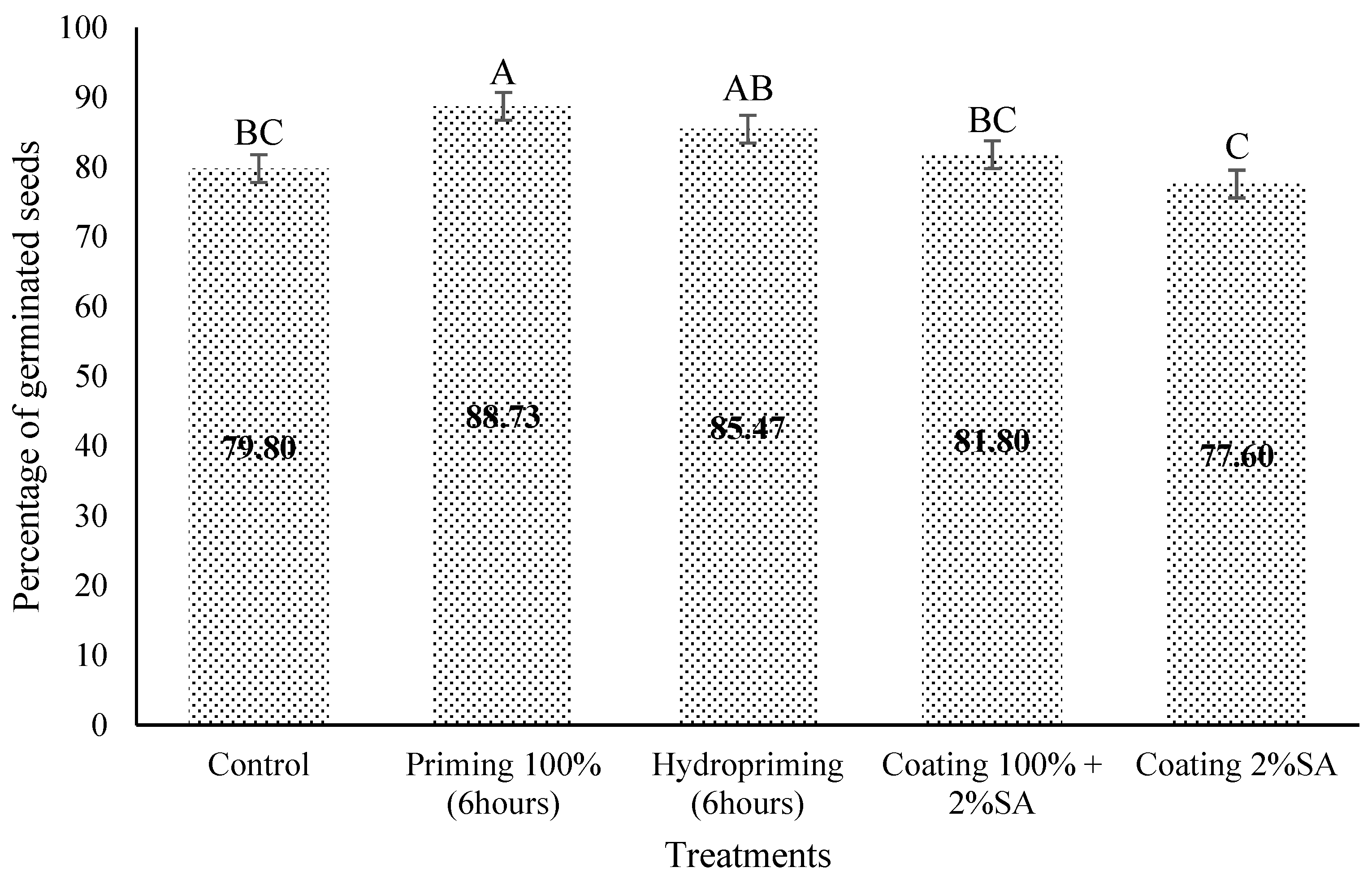
As shown in Figure 2, germination evaluation demonstrates clear differences in the efficacy of each treatment, with seed priming methods showing superior performance (77.60 ± 8.95–81.80 ± 6.65% germination) compared with seed coating treatments, which showed only minimal improvement over the control (79.80 ± 7.69%). Priming the lettuce seeds with 100% pecan extract concentration for a period of 6 h not only reduced pathogen presence but additionally achieved the highest germination rate (88.73 ± 2.87%), significantly higher (p < 0.05) compared to all other treatments, except for hydropriming, with which it showed no significant difference.

3.3. Effect of Different Concentrations of PSE for Coating and Priming on E. coli O157:H7 in Lettuce Seeds

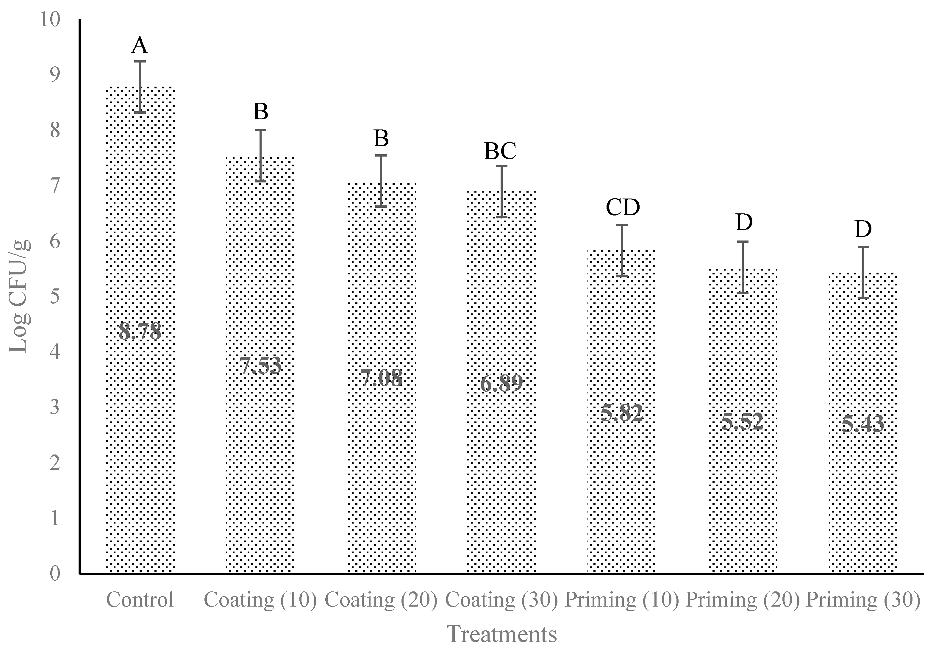
The data presented in Figure 3 illustrates significant differences in the effectiveness of treatments for reducing E. coli levels, measured as Log CFU/g. The control exhibited bacterial concentration at 8.78 ± 0.07 Log CFU/g after inoculation. Seed coating treatments reduced bacterial counts in a dose-dependent manner, with higher concentrations (30 w/v) showing greater efficacy (6.89 ± 0.08 Log CFU/g). Nevertheless, the differences among different concentrations were not statistically significant (p < 0.05). Seed priming treatments (20 and 30 w/v), however, were significantly (p < 0.05) more effective than coating, achieving the highest bacterial reduction across all concentrations (5.52 ± 0.35–5.43 ± 0.71 Log CFU/g), with no significant (p < 0.05) difference between doses.

3.4. Effect of Different Concentrations of PSE for Coating and Priming on Pseudomonas spp. in Lettuce Seeds

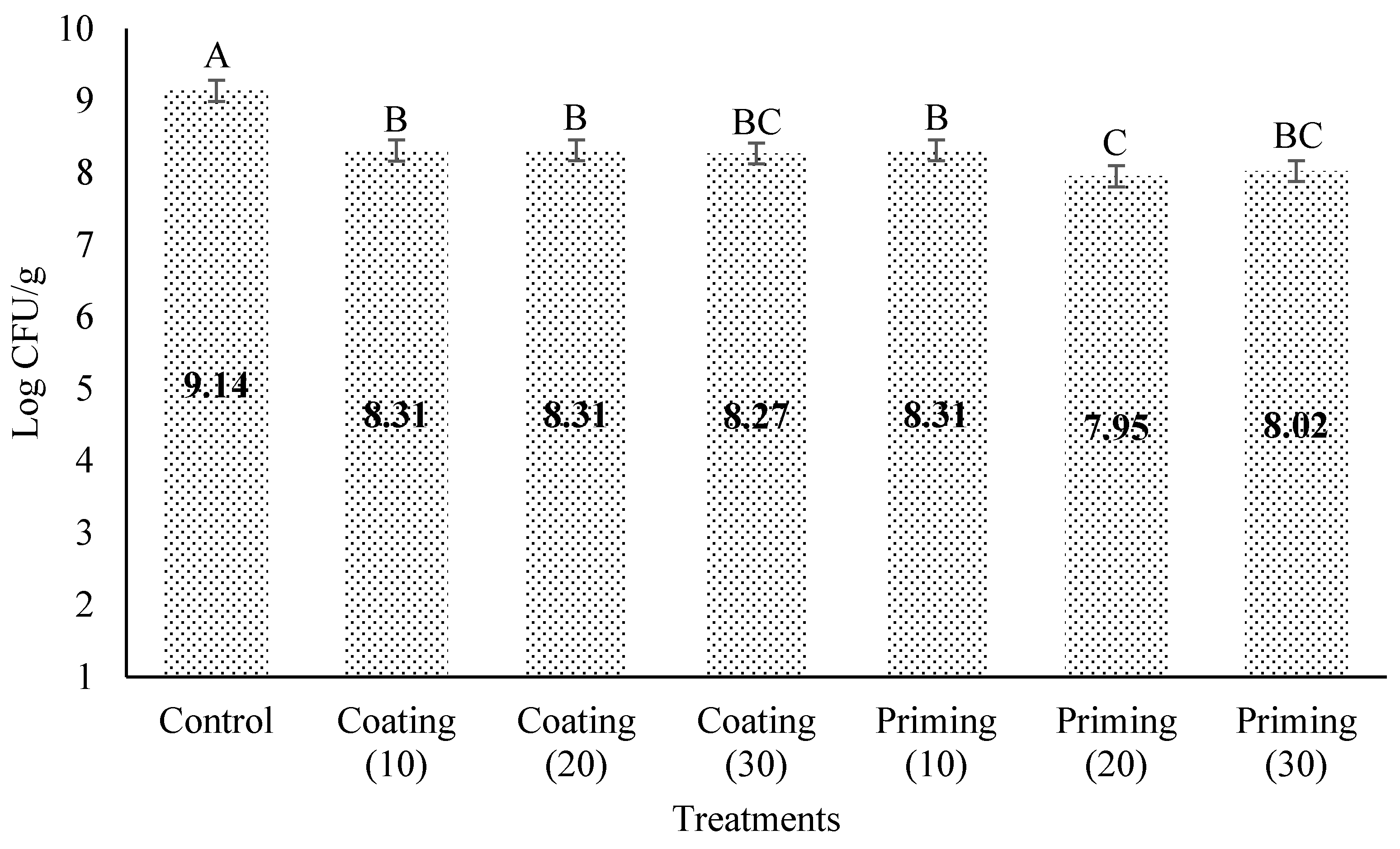
The effect of PSE priming and coating treatments applied in different concentrations (w/v) to lettuce seeds against Pseudomonas spp. was shown in Figure 4. Control treatment showed the bacterial load at 9.14 ± 0.09 Log CFU/g, which was significantly (p < 0.05) different from all other treatments. Coating treatments showed minimal variation across concentrations (8.31± 0.06–8.27 ± 0.003 Log CFU/g), with no significant differences. Seed priming treatments were slightly more effective, particularly at 20 w/v (7.96 ± 0.05 Log CFU/g) and 30 w/v (8.025 ± 0.19 Log CFU/g), yet the differences were marginal. There were no significant differences between treatments. However, the only exceptions were priming (20), which showed a significant (p < 0.05) difference compared to coating (20) and priming (10).

4. Discussion

The effectiveness of PSE as a natural antimicrobial can be attributed to its rich composition of bioactive phenolic compounds, particularly condensed tannins, flavonoids, lignin-derived phenolics, and anthocyanidin pigments. These compounds act synergistically to disrupt bacterial cell membranes, inhibit essential enzymes, and interfere with microbial metabolism, contributing to broad-spectrum antimicrobial activity [23,24,25,26,39]. The results of this study support these mechanisms, as PSE significantly reduced both foodborne pathogens and plant-associated pathogens, including L. monocytogenes, E. coli O157:H7, and Pseudomonas spp. on lettuce seeds without affecting its germination potential. Seed priming and coating applications demonstrated the extract’s efficacy. These highlight its potential as a sustainable alternative to synthetic antimicrobials against plant and food pathogens and for microbial safety improvement in fresh produce and minimally processed food.
Previous studies have applied PSE to reduce L. monocytogenes on food products such as catfish filets and fresh-cut cantaloupe. This supports their potential use as natural antimicrobial agents in food preservation. CaxambÚ et al. [23] found that pecan shell aqueous extract effectively inhibited L. monocytogenes in vitro and reduced bacterial counts in refrigerated lettuce by up to 4 log CFU/g. Another study by Yemmireddy et al. [28] found that PSE at 5 mg/mL significantly reduced L. monocytogenes levels on catfish filets stored at 4 °C, achieving up to 3.97 Log CFU/sample reduction on Day 1. There were reductions in fresh-cut cantaloupes with a maximum of 1.22 Log CFU/sample by Day 5. Similarly, Kharel et al. [29] found that 5% PSE with pullulan (PSE-P) coating reduced L. monocytogenes on blueberries by up to 2.06 Log CFU/g immediately after treatment and significantly suppressed its survival during refrigerated storage. The outcomes of the present study indicate the potential of PSE as a natural antibacterial agent for seeds to reduce Listeria monocytogenes. Further studies were performed to assess the impact of PSE treatment on the germination capacity of lettuce seeds. Germination results highlight the performance of PSE as an effective seed priming agent. These findings are consistent with previous studies that demonstrate how using effective priming agents can significantly enhance germination in lettuce seeds [40]. Such enhancements can possibly be due to improved parameters related to seed priming, such as early biochemical activation of the seeds during the priming process [41].
E. coli and Pseudomonas showed similar results to those found by Yemmireddy et al. [28], in which PSE exhibited antimicrobial activity against E. coli O157:H7 with a minimum inhibitory concentration of 5 mg/mL, indicating that E. coli O157:H7 was the most resistant among the tested pathogens. Unlike L. monocytogenes and Salmonella enterica, E. coli O157:H7 showed no variation in susceptibility across different strains or pecan cultivars, suggesting a pathogen-specific response to pecan shell bioactivity. Van Loo et al. [42] tested commercial liquid smoke from pecan nut shells in inhibiting three foodborne pathogens; they found that roasted pecan shell extracts prepared with acetic acid and methanol exhibited growth inhibition against E. coli. The results of this study demonstrate that coating and priming treatments effectively reduced E. coli levels on lettuce seeds. Furthermore, demonstrating the improved priming efficiency, the priming treatment at 10 w/v concentration generated microbial reductions equivalent to those of the 30 w/v coating treatment. This shows that seed priming could be a more resource-efficient technique, lowering bacteria numbers and minimizing the amount of antimicrobial agent. These results emphasize the importance of delivery method and the benefits of seed priming in the efficacy of natural antimicrobial strategies. Seed priming offers many benefits, including enhanced resistance, stress tolerance, improved germination rates, and overall plant health [43,44,45,46,47,48]. Future work should explore the effectiveness of the PSE coating and priming in different combinations (time and temperature) against E. coli and Pseudomonas. Also, evaluate seed germination, plant growth, and contamination of treated seeds for all pathogens.

5. Conclusions

For applications targeting Listeria monocytogenes, seed coating and seed priming at 4 °C showed significant results in controlling this pathogen compared with the control. Seed priming the lettuce seeds with 100% PSE concentration for 6 h and hydropriming did not negatively affect the germination of seeds. It significantly enhanced the germination rates compared to the untreated control and coating treatments. To control Escherichia coli O157:H7, both seed coating and seed priming treatments presented significantly positive results. However, seed priming showed the most effective result in reducing bacterial levels, showing significant reductions even at the lowest concentration, 1:10 (w/v), compared with the control treatment. PSE priming and coating treatments applied at various concentrations reduced the number of Pseudomonas spp. on lettuce seeds compared to the control, with priming generally showing slightly greater effectiveness than coating. Among all treatments, only priming at 20 w/v showed a statistically significant reduction compared to coating at 20 w/v and priming at 10 w/v. It is noteworthy that the extract is derived from an agricultural byproduct. This adds value to pecan shell waste and supports sustainable processing practices in agriculture and the food industry. Further research with larger sample sizes and additional concentrations could clarify optimal dosing strategies. Overall, these findings support the potential application of PSE as natural antimicrobial agents for food safety interventions, particularly in the decontamination of lettuce seeds and minimally processed or ready-to-eat foods where control of pathogens is critical.

Author Contributions

Conceptualization: A.A. and I.L.; methodology: A.A. and I.L.; validation: A.A. and I.L.; formal analysis: I.L.; investigation: I.L.; resources: A.A.; data curation: I.L.; writing—original draft preparation: I.L.; writing—review and editing: A.A., F.V. and J.H.H.; supervision: A.A.; project administration: A.A.; funding acquisition: A.A. All authors have read and agreed to the published version of the manuscript.

Funding

This research was funded by USDA HATCH Project # LAB94565.

Data Availability Statement

Data are contained within the article.

Conflicts of Interest

The authors declare no conflicts of interest.

Abbreviations

The following abbreviations are used in this manuscript:
PSEPecan Shell Extract
TSBTryptic Soy Broth
TSBYETryptic Soy Broth supplemented with 6% extract
LB, MillerLuria–Bertani, Miller broth/agar
SMACMacConkey Agar with Sorbitol
TTC2, 3, 5-triphenyltetrazolium chloride
SASodium Alginate
DWDistilled Water
PCPecan Shell Extract Coating
PPPecan Shell Extract Priming
PPRPecan Shell Extract Priming at Refrigeration

References

  1. Rajaseger, G.; Chan, K.L.; Yee Tan, K.; Ramasamy, S.; Khin, M.C.; Amaladoss, A.; Kadamb Haribhai, P. Hydroponics: Current trends in sustainable crop production. Bioinformation 2023, 19, 925–938. [Google Scholar] [CrossRef]
  2. Khatri, L.; Kunwar, A.; Bist, D. Hydroponics: Advantages And Challenges in Soilless Farming. Big Data Agric. 2024, 6, 81–88. [Google Scholar] [CrossRef]
  3. Economic Research Service. U.S. Lettuce Production Shifts Regionally by Season. 2023. Available online: https://www.ers.usda.gov/data-products/charts-of-note/chart-detail?chartId=106516 (accessed on 15 July 2025).
  4. The Society of Tympanuchus Cupido Pinnatus, Ltd. Lettuce Market Insights, Size, Share, Growth Analysis, Trends, and Forecast 2024–2031. 2023. Available online: https://www.skyquestt.com/report/lettuce-market (accessed on 15 July 2025).
  5. Ahmed, Z.F.R.; Alnuaimi, A.K.H.; Askri, A.; Tzortzakis, N. Evaluation of Lettuce (Lactuca sativa L.) Production under Hydroponic System: Nutrient Solution Derived from Fish Waste vs. Inorganic Nutrient Solution. Horticulturae 2021, 7, 292. [Google Scholar] [CrossRef]
  6. Center for Crop Diversification, University of Kentucky. Hydroponic Lettuce (CCD-CP-063). 2024. Available online: https://publications.ca.uky.edu/ (accessed on 20 April 2025).
  7. Carvalho, R.; Luís, C.; Nunes, W.; Zanovello, C.; Gomes, M.; Luz, S.; Gadotti, G.I.; Luz, C.; Conill-Gomes, M. Hydroponic lettuce production and minimally processed lettuce. CIGR J. 2015, 2015, 290. [Google Scholar]
  8. Gillani, S.A.; Abbasi, R.; Martinez, P.; Ahmad, R. Comparison of Energy-use Efficiency for Lettuce Plantation under Nutrient Film Technique and Deep-Water Culture Hydroponic Systems. Procedia Comput. Sci. 2023, 217, 11–19. [Google Scholar] [CrossRef]
  9. Majid, M.; Khan, J.N.; Ahmad Shah, Q.M.; Masoodi, K.Z.; Afroza, B.; Parvaze, S. Evaluation of hydroponic systems for the cultivation of Lettuce (Lactuca sativa L., var. Longifolia) and comparison with protected soil-based cultivation. Agric. Water Manag. 2021, 245, 106572. [Google Scholar] [CrossRef]
  10. Sela Saldinger, S.; Rodov, V.; Kenigsbuch, D.; Bar-Tal, A. Hydroponic Agriculture and Microbial Safety of Vegetables: Promises, Challenges, and Solutions. Horticulturae 2023, 9, 51. [Google Scholar] [CrossRef]
  11. Dankwa, A.S.; Machado, R.M.; Perry, J.J. Sources of food contamination in a closed hydroponic system. Lett. Appl. Microbiol. 2020, 70, 55–62. [Google Scholar] [CrossRef]
  12. Riggio, G.M.; Wang, Q.; Kniel, K.E.; Gibson, K.E. Microgreens—A review of food safety considerations along the farm to fork continuum. Int. J. Food Microbiol. 2019, 290, 76–85. [Google Scholar] [CrossRef]
  13. Martín, I.; Gálvez, L.; Guasch, L.; Palmero, D. Fungal Pathogens and Seed Storage in the Dry State. Plants 2022, 11, 3167. [Google Scholar] [CrossRef]
  14. Cottyn, B.; Vanhouteghem, K.; Heyrman, J.; Bleyaert, P.; Vaerenbergh, J.; De Vos, P.; Höfte, M.; Maes, M. Pseudomonads associated with midrib rot and soft rot of butterhead lettuce and endive. Commun. Agric. Appl. Biol. Sci. 2005, 70, 101–109. [Google Scholar]
  15. Patel, N.; Patel, R.; Wyenandt, C.A.; Kobayashi, D.Y. First Report of Pseudomonas cichorii Causing Bacterial Leaf Spot on Romaine Lettuce (Lactuca sativa var. longifolia) and Escarole (Cichorium endivia) in New Jersey. Plant Dis. 2021, 105, 4150. [Google Scholar] [CrossRef] [PubMed]
  16. CDC. Listeria Outbreak Linked to Leafy Greens–February 2023. 2024. Available online: https://www.google.com/url?sa=t&source=web&rct=j&opi=89978449&url=https://www.cdc.gov/listeria/outbreaks/monocytogenes-02-23/index.html&ved=2ahUKEwiNvPHZq96OAxU8s1YBHY7wDWQQFnoECBsQAQ&usg=AOvVaw3vnrVhZSNtn9ABLQaAjgLJ (accessed on 21 May 2025).
  17. CDC. Multistate Outbreak of E. coli O157:H7 Infections Linked to Romaine Lettuce (Final Update). 2019. Available online: https://archive.cdc.gov/www_cdc_gov/ecoli/2019/o157h7-11-19/index.html (accessed on 21 May 2025).
  18. Aguilar-Rito, M.; Arzate-Fernández, A.; García-Núñez, H.; Héctor, T. Establishment of an efficient protocol for in vitro disinfection of seeds of seven Agave spp. species. Rev. Mex. De Fitopatol. Mex. J. Phytopathol. 2023, 42. [Google Scholar] [CrossRef]
  19. Gilbert, G.S.; Diaz, A.; Bregoff, H.A. Seed Disinfestation Practices to Control Seed-Borne Fungi and Bacteria in Home Production of Sprouts. Foods 2023, 12, 747. [Google Scholar] [CrossRef] [PubMed]
  20. Kim, H.; Kim, H.; Bang, J.; Beuchat, L.R.; Ryu, J.-H. Synergistic Effect of Chlorine Dioxide and Drying Treatments for Inactivating Escherichia coli O157:H7 on Radish Seeds. J. Food Prot. 2010, 73, 1225–1230. [Google Scholar] [CrossRef]
  21. Sanna, M.; Gilardi, G.; Gullino, M.L.; Mezzalama, M. Evaluation of physical and chemical disinfection methods of Brassica oleracea seeds naturally contaminated with Xanthomonas campestris pv. campestris. J. Plant Dis. Prot. 2022, 129, 1145–1152. [Google Scholar] [CrossRef]
  22. Di Maro, M.; Gargiulo, L.; Gomez d’Ayala, G.; Duraccio, D. Exploring Antimicrobial Compounds from Agri-Food Wastes for Sustainable Applications. Int. J. Mol. Sci. 2024, 25, 13171. [Google Scholar] [CrossRef]
  23. CaxambÚ, S.; Biondo, E.; Kolchinski, E.; Padilha, R.; Brandelli, A.; Sant Anna, V. Evaluation of the antimicrobial activity of pecan nut [Carya illinoinensis (Wangenh) C. Koch] shell aqueous extract on minimally processed lettuce leaves. Ciência E Tecnol. De Aliment. 2016, 36, 42–45. [Google Scholar] [CrossRef]
  24. Flores-Estrada, R.A.; Gámez-Meza, N.; Medina-Juárez, L.A.; Castillón-Campaña, L.G.; Molina-Domínguez, C.C.; Rascón-Valenzuela, L.A.; García-Galaz, A. Chemical Composition, Antioxidant, Antimicrobial and Antiproliferative Activities of Wastes from Pecan Nut [Carya illinoinensis (Wagenh) K. Koch]. Waste Biomass Valorization 2020, 11, 3419–3432. [Google Scholar] [CrossRef]
  25. Chiao Ying, H.; Gerald, L.R.; Jennifer, C.; Ching Hsuan, L.; Jinn Tsyy, L.; Audrey Chingzu, C. Pecan shell by-products—Phenolic compound contents and antimicrobial properties. AIMS Agric. Food 2020, 5, 218–232. [Google Scholar]
  26. Cason, C.; Yemmireddy, V.K.; Moreira, J.; Adhikari, A. Antioxidant Properties of Pecan Shell Bioactive Components of Different Cultivars and Extraction Methods. Foods 2021, 10, 713. [Google Scholar] [CrossRef]
  27. do Prado, A.C.P.; da Silva, H.S.; da Silveira, S.M.; Barreto, P.L.M.; Vieira, C.R.W.; Maraschin, M.; Ferreira, S.R.S.; Block, J.M. Effect of the extraction process on the phenolic compounds profile and the antioxidant and antimicrobial activity of extracts of pecan nut [Carya illinoinensis (Wangenh) C. Koch] shell. Ind. Crops Prod. 2014, 52, 552–561. [Google Scholar] [CrossRef]
  28. Yemmireddy, V.K.; Cason, C.; Moreira, J.; Adhikari, A. Effect of pecan variety and the method of extraction on the antimicrobial activity of pecan shell extracts against different foodborne pathogens and their efficacy on food matrices. Food Control 2020, 112, 107098. [Google Scholar] [CrossRef]
  29. Kharel, K.; Kraśniewska, K.; Gniewosz, M.; Prinyawiwatkul, W.; Fontenot, K.; Adhikari, A. Antimicrobial screening of pecan shell extract and efficacy of pecan shell extract-pullulan coating against Listeria monocytogenes, Salmonella enterica, and Staphylococcus aureus on blueberries. Heliyon 2024, 10, e29610. [Google Scholar] [CrossRef] [PubMed]
  30. Javed, T.; Afzal, I.; Shabbir, R.; Ikram, K.; Saqlain Zaheer, M.; Faheem, M.; Haider Ali, H.; Iqbal, J. Seed coating technology: An innovative and sustainable approach for improving seed quality and crop performance. J. Saudi Soc. Agric. Sci. 2022, 21, 536–545. [Google Scholar] [CrossRef]
  31. Skrzypczak, D.; Jarzembowski, Ł.; Izydorczyk, G.; Mikula, K.; Hoppe, V.; Mielko, K.A.; Pudełko-Malik, N.; Młynarz, P.; Chojnacka, K.; Witek-Krowiak, A. Hydrogel Alginate Seed Coating as an Innovative Method for Delivering Nutrients at the Early Stages of Plant Growth. Polymers 2021, 13, 4233. [Google Scholar] [CrossRef]
  32. Szabó, L.; Gerber-Lemaire, S.; Wandrey, C. Strategies to Functionalize the Anionic Biopolymer Na-Alginate without Restricting Its Polyelectrolyte Properties. Polymers 2020, 12, 919. [Google Scholar] [CrossRef]
  33. Hecht, H.; Srebnik, S. Structural Characterization of Sodium Alginate and Calcium Alginate. Biomacromolecules 2016, 17, 2160–2167. [Google Scholar] [CrossRef]
  34. Durgadevi, P.; Girigoswami, K.; Girigoswami, A. Biodegradable nanomaterials in boosting seed vigor and germination: Seed coating towards sustainability. Discov. Appl. Sci. 2025, 7, 695. [Google Scholar] [CrossRef]
  35. Dutra, F.; Bianca, D.; Silva, J.M.; Shiguehara, A.P. Seeds technologies: Performance of sodium alginate and calcium chloride as seed coating. Acta Biológica Catarin. 2024, 11, 34–43. [Google Scholar] [CrossRef]
  36. Abdukerim, R.; Li, L.; Li, J.-H.; Xiang, S.; Shi, Y.-X.; Xie, X.-W.; Chai, A.L.; Fan, T.-F.; Li, B.-J. Coating seeds with biocontrol bacteria-loaded sodium alginate/pectin hydrogel enhances the survival of bacteria and control efficacy against soil-borne vegetable diseases. Int. J. Biol. Macromol. 2024, 279, 135317. [Google Scholar] [CrossRef] [PubMed]
  37. Paparella, S.; Araújo, S.S.; Rossi, G.; Wijayasinghe, M.; Carbonera, D.; Balestrazzi, A. Seed priming: State of the art and new perspectives. Plant Cell Rep. 2015, 34, 1281–1293. [Google Scholar] [CrossRef]
  38. MacDonald, M.T.; Mohan, V.R. Chemical Seed Priming: Molecules and Mechanisms for Enhancing Plant Germination, Growth, and Stress Tolerance. Curr. Issues Mol. Biol. 2025, 47, 177. [Google Scholar] [CrossRef]
  39. Ortiz-Quezada, A.G.; Lombardini, L.; Cisneros-Zevallos, L. Chapter 104—Antioxidants in Pecan Nut Cultivars [Carya illinoinensis (Wangenh.) K. Koch]. In Nuts and Seeds in Health and Disease Prevention; Preedy, V.R., Watson, R.R., Patel, V.B., Eds.; Academic Press: San Diego, CA, USA, 2011; pp. 881–889. [Google Scholar]
  40. Lyalina, T.; Shagdarova, B.; Zhuikova, Y.; Il’ina, A.; Lunkov, A.; Varlamov, V. Effect of Seed Priming with Chitosan Hydrolysate on Lettuce (Lactuca sativa) Growth Parameters. Molecules 2023, 28, 1915. [Google Scholar] [CrossRef]
  41. Corbineau, F.; Taskiran-Özbingöl, N.; El-Maarouf-Bouteau, H. Improvement of Seed Quality by Priming: Concept and Biological Basis. Seeds 2023, 2, 101–115. [Google Scholar] [CrossRef]
  42. Van Loo, E.J.; Babu, D.; Crandall, P.G.; Ricke, S.C. Screening of Commercial and Pecan Shell-Extracted Liquid Smoke Agents as Natural Antimicrobials against Foodborne Pathogens. J. Food Prot. 2012, 75, 1148–1152. [Google Scholar] [CrossRef]
  43. Worrall, D.; Holroyd, G.H.; Moore, J.P.; Glowacz, M.; Croft, P.; Taylor, J.E.; Paul, N.D.; Roberts, M.R. Treating seeds with activators of plant defence generates long-lasting priming of resistance to pests and pathogens. New Phytol. 2012, 193, 770–778. [Google Scholar] [CrossRef]
  44. Carmello, C.R.; Cardoso, J.C. Effects of plant extracts and sodium hypochlorite on lettuce germination and inhibition of Cercospora longissima in vitro. Sci. Hortic. 2018, 234, 245–249. [Google Scholar] [CrossRef]
  45. Raza, A.; Tahir, M.; Noor Us, S.; Shah, S.H.; Sarwar, G.; Manzoor, M. Seed priming with zinc ion on growth performance and nutrient acquisition of maize in aridisols. Pak. J. Bot. 2023, 55, 1365–1374. [Google Scholar] [CrossRef] [PubMed]
  46. Huang, P.; He, L.; Abbas, A.; Hussain, S.; Hussain, S.; Du, D.; Hafeez, M.B.; Balooch, S.; Zahra, N.; Ren, X.; et al. Seed Priming with Sorghum Water Extract Improves the Performance of Camelina (Camelina sativa (L.) Crantz.) under Salt Stress. Plants 2021, 10, 749. [Google Scholar] [CrossRef] [PubMed]
  47. Farooq, M.; Akhtar, S.; Imran, S.; Dar, A.H.; Raza, A.; Masroor, A. Seed Priming with Plant Extracts Gives Enhanced Resistance Against Alternaria Solani in Tomato. Pak. J. Phytopathol. 2024, 36, 2411–2422. [Google Scholar]
  48. Gassoumi, W.; Ellouzi, H.; Slimene, I.B.; Kalai, F.Z.; Ayed, R.B.; Zorrig, W.; Debez, A.; Abdelly, C.; Oueslati, S. Plant extracts and plant growth promoting rhizobacteria: Seed bio-priming approach to improve tolerance of Lactuca sativa L. under salt stress. Euro-Mediterr. J. Environ. Integr. 2024, 10, 1901–1918. [Google Scholar] [CrossRef]
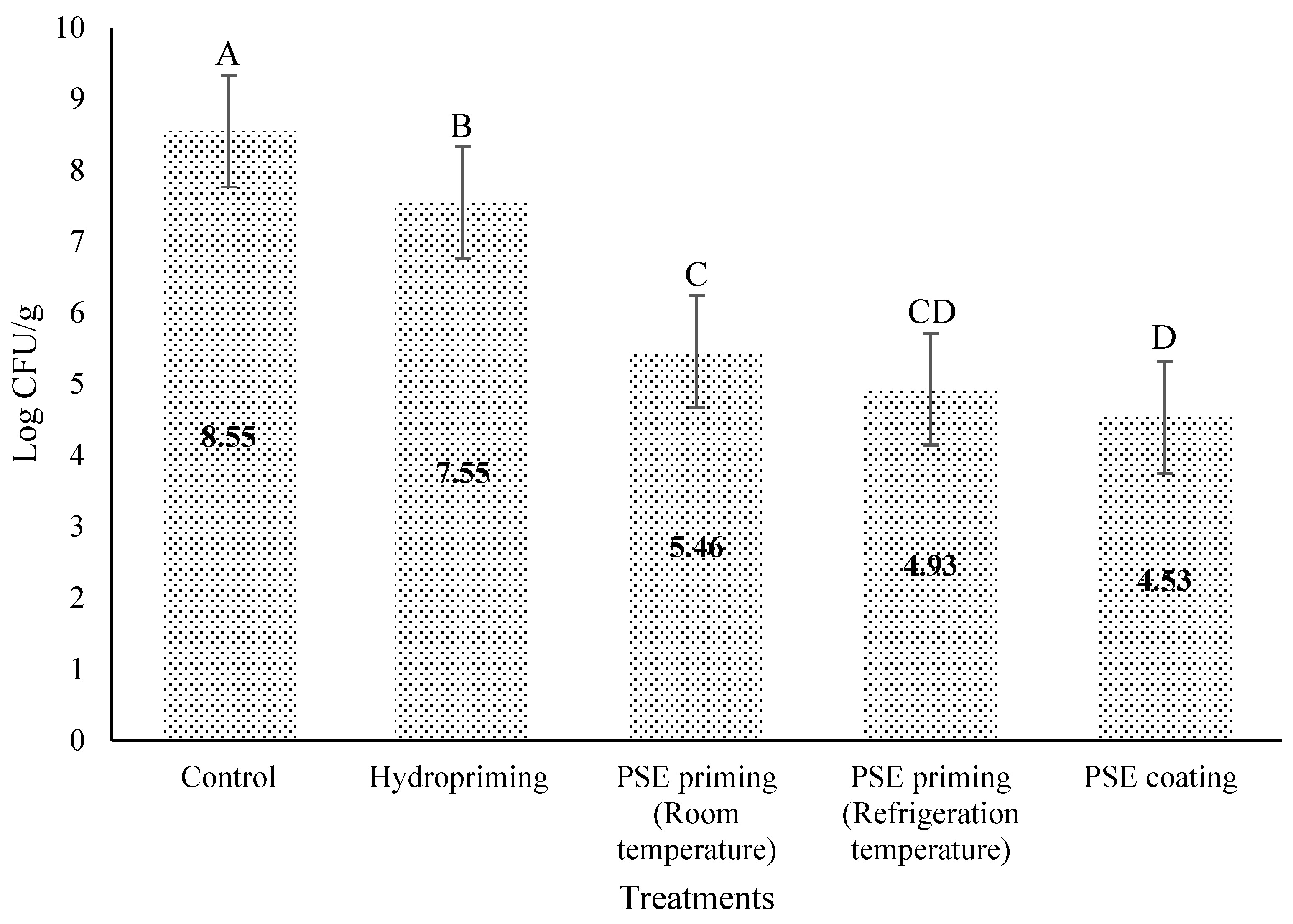
Figure 1. Listeria monocytogenes levels on lettuce seeds after PSE priming and coating treatments. Different letters within a column show a statistically significant difference.
Agronomy 15 01865 g001
Figure 2. Percentage of lettuce germinated seeds after PSE priming and coating treatments. Different letters within a column show a statistically significant difference.
Agronomy 15 01865 g002
Figure 3. E. coli O157:H7 levels on lettuce seeds after PSE priming and coating treatments. Different letters within a column show a statistically significant difference.
Agronomy 15 01865 g003
Figure 4. Pseudomonas spp. levels on lettuce seeds after PSE priming and coating treatments. Different letters within a column show a statistically significant difference.
Agronomy 15 01865 g004
Table 1. PSE coating treatments.
LabelPecan Extract ConcentrationSA
Concentration
A-coating100%2%
BUntreated Seeds
Table 2. PSE priming treatments.
LabelPecan Extract ConcentrationSoaking TimeTemperature
°C
A100%6 h25
B100%6 h4
CSterilized distilled water 6 h25
EUntreated seeds
Table 3. Seed germination parameters.
TreatmentGPEPGE-DAY4GRMGT
Control (No treatment)802720.382.66
PC (2%SA)812710.352.85
Coating (2%SA)772670.352.84
PP (6 h)902840.452.20
Hydropriming (6 h)882820.442.27
GP = Germination percentage, % of seeds that germinated at the end. EP = Energy period, the number of days taken to reach the highest daily germination. (It shows when the maximum daily germination occurred, the “peak” day.) GE = Germination energy, % of seeds germinated by a certain early day (often day 4). GR = germination rate; the reciprocal of the mean time to germination shows the speed of germination. MGT = Mean Germination Time; average time taken for seeds to germinate.
Disclaimer/Publisher’s Note: The statements, opinions and data contained in all publications are solely those of the individual author(s) and contributor(s) and not of MDPI and/or the editor(s). MDPI and/or the editor(s) disclaim responsibility for any injury to people or property resulting from any ideas, methods, instructions or products referred to in the content.