Article

Study on Pathogen Identification and Biocontrol Fungi Screening of Oat Sheath Rot

College of Pratacultural Science, Gansu Agricultural University, Lanzhou 730070, China
*
Author to whom correspondence should be addressed.
Agronomy 2025, 15(8), 1776; https://doi.org/10.3390/agronomy15081776
Submission received: 19 May 2025 / Revised: 8 July 2025 / Accepted: 22 July 2025 / Published: 24 July 2025

Abstract

Oat sheath rot disease significantly reduces commercial oat yields, yet research on its incidence, causative pathogens, and control strategies remains limited, particularly in China. This study investigated the occurrence of oat sheath rot in major oat-producing regions of Northern China. Here, we isolated and identified two species of primary pathogenic fungi, Scopulariopsis brevicaulis and Alternaria alternata. Next, we conducted pathogenicity tests to confirm their role in the progression of oat sheath rot disease. Subsequently, we screened putative biocontrol fungi and identified Trichoderma harzianum and Trichoderma koningii as effective antagonistic biocontrol fungi. Both species demonstrated strong inhibitory effects against two primary pathogens through competitive interactions, with T. koningii achieving 100% inhibition in one test. Overall, T. harzianum and T. koningii both exerted strong inhibitory effects against pathogenic fungi via different forms of competition. Most importantly, infection experiments showed that T. harzianum and T. koningii both exerted strong antifungal effects against the pathogenic fungi that cause oat sheath rot. Taken together, our findings provide a foundation for developing biological control strategies to mitigate oat sheath rot in oat cultivation in China.

1. Introduction

Oat sheath rot disease is known to cause large reductions in commercial oat (Avena sativa) yields. At present, there remains relatively little research on the incidence of oat sheath rot, the extraction and identification of pathogens, or prevention and control measures, especially in China, a major oat exporter. This has resulted in a general failure to establish a corresponding prevention and control system. At the same time, excessive use of chemical agents can produce fungal resistance, leave pesticide residues, and cause other production or environmental issues.
Oat (Avena sativa, Gramineae) is a major cereal crop whose leaves and straw are juicy and tender with good palatability [1]. Moreover, the crude protein, crude fat, and nitrogen-free content of the straw is generally high, and fiber content, which is difficult to digest, is generally low. Oat is an excellent forage plant for supplementary feeding of livestock during the winter and spring and is critical for disaster-resistant livestock protection in pastoral areas [2]. At present, oat is produced on a large scale both as an annual grain and as a forage crop in China. It exhibits significant drought resistance, cold tolerance, adaptability to barren conditions, and salt tolerance [3] and plays a crucial role in agricultural rotation systems in China [4,5].
As the cultivation area of oat crops has expanded, the impact of various diseases on oat yield and quality has intensified, having now become one of the primary limiting factors for further development of the oat industry [6]. Oat diseases directly reduce oat yield and quality, causing decreases in crude protein, fat, and soluble sugars while increasing the crude fiber content of oats, which together cause reduced palatability and digestibility [7].
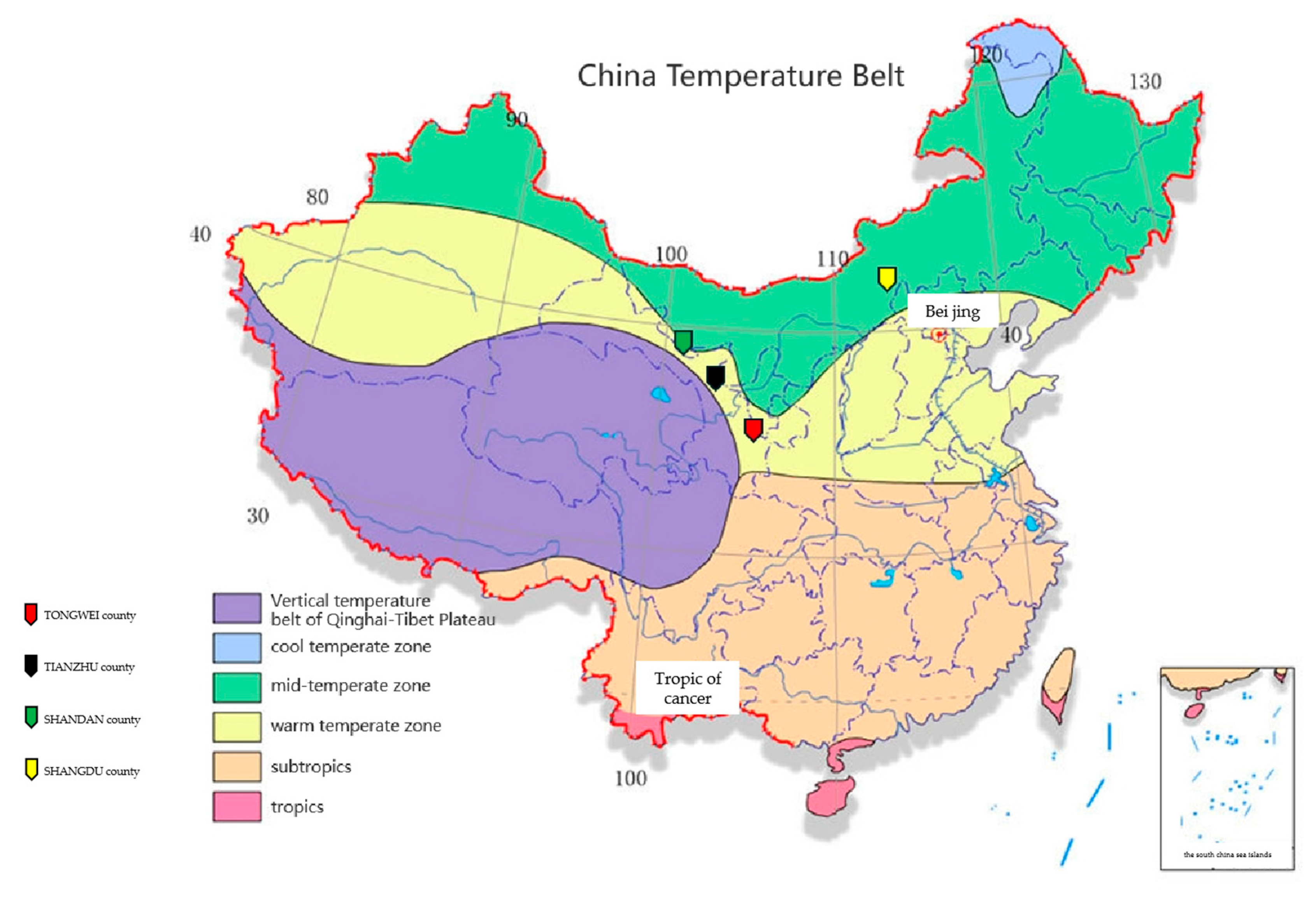
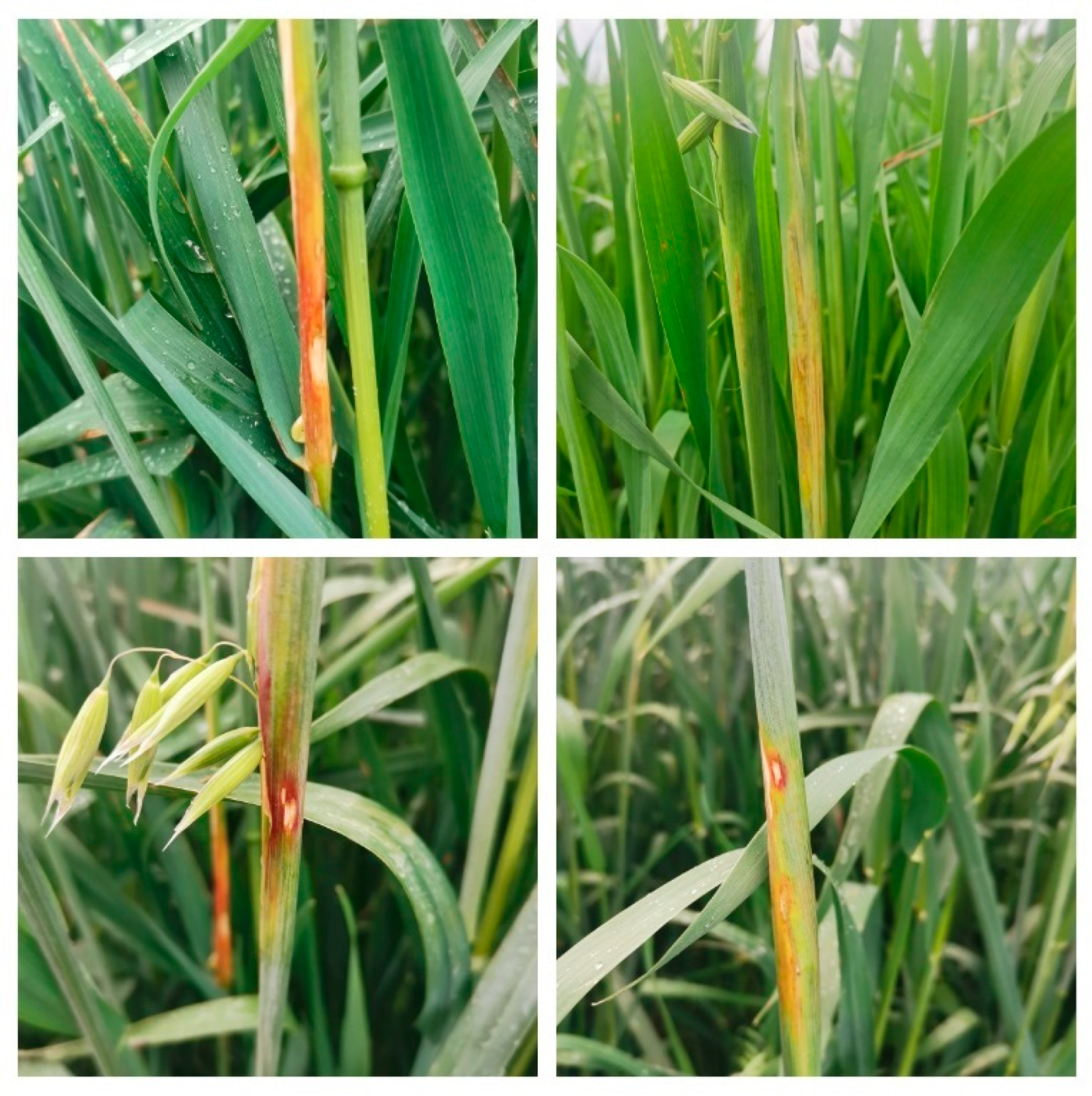
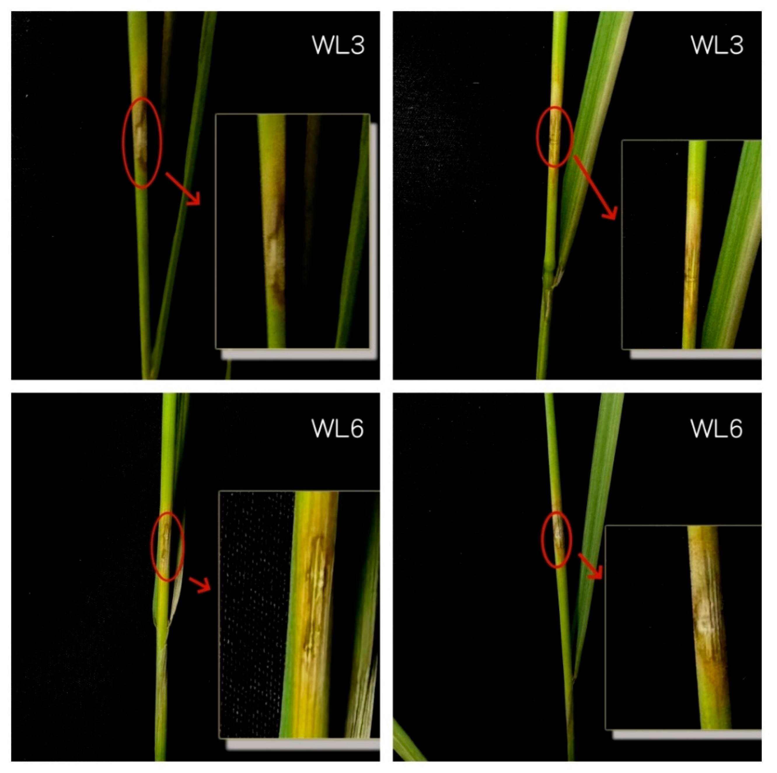
In recent years, a novel disease known as oat sheath rot was found on cultivated oats, wild oats, and highland barley in high-altitude (2400~3200 m) regions of central, western, and southern Gansu Province (i.e., in Tongwei, Tianzhu, and Hezuo) [8]. A previous study found that this oat sheath rot generally occurred sporadically in fields, with rates of diseased ears in severely diseased fields reaching 20%~30% of all plants [9]. The disease mainly occurs during the late growth period [10], which endangers oat leaf sheaths. Once infected, plants show brown and irregularly shaped lesions. In severe cases, the disease can spread to the entire leaf sheath [11,12], resulting in leaf sheath desiccation or necrosis, thereby significantly impacting oat yield [13].
At present, the use of chemical agents to control crop disease remains the main measure used during commercial agricultural production. However, the large-scale use of chemical pesticides is known to cause various problems, including induced resistance of pathogens to specific pesticides, changes in pathogen biotype, increased ineffectiveness of alternate fungicides, high levels of fungicide residues on postharvest products, and other negative effects on the environment and human health [14], including environmental degradation, drug resistance, and large-scale soil hardening. During China’s promotion of eco-friendly agricultural and sustainable development policies, it is necessary to seek greener cultivation methods that can ensure both adequate yield and food and environmental safety [15,16,17,18]. Recent years have identified new ways to prevent and control plant disease by isolating and identifying pathogenic fungi from hosts and applying biocontrol fungal strains to inhibit pathogen proliferation and promote normal host growth [19,20,21].
Therefore, this study conducted research into fungal biocontrol in the main oat-producing areas of China. By investigating disease in the field, we first isolated and identified pathogenic bacteria, then screened efficient and low-toxicity biocontrol fungi. In doing so we identified key pathogens causing oat sheath rot disease in major oat-producing areas and provide a basic protocol for the biological control of this disease.

2. Materials and Methods

2.1. Survey and Sample Collection

2.1.1. Investigation Site

A systematic survey was conducted in Tongwei County (Dingxi City, Gansu Province), Tianzhu County (Wuwei City, Gansu Province), Shandan County (Zhangye City, Gansu Province), and Shangdu County (Wulanchabu City, Inner Mongolia). The distribution of survey sites and general information are shown in Figure 1 and Table 1.

2.1.2. Test Materials

A total of 32 materials were tested; the details are shown in Table 2.

2.1.3. Field Investigation of Sheath Rot

Three oat planting areas in Gansu Province (i.e., Tongwei, Tianzhu, and Shandan Counties) and one in Inner Mongolia (Shangdu County) were investigated in this study. At each site, a 667 m2 plot was selected in an experimental field during the oat growing season. A five-point sampling method was then used to avoid sampling a 5 m band along the edge of the plot, and 20 oat plants were randomly selected at each point for sampling [22].

2.1.4. Disease Classification

The grading standard for oat sheath rot was set according to a framework published by Pushpam et al. (Table 3) [23]. IBM SPSS Statistics 26 software was used to determine the statistical significance of differences in group mean values. Oat sheath rot severity was classified according to the criteria listed below.
Calculation formula:
R = T D ÷ T S × 100 %
S = i = 1 n ( X i × S i ) ÷ ( X n × S n ) × 100
Here, M indicates morbidity; TD, the total number of diseased plants; TS, the total number of survey plants; S, the disease index; Xi, the number of leaf sheaths of each disease grade; Si, the value of severity grade i; Xn, the total number of leaf sheaths investigated; and Sn, the maximum severity grade value.

2.1.5. Resistance Evaluation

Next, oat sheath rot resistance was evaluated as follows (Table 4).

2.2. Pathogen Isolation and Identification

2.2.1. Separation and Purification

After a pair of scissors was sterilized, a small piece of tissue was cut along the edge of a lesion on a diseased plant. First, the lesion sample was soaked in 0.5% sodium hypochlorite for 1–2 min, then washed with 75% alcohol for 30 s before being washed three times in sterile water. Finally, samples were placed on sterilized filter paper to dry, and the lesion was placed downward on a potato glucose agar medium for culturing at 25 °C for 3 days. After the mycelium growth on the plate was visible, a sample of mycelium taken from the edge of the original culture was used to inoculate a new medium for culturing at a constant temperature. After seven days of cultivation, the resulting mycelium was mixed with sterile water to make a fungal spore suspension from which single spores were isolated. Purified colonies were compressed into 5 mm cakes that were inoculated onto an inclined medium that was placed in a refrigerator at 4 °C until further use [25].

2.2.2. Morphological Identification

Pathogen strains isolated and purified at early life stages were cultured on PDA plates at 25 °C for 5–7 days. Next, colony morphology and spore size were observed under a microscope, photographed, and recorded. Finally, we used the morphological taxonomic key developed by Siebold, M et al. for preliminary strain identification [26].

2.2.3. Identification Using Molecular Biological Analyses

In parallel, we extracted DNA from the pathogenic bacteria extracted in the early stage, then sent DNA sequence information regarding the ITS region of the pathogen genome. The primers used in this study were ITS1 (5′-TCCGTAGGTGAACCTGCGG-3′) and ITS4 (5′-TCCTCCGCTTATTGATATGC-3′). Comparative analysis of pathogen ITS sequences was performed using NCBI-BLAST. After preliminary species identification, we developed specific primers for each corresponding strain, as shown in Table 5:
In this process, RNASE was used to process total DNA, and 5 μL was added during the extraction process. After the extraction of DNA was completed, the purity and concentration of DNA were identified by gel electrophoresis of DNA. The amplified PCR product was subjected to agarose gel electrophoresis to obtain an identification gel (Figure 2).
Using these primers we conducted another sequencing analysis as above, after which a phylogenetic tree was constructed using MEGA version 7.0.

2.2.4. Pathogenicity Testing

Next, we evaluated microbe pathogenicity in BBCH35-39 during the oat growing season. The oat variety used for all pathogenicity analyses was Longyan 3 (this variety was selected to reduce the randomness of variety selection in field trials and reduce the chance of trials). We began this procedure by extracting pathogenic fungus in the early stage before transferring it to a new plate, in which it was cultured at 25 °C for 4–5 days. After plate inoculates grew into complete colonies, the pathogenic fungus was inoculated onto oat plants. During inoculation, it was necessary to disinfect and sterilize the oat leaf sheath with alcohol, then use the scalpel to perform a minimally invasive scratch wound on the leaf sheath. Finally, a 5 mm fungi cake was taken, wrapped with a preservative film (to prevent falling or the contamination of the wound), then closely attached to the wound on the oat leaf sheath. We then observed the wound every two days, since, when the fungal cake infects the wound, it creates an obvious lesion. After infection, the pathogenic fungus present at the diseased site was extracted according to the method described in Section 2.2. Finally, we performed two pathogenic fungus extractions to verify whether they yielded the same strain [27].

2.3. Screening of Biocontrol Fungi

2.3.1. Pathogenic Fungi

We tested two strains of pathogenic fungi, Aspergillus brevis and Alternaria spp. Both were extracted from infected young plants before being preserved at the College of Grassland Science, Gansu Agricultural University.

2.3.2. Biocontrol Fungi

The antagonistic (biocontrol) fungi Trichoderma harzianum (HC), Trichoderma koningii (KN), and Gliocladium roseum (FZ) were extracted from young plants before being stored at the pathology laboratory of the College of Prataculture, Gansu Agricultural University.

2.3.3. Screening of Biocontrol Fungi

Preliminary screening of the biocontrol fungi was performed to create 5 mm fungal cakes of activated pathogens and inoculate them in PDA medium. The biocontrol strains were then inoculated around the pathogens using a cross-crossing method. Each treatment was repeated three times. After three days of culturing in a biological incubator held at a constant temperature of 28 °C, the strains of fungus that produced an inhibitory zone against the pathogen were selected for further experiments. Next, the biocontrol fungal strains were rescreened using the plate confrontation method. The specific method used was as follows. First, strains obtained via activation screening were inoculated and stored. One side of the PDA medium was inoculated with a 5 mm sample of sheath rot pathogen located 3 cm from the edge of the plate. At the same time, a sample of a biocontrol fungus obtained from the preliminary screening was inoculated on the other side of the plate as above. The control group was then inoculated with single colonies. Each treatment had three technical replicates before all samples were cultured at 28 °C. The progress of the confrontation was observed and recorded every three days. Finally, those biocontrol strains that showed the best inhibitory effect were selected for further testing.
Formula for calculating antifungal rate:
F ( % ) = C d P d C d × 100
Here, F, fungi suppression rate; Cd, control colony diameter; Pd, processing colony diameter.

2.3.4. Antagonistic Ability of Biocontrol Fungi

Next, the plate confrontation method was used to select a 9 cm culture dish. The biocontrol fungi were pre-cultured in a biochemical incubator at 20 °C for seven days, after which a fungal cake (5 mm) was taken at the edge of the medium and inoculated into the PDA medium (3 cm from the edge of the plate). Simultaneously, the extracted pathogenic fungus was inoculated on the other side of the plate according to the method outlined above. Briefly, a control group was inoculated with single colonies of biocontrol and pathogenic fungi, and each treatment was set up with five replicates. Mycelia were then cultured at 20 °C in a biological incubator, and mycelial growth was observed and recorded every three days. When a colony inhibition zone appeared between two colonies, we measured the width of the colony inhibition zone as well as the colony diameter. When two fungi come into contact, the gap between the pathogen and the biocontrol bacteria is the bacteriostatic zone, and the distance from the edge of the bacteriostatic zone to the other side of the colony was taken to represent the colony diameter.
The calculation formula for the bacteriostatic rate used here is the same as that used in Section 2.3.3.

2.3.5. Mechanism of Action of Biocontrol Fungus on Sheath Rot Pathogen

Next, we co-cultured antagonistic fungi and oat sheath rot pathogens using the confrontation culture method at a constant temperature of 25 °C; here all samples were observed daily. When the two colonies intersected, we selected the pathogen hyphae present at the junction and observed their interactions under an optical microscope [23,28].

2.4. Statistical Analyses

SPSS version 25.0 (IBM SPSS, Chicago, IL, USA) was used for all statistical analyses of experimental data. Duncan multiple range tests were used to assess statistical significance of differences in group means. Finally, Origin 2021 was used for all mapping analyses.

3. Results

3.1. Field Symptoms and Investigation of Oat Sheath Rot Disease

Oat sheath rot is a major disease limiting oat production in China (Figure 3). This disease mostly occurs at the jointing stage and harms the leaf sheath of oats, resulting in irregular brown lesions. In severe cases, it can extend to the entire leaf sheath, causing dry rot of the leaf sheath and potentially death, thereby severely impacting oat production.
In 2023, a field survey of sheath rot was conducted on 32 oat plants cultivated in four counties in Gansu Province and Inner Mongolia. The results of this study showed that the sheath rot disease occurred widely throughout the area surveyed (Table 6). Of the sites under study, Shangdu County (Ulanqab City) had the most serious outbreak of this disease, with an average disease rate of 2.13% and an average disease index of 0.94. This was followed by Tongwei County (Dingxi City), which showed an average disease rate of 1.63% and an average disease index of 0.66. Plants sourced from Shandan County (Zhangye City) had the lowest incidence of this rot, with an average disease rate of 1.13% and an average disease index of 0.44.
The average disease index of each variety was calculated by finding the mean of the sheath rot survey results from the main oat-producing areas of Gansu Province and Inner Mongolia (Figure 4). As shown in Figure 3, we observed significant differences in the average disease index of sheath rot among different varieties from the same survey site (p < 0.05). For example, the disease index of Qingyan 1 in Tongwei County was 2.00 and showed high resistance (HR), while Longyan 3, Aiwo, and Heimeike showed no disease (I), with significant differences among the varieties (p < 0.05). Moreover, the disease index of Wuyou 1 in Shandan County was 1.25 and also showed high resistance (HR), whereas Aiwo, Yanwang, Qingyan 1, and Heimeike did not show disease (I); once again we observed significant differences among varieties (p < 0.05).
In the same variety, we consistently observed significant differences in average disease index at different survey sites (p < 0.05). For example, the average disease index of Evo in Tianzhu County was 0.25, whereas that of Evo in Shangdu County was 2.75. Furthermore, this difference was statistically significant (p < 0.05). In addition, the average disease index of Qingyan 1 in Tongwei County was 2, while its average disease index in Shangdu County was 3.25, and this difference was also significant (p < 0.05).

3.2. Fungal Isolates

Fungal isolates were extracted from all collected strains, and 11 strains with inconsistent appearances were isolated and purified. These pathogenic strains were numbered WL1-WL11.

3.2.1. Determination of Pathogenicity

Next, the isolated pathogenic strains were inoculated into live oats, and we observed that WL3 and WL6 caused different degrees of symptoms. The remaining strains did not cause plant diseases. Symptoms after inoculation by pathogenic fungus are shown in Figure 3. On the third day after inoculation, disease spots began to appear on oat plant stems, approximately 3–7 cm below the flag leaf of the oat leaf sheath. Over time, this lesion area began to expand, the infected site became chlorotic, and light brown lesions gradually emerged. However, none of these symptoms were observed in control plants. Finally, after seven days of inoculation, disease symptoms were significantly aggravated in inoculated plants (Figure 5).
The inoculation sites of strains WL3 and WL6 showed dryness and obvious lesions, indicating that their pathogenicity was strong. These symptoms were not observed in the control. We therefore isolated pathogenic fungi from the WL3 and WL6 lesions for further culturing. Microscopic examination revealed that the obtained isolates were consistent with the inoculated pathogenic fungus. Further molecular identification of reisolated pathogens revealed that they were the same strains as the original pathogens, indicating that both strains induce oat sheath rot.

3.2.2. Morphological Characteristics of Pathogens

We subsequently cultured the two isolated and purified pathogens in PDA medium for seven days, after which the colony and conidial morphologies were observed.
The WL3 strain cultured in PDA medium is shown in Figure 6. This colony was white during its early stages of development and became a powder culture with a yellow hazel edge in the center after a short time. The edge of the colony was not fixed, and the back of the Petri dish had a light yellow appearance. The mycelial surface was smooth. According to these morphological characteristics, the WL3 strain was preliminarily identified as S. brevicaulis.
Next, the WL6 strain cultured on PDA medium is shown in Figure 7. In general, isolated Alternaria spp. can have a variety of different colors, including light green, dark green, light gray, olive-brown-green, brown, and white. Their conidia can also have different shapes, such as long beaks and obtuse shapes with round apices. Moreover, these can have both long and small conidia, and some may have germination tubes and a round perspex and be of a medium size. Other reports of Alternaria isolates showed significant morphological variability in conidial length, conidial width, and the number of septa (transverse/longitudinal). According to the morphological characteristics of WL6 and in comparison to other strains, we initially identified WL6 as Alternaria alternate.
The remaining extracted microbial species were initially identified as Chaetomium globosum, Paecilomyces sp, Thielavia subthermophila, Aspergillus flavus, etc., but only WL3 and WL6 showed good pathogenicity in the later pathogenicity test. Therefore, the remaining extracted fungi were discarded, and only WL3 and WL6 pathogenic fungi were left to continue the study.

3.2.3. Molecular Identification of Pathogens

Using ITS sequencing, we found that the homologous sequence similarity of the two strains was >90%. Phylogenetic tree analysis of the pathogen sequences was then performed using MEGA version 7.0 (Figure 8). These results showed that the gene sequences of the WL3 and WL6 strains were closely related to C. brevis and A. alternata, respectively. When combined with analyses of their morphological characteristics (above) and verification as per Koch’s rule, we confirmed the identification of the pathogenic fungi WL3 and WL6 as Brevicorynespora spp. and Alternaria spp, respectively (sequence ID: WL3: OL589624.1 and WL6: MK370641.1)

3.3. Screening of Biocontrol Fungi

Next, we screened three antagonistic fungi using a cross-crossing method (Figure 9). After three days of culturing, we found that HC and KN exerted obvious antagonistic effects on both pathogens, whereas FZ showed poor antagonistic effects against both pathogens. Thus, we selected two putative biocontrol fungi, HC and KN, from this screening for further analysis.

3.3.1. Pathogen Inhibition Using Biocontrol Fungi

Next, we performed a rescreening using the two biocontrol fungi obtained from the initial screening (Figure 10). The results of the plate confrontation test showed that both HC and KN exerted antagonistic effects on both pathogens. In all cases, the pathogens and antagonistic fungi first grew on their own half of the PDA plate. G. catenatum grew quickly at first and gradually occupied the biocontrol plate. On the third day, the growth rate of G. catenatum decreased, and the antifungal zone gradually appeared. Subsequently, the biocontrol fungi continued to grow, while the pathogens stopped growing. By the fifth day, the biocontrol fungal colonies grew along the pathogen colonies and formed a ring. Over time, the pathogen was gradually suppressed and had been completely covered by the biocontrol fungi by Day 7 (Figure 11). At this point the surface of the culture dish showed the color of the antagonistic fungi. The relative inhibition rates of the two biocontrol strains are listed in Table 7.
The results in Table 7 show that the inhibition rates of HC and KN showed good results by the fifth day of culture. Moreover, by the seventh day of culture, the inhibition rate of both antagonistic fungi was greater than 80%, and the inhibition rate of KN-WL3 and KN-WL6 was 100%.

3.3.2. Competition Between T. harzianum, T. koningii, and Pathogen Hyphae

Figure 12 shows optical microscope images of interactions between the biocontrol fungi T. harzianum and T. koningii and the mycelia of the two pathogens. Here, images were taken after 5 days of constant temperature culturing at 25 °C. Figure 12 (A3,A6) show that the biocontrol fungus tightly wound around the pathogen hyphae, causing the pathogens to distort and break, thereby seriously restricting the normal growth and development of pathogen hyphae. Figure 12 (B3,B6) show that biocontrol mycelia were closely attached to pathogen mycelia; this parallel growth likely prevented the pathogen mycelia from obtaining nutrients and continuing to grow. At this point, the pathogen mycelia began to darken, became thinner, then dissolved.

4. Discussion

The interaction between plant growth and disease is a complex biological process [16]. Plant diseases are often not caused by a single factor, but can be influenced by plant disease resistance, the environment of the planting area, the pathogenicity of the pathogen, and field management measures in place [17,18,19,20]. For example, in a previous study, Linghong [21] examined samples of rice variety Jinyou 463 planted in Wuyuan, Xinfeng, and Taihe Counties, and found that Wuyuan County plants were the most severely affected by rice blast. In addition, meteorological factors are also known to influence plant disease. For example, the temperature, rainfall, and relative humidity of different planting sites can lead to differences in disease occurrence [22]. By investigating the occurrence of powdery mildew in oat plants grown in 13 villages from eight different counties of Gansu Province, a study by Haoyang [
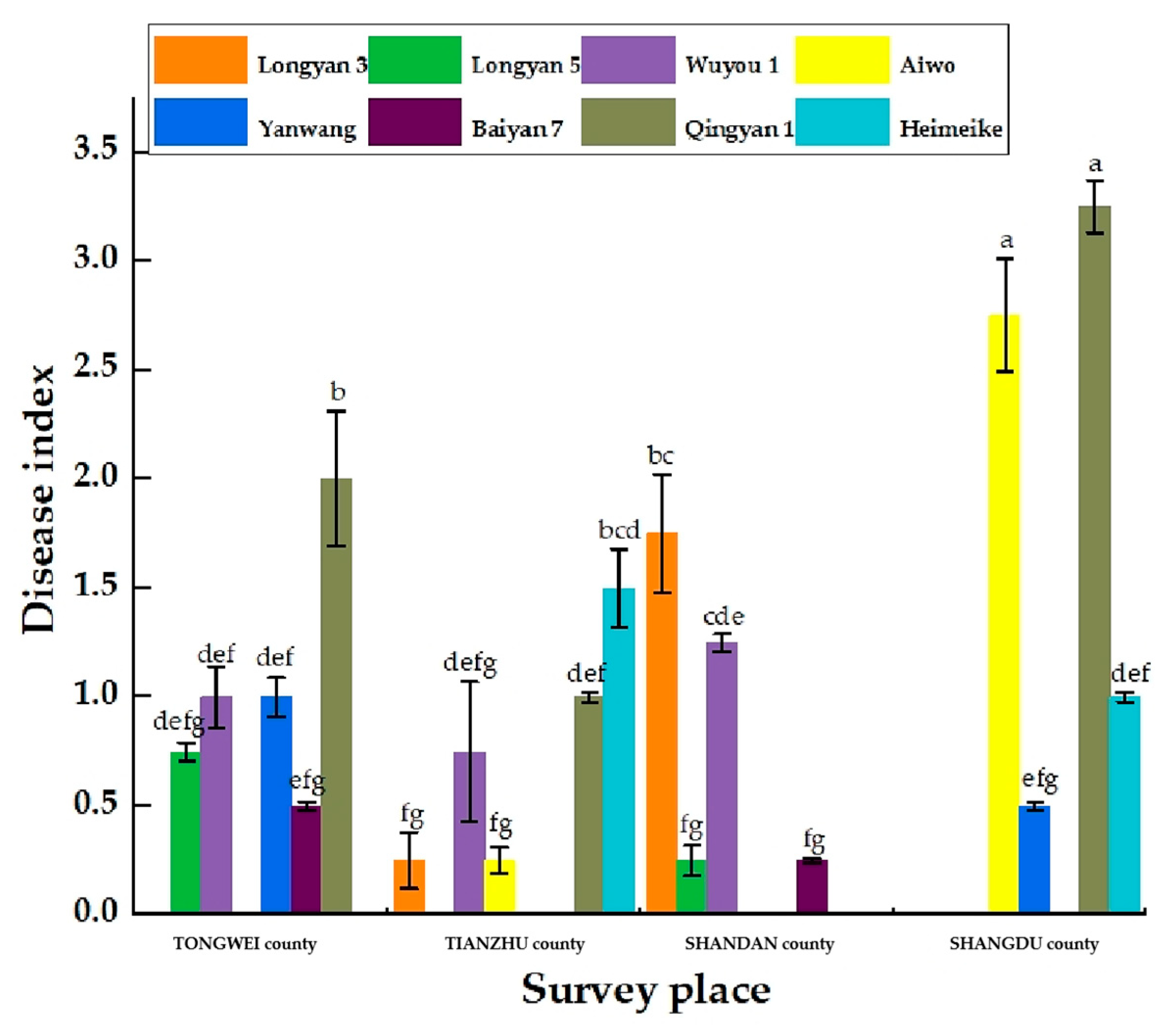
25] found that even when the same variety was present at different survey sites, the disease could take very different formats. Among the oat plants in the six townships, the authors recorded the highest incidence frequency in Majiazhuang Village (Tianzhu County), while the incidence of Baiyan 7 in Zhongtang Village (Yongdeng County) was the highest of the four planting areas.
Overall, meteorological, vegetation, and soil factors are the most tightly connected to the occurrence and prevalence of plant diseases. If the critical period of pathogen proliferation and infection coincides with suitable environmental conditions, the result can be an epidemic of plant disease [21]. Atmospheric humidity and wind speed are also critically important for keeping cereal crops disease-free. As such, climate change can both directly affect pathogens but also make hosts more vulnerable to pathogens [25]. When the temperature rises or rainfall conditions change, pathogen life history, oversummering behavior, and reproductive diffusion ability can change, thereby affecting the occurrence and development of plant disease [22].
Crop plants can be affected by various diseases during any stage of their growth and development, mainly due to pathogen infection [27]. In nature, many fungi can cause crop diseases, including oat sheath rot, with diseases caused by pathogenic fungi accounting for 70–80% of all plant diseases. Furthermore, fungal plant diseases are widely distributed, are generally rapid-onset, are sometimes difficult to spot, and can cause serious harm. Fungal pathogens often invade the apoplast or plant cells to obtain nutrients, thus establishing a parasitic relationship that can cause plant disease [23,28].
Plant–pathogen interactions are an important class of complex biological processes affecting crop production. The resistance of plants to pathogenic microorganisms is not only affected by their own genes but also by their adaptations to different regions and by human protocols for plant management [29,30,31]. This study found slight differences in the types and quantities of pathogens extracted from each plant, which may reflect that pathogen infection is related to variety-specific disease resistance. In addition, disease severity can be related to sowing date, planting density, fertilizer application, plant variety, and irrigation management method. However, the pathogenic factors affecting plant disease and epidemics in different regions can also be quite different, and so the pathogenic microorganisms responsible in different regions may not be the same [31]. In this study, we investigated diseases of the same oat variety in four different regions and found that the main causes of differences in disease emergence and format were environmental factors and management measures that differed among regions.
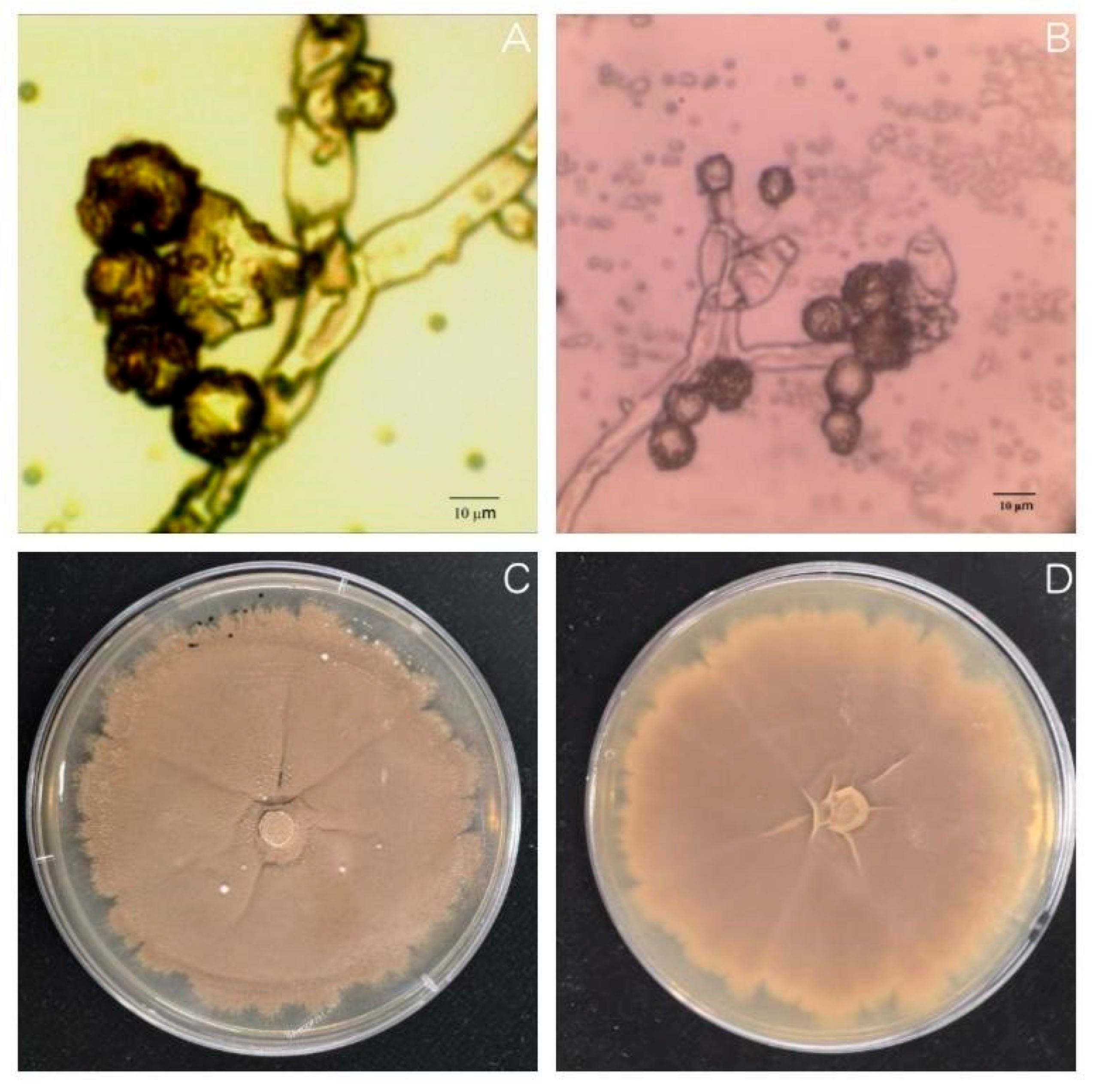
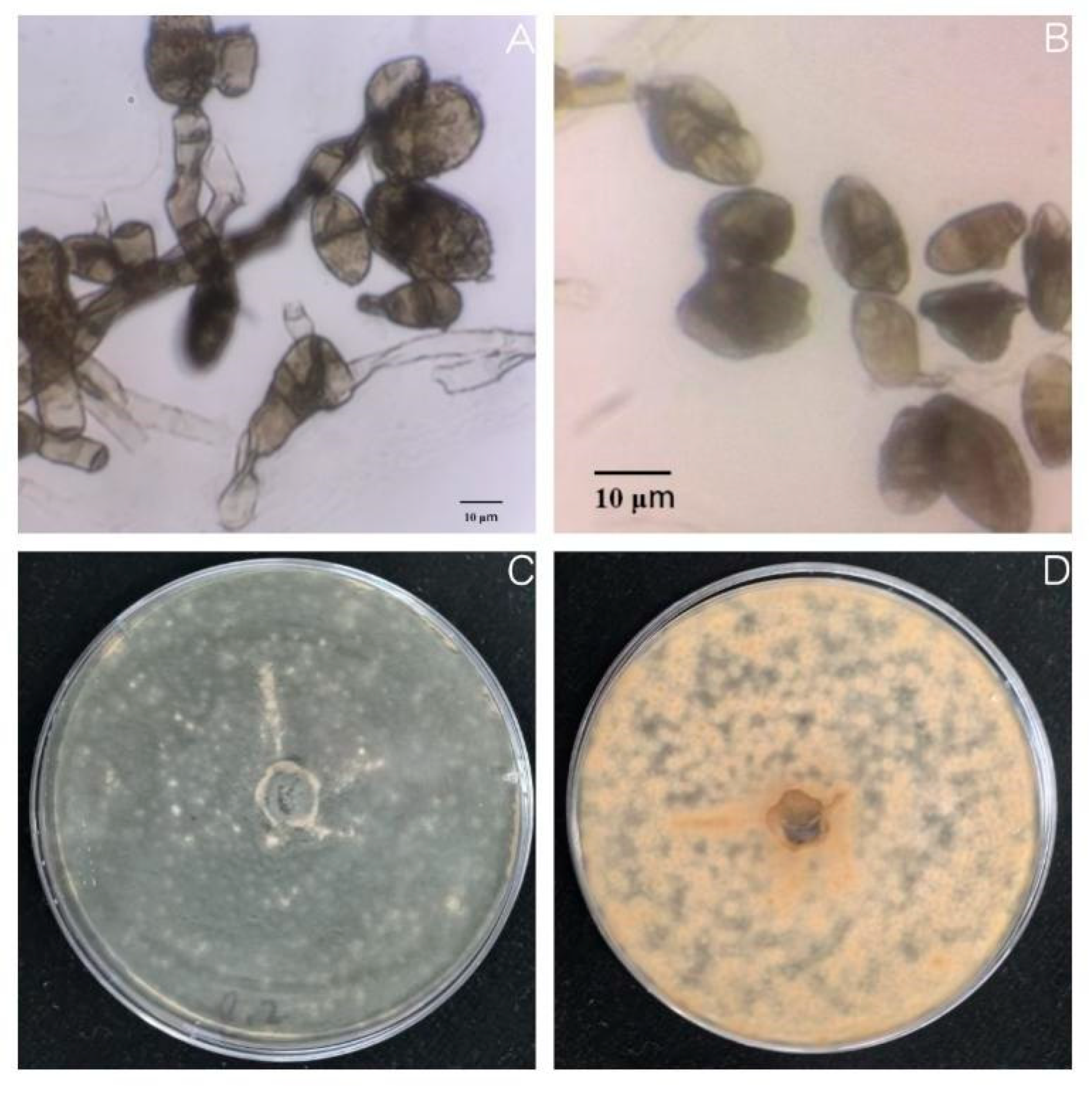
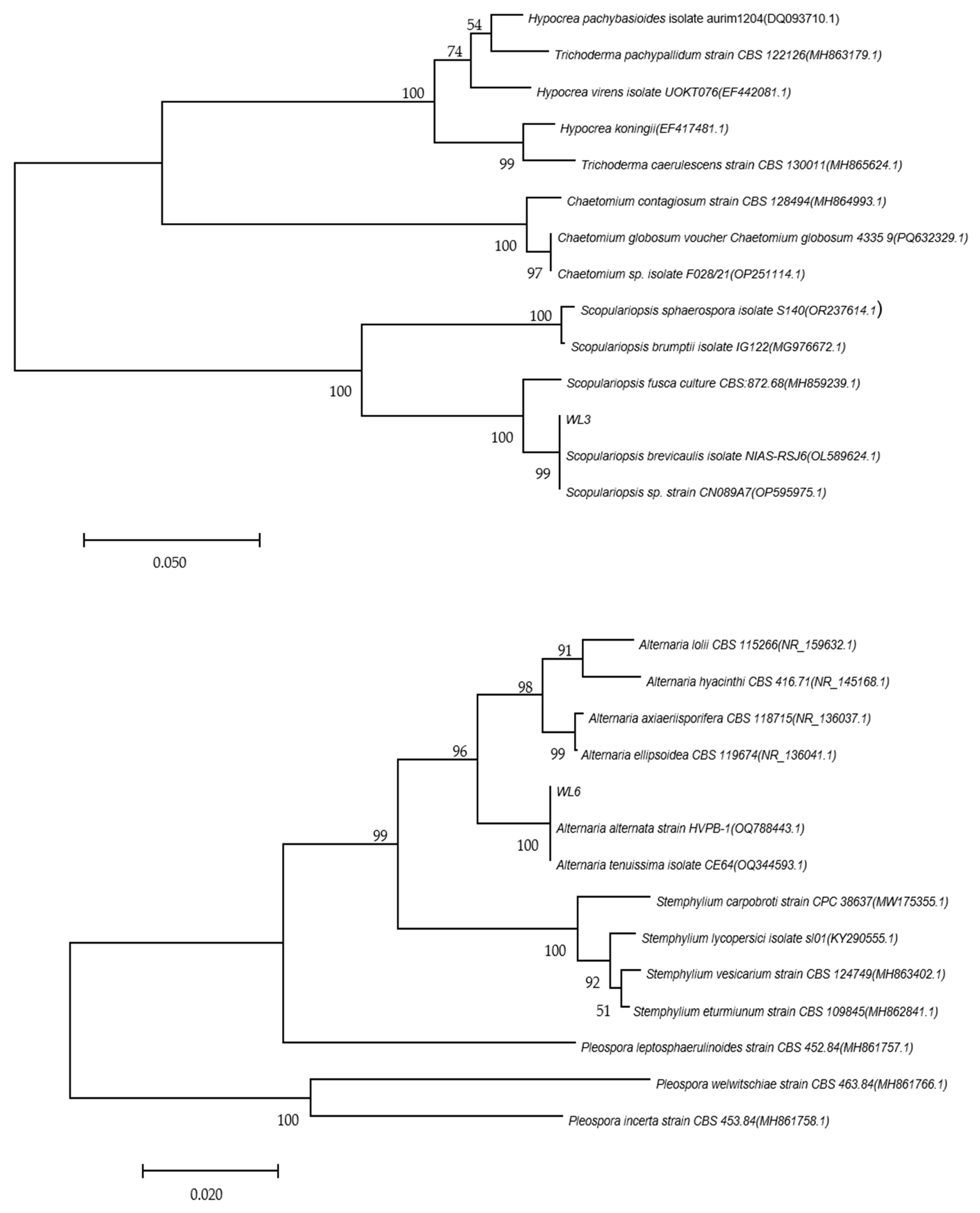
Oat sheath rot caused by S. graminearum has been previously reported in oat crops [8]. However, the two pathogens found in this study that were found to cause oat sheath rot have not been previously identified. This may be because different sites contain different pathogen living environments. In this study, the symptoms of sheath rot caused by pathogens were similar to those observed in the field but were also slightly different. For example, the central color of lesions of susceptible plants cultivated in the laboratory was darker: light beige or light brown, with the halo region being an even darker brown. Moreover, the lesion area overall was smaller than observed in the field. We speculate that these differences merely reflect differences between experimental and field growth environments [32].
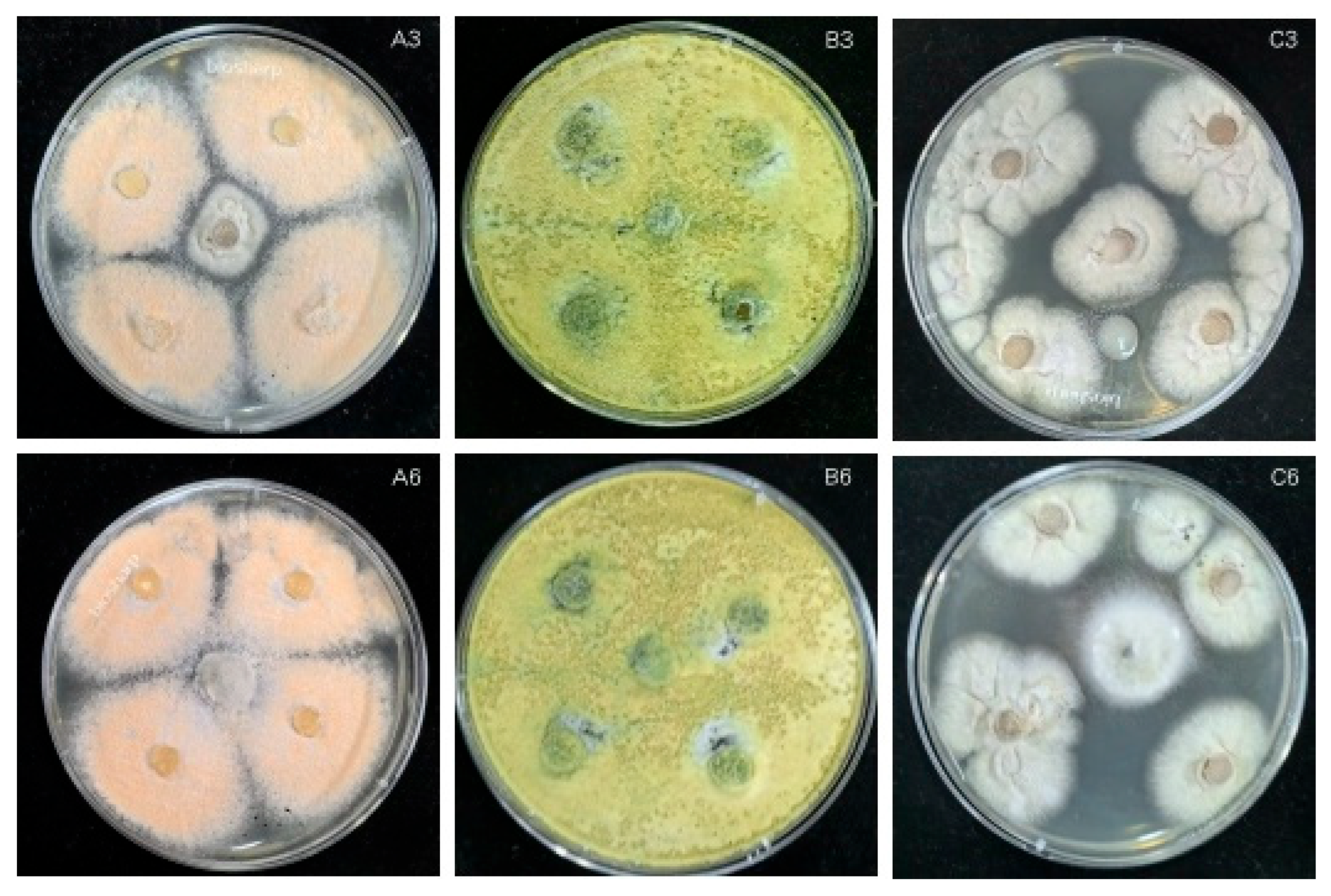
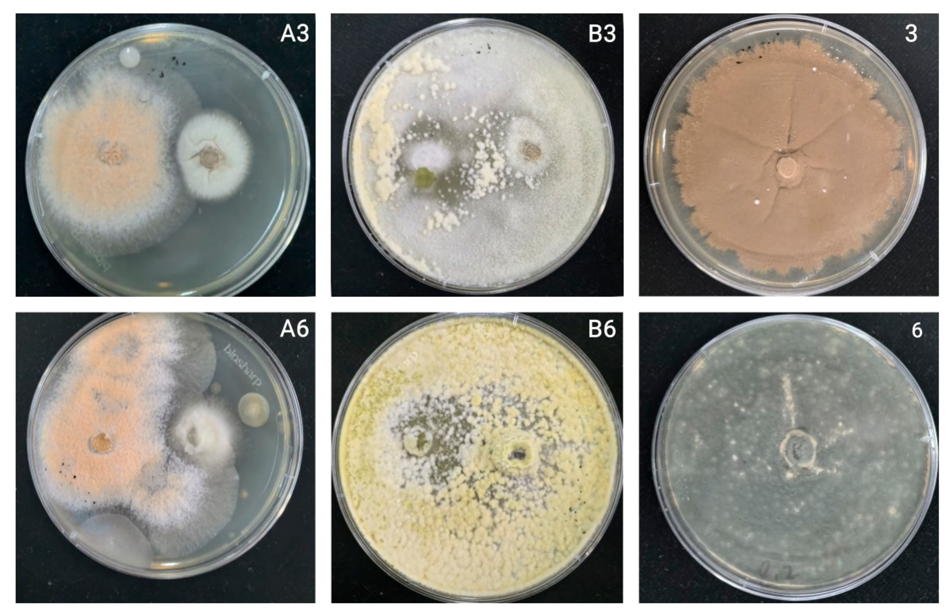
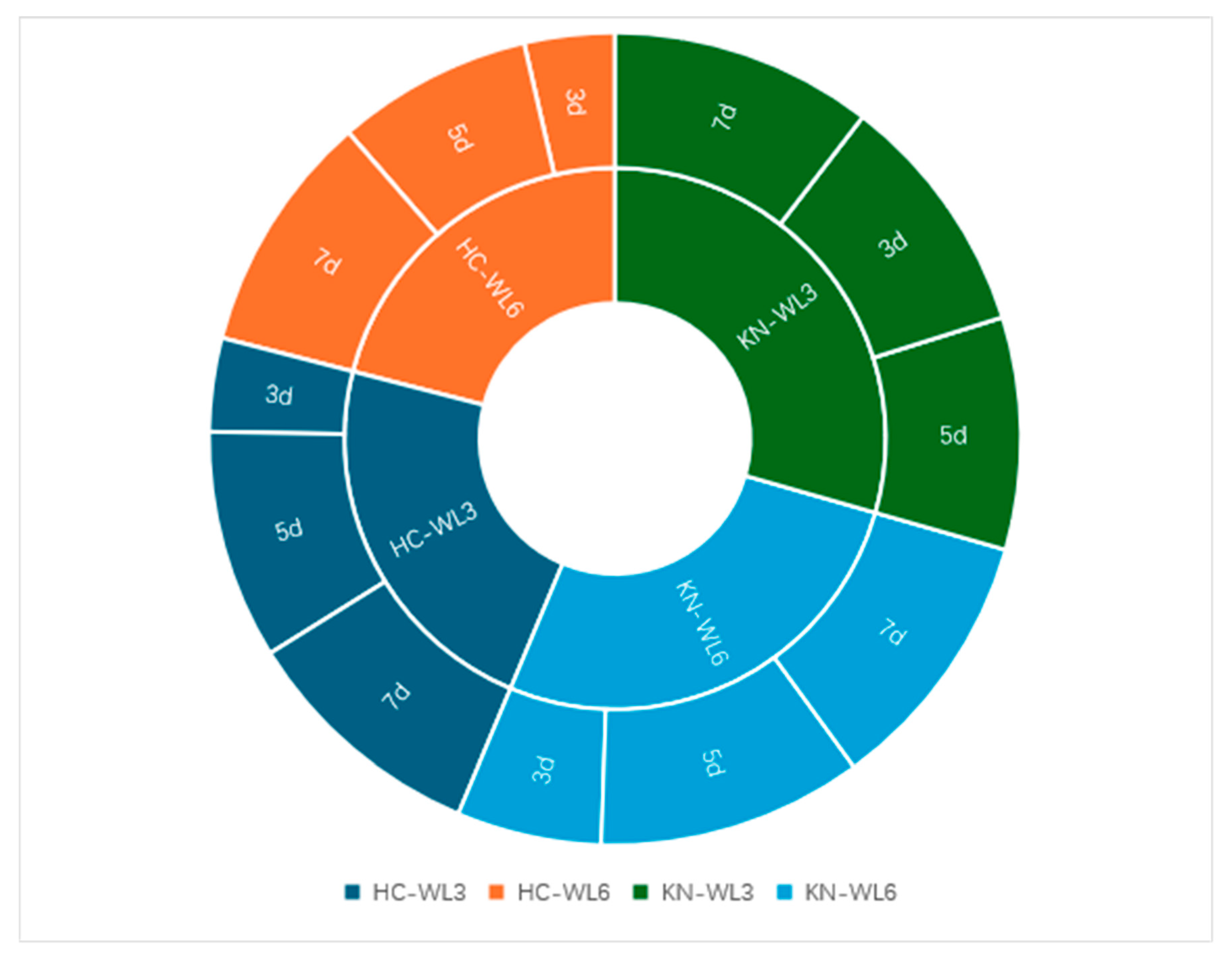
The growth rate of antagonistic fungi is an important indicator that they can compete with pathogenic fungi. Thus, the faster the rate of growth of antagonistic fungi, the more potent its effect on inhibiting the growth of pathogenic fungi [33]. During the seven days of confrontation culture, the relative inhibition rates of the two antagonistic bacteria generally increased (Figure 10). Only KN-WL3 showed a slight downward trend on the fifth day, but this sample showed an upward trend on the seventh day. This result may indicate that the nutrients in its culture medium were not distributed evenly when the PDA plate was manually inverted, resulting in a faster colony growth rate on the nutrient-enriched part of the PDA plate and a slower colony growth rate on the nutrient-scarce part.
Chemical pesticides are fast and effective, but pose significant biological safety and environmental pollution risks when used over the long term. Vogel et al. [34] found that the detection probability of myclobutanil fungicide in rainwater collected during the rainy season in California was as high as 74%, with the concentrations of some samples reaching as high as 0.113 μg L-1. In a similar study, Ju et al. [35] found that the total fungi, total bacteria, and biomass carbon content of fluvio-aquic soil from Henan (China) decreased significantly after myclobutanil spraying. Moreover, Yu et al. [36] showed that triazole fungicides can cause thyroid endocrine disorders and affect gene transcription in zebrafish (Barchydanio rerio) fry.
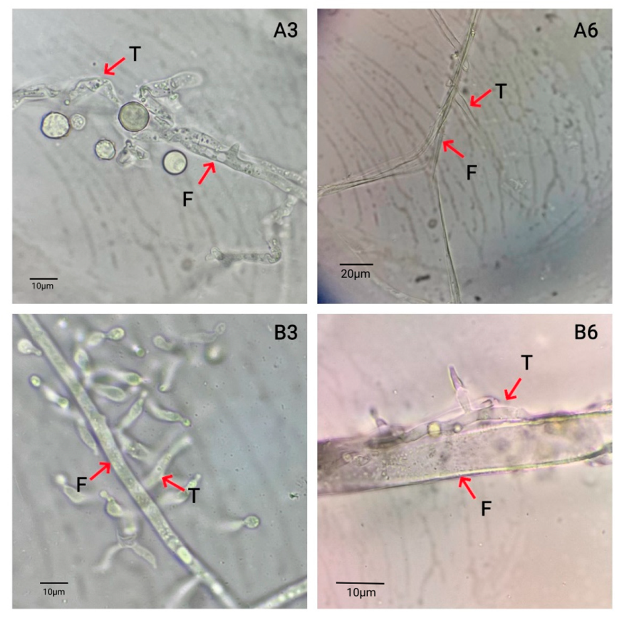
Fungi use many mechanisms to control plant disease, including competitive functions such as parasitism, antagonism and induction of disease resistance. In this study, the antagonistic fungus T. harzianum was found to have a high biological control value; this is significant, since it is known to parasitize a variety of plant pathogens. The biological control effect of T. harzianum has been verified by many previous studies. Bin and Pingzhi [37,38] showed that T. harzianum can improve the rhizosphere soil environment, mainly by regulating nutrient competition, spatial competition, and heavy parasitism on tomato gray mold, leaf mold, Fusarium wilt, and brown spot. Lakhdari et al. [39] showed that T. harzianum could control tomato Fusarium wilt by inhibiting the growth of Fusarium oxysporum mycelia. Another study by Ahmad et al. [40] showed that T. harzianum inhibited the reproduction of tomato root-knot nematodes, while a different study looking at pepper showed that Trichoderma harzianum had an inhibitory effect on cucumber gray mold, powdery mildew, Rhizoctonia solani, Fusarium wilt, root rot, and root-knot nematodes, as well as a strong inhibitory effect on Phytophthora capsici, small sclerotia, Rhizoctonia solani, and other soil-borne pathogens [41]. Reports on the use of Trichoderma to control crop Fusarium wilt have mainly focused on T. harzianum, T. asperellum [42], and T. viride [43]. However, there are relatively few reports of Trichoderma koningii being used to prevent Fusarium wilt. In this study, Trichoderma koningii was used to inhibit oat sheath rot, and it showed a remarkable biocontrol effect.
G. roseum is another important biocontrol fungus. It has many advantages, including a fast growth rate, production of a large number of spores, and strong antagonistic ability. In addition to its heavy parasitism, antibiosis, bacteriolysis, and competitive inhibition effects against pathogens, it can also induce systemic resistance in target plants [44]. One study reported that Gliocladium roseum shows a strong parasitic ability and was able to inhibit a pathogen causing fruit branch disease by more than 60% [45]. Therefore, at present, accelerating the research of biocontrol agents and strengthening their production efficiency are top priorities. Moreover, developing the capacity to culture but inhibit the development of pathogens is required for testing biological control agents [46].
At present, few studies have examined the prevention and control of oat sheath rot. Therefore, our results provide a novel theoretical basis for further prevention and control of oat sheath rot.

5. Conclusions

This study investigated oat sheath rot in oat crops in northern China by isolating and identifying pathogenic fungi, as well as putative biocontrol agents capable of inhibiting their pathogenicity. Accordingly, we identified two pathogenic fungi, Scopulariopsis brevicaulis and Alternaria alternata; to the best of our knowledge, this is the first report that this strain can cause oat sheath rot in China. Moreover, this study also showed that Trichoderma harzianum and Trichoderma koningii both showed good inhibitory effects on the isolated pathogenic fungi in different competitive ways; they may therefore be suitable alternative biocontrol agents for the prevention and treatment of oat sheath rot. In general, the isolation and identification of pathogenic oat sheath rot fungi and the screening of biocontrol fungi is a significant benefit for the oat industry.

Author Contributions

Conceptualization, Y.S., G.Z.; methodology, Y.S., J.C.; investigation, Y.S., L.Z., J.C.; data curation, Y.S., L.Z.; writing—original draft, Y.S.; writing—review and editing, Y.S., G.Z.; visualization, Y.S.; supervision, G.Z.; project administration, G.Z.; funding acquisition, G.Z. All authors have read and agreed to the published version of the manuscript.

Funding

This study was conducted by ‘China Agriculture Research System (CARS-7-C-1)’ and China Agriculture Research System (CARS-34) Funding.

Data Availability Statement

The data presented in this study are available upon request from the corresponding authors.

Acknowledgments

Thanks to Lin Doudou, Liu Xin, and Zhang Guanlu for their contributions in this article.

Conflicts of Interest

The authors declare no conflicts of interest.

References

  1. Li, C.J.; Chen, T.X.; Zhao, G.Q.; Nan, Z.B. Progress in research on diseases of Averra sativa. Acta Prataculturae Sin. 2017, 26, 203–222. [Google Scholar] [CrossRef]
  2. Guo, W.; Chen, Y.; Al-Rewashdy, Y.; Foran, N.; Ma, B.; Yan, W.; Fregeau-Reid, J.; Liu, J.; Ren, C.; Pageau, D.; et al. Effect of nitrogen fertilization on seed-borne Fusarium species in oat. Can. J. Plant Sci. 2017, 98, 38–46. [Google Scholar] [CrossRef]
  3. Acevedo, M.; Jackson, E.W.; Chong, J.; Rines, H.W.; Harrison, S.; Bonman, J.M. Identification and validation of quantitative trait loci for partial resistance to crown rust in oat. Phytopathology 2010, 100, 511–521. [Google Scholar] [CrossRef] [PubMed]
  4. Rasane, P.; Jha, A.; Sabikhi, L.; Kumar, A.; Unnikrishnan, V.S. Nutritional advantages of oats and opportunities for its processing as value added foods—A review. J. Food Sci. Technol. 2015, 52, 662–675. [Google Scholar] [CrossRef] [PubMed]
  5. Mao, L.; Zhang, H.; Yang, Z.; Li, Y.; Shen, Y. Site-specific effects of fertilizer on hay and grain yields of oats: Evidence from large-scale field experiments. J. Sci. Food Agric. 2024, 105, 2429–2439. [Google Scholar] [CrossRef] [PubMed]
  6. Marshall, A.; Cowan, S.; Edwards, S.; Griffiths, I.; White, E. Crops that feed the world 9 oats- a cereal crop for human and livestock feed with industrial applications. Food Secur. 2013, 5, 13–33. [Google Scholar] [CrossRef]
  7. Dietz, J.I.; Schierenbeck, M.; Simón, M.R. Impact of foliar diseases and its interaction with nitrogen fertilization and fungicides mixtures on green leaf area dynamics and yield in oat genotypes with different resistance. Crop Prot. 2019, 121, 80–88. [Google Scholar] [CrossRef]
  8. He, S.Q.; Wen, Z.H.; Wang, S.R.; Zhao, G.Q.; Wang, S.X.; Liu, Y.G.; Jing, Z.Q.; Ma, Y.Q. Dactylobotrys graminicola: A new phytopathogen causing sheath rot of hulless barley and oats. Mycosystema 2015, 34, 331–340. [Google Scholar] [CrossRef]
  9. Imran, M.; Khanal, S.; Zhou, X.-G.; Antony-Babu, S.; Atiq, M. First Report of Fusarium Sheath Rot of Rice Caused by Fusarium incarnatum-equiseti Species Complex in the United States. Plant Dis. 2022, 106, 3206. [Google Scholar] [CrossRef]
  10. Yang, X.; Xu, X.; Wang, S.; Zhang, L.; Shen, G.; Teng, H.; Yang, C.; Song, C.; Xiang, W.; Wang, X.; et al. Identification, Pathogenicity, and Genetic Diversity of Fusarium spp. Associated with Maize Sheath Rot in Heilongjiang Province, China. Int. J. Mol. Sci. 2022, 23, 10821. [Google Scholar] [CrossRef] [PubMed]
  11. Sawant, S.B.; Prabhukarthikeyan, S.R.; Mishra, M.K.; Parameswaran, C.; Keerthana, U.; Senapati, A.K. Understanding the Molecular Basis of Biocontrol Effect of Bacillus cereus RBS-57 on Sheath Rot Disease of Rice Through Protein Profiling. J. Plant Growth Regul. 2024, 43, 4753–4769. [Google Scholar] [CrossRef]
  12. Behura, A.; Parameswaran, C.; Prabhukarthikeyan, S.; Pradhan, C.; Parida, M.; Keerthana, U.; Raghu, S.; Mohapatra, S.; Samantaray, S. Unravelling genetic diversity and population structure of Sarocladium oryzae causing sheath rot disease in rice using hyper-variable SSR markers. Physiol. Mol. Plant Pathol. 2024, 130, 102245. [Google Scholar] [CrossRef]
  13. Sun, Y.; Wang, R.; Qiao, K.; Pan, H.; Wang, F.; Wang, Y.; Liu, J. First Report of Fusarium solani Causing Leaf Sheath Rot of Bush lily in China. Plant Dis. 2021, 106, 1992. [Google Scholar] [CrossRef]
  14. Rahman, F.U.; Zhu, Q.; Wu, Z.; Li, X.; Chen, W.; Xiong, T.; Zhu, X. Current insights into the biocontrol and biotechnological approaches for postharvest disease management of Botrytis cinerea. Postharvest Biol. Technol. 2024, 216, 113055. [Google Scholar] [CrossRef]
  15. Chen, Z.; Zhang, H.; Lv, W.; Zhang, S.; Du, L.; Li, S.; Zhang, H.; Zheng, X.; Zhang, J.; Zhang, T.; et al. Bacillus velezensis SS-20 as a potential and efficient multifunctional agent in biocontrol, saline-alkaline tolerance, and plant-growth promotion. Appl. Soil Ecol. 2025, 205, 105772. [Google Scholar] [CrossRef]
  16. Ansari, M.M.; Bisht, N.; Singh, T.; Mishra, S.K.; Anshu, A.; Singh, P.C.; Chauhan, P.S. Bacillus amyloliquefaciens modulate autophagy pathways to control Rhizoctonia solani infection in rice. Plant Physiol. Biochem. 2025, 218, 109317. [Google Scholar] [CrossRef] [PubMed]
  17. Groves, K.; Li, Y.; Peiris, P.U.S.; Brown, P.H. Efficacy of nematophagous fungi (Arthrobotrys dactyloides and A. oligospora) as biocontrol agents against root-knot nematode (Meloidogyne javanica) in sweetpotato. BioControl 2024, 70, 95–106. [Google Scholar] [CrossRef]
  18. Sawant, S.S.; Bhapkar, S.R.; Choi, E.; Lee, B.; Song, J.; Cho, Y.S.; Park, Y.; Seo, H.-J. Potential of Trichoderma species to control Rosellinia necatrix, the etiological agent of white root rot. Biol. Control 2024, 199, 105664. [Google Scholar] [CrossRef]
  19. Rodríguez-González, Á.; Casquero, P.A.; Mayo-Prieto, S.; Carro-Huerga, G.; Zanfaño, L.; Ramírez-Lozano, D.; Guerra, M.; Vega-Valdés, C.; Cardoza, R.E.; Gutiérrez, S. The efficacy of wild type and two mutants of Trichoderma arundinaceum and Trichoderma brevicompactum spores against Acanthoscelides obtectus and effect on Phaseolus vulgaris seed vitaly. J. Stored Prod. Res. 2025, 110, 102474. [Google Scholar] [CrossRef]
  20. Covarrubias-Rivera, L.; Ragazzo-Sánchez, J.A.; González-Gutiérrez, K.N.; Narváez-Zapata, J.A.; Calderón-Santoyo, M. Isolation and identification of phytopathogenic fungi from blueberry (Vaccinium corymbosum) and their biocontrol by Meyerozyma guilliermondii LMA-Cp01. J. Plant Pathol. 2024, 107, 525–536. [Google Scholar] [CrossRef]
  21. Huang, L.H.; Zhang, T.C.; Wang, R.H.; Xu, X.M.; Xu, S.Z.; Chen, Q.W.; Zhang, G.G.; Lan, B.; Li, X.M. Occurrence of Rice Blast in Main Early Rice Varieties Planteed in Different Ecological Regions of Jiangxi Province in 2009. Acta Agric. Univ. Jiangxiensis 2010, 32, 0485–0488. [Google Scholar] [CrossRef]
  22. Kirk, D.; Cohen, J.M.; Nguyen, V.; Childs, M.L.; Farner, J.E.; Davies, T.J.; Flory, S.L.; Rohr, J.R.; O’COnnor, M.I.; Mordecai, E.A. Impacts of Weather Anomalies and Climate on Plant Disease. Ecol. Lett. 2025, 28, e70062. [Google Scholar] [CrossRef] [PubMed]
  23. Pushpam, R.; Nithya, S.; Nasrin, S.F.; Meena, E.; Ramamoorthy, K.; Parthiban, V.K.; Pillai, M.A. Screening of rice cultivars for resistance to sheath rot (Sarocladium oryzae). Electron. J. Plant Breed. 2019, 10, 377–381. [Google Scholar] [CrossRef]
  24. Rosas, J.E.; Martínez, S.; Bonnecarrère, V.; de Vida, F.P.; Blanco, P.; Malosetti, M.; Jannink, J.; Gutiérrez, L. Comparison of Phenotyping Methods for Resistance to Stem Rot and Aggregated Sheath Spot in Rice. Crop Sci. 2016, 56, 1619–1627. [Google Scholar] [CrossRef]
  25. Sun, H.Y.; Zhang, W.W.; Zeng, L.; Cai, J.K.; Jiao, R.A.; Jin, X.W.; Gong, W.L.; Li, R.; Zhao, G.Q. Effectiveness of bio-medicaments against oat powdery mildew. Pratacultural. Sci. 2019, 36, 2374–2383. [Google Scholar]
  26. Siebold, M.; Von Tiedemann, A. Potential Effects of Global Warming on Oilseed Rape Pathogens in Northern Germany. Fungal Ecol. 2012, 5, 62–72. [Google Scholar] [CrossRef]
  27. Xu, G.; Wu, W.; Zhu, L.; Liang, Y.; Liang, M.; Tan, S.; Chen, H.; Huang, X.; He, C.; Lu, Y.; et al. Whole Genome Sequencing and Biocontrol Potential of Streptomyces luteireticuli ASG80 Against Phytophthora Diseases. Microorganisms 2024, 12, 2255. [Google Scholar] [CrossRef] [PubMed]
  28. Wang, R.; Zhang, H.; Zhang, Q.; Xi, J.; Jiang, K.; Li, J.; Xue, H.; Bi, Y. Isolation, Identification, and Analyzing the Biological Characteristics of Pathogens Causing Stem Rot of Lanzhou Onion During Postharvest Storage and Studying the Influence of Pathogen Infection on the Active Components of Lanzhou Onion. J. Fungi 2024, 10, 789. [Google Scholar] [CrossRef] [PubMed]
  29. Njoroge, S.M.C.; Kabir, Z.; Martin, F.N.; Koike, S.T.; Subbarao, K.V. Comparison of crop rotation for Verticillium wilt management and effect on Pythium species in conventional and organic strawberry production. Plant Dis. 2009, 93, 519–527. [Google Scholar] [CrossRef] [PubMed]
  30. Igarashi, Y.; Iida, T.; Yoshida, R.; Furumai, T. Pteridic acids A and B, novel plant growth promoters with auxin-like activity from Streptomyces hygroscopicus Tp-A0451. J. Antibiot. 2002, 55, 764–767. [Google Scholar] [CrossRef] [PubMed]
  31. Joshi, M.V.; Loria, R. Streptomyces turgidiscabies possesses a functional cytokinin biosynthetic pathway and produces leafy galls. Mol. Plant-Microbe Interact. 2007, 20, 751–758. [Google Scholar] [CrossRef] [PubMed]
  32. Carbone, I.; Kohn, L.M. A method for designing primer sets for speciation studies in filamentous ascomycetes. Mycologia 1999, 91, 553–556. [Google Scholar] [CrossRef]
  33. Hınçal, S.; Yalçın, M. Biological control of some wood-decay fungi with antagonistic fungi. Biodegradation 2023, 34, 597–607. [Google Scholar] [CrossRef] [PubMed]
  34. Vogel, J.R.; Majewski, M.S.; Capel, P.D. Pesticides in Rain in Four Agricultural Watersheds in the United States. J. Environ. Qual. 2008, 37, 1101–1115. [Google Scholar] [CrossRef] [PubMed]
  35. Ju, C.; Xu, J.; Wu, X.; Dong, F.; Liu, X.; Zheng, Y. Effects of Myclobutanil on Soil Microbial Biomass, Respiration, and Soil Nitrogen Transformations. Environ. Pollut. 2016, 208, 811–820. [Google Scholar] [CrossRef] [PubMed]
  36. Yu, L.; Chen, M.; Liu, Y.; Gui, W.; Zhu, G. Thyroid Endocrine Disruption in Zebrafish Larvae Following Exposure to Hexaconazole and Tebuconazole. Aquat. Toxicol. 2013, 138–139, 35–42. [Google Scholar] [CrossRef] [PubMed]
  37. Yao, B.; Wang, A.X.; Li, J.F. Study on the inhibitory effect of Trichoderma harzianum on four tomato pathogenic fungi. J. Northeast. Agric. Univ. 2009, 40, 26–31. [Google Scholar] [CrossRef]
  38. Kang, P.Z.; Zhang, L.R.; Shen, R.Q.; Du, Y.N. Ecological Effect of Trichoderma harzianum Preparations on Rhizosphere Soil Microbes in Facilities Continuous Cropping Tomato and Their Disease Prevention. Agrochemicals 2013, 52, 128–131. [Google Scholar] [CrossRef]
  39. Lakhdari, W.; Dehliz, A.; Mlik, R.; Hammi, H.; Benlamoudi, W.; Acheuk, F.; Doumandji-Mitiche, B. Inhibitory effect of Trichoderma harzianum on mycelial growth of Fusarium oxysporum f. sp. radicis-lycopersici and Alternaria solani. Org. Agric. 2018, 8, 225–230. [Google Scholar] [CrossRef]
  40. Al-Hazmi, A.S.; TariqJaveed, M. Effects of different inoculum densities of Trichoderma harzianum and Trichoderma viride against Meloidogyne javanica on tomato. Saudi J. Biol. Sci. 2016, 23, 288–292. [Google Scholar] [CrossRef] [PubMed]
  41. Vinale, F.; Nigro, M.; Sivasithamparam, K.; Flematti, G.; Ghisalberti, E.L.; Ruocco, M.; Varlese, R.; Marra, R.; Lanzuise, S.; Eid, A.; et al. Harzianic acid: A novel siderophore from Trichoderma harzianum. FEMS Microbiol. Lett. 2013, 347, 123–129. [Google Scholar] [CrossRef] [PubMed]
  42. Veenstra, A.; Rafudeen, M.; Murray, S.L. Trichoderma asperellum isolated from African maize seed directly inhibits Fusarium verticillioides growth in vitro. Eur. J. Plant Pathol. 2019, 153, 279–283. [Google Scholar] [CrossRef]
  43. Lu, C.Y.; Ma, Y.; Wang, Q.; Wang, Q.J.; Wang, G.F.; Mei, Y.C.; Li, C.G.; Guo, D.G. Effects of Trichoderma viride TV41 on spatial distribution of Fusarium oxysporum FW0 around watermelon plant and fusarium wilt control. Inst. Microbiol. 2015, 42, 2159–2167. [Google Scholar] [CrossRef]
  44. García, D.; González-Almario, A.; Cotes, A.M. Controlling Fusarium wilt of cape gooseberry by microbial consortia. Lett. Appl. Microbiol. 2023, 76, ovad072. [Google Scholar] [CrossRef] [PubMed]
  45. Rensink, S.; van Nieuwenhuijzen, E.J.; Sailer, M.F.; Struck, C.; Wösten, H.A.B. Use of Aureobasidium in a sustainable economy. Appl. Microbiol. Biotechnol. 2024, 108, 202. [Google Scholar] [CrossRef] [PubMed]
  46. Hirayama, Y.; Asano, S.; Okayama, K.; Ohki, S.T.; Tojo, M. Weeds as the potential inoculum source of Colletotrichum fructicola responsible for strawberry anthracnose in Nara, Japan. J. Gen. Plant Pathol. 2018, 84, 12–19. [Google Scholar] [CrossRef]
Figure 1. Survey point information overview.
Agronomy 15 01776 g001
Figure 2. Identification glue diagram.
Agronomy 15 01776 g002
Figure 3. The incidence of oat sheath rot in the field. (Note: The picture was taken in the oat field of Shangdu County in 2023).
Agronomy 15 01776 g003
Figure 4. Disease index of oat sheath rot of each variety (where a, b, c, etc. represents its significant difference).
Agronomy 15 01776 g004
Figure 5. Symptoms of oat sheath rot under laboratory conditions. (Note: WL3 and WL6 are the disease symptoms caused by the two pathogens extracted in the early stage).
Agronomy 15 01776 g005
Figure 6. WL3 culture. (Note: (A,B) are the spore photos under the microscope, and (C,D) are the positive and negative sides of the bacteria in the medium, respectively).
Agronomy 15 01776 g006
Figure 7. WL6 culture. (Note: (A,B) are the spore photos under the microscope, and (C,D) are the positive and negative sides of the bacteria in the medium, respectively).
Agronomy 15 01776 g007
Figure 8. Based on the gene sequence, the phylogenetic tree of the two pathogenic fungi WL3 and WL6 was constructed.
Agronomy 15 01776 g008
Figure 9. Preliminary screening of three biocontrol fungi. (Note: In the figure, A, B, and C represent HC, KN, and FZ, respectively; 3 and 6 represent WL3 and WL6, respectively).
Agronomy 15 01776 g009
Figure 10. Inhibitory effect of HC and KN on pathogenic fungi. (Note: A and B represent HC and KN, respectively; 1, 2, 3, 4 represent two kinds of pathogenic fungi; and in the same plate, the left side of the cake is antagonistic fungi, and the right side is pathogenic fungi).
Agronomy 15 01776 g010
Figure 11. The increasing/decreasing trend of the relative inhibition rate on the 3rd, 5th, and 7th day.
Agronomy 15 01776 g011
Figure 12. The competitive relationship between fungal hyphae. (Note: T is an antagonistic fungus, and F is a pathogen).
Agronomy 15 01776 g012
Table 1. Sample plot overview.
Sample AreaElevation (m)Mean Annual Temperature (°C)Rain Fall (mm)Sunshine Hours (h)Frost-Free Period (d)
ProvinceCityCounty
GansuDingxiTongwei23197.75002430147
GansuWuweiTianzhu27701.04002495146
GansuZhangyeShandan21460.23602823155
NeimengguWulanchabuShangdu14003.13502970105
Table 2. Tested oat materials.
NumberingGrowing LocationVariety NameNumberingGrowing LocationVariety Name
1TongweiLongyan 317ShandanLongyan 3
2TongweiLongyan 518ShandanLongyan 5
3TongweiWuyou 119ShandanWuyou 1
4TongweiAiwo20ShandanAiwo
5TongweiYanwang21ShandanYanwang
6TongweiBaiyan 722ShandanBaiyan 7
7TongweiQingyan 123ShandanQingyan 1
8TongweiHeimeike24ShandanHeimeike
9TianzhuLongyan 325ShangduLongyan 3
10TianzhuLongyan 526ShangduLongyan 5
11TianzhuWuyou 127ShangduWuyou 1
12TianzhuAiwo28ShangduAiwo
13TianzhuYanwang29ShangduYanwang
14TianzhuBaiyan 730ShangduBaiyan 7
15TianzhuQingyan 131ShangduQingyan 1
16TianzhuHeimeike32ShangduHeimeike
Table 3. Severity grading standard of oat sheath rot disease.
Severity GradeGrading Standard
0No lesion
IThere are brown spots on the leaf sheath, and the area is not more than 25%.
IIThe lesion area on the leaf sheath ranges from 25% to 50%.
IIIThe lesion area on the leaf sheath ranges from 50% to 75%.
IVLeaf sheath disease, spot area range of 75%.
Table 4. Evaluation method of oat sheath rot resistance [24].
Disease IndexResistance
Disease index = 0Immunity (I)
0.1 < Disease index ≤ 20Highly resistant (HR)
20.1 < Disease index ≤ 40Resistant (R)
40.1 < Disease index ≤ 60 IModerately resistant (MR)
60.1 < Disease index ≤ 80Susceptible (S)
80.1 < Disease index ≤ 100High sensitive (HS)
Table 5. Sequences used for secondary sequencing.
Pathogenic FungusPrimer Name and SequencePCR Length
WL3NS1 (5′-GTAGTCATATGCTTGTCTC-3′)NS4 (5′-CTTCCGTCAATTCCTTTAAG-3′)About 1500 bp
WL6H2P1 (5′-GTTCGCCCACCACTAGGACAAAC-3′)H2P4 (5′-AGACGCCCAACACCAAGCAAAG-3′)
Table 6. Occurrence of sheath rot in the main oat-producing areas.
NumberingPlace of the InvestigationMaterialGrade of the DiseaseDiseased Plant Rate (%)Disease IndexResistant Type
0IIIIIIIV
1Tongwei CountyLongyan 3100000000I
2Tongwei CountyLongyan 598110020.75HR
3Tongwei CountyWuyou 197210031HR
4Tongwei CountyAiwo100000000I
5Tongwei CountyYanwang97210031HR
6Tongwei CountyBaiyan 799010010.5HR
7Tongwei CountyQingyan 196121042HR
8Tongwei CountyHeimeike100000000I
9Tianzhu CountyLongyan 399100010.25HR
10Tianzhu CountyLongyan 5100000000I
11Tianzhu CountyWuyou 199001010.75HR
12Tianzhu CountyAiwo99100010.25HR
13Tianzhu CountyYanwang100000000I
14Tianzhu CountyBaiyan 7100000000I
15Tianzhu CountyQingyan 197210031HR
16Tianzhu CountyHeimeike96220041.5HR
17Shandan CountyLongyan 396130041.75HR
18Shandan CountyLongyan 599100010.25HR
19Shandan CountyWuyou 197120031.25HR
20Shandan CountyAiwo100000000I
21Shandan CountyYanwang100000000I
22Shandan CountyBaiyan 799100010.25HR
23Shandan CountyQingyan 1100000000I
24Shandan CountyHeimeike100000000I
25Shangdu CountyLongyan 3100000000I
26Shangdu CountyLongyan 5100000000I
27Shangdu CountyWuyou 1100000000I
28Shangdu CountyAiwo95122052.75HR
29Shangdu CountyYanwang99010010.5HR
30Shangdu CountyBaiyan 7100000000I
31Shangdu CountyQingyan 192512083.25HR
32Shangdu CountyHeimeike97210031HR
Table 7. The relative inhibition rates of the two biocontrol fungi on the 3rd, 5th, and 7th days against the two pathogens.
AntagonistTimeDiameter of the Pathogen (mm)Diameter of the Pathogen (mm)Control Pathogen Diameter (mm)Antifungal Rate (%)
HC-WL33 d17314360.47
5 d10765581.82
7 d7827190.14
HC-WL63 d22303231.25
5 d13654872.92
7 d9807788.31
KN-WL33 d7814383.72
5 d11775580.00
7 d08771100.00
KN-WL63 d17483246.88
5 d08848100.00
7 d08877100.00
Disclaimer/Publisher’s Note: The statements, opinions and data contained in all publications are solely those of the individual author(s) and contributor(s) and not of MDPI and/or the editor(s). MDPI and/or the editor(s) disclaim responsibility for any injury to people or property resulting from any ideas, methods, instructions or products referred to in the content.

