Article

Inhibitory Effects and Underlying Mechanisms of a Selenium Compound Agent Against the Pathogenic Fungus Sclerotinia sclerotiorum Causing Sclerotinia Stem Rot in Brassica napus

by
Xiaojuan Zhang
1,2,3,4,†,
Yangzi Hou
1,†,
Xiuqi Ma
1,
Xiaomin Sun
5,
Qiao Chen
6,
Lele Wu
1 and
Chenlu Zhang
1,*
1
School of Biological Science and Engineering, Shaanxi University of Technology, Hanzhong 723001, China
2
Shaanxi Province Key Laboratory of Bio-Resources, Hanzhong 723001, China
3
QinLing-Bashan Mountains Bioresources Comprehensive Development C. I. C., Hanzhong 723001, China
4
Qinba State Key Laboratory of Biological Resources and Ecological Environment, Hanzhong 723001, China
5
Hanzhong Agricultural Technology Promotion and Training Center, Hanzhong 723000, China
6
College of Agricultural and Forestry Technology and Biological Engineering, Hanzhong Vocational and Technical College, Hanzhong 723001, China
*
Author to whom correspondence should be addressed.
These authors contributed equally to this work.
Agronomy 2025, 15(8), 1764; https://doi.org/10.3390/agronomy15081764
Submission received: 26 June 2025 / Revised: 21 July 2025 / Accepted: 22 July 2025 / Published: 23 July 2025
(This article belongs to the Special Issue Environmentally Friendly Ways to Control Plant Disease)

Abstract

Sclerotinia sclerotiorum (S. sclerotiorum), a necrotrophic phytopathogen, causes sclerotinia stem rot (SSR) in many crops like oilseed rape, resulting in severe economic losses. Developing eco-friendly compound fungicides has become a critical research priority. This study explored the combination of sodium selenite and cuminic acid to screen for the optimal mixing ratio and investigate its inhibitory effects and mechanisms against S. sclerotiorum. The results demonstrated that synergistic effects were observed with a 1:3 combination ratio of sodium selenite to cuminic acid. After treatment with the selenium compound agent, ultrastructural observations revealed that the hyphae of S. sclerotiorum became severely shriveled, deformed, and twisted. The agent significantly reduced oxalic acid production and the activities of polymethylgalacturonide (PMG) and carboxymethylcellulose enzymes (Cx), while increasing the exocytosis of nucleic acids and proteins from the mycelium. Foliar application of the selenium compound agent significantly reduced lesion areas in rapeseed. Combined with the results of transcriptome sequencing, this study suggests that the compound agent effectively inhibits the growth of S. sclerotiorum by disrupting its membrane system, reducing the activity of cell wall-degrading enzymes, and suppressing protein synthesis, etc. This research provides a foundation for developing environmentally friendly and effective fungicides to control S. sclerotiorum.

Graphical Abstract

1. Introduction

Sclerotinia sclerotiorum (Lib.) de Bary is a devastating plant pathogenic fungus with a worldwide distribution, dominated by a necrotrophic lifestyle [1]. It has a wide host range, infecting more than 600 species of plants worldwide [2]. Sclerotinia stem rot (SSR), caused by S. sclerotiorum, poses a threat to the safe production of various important economic crops such as rapeseed, soybeans, tomatoes, potatoes, etc., resulting in severe yield loss and quality decline of these crops, causing huge economic losses [3]. According to statistics, the yield loss of rapeseed caused by SSR ranges from 10% to 70% [4].
In practice, control methods of SSR are mainly based on chemical fungicides [1]. Due to the lack of cultivars that are immune to SSR, chemicals such as carbendazim and dimetachlone are often used to control SSR. However, the use of chemical fungicides brings a series of problems, such as residual fungicides in soil and water, environmental pollution, ecological imbalance, and harm to human health [3]. Therefore, screening for safe, effective, and environmentally friendly methods for the prevention and control of SSR has attracted a lot of attention, which is also in line with the policies advocated by many countries to promote ecological civilization construction and reduce the use of fertilizers and pesticides.
Trace elements can not only serve as plant nutrients, but research has shown that specific trace elements also have the ability to inhibit various plant diseases [5,6,7,8]. Control of SSR by plant nutrients can effectively reduce fungicide resistance and pesticide residues, and is increasingly valued for its eco-friendliness.
Selenium (Se) is an essential trace element for humans and also a beneficial element for plants. Trace Se can promote plant growth [9,10] and enhance plant resistance to biotic and abiotic stress [11,12,13]. Research has confirmed that a suitable concentration of Se can inhibit the growth of pathogenic fungi and assist plants in defending against varieties of fungal pathogens. Se can inhibit the growth of hyphae of S. sclerotiorum by disrupting the membrane system and interfering with metabolism [3]. Se can also inhibit the formation and germination of S. sclerotiorum sclerotia, reducing the pathogenicity [14]. Sodium selenite (≥10 mg/L) can significantly inhibit the growth of fungal hyphae of fusarium wilt disease in bananas [15]. Applying sodium selenite to the culture medium can significantly inhibit the growth of Phanerochaete chrysosporium [16], and sodium selenite (24 mg/L) can effectively control gray mold disease of tomato [17]. Se has a strong inhibitory effect on spore germination, embryo tube elongation, and mycelial growth and diffusion of Penicillium expansum in culture medium [18]. Se disrupts the integrity of the cell membrane, reduces energy supply, and ultimately induces hyphal apoptosis, thereby inhibiting the proliferation of Phytophthora nicotianae [19]. Özer et al. [20] found that Se has a good inhibitory effect on the mycelial growth of the plant fungal pathogens.
Plants are a natural treasure trove of antibacterial active substances. For example, thymol (a phenolic terpene) and azadirachtin (a limonoid) exhibit strong antifungal activity by disrupting cell membranes and inhibiting chitin synthesis, respectively [21]. Botanical fungicides derived from these natural ingredients have gained attention due to their low toxicity, rapid environmental degradation, and reduced risk of pathogen resistance compared to synthetic chemicals [22].
Cuminic acid is an extract of Cuminum cyminum L. seeds, which belongs to the benzoic acid group of compounds. Previous studies show that cuminic acid has broad-spectrum antifungal activity and good inhibition of the growth of a variety of plant pathogens [23]; meanwhile, it is environment-friendly and easily degradable, which makes it suitable for the development of green and environmentally friendly fungicides of plant origin. Previous studies have shown that cuminic acid exhibits obvious antifungal activity against several plant pathogens; for example, essential oils extracted from Cuminum cyminum seeds (100 mg/mL) can completely inhibit the mycelial growth of S. sclerotiorum [24]; meanwhile, it exhibited low risk for oilseed rape. Wang et al. [25] reported that cuminic acid can inhibit the mycelial growth and spore germination of Phytophthora capsici Leonian, affect mycelial respiration, and increase cell membrane permeability. Cuminic acid inhibits the growth of Fusarium oxysporum f. sp. niveum (FON), the pathogen of watermelon Fusarium Wilt, resulting in a significant decrease in fungal toxins (mainly sickle acid) [26]. The antifungal activity of Valsa mali by cuminic acid was the direct activity of cuminic acid instead of its effect on the pH of the culture medium [27].
The long-term use of a single chemical agent can easily lead to the development of bacterial resistance, thereby reducing the effectiveness of prevention [28]. High-level resistance to fungicides such as carbendazim [29] and dimethachlone [30] has been found in collected isolates of S. sclerotiorum. The combination of two or more drugs can help reduce the resistance of pathogens and improve drug efficacy. Combining safe and environmentally friendly agents with plant-based fungicides could lead to a promising approach in crop protection [1]. However, to date, no reports on the combined effects of Se and cuminic acid on S. sclerotiorum have been reported. Therefore, it was interesting to study the inhibitory effect of the Se and cuminic acid combination on the growth and pathogenicity of S. sclerotiorum, as well as its mechanism.
The main objectives of this research were (1) to examine the effect of different concentrations of sodium selenite and cuminic acid on the growth of S. sclerotiorum and formation of sclerotia, respectively, (2) to determine the inhibitory effect of sodium selenite and cuminic acid in different ratios on the growth of S. Sclerotiorum, and to screen for compound combinations with synergistic effects, (3) to investigate the effects of selenium compound agent on the physiological and biochemical indicators of S. Sclerotiorum mycelia, and to analyze the gene expression changes in S. sclerotiorum after treatment with selenium compound agent at the transcriptome level so as to reveal the molecular mechanisms of S. sclerotiorum inhibition by selenium compound agent. Our research is of great significance for developing safe and ecologically safe fungicides for control of SSR and reducing the harm caused by SSR.

2. Experimental Design

This study investigated the effects of sodium selenite and cuminic acid, alone and in combination, on S. sclerotiorum. First, different concentrations of sodium selenite and cuminic acid were added to the PDA plates. An activated mycelium block of S. sclerotiorum was placed in the center of each plate to measure the inhibition of mycelial growth under the treatments of sodium selenite and cuminic acid, respectively, and to calculate the EC value. The growth of S. sclerotiorum on the PDA plates was registered and used as controls. Compound agent formulations were prepared by mixing sodium selenite and cuminic acid stock solutions in different ratios to evaluate their combined effectiveness using the synergy ratio (SR). Three replicates were set for each treatment. Mycelial morphology after treatment was observed using SEM. Physiological and biochemical indexes of mycelium, including protein, nucleic acid, reducing sugar, pyruvate, glycerin, and oxalic acid contents, as well as pectinase and cellulase activities, were measured. Transcriptomic analysis was conducted on S. sclerotiorum mycelium samples treated with a 1:3 selenium compound agent to elucidate transcriptional changes in S. sclerotiorum under selenium compound agent treatment. The therapeutic effect of the selenium compound agent on SSR in rapeseed was evaluated at both detached leaf and potted plant levels by measuring lesion areas and calculating disease control efficacy. Three replicates were set for each treatment.

3. Materials and Methods

3.1. Strains, Media and Fungicide

S. sclerotiorum (1980) was supplied by Dr. Xiaohui Cheng, Oil Crops Research Institute, Chinese Academy of Agricultural Sciences.
For the potato dextrose agar (PDA) medium, 200 g of potato, 20 g of dextrose, and 20 g of agar were dissolved in 1000 mL of ddH2O. For the potato dextrose broth (PDB) medium, 200 g of potato and 20 g of dextrose were dissolved in 1000 mL of ddH2O. All the media and dishes were autoclaved at 121 °C for 20 min.
Sodium selenite (AR, ≥98%) was supplied by Xi’an chemical reagent station (Xi’an, China). Cuminic acid (AR, ≥98%) was supplied by Beijing Conina Biotechnology Co., LTD. (Beijing, China). Sodium selenite was dissolved in ddH2O to obtain 100 mg/mL stock solution. Cuminic acid was dissolved in methyl alcohol to obtain 100 mg/mL stock solution.

3.2. Sensitivity to Sodium Selenate and Cuminic Acid

Sodium selenite and cuminic acid stock solutions were individually incorporated into PDA medium cooled to approximately 60 °C, resulting in the preparation of PDA media with sodium selenite concentrations of 0, 50, 100, 200, 300, 400, and 500 mg/L, and PDA media with cuminic acid concentrations of 0, 10, 20, 30, 40, 50, 60, and 80 mg/L. Mycelial plugs (5 mm in diameter) cultured for 2 days were placed in the center of PDA medium containing the above different concentrations of agents. The colony diameter was measured after incubation at 22 °C in darkness for 48 h. The inhibition rates of mycelial growth of S. sclerotiorum by different concentrations of sodium selenite and cuminic acid were calculated according to the following formula [31].
M y c e l i a l   g r o w t h   i n h i b i t i o n   % = d c d t / d t 5   mm × 100
where dc and dt represented average colony diameters of the control and treatment groups, respectively.
The toxicity regression equation was calculated using the logarithmic value of agent concentration as the x-axis and the probability value of inhibition rate as the y-axis. The median effective concentration (EC50) as well as EC90, EC25, EC75 were calculated according to the toxicity regression equation [32].
The PDA plates containing the agent and inoculated with S. sclerotiorum were incubated at 22 °C in darkness for 15 days, and the number and fresh weight of sclerotia on each PDA plate were recorded.

3.3. Screening of Compound Agent Formulations

Separately, 100 mg/mL stock solutions of sodium selenite and cuminic acid were prepared. Then, 1 mL sodium selenite stock solution was added to 1, 2, 3, 6, and 10 mL of cuminic acid stock solution, and mixed well to obtain compound agent stock solution at mass ratios of sodium selenite/cuminic acid of 1:1, 1:2, 1:3, 1:6, and 1:10. The concentrations of the selenium compound agent stock solutions at different ratios were all 100 mg/mL. Using stock solutions of each mix (1:1, 1:2, etc.) at 100 mg/mL, PDA media were prepared at different concentrations and tested for S. sclerotiorum mycelial growth. The synergy ratio (SR) was used to evaluate the effectiveness of the combinations of sodium selenite and cuminic acid. SR was calculated according to the following formula [33]:
E C 50 ( t h ) = a + b / a / A 1 + b / B 1
Note: EC50(th) value is a theoretical EC50 value of the mixture; a is the ratio of the original agent A in the mixture; b is the ratio of original agent B in the mixture; A1 is the EC50 of original agent A; and B1 is the EC50 of original agent B.
S R = E C 50 ( t h ) / E C 50 ( m e )
Note: EC50(me) value is a measured EC50 value of the mixture; SR < 0.5 indicates antagonistic interactions; 0.5 ≤ SR ≤ 1.5 indicates additive interactions; SR > 1.5 indicates synergistic interactions.

3.4. Scanning Electron Microscopy (SEM) Observation of Mycelial Morphology

According to the method of Soylu et al. [34], mycelial morphology after treatment with selenium compound agent was observed by SEM. Mycelial plugs cultured for 2 days were placed in the center of PDA medium containing EC50 (38.87 mg/L) selenium combined agent and incubated at 22 °C for 2 days. The control group did not contain the selenium compound agent. Mycelial plugs were cut into small pieces and fixed in 2.5% glutaraldehyde solution overnight, then washed three times using 0.01 M phosphate buffer. The samples were then dehydrated in gradient ethanol (30%, 50%, 70%, 80%, 90%, and 100% ethanol) for 20 min each time and immersed in isoamyl acetate three times for 20 min each time. Finally, the samples were dried with supercritical carbon dioxide. Mycelial morphology was observed using a scanning electron microscope (SEM, ZEISS Sigma 360, Carl Zeiss AG, Oberkochen, Germany).

3.5. Effects of Selenium Compound Agent on Physiological and Biochemical Indexes of Mycelium

Mycelial plugs cultured for 2 days were placed in 200 mL PDB medium and incubated at 22 °C, 180 rpm for 36 h. In the treatment group, selenium compound agent (sodium selenite: cuminic acid as 1:3) was added to the medium to make its concentration reach EC25 (27.60 mg/L), EC50 (38.87 mg/L), and EC75 (54.74 mg/L), while water was added in the control group. The culture medium and mycelium were separated by vacuum filtration after 24 h in a shaking incubator at 22 °C. The culture medium was stored at 4 °C. A total of 1 g of freezing-dried mycelium was weighed and ground with liquid nitrogen, then added to 5.0 mL of Tris-HCl buffer (0.05 moL/L, pH 7.5), centrifuged at 4 °C at 10,000 r/min for 20 min, and the supernatant was collected and stored at −20 °C.
The content of protein and nucleic acid in the culture medium was determined by an ultra-microspectrophotometer. The content of reduced sugar was assayed by 3,5-dinitrosalicylic acid (DNS) colorimetry [35]. The concentration of pyruvate was determined according to Crowther et al. [36]. Glycerin content in mycelium was determined by glycerin copper colorimetry [37]. Iron sulfosalicylic acid-iron complex colorimetric method was used to detect oxalic acid content in culture medium [38]. The activity of mycelial pectinase and cellulase was assayed by Jia et al. [3].

3.6. Transcriptomic Analysis of the Mycelium Treated with Selenium Compound Agent

Mycelial plugs cultured for 2 days were placed in 200 mL PDB medium and incubated at 22 °C, 180 rpm for 36 h. Mycelium (0 h sample) was collected and frozen in liquid nitrogen. Selenium compound agent (sodium selenite: cuminic acid as 1:3) was added to the medium to achieve a concentration equal to EC50 (38.87 mg/L) and incubated at 22 °C. Mycelium at 12 h and 24 h was collected and frozen with liquid nitrogen. Three biological replicates were set up for each timing.
Total RNA of mycelium samples was extracted using TRIzol reagent (Invitro Corp., Carlsbad, CA, USA). The quality and concentration of RNA were assessed via agar-gel electrophoresis and NanoDrop ND1000 spectrophotometer (NanoDrop Technologies, Wilmington, DE, USA). Oligo (dT) beads (Epicentre, Madison, WI, USA) were used to enrich mRNA, and fragmentation buffer was added to break the mRNA into short fragments. Subsequently, double-stranded cDNA was synthesized by reverse transcription of short mRNA fragments. Following purification of the double-stranded cDNA using AMPure XP beads (Beckman Coulter, Brea, CA, USA), end-repair, a-tailing, and adapter ligation were performed. After size selection of the double-stranded cDNA fragments, PCR amplification was conducted to construct the cDNA library. Sequencing was performed on DNBSEQ-T7 platform of IGeneBook Biotechnology Co., Ltd. (Wuhan, China).
After sequencing, fastqc v0.11.5 [39] was used for quality control of the raw reads. Adapter sequences and low-quality reads were removed from the raw reads using cutadapt v1.11 [40] to obtain high-quality clean reads. Clean reads were then aligned to the reference genome of S. sclerotiorum (https://www.ncbi.nlm.nih.gov/datasets/genome/GCF_000146945.2 (accessed on 12 December 2024) [41] using hisat2 v2.0.1-beta [42]. The number of reads mapped to each gene was calculated using featureCounts v1.6.0 [43]. The read counts for each gene were normalized to FPKM (Fragments Per Kilobase of transcript Per Million mapped reads) [44]. Differentially expressed genes (DEGs) were screened by R package edgeR (Bioconductor 3.18, R ≥ 4.3.0) [45] with a threshold of FDR < 0.05 and |log2FC| > 1. GO (Gene Ontology, http://www.geneontology.org/ (accessed on 12 December 2024)) [46] and KEGG (Kyoto Encyclopedia of Genes and Genomes, https://www.kegg.jp/ (accessed on 12 December 2024)) [47] enrichment analyses were performed on the DEGs.
To assess the reliability of the RNA-Seq results, nine genes were selected for quantitative real-time polymerase chain reaction (qRT-PCR) analysis with specific primers (Table S1).

3.7. Therapeutic Effect of Selenium Compound Agent on SSR in Rapeseed

At the detached leaf level: two-month-old rapeseed leaves of similar size and from the same position were placed in a tray covered with double wet gauze, with the petioles wrapped in wet cotton. S. sclerotiorum was inoculated onto the leaf surfaces of the rapeseed plants. After 24 h of incubation in a growth chamber (16 h light, 8 h darkness, and relative humidity ≥ 85%), the selenium compound agent (800 mg/L) was sprayed on the rapeseed leaf surface, followed by further incubation for 48 h, and the control was sprayed with water. The leaf lesion diameter was measured by the cross-patch method to calculate the lesion areas. Three replicates were set up for each treatment.
At the potted plant level: two-month-old rapeseed plants with uniform growth were selected and inoculated with S. sclerotiorum on their leaves. After 24 h, 800 mg/L of combined agent was sprayed on the surface of the plants, and the control was sprayed with water. These plants were then further incubated for 48 h, and the lesion areas were calculated. Three replicates were set up for each treatment.
Disease control efficacy was calculated as follows [48]: control efficacy (%) = [(lesion diameter in control group − lesion diameter in treatment group)/lesion diameter in control group] × 100.

3.8. Statistical Analysis

Data collation was carried out using Microsoft Excel 2016 software. All data from the conducted studies were processed using ANOVA analysis with SPSS 14.0 (SPSS Inc., Chicago, IL, USA). The results were presented as mean values ± SD. The Tukey test was used to analyze the data. The least significant difference of 0.05 was used. The graphs and the correlation analysis were prepared using GraphPad Prism8 software (GraphPad Software, San Diego, CA, USA).

4. Results

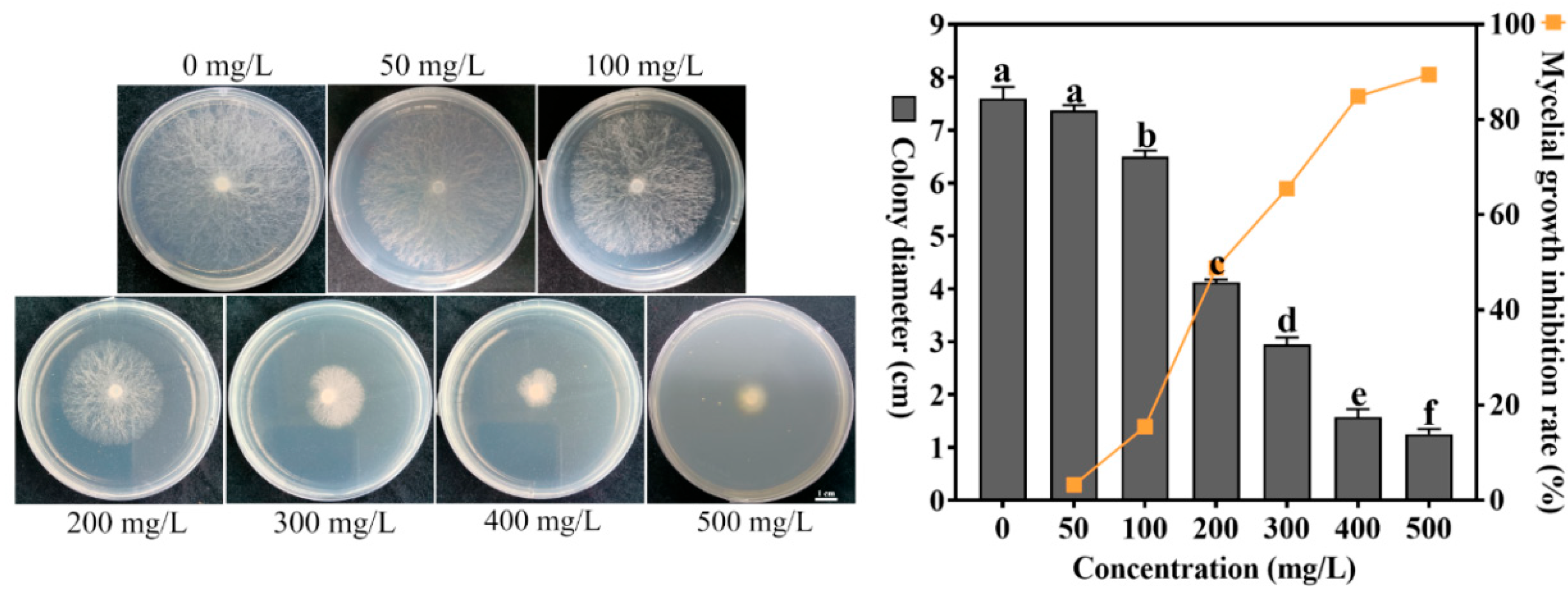
4.1. Effect of Different Concentrations of Sodium Selenite on the Growth of S. sclerotiorum

The effect of different concentrations of sodium selenite (0, 50, 100, 200, 300, 400, and 500 mg/L) on the growth of S. sclerotiorum showed that as the concentration of sodium selenite increased, the colony diameter of S. sclerotiorum gradually decreased (Figure 1), while the inhibition rate of the fungal hypha growth gradually increased, indicating that sodium selenite exhibits a significant inhibitory effect on the mycelial growth of S. sclerotiorum. Under the treatment of 500 mg/L sodium selenite, the inhibitory effect on the growth of S. sclerotiorum was the most significant, with an inhibition rate of 89.43%. The toxicity regression equation was as follows: y = 3.1251x − 2.2107, correlation coefficient r = 0.9974, EC50 was 202.93 mg/L, and EC90 was 521.12 mg/L.
After being treated with different concentrations of sodium selenite (50, 100, 200, 300, 400, and 500 mg/L) for 15 days at 22 °C, no sclerotial formation was observed in any treatments (Figure S1, Table S2), indicating that sodium selenite has a good inhibitory effect on the formation of sclerotia of S. sclerotiorum.

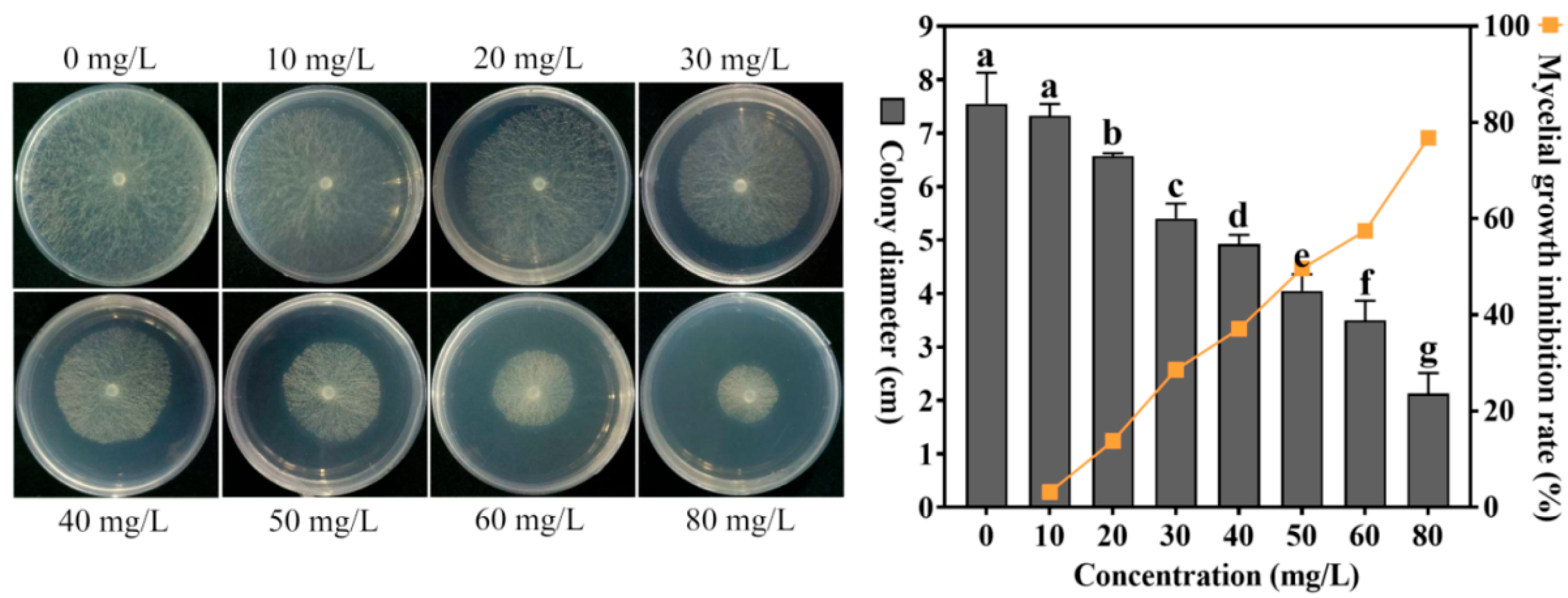
4.2. Effect of Different Concentrations of Cuminic Acid on the Growth of S. sclerotiorum

Studies on the growth of S. sclerotiorum with different concentrations of cuminic acid (0, 10, 20, 30, 40, 50, 60, and 80 mg/L) showed that as the concentration of cuminic acid increased, the colony diameter of S. sclerotiorum gradually decreased (Figure 2), while the inhibition rate of the fungal hypha growth gradually increased, indicating that cuminic acid has a significant inhibitory effect on the growth of mycelium of S. sclerotiorum. Under the treatment of 80 mg/L cuminic acid, the inhibitory effect on the growth of S. sclerotiorum was the most significant, with an inhibition rate of 76.83%. The toxicity regression equation was y = 2.7659x + 0.3307 (correlation coefficient r = 0.9953, EC50 was 48.77 mg/L, and EC90 was 141.56 mg/L).
The study on the effect of cuminic acid on sclerotia formation revealed that after being treated with cuminic acid concentrations of 0, 10, 20, 30, 40, 50, 60, and 80 mg/L, then incubated at 22 °C for 15 days, there were no significant changes observed in both the number of sclerotia and dry weight of sclerotia (Figure S2, Table S3), indicating that prolonged treatment (15 days) with cuminic acid did not exhibit a significant inhibitory effect on sclerotia formation in S. sclerotiorum.

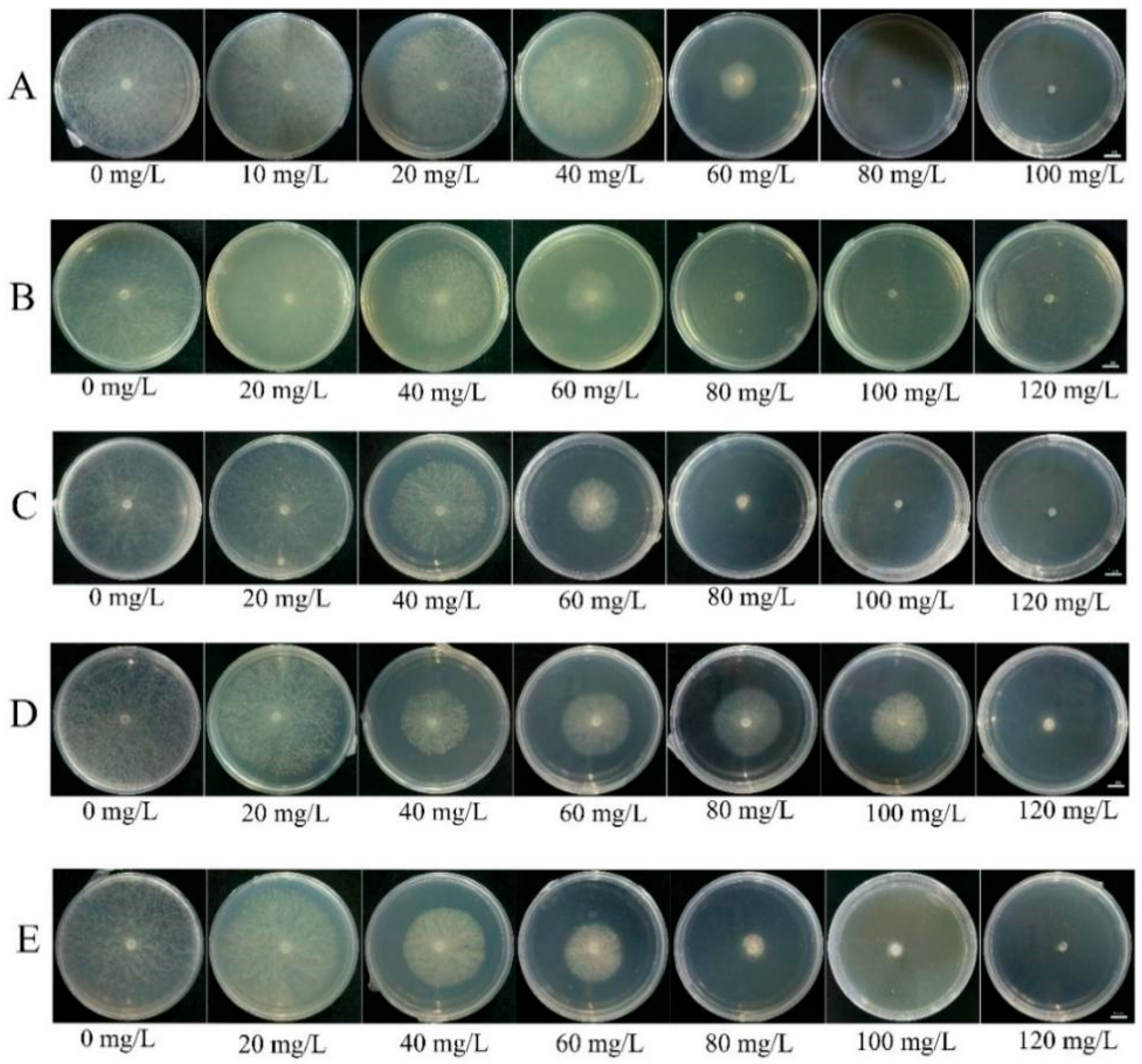
4.3. Compound Agent Formulation Screening

The results of laboratory toxicity assays on S. sclerotiorum with different compounding ratios of sodium selenite to cuminic acid (1:1, 1:2, 1:3, 1:6, 1:10) (Figure 3, Table 1) indicated that the combination with a 1:6 ratio of sodium selenite to cuminic acid exhibited the lowest EC50 value, at 37.63 mg/L; however, this ratio did not demonstrate synergistic activity but rather manifested an additive interaction. The synergistic effect of the combined sodium selenite and cuminic acid formulations was evaluated using the synergism ratio (SR) method. It was found that the formulations with a 1:2 and 1:3 ratio of sodium selenite to cuminic acid demonstrated synergistic effects, with SR values of 1.60 and 1.55, respectively. Although the SR values for the 1:2 and 1:3 combinations were not significantly different, the EC50 for the 1:3 combination (38.87 mg/L) was lower than that for the 1:2 ratio (40.88 mg/L). Therefore, the 1:3 ratio of sodium selenite to cuminic acid was selected for subsequent research.

4.4. Effects of Selenium Compound Agent on the Hyphal Morphology of S. sclerotiorum

SEM observations revealed that under the treatment of selenium compound at EC50 (38.87 mg/L), the mycelium exhibited severe surface shrinkage, deformation, and distortion, whereas control mycelium displayed a smooth, regular morphology. This indicates that the selenium compound agent has a serious impact on the morphology of the mycelium of S. sclerotiorum (Figure 4).

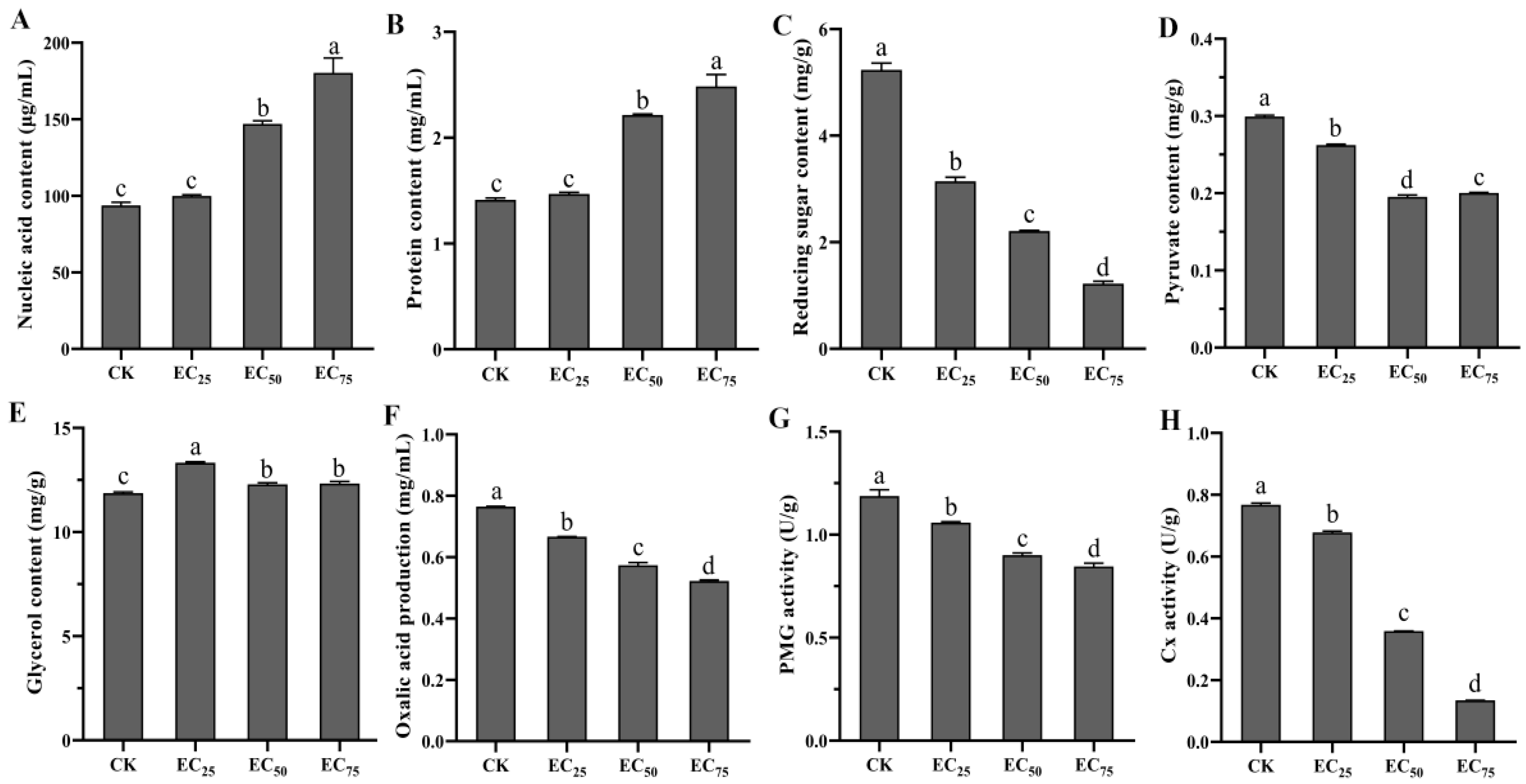
4.5. Effects of Selenium Compound Agent on Physiological and Biochemical Indexes of S. sclerotiorum

A study on the effect of selenium compound agent treatment on the leakage of nucleic acid and protein in the mycelia of S. sclerotiorum revealed that with an increase in the concentration of selenium compound agent, there was a significant increase in the concentrations of nucleic acid and protein in the culture medium (Figure 5A,B). Under treatment with the selenium compound agent at EC75 concentration (54.74 mg/L), the mycelial nucleic acid and protein leakage reached the maximum, and the contents of nucleic acid and protein in the culture medium were 180.23 μg/L and 2.48 mg/mL, respectively. This suggested that treatment with the selenium compound agent resulted in the rupture of S. sclerotiorum cell membranes, leading to the leakage of nucleic acid and protein into the medium.
The reducing sugar content in the mycelia of S. sclerotiorum treated with various concentrations of selenium compound agent was significantly lower than that of the control (p < 0.05). A higher concentration of the selenium compound agent resulted in a lower content of reducing sugar in the mycelial (Figure 5C). This indicated that treatment with selenium compound agent inhibited the synthesis of reducing sugar in the mycelia of S. sclerotiorum. Furthermore, the pyruvate content in the mycelia significantly decreased (Figure 5D), while the glycerol content significantly increased after treatment with the selenium compound agent (Figure 5E).
The production of oxalic acid in mycelia treated with selenium compound agent was significantly lower than that of the control (p < 0.05), indicating that the selenium compound agent had a marked inhibitory effect on oxalic acid synthesis in S. sclerotiorum (Figure 5F). Additionally, the activities of pectinase and cellulase in mycelia decreased significantly after treatment with the selenium compound agent. The results showed that the selenium compound agent could effectively inhibit the activities of pectinase and cellulase of S. sclerotiorum (Figure 5G,H).

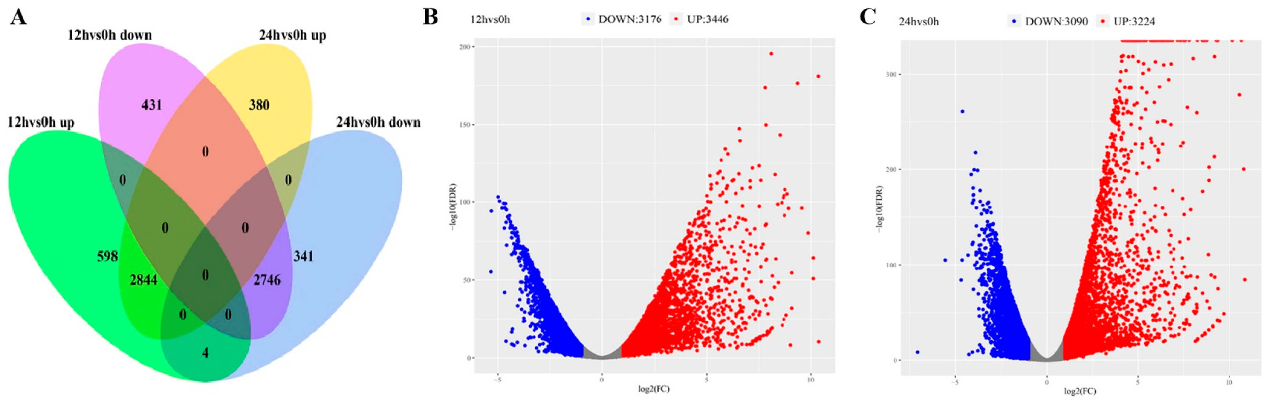
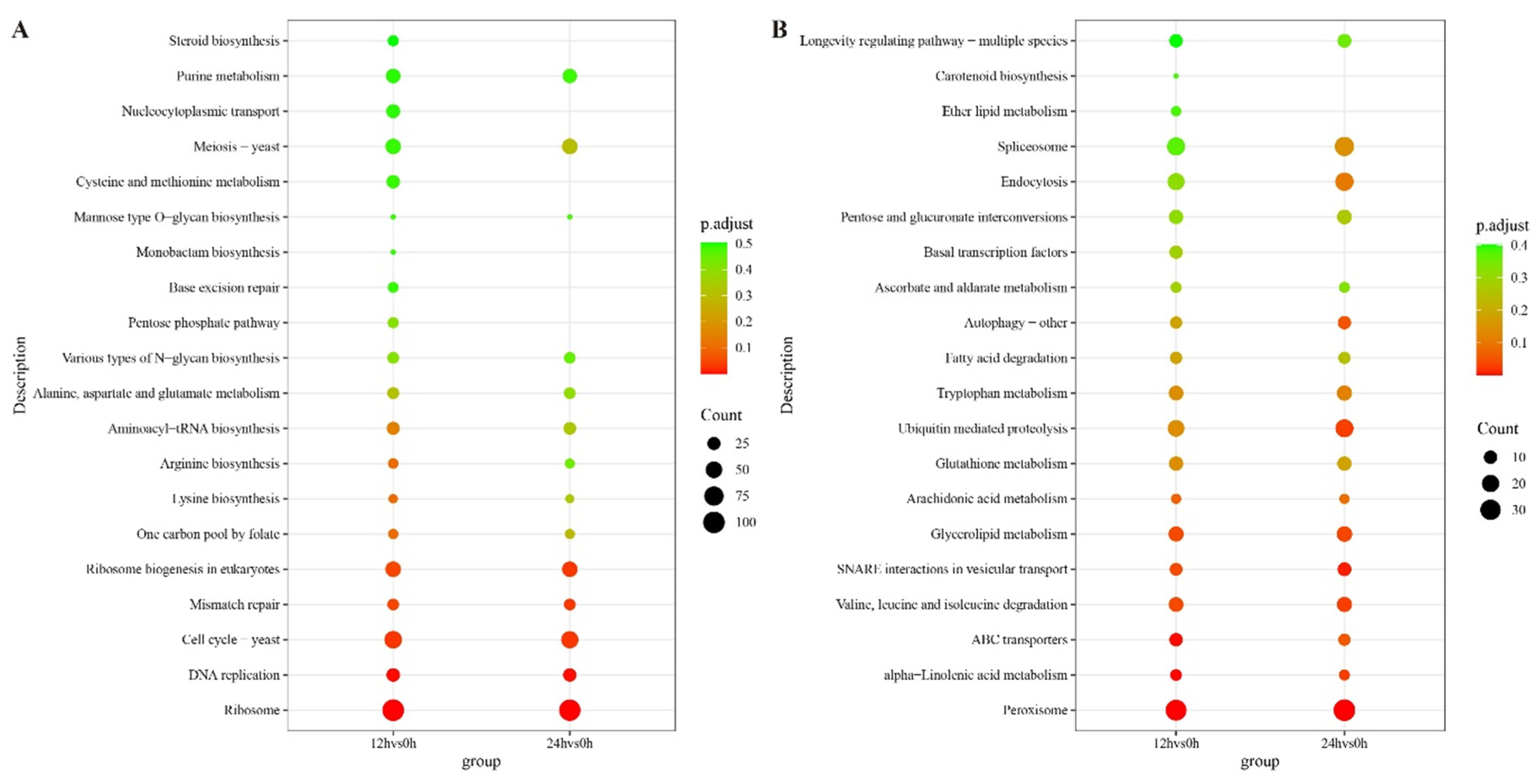
4.6. Transcriptomic Analysis

To elucidate transcriptional changes in S. sclerotiorum under selenium compound agent treatment, RNA-seq analysis was performed on S. sclerotiorum samples collected at 0 h, 12 h, and 24 h post-treatment. The number of clean reads per sample ranged from 36,192,008 to 54,138,988, with GC contents spanning 44.69–46.24% and Q20 scores exceeding 99%. Notably, 97.51–98.57% of clean reads were successfully aligned to the S. sclerotiorum reference genome (Table S4). Principal component analysis (PCA) revealed high biological replicability across time points except for the 24 h-1 sample, which was excluded from subsequent analyses due to inconsistent clustering (Figure S3).
Gene expression was compared between samples treated with selenium compound agent (at 12 h and 24 h) and the control sample (0 h), specifically conducting the comparisons of 12 h vs. 0 h and 24 h vs. 0 h. This analysis identified 6622 and 6314 differentially expressed genes (DEGs), respectively, with the numbers of upregulated/downregulated genes being 3446/3176 and 3224/3090 (Figure 6). Gene Ontology (GO) enrichment analysis of the downregulated DEGs revealed that, in the BP category, seven terms were commonly and significantly enriched in 12 h vs. 0 h and 24 h vs. 0 h groups: translation, ribosome biogenesis, peptide biosynthetic process, organonitrogen compound biosynthetic process, cytoplasmic translation, cellular nitrogen compound metabolic process, and amide biosynthetic process. In the CC category, nine terms were significantly and jointly enriched across the two comparison groups: ribosome, ribosomal subunit, ribonucleoprotein complex, preribosome, non-membrane-bounded organelle, large ribosomal subunit, intracellular non-membrane-bounded organelle, cytosolic ribosome, and cytosolic part. In the MF category, nine shared and significantly enriched terms were identified in both comparison combinations: translation factor activity, structural molecule activity, structural constituent of ribosome, rRNA binding, RNA binding, organic cyclic compound binding, nucleic acid binding, heterocyclic compound binding, and catalytic activity acting on RNA (Figure 7A). Kyoto Encyclopedia of Genes and Genomes (KEGG) pathway analysis identified that the 12 h vs. 0 h and 24 h vs. 0 h groups were enriched in many of the same pathways, including ribosome, DNA replication, cell cycle–yeast, mismatch repair, and ribosome biogenesis in eukaryotes (Figure 8A).
GO enrichment analysis of upregulated DEGs in the comparisons of 12 h vs. 0 h and 24 h vs. 0 h demonstrated that, within the BP category, the two comparison groups were significantly enriched in six terms including response to chemical, protein targeting to peroxisome, protein localization to peroxisome, peroxisomal transport, lipid catabolic process, and establishment of protein localization to peroxisome. In the CC category, DEGs identified in the two comparisons were enriched in many of the same terms, including Pex17p–Pex14p docking complex, peroxisome, peroxisomal part, peroxisomal membrane, peroxisomal matrix, microbody part, microbody membrane, microbody lumen, and microbody. Within the MF category, five terms were significantly and commonly enriched in both comparison sets: ubiquitin protein ligase activity, ubiquitin-protein transferase activity, ubiquitin-like protein transferase activity, ubiquitin-like protein ligase activity, and lipase activity (Figure 7B). KEGG pathway analysis revealed that the peroxisome and alpha-linolenic acid metabolism pathways were significantly and commonly enriched in both comparison groups (Figure 8B).
Through transcriptome analysis, it was found that 12 h and 24 h after the treatment with the selenium compound agent, a large number of genes encoding cellulase and pectinase in the S. sclerotiorum were downregulated in expression, and the key gene SS1G08218 (SsOAH1), which is involved in the biosynthesis of oxalic acid, was also downregulated in expression (Table S5).
In order to validate the reliability of the transcriptome data, nine DEGs were selected for qRT-PCR analysis. The results demonstrated that the expression trends of the genes detected by qRT-PCR were generally consistent with those obtained from transcriptome sequencing (Figure S4), suggesting that the transcriptome data were reliable and accurate.

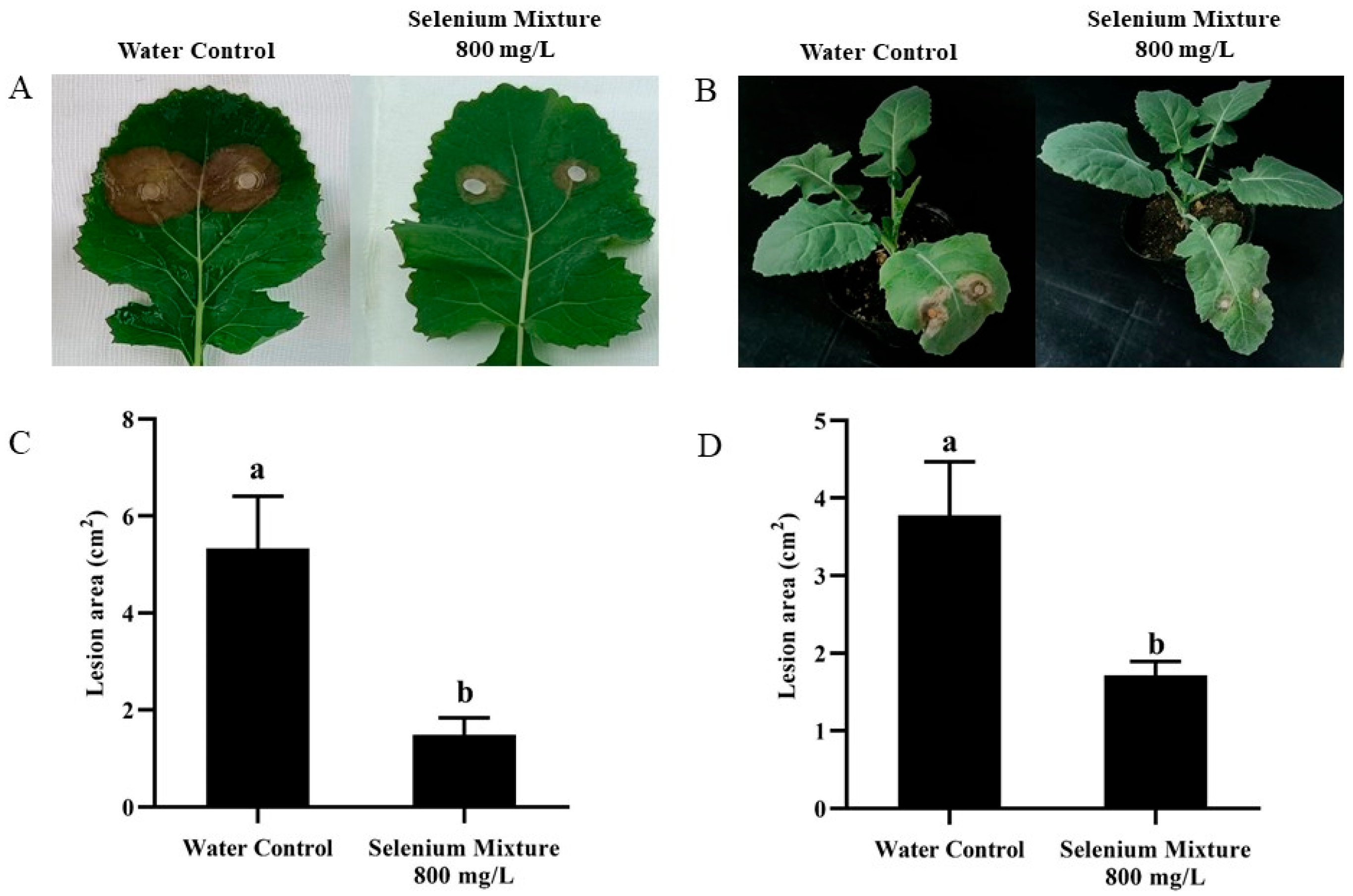
4.7. Control Efficacy of the Selenium Compound Agent on SSR of Rapeseed

Foliar application of an 800 mg/L selenium compound agent significantly reduced lesion areas caused by S. sclerotiorum on detached Brassica napus leaves and in potted plants, with control efficacies of 44.72% and 30.24%, respectively (Figure 9, Table 2). These results demonstrated that the selenium compound agent treatment effectively inhibited S. sclerotiorum infection in rapeseed.

5. Discussion

S. sclerotiorum, as a significant plant pathogen, causes severe economic losses to various crops, including Brassica napus. The long-term use of chemical fungicides has led to problems such as the development of pathogen resistance, environmental pollution, and adverse effects on human health. For instance, S. sclerotiorum has already developed high-level resistance to benzimidazole and dicarboximide fungicides worldwide [49]. Consequently, there is a growing concern for the development of new, safe, and effective fungicides to prevent and control SSR. Both sodium selenite and cuminic acid are environmentally friendly compounds, and the development of their combination as a fungicide not only holds great significance for the control of S. sclerotiorum but also contributes to reducing environmental pollution and promoting sustainable agricultural development.
In this study, we observed that sodium selenite exhibited a significant inhibitory effect on the mycelial growth of S. sclerotiorum. This finding is consistent with previous studies reporting that Se inhibits the mycelial growth of S. sclerotiorum [14]. Cuminic acid demonstrated a more pronounced inhibitory effect on the mycelial growth of S. sclerotiorum compared to sodium selenite. Sclerotia, an important survival form of S. sclerotiorum, exhibit strong adaptability to adverse environments and can survive in the soil for up to 8 years [50]. Therefore, it is highly necessary to investigate the inhibitory effects of agents on the sclerotia of S. sclerotiorum. In our study, sodium selenite effectively inhibited the formation of sclerotia in S. sclerotiorum, whereas cuminic acid showed no significant inhibitory effect. Previous research has also indicated that selenite can prevent sclerotial development [14]. Thus, the combination of sodium selenite with cuminic acid may represent a highly effective control strategy for SSR.
High-dose sodium selenite and cuminic acid applications risk phytotoxicity, growth inhibition, morphological abnormalities, bioaccumulation, and non-target effects [26,51]. Compared with single agents, the combined use of different fungicides can provide better disease control efficacy [33]. Mixtures containing two or three active ingredients may exhibit antagonistic, additive, or synergistic interactions. In this study, to identify safe and effective agents for controlling SSR, we combined sodium selenite and cuminic acid at various ratios to screen for combinations with synergistic effects. The results demonstrated that the combination of sodium selenite and cuminic acid exhibited a synergistic inhibitory effect on S. sclerotiorum, indicating that the combination of these two agents enhances their inhibitory effect on the pathogen, thereby potentially reducing the dosage required and minimizing environmental impact. Both the 1:2 and 1:3 combinations of sodium selenite and cuminic acid displayed synergistic effects; however, the 1:3 ratio combination exhibited the lowest EC50 value. Therefore, the 1:3 ratio combination of sodium selenite and cuminic acid was selected for subsequent investigations in this study.
SEM observations revealed prominent morphological disruptions, including shrinkage, collapse, and deformation, in the mycelia of S. sclerotiorum treated with the selenium compound agent compared to the untreated controls. Sun et al. [52] also similarly demonstrated that cuminic acid treatment induced shrinkage in the mycelia of S. sclerotiorum. In the present study, an increased leakage of nucleic acids and proteins was observed in the mycelium of S. sclerotiorum following treatment with the selenium compound agent, indicating an increase in mycelial cell membrane permeability. Additionally, the treatment with the selenium compound agent resulted in a decrease in the contents of reducing sugars and pyruvate within the mycelia of S. sclerotiorum. The reduction in reducing sugars can lead the mycelium into a sugar-deficient state, inducing the release of stress-activated proteins, disrupting the metabolism and transport of substances, and ultimately inhibiting mycelial growth [53]. The decrease in pyruvate content may block the glycolytic metabolism of fungal mycelia, leading to insufficient cellular energy supply [54]. Furthermore, glycerol accumulation was observed within the mycelial cells of S. sclerotiorum after treatment with the selenium compound agent. Glycerol is an important factor for osmotic regulation in microorganisms, and its accumulation within cells is crucial for maintaining osmotic balance and cell shape [55]. These findings suggest that the selenium compound agent disrupted the integrity of the cell membrane, leading to the efflux of intracellular substances and interfering with the normal metabolic processes of the mycelium, thereby inhibiting mycelial growth.
The major virulence factors of S. sclerotiorum include oxalic acid and plant cell wall-degrading enzymes (such as polygalacturonase and carboxymethyl cellulase). In this study, treatment with the selenium compound agent significantly reduced the production of oxalic acid in the mycelium. Oxalic acid is a crucial virulence factor for S. sclerotiorum in infecting plant cells, and its reduction can decrease the pathogen’s infectivity [56]. Oxalic acid can be synthesized from various precursors. However, in fungi, the most common mechanism involves the hydrolysis of oxaloacetate by oxaloacetate acetylhydrolase (OAH) to form oxalic acid and carbon dioxide. SsOAH1 serves as a key gene in oxalic acid biosynthesis in S. sclerotiorum and is considered crucial for the pathogenicity of S. sclerotiorum [57]. In this study, the expression of SsOAH1 was downregulated at both 12 h and 24 h after treatment with the selenium compound agent. The main components of plant cell walls are cellulose, hemicellulose, and pectins. When S. sclerotiorum infects rapeseed, it produces pectinase and cellulase to degrade the cell walls of rapeseed [58]. We found that treatment with the selenium compound agent effectively suppressed the activities of pectinase and cellulase in S. sclerotiorum. Furthermore, at 12 h and 24 h after treatment with the selenium compound agent, a large number of genes encoding cellulase and pectinase in S. sclerotiorum exhibited downregulated expression. This suggests that after treatment with the selenium compound agent, the damage caused by S. sclerotiorum to the cell walls of oilseed rape can be reduced, thereby decreasing the pathogen’s virulence. Similarly to our findings, Cheng et al. [14] reported that Se caused damage to the pathogenicity of plant pathogens.
Once S. sclerotiorum initiates infection in the plant, the suppression of mycelial growth within the host tissue becomes a primary target for disease management, as it can disrupt the pathogen’s lifecycle [27]. In this study, the selenium compound agent was able to significantly reduce the lesion area of rapeseed and effectively inhibit the expansion of S. sclerotiorum in rapeseed leaves, both at the level of isolated leaves and whole plants. These results indicate that the selenium compound agent has the potential to be applied as a safe and effective pesticide for the control of S. sclerotiorum. The disparity in effective concentrations of selenium compound agent between in vitro and in planta assays originates from differences in biological complexity and environmental dynamics. In in vitro assays, which represent a closed environment, the concentration of compounds remains stable, and microorganisms lack metabolic regulatory mechanisms [59]. Consequently, low concentrations (120 mg/L) can exert antimicrobial effects by directly inhibiting key metabolic pathways in pathogens. In contrast, in planta assays constitute an open system wherein compounds must penetrate the leaf cuticle and undergo translocation via the xylem, resulting in a bioavailability at target sites that may be lower [60]. Therefore, a higher treatment concentration (800 mg/L) was required to maintain therapeutic efficacy.
Transcriptome analysis was conducted to elucidate how the selenium compound agent influences the gene expression characteristics of S. sclerotiorum. The results revealed that the selenium compound agent can extensively affect the transcriptome of S. sclerotiorum. Genes associated with ribosome-related processes (including ribosome biogenesis, ribosome, ribosomal subunit, ribonucleoprotein complex, preribosome, large ribosomal subunit, cytosolic ribosome, ribosome biogenesis in eukaryotes, and structural constituent of ribosome) and protein synthesis-related processes (including translation, peptide biosynthetic process, and cytoplasmic translation) were all downregulated. In particular, a substantial number of genes within the ribosome-related pathway were downregulated, suggesting that treatment with the selenium compound agent impedes protein synthesis in the mycelium, thereby affecting hyphal growth and development. Additionally, genes involved in DNA replication and the cell cycle were also downregulated, indicating that the selenium compound agent may impact cell division in the mycelium of S. sclerotiorum. On the other hand, following treatment with the selenium compound agent, genes related to peroxisomes (including protein targeting to peroxisome, protein localization to peroxisome, peroxisomal transport, establishment of protein localization to peroxisome, peroxisome, peroxisomal part, peroxisomal membrane, and peroxisomal matrix) and the response to chemicals were upregulated in S. sclerotiorum. This suggests that S. sclerotiorum can adjust the upregulated expression of relevant genes to cope with the stress imposed by the selenium compound agent. Nevertheless, the elevated expression levels of these genes failed to alter the inhibited state of cell wall synthesis in S. sclerotiorum.
In summary, this study elucidated the inhibitory effects of the combination of sodium selenite and cuminic acid on S. sclerotiorum and its underlying mechanisms, providing a theoretical basis and empirical support for the development of novel, environmentally friendly, and efficient fungicides. Additionally, Se is an essential trace element in human and animal nutrition [61,62]. Given that approximately 51% of Chinese soils are deficient in Se [63] and that plants serve as the primary dietary source of Se in the food chain [64], the application of selenium compound agents may play a role in enhancing the Se content of oilseed rape and promoting human health. In the future, it would be worthwhile to optimize the formulation of selenium compound agents, investigate their field application techniques, and explore their stability and long-term efficacy under different environmental conditions, with the aim of achieving broader application in agricultural production.

6. Conclusions

In conclusion, the combination of sodium selenite and cuminic acid at a ratio of 1:3 exhibited a synergistic effect in inhibiting the mycelial growth of S. sclerotiorum, accompanied by a relatively low EC50 value. Treatment with the selenium compound agent increased the cell membrane permeability of S. sclerotiorum, disrupting the osmotic balance of the fungal cells and thereby disturbing mycelial growth. Furthermore, the selenium compound agent suppressed the production of oxalic acid and the activities of pectinase and cellulase in S. sclerotiorum, consequently reducing its pathogenicity. Spraying with the selenium compound agent at a concentration of 800 mg/L effectively mitigated the infection of S. sclerotiorum on the leaf surfaces of rapeseed. Treatment with the selenium compound agent downregulated the expression of genes associated with ribosomes, protein synthesis, and cell division in S. sclerotiorum cells, indicating that the selenium compound agent interfered with ribosome biogenesis, the translation process, and the expression of genes related to DNA replication, ultimately inhibiting the growth of S. sclerotiorum. These findings suggest that the selenium compound agent holds potential as a novel fungicide for controlling SSR of rapeseed.

Supplementary Materials

The following supporting information can be downloaded at: https://www.mdpi.com/article/10.3390/agronomy15081764/s1, Figure S1. Effect of different concentrations of sodium selenite on the sclerotia production of S. sclerotiorum; Figure S2. Effect of different concentrations of cuminic acid on the sclerotia production of S. sclerotiorum; Figure S3. Principal component analysis (PCA) for each group of S. sclerotiorum samples; Figure S4. Validation of 9 differentially expressed genes (DEGs) through qRT-PCR; Table S1. Primers used for quantitative real-time polymerase chain reaction (qRT-PCR) analysis; Table S2. Effect of different concentrations of sodium selenite on the sclerotia production of Sclerotinia sclerotiorum (S. sclerotiorum); Table S3. Effect of different concentrations of cuminic acid on the formation of sclerotia; Table S4. Summary of statistics for sequence quality control and genome mapping; Table S5. Changes in expression levels of S. sclerotiorum genes involved in oxalic acid production and degradation of plant cell wall components after selenium compounding agent treatment for 12 h/24 h.

Author Contributions

X.Z. and Y.H.: conceptualization, methodology, writing—original draft; X.M.: writing—review and editing; X.S. and Q.C.: resources, data collection, data interpretation; L.W.: investigation, the literature search, data curation; C.Z.: project administration, writing—review and editing. All authors have read and agreed to the published version of the manuscript.

Funding

This research was funded by Shaanxi University of Technology talent startup project (SLGRCQD2115); Shaanxi Provincial Department of Science and Technology (2024QCY-KXJ-104); the Natural Science Project of Shaanxi Provincial Department of Education (22JK0255); the Qinba Bioremediation and Resource Development Research Innovation and Introduction Base (2025YZ-YIPT-41); Sangin Talents, Shaanxi Provincial First-classTeam “Contaminated Soil Remediation and Resource Utilization Inno-vation” Team at Shaanxi University of Technology.

Institutional Review Board Statement

This article does not contain any studies with human participants or animals performed by any of the authors.

Data Availability Statement

All the experiments in this article were completed before December 2024. The original contributions presented in this study are included in the article/supplementary material. Further inquiries can be directed to the corresponding author.

Acknowledgments

We thank Xiaohui Cheng of the key laboratory of biology and genetic improvement of oil crops and Oil Crops Research Institute of the Chinese Academy of Agricultural Sciences for providing the S. sclerotiorum strain (1980).

Conflicts of Interest

The authors declare no conflicts of interest.

References

  1. Yaderets, V.V.; Karpova, N.V.; Glagoleva, E.V.; Ovchinnikov, A.I.; Petrova, K.S.; Dzhavakhiya, V.V. Inhibition of the Growth and Development of Sclerotinia sclerotiorum (Lib.) De Bary by Combining Azoxystrobin, Penicillium chrysogenum VKM F-4876d, and Bacillus Strains. Agronomy 2021, 11, 2520. [Google Scholar] [CrossRef]
  2. Liang, X.; Rollins, J.A. Mechanisms of broad host range necrotrophic pathogenesis in Sclerotinia sclerotiorum. Phytopathology 2018, 108, 1128–1140. [Google Scholar] [CrossRef] [PubMed]
  3. Jia, W.; Hu, C.X.; Ming, J.J.; Zhao, Y.Y.; Xin, J.; Sun, X.C.; Zhao, X.H. Action of selenium against Sclerotinia sclerotiorum: Damaging membrane system and interfering with metabolism. Pestic. Biochem. Physiol. 2018, 150, 10–16. [Google Scholar] [CrossRef] [PubMed]
  4. Feng, Y.X.; Hu, Y.X.; Fang, P.P.; Zuo, X.J.; Wang, J.X.; Li, J.N.; Qian, W.; Mei, J.Q. Silicon alleviates the disease severity of sclerotinia stem rot in rapeseed. Front. Plant Sci. 2021, 12, 721436. [Google Scholar] [CrossRef] [PubMed]
  5. Boyd, R.S. The defense hypothesis of elemental hyperaccumulation: Status, challenges and new directions. Plant Soil 2007, 293, 153–176. [Google Scholar] [CrossRef]
  6. Hörger, A.C.; Fones, H.N.; Preston, G.M. The current status of the elemental defense hypothesis in relation to pathogens. Front. Plant Sci. 2013, 4, 395. [Google Scholar] [CrossRef] [PubMed]
  7. Pandey, S.S. The role of iron in phytopathogenic microbe–plant interactions: Insights into virulence and host immune response. Plants 2023, 12, 3173. [Google Scholar] [CrossRef] [PubMed]
  8. Elsherbiny, E.A.; Taher, M.A. Silicon induces resistance to postharvest rot of carrot caused by Sclerotinia sclerotiorum and the possible of defense mechanisms. Postharvest Biol. Technol. 2018, 140, 11–17. [Google Scholar] [CrossRef]
  9. Xue, T.L.; Hartikainen, H.; Piironen, V. Antioxidative and growth-promoting effect of selenium on senescing lettuce. Plant Soil 2001, 237, 55–61. [Google Scholar] [CrossRef]
  10. Chu, J.Z.; Yao, X.Q.; Yue, Z.W.; Li, J.M.; Zhao, J.H. The effects of selenium on physiological traits, grain selenium content and yield of winter wheat at different development stages. Biol. Trace Elem. Res. 2013, 151, 434–440. [Google Scholar] [CrossRef] [PubMed]
  11. Lin, L.; Zhou, W.H.; Dai, H.X.; Cao, F.B.; Zhang, G.P.; Wu, F.B. Selenium reduces cadmium uptake and mitigates cadmium toxicity in rice. J. Hazard. Mater. 2012, 235, 343–351. [Google Scholar] [CrossRef] [PubMed]
  12. Mozafariyan, M.; Shekari, L.; Hawrylak-Nowak, B.; Kamelamnesh, M.M. Protective role of selenium on pepper exposed to cadmium stress during reproductive stage. Biol. Trace Elem. Res. 2014, 160, 97–107. [Google Scholar] [CrossRef] [PubMed]
  13. Qing, X.J.; Zhao, X.H.; Hu, C.X.; Wang, P.; Zhang, Y.; Zhang, X.; Wang, P.C.; Shi, H.Z.; Jia, F.; Qu, C. Selenium alleviates chromium toxicity by preventing oxidative stress in cabbage (Brassica campestris, L. ssp. Pekinensis) leaves. Ecotoxicol. Environ. Saf. 2015, 114, 179–189. [Google Scholar] [CrossRef] [PubMed]
  14. Cheng, Q.; Hu, C.X.; Jia, W.; Cai, M.M.; Zhao, Y.Y.; Tang, Y.N.; Yang, D.D.; Zhou, Y.J.; Sun, X.C.; Zhao, X.H. Selenium reduces the pathogenicity of Sclerotinia sclerotiorum by inhibiting sclerotial formation and germination. Ecotoxicol. Environ. Saf. 2019, 183, 109503. [Google Scholar] [CrossRef] [PubMed]
  15. Liu, L.N.; Wang, C.Y.; Yin, K.S.; Ni, M.; Ding, Y.; Li, C.Y.; Zheng, S.J. The Dual Effect of Selenium Application in Reducing Fusarium Wilt Disease Incidence in Banana and Producing Se-Enriched Fruits. Plants 2024, 13, 3435. [Google Scholar] [CrossRef] [PubMed]
  16. Espinosa-Ortiz, E.J.; Gonzalez-Gil, G.; Saikaly, P.E.; van Hullebusch, E.D.; Lens, P.N. Effects of selenium oxyanions on the white-rot fungus Phanerochaete chrysosporium. Appl. Microbiol. Biotechnol. 2015, 99, 2405–2418. [Google Scholar] [CrossRef] [PubMed]
  17. Wu, Z.L.; Yin, X.B.; Bañuelos, G.S.; Lin, Z.Q.; Zhu, Z.; Liu, Y.; Yuan, L.X.; Li, M. Effect of selenium on control of postharvest gray mold of tomato fruit and the possible mechanisms involved. Front. Microbiol. 2016, 6, 1441. [Google Scholar] [CrossRef] [PubMed]
  18. Wu, Z.L.; Yin, X.B.; Lin, Z.Q.; Bañuelos, G.S.; Yuan, L.X.; Liu, Y.; Li, M. Inhibitory effect of selenium against Penicillium expansum and its possible mechanisms of action. Curr. Microbiol. 2014, 69, 192–201. [Google Scholar] [CrossRef] [PubMed]
  19. Ma, X.H.; Xu, J.Y.; Zhao, X.H.; Qu, L.L.; Gao, Y.; Huang, W.X.; Han, D.; Dang, B.J.; Xu, Z.C.; Jia, W. Selenium improves the control efficacy of Phytophthora nicotianae by damaging the cell membrane system and promoting plant energy metabolism. J. Agric. Food Chem. 2024, 72, 5073–5087. [Google Scholar] [CrossRef] [PubMed]
  20. Özer, G.; Türkkan, M.; Sönmez, F.; Kabakcı, H.; Alkan, M.; Derviş, S. In Vitro Evaluation of Selenium Against Some Plant Pathogenic Fungi. Akad. Ziraat Derg. 2024, 13, 99–110. [Google Scholar] [CrossRef]
  21. Wang, J.L.; Ma, Y.; Gao, Q.; Lu, Z.J.; Zhang, J.M. Overview of Active Components and Applications of Plant Derived Fungicides in China. J. Zhejiang Agric. Sci. 2025, 66, 138–145. [Google Scholar] [CrossRef]
  22. Wang, Y.; Zhang, J.; Sun, Y.; Feng, J.T.; Zhang, X. Evaluating the Potential Value of Natural Product Cuminic Acid against Plant Pathogenic Fungi in Cucumber. Molecules 2017, 22, 1914. [Google Scholar] [CrossRef] [PubMed]
  23. Yang, Z.H.; Qiu, Y.G.; Jin, D.J.; Zheng, Y.M.; Cui, Z.N.; Li, J.; Gu, W. Design, synthesis, and antifungal evaluation of novel cuminic acid derivatives as potential laccase inhibitors. J. Agric. Food Chem. 2024, 72, 27726–27736. [Google Scholar] [CrossRef] [PubMed]
  24. Hu, L.F.; He, J.; Feng, J.T.; Zhang, X. Optimization of supercritical CO2 extraction characterization of antifungal activity of essential oils in Cuminum cyminum, L. Aust. J. Crop Sci. 2013, 7, 1809–1813. [Google Scholar]
  25. Wang, Y.; Sun, Y.; Zhang, Y.; Zhang, X.; Feng, J.T. Antifungal activity and biochemical response of cuminic acid against Phytophthora capsici Leonian. Molecules 2016, 21, 756. [Google Scholar] [CrossRef] [PubMed]
  26. Sun, Y.; Wang, Y.; Han, L.R.; Zhang, X.; Feng, J.T. Antifungal activity action mode of cuminic acid from the seeds of Cuminum cyminum, L. against Fusarium oxysporum f. sp. Niveum (FON) causing Fusarium wilt on watermelon. Molecules 2017, 22, 2053. [Google Scholar] [CrossRef] [PubMed]
  27. Wang, Y.; Sun, Y.; Han, L.R.; Zhang, X.; Feng, J.T. Potential use of cuminic acid as a botanical fungicide against Valsa mali. Microb. Pathog. 2017, 106, 9–15. [Google Scholar] [CrossRef] [PubMed]
  28. Ma, H.X.; Feng, X.J.; Chen, Y.; Chen, C.J.; Zhou, M.G. Occurrence and characterization of dimethachlon insensitivity in Sclerotinia sclerotiorum in Jiangsu Province of China. Plant Dis. 2009, 93, 36–42. [Google Scholar] [CrossRef] [PubMed]
  29. Kuang, J.; Wang, J.X.; Zhou, M.G. Monitoring on carbendazim and dimethachlon-resistance of Sclerotinia sclerotiorum obtained from the blight stems of rape in Jiangsu Province. Chin. Agric. Sci. Bull. 2011, 27, 285–291. [Google Scholar] [CrossRef]
  30. Zhou, F.; Zhang, X.L.; Li, J.L.; Zhu, F.X. Dimethachlon resistance in Sclerotinia sclerotiorum in China. Plant Dis. 2014, 98, 1221–1226. [Google Scholar] [CrossRef] [PubMed]
  31. Zhao, W.B.; Zhao, Z.M.; Ma, Y.; Li, A.P.; Zhang, Z.J.; Hu, Y.M.; Zhou, Y.; Wang, R.; Luo, X.F.; Zhang, B.Q.; et al. Antifungal activity and preliminary mechanism of pristimerin against Sclerotinia sclerotiorum. Ind. Crops Prod. 2022, 185, 115124. [Google Scholar] [CrossRef]
  32. Kuang, J.; Hou, Y.P.; Wang, J.X.; Zhou, M.G. Sensitivity of Sclerotinia sclerotiorum to fludioxonil: In vitro determination of baseline sensitivity and resistance risk. Crop Prot. 2011, 30, 876–882. [Google Scholar] [CrossRef]
  33. Wang, W.Q.; Zhang, P.; Meng, R.J.; Zhao, J.J.; Huang, Q.L.; Han, X.Y.; Ma, Z.Q.; Zhang, X.F. Fungitoxicity and synergism of mixtures of fluopicolide and pyraclostrobin against Phytophthora infestans. Crop Prot. 2014, 57, 48–56. [Google Scholar] [CrossRef]
  34. Soylu, E.M.; Kurt, Ş.; Soylu, S. In vitro and in vivo antifungal activities of the essential oils of various plants against tomato grey mould disease agent Botrytis cinerea. Int. J. Food Microbiol. 2010, 143, 183–189. [Google Scholar] [CrossRef] [PubMed]
  35. Zhao, S.L.; Li, P.; Zhang, Q.K.; Zhang, J.H.; Kong, L.Z. Production of reducing sugar from corn stover by dilute acid hydrolysis co-catalyzed with metal salts under microwave radiation. Res. Chem. Intermed. 2013, 39, 3803–3812. [Google Scholar] [CrossRef]
  36. Crowther, T.; Collin, H.A.; Smith, B.; Tomsett, A.B.; O’Connor, D.; Jones, M.G. Assessment of the flavour of fresh uncooked onions by taste-panels and analysis of flavour precursors, pyruvate and sugars. J. Sci. Food Agric. 2005, 85, 112–120. [Google Scholar] [CrossRef]
  37. Duan, Y.B.; Ge, C.Y.; Liu, S.M.; Chen, C.J.; Zhou, M.G. Effect of phenylpyrrole fungicide fludioxonil on morphological and physiological characteristics of Sclerotinia sclerotiorum. Pestic. Biochem. Physiol. 2013, 106, 61–67. [Google Scholar] [CrossRef]
  38. Xu, C.Y.; Hou, Y.P.; Wang, J.X.; Yang, G.F.; Liang, X.Y.; Zhou, M.G. Activity of a novel strobilurin fungicide benzothiostrobin against Sclerotinia sclerotiorum. Pestic. Biochem. Physiol. 2014, 115, 32–38. [Google Scholar] [CrossRef] [PubMed]
  39. Andrews, S. FastQC: A Quality Control Tool for High Throughput Sequence Data. 2010. Available online: https://www.bioinformatics.babraham.ac.uk/projects/fastqc/ (accessed on 12 December 2024).
  40. Martin, M. Cutadapt removes adapter sequences from high-throughput sequencing reads. EMBnet. J. 2011, 17, 10–12. [Google Scholar] [CrossRef]
  41. Amselem, J.; Cuomo, C.A.; van Kan, J.A.L.; Viaud, M.; Benito, E.P.; Couloux, A.; Coutinho, P.M.; de Vries, R.P.; Dyer, P.S.; Fillinger, S.; et al. Genomic analysis of the necrotrophic fungal pathogens Sclerotinia sclerotiorum and Botrytis cinerea. PLoS Genet. 2011, 7, e1002230. [Google Scholar] [CrossRef] [PubMed]
  42. Kim, D.; Langmead, B.; Salzberg, S.L. HISAT: A fast spliced aligner with low memory requirements. Nat. Methods 2015, 12, 357–360. [Google Scholar] [CrossRef] [PubMed]
  43. Liao, Y.; Smyth, G.K.; Shi, W. featureCounts: An efficient general purpose program for assigning sequence reads to genomic features. Bioinformatics 2014, 30, 923–930. [Google Scholar] [CrossRef] [PubMed]
  44. Pertea, M.; Pertea, G.M.; Antonescu, C.M.; Chang, T.C.; Mendell, J.T.; Salzberg, S.L. StringTie enables improved reconstruction of a transcriptome from RNA-seq reads. Nat. Biotechnol 2015, 33, 290–295. [Google Scholar] [CrossRef] [PubMed]
  45. Robinson, M.D.; McCarthy, D.J.; Smyth, G.K. edgeR: A Bioconductor package for differential expression analysis of digital gene expression data. Bioinformatics 2010, 26, 139–140. [Google Scholar] [CrossRef] [PubMed]
  46. Young, M.D.; Wakefield, M.J.; Smyth, G.K.; Oshlack, A. Gene ontology analysis for RNA-seq: Accounting for selection bias. Genome Biol. 2010, 11, R14. [Google Scholar] [CrossRef] [PubMed]
  47. Mao, X.Z.; Cai, T.; Olyarchuk, J.G.; Wei, L.P. Automated genome annotation and pathway identification using the KEGG Orthology (KO) as a controlled vocabulary. Bioinformatics 2005, 21, 3787–3793. [Google Scholar] [CrossRef] [PubMed]
  48. Liang, X.Y.; Zou, L.J.; Lian, W.X.; Wang, M.; Yang, Y.; Zhang, Y. Comparative transcriptome analyses reveal conserved and distinct mechanisms of the SDHI fungicide benzovindiflupyr inhibiting Colletotrichum. Phytopathology 2022, 112, 1255–1263. [Google Scholar] [CrossRef] [PubMed]
  49. Xu, C.Y.; Liang, X.Y.; Hou, Y.P.; Zhou, M.G. Effects of the novel fungicide benzothiostrobin on Sclerotinia sclerotiorum in the laboratory and on Sclerotinia stem rot in rape fields. Plant Dis. 2015, 99, 969–975. [Google Scholar] [CrossRef] [PubMed]
  50. Erental, A.; Dickman, M.B.; Yarden, O. Sclerotial development in Sclerotinia sclerotiorum: Awakening molecular analysis of a “Dormant” structure. Fungal Biol. Rev. 2008, 22, 6–16. [Google Scholar] [CrossRef]
  51. Trippe, R.C., III; Pilon-Smits, E.A.H. Selenium transport and metabolism in plants: Phytoremediation and biofortification implications. J. Hazard. Mater. 2021, 404, 124178. [Google Scholar] [CrossRef] [PubMed]
  52. Sun, Y.; Wang, Y.; Xie, Z.Y.; Guo, E.H.; Han, L.R.; Zhang, X.; Feng, J.T. Activity and biochemical characteristics of plant extract cuminic acid against Sclerotinia sclerotiorum. Crop Prot. 2017, 101, 76–83. [Google Scholar] [CrossRef]
  53. Luo, J.J.; Li, Z.B.; Wang, J.J.; Weng, Q.F.; Chen, S.H.; Hu, M.Y. Antifungal activity of isoliquiritin and its inhibitory effect against Peronophythora litchi Chen through a membrane damage mechanism. Molecules 2016, 21, 237. [Google Scholar] [CrossRef] [PubMed]
  54. Wang, H.X.; Fu, L.; Li, C.L.; Zhang, X.L.; Narcisse, K.E.; Qi, H.S.; Han, C.Y.; Wang, X.Y.; Ma, H.B.; Zhu, C.Y.; et al. Tannic acid exerts antifungal activity in vitro and in vivo against Alternaria alternata causing postharvest rot on apple fruit. Physiol. Mol. Plant Pathol. 2023, 125, 102012. [Google Scholar] [CrossRef]
  55. Hohmann, S. An integrated view on a eukaryotic osmoregulation system. Curr. Genet. 2015, 61, 373–382. [Google Scholar] [CrossRef] [PubMed]
  56. Heller, A.; Witt-Geiges, T. Oxalic acid has an additional, detoxifying function in Sclerotinia sclerotiorum pathogenesis. PLoS ONE 2013, 8, e72292. [Google Scholar] [CrossRef] [PubMed]
  57. Nehela, Y.; Mazrou, Y.S.A.; ELGammal, N.A.; Atallah, O.; Abdelrhim, A.S.; Kumar, S.; Ahmed, T.; Ail, Q.; Makhlouf, A.H.; Hussain, W.A.M. Ornithine enhances common bean growth and defense against white mold disease via interfering with SsOAH and diminishing the biosynthesis of oxalic acid in Sclerotinia sclerotiorum. Front. Plant Sci. 2025, 16, 1483417. [Google Scholar] [CrossRef] [PubMed]
  58. Ding, L.N.; Li, T.; Guo, X.J.; Li, M.; Liu, X.Y.; Cao, J.; Tan, X.L. Sclerotinia stem rot resistance in rapeseed: Recent progress and future prospects. J. Agric. Food Chem. 2021, 69, 2965–2978. [Google Scholar] [CrossRef] [PubMed]
  59. Tóth, D.; Tengölics, R.; Aarabi, F.; Karlsson, A.; Vidal-Meireles, A.; Kovács, L.; Kuntam, S.; Körmöczi, T.; Fernie, A.R.; Hudson, E.P.; et al. Chloroplastic ascorbate modifies plant metabolism and may act as a metabolite signal regardless of oxidative stress. Plant Physiol. 2024, 196, 1691–1711. [Google Scholar] [CrossRef] [PubMed]
  60. Chu, C.C.; Han, C.Y.; Li, H. High-efficiency leucoplast transit peptides for manipulating plastid protein production. Nat. Plants 2025, 11, 1316–1324. [Google Scholar] [CrossRef] [PubMed]
  61. Awual, M.R.; Hasan, M.M.; Ihara, T.; Yaita, T. Mesoporous silica based novel conjugate adsorbent for efficient selenium (IV) detection and removal from water. Microporous Mesoporous Mater. 2014, 197, 331–338. [Google Scholar] [CrossRef]
  62. Vinceti, M.; Filippini, T.; Wise, L.A. Environmental selenium and human health: An update. Curr. Environ. Health Rep. 2018, 5, 464–485. [Google Scholar] [CrossRef] [PubMed]
  63. Dinh, Q.T.; Cui, Z.W.; Huang, J.; Tran, T.A.T.; Wang, D.; Yang, W.X.; Zhou, F.; Wang, M.K.; Yu, D.S.; Liang, D.L. Selenium distribution in the Chinese environment and its relationship with human health: A review. Environ. Int. 2018, 112, 294–309. [Google Scholar] [CrossRef] [PubMed]
  64. Natasha; Shahid, M.; Niazi, N.K.; Khalid, S.; Murtaza, B.; Bibi, I.; Rashid, M.I. A critical review of selenium biogeochemical behavior in soil-plant system with an inference to human health. Environ. Pollut. 2018, 234, 915–934. [Google Scholar] [CrossRef] [PubMed]
Figure 1. Effect of different concentrations of sodium selenite on the growth of Sclerotinia sclerotiorum (S. sclerotiorum). ANOVA, Tukey’s test. Different lowercase letters indicate significant differences (p < 0.05).
Agronomy 15 01764 g001
Figure 2. Effect of different concentrations of cuminic acid on the growth of S. sclerotiorum mycelial. ANOVA, Tukey’s test. Different lowercase letters indicate significant differences (p < 0.05).
Agronomy 15 01764 g002
Figure 3. Effect of selenite compound agent on the mycelial growth of S. sclerotiorum at different dosages (0–120 mg/mL) and different ratios of sodium selenite and cuminic acid: 1:1 (A), 1:2 (B), 1:3 (C), 1:6 (D), and 1:10 (E).
Agronomy 15 01764 g003
Figure 4. Scanning electron microscopy (SEM) images of mycelium morphology of S. sclerotiorum under selenium compound agent treatments. (A) control (without selenium compound agent treatment); (B) selenium compound agent (EC50, 38.87 mg/L).
Agronomy 15 01764 g004
Figure 5. Effect of the selenium compound agent on the leakage of mycelial nucleic acid (A), protein (B), reducing sugar (C), pyruvate (D), glycerol (E) content and oxalic acid production (F), PMG (G), and Cx (H) activity of S. sclerotiorum mycelia. CK is 0 mg/L; EC25 is 27.60 mg/L; EC50 is 38.87 mg/L; and EC75 is 54.74 mg/L. Values are means ± SD. ANOVA, Tukey’s test. Different lowercase letters indicate significant differences (p < 0.05).
Agronomy 15 01764 g005
Figure 6. Overview of the differentially expressed genes (DEGs). (A) Venn diagram displaying the upregulated and downregulated genes after selenium compound agent treatment for 12 h/24 h compared to that of the control 0 h. (B,C) Volcano plots of global gene expression patterns. Each dot represents a gene. Blue dots are downregulated genes, while red dots are upregulated ones, and black dots are genes without significant difference.
Agronomy 15 01764 g006
Figure 7. Gene ontology classifications of DEGs. Downregulation of genes (A). Upregulation of genes (B).
Agronomy 15 01764 g007
Figure 8. Kyoto Encyclopedia of Genes and Genomes classifications of DEGs. Downregulation of genes (A). Upregulation of genes (B).
Agronomy 15 01764 g008
Figure 9. Control efficacy of sclerotinia stem rot (SSR) in detached Brassica napus leaves (A,C) and potted plants (B,D) of rapeseed by selenium compound agent. Values are reported as means ± SD. ANOVA, Tukey’s test. Different lowercase letters indicate significant differences (p < 0.05).
Agronomy 15 01764 g009
Table 1. Combined virulence determination of sodium selenite and cuminic acid compounded against S. sclerotiorum.
Ratios of Sodium Selenite and Cuminic AcidVirulence EquationCorrelation CoefficientEC50(th) w (mg/L)EC50(me) x
(mg/L)
EC90(me) y
(mg/L)
SR z
Sodium selenitey = 3.1251x − 2.21070.9974-202.93521.12-
Cuminic acidy = 2.7659x + 0.33070.9953-48.77141.56-
1:1y = 4.7524x − 3.43450.928978.6459.54110.691.32
1:2y = 4.7942x − 2.72600.917265.3140.8875.601.60
1:3y = 4.5355x − 2.20950.964760.2038.8774.441.55
1:6y = 2.1448x + 1.62080.950554.7137.63148.701.45
1:10y = 2.3250x + 1.19690.961452.3943.23153.561.21
w EC50 = effective concentration causing 50% mycelial growth inhibition. EC50 (th) indicates the theoretical EC50 values of the mixtures. x EC50 (me) indicates the measured EC50 values of the fungicides. y EC90 = effective concentration causing 90% mycelial growth inhibition. EC90 (me) indicates the measured EC90 values of the mixtures. z Synergy ratio (SR) = ratio between expected and observed EC50 values. SR = EC50(th)/EC50(me). An SR < 0.5 indicates an antagonistic interaction between the fungicides, an SR between 0.5 and 1.5 indicates an additive interaction, and an SR > 1.5 indicates a synergistic interaction.
Table 2. Control efficacy of selenium compound agent against SSR in detached leaves and potted plants of rapeseed.
TreatmentDetached Leaves of RapeseedPotted Plants of Rapeseed
Lesion Area
(cm2) a
Control Efficacy
(%) b
Lesion Area (cm2)Control Efficacy
(%)
Selenium Mixture 800 mg/L1.49 ± 0.3544.72 ± 3.121.71 ± 0.1830.24 ± 3.48
Water Control5.33 ± 1.08-3.78 ± 0.69-
a Lesion area was calculated by approximation to a circle using average diameter. b Control efficacy = [(lesion diameter in the water control − lesion diameter in the treatment)/(lesion diameter in the water control)] × 100%. All values are reported as the means ± SD.
Disclaimer/Publisher’s Note: The statements, opinions and data contained in all publications are solely those of the individual author(s) and contributor(s) and not of MDPI and/or the editor(s). MDPI and/or the editor(s) disclaim responsibility for any injury to people or property resulting from any ideas, methods, instructions or products referred to in the content.

