Review

Molecular Mechanisms Underlying Root Nodule Formation and Activity

Department of Biochemistry and Biotechnology, Poznan University of Life Sciences, Dojazd 11, 60-632 Poznan, Poland
*
Author to whom correspondence should be addressed.
Agronomy 2025, 15(7), 1552; https://doi.org/10.3390/agronomy15071552
Submission received: 30 May 2025 / Revised: 22 June 2025 / Accepted: 24 June 2025 / Published: 26 June 2025

Abstract

Symbiotic interactions between legumes and a group of soil bacteria, known as rhizobia, lead to the formation of a specialized organs called root nodules. Inside them, atmospheric nitrogen (N2) is fixed by bacteria and reduced to forms available to plants, catalyzed by the nitrogenase enzyme complex. The development of a symbiotic relationship between legumes and nodule bacteria is a multi-stage, precisely regulated process, characterized by a high specificity of partner selection. Nodulation involves the enhanced expression of certain plant genes, referred to as early- and late-nodulin genes. Many nodulin genes encode hydroxyproline-rich glycoproteins (HRGPs) and proline-rich proteins (PRPs) which are involved in various processes, including infection thread formation, cell signaling, and defense responses, thereby affecting nodule formation and function. Cyclophilins (CyPs) belong to a family of proteins with peptidyl-prolyl cistrans isomerase activity. Proteins with cyclophilin domain can be found in the cytoplasm, endoplasmic reticulum, nucleus, chloroplast, and mitochondrion. They are involved in various processes, such as protein folding, cellular signaling, mRNA maturation, and response to biotic and abiotic stress. In this review, we aim to summarize the molecular processes involved in the development of symbiosis and highlight the potential role of cyclophilins (peptidyl-prolyl cis-trans isomerases) in this process.

1. Introduction

The establishment of efficient cooperation between rhizobia and legumes begins with the exchange of chemical signals between the future symbionts. The first stage of infection is the adsorption of bacteria on the surface of root hairs. This process involves numerous structures present on the surface of both future symbiosis partners, such as fimbriae, polysaccharides, and plant lectins [1]. After the bacteria have colonized the root surface, molecular signals are exchanged to specifically select symbionts. The basic chemical compounds involved in this process include plant flavonoids and isoflavonoids, as well as Nod factors (NFs) synthesized by rhizobia [2,3]. Nodulation involves the enhanced expression of specific plant genes, referred to as early- and late-nodulin genes. These nodulin genes are differentially expressed during the development of the symbiotic interaction. Early nodulin genes are expressed at the stage when nodule structures are formed and late nodulins become detectable as nitrogen fixation begins. Many nodulin genes encode hydroxyproline-rich glycoproteins (HRGPs) and proline-rich proteins (PRPs) (Table 1). Some of them are infection thread and cell wall components affecting nodule formation and function. Peptide bonds in protein structures are synthesized by ribosomes mainly in the trans conformation. This conformation is energetically more favorable due to the possible steric hindrance of the side chains of the following amino acid residues. An exception is the peptide bond Xaa–Pro (Xaa represents any amino acid), which can exist in both the trans and cis conformation [4]. Isomerization of this peptide bond is slow, and the cis peptidyl-prolyl conformation is present at a small percentage in proteins [5]. However, such a conformational change may significantly affect the structure and function of a given protein, as can be seen for the largest subunit of RNA polymerase II [6,7,8].
Cyclophilins (CyPs), together with FK506-binding proteins (FKBPs) and parvulins, are proteins with peptidyl-prolyl cistrans isomerase activity. Although these proteins lack sequence homology, they exhibit peptidyl-prolyl cistrans isomerase activity. Cyclophilins participate in various cellular processes including signaling, interactions with pathogens, mRNA maturation, protein degradation, and apoptosis, as well as in response to different stress stimuli [10,11,12,13,14,15,16]. It has been shown that some CyP transcripts accumulate in response to heat shock, low temperature treatment, and wounding and in symbiotic plant–microbe interactions [3,17,18,19,20].
Cyclophilins can be divided into two groups of proteins: single-domain and modular proteins with a CyP domain. Single-domain proteins consist of a catalytic domain with a highly conserved structure (the cyclophilin domain CyP, which is approximately 120 amino acids in length). Modular cyclophilins contain, in addition to the CyP, other domains. For example, an A. thaliana AtCyP40 has a CyP domain and the tetratricopeptide repeat domain (TPR); AtCyP59 contains a CyP domain followed by an RNA recognition motif (RRM) and a C-terminal domain enriched in charged amino acids [21]. In modular plant cyclophilins, the TPR and WD40 repeats are commonly observed. These domains have been reported to facilitate protein–protein interactions in the cell. The large diversity of protein forms with the CyP domain indicates their involvement in numerous cellular processes. However, little is known about the function of individual cyclophilins. More information can be obtained from genome or transcriptome analysis, which is both an advantage and a disadvantage nowadays. In this review, we aim to summarize the molecular processes involved in the development of symbiosis and highlight the potential role of cyclophilins in this process.

2. Organogenesis and Function of Root Nodules

Symbiotic interactions between leguminous plants (Fabaceae) and a group of soil bacteria collectively known as rhizobia result in the formation of specialized structures called root nodules on the roots of the host plant. Within these nodules, atmospheric nitrogen (N2) is fixed and reduced in a process catalyzed by the nitrogenase enzymatic complex to forms that can be assimilated by the plant [22]. The interaction confers mutual benefits: the macrosymbiont (the plant) obtains access to readily assimilable ammonium ions while concurrently undergoing continuous, albeit low-intensity, stimulation of its defense mechanisms. In return, the microsymbiont (the bacteria) receives energy-rich compounds and carbon skeletons in the form of dicarboxylates while being sheltered from environmental stresses common in the soil, including competition from other microorganisms [23]. Establishing an effective symbiotic relationship between legumes and nodule-forming bacteria is a multi-stage and tightly regulated process. It is also characterized by a high degree of partner specificity.

3. The Selection of a Symbiotic Partner and the Exchange of Chemical Signals Are Specific Processes

Efficient cooperation between rhizobia and legumes is established through the exchange of chemical signals between the future symbionts. The initial stage in the establishment of symbiosis between leguminous plants and Rhizobium spp. involves colonization of the rhizosphere, a process in which chemotaxis and bacterial motility play pivotal roles [24]. Rhizobia migrate towards nutrient-rich zones in response to chemoattractant signals, which are detected by chemoreceptor proteins that interact with histidine kinase, initiating a signal transduction cascade that culminates in flagellum-driven movement [25].
Recent studies have demonstrated that the principal determinants of the directional migration of Rhizobium spp. towards the plant root system are surface-associated motility and signaling molecules belonging to the acyl-homoserine lactone (AHL) family, independent of flagellar motility [26]. The FadL–ExoFQP system is central to this mechanism, being responsible for the biosynthesis and degradation of extracellular polysaccharides (EPSs), as well as for modulating extracellular concentrations of long-chain AHLs. Specifically, FadL facilitates the uptake of long-chain AHLs, while the ExoFQP complex mediates the secretion of short-chain AHL molecules [26].
The dynamic regulation of EPS synthesis and degradation influences the local concentration of AHLs, thereby modulating the efficiency of quorum sensing. EPSs may facilitate the accumulation of AHLs in the immediate vicinity of bacterial cells, enabling the quorum sensing threshold to be reached. In contrast, EPS degradation promotes AHL diffusion and alters the bioavailability of AHLs. The experimental application of a synthetic cocktail of long-chain AHLs has been shown to enhance Rhizobium surface motility and promote its migration from the rhizosphere to the root surface in a quorum sensing-dependent manner [26].
LuxI-family enzymes regulate the synthesis of AHLs, while LuxR-type receptor proteins mediate their perception. Once the threshold concentration of AHLs (typically 10 ng/L to 10 μg/L) is reached, the LuxR–AHL complex activates the transcription of target genes involved in the regulation of bacterial motility [26]. Once the bacteria have colonized the root surface, molecular signals are exchanged. The main chemical compounds involved in this process are plant flavonoids and isoflavonoids, as well as Nod factors (NFs), which are synthesized by rhizobia [27]. Flavonoids are a group of plant secondary metabolites that are synthesized in response to stress conditions, including nitrogen deficiency. The secretion of flavonoids can occur via membrane-associated proteins such as ATP-binding cassette (ABC) transporters and MATE transporters [28,29]. These compounds act as chemoattractants for rhizobia. Flavonoids are secreted into the soil in the proximity of the root apex. However, their optimal concentration has been identified in the maturation zone, where root hairs are formed [25,30,31,32]. The exchange of molecular signals may also be involved with other chemical compounds produced by plants. These include betaines, aldonic acids, xanthones, and jasmonic acid [33,34].
Furthermore, research on Lotus japonicus has revealed that specific phenolic acids, such as ferulic acid and caffeic acid, which are secreted into the substrate, may also play a role in symbiosis with a specific type of rhizobia [35]. Legumes secrete a mixture of chemicals into the substrate that is specific to their species. The composition of this mixture guarantees a high level of specificity in the selection of a symbiotic partner [31]. The interaction between host plants and their microsymbionts is influenced in part by the structural and functional diversity of flavonoid compounds released in the rhizosphere. While certain flavonoids serve as positive signals promoting nod gene expression, others function as negative regulators. The dynamic equilibrium between these opposing flavonoid activities appears to fine-tune the extent of nod gene activation, ensuring an appropriate symbiotic response [36].
Some of the substances secreted by legumes into the substrate activate the bacterial transcriptional regulator NodD, which is constitutive expressed in many rhizobial species [37]. Although plants continuously produce a wide array of flavonoids, only a limited number of flavonoids have been detected in the rhizosphere, suggesting their potential role as key signaling molecules. Currently, the set of identified flavonoids is largely confined to luteolin, hesperetin, genistein, naringenin, and daidzein, with minimal representation from other flavonoid types [38]. Chemical signals sent by the macrosymbiont influence the NodD protein, causing it to interact with conserved motifs in DNA called nod-box sequences. This interaction initiates the transcription of nodulation genes, which are responsible for the synthesis of bacterial signaling molecules termed Nod factors (NFs) [39]. Nod factors belong to lipochitooligosaccharides (LCOs) constructed from N-acetyl-D-glucosamines linked by β-1,4 bonds and having a 16- or 18-carbon fatty acid chain with a variable number of double bonds at the non-reducing end [40,41]. The NFs’ structure is the second factor determining species specificity in the selection of symbiotic partners. Two groups of genes are responsible for this. The first of these are the so-called common nodulation genes, which are part of the nodABC operon present in most rhizobia and which encode enzymes involved in the synthesis of the N-acetylglucosamine skeleton. The second group of genes responsible for the synthesis of Nod factors includes the nod, noe, and nol genes. The products of these genes are involved in chemical modifications of the backbone, such as sulfidation, acetylation, fucosylation, and glycosylation. The length of the N-acetylglucosamine skeleton and the presence of specific modifications at defined positions determine species specificity in the selection of symbiotic partners [40,42,43,44]. Rhizobia synthesize several types of Nod factors. For example, a mixture of four large and four smaller lipochitooligosaccharides is secreted by Mesorhizobium loti, which enters into symbiosis with L. japonicus. These lipochitooligosaccharides differ in the type of fatty acid and the number of carbamoyl groups (-CONH2) at the non-reducing end and the number of acetyl groups attached to the fucose at the reducing end [45].
The host recognizes Nod factors through Nod Factor Receptors (NFRs), which have a high affinity for them [46,47]. Three Nod factor receptors (NFR1, NFR5, and NFRe) have been identified in the plasma membrane of L. japonicus rhizoderma, functioning as transmembrane proteins. It is thought that the NFR1 and NFR5 receptors are essential for Nod factor perception. In turn, NFRe is involved in the amplification of NFR1 and NFR5-derived signaling in rhizodermis cells [27]. Both NFR1 and NFR5, as well as NFRe, belong to the Lysine Motif-Receptor-Like Kinase, which consists of three domains: LysM, which is located on the outer side of the plasmalemma and is responsible for interacting with Nod factors; the transmembrane domain; and an intracellular domain with kinase activity. Studies using purified receptors have shown that each receptor can directly and independently bind bacterial LCOs [48,49]. RinRK1 (Rhizobia Infection Receptor-like Kinase 1), a leucine-rich repeat receptor-like kinase (LRR-RLK), interacts with the extracellular domains of Nod factor receptors (NFR1 and NFR5), facilitating their accumulation at root hair tips in response to rhizobial signals or Nod factors. Additionally, Flotillin 1 (Flot1), a membrane nanodomain-organizing protein, associates with the kinase domains of NFR1, NFR5, and RinRK1. RinRK1 enhances the interactions between Flot1 and the Nod factor receptors, and the presence of both RinRK1 and Flot1 is essential for the targeted localization of Nod factor receptors at root hair apexes upon Nod factor stimulation [50].
In addition to Nod factors, the specificity of symbiotic partner selection is determined by exopolysaccharides (EPSs) and lipopolysaccharides (LPSs) produced by rhizobia. The structure of EPSs varies depending on the species and strain of bacteria, suggesting their potential participation in symbiont recognition processes. Exopolysaccharides belong to homo- or heterosaccharides, which can be secreted into the environment or retained on the bacterial surface as capsular polysaccharides (CPSs) [51,52] (Scheme 1).
The plant’s ability to recognize exopolysaccharides on the surface of rhizobia acts as an additional checkpoint, regulating the progression of the infection and facilitating the transportation of rhizobia into the root. Perception of Nod factors initiates the transcription of a gene that encodes the EPR3 protein, which acts as a specific EPS receptor. EPR3 is a transmembrane protein with a structure similar to the NFR1 receptor [53]. Research using plants with a mutation in the sequence encoding this receptor has shown an impairment in the formation of an infectious thread and the establishment of an ineffective symbiosis between L. japonicus and M. loti [54,55]. This result suggests that the LysM-type transmembrane kinase, also known as the EPR3 protein, is involved in the recognition of bacterial exopolysaccharides. The process of symbiosis formation between legumes and rhizobia is characterized by a high specificity of selection of future symbionts. This involves the exchange of complementary chemical signals, including plant secondary metabolites such as flavonoids, isoflavonoids, and some phenolic acids, as well as bacterially produced polysaccharides and Nod factors.

4. The Early Stages of the Infection

The interaction between Nod factors and rhizodermal membrane receptors activates two parallel signaling pathways: one leading to the formation of the infectious thread and the other stimulating root nodule organogenesis [56]. The perception of Nod factors is accompanied by an activation of SYMRK (Symbiosis Receptor-Like Kinase), which belongs to the Leucine-Rich Repeat-Receptor-Like Kinase (LRR-RLK) subfamily [36]. Nod factor perception leads to signal transduction, resulting in fluctuations in calcium ion concentration in the nucleoplasm [57]. These changes in Ca2+ concentration are caused by CASTOR and POLUX potassium channels located in the nuclear envelope. Activation of these channels affects the membrane potential of the nuclear envelope, leading to the activation of calcium channels [58]. Some nucleoporins, such as Nup85 and Nup133, are also involved in oscillatory changes in calcium ion concentration [59,60]. Changes in Ca2+ concentration activate the nuclear-localized calmodulin-dependent kinase CCaMK, which is involved in the phosphorylation of the transcription activator protein CYCLOPS. CYCLOPS is part of a protein complex that includes GRAS transcription factors (NSP1, NSP2, and DELLA), which activate the expression of genes that encode early nodulins and numerous transcription factors. These transcription factors play a crucial role in activating developmental programs that are essential for root nodule organogenesis and infection thread formation [39,44]. Additionally, the formation of infectious threads depends on the kinase activity of NFR receptors. The binding of Nod factors synthesized by complementary rhizobia to their receptors leads to the expression of Nap1 and Pir1 genes, which encode proteins that are part of the SCAR/WAVE complex. This complex is responsible for cytoskeletal reorganization in root cells [61], followed by the expression of the Cerberus gene, which encodes an E3 ubiquitin ligase. The presence of this ligase enables the elongation of the infectious threads [62].

5. Infection of the Root by Rhizobia

The interaction between leguminous plants and rhizobia begins with the colonization of the root zone by the bacteria, a critical step in the initiation of endosymbiosis. Depending on the host plant species, rhizobia can infect plant cells through different pathways: via root hairs, involving specialized structures known as infection threads (ITs), or by an alternative route—intercellular invasion through the apoplastic spaces between epidermal cells [63]. Two main mechanisms of this type of infection are recognized: “crack entry”, in which rhizobia penetrate through natural cracks formed during the emergence of lateral roots and subsequently establish infection pockets or intracellular infection threads, and actual intercellular invasion, in which rhizobia traverse intact epidermal layers, typically at the junctions between root hair cells and neighboring epidermal cells [63]. Among the studied legume genera, approximately 75% utilize the root hair-mediated entry route, while the remaining 25% favor the intercellular mode of infection [64].
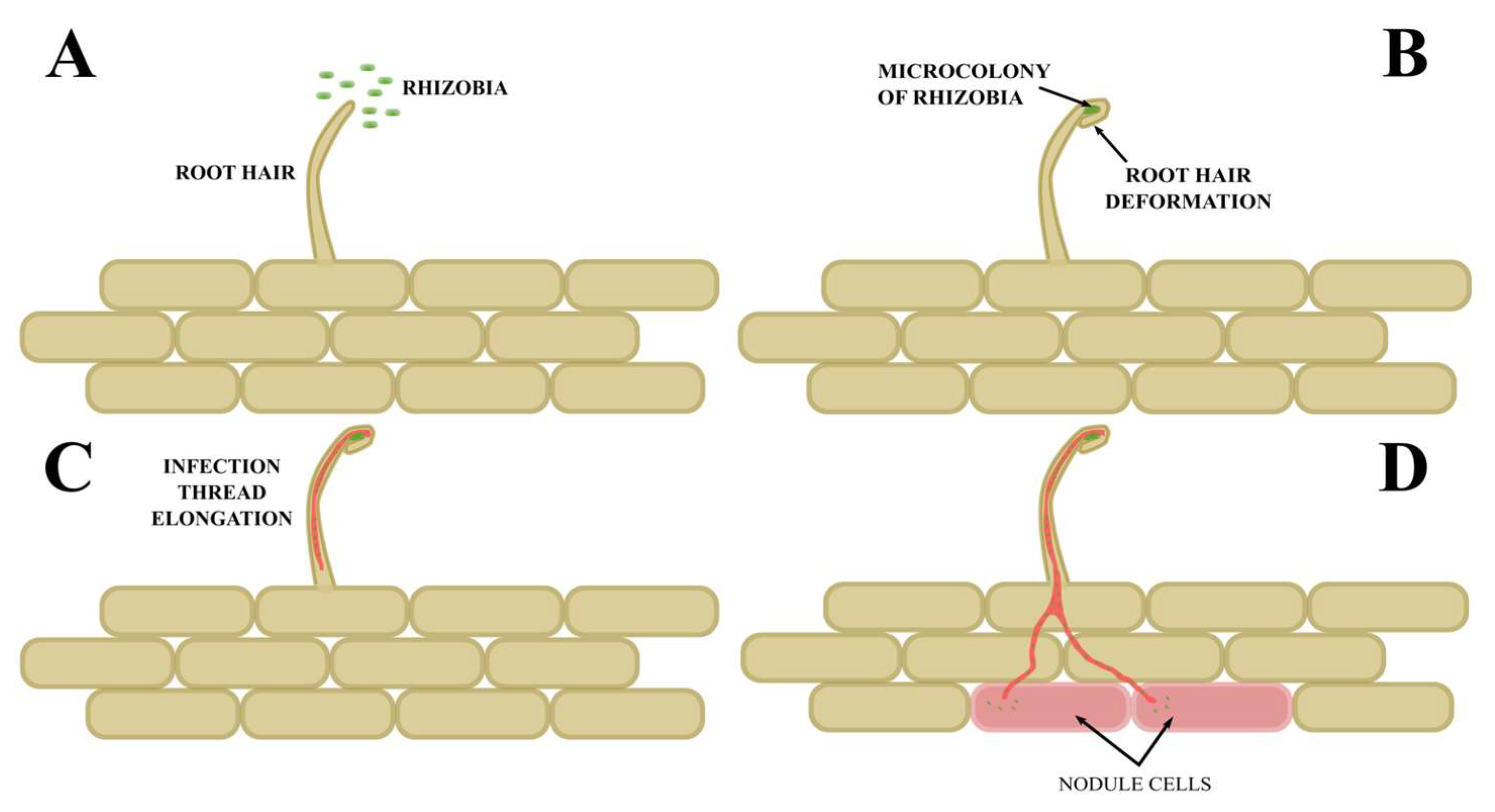
The symbiotic interaction is initiated by the secretion of flavonoids by legume roots into the rhizosphere. These compounds are recognized by rhizobia, which in response synthesize specific Nod factors—lipochitooligosaccharides—that trigger a cascade of transcriptional changes in the host plant roots [40]. The perception of Nod factors by root epidermal receptors induces root hair curling, enabling bacterial entrapment and subsequent infection. Following this, infection threads undergo polarized tip growth guided by the trajectory of the migrating plant nucleus. In Lotus japonicus, this polarized growth is mediated by the guanine nucleotide exchange factor (GEF) LjSPK1, which activates the Rho-family GTPase LjROP6 [65]. This process further stimulates cell divisions in the root cortex, leading to the formation of nodule primordia [66]. During infection, modifications and partial degradation of the plant cell wall occur, facilitating rhizobial penetration either intercellularly or via the interior of root hairs [67,68,69] (Scheme 2).
Once the bacteria are trapped within the curled root hair, rhizobia proliferate and form microcolonies [70], resulting in a local increase in the concentration of Nod factors. This, in turn, stimulates the formation of the infection thread (IT)—a plant-derived structure that develops as a result of reversed tip growth of the root hair, loosening of the cell wall, and invagination of the plasma membrane [71]. The elongation of the infection thread depends on the targeted delivery of secretory vesicles that supply the thread tip with structural components and signaling molecules [72]. The transcription factor MYB3R1, activated by Nod factor signaling, directly regulates the expression of the AUR1 gene through two mitosis-specific cis-acting elements, ensuring proper orientation of pre-infection structures and directing the deposition of cell wall material for infection thread elongation in a process analogous to cell plate formation during cytokinesis [73].
The migrating plant nucleus, which follows the extending IT, remains connected to the tip of the thread by a cytoplasmic bridge composed of cytoskeletal elements and vesicles [74]. In the later stages of infection, a protein complex known as the “infectosome” forms in epidermal and cortical cells. This complex consists of Vapyrin (VPY), Lumpy Infections (LIN), Rhizobium-directed Polar Growth (RPG), and the exocyst subunit EXO70H4. VPY, localized to the trans-Golgi network and early endosomes, interacts with the E3 ligase LIN via its ankyrin repeat domain, which likely leads to the stabilization of the VPY protein [75,76]. The infectosome regulates the development of infection threads, most likely through polarized exocytosis. It plays a crucial role by facilitating polarized growth of the infection thread through coordinated endocytic and exocytic processes.
When the infection thread reaches the base of the root hair cell, localized cell wall degradation occurs, allowing rhizobia to be released into adjacent plant cells via a SymRK (Symbiosis Receptor-like Kinase)-dependent mechanism, which enables bacterial endocytosis and further nodule development [67]. At later stages, infection threads grow toward the developing nodule primordia, and rhizobia are released into the nodule interior as infection droplets via an endocytosis-like mechanism [77]. Rhizobia are then internalized by plant cells into organelle-like structures called symbiosomes, which consist of differentiating bacteroids surrounded by a symbiosome membrane [78].
The capacity of legumes to efficiently fix atmospheric nitrogen hinges on the successful initiation of endosymbiosis and the accurate differentiation and maturation of symbiosomes. Numerous studies have highlighted that the synchronized reorganization of the plant cell wall, endomembrane network, and cytoskeletal elements serves as a structural and functional framework for rhizobial invasion, nodule organogenesis, symbiosome integrity, and overall symbiotic performance [68,78].
The plant cell wall and membrane systems are fundamental in orchestrating the developmental trajectory of the legume–rhizobia symbiosis. The cell wall’s structural flexibility and its susceptibility to enzymatic remodeling are prerequisites for successful rhizobial penetration into host tissues [68,79].
Similarly, biological membranes—especially the plasma membrane and elements of the endomembrane system—play critical roles in symbiotic communication and development. The plasma membrane functions not only as a selective barrier for solute exchange but also as a sensory interface that perceives rhizobial Nod factors, thereby triggering downstream signaling cascades that prepare host cells for infection [80]. The endomembrane system, encompassing organelles such as the endoplasmic reticulum, Golgi apparatus, and endosomes, facilitates targeted vesicular trafficking of structural and signaling molecules necessary for infection thread initiation and nodule morphogenesis [81].
The functional interplay among the cell wall, plasma membrane, and cytoskeleton—collectively referred to as the Wall–Membrane–Cytoskeleton (WMC) continuum—is essential for mediating rhizobial infection and driving the development of root nodule structures in leguminous hosts [68,79].
At the onset of symbiotic engagement, root hairs, which are tubular projections of epidermal cells, serve as the primary sites for microbial recognition and attachment [82].
During root hair elongation, actin filaments form longitudinal bundles extending from the basal region toward the subapical domain, encircling the vacuole. In parallel, endoplasmic microtubules undergo dynamic rearrangements in response to developmental cues, whereas cortical microtubules maintain a relatively stable configuration [83].
This specialized cytoskeletal architecture, along with a coordinated membrane trafficking apparatus, supports both the polar growth of root hairs and their readiness for microbial colonization, setting the stage for infection and nodule formation.
Upon rhizobial recognition and Nod factor perception, host cells undergo extensive cytoskeletal remodeling. Both actin microfilaments and microtubules are reorganized to facilitate the initiation and progression of infection threads (ITs).
The actin cytoskeleton exhibits a biphasic response, characterized by transient depolymerization followed by partial repolymerization, which creates a conducive framework for IT initiation [84]. Key regulatory genes, such as Nap1 and Pir1, which encode actin-binding proteins, are critical for proper IT formation; their loss-of-function mutations lead to malformed root hairs and reduced infection efficiency [61].
The SYFO1 protein accumulates near the migrating nucleus and guides the orientation of actin filaments toward the apex of the root hair. By binding directly to actin, SYFO1 promotes the polarized elongation and directed growth of infection threads [85].
In parallel, microtubules undergo rapid disassembly and reassembly in response to the perception of the Nod factor. The depolymerization cascade initiates in the endoplasmic microtubule network and subsequently affects the cortical array, which is then rapidly rebuilt to accommodate the demands of the infection. In Medicago truncatula, the DREPP protein facilitates microtubule fragmentation during early infection events, thereby promoting bacterial ingress and IT extension [86].
In conclusion, the reconfiguration of the cytoskeleton is indispensable for the establishment and maintenance of symbiotic infection. This dynamic process underpins root hair polarization, nuclear migration, and infection thread formation, which collectively drive efficient rhizobial colonization and the ontogeny of nitrogen-fixing root nodules.

6. Types of Root Nodules

As the infection thread develops, the cells in the primary root cortex next to the infected root hair start to specialize. In this differentiated region, cell divisions lead to the formation of root primordia. Depending on the species, primordia formation occurs in either outer or inner layers of the cortex, which correlates with the type of root nodule formed. For instance, in soybeans (Glycine max), the initial cell divisions take place in the cortex cells adjacent to the rhizodermis. In plants belonging to the genus Lotus, cell differentiation occurs in the middle layers of the cortex. The development of the primodium leads to the formation of determinate root nodules in both soybeans and Lotus, i.e., those in which a temporary nodule meristem functions. In contrast, in plants belonging to the Medicago and Trifolium genera, among others, the first divisions occur in the inner layers of the root cortex. This leads to the formation of indeterminate root nodules, whose development is controlled by persistent meristem.
When the infectious thread reaches the root nodules, rhizobia are released. This process is similar to endocytosis and involves so-called infection droplets forming from the apical fragment of the thread [87]. Rhizobia entering the cells of the macrosymbiont are surrounded by a membrane derived from the host cell membrane, and together with it, they form organelle-like hybrid structures called symbiosomes. Inside the symbiosome, the rod-shaped rhizobium cell divides along with the division of the membrane (symbiosome multiplication), and over time, the divisions cease and the vegetative forms of rhizobia differentiate into bacteroids capable of nitrogen fixation.
Bacteroids inside root nodules can occur in three morphotypes: U, S, and E [88,89]. Bacteroids with minor morphological modifications, compared to free-living bacteria, characterize the U (undifferentiated) morphotype. These bacteroids are capable of returning to life in the soil after the root nodule decomposes. In contrast, bacteria with morphotypes S (spherical) and E (enlarged) cannot return to life in the soil due to increased cell membrane permeability and DNA endoreplication [90]. The differentiation of bacteroids is caused by nodule-specific cysteine-rich peptides (NCRs) produced by the macrosymbiont [91,92,93]. NCR peptides have been found to control the irreversible differentiation of bacteroids in leguminous plants belonging to the IRLC (Inverted Repeat-Lacking Clade), such as M. truncatula [94,95]. These peptides increase the permeability of bacteroid cell membranes and affect the cell division protein FtsZ, which is responsible for septum formation [96,97].
The growth and development of the primordium results in the formation of a mature root nodule that contains bacteroids. Due to the type of meristem that determines nodule development, two main types of root nodules can be distinguished. The first type, found in plants such as Trifolium and Medicago, is referred to as an indeterminate root nodule and is characterized by an elongated, cylindrical shape resulting from the presence of a persistent meristem. This type of meristem functions throughout the organ’s entire life. Constantly dividing cells within the meristem at the apex of the indeterminate root nodule result in five distinct developmental zones that form its anatomical structure. The meristem (zone I) is located closest to the tip of the nodule and is composed of small, cytoplasm-rich cells that give rise to all the tissues of the developing organ. Near the meristem is the so-called infection zone. Known also as the infection thread penetration zone or zone II, the infection zone is formed by larger cells. These cells cease division as a result of the penetration of the infection thread. Here, rhizobia are released from the infection thread and then differentiated into bacteroids. Further away from the meristem is the transition zone (zone III), where there is a rapid increase in starch content and the final differentiation of bacteroid tissue occurs. This specialized parenchyma tissue occupies a central position in the nodule. The atmospheric nitrogen fixation zone (zone III) contains fully differentiated cells, where bacteroids surrounded by a peribacteroid membrane of plant origin form symbiosomes. Here, atmospheric nitrogen (N2) is reduced to ammonium ions (NH4+) by the bacterial nitrogenase complex. Closer to the root is the aging zone (zone IV), where bacteroids that can no longer reduce N2 undergo degradation and infected host cells enter the apoptosis pathway. These dead cells are then colonized by rod-shaped rhizobia, which previously inhabited the infectious thread in the saprophytic zone (zone V). Unlike rhizobia, which transform into bacteroids, bacteria colonizing the apoplast and saprophytic zone return to the soil after nodule decomposition [98,99,100,101].
Plants from subtropical and tropical climates, such as Lotus and Phaseolus, are known to have determinate root nodules. In contrast to indeterminate nodules, they are defined by a spherical shape resulting from the early cessation of cell division and an increase in cell volume [102,103,104,105]. Mature determinate nodules contain bacteroid tissue with a homogeneous population of bacteroids, as the differentiation of infected nodule cells occurs almost simultaneously. Additionally, the absence of a meristem that functions continuously leads to a limited operational lifespan. Subsequently, the nodules are replaced by new nodules on young roots [106].

7. Biological Nitrogen Fixation

The primary function of rhizobia, when they are transformed into bacteroids inside the root nodule, is to bind and reduce nitrogen (N2). This process, catalyzed by the nitrogenase enzymatic complex, requires 16 ATP molecules for the reduction of one nitrogen molecule. During the nitrogen fixation, the high activity of the nitrogenase complex is maintained by ensuring a low oxygen concentration inside the root nodule and providing the bacteroids with a constant source of carbon in the form of dicarboxylic acids, mainly malic and succinic acids [107]. The ammonia produced by the nitrogenase complex is supplied to the macrosymbiont in two ways. It can be supplied directly through ammonium channels in the peribacteroidal membrane. Alternatively, it is supplied indirectly in the form of amino acids synthesized by bacteroids [108]. Furthermore, nitrogen assimilation may occur in small, uninfected cells of the bacteroid tissue through the conversion of amino acids to ureides, which are then exported via the vascular bundles [109,110].
The most common nitrogenase complex found in rhizobia is composed of two proteins. The first of these is a Fe protein functioning as a dimer, encoded by the nifH gene, and called dinitrogenase reductase. It is responsible for binding ATP and supplying reducing potential in the form of electrons to nitrogenase. This reducing potential, derived from oxidative processes and indirectly from photosynthesis, is transferred to dinitrogenase reductase via flavodoxin and ferredoxin [111,112]. Nitrogenase belongs to the molybdenum–iron protein family, which binds N2 and catalyzes its reduction to ammonia. In bacteroids, nitrogenase functions as a tetramer composed of two α-subunits and two β-subunits, which are encoded by the nifD and nifK genes [113,114].
The nitrogenase enzyme complex is irreversibly inactivated by oxygen (O2). Nevertheless, root nodule cells—including those containing differentiated bacteroids—require an adequate oxygen supply to sustain mitochondrial respiration. Furthermore, rhizobia are aerobic bacteria. This apparent paradox is resolved by the establishment of microaerobic conditions within the nodules. The maintenance of a low internal oxygen concentration is achieved through several mechanisms. One such mechanism is the structure of the root nodule, particularly the cortex. Owing to lignification, the compact arrangement of cortical endodermal cells, the absence of turgor, and the limited intercellular space within the inner cortex, the cortex acts as an effective barrier to oxygen diffusion, thereby protecting the bacteroids [115,116].
Additionally, a substantial accumulation of leghemoglobin has been observed in the cytoplasm of cells within the infected zone, accounting for up to 40% of the total cellular protein content. The high leghemoglobin content facilitates the maintenance of oxygen concentrations in the bacteroid-inhabited zone at a safe level, typically ranging between 3 and 22 nM [117]. Leghemoglobin, a hemoprotein with a high affinity for oxygen, functions by delivering O2 directly to mitochondria and bacteroids while simultaneously restricting the concentration of free oxygen within the cytoplasm [118]. Moreover, bacteroids exhibit physiological adaptations for microaerobic respiration, notably the presence of cytochrome cbb3 oxidase, an enzyme complex characterized by a high affinity for oxygen [119]. Legume nodules express multiple leghemoglobins (Lbs) and non-symbiotic hemoglobins (Glbs), but the regulation of their expression is unclear. Research conducted on the Lotus japonicus plant has shown the diverse regulation of genes encoding hemoglobin (including Lbs and other globins) at different stages of nodule development—from developing to senestence—and under the influence of varying nitrate levels. It has been established that this regulation is mediated, among others factors, by the NLP4 transcription factor, which plays a crucial role in the response of nodules to the presence of nitrates [120,121].
Furthermore, leghemoglobin deficiency led to increased oxidative stress in the nodule and elevated levels of nitric oxide (NO), even in the absence of nitrates, suggesting the existence of an alternative, nitrate-independent NO production pathway [117,122].

8. Autoregulation of Nodulation (AON)

Through the establishment of symbiosis with rhizobia, leguminous plants gain access to readily assimilable forms of nitrogen. However, the formation of a novel organ—the root nodule—and its subsequent functioning entail substantial energetic and material costs. The carbon expenditure associated with nitrogen fixation per unit of nitrogen assimilated (expressed as grams of carbon per gram of nitrogen) exhibits considerable variability depending on the species, developmental stage, and environmental parameters, with reported values ranging from 1.4 to 12 g C per g N fixed [123].
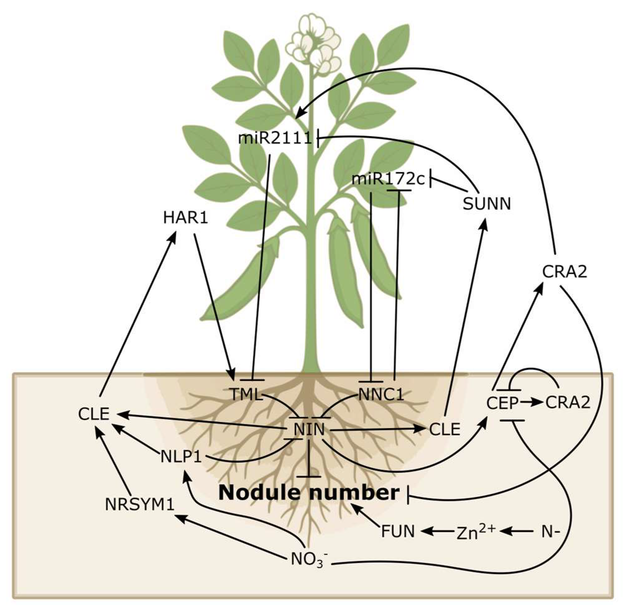
To maintain a balance between the carbon invested in the symbiosis and the nitrogen acquired—thus ensuring the stable growth and development of the host plant (macrosymbiont)—legumes exert stringent control over both the number and activity of nodules. A key regulatory mechanism is known as autoregulation of nodulation (AON), whereby the formation of initial nodules suppresses the development of additional nodules in the same root region. This process involves a long-distance signal exchange between the root and the shoot [124,125,126] (Scheme 3).
The autoregulation of nodulation (AON) system integrates both local root-level and systemic shoot–root signaling pathways to control nodule formation. It involves key components, including plant hormones, NIN-mediated recognition of rhizobia and nitrate (NO3), the negative regulatory SUNN pathway, the positive CEP-CRA2 pathway, and the miR2111/TML module.
Root nodule formation and symbiotic nitrogen fixation (SNF) are highly dependent on energy. Leguminous plants have developed complex regulatory mechanisms that allow them to adjust the intensity of these processes according to the amount of nitrogen available in the soil.
Nitrates (NO3), the primary form of assimilable nitrogen, enhance the action of AON by activating the expression of CLE peptides such as CLE35 in Medicago truncatula [127], as well as nitrate-dependent CLEs such as GmNIC1a and GmNIC1b in soybeans [128]. Nitrates also activate a signaling pathway dependent on NLP (NIN-like proteins) transcription factors such as NLP1, which bind to CLE and CEP gene promoters to limit nodulation and nitrogenase activity [127,128,129].
Recent studies have shown that nitrate signaling may utilize AON components, including CLE-RS2 and HAR1, as inhibitors of nodule development when mineral nitrogen levels are high [130,131]. This enables the plant to suppress costly symbiosis when soil nitrogen levels are sufficient for growth [132,133].
The CRA2 (Compact Root Architecture 2) receptor positively regulates nodule formation under nitrogen deficiency and also participates in AON in a nitrate-dependent manner by receiving CEP (C-terminally Encoded Peptide) signals such as CEP1 and CEP2. This optimizes the ratio between lateral roots and root hairs [134,135,136].
Under conditions of high nitrate concentration, NLP1 acts in antagonism to CRA2, thereby inhibiting CEP1 expression and limiting nodulation [129]. The final effect depends on the interactions between positive and negative regulators in the root and shoot.
Recent discoveries highlight the significant role of zinc as a second messenger in plants involved in regulating SNF efficiency. As an essential micronutrient, zinc serves as an intracellular signal in response to variation in soil nitrogen availability [137].
A FUN (Fixation Under Nitrate) mutant has been identified that maintains nitrogenase function and SNF activity even under conditions of high nitrogen availability. When soil nitrogen levels are low, zinc accumulates in the nodules, inducing the formation of inactive FUN protein filaments and thereby enabling efficient nitrogen fixation. Under high nitrogen (N) conditions, zinc content decreases, FUN is activated, and the expression of genes associated with SNF inhibition is regulated, accelerating nodule senescence [137].
High nitrate levels accelerate nodule senescence, leading to a decrease in nitrogenase activity and binding capacity. This phenomenon is associated with increased oxidative stress, loss of symbiont integrity, and reduced allocation of assimilates [138,139,140]. Key regulators of this process include the transcriptional factor LjNAC094, which acts downstream of NLP1/4, and SNAP factors (SANP1–4) in soybeans, which modify the expression of senescence genes and limit nitrogenase activity [141,142].
The nodulation autoregulation mechanism also involves root nodule senescence processes in response to high nitrate levels in the soil, which are modulated by transcription factors.
The senescence of nodules involves specific transcription factors, including proteins belonging to the bHLH and NAC families. These factors regulate the expression of genes associated with programmed cell death (PCD) and the accumulation of reactive oxygen species (ROS). This process leads to the degradation of nodule structures and a decrease in nitrogenase activity [143,144]. bHLH proteins, such as MrbHLH2, which was identified in M. truncatula root nodules, may act as negative regulators of root nodule senescence. They achieve this by inhibiting the expression of cysteine protease genes, such as MtCP77, which delays programmed cell death (PCD) and lessens oxidative stress [143]. On the other hand, NAC proteins have been found to promote root nodule senescence (for example, MtNAC969) or activate the expression of cysteine protease genes resulting in the degradation of nodule cells and accelerating their senescence [144,145].
Excess mineral nitrogen (especially in the form of nitrates) strongly inhibits nitrogen fixation and accelerates root nodule senescence. In Lotus japonicus, the transcription factor LjNAC094 has been shown to act downstream of the NLP1/NLP4 pathway, regulating the expression of senescence-associated genes (SAGs) via NAC-binding motifs [142].

9. Cyclophilins

Cyclophilins constitute a group of proteins with peptidyl-prolyl cistrans isomerase activity (PPIase) involved in the folding of target proteins (they catalyze the reaction in both directions) [146,147,148,149]. These isomerases belong to the family of immunophilins, which consists of two groups: cyclophilins (CyPs) and FK506-binding proteins (FKBPs) [150,151]. Another group of proteins with PPIase activity is parvulins, but they do not belong to the immunophilin family, as they are not sensitive to any specific immunosuppressive drug [152].
Many proline-rich (PRPs) and hydroxyproline-rich glycoproteins (HRGPs) are involved in the formation of root nodules [153,154,155,156]. Some of them are cell wall components that affect nodule formation and function. In situ hybridization experiments indicate that cyclophilin (CyPA) is highly expressed in meristematic tissues of Lupinus luteus, with the highest level observed in the nodule meristem zone [17]. The activity of the LlCyP promoter was analyzed in the model plant Lotus japonicus using site-directed mutagenesis [19]. In contrast to indeterminate nodules (L. luteus), the determinate nodules (L. luteus) do not have a strictly defined meristematic zone [157,158]. The activity of the LlCyP promoter was detected in root nodule parenchyma, which is probably related to the need for a large number of cyclophilins that catalyze isomerization around Xaa–Pro bonds during protein folding, enabling HRGPs and PRPs to attain their native structure [17]. Gene expression analysis revealed that, despite the presence of nodulin-specific cis elements in their promoter regions, only a few genes coding for L. japonicus cyclophilins are induced during the early stages of symbiosis. It concerns LjCYP19 (localized in cytoplasm) and LjCYP18 (localized in chloroplasts). Their expression in roots two days after infection with M. loti is twice as high as in the roots of plants grown axenically [159]. Cyclophilins localized in the nucleus, such as LjCYP7 and LjCYP20, as well as LjCYP34, which is localized in the endoplasmic reticulum, are also induced two days after infection. However, this induction is significantly lower. The cyclophilins LjCYP7, LjCYP19, LjCYP21, and LjCYP34 have different structures than their counterparts in Arabidopsis thaliana. However, such proteins can also be found in other plants belonging to the Fabaceae family, suggesting that they are specific to this family. The configurational change of the peptide bond (trans/cis) significantly affects the tertiary structure of the protein. It follows that peptidylprolyl isomerases play a key role in imparting specific properties to proline-rich polypeptides. Many proteins expressed during the development of the symbiotic system contain numerous proline residues in their structure. Proteins rich in proline and hydroxyproline are involved in the construction of the protective layer of cells of the parenchyma of the cortex of the nodule. Early nodulins are also characterized by a high content of proline residues. Therefore, proteins rich in proline play a crucial role in the formation and functioning of the symbiotic system. It can be assumed that cyclophilins play a crucial role in the process of diazotrophy.

Author Contributions

Conceptualization, K.N. and P.O.; writing—original draft preparation, K.N. and P.O. All authors have read and agreed to the published version of the manuscript.

Funding

This work was supported by the State Committee for Scientific Research (KBN) grant No. 3 P06A 037 25.

Data Availability Statement

No new data were created or analyzed in this study.

Conflicts of Interest

The authors declare no conflicts of interest.

References

  1. Aboagye, I.A.; Oba, M.; Koenig, K.M.; Zhao, G.Y.; Beauchemin, K.A. Use of Gallic Acid and Hydrolyzable Tannins to Reduce Methane Emission and Nitrogen Excretion in Beef Cattle Fed a Diet Containing Alfalfa Silage. J. Anim. Sci. 2019, 97, 2230–2244. [Google Scholar] [CrossRef] [PubMed]
  2. Adrain, C.; Slee, E.A.; Harte, M.T.; Martin, S.J. Regulation of Apoptotic Protease Activating Factor-1 Oligomerization and Apoptosis by the WD-40 Repeat Region. J. Biol. Chem. 1999, 274, 20855–20860. [Google Scholar] [CrossRef]
  3. Ahn, J.C.; Kim, D.-W.; You, Y.N.; Seok, M.S.; Park, J.M.; Hwang, H.; Kim, B.-G.; Luan, S.; Park, H.-S.; Cho, H.S. Classification of Rice (Oryza sativa L. japonica Nipponbare) Immunophilins (FKBPs, CYPs) and Expression Patterns under Water Stress. BMC Plant Biol. 2010, 10, 253. [Google Scholar] [CrossRef]
  4. Stewart, D.E.; Sarkar, A.; Wampler, J.E. Occurrence and Role of Cis Peptide Bonds in Protein Structures. J. Mol. Biol. 1990, 214, 253–260. [Google Scholar] [CrossRef]
  5. Cheng, H.N.; Bovey, F.A. Cis-Trans Equilibrium and Kinetic Studies of Acetyl-L-Proline and Glycyl-L-Proline. Biopolymers 1977, 16, 1465–1472. [Google Scholar] [CrossRef] [PubMed]
  6. Hanes, S.D. The Ess1 Prolyl Isomerase: Traffic Cop of the RNA Polymerase II Transcription Cycle. Biochim. Biophys. Acta 2014, 1839, 316–333. [Google Scholar] [CrossRef] [PubMed]
  7. Lu, K.P.; Hanes, S.D.; Hunter, T. A Human Peptidyl-Prolyl Isomerase Essential for Regulation of Mitosis. Nature 1996, 380, 544–547. [Google Scholar] [CrossRef]
  8. Joseph, J.D.; Yeh, E.S.; Swenson, K.I.; Means, A.R. Winkler, null The Peptidyl-Prolyl Isomerase Pin1. Prog. Cell Cycle Res. 2003, 5, 477–487. [Google Scholar]
  9. Hirsch, A.M.; Larue, T.A.; Doyle, J. Is the Legume Nodule a Modified Root or Stem or an Organ Sui Generis? Crit. Rev. Plant Sci. 1997, 16, 361–392. [Google Scholar] [CrossRef]
  10. Arévalo-Rodríguez, M.; Heitman, J. Cyclophilin A Is Localized to the Nucleus and Controls Meiosis in Saccharomyces Cerevisiae. Eukaryot. Cell 2005, 4, 17–29. [Google Scholar] [CrossRef]
  11. Andreeva, L.; Heads, R.; Green, C.J. Cyclophilins and Their Possible Role in the Stress Response. Int. J. Exp. Pathol. 1999, 80, 305–315. [Google Scholar] [CrossRef]
  12. Lin, D.-T.; Lechleiter, J.D. Mitochondrial Targeted Cyclophilin D Protects Cells from Cell Death by Peptidyl Prolyl Isomerization. J. Biol. Chem. 2002, 277, 31134–31141. [Google Scholar] [CrossRef]
  13. Pushkarsky, T.; Yurchenko, V.; Vanpouille, C.; Brichacek, B.; Vaisman, I.; Hatakeyama, S.; Nakayama, K.I.; Sherry, B.; Bukrinsky, M.I. Cell Surface Expression of CD147/EMMPRIN Is Regulated by Cyclophilin 60. J. Biol. Chem. 2005, 280, 27866–27871. [Google Scholar] [CrossRef] [PubMed]
  14. Nuc, K.; Lesniewicz, K.; Nuc, P.; Slomski, R. Yellow Lupine Cyclophilin Interacts with Nucleic Acids. Protein Pept. Lett. 2008, 15, 719–723. [Google Scholar] [CrossRef]
  15. Trivedi, D.K.; Ansari, M.W.; Tuteja, N. Multiple Abiotic Stress Responsive Rice Cyclophilin: (OsCYP-25) Mediates a Wide Range of Cellular Responses. Commun. Integr. Biol. 2013, 6, e25260. [Google Scholar] [CrossRef] [PubMed]
  16. Bannikova, O.; Zywicki, M.; Marquez, Y.; Skrahina, T.; Kalyna, M.; Barta, A. Identification of RNA Targets for the Nuclear Multidomain Cyclophilin atCyp59 and Their Effect on PPIase Activity. Nucleic Acids Res. 2013, 41, 1783–1796. [Google Scholar] [CrossRef] [PubMed]
  17. Nuc, K.; Nuc, P.; Słomski, R. Yellow Lupine Cyclophilin Transcripts Are Highly Accumulated in the Nodule Meristem Zone. Mol. Plant Microbe Interact. 2001, 14, 1384–1394. [Google Scholar] [CrossRef]
  18. Trupkin, S.A.; Mora-García, S.; Casal, J.J. The Cyclophilin ROC1 Links Phytochrome and Cryptochrome to Brassinosteroid Sensitivity. Plant J. 2012, 71, 712–723. [Google Scholar] [CrossRef]
  19. Nuc, K.; Olejnik, P.; Samardakiewicz, M.; Nuc, P. Functional Analysis of the Lupinus Luteus Cyclophilin Gene Promoter Region in Lotus japonicus. Agriculture 2021, 11, 435. [Google Scholar] [CrossRef]
  20. Aloui, A.; Recorbet, G.; Gollotte, A.; Robert, F.; Valot, B.; Gianinazzi-Pearson, V.; Aschi-Smiti, S.; Dumas-Gaudot, E. On the Mechanisms of Cadmium Stress Alleviation in Medicago truncatula by Arbuscular Mycorrhizal Symbiosis: A Root Proteomic Study. Proteomics 2009, 9, 420–433. [Google Scholar] [CrossRef]
  21. Gullerova, M.; Barta, A.; Lorkovic, Z.J. AtCyp59 Is a Multidomain Cyclophilin from Arabidopsis thaliana That Interacts with SR Proteins and the C-Terminal Domain of the RNA Polymerase II. RNA 2006, 12, 631–643. [Google Scholar] [CrossRef] [PubMed]
  22. Poole, P.; Ramachandran, V.; Terpolilli, J. Rhizobia: From Saprophytes to Endosymbionts. Nat. Rev. Microbiol. 2018, 16, 291–303. [Google Scholar] [CrossRef]
  23. Mitsch, M.J.; diCenzo, G.C.; Cowie, A.; Finan, T.M. Succinate Transport Is Not Essential for Symbiotic Nitrogen Fixation by Sinorhizobium meliloti or Rhizobium leguminosarum. Appl. Environ. Microbiol. 2017, 84, e01561-17. [Google Scholar] [CrossRef]
  24. Aroney, S.T.N.; Poole, P.S.; Sánchez-Cañizares, C. Rhizobial Chemotaxis and Motility Systems at Work in the Soil. Front. Plant Sci. 2021, 12. [Google Scholar] [CrossRef] [PubMed]
  25. Compton, K.K.; Scharf, B.E. Rhizobial Chemoattractants, the Taste and Preferences of Legume Symbionts. Front. Plant Sci. 2021, 12, 686465. [Google Scholar] [CrossRef]
  26. Ji, Y.-Y.; Zhang, B.; Zhang, P.; Chen, L.-C.; Si, Y.-W.; Wan, X.-Y.; Li, C.; Wang, R.-H.; Tian, Y.; Zhang, Z.; et al. Rhizobial Migration toward Roots Mediated by FadL-ExoFQP Modulation of Extracellular Long-Chain AHLs. ISME J. 2023, 17, 417–431. [Google Scholar] [CrossRef] [PubMed]
  27. Roy, S.; Liu, W.; Nandety, R.S.; Crook, A.; Mysore, K.S.; Pislariu, C.I.; Frugoli, J.; Dickstein, R.; Udvardi, M.K. Celebrating 20 Years of Genetic Discoveries in Legume Nodulation and Symbiotic Nitrogen Fixation. Plant Cell 2020, 32, 15–41. [Google Scholar] [CrossRef]
  28. Biała-Leonhard, W.; Zanin, L.; Gottardi, S.; de Brito Francisco, R.; Venuti, S.; Valentinuzzi, F.; Mimmo, T.; Cesco, S.; Bassin, B.; Martinoia, E.; et al. Identification of an Isoflavonoid Transporter Required for the Nodule Establishment of the Rhizobium-Fabaceae Symbiotic Interaction. Front. Plant Sci. 2021, 12, 758213. [Google Scholar] [CrossRef]
  29. Sugiyama, A.; Shitan, N.; Yazaki, K. Involvement of a Soybean ATP-Binding Cassette-Type Transporter in the Secretion of Genistein, a Signal Flavonoid in Legume-Rhizobium Symbiosis. Plant Physiol. 2007, 144, 2000–2008. [Google Scholar] [CrossRef]
  30. Berillo, D.; Kozhahmetova, M.; Lebedeva, L. Overview of the Biological Activity of Anthraquinons and Flavanoids of the Plant Rumex Species. Molecules 2022, 27, 1204. [Google Scholar] [CrossRef]
  31. Liu, C.-W.; Murray, J.D. The Role of Flavonoids in Nodulation Host-Range Specificity: An Update. Plants 2016, 5, 33. [Google Scholar] [CrossRef] [PubMed]
  32. Weston, L.A.; Mathesius, U. Flavonoids: Their Structure, Biosynthesis and Role in the Rhizosphere, Including Allelopathy. J. Chem. Ecol. 2013, 39, 283–297. [Google Scholar] [CrossRef]
  33. Cooper, J.E. Early Interactions between Legumes and Rhizobia: Disclosing Complexity in a Molecular Dialogue. J. Appl. Microbiol. 2007, 103, 1355–1365. [Google Scholar] [CrossRef]
  34. Skorupska, A.; Wielbo, J.; Kidaj, D.; Marek-Kozaczuk, M. Enhancing Rhizobium–Legume Symbiosis Using Signaling Factors. In Microbes for Legume Improvement; Khan, M.S., Musarrat, J., Zaidi, A., Eds.; Springer: Vienna, Austria, 2010; pp. 27–54. ISBN 978-3-211-99753-6. [Google Scholar]
  35. Shimamura, M.; Kumaki, T.; Hashimoto, S.; Saeki, K.; Ayabe, S.-I.; Higashitani, A.; Akashi, T.; Sato, S.; Aoki, T. Phenolic Acids Induce Nod Factor Production in Lotus japonicus-Mesorhizobium Symbiosis. Microbes Environ. 2022, 37, ME21094. [Google Scholar] [CrossRef] [PubMed]
  36. Yang, J.; Lan, L.; Jin, Y.; Yu, N.; Wang, D.; Wang, E. Mechanisms Underlying Legume–Rhizobium Symbioses. J. Integr. Plant Biol. 2022, 64, 244–267. [Google Scholar] [CrossRef]
  37. Mbengue, M.D.; Hervé, C.; Debellé, F. Nod Factor Signaling in Symbiotic Nodulation. In Advances in Botanical Research; Frendo, P., Frugier, F., Masson-Boivin, C., Eds.; Regulation of Nitrogen-Fixing Symbioses in Legumes; Academic Press: Cambridge, MA, USA, 2020; Volume 94, pp. 1–39. [Google Scholar]
  38. Bag, S.; Mondal, A.; Majumder, A.; Mondal, S.K.; Banik, A. Flavonoid Mediated Selective Cross-Talk between Plants and Beneficial Soil Microbiome. Phytochem. Rev. 2022, 21, 1739–1760. [Google Scholar] [CrossRef] [PubMed]
  39. Limpens, E.; van Zeijl, A.; Geurts, R. Lipochitooligosaccharides Modulate Plant Host Immunity to Enable Endosymbioses. Annu. Rev. Phytopathol. 2015, 53, 311–334. [Google Scholar] [CrossRef]
  40. Ghantasala, S.; Roy Choudhury, S. Nod Factor Perception: An Integrative View of Molecular Communication during Legume Symbiosis. Plant Mol. Biol. 2022, 110, 485–509. [Google Scholar] [CrossRef]
  41. Nelson, M.S.; Sadowsky, M.J. Secretion Systems and Signal Exchange between Nitrogen-Fixing Rhizobia and Legumes. Front. Plant Sci. 2015, 6, 491. [Google Scholar] [CrossRef]
  42. Downie, J.A. The Roles of Extracellular Proteins, Polysaccharides and Signals in the Interactions of Rhizobia with Legume Roots. FEMS Microbiol. Rev. 2010, 34, 150–170. [Google Scholar] [CrossRef]
  43. Perret, X.; Staehelin, C.; Broughton, W.J. Molecular Basis of Symbiotic Promiscuity. Microbiol. Mol. Biol. Rev. 2000, 64, 180–201. [Google Scholar] [CrossRef] [PubMed]
  44. Zipfel, C.; Oldroyd, G.E.D. Plant Signalling in Symbiosis and Immunity. Nature 2017, 543, 328–336. [Google Scholar] [CrossRef] [PubMed]
  45. Bek, A.S.; Sauer, J.; Thygesen, M.B.; Duus, J.Ø.; Petersen, B.O.; Thirup, S.; James, E.; Jensen, K.J.; Stougaard, J.; Radutoiu, S. Improved Characterization of Nod Factors and Genetically Based Variation in LysM Receptor Domains Identify Amino Acids Expendable for Nod Factor Recognition in Lotus Spp. Mol. Plant Microbe Interact. 2010, 23, 58–66. [Google Scholar] [CrossRef] [PubMed]
  46. Geurts, R.; Bisseling, T. Rhizobium Nod Factor Perception and Signalling. Plant Cell 2002, 14, S239–S249. [Google Scholar] [CrossRef]
  47. Oldroyd, G.E.D.; Murray, J.D.; Poole, P.S.; Downie, J.A. The Rules of Engagement in the Legume-Rhizobial Symbiosis. Annu. Rev. Genet. 2011, 45, 119–144. [Google Scholar] [CrossRef]
  48. Bozsoki, Z.; Cheng, J.; Feng, F.; Gysel, K.; Vinther, M.; Andersen, K.R.; Oldroyd, G.; Blaise, M.; Radutoiu, S.; Stougaard, J. Receptor-Mediated Chitin Perception in Legume Roots Is Functionally Separable from Nod Factor Perception. Proc. Natl. Acad. Sci. USA 2017, 114, E8118–E8127. [Google Scholar] [CrossRef]
  49. Murakami, E.; Cheng, J.; Gysel, K.; Bozsoki, Z.; Kawaharada, Y.; Hjuler, C.T.; Sørensen, K.K.; Tao, K.; Kelly, S.; Venice, F.; et al. Epidermal LysM Receptor Ensures Robust Symbiotic Signalling in Lotus japonicus. eLife 2018, 7, e33506. [Google Scholar] [CrossRef]
  50. Zhou, N.; Li, X.; Zheng, Z.; Liu, J.; Downie, J.A.; Xie, F. RinRK1 Enhances NF Receptors Accumulation in Nanodomain-like Structures at Root-Hair Tip. Nat. Commun. 2024, 15, 3568. [Google Scholar] [CrossRef] [PubMed]
  51. Skorupska, A.; Janczarek, M.; Marczak, M.; Mazur, A.; Król, J. Rhizobial Exopolysaccharides: Genetic Control and Symbiotic Functions. Microb. Cell Fact. 2006, 5, 7. [Google Scholar] [CrossRef]
  52. Fraysse, N.; Couderc, F.; Poinsot, V. Surface Polysaccharide Involvement in Establishing the Rhizobium–Legume Symbiosis. Eur. J. Biochem. 2003, 270, 1365–1380. [Google Scholar] [CrossRef]
  53. Acosta-Jurado, S.; Fuentes-Romero, F.; Ruiz-Sainz, J.-E.; Janczarek, M.; Vinardell, J.-M. Rhizobial Exopolysaccharides: Genetic Regulation of Their Synthesis and Relevance in Symbiosis with Legumes. Int. J. Mol. Sci. 2021, 22, 6233. [Google Scholar] [CrossRef]
  54. Kawaharada, Y.; Kelly, S.; Nielsen, M.W.; Hjuler, C.T.; Gysel, K.; Muszyński, A.; Carlson, R.W.; Thygesen, M.B.; Sandal, N.; Asmussen, M.H.; et al. Receptor-Mediated Exopolysaccharide Perception Controls Bacterial Infection. Nature 2015, 523, 308–312. [Google Scholar] [CrossRef] [PubMed]
  55. Kawaharada, Y.; Nielsen, M.W.; Kelly, S.; James, E.K.; Andersen, K.R.; Rasmussen, S.R.; Füchtbauer, W.; Madsen, L.H.; Heckmann, A.B.; Radutoiu, S.; et al. Differential Regulation of the Epr3 Receptor Coordinates Membrane-Restricted Rhizobial Colonization of Root Nodule Primordia. Nat. Commun. 2017, 8, 14534. [Google Scholar] [CrossRef]
  56. Madsen, L.H.; Tirichine, L.; Jurkiewicz, A.; Sullivan, J.T.; Heckmann, A.B.; Bek, A.S.; Ronson, C.W.; James, E.K.; Stougaard, J. The Molecular Network Governing Nodule Organogenesis and Infection in the Model Legume Lotus japonicus. Nat. Commun. 2010, 1, 10. [Google Scholar] [CrossRef] [PubMed]
  57. Sieberer, B.J.; Chabaud, M.; Timmers, A.C.; Monin, A.; Fournier, J.; Barker, D.G. A Nuclear-Targeted Cameleon Demonstrates Intranuclear Ca2+ Spiking in Medicago truncatula Root Hairs in Response to Rhizobial Nodulation Factors. Plant Physiol. 2009, 151, 1197–1206. [Google Scholar] [CrossRef] [PubMed]
  58. Kim, S.; Zeng, W.; Bernard, S.; Liao, J.; Venkateshwaran, M.; Ane, J.-M.; Jiang, Y. Ca2+-Regulated Ca2+ Channels with an RCK Gating Ring Control Plant Symbiotic Associations. Nat. Commun. 2019, 10, 3703. [Google Scholar] [CrossRef]
  59. Kanamori, N.; Madsen, L.H.; Radutoiu, S.; Frantescu, M.; Quistgaard, E.M.H.; Miwa, H.; Downie, J.A.; James, E.K.; Felle, H.H.; Haaning, L.L.; et al. A Nucleoporin Is Required for Induction of Ca2+ Spiking in Legume Nodule Development and Essential for Rhizobial and Fungal Symbiosis. Proc. Natl. Acad. Sci. USA 2006, 103, 359–364. [Google Scholar] [CrossRef]
  60. Saito, K.; Yoshikawa, M.; Yano, K.; Miwa, H.; Uchida, H.; Asamizu, E.; Sato, S.; Tabata, S.; Imaizumi-Anraku, H.; Umehara, Y.; et al. NUCLEOPORIN85 Is Required for Calcium Spiking, Fungal and Bacterial Symbioses, and Seed Production in Lotus japonicus. Plant Cell 2007, 19, 610–624. [Google Scholar] [CrossRef]
  61. Yokota, K.; Fukai, E.; Madsen, L.H.; Jurkiewicz, A.; Rueda, P.; Radutoiu, S.; Held, M.; Hossain, M.S.; Szczyglowski, K.; Morieri, G.; et al. Rearrangement of Actin Cytoskeleton Mediates Invasion of Lotus japonicus Roots by Mesorhizobium Loti. Plant Cell 2009, 21, 267–284. [Google Scholar] [CrossRef]
  62. Yano, K.; Shibata, S.; Chen, W.-L.; Sato, S.; Kaneko, T.; Jurkiewicz, A.; Sandal, N.; Banba, M.; Imaizumi-Anraku, H.; Kojima, T.; et al. CERBERUS, a Novel U-Box Protein Containing WD-40 Repeats, Is Required for Formation of the Infection Thread and Nodule Development in the Legume-Rhizobium Symbiosis. Plant J. 2009, 60, 168–180. [Google Scholar] [CrossRef]
  63. Ibáñez, F.; Wall, L.; Fabra, A. Starting Points in Plant-Bacteria Nitrogen-Fixing Symbioses: Intercellular Invasion of the Roots. J. Exp. Bot. 2017, 68, 1905–1918. [Google Scholar] [CrossRef]
  64. Quilbé, J.; Montiel, J.; Arrighi, J.-F.; Stougaard, J. Molecular Mechanisms of Intercellular Rhizobial Infection: Novel Findings of an Ancient Process. Front. Plant Sci. 2022, 13, 922982. [Google Scholar] [CrossRef]
  65. Liu, J.; Liu, M.X.; Qiu, L.P.; Xie, F. SPIKE1 Activates the GTPase ROP6 to Guide the Polarized Growth of Infection Threads in Lotus japonicus. Plant Cell 2020, 32, 3774–3791. [Google Scholar] [CrossRef]
  66. Luo, Z.; Liu, H.; Xie, F. Cellular and Molecular Basis of Symbiotic Nodule Development. Curr. Opin. Plant Biol. 2023, 76, 102478. [Google Scholar] [CrossRef]
  67. Su, C. Pectin Modifications at the Symbiotic Interface. New Phytol. 2023, 238, 25–32. [Google Scholar] [CrossRef]
  68. Su, C.; Zhang, G.; Rodriguez-Franco, M.; Hinnenberg, R.; Wietschorke, J.; Liang, P.; Yang, W.; Uhler, L.; Li, X.; Ott, T. Transcellular Progression of Infection Threads in Medicago truncatula Roots Is Associated with Locally Confined Cell Wall Modifications. Curr. Biol. 2023, 33, 533–542.e5. [Google Scholar] [CrossRef]
  69. Xie, F.; Murray, J.D.; Kim, J.; Heckmann, A.B.; Edwards, A.; Oldroyd, G.E.D.; Downie, J.A. Legume Pectate Lyase Required for Root Infection by Rhizobia. Proc. Natl. Acad. Sci. USA 2012, 109, 633–638. [Google Scholar] [CrossRef]
  70. Tsyganova, A.V.; Brewin, N.J.; Tsyganov, V.E. Structure and Development of the Legume-Rhizobial Symbiotic Interface in Infection Threads. Cells 2021, 10, 1050. [Google Scholar] [CrossRef]
  71. Fournier, J.; Teillet, A.; Chabaud, M.; Ivanov, S.; Genre, A.; Limpens, E.; de Carvalho-Niebel, F.; Barker, D.G. Remodeling of the Infection Chamber before Infection Thread Formation Reveals a Two-Step Mechanism for Rhizobial Entry into the Host Legume Root Hair. Plant Physiol. 2015, 167, 1233–1242. [Google Scholar] [CrossRef]
  72. Liu, C.-W.; Breakspear, A.; Stacey, N.; Findlay, K.; Nakashima, J.; Ramakrishnan, K.; Liu, M.; Xie, F.; Endre, G.; de Carvalho-Niebel, F.; et al. A Protein Complex Required for Polar Growth of Rhizobial Infection Threads. Nat. Commun. 2019, 10, 2848. [Google Scholar] [CrossRef]
  73. Gao, J.-P.; Jiang, S.; Su, Y.; Xu, P.; Wang, J.; Liang, W.; Liu, C.-W.; Murray, J.D. Intracellular Infection by Symbiotic Bacteria Requires the Mitotic Kinase AURORA1. Proc. Natl. Acad. Sci. USA 2022, 119, e2202606119. [Google Scholar] [CrossRef]
  74. Fournier, J.; Timmers, A.C.J.; Sieberer, B.J.; Jauneau, A.; Chabaud, M.; Barker, D.G. Mechanism of Infection Thread Elongation in Root Hairs of Medicago truncatula and Dynamic Interplay with Associated Rhizobial Colonization. Plant Physiol. 2008, 148, 1985–1995. [Google Scholar] [CrossRef]
  75. Deng, J.-L.; Zhao, L.; Wei, H.; Ye, H.-X.; Yang, L.; Sun, L.; Zhao, Z.; Murray, J.D.; Liu, C.-W. A Deeply Conserved Amino Acid Required for VAPYRIN Localization and Function during Legume–Rhizobial Symbiosis. New Phytol. 2024, 243, 14–22. [Google Scholar] [CrossRef]
  76. Liu, M.; Jia, N.; Li, X.; Liu, R.; Xie, Q.; Murray, J.D.; Downie, J.A.; Xie, F. CERBERUS Is Critical for Stabilization of VAPYRIN during Rhizobial Infection in Lotus japonicus. New Phytol. 2021, 229, 1684–1700. [Google Scholar] [CrossRef]
  77. Genre, A.; Timmers, T. The Symbiotic Role of the Actin Filament Cytoskeleton. New Phytol. 2019, 221, 611–613. [Google Scholar] [CrossRef]
  78. Jhu, M.-Y.; Oldroyd, G.E.D. Dancing to a Different Tune, Can We Switch from Chemical to Biological Nitrogen Fixation for Sustainable Food Security? PLOS Biol. 2023, 21, e3001982. [Google Scholar] [CrossRef]
  79. Wolf, S. Cell Wall Signaling in Plant Development and Defense. Annu. Rev. Plant Biol. 2022, 73, 323–353. [Google Scholar] [CrossRef]
  80. Medina-Puche, L.; Lozano-Durán, R. Plasma Membrane-to-Organelle Communication in Plant Stress Signaling. Curr. Opin. Plant Biol. 2022, 69, 102269. [Google Scholar] [CrossRef]
  81. Zeng, Y.; Liang, Z.; Liu, Z.; Li, B.; Cui, Y.; Gao, C.; Shen, J.; Wang, X.; Zhao, Q.; Zhuang, X.; et al. Recent Advances in Plant Endomembrane Research and New Microscopical Techniques. New Phytol. 2023, 240, 41–60. [Google Scholar] [CrossRef]
  82. Cai, G.; Ahmed, M.A. The Role of Root Hairs in Water Uptake: Recent Advances and Future Perspectives. J. Exp. Bot. 2022, 73, 3330–3338. [Google Scholar] [CrossRef]
  83. Pei, W.; Du, F.; Zhang, Y.; He, T.; Ren, H. Control of the Actin Cytoskeleton in Root Hair Development. Plant Sci. 2012, 187, 10–18. [Google Scholar] [CrossRef] [PubMed]
  84. Zepeda, I.; Sánchez-López, R.; Kunkel, J.G.; Bañuelos, L.A.; Hernández-Barrera, A.; Sánchez, F.; Quinto, C.; Cárdenas, L. Visualization of Highly Dynamic F-Actin Plus Ends in Growing Phaseolus Vulgaris Root Hair Cells and Their Responses to Rhizobium Etli Nod Factors. Plant Cell Physiol. 2014, 55, 580–592. [Google Scholar] [CrossRef] [PubMed]
  85. Liang, P.; Schmitz, C.; Lace, B.; Ditengou, F.A.; Su, C.; Schulze, E.; Knerr, J.; Grosse, R.; Keller, J.; Libourel, C.; et al. Formin-Mediated Bridging of Cell Wall, Plasma Membrane, and Cytoskeleton in Symbiotic Infections of Medicago truncatula. Curr. Biol. 2021, 31, 2712–2719.e5. [Google Scholar] [CrossRef]
  86. Su, C.; Klein, M.-L.; Hernández-Reyes, C.; Batzenschlager, M.; Ditengou, F.A.; Lace, B.; Keller, J.; Delaux, P.-M.; Ott, T. The Medicago truncatula DREPP Protein Triggers Microtubule Fragmentation in Membrane Nanodomains during Symbiotic Infections. Plant Cell 2020, 32, 1689–1702. [Google Scholar] [CrossRef] [PubMed]
  87. Harrison, M.J.; Ivanov, S. Exocytosis for Endosymbiosis: Membrane Trafficking Pathways for Development of Symbiotic Membrane Compartments. Curr. Opin. Plant Biol. 2017, 38, 101–108. [Google Scholar] [CrossRef]
  88. Kondorosi, E.; Mergaert, P.; Kereszt, A. A Paradigm for Endosymbiotic Life: Cell Differentiation of Rhizobium Bacteria Provoked by Host Plant Factors. Annu. Rev. Microbiol. 2013, 67, 611–628. [Google Scholar] [CrossRef]
  89. Oono, R.; Schmitt, I.; Sprent, J.I.; Denison, R.F. Multiple Evolutionary Origins of Legume Traits Leading to Extreme Rhizobial Differentiation. New Phytol. 2010, 187, 508–520. [Google Scholar] [CrossRef]
  90. Mergaert, P.; Uchiumi, T.; Alunni, B.; Evanno, G.; Cheron, A.; Catrice, O.; Mausset, A.-E.; Barloy-Hubler, F.; Galibert, F.; Kondorosi, A.; et al. Eukaryotic Control on Bacterial Cell Cycle and Differentiation in the Rhizobium-Legume Symbiosis. Proc. Natl. Acad. Sci. USA 2006, 103, 5230–5235. [Google Scholar] [CrossRef]
  91. Kereszt, A.; Mergaert, P.; Kondorosi, E. Bacteroid Development in Legume Nodules: Evolution of Mutual Benefit or of Sacrificial Victims? Mol. Plant Microbe Interact. 2011, 24, 1300–1309. [Google Scholar] [CrossRef]
  92. Montiel, J.; Downie, J.A.; Farkas, A.; Bihari, P.; Herczeg, R.; Bálint, B.; Mergaert, P.; Kereszt, A.; Kondorosi, É. Morphotype of Bacteroids in Different Legumes Correlates with the Number and Type of Symbiotic NCR Peptides. Proc. Natl. Acad. Sci. USA 2017, 114, 5041–5046. [Google Scholar] [CrossRef]
  93. Van de Velde, W.; Guerra, J.C.P.; Keyser, A.D.; De Rycke, R.; Rombauts, S.; Maunoury, N.; Mergaert, P.; Kondorosi, E.; Holsters, M.; Goormachtig, S. Aging in Legume Symbiosis. A Molecular View on Nodule Senescence in Medicago truncatula. Plant Physiol. 2006, 141, 711–720. [Google Scholar] [CrossRef]
  94. Alunni, B.; Kevei, Z.; Redondo-Nieto, M.; Kondorosi, A.; Mergaert, P.; Kondorosi, E. Genomic Organization and Evolutionary Insights on GRP and NCR Genes, Two Large Nodule-Specific Gene Families in Medicago truncatula. Mol. Plant Microbe Interact. 2007, 20, 1138–1148. [Google Scholar] [CrossRef]
  95. Czernic, P.; Gully, D.; Cartieaux, F.; Moulin, L.; Guefrachi, I.; Patrel, D.; Pierre, O.; Fardoux, J.; Chaintreuil, C.; Nguyen, P.; et al. Convergent Evolution of Endosymbiont Differentiation in Dalbergioid and Inverted Repeat-Lacking Clade Legumes Mediated by Nodule-Specific Cysteine-Rich Peptides. Plant Physiol. 2015, 169, 1254–1265. [Google Scholar] [CrossRef] [PubMed]
  96. Farkas, A.; Maróti, G.; Dürgő, H.; Györgypál, Z.; Lima, R.M.; Medzihradszky, K.F.; Kereszt, A.; Mergaert, P.; Kondorosi, É. Medicago truncatula Symbiotic Peptide NCR247 Contributes to Bacteroid Differentiation through Multiple Mechanisms. Proc. Natl. Acad. Sci. USA 2014, 111, 5183–5188. [Google Scholar] [CrossRef] [PubMed]
  97. Kirkpatrick, C.L.; Viollier, P.H. New(s) to the (Z-)Ring. Curr. Opin. Microbiol. 2011, 14, 691–697. [Google Scholar] [CrossRef] [PubMed]
  98. de Bruijn, F.J. Structure of Indeterminate Medicago truncatula Nodules: Introduction. In The Model Legume Medicago truncatula; John Wiley & Sons, Ltd.: Hoboken, NJ, USA, 2020; pp. 706–707. ISBN 978-1-119-40914-4. [Google Scholar]
  99. Monahan-Giovanelli, H.; Pinedo, C.A.; Gage, D.J. Architecture of Infection Thread Networks in Developing Root Nodules Induced by the Symbiotic Bacterium Sinorhizobium meliloti on Medicago truncatula. Plant Physiol. 2006, 140, 661–670. [Google Scholar] [CrossRef]
  100. Timmers, A.C.; Soupène, E.; Auriac, M.C.; de Billy, F.; Vasse, J.; Boistard, P.; Truchet, G. Saprophytic Intracellular Rhizobia in Alfalfa Nodules. Mol. Plant Microbe Interact. 2000, 13, 1204–1213. [Google Scholar] [CrossRef]
  101. Vasse, J.; de Billy, F.; Camut, S.; Truchet, G. Correlation between Ultrastructural Differentiation of Bacteroids and Nitrogen Fixation in Alfalfa Nodules. J. Bacteriol. 1990, 172, 4295–4306. [Google Scholar] [CrossRef]
  102. Newcomb, W.; Sippell, D.; Peterson, R.L. The Early Morphogenesis of Glycine Max and Pisum Sativum Root Nodules. Can. J. Bot. 1979, 57, 2603–2616. [Google Scholar] [CrossRef]
  103. Calvert, H.E.; Pence, M.K.; Pierce, M.; Malik, N.S.A.; Bauer, W.D. Anatomical Analysis of the Development and Distribution of Rhizobium Infections in Soybean Roots. Can. J. Bot. = J. Can. De. Bot. 1984, 62, 2375–2384. [Google Scholar] [CrossRef]
  104. Mathews, A.; Carroll, B.J.; Gresshoff, P.M. Development of Bradyrhizobium Infections in Supernodulating and Non-Nodulating Mutants of Soybean (Glycine Max [L.] Merrill). Protoplasma 1989, 150, 40–47. [Google Scholar] [CrossRef]
  105. Turgeon, B.G.; Bauer, W.D. Early Events in the Infection of Soybean by Rhizobium Japonicum. Time Course and Cytology of the Initial Infection Process. Can. J. Bot. 1982, 60, 152–161. [Google Scholar] [CrossRef]
  106. Rolfe, B.G.; Gresshoff, P.M. Genetic Analysis of Legume Nodule Initiation. Annu. Rev. Plant Physiol. Plant Mol. Biol. 1988, 39, 297–319. [Google Scholar] [CrossRef]
  107. Kazmierczak, T.; Yang, L.; Boncompagni, E.; Meilhoc, E.; Frugier, F.; Frendo, P.; Bruand, C.; Gruber, V.; Brouquisse, R. Chapter Seven–Legume Nodule Senescence: A Coordinated Death Mechanism between Bacteria and Plant Cells. In Advances in Botanical Research; Frendo, P., Frugier, F., Masson-Boivin, C., Eds.; Regulation of Nitrogen-Fixing Symbioses in Legumes; Academic Press: Cambridge, MA, USA, 2020; Volume 94, pp. 181–212. [Google Scholar]
  108. Day, D.A.; Poole, P.S.; Tyerman, S.D.; Rosendahl, L. Ammonia and Amino Acid Transport across Symbiotic Membranes in Nitrogen-Fixing Legume Nodules. Cell Mol. Life Sci. 2001, 58, 61–71. [Google Scholar] [CrossRef] [PubMed]
  109. Gibson, K.E.; Kobayashi, H.; Walker, G.C. Molecular Determinants of a Symbiotic Chronic Infection. Annu. Rev. Genet. 2008, 42, 413–441. [Google Scholar] [CrossRef]
  110. White, J.; Prell, J.; James, E.K.; Poole, P. Nutrient Sharing between Symbionts. Plant Physiol. 2007, 144, 604–614. [Google Scholar] [CrossRef]
  111. Egener, T.; Martin, D.E.; Sarkar, A.; Reinhold-Hurek, B. Role of a Ferredoxin Gene Cotranscribed with the nifHDK Operon in N(2) Fixation and Nitrogenase “Switch-off” of Azoarcus Sp. Strain BH72. J. Bacteriol. 2001, 183, 3752–3760. [Google Scholar] [CrossRef]
  112. Ledbetter, R.N.; Garcia Costas, A.M.; Lubner, C.E.; Mulder, D.W.; Tokmina-Lukaszewska, M.; Artz, J.H.; Patterson, A.; Magnuson, T.S.; Jay, Z.J.; Duan, H.D.; et al. The Electron Bifurcating FixABCX Protein Complex from Azotobacter Vinelandii: Generation of Low-Potential Reducing Equivalents for Nitrogenase Catalysis. Biochemistry 2017, 56, 4177–4190. [Google Scholar] [CrossRef]
  113. Dixon, R.O.D.; Wheeler, C.T. Nitrogen Fixation in Plants; Blackie Chapman and Hall: Glasgow, NY, USA, 1986; ISBN 978-0-412-01381-2. [Google Scholar]
  114. Igarashi, R.Y.; Seefeldt, L.C. Nitrogen Fixation: The Mechanism of the Mo-Dependent Nitrogenase. Crit. Rev. Biochem. Mol. Biol. 2003, 38, 351–384. [Google Scholar] [CrossRef]
  115. Minchin, F.R. Regulation of Oxygen Diffusion in Legume Nodules. Soil. Biol. Biochem. 1997, 29, 881–888. [Google Scholar] [CrossRef]
  116. Tjepkema, J.D.; Yocum, C.S. Measurement of Oxygen Partial Pressure within Soybean Nodules by Oxygen Microelectrodes. Planta 1974, 119, 351–360. [Google Scholar] [CrossRef]
  117. Minguillón, S.; Román, Á.; Pérez-Rontomé, C.; Wang, L.; Xu, P.; Murray, J.D.; Duanmu, D.; Rubio, M.C.; Becana, M. Dynamics of Hemoglobins during Nodule Development, Nitrate Response, and Dark Stress in Lotus japonicus. J. Exp. Bot. 2024, 75, 1547–1564. [Google Scholar] [CrossRef] [PubMed]
  118. Ott, T.; van Dongen, J.T.; Günther, C.; Krusell, L.; Desbrosses, G.; Vigeolas, H.; Bock, V.; Czechowski, T.; Geigenberger, P.; Udvardi, M.K. Symbiotic Leghemoglobins Are Crucial for Nitrogen Fixation in Legume Root Nodules but Not for General Plant Growth and Development. Curr. Biol. 2005, 15, 531–535. [Google Scholar] [CrossRef] [PubMed]
  119. Pitcher, R.S.; Watmough, N.J. The Bacterial Cytochrome Cbb3 Oxidases. Biochim. Biophys. Acta 2004, 1655, 388–399. [Google Scholar] [CrossRef] [PubMed]
  120. Nishida, H.; Tanaka, S.; Handa, Y.; Ito, M.; Sakamoto, Y.; Matsunaga, S.; Betsuyaku, S.; Miura, K.; Soyano, T.; Kawaguchi, M.; et al. A NIN-LIKE PROTEIN Mediates Nitrate-Induced Control of Root Nodule Symbiosis in Lotus japonicus. Nat. Commun. 2018, 9, 499. [Google Scholar] [CrossRef]
  121. Nishida, H.; Nosaki, S.; Suzuki, T.; Ito, M.; Miyakawa, T.; Nomoto, M.; Tada, Y.; Miura, K.; Tanokura, M.; Kawaguchi, M.; et al. Different DNA-Binding Specificities of NLP and NIN Transcription Factors Underlie Nitrate-Induced Control of Root Nodulation. Plant Cell 2021, 33, 2340–2359. [Google Scholar] [CrossRef]
  122. Pathak, P.K.; Yadav, N.; Kaladhar, V.C.; Jaiswal, R.; Kumari, A.; Igamberdiev, A.U.; Loake, G.J.; Gupta, K.J. The Emerging Roles of Nitric Oxide and Its Associated Scavengers-Phytoglobins-in Plant Symbiotic Interactions. J. Exp. Bot. 2024, 75, 563–577. [Google Scholar] [CrossRef]
  123. Schwember, A.R.; Schulze, J.; Del Pozo, A.; Cabeza, R.A. Regulation of Symbiotic Nitrogen Fixation in Legume Root Nodules. Plants 2019, 8, 333. [Google Scholar] [CrossRef]
  124. Nishida, H.; Suzaki, T. Two Negative Regulatory Systems of Root Nodule Symbiosis: How Are Symbiotic Benefits and Costs Balanced? Plant Cell Physiol. 2018, 59, 1733–1738. [Google Scholar] [CrossRef]
  125. Reid, D.E.; Ferguson, B.J.; Hayashi, S.; Lin, Y.-H.; Gresshoff, P.M. Molecular Mechanisms Controlling Legume Autoregulation of Nodulation. Ann. Bot. 2011, 108, 789–795. [Google Scholar] [CrossRef]
  126. Suzaki, T.; Yoro, E.; Kawaguchi, M. Leguminous Plants: Inventors of Root Nodules to Accommodate Symbiotic Bacteria. Int. Rev. Cell Mol. Biol. 2015, 316, 111–158. [Google Scholar] [CrossRef] [PubMed]
  127. Luo, Z.; Lin, J.; Zhu, Y.; Fu, M.; Li, X.; Xie, F. NLP1 Reciprocally Regulates Nitrate Inhibition of Nodulation through SUNN-CRA2 Signaling in Medicago truncatula. Plant Comm. 2021, 2, 100183. [Google Scholar] [CrossRef] [PubMed]
  128. Fu, M.; Yao, X.; Li, X.; Liu, J.; Bai, M.; Fang, Z.; Gong, J.; Guan, Y.; Xie, F. GmNLP1 and GmNLP4 Activate Nitrate-Induced CLE Peptides NIC1a/b to Mediate Nitrate-Regulated Root Nodulation. Plant J. 2024, 119, 783–795. [Google Scholar] [CrossRef]
  129. Luo, Z.; Moreau, C.; Wang, J.; Frugier, F.; Xie, F. NLP1 Binds the CEP1 Signalling Peptide Promoter to Repress Its Expression in Response to Nitrate. New Phytol. 2022, 234, 1547–1552. [Google Scholar] [CrossRef]
  130. Nishida, H.; Ito, M.; Miura, K.; Kawaguchi, M.; Suzaki, T. Autoregulation of Nodulation Pathway Is Dispensable for Nitrate-Induced Control of Rhizobial Infection. Plant Signal. Behav. 2020, 15, 1733814. [Google Scholar] [CrossRef]
  131. Okuma, N.; Soyano, T.; Suzaki, T.; Kawaguchi, M. MIR2111-5 Locus and Shoot-Accumulated Mature miR2111 Systemically Enhance Nodulation Depending on HAR1 in Lotus japonicus. Nat. Commun. 2020, 11, 5192. [Google Scholar] [CrossRef] [PubMed]
  132. Dai, H.; Wei, S.; Li, J.; Kong, W.; Wang, B.; Pei, J.; Wu, J. Fertilization Effects on Symbiotic and Free-Living Biological Nitrogen Fixations: Similar Effects but Different Mechanisms. Appl. Soil. Ecol. 2024, 202, 105590. [Google Scholar] [CrossRef]
  133. Ferguson, B.J.; Mens, C.; Hastwell, A.H.; Zhang, M.; Su, H.; Jones, C.H.; Chu, X.; Gresshoff, P.M. Legume Nodulation: The Host Controls the Party. Plant Cell Environ. 2019, 42, 41–51. [Google Scholar] [CrossRef]
  134. Imin, N.; Mohd-Radzman, N.A.; Ogilvie, H.A.; Djordjevic, M.A. The Peptide-Encoding CEP1 Gene Modulates Lateral Root and Nodule Numbers in Medicago truncatula. J. Exp. Bot. 2013, 64, 5395–5409. [Google Scholar] [CrossRef]
  135. Laffont, C.; Huault, E.; Gautrat, P.; Endre, G.; Kalo, P.; Bourion, V.; Duc, G.; Frugier, F. Independent Regulation of Symbiotic Nodulation by the SUNN Negative and CRA2 Positive Systemic Pathways. Plant Physiol. 2019, 180, 559–570. [Google Scholar] [CrossRef]
  136. Mohd-Radzman, N.A.; Laffont, C.; Ivanovici, A.; Patel, N.; Reid, D.; Stougaard, J.; Frugier, F.; Imin, N.; Djordjevic, M.A. Different Pathways Act Downstream of the CEP Peptide Receptor CRA2 to Regulate Lateral Root and Nodule Development. Plant Physiol. 2016, 171, 2536–2548. [Google Scholar] [CrossRef] [PubMed]
  137. Lin, J.; Bjørk, P.K.; Kolte, M.V.; Poulsen, E.; Dedic, E.; Drace, T.; Andersen, S.U.; Nadzieja, M.; Liu, H.; Castillo-Michel, H.; et al. Zinc Mediates Control of Nitrogen Fixation via Transcription Factor Filamentation. Nature 2024, 631, 164–169. [Google Scholar] [CrossRef]
  138. Du, M.; Gao, Z.; Li, X.; Liao, H. Excess Nitrate Induces Nodule Greening and Reduces Transcript and Protein Expression Levels of Soybean Leghaemoglobins. Ann. Bot. 2020, 126, 61–72. [Google Scholar] [CrossRef]
  139. Yang, Z.; Zheng, J.; Zhou, H.; Chen, S.; Gao, Z.; Yang, Y.; Li, X.; Liao, H. The Soybean β-Expansin Gene GmINS1 Contributes to Nodule Development in Response to Phosphate Starvation. Physiol. Plant. 2021, 172, 2034–2047. [Google Scholar] [CrossRef]
  140. Zhou, S.; Zhang, C.; Huang, Y.; Chen, H.; Yuan, S.; Zhou, X. Characteristics and Research Progress of Legume Nodule Senescence. Plants 2021, 10, 1103. [Google Scholar] [CrossRef] [PubMed]
  141. Wang, L.; Tian, T.; Liang, J.; Li, R.; Xin, X.; Qi, Y.; Zhou, Y.; Fan, Q.; Ning, G.; Becana, M.; et al. A Transcription Factor of the NAC Family Regulates Nitrate-Induced Legume Nodule Senescence. New Phytol. 2023, 238, 2113–2129. [Google Scholar] [CrossRef]
  142. Wang, X.; Qiu, Z.; Zhu, W.; Wang, N.; Bai, M.; Kuang, H.; Cai, C.; Zhong, X.; Kong, F.; Lü, P.; et al. The NAC Transcription Factors SNAP1/2/3/4 Are Central Regulators Mediating High Nitrogen Responses in Mature Nodules of Soybean. Nat. Commun. 2023, 14, 4711. [Google Scholar] [CrossRef]
  143. Deng, J.; Zhu, F.; Liu, J.; Zhao, Y.; Wen, J.; Wang, T.; Dong, J. Transcription Factor bHLH2 Represses CYSTEINE PROTEASE77 to Negatively Regulate Nodule Senescence. Plant Physiol. 2019, 181, 1683–1703. [Google Scholar] [CrossRef] [PubMed]
  144. Sauviac, L.; Rémy, A.; Huault, E.; Dalmasso, M.; Kazmierczak, T.; Jardinaud, M.-F.; Legrand, L.; Moreau, C.; Ruiz, B.; Cazalé, A.-C.; et al. A Dual Legume-Rhizobium Transcriptome of Symbiotic Nodule Senescence Reveals Coordinated Plant and Bacterial Responses. Plant Cell Environ. 2022, 45, 3100–3121. [Google Scholar] [CrossRef]
  145. Yu, H.; Xiao, A.; Wu, J.; Li, H.; Duan, Y.; Chen, Q.; Zhu, H.; Cao, Y. GmNAC039 and GmNAC018 Activate the Expression of Cysteine Protease Genes to Promote Soybean Nodule Senescence. Plant Cell 2023, 35, 2929–2951. [Google Scholar] [CrossRef]
  146. Fanghänel, J.; Fischer, G. Insights into the Catalytic Mechanism of Peptidyl Prolyl Cis/Trans Isomerases. Front. Biosci. 2004, 9, 3453–3478. [Google Scholar] [CrossRef] [PubMed]
  147. Schiene, C.; Fischer, G. Enzymes That Catalyse the Restructuring of Proteins. Curr. Opin. Struct. Biol. 2000, 10, 40–45. [Google Scholar] [CrossRef]
  148. Romano, P.G.N.; Horton, P.; Gray, J.E. The Arabidopsis Cyclophilin Gene Family. Plant Physiol. 2004, 134, 1268–1282. [Google Scholar] [CrossRef] [PubMed]
  149. Fischer, G.; Bang, H.; Mech, C. Determination of enzymatic catalysis for the cis-trans-isomerization of peptide binding in proline-containing peptides. Biomed. Biochim. Acta 1984, 43, 1101–1111. [Google Scholar] [PubMed]
  150. Handschumacher, R.E.; Harding, M.W.; Rice, J.; Drugge, R.J.; Speicher, D.W. Cyclophilin: A Specific Cytosolic Binding Protein for Cyclosporin A. Science 1984, 226, 544–547. [Google Scholar] [CrossRef] [PubMed]
  151. Bierer, B.E.; Mattila, P.S.; Standaert, R.F.; Herzenberg, L.A.; Burakoff, S.J.; Crabtree, G.; Schreiber, S.L. Two Distinct Signal Transmission Pathways in T Lymphocytes Are Inhibited by Complexes Formed between an Immunophilin and Either FK506 or Rapamycin. Proc. Natl. Acad. Sci. USA 1990, 87, 9231–9235. [Google Scholar] [CrossRef]
  152. He, Z.; Li, L.; Luan, S. Immunophilins and Parvulins. Superfamily of Peptidyl Prolyl Isomerases in Arabidopsis. Plant Physiol. 2004, 134, 1248–1267. [Google Scholar] [CrossRef]
  153. Arsenijevic-Maksimovic, I.; Broughton, W.J.; Krause, A. Rhizobia Modulate Root-Hair-Specific Expression of Extensin Genes. MPMI 1997, 10, 95–101. [Google Scholar] [CrossRef]
  154. Muñoz, J.A.; Palomares, A.J.; Ratet, P. Plant Genes Induced in the Rhizobium-Legume Symbiosis. World J. Microbiol. Biotechnol. 1996, 12, 189–202. [Google Scholar] [CrossRef]
  155. Sherrier, D.J.; VandenBosch, K.A. Localization of Repetitive Proline-Rich Proteins in the Extracellular Matrix of Pea Root Nodules. Protoplasma 1994, 183, 148–161. [Google Scholar] [CrossRef]
  156. Wilson, R.C.; Long, F.; Maruoka, E.M.; Cooper, J.B. A New Proline-Rich Early Nodulin from Medicago truncatula Is Highly Expressed in Nodule Meristematic Cells. Plant Cell 1994, 6, 1265–1275. [Google Scholar] [CrossRef] [PubMed]
  157. Ferguson, B.J.; Indrasumunar, A.; Hayashi, S.; Lin, M.-H.; Lin, Y.-H.; Reid, D.E.; Gresshoff, P.M. Molecular Analysis of Legume Nodule Development and Autoregulation. J. Integr. Plant Biol. 2010, 52, 61–76. [Google Scholar] [CrossRef] [PubMed]
  158. Suzaki, T.; Yano, K.; Ito, M.; Umehara, Y.; Suganuma, N.; Kawaguchi, M. Positive and Negative Regulation of Cortical Cell Division during Root Nodule Development in Lotus japonicus Is Accompanied by Auxin Response. Development 2013, 139, 3997–4006. [Google Scholar] [CrossRef] [PubMed]
  159. Nuc, K. Cyklofiliny “Lotus japonicus”; Wydawnictwo Uniwersytetu Przyrodniczego: Poznań, Poland, 2015; ISBN 978-83-7160-819-3. [Google Scholar]
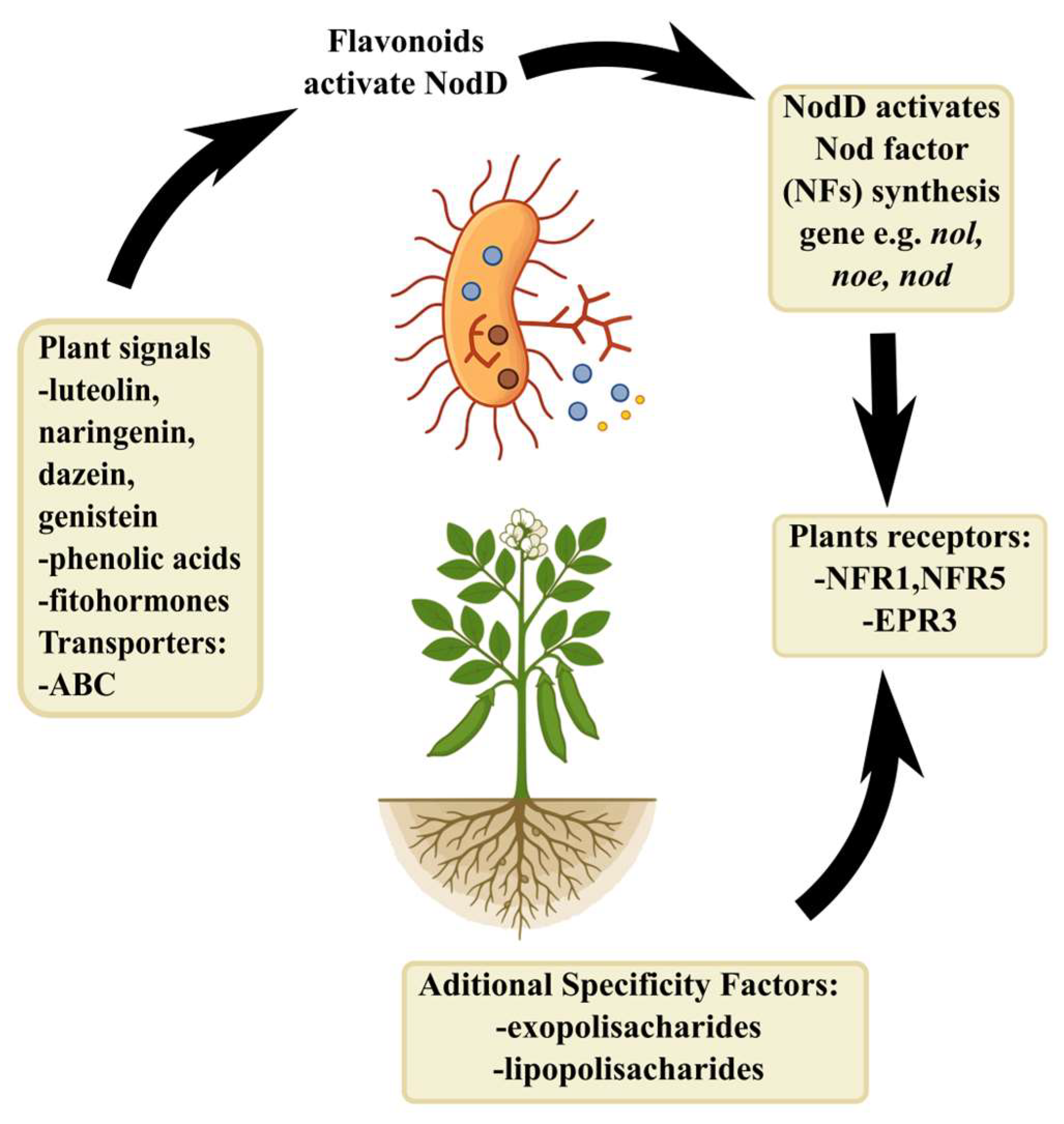
Scheme 1. The exchange of chemical signals underlying the initiation of the symbiosis process. NodD—Nodulation protein D; NF-Nod Factor; NFR1 and NFR5—Nod factor receptors 1 and 5; EPR3—exopolysaccharide receptor 3; ABC transporter—ATP-binding cassette transporter.
Agronomy 15 01552 sch001
Scheme 2. Early stages of symbiotic interaction. Root hair infection and primodium colonization. (A) Root exudates attract rhizobia to the root hair apex. (B) Root tip deformation is caused by high NF concentrations. A small population of dividing bacteria is formed. This leads to the formation of a microcolony. (C) Local degradation of root hair cell wall and cell membrane invagination caused by dividing rhizobia leads to infection thread elongation. (D) Release of rhizobia from infection thread by a process similar to exocytosis formation of symbiosomes.
Agronomy 15 01552 sch002
Scheme 3. Model of nodulation autoregulation signaling pathway. Arrows indicate upregulation, while lines indicate downregulation and/or degradation.
Agronomy 15 01552 sch003
Table 1. Examples of proline-rich proteins coded by nodulin genes [9].
Name of the ProteinFunctionProduct Location
ENOD 2presumed cell wall proteininner cortex (nodule parenchyma), may be nodule specific
ENOD 5related to arabinogalactans in plasma membranenodule primordium, zones 3 and 4 of mature indeterminate nodules, in infected cells only
ENOD l0presumed cell wall proteinnodule primordium, zone 2 of mature indeterminate nodules
ENOD 11presumed cell wall proteinroot hair/epidermal cells, nodule primordium, invasion zone of mature nodules
ENOD l2presumed cell wall proteinroot hair/epidermal cells, nodule primordium, invasion zone of mature nodules
PrP462-kDa proline-rich proteinmeristem of mature nodule, nodule specific
Disclaimer/Publisher’s Note: The statements, opinions and data contained in all publications are solely those of the individual author(s) and contributor(s) and not of MDPI and/or the editor(s). MDPI and/or the editor(s) disclaim responsibility for any injury to people or property resulting from any ideas, methods, instructions or products referred to in the content.