Article

Physiological and Transcriptomic Analysis of a Sepal Mutant in Phalaenopsis

Key Laboratory of East China Urban Agriculture, Shandong Engineering Research Center of Ecological Horticultural Plant Breeding, Shandong Academy of Agricultural Sciences, Institute of Leisure Agriculture, Jinan 250100, China
*
Author to whom correspondence should be addressed.
Agronomy 2025, 15(6), 1361; https://doi.org/10.3390/agronomy15061361
Submission received: 27 April 2025 / Revised: 23 May 2025 / Accepted: 30 May 2025 / Published: 31 May 2025

Abstract

MADS-box transcription factors have undergone in-depth investigations regarding their function in regulating the development of plant floral organs. Flower type mutants serve as critical biological models for investigating the regulatory mechanisms of MADS-box genes in floral organ development, while simultaneously constituting essential genetic resources for molecular breeding programs. In this work, we examined a lip-like sepal of the peloric mutant in Phalaenopsis ‘Huayang’, which exhibited changes in both the morphology and color of the sepals. Our cryo-SEM investigations revealed that the mutation type belonged to a sepal labellum-like variation in Phalaenopsis ‘Huayang’. Nine glycosylated anthocyanins were identified and their contents were significantly upregulated in the Se-red of mutant flowers. Transcriptomic analysis identified 9408 differentially expressed genes, including 4934 upregulated and 4474 downregulated genes. In addition, 57 MADS-box genes were identified and classed into five groups (Mα, Mβ, Mγ, MIKC*, and MIKCC) according to a phylogenetic comparison with Arabidopsis homologs. Furthermore, 29 MADS genes were screened from the MIKCC group, and these genes may play a crucial role in the regulation of floral organ development. Through real-time PCR analysis and protein interaction analysis, we identified three genes that were upregulated in the mutant, which may be involved in sepal development. The subcellular localization results demonstrated that three genes were found within the nucleus. Taken together, our results elucidated the molecular mechanism of sepal variation in Phalaenopsis ‘Huayang’. Our results could enhance our comprehension of the regulatory mechanisms underlying floral patterning and promote the molecular breeding process of Phalaenopsis.

1. Introduction

The development of floral organs has always played an important role in plant developmental biology. The development of flower organs in higher plants is a very complex process, and its regulatory mechanism involves multiple transcription factor families, including MADS-box [1], MYB [2], LEAFY [3], and NAC [4]. The molecular mechanisms of MADS-box transcription factors in regulating flower development have been extensively characterized in dicotyledonous model plants such as Arabidopsis thaliana and Antirrhinum majus [5,6]. However, among monocotyledonous species, comprehensive studies are currently limited to cereal crops like rice and maize [7,8]. Notably, Phalaenopsis is a typical monocotyledonous plant belonging to the Orchidaceae family, which is a high-value ornamental plant in the global horticulture market due to its diverse colors and unique flower shapes. Unlike most radiation symmetric flowers, the floral structure of Phalaenopsis is zygomorphic. It consists of three sepals in the first floral whorl. As development commences, two petals are formed, while the third petal transforms into a labellum at an early stage. The labellum, which is a highly modified part of the second floral whorl, is a distinctive trait that is peculiar to orchids [9,10]. In addition, the male and female reproductive parts of Phalaenopsis are merged into a structure, namely the gynostemium or column [11]. Thus, Phalaenopsis serves as an outstanding model for investigating the mechanism by which MADS-box genes regulate the specification of diverse and highly specialized structures within the Orchidaceae family. However, our research on how these genes regulate the variation mechanism of Phalaenopsis is still incomplete.
Based on their sequence associations and structural properties, the members of the MADS-box gene family in plants can be categorized as type I and type II [12]. The biological functions of type I MADS-box genes remain ambiguous, whereas type II MADS genes have been well documented as critical regulators in floral morphogenesis [13]. In plants, type II MADS-box proteins can alternatively be called MIKC-type MADS-box proteins, which consist of MIKCC and MIKC* proteins [14,15]. The MIKC* protein primarily contributes to the development of pollen, while the MIKCC protein is mainly associated with flower transformation, the development of floral organs, and fruit ripening [15,16]. With the exception of APETALA2 (AP2), all genes involved in the ABCDE model are MIKCC genes belonging to different functional categories [17]. In addition to the ‘ABCDE model’, ‘orchid code’ [18], ‘HOT model’ [19], and ‘perianth (P) code model’ [20] are commonly acknowledged as hypotheses used to elucidate the distinctive features of the perianth organs in orchids. The genes in these models have specific functions in regulating the development of orchid flower organs, and the ectopic expression or structural changes in these genes can cause the homologous transformation of flower organs. In peloric varieties of Chinese Cymbidium, the exon deletions in CsAP3-2 indicated that the peloria was caused by a loss of B-class gene function [21]. In the gylp (gynostemium-like petal) mutant of Phalaenopsis equestris, petal-to-gynostemium homeotic transformation was accompanied by ectopic PeMADS1 expression in modified floral organs [22]. Furthermore, the regulatory function of MADS-box transcription factors on flower organ development was further identified based on various types of mutant materials from Orchidaceae. Through the analysis of gene expression patterns in the peloric mutant of Phalaenopsis, PeMADS2, PeMADS4, and PeMADS5 of the class B-clade genes were identified as being involved in the development of the outer perianth, lip, and inner perianth, respectively [23]. However, these findings are largely isolated, and the molecular networks underlying labellum specification and their interactions with other MADS-box members remain uncharacterized.
Through the identification of mutants showing ‘lip-like sepal’ and ‘lip-like petal’ characteristics, Huang et al. (2015) determined that PhAGL6a, PhAGL6b, and PhMADS4 could potentially have significant functions in the formation of the labellum in Phalaenopsis [24]. In this research, we reported a lip-like sepal of a peloric mutant in Phalaenopsis ‘Huayang’, which exhibited changes in both the morphology and color of the sepals. Based on the morphological research, we first identified the types of variations that occurred. Through the analysis of transcriptome sequencing data, we conducted a comparison between the wild-type and the peloric mutant, and subsequently identified genes that exhibited significant differential expression in floral organs. This study elucidated the functional specialization and differentiation of floral organs in Phalaenopsis, as well as predicting the potential molecular regulatory network that controls their morphogenesis process, providing crucial molecular targets for orchid floral patterning research and laying a foundation for developing novel floral traits through molecular breeding.

2. Materials and Methods

2.1. Plant Materials and Growth Conditions

Wild-type and mutant-type plants of Phalaenopsis ‘Huayang’ were used as the experimental materials for this study, and were cultivated in the intelligent orchid greenhouse of the Shandong Academy of Agricultural Sciences (Jinan, China). The wild-type plants of Phalaenopsis ‘Huayang’, as commercial cultivars, were purchased from flower markets. The mutant-type plants of Phalaenopsis ‘Huayang’, as a natural mutant material, were selected during the cultivation process. We carried out tissue culture and propagation for the mutant-type plants through the method of flower stalk propagation, obtained plants with stable variations, and used them for the research in this experiment. All plants were cultivated with the sphagnum moss developed specifically for Phalaenopsis. When sphagnum moss became moderately dry, it was irrigated with a 2000- to 2500-fold diluted solution of water-soluble fertilizer (N-P2O5-K2O = 20-20-20) every 7–10 days. Additionally, a 1000-fold diluted agricultural amino acid solution was applied every 2–3 weeks. The growth conditions for all plants were maintained under natural light and controlled temperature from 27 to 30 °C. Tobacco (Nicotiana benthamiana) was grown in greenhouses under natural light at a temperature of 25 °C and then utilized for subcellular localization.

2.2. Scanning Electron Microscopy

Dissected blossom apex specimens were subjected to 24 h chemical fixation using a mixed aldehyde solution (3% glutaraldehyde mixed with 2% formaldehyde) under vacuum conditions. The tissue blocks were washed three times with 0.1 M phosphate buffer (PB, pH 7.4), with a duration of 15 min for each washing step. Subsequently, the tissue blocks were immersed in 0.1 M phosphate buffer (PB, pH 7.4) with 1% osmium tetroxide (OsO4) and kept at room temperature for 1–2 h. Following OsO4 exposure, the tissue blocks underwent a triple re-washing process. Then, dehydration was performed as follows: the tissue blocks were sequentially treated with 30%, 50%, 70%, 80%, 90%, and 95% ethanol for 15 min intervals each, followed by two 15 min treatments with 100% ethanol and a final 15 min treatment with isoamyl acetate. After undergoing critical-point drying (K850, Quorum), the specimens were mounted onto metal stubs utilizing conductive carbon adhesive disks, followed by a 30 s gold deposition process through magnetron sputtering coating. The prepared specimens were examined by means of a SU8100 (HITACHI, Hitachi, Tokyo, Japan) scanning electron microscope. Under a 300× magnification of the electron microscope with a 100 μm scale, the number of cells observed in each 2560 × 1920 field of view was 30–60. All samples were performed with three biological replicates.

2.3. Metabolite Extraction

The lyophilized specimen was subjected to mechanical pulverization (30 Hz, 1.5 min) followed by cryopreservation at −80 °C. Aliquots comprising 50 mg of the homogenized material underwent dual-phase extraction using 0.5 mL of acidified methanol-water solution (MeOH:H2O:HCl, 500:500:1, v/v/v). The extraction protocol involved sequential vortex mixing (5 min) and ultrasonic treatment (5 min), followed by refrigerated centrifugation (4 °C, 12,000× g, 3 min). To ensure maximum extraction efficiency, the insoluble matrix underwent a secondary extraction cycle under identical parameters. Combined supernatants were subsequently filtered through 0.22 μm microporous membranes (Anpel, Shanghai, China) before LC-MS/MS analysis [25].

2.4. HPLC-MS/MS Analysis

The UPLC-ESI-MS/MS system (ExionLCTM AD UPLC coupled with a 6500 Q TRAP mass spectrometer; SCIEX, Framingham, MA, USA) was employed for sample analysis. The separation was carried out using a Waters ACQUITY BEH C18 analytical column (Waters, Milford, MA, USA) (1.7 µm, 2.1 × 100 mm) kept at 40 °C. A binary mobile phase system with acid-modified solvents containing 0.5% formic acid in both water (A) and methanol (B) was used. The elution gradient was multi-step: it started at 95% A, changed linearly to 50% A from 0 to 6 min, and then reduced to 5% A at 12 min and was held for 120 s. Column re-equilibration started at 14 min by reverting to the initial 95% A composition, followed by a 2 min stabilization. The operation had a constant flow rate of 350 µL/min and a sample injection volume of 2 µL.
The analysis of anthocyanins was performed using a QTRAP® 6500+ hybrid mass spectrometer (SCIEX, Suzhou, China) configured with electrospray ionization (ESI) with positive polarity. Instrument operation involved dual-mode acquisition through synchronized linear ion trap (LIT) and triple quadrupole (QQQ) functionalities. Targeted compound detection was achieved through a scheduled multiple-reaction monitoring (MRM) methodology with optimized transition parameters. Instrument operation and data collection were carried out using Analyst 1.6.3 software (SCIEX, Framingham, MA, USA), and metabolite quantification was carried out with Multiquant 3.0.3 (SCIEX) [26,27]. All experiments were carried out with three biological replicates and triplicate technical replicates.

2.5. RNA-Seq and DEG Analysis

The cDNA-based libraries of wild-type Phalaenopsis ‘Huayang’ and its mutant plants were constructed using RNAs sourced from equimolar mixtures. These RNAs were isolated separately from the mature flowers of five individual plants in each of the wild-type and mutant groups. The library preparation was processed using the BGISEQ-500 sequencing system (BGI Group, Beijing, China) following the manual. Then, quality control steps were carried out to remove low-fidelity sequences, finally obtaining paired-end reads of high quality for downstream analysis. Gene expression levels were quantified by calculating the FPKM metric. Significantly differentially expressed genes were selected based on a threshold of q value ≤ 0.001 and |fold-change| ≥ 2 [28]. The functional annotation of DEGs was conducted using the Blast2GO software (basic 6.0 version) suite based on the Gene Ontology (GO) database.

2.6. Multiple Sequence Alignment and Phylogenetic Analysis

A total of 276 full-length protein sequences were subjected to alignment via ClustalW, including 100 AtMADS from Arabidopsis thaliana, 64 OsMADS from Oryza sativa, 55 PeMADS from Phalaenopsis equestris, and 57 MADS from Phalaenopsis ‘Huayang’. Phylogenetic analysis was conducted using the neighbor-joining algorithm in MEGA-X (https://www.megasoftware.net/ (accessed on 12 May 2024)) with default parameters, which were based on the alignment. The tree topology and reliability were evaluated using 1000 bootstrap replications.

2.7. Protein Interaction Network Prediction

Protein interaction networks of MADS transcription factors were systematically analyzed using the STRING database v12.0 (https://string-db.org/ (accessed on 2 March 2025)) with Arabidopsis thaliana as the reference species. Predicted interactions were further visualized and topologically characterized through Cytoscape v3.10.1 (https://cytoscape.org/ (accessed on 15 March 2025)).

2.8. Heatmap Construction

To further identify differentially expressed genes in sepal variation, we excavated the transcriptome data. The transcript abundances for each sample were computed and measured in terms of reads per kilobase per million mapped reads (RPKM). Subsequently, the mean RPKM values underwent a log transformation. These transformed values were then employed to create a heatmap through the utilization of GraphPad Prism 10.0, with the hierarchical clustering technique being applied in the process [29].

2.9. Expression Profile Analysis

Total RNA isolation from plant samples was performed with an RNA extraction kit (HuaYueYang, Shenzhen, China). Subsequently, cDNA synthesis was achieved with a reverse transcription kit (Vazyme, Nanjing, China). The qRT-PCR was conducted on a Bio-Rad CFX-96 PCR System (Bio-Rad Laboratories, Hercules, CA, USA) coupled with the 2×RealStar Fast SYBR Green master mix. The tubulin gene served as the reference gene. The 2−ΔΔCt method was implemented for the relative quantification of gene expression levels, as described previously [30]. Three independent biological replicates were conducted for each experiment, with triplicate technical measurements performed per biological replicate.

2.10. Subcellular Localization Analysis

The CDS regions of CL9095.4, CL1003.1, and Unigene32429 (excluding stop codons) were amplified via PCR and inserted into modified 35S::GFP/PHB vectors. The vectors for the fusion and control of GFP were introduced into Agrobacterium GV3101 via electroporation. Subsequently, the transformed GV3101 was infiltrated into the leaves of Nicotiana tabacum. The GFP and DAPI fluorescence signals were analyzed 48 h later with an Olympus FV1200 fluorescence microscope (OLYMPUS, Tokyo, Japan). Experiments were triplicated.

2.11. Statistical Analysis

All data are presented as the means of three replicates with standard deviations. Primers for quantitative real-time PCR (qRT-PCR) were designed using Primer 5.0. The data were analyzed using SPSS 24.0 (IBM Corporation, New York, NY, USA), and p < 0.05 was considered statistically significant. The volcano plots and histograms were drawn using TBtools-II (v2.112). The heatmaps and bar charts were drawn using GraphPad Prism 10.0. The phylogenetic tree was constructed using MEGA-X.

3. Results

3.1. Morphological Study of Sepal Mutants in Phalaenopsis ‘Huayang’

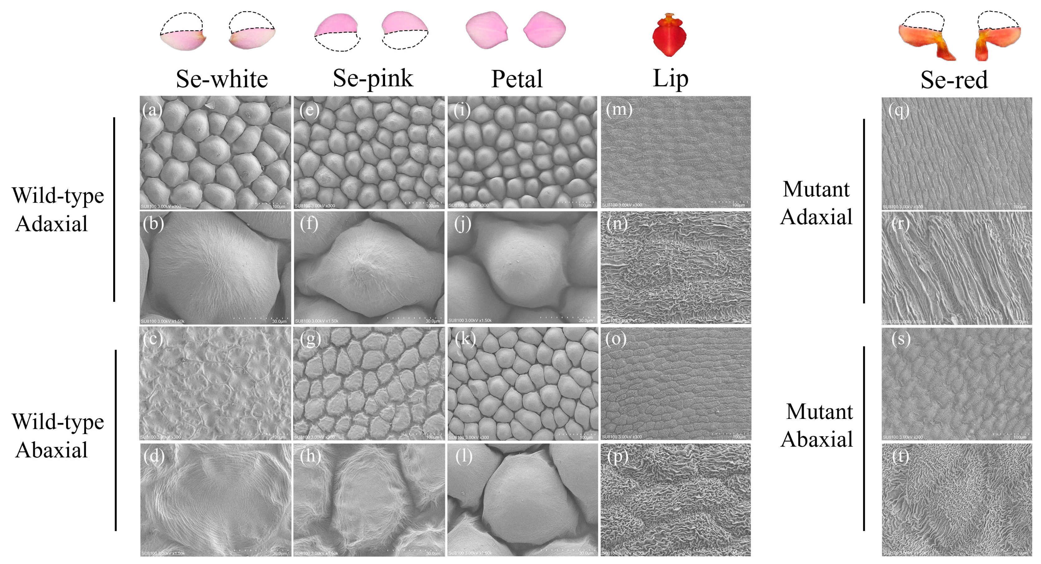
The wild-type flower of Phalaenopsis ‘Huayang’ shows three sepals, two petals, one lip, and one gynostemium. However, the mutant flower has conversions of sepals to lip-like structures (Figure 1A,B). Here, the normal sepals of wild-type flowers were dissected into Se-white and Se-pink, and the lip-like sepals of mutant flowers were dissected into Se-red and Se-pink (Figure 1C). To investigate the morphological discrepancies between wild-type and mutant floral organs, cryo-scanning electron microscopy (cryo-SEM) was employed for a qualitative analysis of epidermal cell specimens. In wild-type flowers, the shapes of the epidermal cells and the ultrastructural characteristics differ among the Se-white, Se-pink, petal, and lip (Figure 2). On the epidermis of both the adaxial and abaxial sides of the wild-type flowers, se-white and se-pink cells are conical/tabular; petal cells are conical. Nevertheless, within the lip of wild-type flowers, cuticular folding at the ultrastructural level was present on both epidermal surfaces. In contrast, the Se-red of mutant flowers lacks conical-to-tabular-shaped cells. Nevertheless, similarly to the ultrastructural cuticular folding observed on the lip, the Se-red cells also exhibit these structures.

3.2. Identification and Quantification of Anthocyanin Components in Phalaenopsis ‘Huayang’

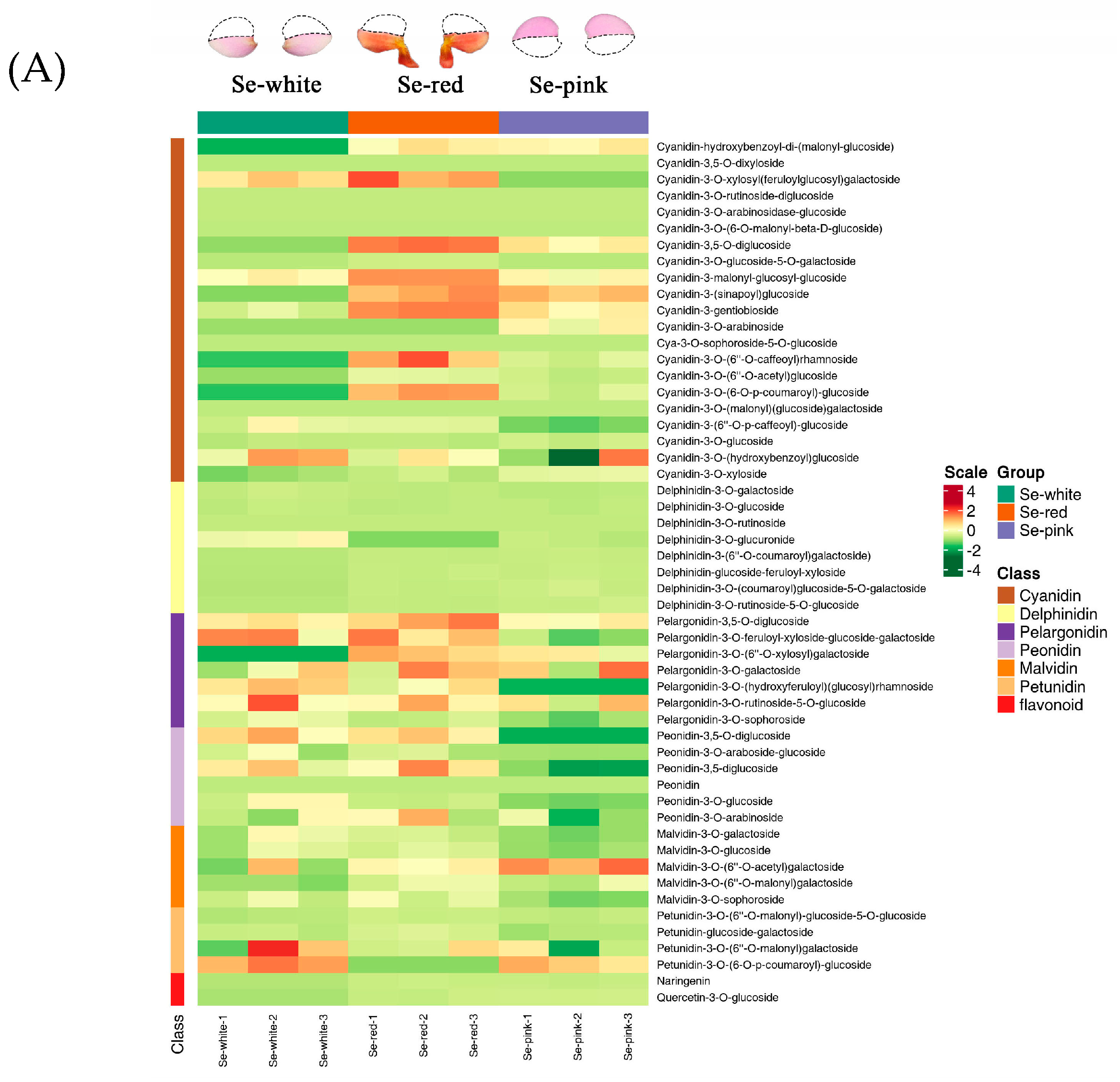
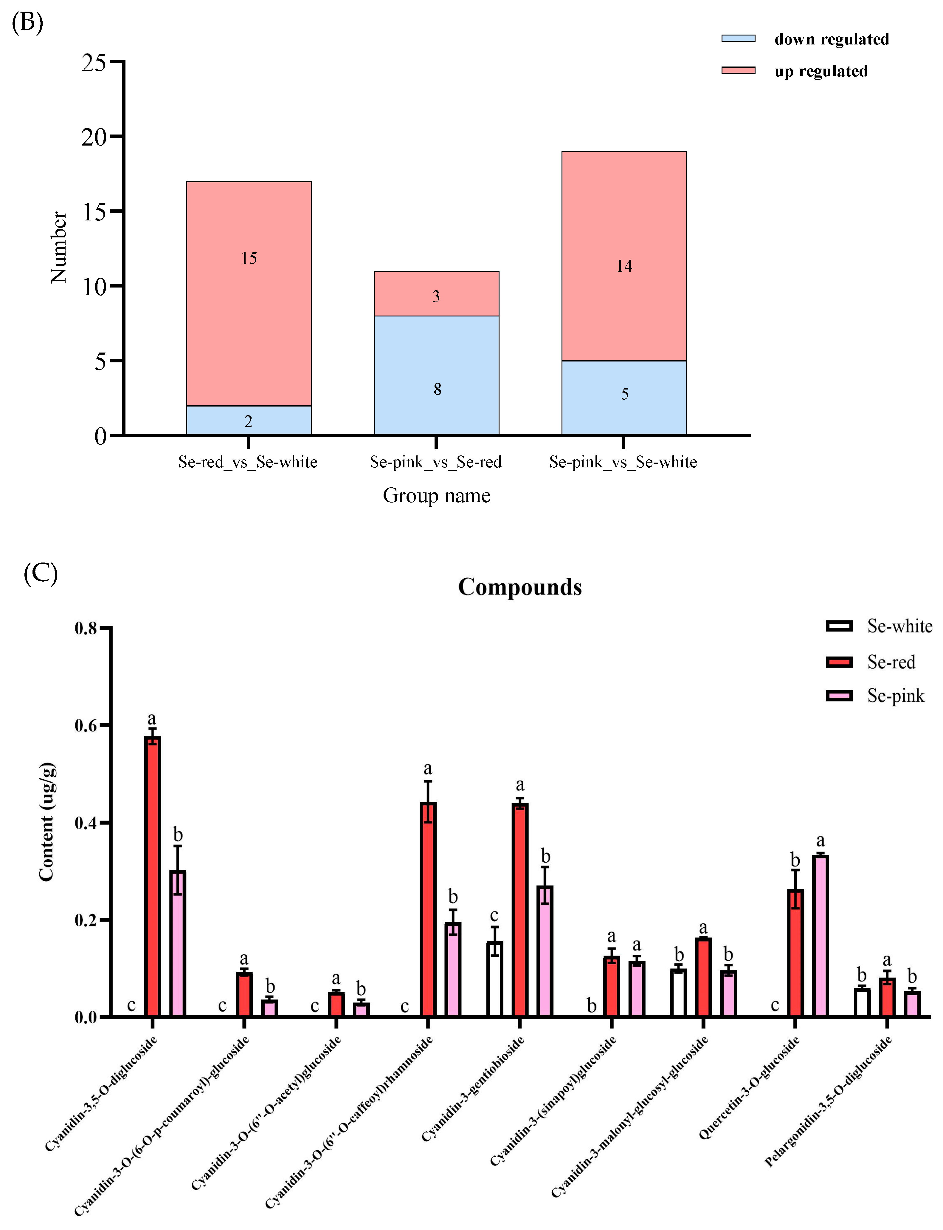
In this study, the sepals of the mutant flowers transformed into lip-like structures, and their color also changed. To conduct an in-depth analysis of the metabolic mechanism underlying the color variation, liquid chromatography–tandem mass spectrometry (LC-MS/MS) was utilized to analyze the anthocyanin metabolites. As a result, 53 anthocyanin compounds were recognized, as presented in Supplementary Table S1. Through the application of unit variance scaling (UV) and hierarchical cluster analysis (HCA) to these compounds, 21 cyanidin compounds, 8 delphinidin compounds, 7 pelargonidin compounds, 6 peonidin compounds, 5 malvidin compounds, 4 petunidin compounds, 1 Naringenin, and 1 Quercetin-3-O-glucoside were identified (Figure 3A and Supplementary Figure S1). In the comparison of Se-red vs. Se-white and Se-pink vs. Se-white, the number of metabolites with up-regulated expression was significantly higher than that of metabolites with down-regulated expression. However, in the comparison of Se-pink vs. Se-red, the number of metabolites with down-regulated expression was significantly higher than that of metabolites with up-regulated expression (Figure 3B and Supplementary Table S2). Additionally, the study results showed significant differences in the contents of nine differential metabolites in Se-white, Se-red, and Se-pink, which are involved in seven Cyanidin-based compounds, 1 Quercetin-3-O-glucoside and Pelargonidin-3,5-O-diglucoside. In particular, in the Se-red of mutant flowers, the content of nine differential metabolites was significantly upregulated compared with the Se-white of the wild-type flower. Based on these findings, we propose that nine differentially accumulated metabolites (DAMs) might play a crucial role in the color transformation of Phalaenopsis ‘Huayang’ from Se-white to Se-red (Figure 3C and Supplementary Table S3). This speculation indicates that these differential metabolites could be key factors triggering the change in flower color, providing a theoretical basis for further exploring the molecular mechanism of color formation in Phalaenopsis ‘Huayang’.

3.3. Transcriptome Sequencing and Differentially Expressed Genes

To investigate the molecular mechanism underlying the lip-like sepals of the mutant flower, a comprehensive assessment of the transcriptomes from the sequencing libraries of both wild-type and mutant flowers was carried out. The wild-type (WT) and mutant samples generated 48.50 million and 47.91 million total raw reads, respectively. After eliminating low-quality sequences, conducting adapter filtering, and excluding ambiguous reads, we acquired roughly 44.75 million and 43.87 million total clean reads in the wild-type (WT) and the mutant samples, respectively. Subsequently, these total clean reads were aligned to the genomic database of Phalaenopsis equestris. The alignment rates were 75.82% and 75.67%, respectively (Supplementary Table S4). The differential gene expression analysis was carried out between the WT and mutant flowers. A statistical analysis with stringent thresholds (p value ≤ 0.001, |fold-change| ≥ 2) revealed 9408 significant DEGs, comprising 4934 upregulated and 4474 downregulated transcripts (Figure 4A). The functions of these DEGs were categorized based on the Gene Ontology (GO) database by utilizing the Blast2GO software package (Figure 4B). These DEGs were associated with biological processes (669), cellular components (843), and molecular function (859).

3.4. Identification of MADS Genes in Phalaenopsis ‘Huayang’

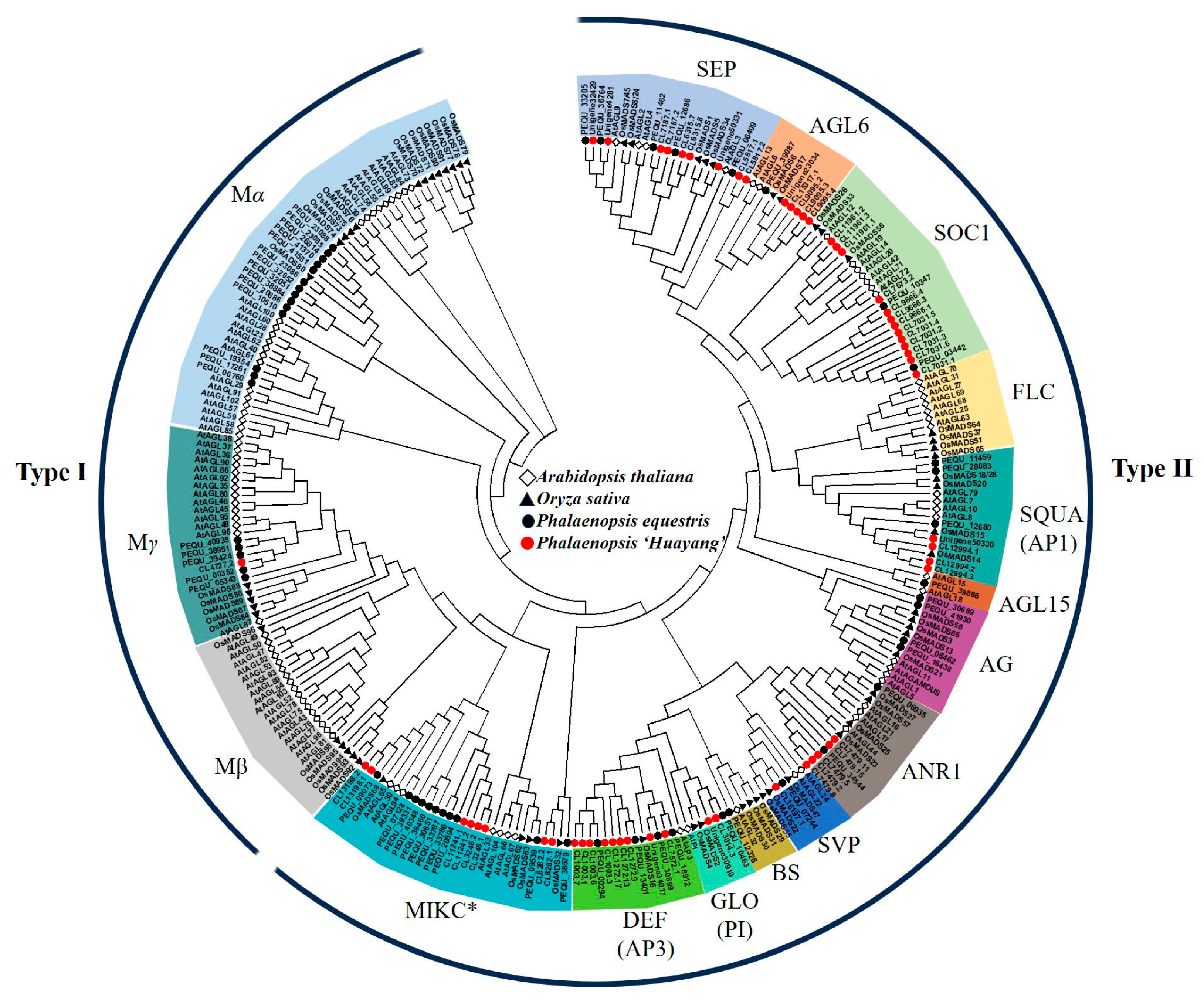
MADS-box transcription factors have been found to be of great significance in controlling floral organ formation and development [31]. Based on the functional annotation information of the transcriptome sequencing data we previously obtained from the petals of wild-type and mutant flowers, we obtained 57 MADS-box transcription factors. A neighbor-joining phylogenetic tree was constructed to elucidate the evolutionary relationships and taxonomic categorization of MADS-box proteins within Phalaenopsis. Briefly, 276 MADS-box proteins were analyzed, including 100 from Arabidopsis thaliana, 64 from Oryza sativa, 55 from Phalaenopsis equestris, and 57 from Phalaenopsis ‘Huayang’. As shown in Figure 5, the phylogenetic tree was constructed using the neighbor-joining method with 1000 bootstrap replications. The 16 subfamilies are marked with different colors. The rectangles represent MADS-box proteins from Arabidopsis thaliana, the black triangles represent MADS-box proteins from Oryza sativa, the black circles represent MADS-box proteins from Phalaenopsis equestris, and the red circles represent MADS-box proteins from Phalaenopsis ‘Huayang’.
The 276 identified MADS-box proteins were systematically categorized through phylogenetic analysis into two principal classes: Type I and Type II. Based on their M-domain architectures and conserved domain characterization, Type I MADS-box proteins were phylogenetically divided into three evolutionarily conserved clades, namely the Mα, Mβ, and Mγ subgroups. The Mα subfamily, with 23 AtMADS, 13 OsMADS, and 14 PeMADS members, was associated with floral organ identity and developmental timing. The Mβ subfamily consisted of 17 AtMADS and 6 OsMADS members, which play crucial roles in plant development, particularly in reproductive organogenesis and floral patterning. The Mγ subfamily has a specialized role in P. ‘Huayang’, with only 1 member compared to 14 AtMADS and 5 OsMADS members. The Type II MADS-box proteins were categorized into MIKC* and MIKCC clades based on their structural characteristics. Among the MIKC* subgroup, there were eight MADS-box proteins in P. ‘Huayang’, playing crucial roles in plant development, particularly in reproductive processes. Furthermore, the MIKCC subgroup was subdivided into 12 subfamilies: SEP, AGL6, SOC1, FLC, SQUA(AP1), AGL15, AG, ANR1, SVP, BS, GLO (PI), and DEF (AP3) (Supplementary Table S5). The ABCDE model is associated with the development of flower organs and consists of A-class genes (SQUA), B-class genes (DEF and GLO), C/D-class genes (AG), and E-class genes (SEP) [32]. In summary, the subdivision of the MIKCC subgroup into specific subfamilies and their associations with the ABCDE model provide insights into the genetic regulatory mechanisms underlying sepal variation in P. ‘Huayang’.

3.5. Identification of Differentially Expressed Genes (DEGs) Associated with Morphological Mutations in Sepals

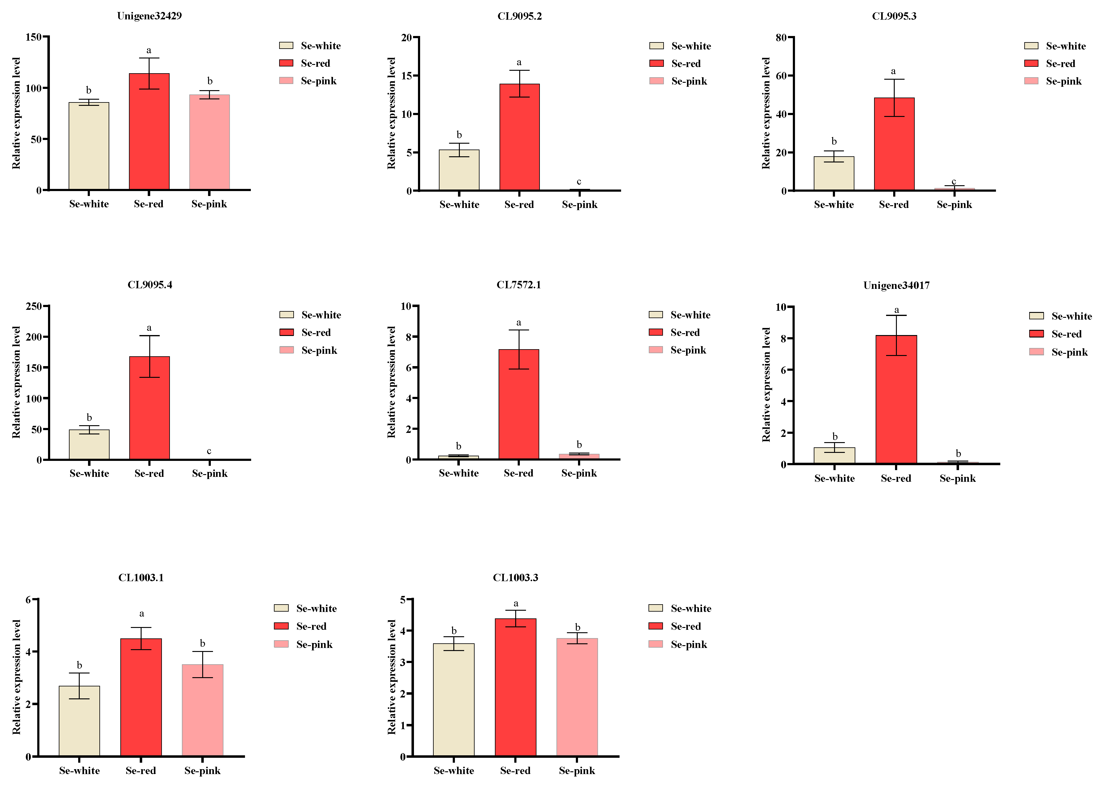
MIKCC proteins act as key regulators in the process of floral organ differentiation. Based on their evolutionary history, they can be further classified into 12 distinct subgroups. These include SQUA (AP1), GLO (PI), DEF (AP3), CD (AG), E (SEP), and AGL6, all of which potentially play significant roles in the regulation of floral organ development [33]. To further identify differentially expressed genes in sepal variation in Phalaenopsis ‘Huayang’, the expression profiles of 29 MADS-box genes of the MIKCC subgroup were quantified by RPKM, and the data were retrieved from transcriptomic datasets. Subsequently, a heatmap analysis was employed to vividly present and visualize the expression profiles of these genes in Se-white, Se-red, and Se-pink. Our findings indicate that among the 29 MADS-box genes in the SEP, AGL6, and DEF (AP3) subfamilies involved in flower development, the expression levels of ten genes were higher in the Se-red of mutant flowers, while they were lower in the Se-white and Se-pink of wild-type flowers (Figure 6 and Supplementary Table S6). The eight significantly differentially expressed genes verified by qRT-PCR included the SEP-clade gene Unigene32429, the AGL6-clade gene CL9095.2/CL9095.3/CL9095.4, and the DEF-clade gene Unigene34017/CL7572.1/CL1003.1/CL1003.3. In the Se-red of mutant flowers, the expression patterns of eight genes were significantly upregulated compared with the Se-white and Se-pink of the wild-type flower (Figure 7 and Supplementary Table S7).

3.6. Analysis of Protein Interactions and Putative Functions of PaMADSs

The functions of MADS-box family members in Arabidopsis thaliana have been well characterized. Given that proteins with high homology usually cluster together and tend to have similar functions [34], the STRING database (version 12.0) was utilized to deduce the potential functions and the hypothesized interaction network of the 29 MADS proteins in Phalaenopsis ‘Huayang’ (Figure 8). AtAP3 (high similarity with CL1003.1) occupied the central position within the regulatory network and exhibited the highest degree of connectivity. It played a critical role in the genetic regulation of flower development and was essential for the normal growth of petals and stamens in wild-type flowers [35]. In addition, flower development genes may be regulated through a ternary complex consisting of the AP3/PI heterodimer in conjunction with APETALA1 or SEPALLATA3 [36]. In this study, the up-regulated AtAP3 (CL1003.1) in the Se-red of mutant flowers was found to interact with the up-regulated AtSEP3 (Unigene32429) and AtAGL6 (CL9095.4), forming a ternary complex that may regulate genes associated with sepal variation in Phalaenopsis ‘Huayang’. This finding directly aligns with the research objective of exploring the key gene-regulatory networks underlying sepal variation in Phalaenopsis ‘Huayang’. The identified ternary complex is hypothesized to play a critical role in regulating genes associated with sepal variation traits, providing mechanistic insights into the genetic basis of floral organ differentiation in orchids.

3.7. Subcellular Localization of Three Key MADS Genes

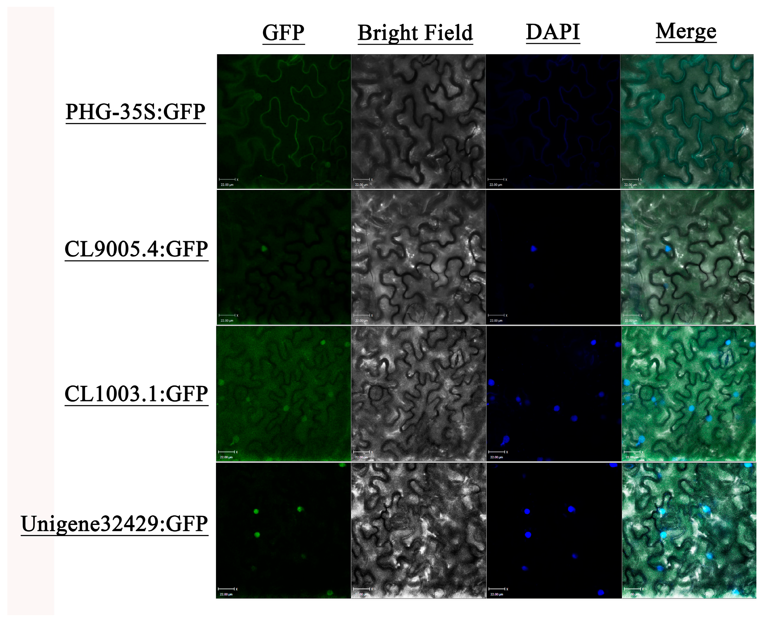
To investigate the subcellular distribution patterns of CL9095.4, CL1003.1, and Unigene32429, the recombinant fusion constructs (CL9095.4-GFP, CL1003.1-GFP, and Unigene32429-GFP), along with the control vector PHB-GFP, were transiently expressed in the leaf epidermal cells of tobacco (Nicotiana benthamiana) (Supplementary Table S8). The fluorescence of green fluorescent protein (GFP) in the control vector was distinctly observed to be distributed within both the nucleus and cyto-membrane. In contrast, for the CL9095.4-GFP/CL1003.1-GFP/Unigene32429-GFP vector, a robust fluorescence signal was detected specifically in the nucleus of tobacco cells. Therefore, based on these findings, we hypothesized that the three key MADS genes were concurrently located and exerted their functions within the nucleus (Figure 9).

4. Discussion

Flower variations occur frequently in Phalaenopsis, including the gylp (gynostemium-like petal) mutant [22], the big lip mutant [24], and the labellum-like petal and lip-like sepal of the peloric mutant [37,38]. Among them, the lip-like sepal of the peloric mutant is relatively rare. The peloric mutant of orchid plays a significant role in the exploration of floral development from both morphological and molecular perspectives [39]. In this research, we discovered a lip-like sepal of peloric mutant in Phalaenopsis ‘Huayang’, which exhibited changes in both the morphology and color of the sepals. Through morphological characterization and transcriptome-wide analysis, we elucidated the molecular mechanism of sepal variation in Phalaenopsis ‘Huayang’.

4.1. Morphological and Anthocyanin Analysis of Lip-like Sepal of Peloric Mutant in Phalaenopsis ‘Huayang’

Our cryo-SEM revealed a strong contrast between wild-type flowers and mutant flowers. The morphological characteristics of epidermal cells and the ultrastructural features of the Se-red of mutant flowers were consistent with the lip structure of the wild-type flowers, indicating that the mutation type belongs to the sepal labellum-like variation. These results are similar to the ultrastructural cuticular of the lip of the Phalaenopsis OX Red Shoes [40]. These results lay the groundwork for exploring the genetic mechanisms of sepal labellum-like variation, which advances our understanding of floral development and evolution in orchids. Floral color serves as a critical criterion for assessing the ornamental value of Phalaenopsis, and the differences in the anthocyanin types and contents of petals directly affect the presentation of color [41]. While 53 anthocyanin compounds were identified in Phalaenopsis, nine metabolites, including Cyanidin-3,5-O-diglucoside, Cyanidin-3-O-(6-O-p-coumaroyl)-glucoside, Cyanidin-3-O-(6″-O-caffeoyl)rhamnoside, Cyanidin-3-O-(6″-O-caffeoyl)rhamnoside, Cyanidin-3-gentiobioside, Cyanidin-3-(sinapoyl)glucoside, Cyanidin-3-malonyl-glucosyl-glucoside, Quercetin-3-O-glucoside, and Pelargonidin-3,5-O-diglucoside, showed significant upregulation in the Se-red of mutant flowers but not in the Se-white of wild-type flowers. We speculated that nine anthocyanin compounds were the key metabolic substances for the color variation in sepals in the Se-red of mutant flowers. This finding not only clarifies the molecular basis of the sepal coloration of Phalaenopsis ‘Huayang’ but also offers valuable targets for breeding.

4.2. Transcriptome Analysis and Identification of MADS-Box Genes Reveal Sepal Variation in Phalaenopsis ‘Huayang’

Transcriptome analysis can be effectively used to recognize the essential genes involved in the formation of mutants. In our study, we conducted transcriptome sequencing on the wild-type flowers and mutant flowers of Phalaenopsis. A total of 4934 up-regulated differentially expressed genes (DEGs) and 4474 down-regulated DEGs were identified using differential gene expression analysis and were found to be associated with biological processes, cellular components, and molecular function in Phalaenopsis. Previous research has indicated that MADS-box transcription factor family members are crucial for the development of flower structures and have drawn significant attention across multiple plant species; for instance, Arabidopsis thaliana with 100 genes [42] and Oryza sativa with 64 genes [43]. The disparities in the numbers of MADS-box genes across different species might be associated with the events of genome-wide duplication (WGD) during the evolutionary process [44].
In this study, a total of 57 MADS-box genes were comprehensively identified from Phalaenopsis ‘Huayang’, which were further classified into two primary subgroups: type I and type II. Then, based on the characteristics of the M domain of MADS-box proteins, the type I members were further categorized into three distinct subgroups, Mα, Mβ, and Mγ, which play crucial roles in various biological processes, particularly in plant development and reproduction [45]. In this study, only one gene from the Mγ subfamily was identified. In terms of the type II members, they were separated into the MIKC* subgroup and the MIKCC subgroup. In addition, we categorized the MIKCC-type genes into 12 subfamilies. The functions of MIKCC-type genes vary; for example, the SOC1, FLC, and SVP genes regulate flowering time [46,47,48]. The ABCDE model associated with the development of flower organs is made up of A (SQUA), B (DEF/GLO), CD (AG), and E (SEP) genes, all of which belong to the MIKCC subgroups [17]. We identified 29 genes of Phalaenopsis ‘Huayang’ related to floral organ development, which are involved in five subfamilies of AP1, PI, AP3, SEP, and AGL6, and they are crucial for studying the formation of mutant flowers of Phalaenopsis. The ectopic overexpression of PhAGL6b in the petal or sepal induces a homeotic transformation in the lip-like structures in Phalaenopsis ‘KHM190’ cultivars [49]. In peloric varieties of Chinese Cymbidium, the exon deletions in CsAP3-2 indicated that the peloria was caused by the loss of B-class gene function [21].
The MIKCC-class genes are critical in controlling the morphological and physiological properties of floral organs [50]. To uncover the regulatory mechanisms of the 29 genes belonging to five subgroups (AP1, PI, AP3, SEP, and AGL6) during the process of sepal variation in Phalaenopsis, we conducted differential expression patterns in multiple floral morphogens of Phalaenopsis (Se-red of mutant flower, and Se-white and Se-pink of wild-type flowers). In our study, eight genes highly expressed in the Se-red of mutant flowers were identified by qRT-PCR, illustrating that they have significant and potential function in the process of sepal labialization development. Similar results were reported in Cymbidium sinense [21], Phalaenopsis equestris [51], and Hippophae rhamnoides [13]. In addition, in Phalaenopsis, PhAGL6a, PhAGL6b, and PhMADS4, a high level of expression was demonstrated specifically within the labellum. Moreover, these genes were found to be up-regulated in the lip-like petals and sepals of peloric-mutant flowers [24]. Based on the transformation of the sepal mutant into a lip-like structure, we identified eight MADS-box genes that likely function as key regulatory components in labellum development. These genes could serve as key candidates for genetic engineering approaches aimed at breeding novel Phalaenopsis cultivars with unique floral morphologies. Our study elucidates the fundamental molecular mechanisms underlying the sepal mutant phenotype in Phalaenopsis.

4.3. Regulatory Network and Interaction Patterns of MADS-Box Transcription Factors Reveal Sepal Variation in Phalaenopsis ‘Huayang’

Previous studies have indicated that genes of the MADS transcription factor family regulate plant function through interactions [52]. Network analysis has demonstrated that the members of most MADS-box proteins within the MIKCC family exhibit protein–protein interactions in Phalaenopsis ‘Huayang’. AtAP3 (high similarity with CL1003.1) occupied the central position within the regulatory network and exhibited the highest degree of connectivity. It served a pivotal function in modulating the genetic mechanisms of flower development and was essential for the normal growth of petals and stamens in wild-type flowers. In addition, the up-regulated AtAP3 (CL1003.1) in the mutant flower interacts with up-regulated AtSEP3 (Unigene32429) and AtAGL6 (CL9095.4) to form a ternary complex that may regulate genes associated with sepal development. Similar interaction patterns were confirmed in Dendrobium and Phalaenopsis equestris [53,54]. The CsAP3-2 gene in Cymbidium sinense, expressed specifically in the inner floral whorls, cooperates with the labellum-specific CsAGL6-2 protein to mediate the asymmetric development of the inner tepals [21]. Previous research has demonstrated that Phalaenopsis employs multimeric complexes (ranging from dimers to tetramers) through interactions between PeSEP proteins and other MADS-box transcription factors. The involvement of these four SEP paralogs leads to distinct patterns being exhibited during the processes of floral transition, morphogenesis, and ovule development in Phalaenopsis [40]. In this study, the predicted network interaction patterns of the three proteins, AP3, SEP3, and AGL6, are key patterns for regulating sepal variation in Phalaenopsis ‘Huayang’, laying a solid foundation for the subsequent functional verification.

5. Conclusions

In this study, we first identified mutation types belonging to a sepal labellum-like variation using cryo-SEM. The identification and quantification results of anthocyanins revealed that nine anthocyanin compounds were the key metabolic substances for the color variation in sepals in the Se-red of mutant flowers. These findings enrich our understanding of the molecular basis of floral color and morphological development in Phalaenopsis. Through transcriptome sequencing analysis, a total of 57 MADS-box genes were identified and subsequently categorized into five distinct groups (Mα, Mβ, Mγ, MIKC*, and MIKCC) according to a phylogenetic comparison with Arabidopsis homologs. Furthermore, 29 MADS genes were screened from the MIKCC group, which may play a crucial role in the development of sepal variation in Phalaenopsis. The results of heatmap analysis and qRT-PCR showed that the expression levels of eight genes were significantly upregulated in the Se-red of mutant flowers compared with the Se-white and Se-pink of wild-type flowers. These genes could serve as key candidates for genetic engineering approaches aimed at breeding novel Phalaenopsis cultivars with unique floral morphologies. Through the analysis of the protein interaction network, based on the transformation of sepal mutants into labellum-like structures, we identified that the MADS-box genes AP3 (CL1003.1), SEP3 (Unigene32429), and AGL6 (CL9095.4) are potential regulatory components for sepal variation in Phalaenopsis ‘Huayang’. The results provide new insights into how gene networks contribute to orchid floral diversity, and especially labellum development. Meanwhile, the experimental findings establish a robust foundation for subsequent investigations into the developmental mechanisms of floral organs in Phalaenopsis.

Supplementary Materials

The following supporting information can be downloaded at: https://www.mdpi.com/article/10.3390/agronomy15061361/s1, Figure S1: Qualitative analysis of pigments in Phalaenopsis ‘Huayang’; Table S1: Identification of anthocyanin components in Phalaenopsis ‘Huayang’; Table S2: Number of differentially regulated pigment metabolites in different groups; Table S3: Histogram data of differential metabolite contents; Table S4: Summary of sequencing reads in WT and mutants of Phalaenopsis ‘Huayang’; Table S5: The number of each type of MADS-box gene in Phalaenopsis ‘Huayang’; Table S6: FPKM value of relative expression of MADS genes; Table S7: Primers used for qRT-PCR; Table S8: Primers used for subcellular localization.

Author Contributions

Conceptualization and methodology, X.L.; software and validation, Y.W.; formal analysis and methodology, F.D.; data curation and resources, J.Z.; writing—original draft preparation, Y.Q.; writing—review and editing, Y.Q. and X.L.; project administration, X.L. All authors have read and agreed to the published version of the manuscript.

Funding

This research was funded by Key R&D Program of Shandong Province, China, grant number 2023LZGCQY005.

Data Availability Statement

The data presented in this study are available in the Supplementary Materials.

Acknowledgments

We appreciate the support from the Key R&D Program of Shandong Province.

Conflicts of Interest

The authors declare no conflicts of interest.

References

  1. Dreni, L.; Zhang, D. Flower Development: The Evolutionary History and Functions of the AGL6 Subfamily MADS-Box Genes. J. Exp. Bot. 2016, 67, 1625–1638. [Google Scholar] [CrossRef] [PubMed]
  2. Chopy, M.; Binaghi, M.; Cannarozzi, G.; Halitschke, R.; Boachon, B.; Heutink, R.; Bomzan, D.P.; Jäggi, L.; van Geest, G.; Verdonk, J.C.; et al. A Single MYB Transcription Factor with Multiple Functions during Flower Development. New Phytol. 2023, 239, 2007–2025. [Google Scholar] [CrossRef] [PubMed]
  3. Jin, R.; Klasfeld, S.; Zhu, Y.; Fernandez Garcia, M.; Xiao, J.; Han, S.K.; Konkol, A.; Wagner, D. LEAFY Is a Pioneer Transcription Factor and Licenses Cell Reprogramming to Floral Fate. Nat. Commun. 2021, 12, 626. [Google Scholar] [CrossRef] [PubMed]
  4. Xiong, H.; He, H.; Chang, Y.; Miao, B.; Liu, Z.; Wang, Q.; Dong, F.; Xiong, L. Multiple Roles of NAC Transcription Factors in Plant Development and Stress Responses. J. Integr. Plant Biol. 2025, 67, 510–538. [Google Scholar] [CrossRef]
  5. Smyth, D.R. How Flower Development Genes Were Identified Using Forward Genetic Screens in Arabidopsis thaliana. Genetics 2023, 224, iyad102. [Google Scholar] [CrossRef]
  6. Egea-Cortines, M.; Saedler, H.; Sommer, H. Ternary Complex Formation between the MADS-Box Proteins SQUAMOSA, DEFICIENS and GLOBOSA Is Involved in the Control of Floral Architecture in Antirrhinum majus. EMBO J. 1999, 18, 5370–5379. [Google Scholar] [CrossRef]
  7. Sugiyama, S.H.; Yasui, Y.; Ohmori, S.; Tanaka, W.; Hirano, H.Y. Rice Flower Development Revisited: Regulation of Carpel Specification and Flower Meristem Determinacy. Plant Cell Physiol. 2019, 60, 1284–1295. [Google Scholar] [CrossRef]
  8. Feng, H.; Fan, W.; Liu, M.; Huang, J.; Li, B.; Sang, Q.; Song, B. Cross-Species Single-Nucleus Analysis Reveals the Potential Role of Whole-Genome Duplication in the Evolution of Maize Flower Development. BMC Genom. 2025, 26, 3. [Google Scholar] [CrossRef]
  9. Rudall, P.J.; Bateman, R.M.; Bateman, R.M. Roles of Synorganisation, Zygomorphy and Heterotopy in Floral Evolution: The Gynostemium and Labellum of Orchids and Other Lilioid Monocots. Biol. Rev. 2002, 77, 403–441. [Google Scholar] [CrossRef]
  10. Cozzolino, S.; Widmer, A. Orchid Diversity: An Evolutionary Consequence of Deception? Trends Ecol. Evol. 2005, 20, 487–494. [Google Scholar] [CrossRef]
  11. Tsai, W.C.; Pan, Z.J.; Hsiao, Y.Y.; Chen, L.J.; Liu, Z.J. Evolution and Function of MADS-Box Genes Involved in Orchid Floral Development. J. Syst. Evol. 2014, 52, 397–410. [Google Scholar] [CrossRef]
  12. Alvarez-Buylla, E.R.; Pelaz, S.; Liljegren, S.J.; Gold, S.E.; Burgeff, C.; Ditta, G.S.; Ribas de Pouplana, L.; Martínez-Castilla, L.; Yanofsky, M.F. An Ancestral MADS-Box Gene Duplication Occurred before the Divergence of Plants and Animals. Proc. Natl. Acad. Sci. USA 2000, 97, 5328–5333. [Google Scholar] [CrossRef] [PubMed]
  13. Cong, D.; Zhao, X.; Ni, C.; Li, M.; Han, L.; Cheng, J.; Liu, H.; Liu, H.; Yao, D.; Liu, S.; et al. The SEPALLATA-like Gene HrSEP1 in Hippophae rhamnoides Regulates Flower Development by Interacting with Other MADS-Box Subfamily Genes. Front. Plant Sci. 2024, 15, 1503346. [Google Scholar] [CrossRef] [PubMed]
  14. Kaufmann, K.; Melzer, R. MIKC-Type MADS-Domain Proteins: Structural Modularity, Protein Interactions and Network Evolution in Land Plants. Gene 2005, 347, 183–198. [Google Scholar] [CrossRef]
  15. Smaczniak, C.; Immink, R.G.H.; Angenent, G.C.; Kaufmann, K. Developmental and Evolutionary Diversity of Plant MADS-Domain Factors: Insights from Recent Studies. Development 2012, 139, 3081–3098. [Google Scholar] [CrossRef]
  16. Verelst, W.; Saedler, H.; Münster, T. MIKC* MADS-Protein Complexes Bind Motifs Enriched in the Proximal Region of Late Pollen-Specific Arabidopsis Promoters. Plant Physiol. 2007, 143, 447–460. [Google Scholar] [CrossRef]
  17. Wollmann, H.; Mica, E.; Todesco, M.; Long, J.A.; Weigel, D. On Reconciling the Interactions between APETALA2, MiR172 and AGAMOUS with the ABC Model of Flower Development. Development 2010, 137, 3633–3642. [Google Scholar] [CrossRef]
  18. Mondragón-Palomino, M.; Theißen, G. MADS about the Evolution of Orchid Flowers. Trends Plant Sci. 2008, 13, 51–59. [Google Scholar] [CrossRef]
  19. Pan, Z.J.; Cheng, C.C.; Tsai, W.C.; Chung, M.C.; Chen, W.H.; Hu, J.M.; Chen, H.H. The Duplicated B-Class MADS-Box Genes Display Dualistic Characters in Orchid Floral Organ Identity and Growth. Plant Cell Physiol. 2011, 52, 1515–1531. [Google Scholar] [CrossRef]
  20. Hsu, H.-F.; Hsu, W.-H.; Lee, Y.-I.; Mao, W.-T.; Yang, J.-Y.; Li, J.-Y.; Yang, C.-H. Model for Perianth Formation in Orchids. Nat. Plants 2015, 1, 15046. [Google Scholar] [CrossRef]
  21. Su, S.; Shao, X.; Zhu, C.; Xu, J.; Lu, H.; Tang, Y.; Jiao, K.; Guo, W.; Xiao, W.; Liu, Z.; et al. Transcriptome-Wide Analysis Reveals the Origin of Peloria in Chinese Cymbidium (Cymbidium sinense). Plant Cell Physiol. 2018, 59, 2064–2074. [Google Scholar] [CrossRef] [PubMed]
  22. Chen, Y.Y.; Lee, P.F.; Hsiao, Y.Y.; Wu, W.L.; Pan, Z.J.; Lee, Y.I.; Liu, K.W.; Chen, L.J.; Liu, Z.J.; Tsai, W.C. C-and D-Class MADS-Box Genes from Phalaenopsis equestris (Orchidaceae) Display Functions in Gynostemium and Ovule Development. Plant Cell Physiol. 2012, 53, 1053–1067. [Google Scholar] [CrossRef] [PubMed]
  23. Tsai, W.C.; Kuoh, C.S.; Chuang, M.H.; Chen, W.H.; Chen, H.H. Four DEF-like MADS Box Genes Displayed Distinct Floral Morphogenetic Roles in Phalaenopsis Orchid. Plant Cell Physiol. 2004, 45, 831–844. [Google Scholar] [CrossRef] [PubMed]
  24. Huang, J.Z.; Lin, C.P.; Cheng, T.C.; Chang, B.C.H.; Cheng, S.Y.; Chen, Y.W.; Lee, C.Y.; Chin, S.W.; Chen, F.C. A de Novo Floral Transcriptome Reveals Clues into Phalaenopsis Orchid Flower Development. PLoS ONE 2015, 10, e0123474. [Google Scholar] [CrossRef]
  25. Di Paola-Naranjo, R.D.; Sánchez-Sánchez, J.; González-Paramás, A.M.; Rivas-Gonzalo, J.C. Liquid Chromatographic-Mass Spectrometric Analysis of Anthocyanin Composition of Dark Blue Bee Pollen from Echium plantagineum. J. Chromatogr. A 2004, 1054, 205–210. [Google Scholar] [CrossRef]
  26. De Ferrars, R.M.; Czank, C.; Saha, S.; Needs, P.W.; Zhang, Q.; Raheem, K.S.; Botting, N.P.; Kroon, P.A.; Kay, C.D. Methods for Isolating, Identifying, and Quantifying Anthocyanin Metabolites in Clinical Samples. Anal. Chem. 2014, 86, 10052–10058. [Google Scholar] [CrossRef]
  27. Acevedo De la Cruz, A.; Hilbert, G.; Rivière, C.; Mengin, V.; Ollat, N.; Bordenave, L.; Decroocq, S.; Delaunay, J.C.; Delrot, S.; Mérillon, J.M.; et al. Anthocyanin Identification and Composition of Wild Vitis Spp. Accessions by Using LC-MS and LC-NMR. Anal. Chim. Acta 2012, 732, 145–152. [Google Scholar] [CrossRef]
  28. Wang, L.; Feng, Z.; Wang, X.; Wang, X.; Zhang, X. DEGseq: An R Package for Identifying Differentially Expressed Genes from RNA-Seq Data. Bioinformatics 2009, 26, 136–138. [Google Scholar] [CrossRef]
  29. Chen, C.; Chen, H.; Zhang, Y.; Thomas, H.R.; Frank, M.H.; He, Y.; Xia, R. TBtools: An Integrative Toolkit Developed for Interactive Analyses of Big Biological Data. Mol. Plant 2020, 13, 1194–1202. [Google Scholar] [CrossRef]
  30. Livak, K.J.; Schmittgen, T.D. Analysis of Relative Gene Expression Data Using Real-Time Quantitative PCR and the 2−ΔΔCT Method. Methods 2001, 25, 402–408. [Google Scholar] [CrossRef]
  31. Callens, C.; Tucker, M.R.; Zhang, D.; Wilson, Z.A. Dissecting the Role of MADS-Box Genes in Monocot Floral Development and Diversity. J. Exp. Bot. 2018, 69, 2435–2459. [Google Scholar] [CrossRef] [PubMed]
  32. Su, C.L.; Chen, W.C.; Lee, A.Y.; Chen, C.Y.; Chang, Y.C.A.; Chao, Y.T.; Shih, M.C. A Modified ABCDE Model of Flowering in Orchids Based on Gene Expression Profiling Studies of the Moth Orchid Phalaenopsis aphrodite. PLoS ONE 2013, 8, e80462. [Google Scholar] [CrossRef] [PubMed]
  33. Wang, Y.; Yang, T.; Li, Y.; Hou, J.; He, J.; Ma, N.; Zhou, X. Genome-Wide Identification and Expression Analysis of MIKCC Genes in Rose Provide Insight into Their Effects on Flower Development. Front. Plant Sci. 2022, 13, 1059925. [Google Scholar] [CrossRef] [PubMed]
  34. Han, L.; Zou, H.; Zhou, L.; Wang, Y. Transcriptome-Based Identification and Expression Analysis of the Glutathione S-Transferase (GST) Family in Tree Peony Reveals a Likely Role in Anthocyanin Transport. Hortic. Plant J. 2022, 8, 787–802. [Google Scholar] [CrossRef]
  35. Huang, F.; Zhang, Y.; Hou, X. BcAP3, a MADS Box Gene, Controls Stamen Development and Male Sterility in Pak-Choi (Brassica rapa ssp. Chinensis). Gene 2020, 747, 144698. [Google Scholar] [CrossRef]
  36. Chen, D.; Yan, W.; Fu, L.Y.; Kaufmann, K. Architecture of Gene Regulatory Networks Controlling Flower Development in Arabidopsis thaliana. Nat. Commun. 2018, 9, 4534. [Google Scholar] [CrossRef]
  37. Mondragõn-Palomino, M.; Theißen, G. Conserved Differential Expression of Paralogous DEFICIENS- and GLOBOSA-like MADS-Box Genes in the Flowers of Orchidaceae: Refining the “Orchid Code”. Plant J. 2011, 66, 1008–1019. [Google Scholar] [CrossRef]
  38. Mitoma, M.; Kajino, Y.; Hayashi, R.; Endo, M.; Kubota, S.; Kanno, A. Molecular Mechanism Underlying Pseudopeloria in Habenaria radiata (Orchidaceae). Plant J. 2019, 99, 439–451. [Google Scholar] [CrossRef]
  39. Lucibelli, F.; Valoroso, M.C.; Theißen, G.; Nolden, S.; Mondragon-palomino, M.; Aceto, S. Extending the Toolkit for Beauty: Differential Co-expression of Drooping Leaf-like and Class B MADS-box Genes during Phalaenopsis Flower Development. Int. J. Mol. Sci. 2021, 22, 7025. [Google Scholar] [CrossRef]
  40. Pan, Z.J.; Chen, Y.Y.; Du, J.S.; Chen, Y.Y.; Chung, M.C.; Tsai, W.C.; Wang, C.N.; Chen, H.H. Flower Development of Phalaenopsis Orchid Involves Functionally Divergent SEPALLATA-like Genes. New Phytol. 2014, 202, 1024–1042. [Google Scholar] [CrossRef]
  41. Liang, C.Y.; Rengasamy, K.P.; Huang, L.M.; Hsu, C.C.; Jeng, M.F.; Chen, W.H.; Chen, H.H. Assessment of Violet-Blue Color Formation in Phalaenopsis Orchids. BMC Plant Biol. 2020, 20, 212. [Google Scholar] [CrossRef] [PubMed]
  42. Pařenicová, L.; De Folter, S.; Kieffer, M.; Horner, D.S.; Favalli, C.; Busscher, J.; Cook, H.E.; Ingram, R.M.; Kater, M.M.; Davies, B.; et al. Molecular and Phylogenetic Analyses of the Complete MADS-Box Transcription Factor Family in Arabidopsis: New Openings to the MADS World. Plant Cell 2003, 15, 1538–1551. [Google Scholar] [CrossRef] [PubMed]
  43. Arora, R.; Agarwal, P.; Ray, S.; Singh, A.K.; Singh, V.P.; Tyagi, A.K.; Kapoor, S. MADS-Box Gene Family in Rice: Genome-Wide Identification, Organization and Expression Profiling during Reproductive Development and Stress. BMC Genom. 2007, 8, 242. [Google Scholar] [CrossRef] [PubMed]
  44. Zhao, J.; Xu, Y.; Zhang, Z.; Zhao, M.; Li, K.; Wang, F.; Sun, K. Genome-Wide Analysis of the MADS-Box Gene Family of Sea Buckthorn (Hippophae rhamnoides ssp. Sinensis) and Their Potential Role in Floral Organ Development. Front. Plant Sci. 2024, 15, 1387613. [Google Scholar]
  45. Chen, Q.; Li, J.; Yang, F. Genome-Wide Analysis of the Mads-Box Transcription Factor Family in Solanum melongena. Int. J. Mol. Sci. 2023, 24, 826. [Google Scholar] [CrossRef]
  46. Sri, T.; Gupta, B.; Tyagi, S.; Singh, A. Homeologs of Brassica SOC1, a Central Regulator of Flowering Time, Are Differentially Regulated Due to Partitioning of Evolutionarily Conserved Transcription Factor Binding Sites in Promoters. Mol. Phylogenet. Evol. 2020, 147, 106777. [Google Scholar] [CrossRef]
  47. Mattioli, R.; Francioso, A.; Trovato, M. Proline Affects Flowering Time in Arabidopsis by Modulating FLC Expression: A Clue of Epigenetic Regulation? Plants 2022, 11, 2348. [Google Scholar] [CrossRef]
  48. Wang, Y.; Severing, E.I.; Koornneef, M.; Aarts, M.G.M. FLC and SVP Are Key Regulators of Flowering Time in the Biennial/Perennial Species Noccaea Caerulescens. Front. Plant Sci. 2020, 11, 582577. [Google Scholar] [CrossRef]
  49. Huang, J.Z.; Lin, C.P.; Cheng, T.C.; Huang, Y.W.; Tsai, Y.J.; Cheng, S.Y.; Chen, Y.W.; Lee, C.P.; Chung, W.C.; Chang, B.C.H.; et al. The Genome and Transcriptome of Phalaenopsis Yield Insights into Floral Organ Development and Flowering Regulation. PeerJ 2016, 4, e2017. [Google Scholar] [CrossRef]
  50. Yang, D.; Yang, J.; Wan, J.; Xu, Y.; Li, L.; Rong, J.; Chen, L.; He, T.; Zheng, Y. Genome-Wide Identification of MIKCc-Type MADS-Box Family Gene and Floral Organ Transcriptome Characterization in Ma Bamboo (Dendrocalamus latiflorus Munro). Genes 2023, 14, 78. [Google Scholar] [CrossRef]
  51. Tsai, W.C.; Lee, P.F.; Chen, H.I.; Hsiao, Y.Y.; Wei, W.J.; Pan, Z.J.; Chuang, M.H.; Kuoh, C.S.; Chen, W.H.; Chen, H.H. PeMADS6, a GLOBOSA/PISTILLATA-like Gene in Phalaenopsis equestris Involved in Petaloid Formation, and Correlated with Flower Longevity and Ovary Development. Plant Cell Physiol. 2005, 46, 1125–1139. [Google Scholar] [CrossRef] [PubMed]
  52. Zhou, L.; Iqbal, A.; Yang, M.; Yang, Y. Research Progress on Gene Regulation of Plant Floral Organogenesis. Genes 2025, 16, 79. [Google Scholar] [CrossRef] [PubMed]
  53. Chen, Y.; Shen, Q.; Lyu, P.; Lin, R.; Sun, C. Identification and Expression Profiling of Selected MADS-Box Family Genes in Dendrobium officinale. Genetica 2019, 147, 303–313. [Google Scholar] [CrossRef] [PubMed]
  54. Xu, Q.; Yang, Z.; Jia, Y.; Wang, R.; Zhang, Q.; Gai, R.; Wu, Y.; Yang, Q.; He, G.; Wu, J.H.; et al. PeNAC67-PeKAN2-PeSCL23 and B-Class MADS-Box Transcription Factors Synergistically Regulate the Specialization Process from Petal to Lip in Phalaenopsis equestris. Mol. Hortic. 2024, 4, 15. [Google Scholar] [CrossRef]
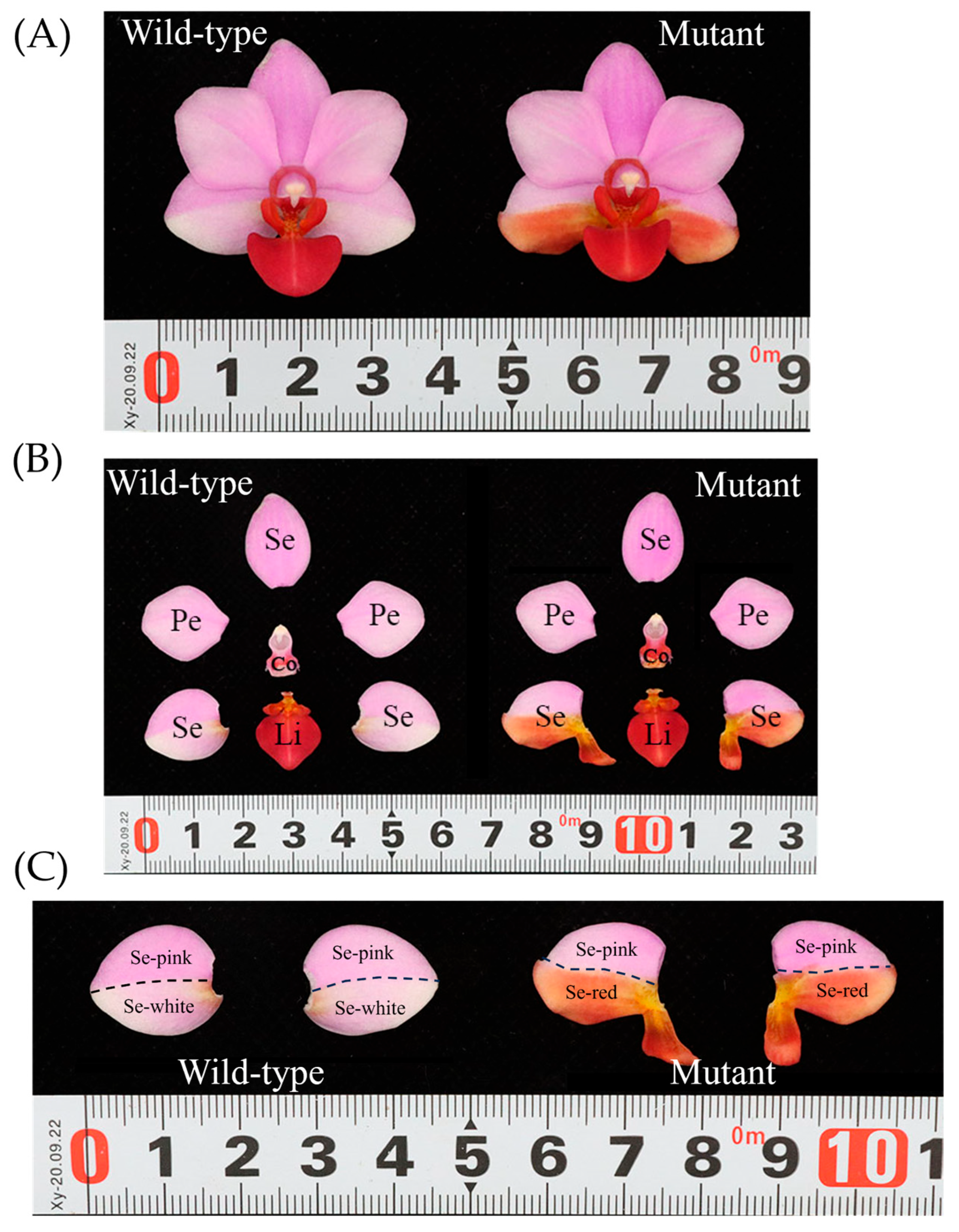
Figure 1. Flower morphology of Phalaenopsis ‘Huayang’. (A) Wild-type flower and mutant flower of Phalaenopsis. (B) Wild-type flower and mutant flower dissected into Se (sepal); Pe (petal); Li (lip); and Co (column). (C) The normal sepals of wild-type flowers were dissected into Se-white and Se-pink; the lip-like sepals of mutant flowers were dissected into Se-red and Se-pink.
Agronomy 15 01361 g001
Figure 2. Cell shapes of flowers of wild-type flower and mutant flower of Phalaenopsis using cryo-scanning electron microscopy (cryo-SEM). Adaxial and abaxial epidermis in (ad) Se-white, (eh) Se-pink, (il) petal, (mp) lip, and (qt) Se-red. Bars: 30 µm and 100 µm.
Agronomy 15 01361 g002
Figure 3. Metabolome analysis of wild-type flower and mutant flower of Phalaenopsis. (A) Heat map of 53 anthocyanin contents. (B) Number of differentially regulated pigment metabolites in different groups. (C) Histogram of differential metabolite contents. Bars with different lowercase letters are significantly different (p < 0.05).
Agronomy 15 01361 g003aAgronomy 15 01361 g003b
Figure 4. RNA-Seq analysis of wild-type flower and mutant flower. (A) Volcano plot of differentially expressed genes (DEGs). The X axis represents the log2-transformed fold change, the Y axis represents the −log10-transformed significance, the red points represent upregulated DEGs, the blue points represent downregulated DEGs, and the gray points represent non-DEGs. (B) GO classification of DEGs. The X-axis represents the GO term; the Y-axis represents the number of differentially expressed genes.
Agronomy 15 01361 g004
Figure 5. Phylogenetic tree showing relationships among MADS-box proteins of Arabidopsis thaliana, Oryza sativa, Phalaenopsis equestris, and Phalaenopsis ‘Huayang’.
Agronomy 15 01361 g005
Figure 6. Relative expression of MADS genes. All genes are classified into different subfamilies, and the color scale indicates relative expression levels from high (red) to low (green).
Agronomy 15 01361 g006
Figure 7. Quantitative real-time PCR verification of expression levels of eight DEGs identified via RNA sequencing. The y-axis on the right shows the relative gene expression levels (2−ΔΔCt) analyzed by qRT-PCR. The x-axis represents the different samples. Bars with different lowercase letters are significantly different (p < 0.05).
Agronomy 15 01361 g007
Figure 8. Interaction networks of the MADS-box proteins based on their homology to MADS-box proteins of A. thaliana. (A) The interaction network of the 29 MADS proteins. The blue arrows indicate the genes with down-regulated expression in the Se-red of mutant flowers, and the red arrows indicate the genes with up-regulated expression in the Se-red of mutant flowers. (B) The interaction network of three up-regulated MADS proteins in the Se-red of mutant flowers. The line color indicates the type of interaction evidence. The more colors on the lines, the greater the likelihood of their interaction.
Agronomy 15 01361 g008
Figure 9. Subcellular localization of three key MADS genes. The GFP fluorescence is shown in green and the DAPI fluorescence is shown in blue. The nuclei were indicated by DAPI staining. The GFP fluorescence was detected 50 h after infiltration. The scale bar is 22 μm.
Agronomy 15 01361 g009
Disclaimer/Publisher’s Note: The statements, opinions and data contained in all publications are solely those of the individual author(s) and contributor(s) and not of MDPI and/or the editor(s). MDPI and/or the editor(s) disclaim responsibility for any injury to people or property resulting from any ideas, methods, instructions or products referred to in the content.