Article

Potential of Salvia discolor Extract Against Common Agricultural Pathogens †

1
Department of Pharmacy, University of Genova, Viale Cembrano 4, 16148 Genova, Italy
2
CeRSAA Centro di Sperimentazione e Assistenza Agricola, Regione Rollo 98, 17031 Albenga, Italy
3
Department of Pharmacy, University of Salerno, Via Giovanni Paolo II 132, 84084 Salerno, Italy
*
Authors to whom correspondence should be addressed.
The article originates from part of the Ph.D. dissertation of the first author (P.D.): Devi, P. Chemical and biological investigation of some species of Salvia. University of Genoa, Genova, Italy, 2025.
Agronomy 2025, 15(6), 1268; https://doi.org/10.3390/agronomy15061268
Submission received: 30 April 2025 / Revised: 18 May 2025 / Accepted: 20 May 2025 / Published: 22 May 2025

Abstract

:
Phytopathogenic fungi and bacteria pose a serious threat to global agriculture, leading to significant economic losses and potential health risks. Consequently, the search for natural alternatives to synthetic agrochemicals has garnered increasing scientific attention, with plant extracts emerging as promising environmentally friendly solutions. In this context, the surface extract of Salvia discolor, obtained using dichloromethane, was analyzed for its bioactive potential. Chemical profiling revealed a rich composition of terpenoids and flavonoids. The antimicrobial potential of the ground extract was evaluated against nine phytopathogenic fungi (Alternaria solani, Botrytis cinerea, Colletotrichum lindemuthianum, Fusarium solani, Fusarium oxysporum f. sp. lactucae race 1, Phoma betae, Phaeomoniella chlamydospora, Pythium dissotocum, and Stemphylium sp.), and two phytopathogenic bacteria (Clavibacter michiganesis subsp. michiganesis and Pectobacterium carotovorum subsp. carotovorum), selected from common pathogens of agricultural interest. Complete inhibition of P. chlamydospora at 1000 µg mL−1 and strong activity against P. dissotocum, F. solani and B. cinerea was observed, and low inhibition (<40%) against C. lindemuthianum and F. oxysporum f. sp. lactucae race 1. However, the extract showed promising results in the post-harvest protection of tomatoes against gray mold. Moderate antibacterial activity was seen against C. michiganensis subsp. michiganensis. These findings indicate that S. discolor extract has the potential to serve as an effective natural crop protection agent, though further optimization may be needed for broader application.

1. Introduction

Significant losses in the agricultural sector are caused by phytopathogenic fungi and bacteria, which harm food production and cause health problems [1,2]. The most prevalent fungal genera affecting crops on a global scale are Alternaria, Botrytis, Fusarium, Pythium, and Stemphylium [3,4,5]. The most problematic and widespread plant pathogenic bacteria mainly originate from Gram-negative genera, including Xanthomonas, Pseudomonas, Xylella, and Erwinia, and Gram-positive genera such as Arthrobacter, Clavibacter, Curtobacterium, and Rhodococcus [6]. The presence of these organisms in agricultural systems poses a significant challenge to the implementation of effective pest management and crop protection strategies, resulting in considerable economic losses due to reduced yields and low-quality products [7,8,9]. Pathogenic fungal diseases account for approximately 65–70% of all crop losses caused by pathogens [10,11].
Chemical pesticides are commonly used for their effectiveness in reducing pest-related crop damage [12]. However, they pose severe health risks to humans, including fetal impairments and cancers, and are non-biodegradable, persisting in the environment for years [13,14]. The increased production of synthetic substances for agricultural use has underscored the need to develop sustainable disease control strategies that minimize risks to humans and the environment [15]. Research into natural products as potential fungicides and bactericides offers a promising, environmentally friendly alternative to mitigate the harmful effects of these pathogens and safeguard crop yield and quality. This approach could facilitate the identification of safer alternatives to traditional pesticides [16]. Plants produce various metabolites with bactericidal, fungicidal, insecticidal, and nematocidal properties to protect themselves against phytophagous organisms and diseases [17]. Several studies have demonstrated the potential of these natural substances to be integrated with traditional crop management practices, addressing the urgent need for sustainable agriculture, safe food production, and organic farming, which are advocated for by governmental bodies and major retailers [18,19,20].
The genus Salvia comprises nearly one thousand species of herbs, shrubs, and perennial plants, making it the largest genus within the Lamiaceae [21]. These plants are notably abundant in various natural compounds, including monoterpenoids, diterpenoids, phenolic acids, triterpenoids, flavonoids, and saccharides [22]. Historically, the Salvia genus has been used for diverse applications, such as in herbal beverages, culinary appetizers, and addressing ailments like colds, stomachaches, and gastrointestinal discomforts [23]. Furthermore, extensive research describes the biological activity of this genus’s members, including their anticancer, antioxidant, antihypertensive, hypoglycemic, hypolipidemic, memory-enhancing, antiviral, antimicrobial, and phytotoxic properties [22,24,25,26]. Phytochemicals derived from Salvia have demonstrated significant potential in combating phytopathogens responsible for diseases and substantial economic losses in agricultural systems globally [27]. In particular, surface extracts play an important role in plant defense by providing toxic, antifeedant, antifungal and antibacterial protection against herbivores and pathogens, thanks to the secretion of defense compounds onto the cuticular layer [28,29], and lipophilic extracts of several Salvia species have shown efficacy against several plant pathogens [24,25,26]. Due to their natural properties, several sage-derived antifungal compounds have been classified as “generally recognized as safe” (GRAS) for food preservation and crop protection [30]. Despite the promising potential of natural products, there is still a considerable gap in understanding the full spectrum of bioactive compounds found in Salvia species and their efficacy against a wide range of plant pathogens [27].
Addressing this gap, our study seeks to characterize the phytochemical profile of the surface dichloromethane extract of Salvia discolor Kunth [31,32,33], and to evaluate its bioactivity against phytopathogens, thus offering valuable insights into the development of novel, sustainable crop protection strategies [34]. Nine phytopathogenic fungi (Fusarium solani, Phoma betae, Phaeomoniella chlamydospora, Colletotrichum lindemuthianum, Fusarium oxysporum f. sp. lactucae race 1, Pythium dissotocum, Alternaria solani, Stemphylium sp., and Botrytis cinerea), and two phytopathogenic bacteria (Clavibacter michiganensis subsp. michiganensis and Pectobacterium carotovorum subsp. carotovorum), selected from the more common pathogens of agricultural interest, were tested. These pathogens induce various diseases, ranging from anthracnose and wilt diseases to bacterial canker and soft rot, resulting in more than 60% losses in agricultural production [10,35]. Fusarium solani is the main cause of root rot in legumes. This pathogen significantly affects the production of several legumes and vegetables, including beans, soybeans, peas, lentils, tomatoes, and cucurbits [36,37]. Phoma betae is known to cause leaf spots and root rot disease in beets and various other plants [36,38]. Phaeomoniella chlamydospora is identified as the principal fungal pathogen causing vascular diseases in grapevines, particularly within the Esca complex and Petri disease [39]. Colletotrichum lindemuthianum is classified as a hemibiotrophic fungus responsible for anthracnose, affecting common beans (Phaseolus vulgaris) and other Phaseolus species [40]. Fusarium oxysporum is a global ascomycete fungus, usually saprophytic and nonpathogenic, commonly found in soil. Certain strains cause wilt disease by invading the plant’s vascular system, resulting in severe wilting and often the death of the host plants [41,42]. Pythium dissotocum is a major pathogen responsible for seedling damping off and causes a range of severe diseases in various plants, affecting everything from young annual vegetable seedlings to mature forest trees [43,44]. Alternaria solani, a necrotrophic fungus, causes early bird disease in tomatoes, significantly impacting stems, foliage, and fruits, leading to substantial economic losses [45]. Stemphylium sp. causes gray leaf spots in tomato crops, resulting in significant defoliation and yield losses. Symptoms, visible on both seedlings and plants, manifest as circular to elongated dark specks on leaves, enlarging and becoming gray with age [46]. Botrytis cinerea affects numerous plant species, including grapevines, fruits, vegetables and ornamentals, and can infect almost all plant parts, from stems to leaves, flowers, and fruits, leading to gray mold disease [47,48]. Clavibacter michiganensis subsp. michiganensis is a Gram-positive bacterium that causes bacterial canker in tomatoes, which is particularly destructive in greenhouse conditions [49]. Pectobacterium carotovorum subsp. carotovorum, a Gram-negative pathogen, induces severe soft rot in various economically important vegetables, including carrots, cabbage, cucumbers, eggplants, garlic, onions, peppers, potatoes, radishes, sweet potatoes, pumpkins, and tomatoes [50].
Salvia discolor is cultivated as an ornamental in the Ligurian region (Italy) (information about the species can be found in the Supplementary Materials). Ornamental plant cultivation produces waste biomass, and this study is part of our research on the potential extractive use of waste biomass from local crops in the vision of sustainable agriculture [51,52].

2. Materials and Methods

2.1. General Experimental Procedures

Optical rotations, LC, TLC and MPLC chromatography, semi-preparative HPLC, and NMR experiments were performed as previously described [53,54].
MPLC chromatography was carried out using normal phase Si60 cartridges Supervarioflash and LiChroprep RP-18 (40–63 μm) (Merck, Darmstadt, Germany) in conjunction with a spot liquid chromatography system (Armen Instrument, Saint Ave, France). For TLC analysis, Silica gel 60 F254-coated aluminum sheets (Merck, dimensions: 20 × 20 cm, layer thickness: 0.2 mm) were used. The mobile phase composition was CHCl3/CH3OH/HCOOH (10:0.5:0.1), and spot visualization involved spraying with 50% H2SO4, followed by gentle heating. Flash chromatography was performed over an Isolera® Biotage® purification system (Biotage, Uppsala, Sweden) equipped with a Biotage flash SNAP KP-Sil column of 50 g, a flow rate of 35 mL/min, and a collection volume of 9 mL. Fractions were collected according to UV absorbance at 254 and 280 nm.
Semi-preparative HPLC employed a Waters W600 pump equipped with a Rheodyne Delta 600 injector, a 2414 refractive index detector, and a 2998 photodiode array detector (all Waters Corporation, Milford, MA, USA). A C18 column, Symmetry Prep C18, 7.8 × 300 mm i.d., with a 7 μm particle size (Waters), was used at room temperature, with a flow rate of 1.03 mL min−1. The sample loop was 100 μL, and the eluents were A: H2O, B: CH3OH; gradient: B 50% at time 0, B 100% at 61 min, B 100% at 75 min, B 100% at 100 min. A Bruker DRX-600 spectrometer (Bruker BioSpin GmBH, Rheinstetten, Germany) coupled with a Bruker 5 mm TCI CryoProbe at 300 K was used for NMR investigations. Every 2D NMR spectrum was obtained in CDCl3 (99.8%, Sigma-Aldrich, Milano, Italy), and the HSQC, COSY, HMBC, and ROESY spectra were obtained employing conventional pulse sequences and phase cycling. UXNMR software was employed to process the NMR data. tmix = 400 ms was used to acquire the ROESY spectra.
HRESIMS data were obtained in both the positive and negative ion mode on a Q Exactive Plus mass spectrometer, Orbitrap-based FT-MS system, equipped by an ESI source (Thermo Fischer Scientific Inc., Bremen, Germany). Following the manufacturer’s instructions, the Orbitrap mass analyzer was calibrated with a combination of caffeine, sodium dodecyl sulfate, sodium taurocholate, methionine-arginine-phenylalanine-alanine-acetate (MRFA), and Ultramark 1621. Information was collected and analyzed employing the software supplied by the manufacturer.

2.2. Plant Material

The fresh aerial parts of S. discolor were obtained from Consiglio per la ricerca in agricoltura e l’analisi dell’economia agraria (CREA), Sanremo (IM), Italy. The plant material was identified by Dr. Andrea Copetta and a voucher specimen (HMGBH.e/7290.2024.001) was deposited at Herbarium of Giardini Botanici Hanbury (La Mortola, Ventimiglia, Italy).

2.3. Chemicals

Extraction and purification solvents were acquired from VWR International Srl, (Milano, Italy). CDCl3 (99.8%) was acquired from Sigma-Aldrich (Milano, Italy).

2.4. Extraction and Isolation

1.2 Kg of freshly harvested aerial parts of S. discolor were briefly immersed in CH2Cl2 for 20 s [26] affording 53 g of dry residue. This extract underwent n-hexane partitioning, yielding an n-hexane-soluble (26.6 g) and an n-hexane-insoluble portion (23.2 g).
The n-hexane-soluble portion was separated by CC on silica gel (MPLC; monitoring by TLC), eluted with mixtures of n-hexane/CHCl3/CH3OH (from 1:1:0 to 0:0:1, 2.28 L) to afford 37 fractions (I1–I37). Fraction I26 (130.0 mg), eluted with a mixtures of CHCl3/CH3OH from 1.47 to 1.56 L, was purified by semi-preparative RP-HPLC, affording 1 (2.9 mg; tR = 75.5 min).
The n-hexane-insoluble extract was then treated with aqueous CH3OH (80%) to obtain a methanol-soluble fraction (7.2 g), and a methanol-insoluble fraction, mainly constituted by triterpene compounds, particularly ursolic and oleanolic acids (10.2 g). The methanol-soluble fraction was separated by CC on silica gel (MPLC; monitoring by TLC), eluted with mixtures of n-hexane/CHCl3/CH3OH (from 1:1:0 to 0:0:1, 1.65 L) to afford 11 fractions (II1–II11). Fraction II5 (1.2 g), eluted with CHCl3 from 0.51 to 0.57 L, was re-chromatographed on silica gel (MPLC; monitoring by TLC) eluted with mixtures of n-hexane/CHCl3/CH3OH (from 1:1:0 to 0:0:1, 1.68 L), affording 17 fractions (III1–III17). Fraction III8 (120.0 mg), eluted CHCl3 from 0.81 to 0.84 L, was further purified by semi-preparative RP-HPLC, affording 2 (0.7 mg; tR = 63.0 min) and 3 (3.1 mg; tR = 86.5 min). Fraction III10 (175.2 mg), eluted with CHCl3 from 0.87 to 0.90 L, was further purified by semi-preparative RP-HPLC, affording 4 (2.6 mg; tR = 52.0 min).
Fraction II6 (741.0 mg), eluted with CHCl3 from 0.57 to 0.60 L, was subjected to Isolera Biotage CC eluted with n-hexane/ethyl acetate (from 72:28 to 28:72, 0.89 L), affording 7 fractions (IV1–IV7). Fraction IV4 (128.7 mg), eluted with n-hexane/ethyl acetate 58:42 from 0.4 to 0.6 L, was purified by semi-preparative RP-HPLC affording 5 (1.0 mg; tR = 37.5 min).
Fraction II7 (0.7 g), eluted with CHCl3 from 0.60 to 0.74 L, after MPLC separation, afforded 12 fractions (V1–V12). Fraction V6 (103.5 mg), eluted with CHCl3 from 0.87 to 0.93 L, was purified by semipreparative RP-HPLC affording 6 (1.0 mg; tR = 35.1 min) and 7 (2.0 mg, tR = 60.5).
Fraction II9 (1.2 g), eluted with CHCl3/CH3OH 95:05 from 1.14 to 1.20 L, was subjected to Isolera Biotage CC eluting with n-hexane/ethyl acetate (from 1:1 to 0:1, 1.02 L), affording 12 fractions (VI1–VI12). Fraction VI6 (131.7 mg), eluted with n-hexane/ethyl acetate 87:13 from 0.24 to 0.30 L, was purified by semipreparative RP-HPLC affording 8 (1.4 mg; tR = 51.5 min) and 9 (1.2 mg; tR = 69.5 min). Fraction VI7 (159.8 mg), eluted with n-hexane/ethyl acetate 87:13 from 0.30 to 0.39 L, was purified by semi-preparative RP-HPLC affording 10 (3.7 mg; tR = 60.0 min).
The purity of the isolated compounds was confirmed by TLC and HPLC (purity > 95% for compounds 18. Compound 9 showed purity > 90%).

2.5. Microorganisms

Phytopathogenic fungi species A. solani, C. lindemuthianum, F. solani, F. oxysporum f. sp. lactucae race 1, P. betae, P. chlamydospora, P. dissotocum, and Stemphylium sp. were isolated from symptomatic plants of Solanum tuberosum, Vitis vinifera, Phaseolus vulgaris, Curcurbita pepo, Lactuca sativa, Spinacia oleracea, Apium graveolens, and S. lycopersicum, respectively, at CeRSAA (Albenga (SV), Italy). Four strains of B. cinerea were isolated from Lavandula angustifolia (strain 1), S. lycopersicum (strain 2), Viburnum opulus (strain 4), and Fragaria x ananassa (strain 11). Bacterial strains of C. michiganensis subsp. michiganensis and P. carotovorum subsp. carotovorum were isolated from symptomatic plants of Solanum lycopersicum and Brassica oleracea, respectively.

2.6. Poisoned Food Technique

The antifungal activity of the dichloromethane surface extract of S. discolor was evaluated against several plant pathogenic fungi using the poisoned food technique [55]. The inhibitory effect on mycelium growth of A. solani, B. cinerea, C. lindemuthianum, F. solani, F. oxysporum f.sp. lactucae race 1, P. betae, P. chlamydospora, P. dissotocum, and Stemphylium sp. has been tested. Culture medium was prepared by autoclaving 39 g of potato dextrose agar (Merck) in 1 L of distilled water at 121 °C for 15 min. After cooling to 50 °C, the crude surface extract [dissolved in 1% v/v DMSO (Sigma Aldrich, St. Louis, MO, USA)] was added to the medium at concentrations of 5, 100, 250, 500, 750, and 1000 μg mL−1. The range of extract concentrations (5–1000 µg/mL) was selected based on previously published studies on the antifungal activity of plant extracts, which commonly utilized similar concentration ranges to evaluate the effect at different doses [52,56,57]. The medium was then supplemented with streptomycin sulfate (5 μg mL−1) and solidified in 90 mm diameter Petri dishes. Four synthetic fungicides, Ortiva® (Syngenta, Milano, Italy) (azoxystrobin 23%) corresponding to a concentration of azoxystrobin 250 μg mL−1, Switch® (Syngenta, Milano, Italy) (cyprodinil 37% + fludioxonil 25%) corresponding to a concentration of cyprodinil 375 μg mL−1 and fludioxonil 250 μg mL−1, Score® 25 EC (Syngenta, Milano, Italy) (difenoconazole 23.6%) corresponding to a concentration of difenoconazole 250 μg mL−1, and Ridomil Gold®R WG (metalaxyl-m 2%+ copper 14.19%) corresponding to a concentration of metalaxyl-m 20 μg mL−1 and copper 141.9 μg mL−1, were included as controls at the manufacturer-recommended concentrations. Sterile agar plugs (approximately 2 × 2 mm) of the test fungi were placed centrally on the prepared media in Petri dishes, which were incubated in the dark at 25 °C. Each concentration of the extract was tested in triplicate, with additional controls consisting of media treated with 1% v/v DMSO and untreated media. The diameter of fungal growth was measured when the mycelium fully colonized the untreated plate. The inhibition percentage of growth, compared to the untreated control, was calculated using the formula I = [(C − T)/C] × 100, where I is the inhibition percentage, C and T are the mean diameters (cm) of the fungal colonies in the control (untreated) plate and the treated plate, respectively [58]. The experiments were repeated three times, yielding consistent results across all trials.

2.7. Efficacy of S. discolor Extract Against Post-Harvest Diseases in Tomato Fruit

Based on the in vitro results, the concentrations of 100, 500, and 1000 μg mL−1 of dichloromethane extract were selected as the optimal levels for subsequent in vivo experiments on tomato fruits against B. cinerea. The experiments were carried out according to the method described by Liu and co-workers [59] with a few modifications. Healthy tomatoes var. Maraskino (Seminis, Vegetables, Bayer, Leverkusen, Germany) were obtained from organic local farms in Albenga (SV) and washed with tap water to remove surface dust. The fruit was then disinfected by immersion in a 1% v/v sodium hypochlorite solution for 20 min, followed by two rinses with distilled water and air drying at 25 ± 2 °C. Tomato fruits were wounded at the equator using a sterile scalpel, creating incisions 3 mm deep and 3 mm wide. These wounds served as the sites for applying the dichloromethane extract and subsequent inoculation with B. cinerea. Preventive and curative treatments were applied during the tests. For the preventive treatments, tomato fruits were first treated with dichloromethane extract dissolved in 1% v/v DMSO at concentrations of 100, 500, and 1000 μg mL−1, applied using a nebulizer. Control treatments included 1% v/v DMSO and distilled water, while Teldor® (Bayer Cropscience s.r.l, Milano, Italy) (fenhexamid 42.7%) corresponding to a concentration of fenhexamid 750 μg mL−1, and 1% v/v peracetic acid served as reference treatments. After 4 h, B. cinerea conidial suspension (5.7 × 10⁴ spores mL−1) was applied to the fruits. For the curative treatments, the process was reversed: tomato wounds were first inoculated with the B. cinerea conidial suspension, followed by a 10 min waiting period. The dichloromethane extract, the control, and the reference solutions were then applied. Treated fruits were placed in plastic boxes (200 × 130 × 50 mm) and maintained at high relative humidity (~95%). Fruits treated with extract and positive and negative controls were kept in a climatic chamber at 18 ± 2 °C with a 14:10 dark: light ratio. The percentage of decay incidence caused by B. cinerea was assessed 7 days post-inoculation for both treatments. Each treatment was conducted in triplicate, and the experiment was repeated three times, for a total of 27 fruits per treatment [60].

2.8. Antibacterial Assay

To evaluate the antibacterial effect of S. discolor dichloromethane extract, two phytopathogenic bacteria (C. michiganensis subsp. michiganensis and P. carotovorum subsp. carotovorum) were utilized. A sterile stock solution of the dichloromethane extract in dimethyl sulfoxide (DMSO) was prepared and stored at −20 °C. This stock solution was subsequently diluted 1:10 with Mueller–Hinton broth medium (Merck-Millipore, Burlington, MA, USA), resulting in a final concentration of 2000 μg mL−1, which is twice the highest concentration tested. A sample of 100 μL of this solution was transferred to the wells in the first column of a microtiter plate. Similarly, sterile stock solutions of streptomycin sulfate (VWR Life Science, Radnor, PA, USA), bacitracin (Sigma-Aldrich, Milano, Italy), and ampicillin (Sigma-Aldrich, Milano, Italy) were prepared in DMSO and diluted 1:10 with Mueller–Hinton broth, achieving a concentration of 64 μg mL−1, double the highest test concentration. Each antibiotic solution (100 μL) was also transferred to the wells of the first column of a microtiter plate. One-day-old bacterial cultures were prepared by diluting them in Buffered Peptone Water (VWR Life Science, Radnor, PA, USA) until the suspension reached 0.5 on the McFarland scale. This bacterial suspension was then further diluted to 1/150 in Mueller–Hinton broth (Merck-Millipore, Burlington, MA, USA), achieving a final concentration of approximately 5 × 10⁵ cells mL−1. A microdilution procedure was used to determine the MIC (minimum inhibitory concentration value) reported by the Clinical and Laboratory Standards Institute using Mueller–Hinton broth as the test medium [61]. In summary, a 96-well microplate was prepared with 2-fold serial dilutions of the dichloromethane extract (0.976 to 1000 µg mL−1), and 50 µL of the inoculum generated as described above was introduced to corresponding amounts of various concentrations of the dichloromethane extract. At the same time, the inoculum was added to equivalent volumes of ampicillin and streptomycin sulfate spread over a 96-well microplate and prepared from 2-fold serial dilutions ranging from 0.0312 to 32 µg mL−1. The activity of DMSO was tested in the last second row, ranging from 0.0488 to 50 µL mL−1. Not-inoculated sterile Mueller–Hinton broth (MHB) medium served as a blank control in the final row, whereas drug-free wells were used in the final column to confirm bacterial growth. Bacterial growth was assessed by visual inspection of turbidity in the wells. The minimum inhibitory concentration (MIC) was determined by measuring the lowest concentration of chemicals that prevented observable growth after a 24 h incubation period at 35 °C. Each MIC determination was conducted in triplicate [61].

2.9. Statistical Analysis

Analysis of variance (ANOVA) was used to evaluate the effects of treatments, followed by Tukey’s HSD test for post hoc mean separation at a significance level of p ≤ 0.05. Each assay included three replicates per treatment. The assumptions underlying ANOVA were verified and met. Descriptive statistics and visual inspection of the data suggested that the data were approximately normally distributed. Given the small sample size per group, visual assessment was considered more appropriate than formal normality tests, which can be unreliable with very small samples. Homogeneity of variances was assessed with Levene’s test. The version of the test based on the median, which is more robust to non-normality and outliers, indicated that the assumption of equal variances was met. This supports the validity of the ANOVA results. ANOVA was performed using Statistica software, version 8.0 (StatSoft Inc., Tulsa, OK, USA). Generalized linear models (GLMs) were constructed using the lme4 package version 1.1.37 in R [62,63] to investigate the percentage of inhibition, the administered dose, and fungal species identity. Various model configurations were tested, including the additive effect of the predictors and the interaction between the variables. In addition, the effect of the quantitative variable dose was evaluated in its original form as well as in logarithmic, second-degree, and third-degree polynomial transformations. The model with the highest coefficient of determination (R2) and statistical significance was selected for further analysis. Predicted inhibition values were computed using the ggpredict function from the ggeffects package [64]. For each fungal species, the median effective dose (ED50) [65] and corresponding 95% confidence intervals were estimated using MATLAB R2024b (Microsoft, Redmond, WA, USA). Additionally, a third-degree polynomial was fitted to the data using the polyfit function, with absolute dose values on the x-axis and inhibition percentages on the y-axis. This polynomial degree was chosen to capture non-linear trends while minimizing the risk of overfitting. The resulting equation was solved using the roots function, and 95% confidence intervals were calculated using the polyconf function. Model fit quality was evaluated using the coefficient of determination (R2).

3. Results

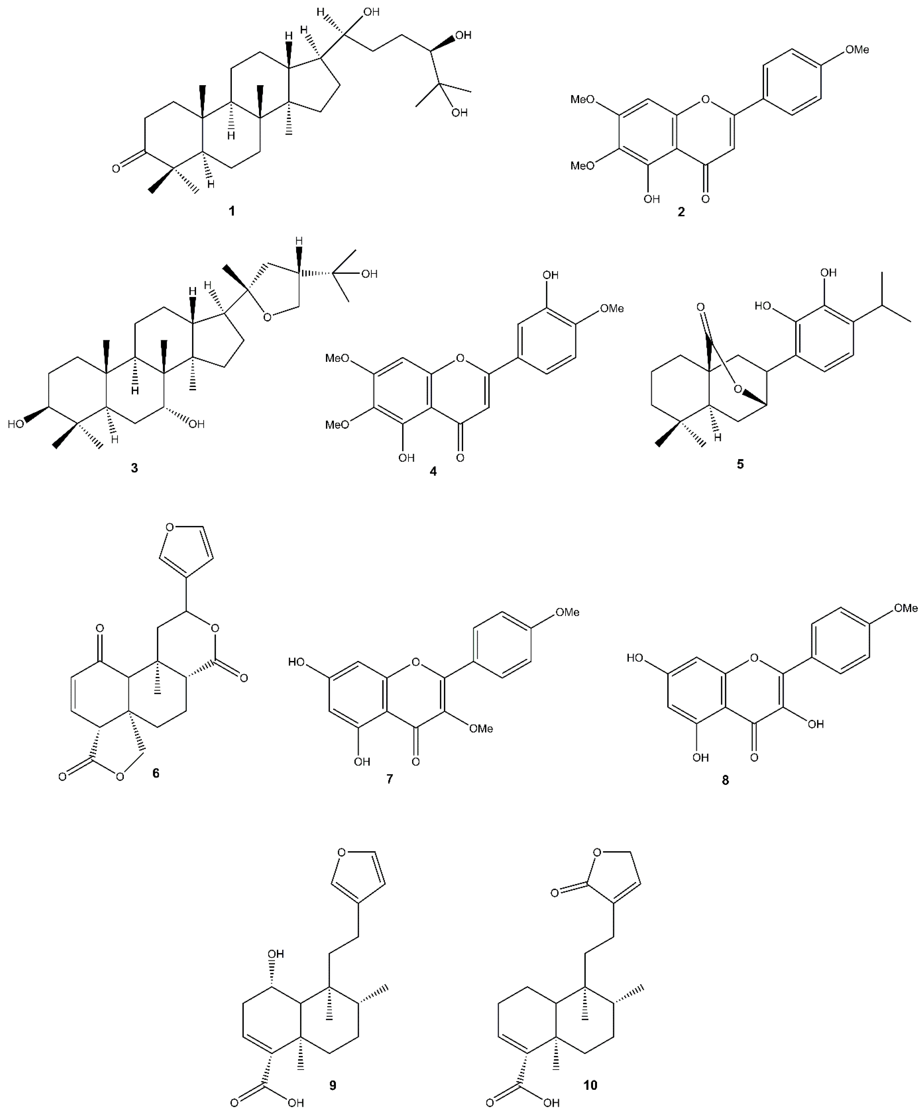
3.1. Chemical Analysis

The dichloromethane extract obtained from the plant surface of S. discolor afforded some known compounds, gardaubryone C (1) [66], salvigenin (2) [67], salvilymitol (3) [68], eupatorin (4) [69], carnosol (5) [70], an ent-clerodane diterpene dilactone, rhynchosperin A (6) [71,72], ermanin (7) [73], kaempferide (8) [74], divinatorin A (9) [75], and patagonic acid (10) [76] (Figure 1), which were identified through NMR analysis (Figures S1–S10, Supplementary Materials), including 1H and 13C NMR, TOCSY, HSQC, and HMBC experiments, and HRESIMS data.

3.2. Antifungal Assays

The antifungal efficacy of S. discolor dichloromethane extract was assessed in vitro at various concentrations against nine phytopathogenic fungi (A. solani, B. cinerea, C. lindemuthianum, F. solani, F. oxysporum f. sp. lactucae race 1, P. betae, P. chlamydospora, P. dissotocum, and Stemphylium sp.). The activity was compared with four synthetic fungicides and expressed as a percentage of inhibition of mycelial growth. The percentage of growth inhibition was determined by comparing mycelial growth in the presence of different concentrations of the tested compounds with growth in the control condition, where no compounds were applied. Representative figures illustrating the inhibition of fungal mycelial growth after inoculation onto a medium containing the extract are provided in the Supplementary Materials (Figures S11–S14). Complete inhibition of P. chlamydospora was observed at the extract concentration of 1000 μg mL−1 (Table 1), statistically comparable to the standard fungicides Switch® (cyprodinil 375 μg mL−1 and fludioxonil 250 μg mL−1), Score® 25 EC (difeconazole 250 μg mL−1), and Ridomil Gold®R WG (metalaxyl-m 20 μg mL−1 and copper 141.9 μg mL−1). At a lower concentration, 100 μg mL−1, a 59% inhibition rate was recorded, and at higher concentrations (250, 500, and 750 μg mL−1) 77.1%, 80.9%, and 88.6% inhibition rates, respectively, were observed (Table 1).
At the concentration of 1000 μg mL−1 the extract achieved a 91.4% inhibition rate against F. solani, a better result than with Score® 25 EC (difenoconazole, 250 μg mL−1) and Ridomil Gold® WG (metalaxyl-m 20 μg mL−1 and copper 141.9 μg mL−1), but lower than that obtained with Ortiva® (azoxystrobin 250 μg mL−1). At the concentration of 1000 μg mL−1, the extract achieved a 91.4% inhibition rate against P. dissotocum, a better result than with Score® 25 EC (difenoconazole, 250 μg mL−1), but lower than other fungicides. The extract exhibited 70.5% inhibition against P. betae at a concentration of 1000 μg mL−1, though this was not comparable to synthetic fungicides. Low inhibitory effects were observed against A. solani, Stemphylium sp., and B. cynerea, and the least inhibition was observed against C. lindemuthianum and F. oxysporum f. sp. lactucae race 1, with less than 40% inhibition across the tested concentrations.
The GLM highlighted statistically significant effects for the interaction between species and dose, albeit with differences between species in the estimated effect. In particular, the treatment was more effective on P. chlamydospora (estimated effect of dose = 0.098) and F. solani (estimated effect of dose = 0.084), while it was very low for other species, including C. lindemuthianum (estimated effect of dose = 0.012) and F. oxysporum f. sp. lactucae (estimated effect of dose = 0.004) (Figure S15, Supplementary Material).
Furthermore, ED50, the dose required to inhibit 50% of fungal growth, was calculated (Table 2). Among the tested fungi, P. chlamydospora was the most sensitive to the extract, with the lowest ED50 value of 71.73 μg mL−1. Antifungal activity was also observed against F. solani (ED50 185.55 μg mL−1), and lower activity was observed against P. betae (ED50 277.41 μg mL−1) and P. dissotocum (ED50 530.82 μg mL−1). ED50 values for C. lindemuthianum, F. oxysporum f. sp. lactucae (race 1), and B. cynerea could not be determined, indicating very limited efficacy.
A further evaluation of the extract’s efficacy against three additional strains of B. cinerea from different diseased crops is presented in Table 3. For strain 1 (L. angustifolia), only 20% inhibition was observed at the highest extract concentration. In field conditions, crops like L. angustifolia and V. opulus typically receive fewer treatments due to economic constraints, making frequent applications impractical. In contrast, for strain 2 (S. lycopersicum) and strain 11 (F. x ananassa), inhibition rates at 750 and 1000 μg mL−1 were significantly higher than those obtained with Ridomil Gold®R WG (metalaxyl-m 20 μg mL−1 and copper 141.9 μg mL−1) and Ortiva® (azoxystrobin 250 μg mL−1). These strains have a higher potential for developing resistance due to their origin from crops subjected to high-load treatments. The inhibition levels for all tested B. cinerea strains were still lower compared to those achieved with Switch® (cyprodinil 375 μg mL−1 and fludioxonil 250 μg mL−1).
ED50 values (Table 4) revealed considerable variation in sensitivity among the tested B. cinerea strains. Strain 2 was the most responsive, with the lowest ED50 value of 566.10 μg mL−1. Strain 11 showed a moderate susceptibility, with ED50 583.20 μg mL−1. Strains 4 and 1 showed substantial resistance or insensitivity to the extract.

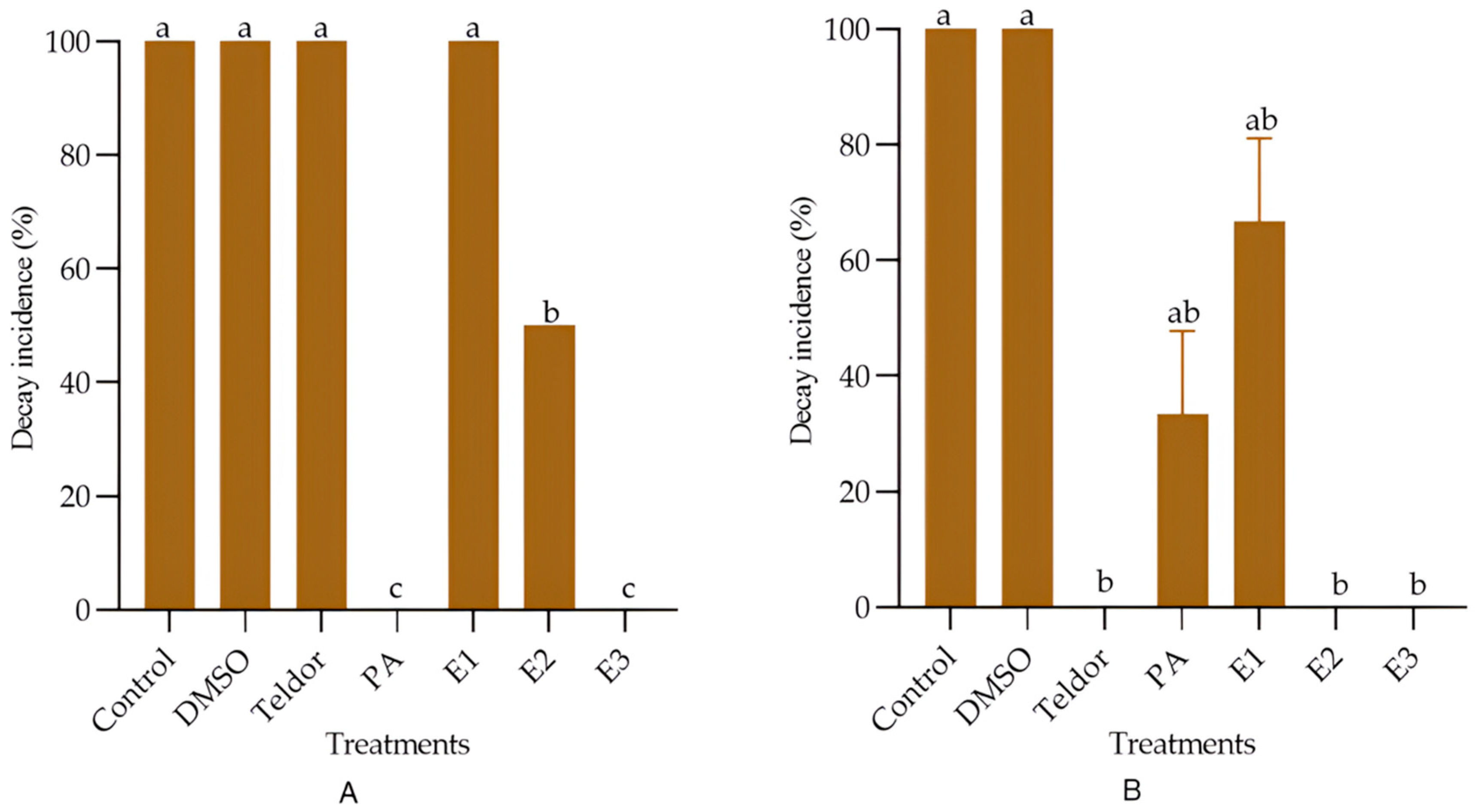
3.3. Effect of the Dichloromethane Extract Against Post-Harvest Diseases in Tomato Fruit

The effects of the S. discolor extract and of reference controls varied between curative and preventive treatments against decay incidence caused by B. cinerea (Figure 2). Teldor® (fenhexamid 750 μg mL−1) was ineffective when used as a curative treatment but achieved 100% activity as a preventive treatment. Peracetic acid showed potent efficacy in the curative treatment, while its ability to control decay incidence in the preventive treatment was moderate. The dichloromethane extract at a concentration of 1000 μg mL−1 significantly protected tomatoes from gray mold disease in both preventive and curative applications. Interestingly, its effect at 500 μg mL−1 and 1000 μg mL−1 was statistically comparable to Teldor® when used in preventive treatment. Moreover, small activity of the extract was observed at 100 μg mL−1 in preventive treatment comparable to the effect of peracetic acid.

3.4. Antibacterial Assays

The S. discolor extract was tested against two species of phytopathogenic bacteria (C. michiganensis subsp. michiganensis, and P. carotovorum subsp. carotovorum). Bacitracin and streptomycin sulfate were used as standard agents against Gram-positive bacteria [77,78]. Ampicillin and streptomycin sulfate were also used as reference antibiotics against Gram-negative bacteria [79,80,81,82]. The crude extract showed an MIC value of 500 μg mL−1 against C. michiganensis subsp. michiganensis and 1000 μg mL−1 against P. carotovorum subsp. carotovorum. Bacitracin showed an MIC value of 0.0312 μg mL−1 against C. michiganensis subsp. michiganensis. Streptomycin sulfate showed an MIC value of 0.25 μg mL−1 against both C. michiganensis subsp. michiganensis and P. carotovorum subsp. carotovorum. Ampicillin showed an MIC value of 0.0625 μg mL−1 against P. carotovorum subsp. carotovorum. Growth of each bacterium was observed in drug-free wells (growth control), and no growth was observed in wells containing uninoculated sterile Mueller–Hinton broth (MHB) as a blank control.

4. Discussion

The importance of safeguarding against phytopathogens has been acknowledged for centuries, dating back to the ancient use of chemical and organic pesticides [12]. Excessive pesticide application can result in the accumulation of chemical residues on crop surfaces, promoting the development of resistant pest populations. This not only poses potential health risks to consumers but also adversely affects environmental health by disrupting ecosystems and contaminating soil and water sources [83,84]. Even though researchers are always finding and developing novel active compounds for crop protection, the process of discovery is still difficult [85], as active substances have to live up to ever-higher consumer standards and legal restrictions [86]. In the present study, the surface dichloromethane extract of Salvia discolor was analyzed to identify its chemical constituents and evaluate its biological activity against phytopathogenic organisms. Chemical analysis of the surface extract revealed the presence of several known terpenoids and flavonoids. Among these, diterpenoids are particularly noteworthy due to their broad spectrum of biological activities, including cytotoxic, antimicrobial, antiprotozoal, antioxidant, phytotoxic, and insecticidal effects, highlighting their importance in plant defense mechanisms against phytopathogens [22,87]. Previous research has shown that extracts and essential oils from Salvia and other plant species have significant effects against fungal phytopathogens [19,27,56,88]. The extract of S. discolor displayed the best antifungal efficacy at higher concentrations against P. chlamydospora, and was also active against F. solani with an over 90% inhibition of mycelial growth. The activity of the extract could be attributed to the presence of diterpenes, as previous studies have demonstrated the antifungal properties of these compounds [7]. These results are very promising as P. chlamydospora is the primary pathogen responsible for Esca disease in grapevines and the tested extract showed that 50% of growth can already be inhibited at a concentration of 71.73 μg mL−1, highlighting its potential as an effective antifungal agent [89]. These findings are consistent with the results of Cobos et al. [90], who tested various natural antifungals against P. chlamydospora, showing that chitosan oligosaccharide (1000 µg mL−1) and garlic extract both achieved the 100% inhibition of in vitro mycelial growth. Furthermore, the extract showed low effects against A. solani, while previous research reported moderate antifungal activity of ethanolic extracts of S. fructicosa against two strains of the same pathogen [91].
Botrytis cinerea poses a significant threat to crop production in various fields and greenhouse crops. The extract of S. discolor showed low efficacy against strains of B. cinerea from heavily treated crops such as S. lycopersicum (strain 2) and Fragaria × ananassa (strain 11), with inhibition levels lower than those achieved by the synthetic fungicide Switch®. Similar activity was observed by the crude extract of S. somalensis against highly treated strains of B. cinerea [52]. The extract of S. discolor was inactive against the other tested strains 1 and 4 obtained from Lavandula angustifolia and Viburnum opulus, respectively. This reduced efficacy may result from resistance developed in these crops due to frequent pesticide treatments.
The efficacy of the extract of S. discolor was also tested against gray mold disease in postharvest tomato fruits. At 500 µg mL−1, the extract showed moderate efficacy in curative applications, while at 1000 µg mL−1 no fruit decay was observed. These findings are significant, as the use of chemical treatments on postharvest fruits can leave harmful residues, making them unsafe for consumption [92].
Finally, the surface extract also exhibited moderate inhibition against the phytopathogenic bacterium C. michiganensis subsp. michiganensis (MIC value 500 µg mL−1), which causes bacterial wilt and canker in tomatoes. This bacterium is widely considered to be the most important bacterial disease affecting tomatoes, resulting in significant commercial losses worldwide [93]. Although the MIC values were higher than those of standard antibiotics, the efficacy of the extract against this bacterium suggests its potential as a natural antibacterial agent, especially considering increasing antibiotic resistance and the prohibition of antibiotic use in agriculture in Europe. Furthermore, except for copper, no antibacterial chemicals are available for managing bacterial diseases in plants [94]. Our results are consistent with the MIC value of 500 µg mL−1 shown by the aqueous methanol (1:1, v/v) leaf extract of Ginkgo biloba [95] and the Silybum marianum aqueous extract [96] against C. michiganensis subsp. michiganensis. Other plant extracts showed similar results: Rubus, Anvillea and Pistacia aqueous extracts had MICs equal to 3125 µg mL−1 [97]. In a recent study, the essential oil of Salvia hydrangea showed an MIC value of 500 µL mL−1 against the same pathogen [98]. On the other hand, the activity of the surface extract against P. carotovorum subsp. carotovorum (MIC value 1000 µg mL−1) was weak compared to other results in the literature [99,100,101,102].

5. Conclusions

The current study proves that the surface extract of Salvia discolor has potential as a crop protectant. These results validate the first goal of assessing S. discolor as an alternative, sustainable substitute for traditional agrochemicals. To realize its full potential, further research on formulation strategies, synergistic blends, and applications under field conditions is needed. This would facilitate its use in integrated pest management programs and support the development of safer, environmentally friendly options for modern agriculture.

Supplementary Materials

The following supporting information can be downloaded at: https://www.mdpi.com/article/10.3390/agronomy15061268/s1: Figure S1. 1H NMR (600 MHz, CDCl3) spectrum of compound 1; Figure S2. 1H NMR (600 MHz, CDCl3) spectrum of compound 2; Figure S3. 1H NMR (600 MHz, CDCl3) spectrum of compound 3; Figure S4. 1H NMR (600 MHz, CDCl3) spectrum of compound 4; Figure S5. 1H NMR (600 MHz, CDCl3) spectrum of compound 5; Figure S6. 1H NMR (600 MHz, CDCl3) spectrum of compound 6; Figure S7. 1H NMR (600 MHz, CDCl3) spectrum of compound 7; Figure S8. 1H NMR (600 MHz, CDCl3) spectrum of compound 8; Figure S9. 1H NMR (600 MHz, CDCl3) spectrum of compound 9; Figure S10. 1H NMR (600 MHz, CDCl3) spectrum of compound 10; Figure S11. Inhibition of growth of Fusarium solani mycelium 5 days after inoculation on poisoned media; Figure S12. Inhibition of growth of Phaeomoniella chlamydospora mycelium 5 days after inoculation on poisoned media; Figure S13. Inhibition of growth of Pythium dissocotum mycelium 4 days after inoculation on poisoned media; Figure S14. Inhibition of growth of Botrytis cinerea (strain 11) mycelium 5 days after inoculation on poisoned media; Figure S15. Effect of treatment dose on fungal growth inhibition across different species; Information about Salvia discolor Kunth F.W.H.von Humboldt.

Author Contributions

Conceptualization, A.P.L., N.D.T. and A.B.; methodology, A.P.L., A.M. and A.B.; validation, A.P.L., N.D.T. and A.B.; formal analysis, P.D. and A.P.L.; investigation, P.D., A.P.L., V.P. and V.I.; resources, A.B.; data curation, A.P.L.; writing—original draft preparation, P.D. and A.P.L.; writing—review and editing, A.P.L., N.D.T. and A.B.; supervision, A.P.L., N.D.T. and A.B.; project administration, A.B.; funding acquisition, A.B. All authors have read and agreed to the published version of the manuscript.

Funding

This research received no external funding.

Data Availability Statement

The original contributions presented in this study are included in the article. Further inquiries can be directed to the corresponding authors.

Conflicts of Interest

The authors declare no conflicts of interest.

References

  1. Doehlemann, G.; Ökmen, B.; Zhu, W.; Sharon, A. Plant Pathogenic Fungi. In The Fungal Kingdom; Heitman, J., Howlett, B.J., Crous, P.W., Stukenbrock, E.H., James, T.Y., Gow, N.A.R., Eds.; American Society for Microbiology: Washington, DC, USA, 2017; pp. 701–726. [Google Scholar]
  2. Savary, S.; Willocquet, L.; Pethybridge, S.J.; Esker, P.; McRoberts, N.; Nelson, A. The global burden of pathogens and pests on major food crops. Nat. Ecol. Evol. 2019, 3, 430–439. [Google Scholar] [CrossRef] [PubMed]
  3. Fillinger, S.; Elad, Y. Botrytis—The Fungus, The Pathogen and Its Management in Agricultural Systems; Springer: Heidelberg, Germany, 2016. [Google Scholar]
  4. Yang, L.-N.; He, M.-H.; Ouyang, H.-B.; Zhu, W.; Pan, Z.-C.; Sui, Q.-J.; Shang, L.-P.; Zhan, J. Cross-resistance of the pathogenic fungus Alternaria alternata to fungicides with different modes of action. BMC Microbiol. 2019, 19, 205. [Google Scholar] [CrossRef] [PubMed]
  5. Zuriegat, Q.; Zheng, Y.; Liu, H.; Wang, Z.; Yun, Y. Current progress on pathogenicity-related transcription factors in Fusarium oxysporum. Mol. Plant Pathol. 2021, 22, 882–895. [Google Scholar] [CrossRef]
  6. Gakuubi, M.M.; Wagacha, J.M.; Dossaji, S.F.; Wanzala, W. Chemical composition and antibacterial activity of essential oils of Tagetes minuta (Asteraceae) against selected plant pathogenic bacteria. Int. J. Microbiol. 2016, 2016, 7352509. [Google Scholar] [CrossRef]
  7. López-Cabeza, R.; Rodríguez-Sabina, S.; Reyes, C.P.; Expósito, D.G.; Giménez, C.; Jiménez, I.A.; Cabrera, R.; Bazzocchi, I.L. Bio-guided isolation of aromatic abietane diterpenoids from Salvia canariensis as biopesticides in the control of phytopathogenic fungi. Pest Manag. Sci. 2024, 80, 2199–2207. [Google Scholar] [CrossRef]
  8. El-Baky, N.A.; Amara, A. Recent approaches towards control of fungal diseases in plants: An updated review. J. Fungi 2021, 7, 900. [Google Scholar] [CrossRef]
  9. Jain, A.; Sarsaiya, S.; Wu, Q.; Lu, Y.; Shi, J. A review of plant leaf fungal diseases and its environment speciation. Bioengineered 2019, 10, 409–424. [Google Scholar] [CrossRef]
  10. Fisher, M.C.; Henk, D.A.; Briggs, C.J.; Brownstein, J.S.; Madoff, L.C.; McCraw, S.L.; Gurr, S.J. Emerging fungal threats to animal, plant and ecosystem health. Nature 2012, 484, 186–194. [Google Scholar] [CrossRef] [PubMed]
  11. Al-Goul, S.T. The impact of eco-friendly nanoparticles on the management of phytopathogenic fungi: A comprehensive review. J. Plant Pathol. 2025, 107, 265–290. [Google Scholar] [CrossRef]
  12. Nicolopoulou-Stamati, P.; Maipas, S.; Kotampasi, C.; Stamatis, P.; Hens, L. Chemical pesticides and human health: The urgent need for a new concept in agriculture. Front. Public Health 2016, 4, 148. [Google Scholar] [CrossRef]
  13. Kalliora, C.; Mamoulakis, C.; Vasilopoulos, E.; Stamatiades, G.A.; Kalafati, L.; Barouni, R.; Karakousi, T.; Abdollahi, M.; Tsatsakis, A. Association of pesticide exposure with human congenital abnormalities. Toxicol. Appl. Pharmacol. 2018, 346, 58–75. [Google Scholar] [CrossRef] [PubMed]
  14. Khursheed, A.; Rather, M.A.; Jain, V.; Wani, A.R.; Rasool, S.; Nazir, R.; Malik, N.A.; Majid, S.A. Plant based natural products as potential ecofriendly and safer biopesticides: A comprehensive overview of their advantages over conventional pesticides, limitations and regulatory aspects. Microb. Pathog. 2022, 173, 105854. [Google Scholar] [CrossRef] [PubMed]
  15. Tian, Z.; Wang, J.-W.; Li, J.; Han, B. Designing future crops: Challenges and strategies for sustainable agriculture. Plant J. 2021, 105, 1165–1178. [Google Scholar] [CrossRef]
  16. PAN. PAN 2023–2024 Annual Report; PAN: Berkeley, CA, USA, 2024. [Google Scholar]
  17. Košćak, L.; Lamovšek, J.; Đermić, E.; Prgomet, I.; Godena, S. Microbial and plant-based compounds as alternatives for the control of phytopathogenic bacteria. Horticulturae 2023, 9, 1124. [Google Scholar] [CrossRef]
  18. Jiménez-Reyes, M.F.; Carrasco, H.; Olea, A.; Silva-Moreno, E. Natural compounds: A sustainable alternative for controlling phytopathogens. J. Chil. Chem. Soc. 2019, 64, 4459–4465. [Google Scholar] [CrossRef]
  19. Kursa, W.; Jamiołkowska, A.; Wyrostek, J.; Kowalski, R. Antifungal effect of plant extracts on the growth of the cereal pathogen Fusarium spp.—An in vitro study. Agronomy 2022, 12, 3204. [Google Scholar] [CrossRef]
  20. Steglińska, A.; Bekhter, A.; Wawrzyniak, P.; Kunicka-Styczyńska, A.; Jastrząbek, K.; Fidler, M.; Śmigielski, K.; Gutarowska, B. Antimicrobial activities of plant extracts against Solanum tuberosum L. phytopathogens. Molecules 2022, 27, 1579. [Google Scholar] [CrossRef] [PubMed]
  21. Bisio, A.; Pedrelli, F.; D’Ambola, M.; Labanca, F.; Schito, A.M.; Govaerts, R.; De Tommasi, N.; Milella, L. Quinone diterpenes from Salvia species: Chemistry, botany, and biological activity. Phytochem. Rev. 2019, 18, 665–842. [Google Scholar] [CrossRef]
  22. Jassbi, A.R.; Zare, S.; Firuzi, O.; Xiao, J. Bioactive phytochemicals from shoots and roots of Salvia species. Phytochem. Rev. 2016, 15, 829–867. [Google Scholar] [CrossRef]
  23. Sharifi-Rad, M.; Ozcelik, B.; Altın, G.; Daşkaya-Dikmen, C.; Martorell, M.; Ramírez-Alarcón, K.; Alarcón-Zapata, P.; Morais-Braga, M.F.B.; Carneiro, J.N.P.; Alves Borges Leal, A.L.; et al. Salvia spp. plants-from farm to food applications and phytopharmacotherapy. Trends Food Sci. Technol. 2018, 80, 242–263. [Google Scholar] [CrossRef]
  24. Bisio, A.; Damonte, G.; Fraternale, D.; Giacomelli, E.; Salis, A.; Romussi, G.; Cafaggi, S.; Ricci, D.; De Tommasi, N. Phytotoxic clerodane diterpenes from Salvia miniata Fernald (Lamiaceae). Phytochemistry 2011, 72, 265–275. [Google Scholar] [CrossRef]
  25. Bisio, A.; Fraternale, D.; Damonte, G.; Millo, E.; Lanteri, A.P.; Russo, E.; Romussi, G.; Parodi, B.; Ricci, D.; De Tommasi, N. Phytotoxic Activity of Salvia x jamensis. Nat. Prod. Commun. 2009, 4, 1621–1630. [Google Scholar] [CrossRef] [PubMed]
  26. Bisio, A.; Fraternale, D.; Giacomini, M.; Giacomelli, E.; Pivetti, S.; Russo, E.; Caviglioli, G.; Romussi, G.; Ricci, D.; De Tommasi, N. Phytotoxicity of Salvia spp. exudates. Crop Prot. 2010, 29, 1434–1446. [Google Scholar] [CrossRef]
  27. Zaccardelli, M.; Pane, C.; Caputo, M.; Durazzo, A.; Lucarini, M.; Silva, A.M.; Severino, P.; Souto, E.B.; Santini, A.; De Feo, V. Sage species case study on a spontaneous Mediterranean plant to control phytopathogenic fungi and bacteria. Forests 2020, 11, 704. [Google Scholar] [CrossRef]
  28. LoPresti, E.F. Chemicals on plant surfaces as a heretofore unrecognized, but ecologically informative, class for investigations into plant defence. Biol. Rev. 2015, 91, 1102–1117. [Google Scholar] [CrossRef]
  29. Muller, C.; Riederer, M. Plant surface properties in chemical ecology. J. Chem. Ecol. 2005, 31, 2621–2651. [Google Scholar] [CrossRef]
  30. Marchev, A.; Haas, C.; Schulz, S.; Georgiev, V.; Steingroewer, J.; Bley, T.; Pavlov, A. Sage in vitro cultures: A promising tool for the production of bioactive terpenes and phenolic substances. Biotechnol. Lett. 2014, 36, 211–221. [Google Scholar] [CrossRef]
  31. Jenks, A.A.; Walker, J.B.; Kim, S.-C. Phylogeny of New World Salvia subgenus Calosphace (Lamiaceae) based on cpDNA (psbA-trnH) and nrDNA (ITS) sequence data. J. Plant Res. 2013, 126, 483–496. [Google Scholar] [CrossRef] [PubMed]
  32. Martínez-Gordillo, M.; Martínez-Ambriz, E.; García-Peña, M.R.; Cantú-Morón, E.A.; Fragoso-Martínez, I. Lamiaceae Martinov; Universidad Nacional Autónoma de México: Ciudad de México, México, 2019; Volume 2019, p. 119. [Google Scholar]
  33. POWO. Plants of the World Online. Available online: https://powo.science.kew.org (accessed on 24 January 2025).
  34. Devi, P. Chemical and Biological Investigation of Some Species of Salvia. Ph.D. Thesis, University of Genoa, Genova, Italy, 2025. [Google Scholar]
  35. Almasoudi, N.M.; Al-Qurashi, A.D.; Elsayed, M.I.; Abo-Elyousr, K.A.M. Native bacterial bioagents for management of potato soft rot disease caused by Pectobacterium carotovorum subsp. carotovorum. Egypt. J. Biol. Pest Control 2024, 34, 31. [Google Scholar] [CrossRef]
  36. Bodah, E. Root rot diseases in plants: A review of common causal agents and management strategies. Agric. Res. Technol. 2017, 5, 56–63. [Google Scholar] [CrossRef]
  37. Kong, W.; Huo, H.; Gu, Y.; Cao, Y.; Wang, J.; Liang, J.; Niu, S. Antifungal activity of camphor against four phytopathogens of Fusarium. S. Afr. J. Bot. 2022, 148, 437–445. [Google Scholar] [CrossRef]
  38. Sultana, F.; Hossain, M.M. Diseases of vegetables caused by Phoma spp. In Phoma: Diversity, Taxonomy, Bioactivities, and Nanotechnology; Rai, M., Zimowska, B., Kövics, G.J., Eds.; Springer International Publishing: Cham, Switzerland, 2022; pp. 91–119. [Google Scholar]
  39. Andolfi, A.; Mugnai, L.; Luque, J.; Surico, G.; Cimmino, A.; Evidente, A. Phytotoxins produced by fungi associated with grapevine trunk diseases. Toxins 2011, 3, 1569–1605. [Google Scholar] [CrossRef]
  40. Khaledi, N.; Hassani, F. Antifungal activity of Bunium persicum essential oil and its constituents on growth and pathogenesis of Colletotrichum lindemuthianum. J. Plant Prot. Res. 2018, 58, 431–441. [Google Scholar] [CrossRef]
  41. Rosado-Álvarez, C.; Molinero-Ruiz, L.; Rodríguez-Arcos, R.; Basallote-Ureba, M.J. Antifungal activity of asparagus extracts against phytopathogenic Fusarium oxysporum. Sci. Hortic. 2014, 171, 51–57. [Google Scholar] [CrossRef]
  42. Minerdi, D.; Sadeghi, S.J.; Pautasso, L.; Morra, S.; Aigotti, R.; Medana, C.; Gilardi, G.; Gullino, M.L.; Gilardi, G. Expression and role of CYP505A1 in pathogenicity of Fusarium oxysporum f. sp. lactucae. Biochim. Biophys. Acta 2020, 1868, 140268. [Google Scholar] [CrossRef]
  43. Martin, F.N.; Loper, J.E. Soilborne plant diseases caused by Pythium spp.: Ecology, epidemiology, and prospects for biological control. Crit. Rev. Plant Sci. 1999, 18, 111–181. [Google Scholar] [CrossRef]
  44. Kasteel, M.; Ketelaar, T.; Govers, F. Fatal attraction: How Phytophthora zoospores find their host. Semin. Cell Dev. Biol. 2023, 148, 13–21. [Google Scholar] [CrossRef]
  45. Karačić, V.; Miljaković, D.; Marinković, J.; Ignjatov, M.; Milošević, D.; Tamindžić, G.; Ivanović, M. Bacillus species: Excellent biocontrol agents against tomato diseases. Microorganisms 2024, 12, 457. [Google Scholar] [CrossRef]
  46. Zorzi Tomazoni, E.; Schiavo Griggio, G.; Pessin Broilo, E.; da Silva Ribeiro, R.T.; Gonçalves Soares, G.L.; Schwambach, J. Screening for inhibitory activity of essential oils on fungal tomato pathogen Stemphylium solani Weber. Biocatal. Agric. Biotechnol. 2018, 16, 364–372. [Google Scholar] [CrossRef]
  47. Orozco-Mosqueda, M.d.C.; Kumar, A.; Fadiji, A.E.; Babalola, O.O.; Puopolo, G.; Santoyo, G. Agroecological management of the grey mould fungus Botrytis cinerea by plant growth-promoting bacteria. Plants 2023, 12, 637. [Google Scholar] [CrossRef]
  48. Nikolova, M.T.; Yordanov, P.; Slavov, S.; Berkov, S. Antifungal activity of plant extracts against phytopathogenic fungi: Antifungal activity of plant extracts. J. Biosci. Biotechnol. 2017, 6, 155–161. [Google Scholar]
  49. Nandi, M.; Macdonald, J.; Liu, P.; Weselowski, B.; Yuan, Z.-C. Clavibacter michiganensis ssp. michiganensis: Bacterial canker of tomato, molecular interactions and disease management. Mol. Plant Pathol. 2018, 19, 2036–2050. [Google Scholar] [CrossRef] [PubMed]
  50. Abd-El-Khair, H.; Abdel-Gaied, T.G.; Mikhail, M.S.; Abdel-Alim, A.I.; El-Nasr, H.I.S. Biological control of Pectobacterium carotovorum subsp. carotovorum, the causal agent of bacterial soft rot in vegetables, in vitro and in vivo tests. Bull. Natl. Res. Cent. 2021, 45, 37. [Google Scholar] [CrossRef]
  51. Iobbi, V.; Donadio, G.; Lanteri, A.P.; Maggi, N.; Kirchmair, J.; Parisi, V.; Minuto, G.; Copetta, A.; Giacomini, M.; Bisio, A.; et al. Targeted metabolite profiling of Salvia rosmarinus Italian local ecotypes and cultivars and inhibitory activity against Pectobacterium carotovorum subsp. carotovorum. Front. Plant Sci. 2024, 15, 1164859. [Google Scholar] [CrossRef]
  52. Iobbi, V.; Lo Vetere, M.; Lanteri, A.P.; Reinhardt, J.K.; Danton, O.; Keller, M.; Hamburger, M.; Salis, A.; Damonte, G.; Potterat, O.; et al. Antifungal potential of carnosic acid from Salvia somalensis against phytopathogenic fungi. Agronomy 2024, 14, 1444. [Google Scholar] [CrossRef]
  53. Bisio, A.; De Tommasi, N.; Romussi, G. Diterpenoids from Salvia wagneriana. Planta Med. 2004, 70, 452–457. [Google Scholar] [CrossRef] [PubMed]
  54. Bisio, A.; Fraternale, D.; Schito, A.M.; Parricchi, A.; Dal Piaz, F.; Ricci, D.; Giacomini, M.; Ruffoni, B.; De Tommasi, N. Establishment and analysis of in vitro biomass from Salvia corrugata Vahl. and evaluation of antimicrobial activity. Phytochemistry 2016, 122, 276–285. [Google Scholar] [CrossRef]
  55. Gakuubi, M.M.; Maina, A.W.; Wagacha, J.M. Antifungal activity of essential oil of Eucalyptus camaldulensis Dehnh. against selected Fusarium spp. Int. J. Microbiol. 2017, 2017, 8761610. [Google Scholar] [CrossRef]
  56. Bayar, Y.; Yilar, M. The antifungal and phytotoxic effect of different plant extracts of Salvia virgata Jacq. Fresenius Environ. Bull. 2019, 28, 3492–3497. [Google Scholar]
  57. Kalboush, Z.; Hassan, A. Antifungal potential and characterization of plant extracts against Fusarium fujikuroi on rice. J. Plant Prot. Pathol. 2019, 10, 369–376. [Google Scholar] [CrossRef]
  58. Balouiri, M.; Sadiki, M.; Ibnsouda, S.K. Methods for in vitro evaluating antimicrobial activity: A review. J. Pharm. Anal. 2016, 6, 71–79. [Google Scholar] [CrossRef] [PubMed]
  59. Liu, J.; Tian, S.; Meng, X.; Xu, Y. Effects of chitosan on control of postharvest diseases and physiological responses of tomato fruit. Postharvest Biol. Technol. 2007, 44, 300–306. [Google Scholar] [CrossRef]
  60. Fernández-Ortuño, D.; Grabke, A.; Bryson, P.K.; Amiri, A.; Peres, N.A.; Schnabel, G. Fungicide resistance profiles in Botrytis cinerea from strawberry fields of seven southern U.S. States. Plant Dis. 2014, 98, 825–833. [Google Scholar] [CrossRef] [PubMed]
  61. Dogra, D.; Julka, J.M.; Kumar, A. Insight into antimicrobic evaluation techniques and their role in better assessment of antimicrobic agents: A review. Asian J. Pharm. Clin. Res. 2021, 15, 6–13. [Google Scholar] [CrossRef]
  62. Bates, D.; Mächler, M.; Bolker, B.; Walker, S. Fitting linear mixed-effects models using lme4. J. Stat. Softw. 2015, 67, 1–48. [Google Scholar] [CrossRef]
  63. R Core Team. R: A Language and Environment for Statistical Computing; R Foundation for Statistical Computing: Vienna, Austria, 2025. [Google Scholar]
  64. Lüdecke, D. ggeffects: Tidy data frames of marginal effects from regression models. J. Open Source Softw. 2018, 3, 772. [Google Scholar] [CrossRef]
  65. Tippannanavar, M.; Verma, A.; Kumar, R.; Gogoi, R.; Kundu, A.; Patanjali, N. Preparation of nanofungicides based on imidazole drugs and their antifungal evaluation. J. Agric. Food. Chem. 2020, 68, 4566–4578. [Google Scholar] [CrossRef]
  66. Grougnet, R.; Magiatis, P.; Mitaku, S.; Skaltsounis, A.-L.; Cabalion, P.; Tillequin, F.; Michel, S. Dammarane Triterpenes from Gardenia aubryi Vieill. Helv. Chim. Acta 2011, 94, 656–661. [Google Scholar] [CrossRef]
  67. Reza Jassbi, A.R.; Zamanizadehnajari, S.; Aberoomand Azar, P.; Tahar, S. Antibacterial Diterpenoids from Astragalus brachystachys. Z. Naturforsch. C 2002, 57, 1016–1021. [Google Scholar] [CrossRef]
  68. Pedreros, S.; Rodríguez, B.; De La Torre, M.C.; Bruno, M.; Savona, G.; Perales, A.; Torres, M.R. Dammarane triterpenes of Salvia hierosolymitana. Phytochemistry 1990, 29, 919–922. [Google Scholar] [CrossRef]
  69. Nagao, T.; Abe, F.; Kinjo, J.; Okabe, H. Antiproliferative constituents in plants 10. Flavones from the leaves of Lantana montevidensis Briq. and consideration of structure-activity relationship. Biol. Pharm. Bull. 2002, 25, 875–879. [Google Scholar] [CrossRef]
  70. Pukalskas, A.; van Beek, T.A.; de Waard, P. Development of a triple hyphenated HPLC-radical scavenging detection-DAD-SPE-NMR system for the rapid identification of antioxidants in complex plant extracts. J. Chromatogr. A 2005, 1074, 81–88. [Google Scholar] [CrossRef]
  71. Fan, X.; Zi, J.; Zhu, C.; Xu, W.; Cheng, W.; Yang, S.; Guo, Y.; Shi, J. Chemical Constituents of Heteroplexis micocephala. J. Nat. Prod. 2009, 72, 1184–1190. [Google Scholar] [CrossRef]
  72. Seto, M.; Miyase, T.; Akira, U. Ent-clerodane diterpenoids from Rhynchospermum verticillatum. Phytochemistry 1987, 26, 3289–3292. [Google Scholar] [CrossRef]
  73. Calvert, D.J.; Cambie, R.C.; Davis, B.R. 13C NMR spectra of polymethoxy- and methylenedioxyflavonols. Org. Magn. Reson. 1979, 12, 583–586. [Google Scholar] [CrossRef]
  74. Nguyen, V.S.; Shi, L.; Luan, F.Q.; Wang, Q.A. Synthesis of kaempferide Mannich base derivatives and their antiproliferative activity on three human cancer cell lines. Acta Biochim. Pol. 2015, 62, 547–552. [Google Scholar] [CrossRef]
  75. Bigham, A.; Munro, T.; Rizzacasa, M.; Robins-Browne, R. Divinatorins A−C, new neoclerodane diterpenoids from the controlled sage Salvia divinorum. J. Nat. Prod. 2003, 66, 1242–1244. [Google Scholar] [CrossRef]
  76. Rivera, A.P.; Faini, F.; Castillo, M. 15α-Hydroxy-β-amyrin and patagonic acid from Baccharis magellanica and Baccharis patagonica. J. Nat. Prod. 1988, 51, 155–157. [Google Scholar] [CrossRef]
  77. Dowling, P.M. Peptide Antibiotics. In Antimicrobial Therapy in Veterinary Medicine; Dowling, P.M., Prescott, J.F., Baptiste, K.E., Eds.; John Wiley & Sons, Inc.: Hoboken, NJ, USA, 2024; pp. 187–201. [Google Scholar]
  78. Gustafson, R.H. Antibiotics use in agriculture: An overview. In Antibiotics use in Agriculture; American Chemical Society: Washington, DC, USA, 1986; pp. 1–6. [Google Scholar]
  79. Németh, J. Practice of applying streptomycin to control fireblight in Hungary. EPPO Bull. 2004, 34, 381–382. [Google Scholar] [CrossRef]
  80. Cameron, A.; Sarojini, V. Pseudomonas syringae pv. actinidiae: Chemical control, resistance mechanisms and possible alternatives. Plant Pathol. 2014, 63, 1–11. [Google Scholar] [CrossRef]
  81. Verhaegen, M.; Bergot, T.; Liebana, E.; Stancanelli, G.; Streissl, F.; Mingeot-Leclercq, M.-P.; Mahillon, J.; Bragard, C. On the use of antibiotics to control plant pathogenic bacteria: A genetic and genomic perspective. Front. Microbiol. 2023, 14, 1221478. [Google Scholar] [CrossRef] [PubMed]
  82. Miller, S.A.; Ferreira, J.P.; LeJeune, J.T. Antimicrobial use and resistance in plant agriculture: A one health perspective. Agriculture 2022, 12, 289. [Google Scholar] [CrossRef]
  83. Tudi, M.; Daniel Ruan, H.; Wang, L.; Lyu, J.; Sadler, R.; Connell, D.; Chu, C.; Phung, D.T. Agriculture development, pesticide application and its impact on the environment. Int. J. Environ. Res. Public Health 2021, 18, 1112. [Google Scholar] [CrossRef] [PubMed]
  84. Alewu, B.; Nosiri, C. Pesticides and human health. In Pesticides in the Modern World—Effects of Pesticides Exposure; Margarita, S., Ed.; IntechOpen: Rijeka, Croatia, 2011; pp. 231–250. [Google Scholar]
  85. Sparks, T.C.; Bryant, R.J. Crop protection compounds—Trends and perspective. Pest Manag. Sci. 2021, 77, 3608–3616. [Google Scholar] [CrossRef]
  86. Lamberth, C.; Jeanmart, S.; Luksch, T.; Plant, A. Current challenges and trends in the discovery of agrochemicals. Science 2013, 341, 742–746. [Google Scholar] [CrossRef]
  87. Wu, Y.B.; Ni, Z.Y.; Shi, Q.W.; Dong, M.; Kiyota, H.; Gu, Y.C.; Cong, B. Constituents from Salvia species and their biological activities. Chem. Rev. 2012, 112, 5967–6026. [Google Scholar] [CrossRef]
  88. Ahmad, H.; Matsubara, Y.-I. Antifungal effect of Lamiaceae herb water extracts against Fusarium root rot in Asparagus. J. Plant Dis. Prot. 2020, 127, 229–236. [Google Scholar] [CrossRef]
  89. Del Frari, G.; Gobbi, A.; Aggerbeck, M.R.; Oliveira, H.; Hansen, L.H.; Ferreira, R.B. Fungicides and the grapevine wood mycobiome: A case study on tracheomycotic ascomycete Phaeomoniella chlamydospora reveals potential for two novel control strategies. Front. Plant Sci. 2019, 10, 1405. [Google Scholar] [CrossRef]
  90. Cobos, R.; Mateos, R.M.; Álvarez-Pérez, J.M.; Olego, M.A.; Sevillano, S.; González-García, S.; Garzón-Jimeno, E.; Coque, J.J. Effectiveness of natural antifungal compounds in controlling infection by grapevine trunk disease pathogens through pruning wounds. Appl. Environ. Microbiol. 2015, 81, 6474–6483. [Google Scholar] [CrossRef]
  91. Goussous, S.J.; Mas’ad, I.S.; Abu El-Samen, F.M.; Tahhan, R.A. In vitro inhibitory effects of rosemary and sage extracts on mycelial growth and sclerotial formation and germination of Sclerotinia sclerotiorum. Arch. Phytopathol. Plant Protect. 2013, 46, 890–902. [Google Scholar] [CrossRef]
  92. Munir, S.; Azeem, A.; Sikandar Zaman, M.; Zia Ul Haq, M. From field to table: Ensuring food safety by reducing pesticide residues in food. Sci. Total Environ. 2024, 922, 171382. [Google Scholar] [CrossRef] [PubMed]
  93. Gartemann, K.H.; Kirchner, O.; Engemann, J.; Gräfen, I.; Eichenlaub, R.; Burger, A. Clavibacter michiganensis subsp. michiganensis: First steps in the understanding of virulence of a Gram-positive phytopathogenic bacterium. J. Biotechnol. 2003, 106, 179–191. [Google Scholar] [CrossRef] [PubMed]
  94. Badosa, E.; Planas, M.; Feliu, L.; Montesinos, L.; Bonaterra, A.; Montesinos, E. Synthetic peptides against plant pathogenic bacteria. Microorganisms 2022, 10, 1784. [Google Scholar] [CrossRef] [PubMed]
  95. Sánchez-Hernández, E.; González-García, V.; Palacio-Bielsa, A.; Lorenzo-Vidal, B.; Buzón-Durán, L.; Martín-Gil, J.; Martín-Ramos, P. Antibacterial activity of Ginkgo biloba extracts against Clavibacter michiganensis subsp. michiganensis, Pseudomonas spp., and Xanthomonas vesicatoria. Horticulturae 2023, 9, 461. [Google Scholar] [CrossRef]
  96. Siddique, M.; Din, N.; Ahmad, M.; Ali, A.; Naz, I.; Alam, S.S.; Ullah, N. Bioefficacy of some aqueous phytoextracts against Clavibacter michiganensis subsp. michiganensis (Smith), the cause of bacterial canker of tomato. Gesunde Pflanz. 2020, 72, 207–217. [Google Scholar] [CrossRef]
  97. Talibi, I.; Amkraz, N.; Askarne, L.; Msanda, F.; Saadi, B.; Boudyach, H.; Boubaker, H. Antibacterial activity of moroccan plants extracts against Clavibacter michiganensis subsp. michiganensis, the causal agent of tomatoes’ bacterial canker. J. Med. Plant. Res. 2011, 5, 4332–4338. [Google Scholar]
  98. Temel, I.; Dönmez, M.; Temtek, E. Control of bacterial cancer and wilt agent Clavibacter michiganensis subsp. michiganensis using different plant essential oils. J. Essent. Oil Bear. Plants 2023, 26, 814–829. [Google Scholar] [CrossRef]
  99. Cai, J.; Wang, S.; Gao, Y.; Wang, Q. Antibacterial activity and mechanism of Polygonum orientale L. Essential oil against Pectobacterium carotovorum subsp. carotovorum. Foods 2022, 11, 1585. [Google Scholar] [CrossRef] [PubMed]
  100. Ashmawy, N.; Behiry, S.; Ali, H.; Salem, M. Evaluation of Tecoma stans and Callistemon viminalis extracts against potato soft rot bacteria in vitro. J. Pure Appl. Microbiol. 2014, 8, 667–673. [Google Scholar]
  101. Shaheen, H.A.; Issa, M.Y. In vitro and in vivo activity of Peganum harmala L. alkaloids against phytopathogenic bacteria. Sci. Hortic. 2020, 264, 108940. [Google Scholar] [CrossRef]
  102. Suaza-Gaviria, V.; Mesa-Vanegas, A.M.; Ocampo-Jiménez, O.; Monsalve-Fonnegra, Z.I. Antioxidant activity and phytopathogenic control of extracts and fraction from Struthanthus calophyllus A.C.Sm. (Loranthaceae). Chem. Biodivers. 2023, 20, e202200830. [Google Scholar] [CrossRef] [PubMed]
Figure 1. Compounds isolated from S. discolor.
Agronomy 15 01268 g001
Figure 2. Effect of Salvia discolor dichloromethane extract on post-harvest disease caused by B. cinerea compared to Teldor® and peracetic acid (PA). (A) Curative treatment. (B) Preventive treatment where E1 (100 μg mL−1), E2 (500 μg mL−1), and E3 (1000 μg mL−1) are different concentrations of the dichloromethane extract. Values followed by different letters are significantly different from each other according to Tuckey’s test at p ≤ 0.05.
Agronomy 15 01268 g002
Table 1. Percentage of inhibition of mycelial growth of nine phytopathogenic fungi treated with different concentrations of S. discolor dichloromethane extract compared to four standard fungicides 1.
TreatmentConcentration
(μg mL−1)
Fusarium solaniPhoma betaePhaeomoniella chlamydosporaColletotrichum lindemuthianumFusarium oxysporum
f. sp. lactucae Race 1
Pythium dissotocumAlternaria solaniStemphylium sp.Botrytis cinerea (Strain 4)
Crude extract56.67 ± 1.55 b11.42 ± 0.04 b31.42 ± 0.04 b5.71 ± 0.03 b0.95 ± 1.64 a16.19 ± 1.64 b2.85 ± 0.04 b4.74 ± 1.56 b4.76 ± 1.63 b
Crude extract10043.81 ± 1.60 c28.57 ± 0.03 c59.04 ± 1.60 c15.23 ± 1.56 c8.57 ± 0.04 b27.61 ± 1.60 c19.40 ± 1.59 c28.57 ± 0.04 c14.28 ± 0.01 c
Crude extract25054.29 ± 0.04 d48.57 ± 0.04 d77.14 ± 0.04 d18.09 ± 1.63 d8.57 ± 0.03 b27.61 ± 1.55 c29.52 ± 1.55 d28.57 ± 0.04 c18.09 ± 1.61 d
Crude extract50071.43 ± 0.04 e57.14 ± 0.03 e80.86 ± 1.61 e22.85 ± 0.04 e11.42 ± 0.04 c51.42 ± 0.02 d37.14 ± 0.03 e40.0 ± 0.03 d34.28 ± 0.04 e
Crude extract75080.00 ± 0.03 f57.14 ± 0.03 e88.57 ± 0.04 f28.54 ± 0.03 f23.80 ± 0.03 d64.74 ± 1.59 e40.00 ± 0.04 f51.42 ± 0.02 e37.14 ± 0.02 f
Crude extract100091.43 ± 0.04 g70.51 ± 0.02 f100 ± 0.00 g31.42 ± 0.04 g28.57 ± 0.04 e91.42 ± 0.0 fg55.23 ± 1.63 g57.14 ± 0.01 f42.54 ± 0.02 g
Cyprodinil + Fludioxonil375 + 250100 ± 0.00 i100 ± 0.00 i100 ± 0.00 g100 ± 0.00 j91.42 ± 0.04 h100 ± 0.00 g100 ± 0.00 h100 ± 0.00 g100 ± 0.00 j
Metalaxyl-m + copper20 + 141.997.14 ± 0.03 h94.28 ± 0.00 h100 ± 0.00 g100 ± 0.00 j88.57 ± 0.04 g100 ± 0.00 g100 ± 0.00 h100 ± 0.00 g97.14 ± 0.00 ij
Difenoconazole25097.14 ± 0.03 h100 ± 0.00 i100 ± 0.00 g82.85 ± 0.0 i88.57 ± 0.04 g84.66 ± 1.63 f100 ± 0.00 h98.08 ± 0.02 g93.34 ± 0.02 i
Azoxystrobin25080.01 ± 0.04 f88.57 ± 0.03 g80.00 ± 0.04 e42.85 ± 0.04 h55.23 ± 0.03 f100 ± 0.00 g28.57 ± 0.04 d100 ± 0.00 g65.71 ± 0.03 h
DMSO100.00 ± 0.00 a0.00 ± 0.00 a0.00 ± 0.00 a0.00 ± 0.00 a0.00 ± 0.00 a0.00 ± 0.00 a0.00 ± 0.00 a0.00 ± 0.00 a0.00 ± 0.00 a
Control-0.00 ± 0.00 a0.00 ± 0.00 a0.00 ± 0.00 a0.00 ± 0.00 a0.00 ± 0.00 a0.00 ± 0.00 a0.0 ± 0.000 a0.00 ± 0.00 a0.00 ± 0.00 a
1 Data expressed as mean inhibition percentages ± standard deviation. Superscript letters denote significant differences at p ≤ 0.05, as determined by Tukey’s test.
Table 2. Median effective dose (ED50) of the extract against the selected fungal species.
Fungal SpeciesEquationED50 (μg mL−1) aR2
Fusarium solaniy = 2.291 × 10−7x3 − 0.0004312x2 + 0.2835x + 10.78185.55 (127.11–263.02)0.97262
Phoma betaey = 2.229 × 10−7x3 − 0.0004022x2 + 0.24x + 9.606277.41 (258.11–299.41)0.99896
Phaeomoniella chlamydosporay = 2.693 × 10−7x3 − 0.0004788x2 + 0.278x + 32.4371.73 (45.63–100.84)0.98336
Colletotrichum lindemuthianumy = 2.693 × 10−7x3 − 0.0004788x2 + 0.278x + 32.43NA b0.97038
Fusarium oxysporum f. sp. lactucae race 1y = 2.693 × 10−7x3 − 0.0004788x2 + 0.278x + 32.43NA b0.94038
Pythium dissotocumy = 3.037 × 10−8x3 − 2.067 × 10−5x2 + 0.06409x + 17.26530.82 (430.21–623.86)0.98556
Alternaria solaniy = 1.812 × 10−7x3 − 0.0003042x2 + 0.1756x + 2.757945.87 (923.57–965.98)0.99751
Stemphylium sp.y = 8.645 × 10−8x3 − 0.0001666x2 + 0.1295x + 8.679803.63 (412.44 c)0.93221
Botrytis cinerea (strain 4)y = 8.645 × 10−8x3 − 0.0001666x2 + 0.1295x + 8.679NA b0.98048
a Values in parenthesis are for fiducial limits (min.–max.). b The ED50 could not be determined when the fitted polynomial did not intersect with the 50% inhibition level within the experimental dosage range. c The upper bound of the confidence interval is missing because the corresponding root falls outside the range of the dosage data.
Table 3. Percentage of inhibition of mycelial growth of four strains of Botrytis cinerea treated with different concentrations of S. discolor dichloromethane extract compared to four standard fungicides 1.
TreatmentConcentration
μg mL−1
Botrytis cinerea
(Strain 4)
Botrytis cinerea
(Strain 1)
Botrytis cinerea
(Strain 2)
Botrytis cinerea
(Strain 11)
Crude extract54.76 ± 1.63 b0.00 ± 0.00 a0.00 ± 0.00 a0.00 ± 0.00 a
Crude extract10014.28 ± 0.01 c0.00 ± 0.00 a0.00 ± 0.00 a5.71 ± 0.00 b
Crude extract25018.09 ± 1.61 d2.85 ± 0.00 a2.85 ± 0.00 b5.71 ± 0.00 b
Crude extract50034.28 ± 0.04 e8.57 ± 0.00 a48.57 ± 0.04 c37.14 ± 0.04 c
Crude extract75037.14 ± 0.02 f8.57 ± 0.04 a65.73 ± 0.03 e71.42 ± 0.01 f
Crude extract100042.54 ± 0.02 g20.0 ± 0.03 b74.97 ± 0.02 f77.14 ± 0.04 g
Cyprodinil + Fludioxonil375 + 250100 ± 0.00 j100 ± 0.00 e100 ± 0.00 g100 ± 0.00 h
Metalaxyl-m + copper20 + 141.997.14 ± 0.00 ij68.56 ± 0.02 d58.19 ± 0.03 d59.04 ± 1.59 e
Difenoconazole25093.34 ± 0.02 i100 ± 0.00 e100 ± 0.00 g100 ± 0.00 h
Azoxystrobin25065.71 ± 0.03 h45.71 ± 0.04 c48.57 ± 0.00 c51.4 ± 0.00 d
DMSO100.00 ± 0.00 a0.00 ± 0.00 a0.00 ± 0.00 a0.00 ± 0.00 a
Control-0.00 ± 0.00 a0.00 ± 0.00 a0.00 ± 0.00 a0.00 ± 0.00 a
1 Data expressed as mean inhibition percentages ± standard deviation. Superscript letters denote significant differences at p ≤ 0.05, as determined by Tukey’s test.
Table 4. Median effective dose (ED50) of the extract against Botrytis cinerea selected strains.
Fungal SpeciesEquationED50 (μg mL−1) aR2
Botrytis cinerea (strain 4)y = 1.436 × 10−8x3 − 5.365 × 10−5x2 + 0.07685x + 4.984NA b0.98048
Botrytis cinerea (strain 1)y = 3.963 × 10−8x3 − 4.762 × 10−5x2 + 0.0287x − 1.159NA b0.95867
Botrytis cinerea (strain 2)y = −2.678 × 10−7x3 + 0.0003832x2 − 0.0418x − 0.5586566.10 (496.26–641.74)0.97996
Botrytis cinerea (strain 11)y = −2.617 × 10−7x3 + 0.0004002x2 − 0.06393x + 3.087583.20 (540.15–627.90)0.99245
a Values in parenthesis are for fiducial limits (min.–max.). b The ED50 could not be determined when the fitted polynomial did not intersect with the 50% inhibition level within the experimental dosage range.
Disclaimer/Publisher’s Note: The statements, opinions and data contained in all publications are solely those of the individual author(s) and contributor(s) and not of MDPI and/or the editor(s). MDPI and/or the editor(s) disclaim responsibility for any injury to people or property resulting from any ideas, methods, instructions or products referred to in the content.

