Article

Genome-Wide Identification of BnaPDAT Family in Brassica napus and the Effect of BnaA02.PDAT1 on Seed Oil Content

1
College of Agriculture, Hunan Agricultural University, Changsha 410128, China
2
Hunan Branch of National Oilseed Crops Improvement Center, Changsha 410128, China
3
Southern Regional Collaborative Innovation Center for Grain and Oil Crops in China, Changsha 410128, China
*
Authors to whom correspondence should be addressed.
Agronomy 2025, 15(5), 1204; https://doi.org/10.3390/agronomy15051204
Submission received: 20 March 2025 / Revised: 18 April 2025 / Accepted: 24 April 2025 / Published: 16 May 2025
(This article belongs to the Section Plant-Crop Biology and Biochemistry)

Abstract

:
Studies in multiple species have shown that phospholipid:diacylglycerol acyltransferase (PDAT) and oil bodies are important factors affecting plant oil accumulation. Although the PDAT gene family has been extensively studied in many plants, it has not yet been systematically analyzed in Brassica napus. In this study, we identified four PDAT family members in B. napus, which were divided into two subfamilies based on phylogenetic analysis. These members share conserved motifs and gene structures, with multiple cis-acting elements related to plant hormones and abiotic stress in their promoter regions. Transcriptome sequencing revealed that most BnaPDAT genes are highly expressed during the late stages of seed development, with expression differences under various abiotic stresses and in materials with varying oleic acid content. To further investigate the effects of the PDAT gene on seed oil content and fatty acid composition in Brassica napus, we constructed transgenic plants overexpressing BnaA02.PDAT1 under the control of the 35S promoter. The results showed that compared to wild type (WT), the thousand-seed weight of BnaA02.PDAT1 transgenic plants increased significantly by 12.95–14.76%. Additionally, the total oil content in transgenic seeds was 1.86–2.77% higher than that of WT. Furthermore, the fatty acid composition in the seeds was also altered. This study confirms the critical role of BnaPDAT genes in B. napus seed development and their impact on oil accumulation.

1. Introduction

Brassica napus L. is one of the world’s most important oilseed crops, with its seed oil accounting for approximately 16% of global plant oil consumption by humans [1]. The oil in rapeseed is not only a crucial source of edible oil but also a key raw material for biodiesel and industrial applications [2,3]. The seed oil of B. napus predominantly consists of oleic acid (C18:1, >60%) and is rich in linoleic acid (C18:2, ~22%) and α-linolenic acid (C18:3, ~10%), which is beneficial for human health. With an increasing population and limited arable land, the improvement of oil content is emerging as a critical goal of oil crop breeding. Plant oil synthesis is a complex biochemical process involving fatty acid synthesis, desaturation, modification, and ultimate storage in lipid droplets as triacylglycerols (TAG) [4]. In higher plants, TAG can be synthesized in the cytosol and stored in plastids like chloroplasts [5].
There are two pathways for TAG synthesis. One is the acyl-CoA-dependent pathway. Here, glycerol-3-phosphate acyltransferase (GPAT) moves an acyl group from acyl-CoA or acyl carrier protein (ACP) to the sn-1 position of glycerol-3-phosphate, forming lysophosphatidic acid (LPA). This is the first step in synthesizing various glycerolipids, including membrane lipids and triacylglycerol [6,7]. Finally, diacylglycerol acyltransferase (DGAT) catalyzes the acylation at the sn-3 position to produce TAG. DGAT is also considered a rate-limiting enzyme for oil storage in plants [8]. The other pathway is the acyl-CoA-independent one [9], where TAG can also be synthesized from phospholipids: phospholipid:diacylglycerol acyltransferase (PDAT) catalyzes the acyl transfer between phosphatidylcholine (PC) and diacylglycerol (DAG), transferring the acyl group from PC to DAG to generate TAG. This process does not depend on acyl-CoA, so it is considered an important additional route for TAG synthesis [10]. PDAT was first discovered by Voelker in yeast and was later studied in species including Arabidopsis thaliana (L.) Heynh, Helianthus annuus L., and Camellia oleifera Abel [11,12]. It was found that there were differences in PDAT functions in different species [13]. Studies in Arabidopsis have demonstrated that AtPDAT1 and AtDGAT1 display functional redundancy during seed development, but AtPDAT1 plays a more substantial role in TAG synthesis in leaves (PDAT1 mutation reduced TAG by 57% compared to only 31% reduction in DGAT1 mutants) [14]. PDAT enzymes from different species show distinct substrate preferences: flax (Linum usitatissimum) PDATs preferentially incorporate α-linolenic acid into TAG [15], while castor bean (Ricinus communis) PDATs facilitate hydroxyl fatty acid accumulation [16]. Additionally, PDATs participate in stress responses, with camelina (Camelina sativa) PDATs showing significant upregulation under drought and salt stress [17] and AtPDAT1 overexpression enhancing temperature adaptation in Arabidopsis [18].
B. napus (genome AnAnCnCn), an allotetraploid derived from hybridization between Brassica rapa (An) and Brassica oleracea (Cn) followed by chromosome doubling [19,20], is a globally significant oilseed crop. However, systematic studies on the PDAT gene family in B. napus remain limited. A comprehensive analysis of the PDAT gene family in B. napus will facilitate deeper understanding of its mechanistic roles in lipid biosynthesis, enable identification of potential genes influencing seed oil content and fatty acid composition, and provide theoretical foundations for genetic engineering approaches to improve seed quality. This study employs bioinformatics to analyze the physicochemical properties, structural features, tissue-specific expression patterns, responses to abiotic stresses, and cis-regulatory elements of the PDAT family in B. napus. Additionally, we investigate the effects of overexpressing BnaA02.PDAT1 on seed oil content and fatty acid composition. Our findings provide a theoretical foundation for elucidating the functional roles of PDAT genes in B. napus lipid metabolism.

2. Materials and Methods

2.1. Plant Materials

The B. napus varieties “Gaoyousuan No.1”, “Xiangyou No.15”, and “Zhongshuang 11” were provided by the Oil Crops Research Institute of Hunan Agricultural University. Seeds were sown in pots containing soil, and two-week-old seedlings were transplanted to pots containing soil in growth room under the conditions of 16 h light (25 °C)/8 h dark (20 °C) with 25,000 lx and relative humidity of 70%. The planting soil consisted of three parts of peat, three parts of vermiculite, and one part of perlite. The plants were also grown in pots with regular watering under natural conditions from autumn through the spring seasons in Changsha, China.

2.2. Identification and Characterization of PDAT Family Members

The PDAT gene sequences of A. thaliana, B. napus, B. rapa, and B. olerace were retrieved from the Ensembl Plants database (http://plants.ensembl.org/index.html, accessed on 25 June 2024) [21], including full-length DNA sequences, coding sequences (CDS), and amino acid (aa) sequences. Using A. thaliana AtPDAT1 (AT5G13640) and AtPDAT2 (AT3G44830) as query sequences, BLAST (version 2.16.0) analysis was performed against the genomes of B. rapa, B. oleracea, and B. napus, yielding candidate sequences with an E-value ≤ 1 × 10⁻¹⁰ after duplicate removal. The SMART database (http://smart.embl.de/, accessed on 25 June 2024) was employed to verify the presence of the complete lecithin:cholesterol acyltransferase (LCAT) functional domain (PF02450) in candidate sequences [22], which were subsequently named based on homology to A. thaliana PDAT family members. Physicochemical properties of PDAT proteins were analyzed using ExPASy ProtParam (https://web.expasy.org/protparam/, accessed on 25 June 2024) [23], while subcellular localization was predicted via PredictProtein (https://predictprotein.org/, accessed on 25 June 2024) [24].

2.3. Phylogenetic, Conserved Domain, and Conserved Motif Analyses of PDAT Family Members

The phylogenetic tree of PDAT proteins from A. thaliana, B. rapa, B. olerace, and B. napus was constructed using the neighbor-joining (NJ) method in MEGA 11. Parameter settings were as follows: validation parameter bootstrap was repeated 1000 times in p-distance model mode [25]. The protein structures, conserved domains and motifs, and gene structures were analyzed using MEME (Multiple Em for Motif Elicitation- http://meme-suite.org/, accessed on 25 June 2024), GSDS (http://gsds.gao-lab.org/, accessed on 15 June 2024), and SMART (Gene Structure Display Server http://gsds.gao-lab.org/, accessed on 15 June 2024) and visualized with TBtools [26].

2.4. Chromosome Location and Collinearity Analysis of PDAT Genes

The intra- and interspecies syntenic relationships and chromosomal localization were visualized using the Advance Circos, Dual Synteny Plot for MCscanX, and Visualize Gene Structure tools in TBtools (version 2.154) software [27].

2.5. Promoter cis-Acting Element Analysis of BnaPDAT Genes

The 2000 base pairs (bp) upstream promoter sequences of BnaPDAT genes were extracted using TBtools software and analyzed for cis-acting elements via the PlantCARE database (http://bioinformatics.psb.ugent.be/webtools/plantcare/html/, accessed on 28 June 2024) [28].

2.6. Transcriptomic Data Acquisition and qRT-PCR Analysis

Transcriptomic data from different tissues were downloaded from the BnTIR database (https://yanglab.hzau.edu.cn/bntir, accessed on 28 June 2024), along with previously generated transcriptomic datasets of high- and low-oleic acid materials from our research group [29,30]. The extracted data were normalized by log2, respectively, and the heatmaps were generated by TBtools. For qRT-PCR analysis, total RNA was extracted from B. napus tissues using the RNA extraction kit from Vazyme Biotech Co., Ltd. (Nanjing, China) and reverse-transcribed into cDNA using reverse transcription kits from Takara Biomedical Technology Co., Ltd. (Beijing, China). Specific qPCR primers were designed based on BnaPDAT reference sequences using the NCBI Primer-BLAST tool (https://www.ncbi.nlm.nih.gov/tools/primer-blast/, accessed on 25 June 2024) (Supplementary Table S1). Additionally, validated qPCR primer sets exhibited amplification efficiencies of 90–100%. Melting curve analyses confirmed amplification specificity through the observation of single distinct peaks for all targets, with no primer-dimer formation or non-specific products detected (Supplementary Figure S1). Gene expression was examined via a quantitative real-time polymerase chain reaction (qRT-PCR) using the Taq Pro Universal SYBR qPCR Master Mix (Vazyme, Nanjing, China). The qRT-PCR reaction program was as follows: 95 °C for 5 min; 95 °C for 30 s, 60 °C for 1 min, and 72 °C for 10 min; 30 cycles; following the manufacturer’s protocol. Data were analyzed via the 2−ΔΔCt method [31], with Bnactin gene as the internal reference gene for normalizing relative expression levels [32]. At the same time, the relative expression in root was normalized to the value 1.0. We performed qRT-PCR on three independent biological replicates, each consisting of three technical replicates.

2.7. Construction of Overexpression Vector and Genetic Transformation in B. napus

Total RNA was extracted from B. napus tissues using the RNA isolation kit from Vazyme Biotech Co., Ltd. (Nanjing, China), followed by reverse transcription into cDNA using the TransScript cDNA Synthesis SuperMix kit (Takara Biomedical Technology Co., Ltd., Beijing, China). The full-length coding sequence (CDS) of BnaA02.PDAT1 was amplified from cDNA via PCR using the forward primer 5′-TCTCGAGCTTTCGCGAGCTCATGCCCCTTTTTCAGCG-3′ and reverse primer 5′-CAGGTCGACTCTAGAGGATCCTCACAGCTTCAAGTCAATACGC-3′. The purified BnaA02.PDAT1 CDS was digested with XbaI and BamHI, then inserted into the 35S promoter-driven overexpression vector and transformed into the B. napus cultivar GYS1 via Agrobacterium-mediated transformation. The Agrobacterium-mediated transformation process for B. napus refers to the method described by Dai [33]. Transgenic plants were confirmed by PCR using primers specific to the PC1300S vector (5′-TTTACAATGTGGACGGAGATG-3′) and BnaA02.PDAT1 (BnaA02.PDAT1-OE-F: 5′-GCACCCCAGGCTTTACACTT-3′).

2.8. Determination of Seed Oil Content and Fatty Acid Composition

The analysis of fatty acids (FA) was performed using an Agilent 7890B Gas Chromatograph (GC; Agilent, Santa Clara, CA, USA). A total of 0.2 g of mature seeds was ground and added to a 5 mL glass tube containing 2 mL of a petroleum ether and ether solution (1:1, v/v). The mixture was vortexed for 10 min and then allowed to stand for 40 min. Subsequently, 2 mL of a KOH methanol solution (0.4 mol/L) was added, and the sample was mixed. Methanol transesterification proceeded for 30 min, after which 2 mL of distilled water was slowly added along the tube wall. The mixture was thoroughly shaken, allowed to settle, and the supernatant was collected. Finally, 1 mL of the supernatant was transferred to a GC vial for analysis. The corresponding peaks of each fatty acid (FA) were identified based on their characteristic retention times [34]. For oil content determination, a Soxhlet extraction apparatus was employed using petroleum ether as the extraction solvent [35].

2.9. Statistical Analysis

All results in this study were performed in three replicates unless otherwise specified. Data are expressed as the mean of triplicate values, and the error represents the SEM (Standard Error of Mean). Statistical analysis and plotting were performed using GraphPad Prism8 (V8.4.3, GraphPad, Changchun, China). The statistical significance of the differences was confirmed by one-way ANOVA.

3. Results

3.1. Identification and Characterization of PDAT Genes in B. napus, B. rapa, and B. olerace

This study identified four PDAT family genes in B. napus, two in B. rapa, and two in B. olerace using a combination of Blastp and HMMER approaches (Table 1). Physicochemical property analysis revealed that the amino acid (aa) lengths of the PDAT family members ranged from 597 to 671 residues, with predicted isoelectric points (pI) between 5.98 and 8.69, indicating that most members are alkaline proteins. Their molecular weights varied from 65.69 to 74.15 kDa. Subcellular localization predictions indicated that the majority of PDAT family members are localized to periplasm, while AtPDAT2 was uniquely predicted to localize to the outer membrane. The instability index of the proteins ranged from 35.51 to 49.02, with all members classified as stable proteins except AtPDAT2, which was identified as unstable. Hydrophobic index predictions confirmed that all PDAT family members are hydrophilic proteins (Table 1).

3.2. Phylogenetic and Gene Structure Characterization of PDAT

To elucidate the evolutionary relationships between PDAT genes in B. napus and those in A. thaliana, B. rapa, and B. olerace, this study constructed a phylogenetic tree using MEGA11 software based on 10 PDAT protein sequences. Phylogenetic analysis revealed that the plant PDAT family can be divided into two subfamilies: PDAT1 and PDAT2. The PDAT1 subfamily included all PDAT proteins from B. napus, B. oleracea, and B. rapa, while the PDAT2 subfamily contained only one A. thaliana PDAT protein (AtPDAT2). These results suggest that PDAT2 may have been gradually lost during evolution (Figure 1A). The protein sequence identities between BnaA02.PDAT1, BnaC02.PDAT1, BnaC09.PDAT1, BnaA10.PDAT1, and AtPDAT1 are 88.6%, 89.5%, 89.1%, and 90% (Supplementary Figure S2). Further analysis of conserved motifs in the B. napus PDAT family using the online MEME program revealed that all PDAT members share 10 conserved motifs, except for BraA02.PDAT1, which lacks motif8 (Figure 1B). Domain analysis confirmed that all PDAT proteins belong to the LCAT superfamily (Pfam: PM02450) (Supplementary Figure S3). Gene structure prediction demonstrated high conservation across PDAT genes, with all members containing six exons and five introns (Figure 1C). This structural conservation may be closely linked to the critical role of the PDAT gene family in plant lipid biosynthesis.

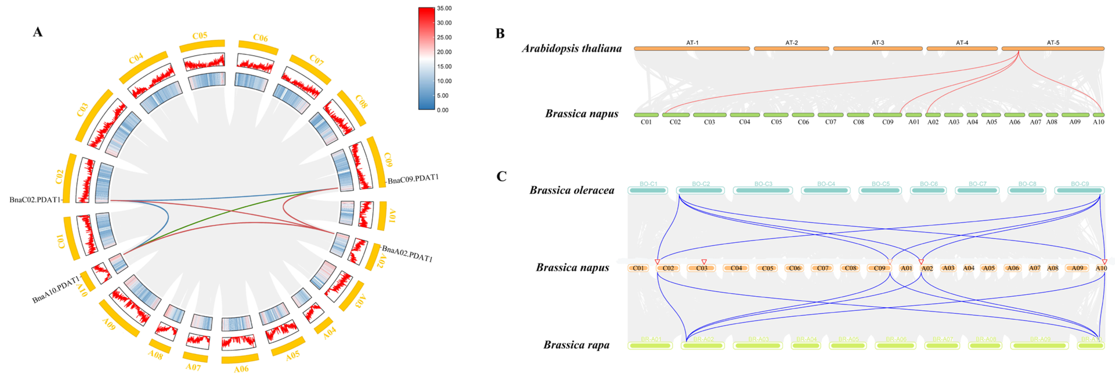
3.3. Chromosomal Localization and Collinearity Analysis of PDAT

Chromosomal localization results revealed that the five BnaPDAT genes in B. napus are located on chromosomes A02, A10, C02, C03, and C09. Additionally, the two BoPDAT genes in B. oleracea were mapped to chromosomes C2 and C9, while the two BraPDAT genes in B. rapa were localized to chromosomes A02 and A10. In A. thaliana, the two AtPDAT genes were positioned on chromosomes Chr3 and Chr5 (Figure 2). Intra-species collinearity analysis, which investigates the distribution and duplication of homologous genes within a single species, was performed for the B. napus PDAT gene family (Figure 3A). Among the four BnaPDAT genes, six collinear gene pairs were detected, with no evidence of tandem duplications, indicating that these four BnaPDAT genes are homologous. To further explore the evolutionary history of the B. napus PDAT gene family, inter-species collinearity analysis was conducted among B. napus, B. rapa, B. oleracea, and A. thaliana (Figure 3B,C). The results showed eight, eight, and four collinear gene pairs between BnaPDAT and BoPDAT, BraPDAT, and AtPDAT, respectively. The BnaPDAT gene family underwent whole-genome duplication (WGD) events during its evolution, which served as the primary driver for its expansion in B. napus. Notably, no collinearity was observed between AtPDAT2 and BnaPDAT genes, suggesting that AtPDAT2 may have been lost during evolution or undergone significant genomic repositioning, leading to the disruption of collinearity with BnaPDAT genes.

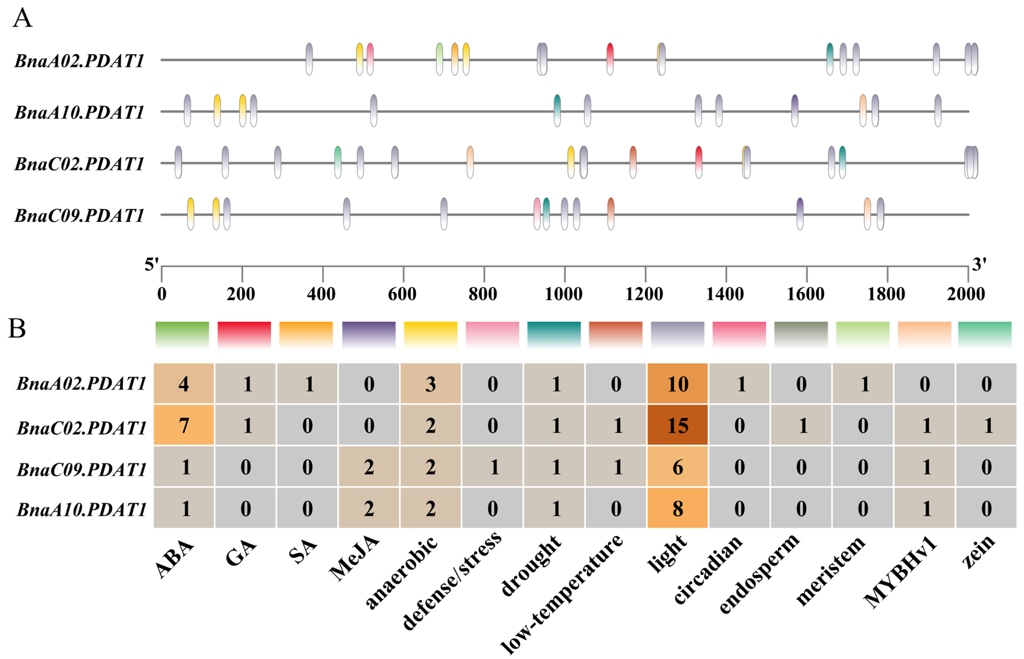
3.4. cis-Element Analyses of BnaPDAT Genes

Cis-acting elements play a critical role in regulating gene expression, and genes with similar functions may share identical regulatory elements in their promoters. To investigate whether BnaPDAT genes are involved in plant growth, development, hormone responses, and stress adaptation, we analyzed cis-regulatory elements within the 2000 bp upstream regions of BnaPDAT promoters using PlantCARE (Figure 4). The results revealed four types of cis-acting elements in the promoter regions of BnaPDAT genes, including elements associated with plant growth and development, abiotic stress responses, hormone responses, and core promoter elements essential for eukaryotic transcription. All four BnaPDAT gene promoters contained light-responsive elements, and hormone-responsive elements were widely distributed across these promoters. Specifically, the promoter regions of all members harbored abscisic acid (ABA)-responsive elements, two members contained methyl jasmonate (MeJA)-responsive elements, two members possessed gibberellin (GA)-responsive elements, and one member included a salicylic acid (SA)-responsive element. Additionally, some BnaPDAT promoters contained abiotic stress-related elements, such as those responsive to drought, low temperature, anaerobic induction, and wounding. Notably, all BnaPDAT members carried drought- and anaerobic induction-responsive elements, further suggesting their potential roles in mediating plant developmental processes and abiotic stress adaptation.

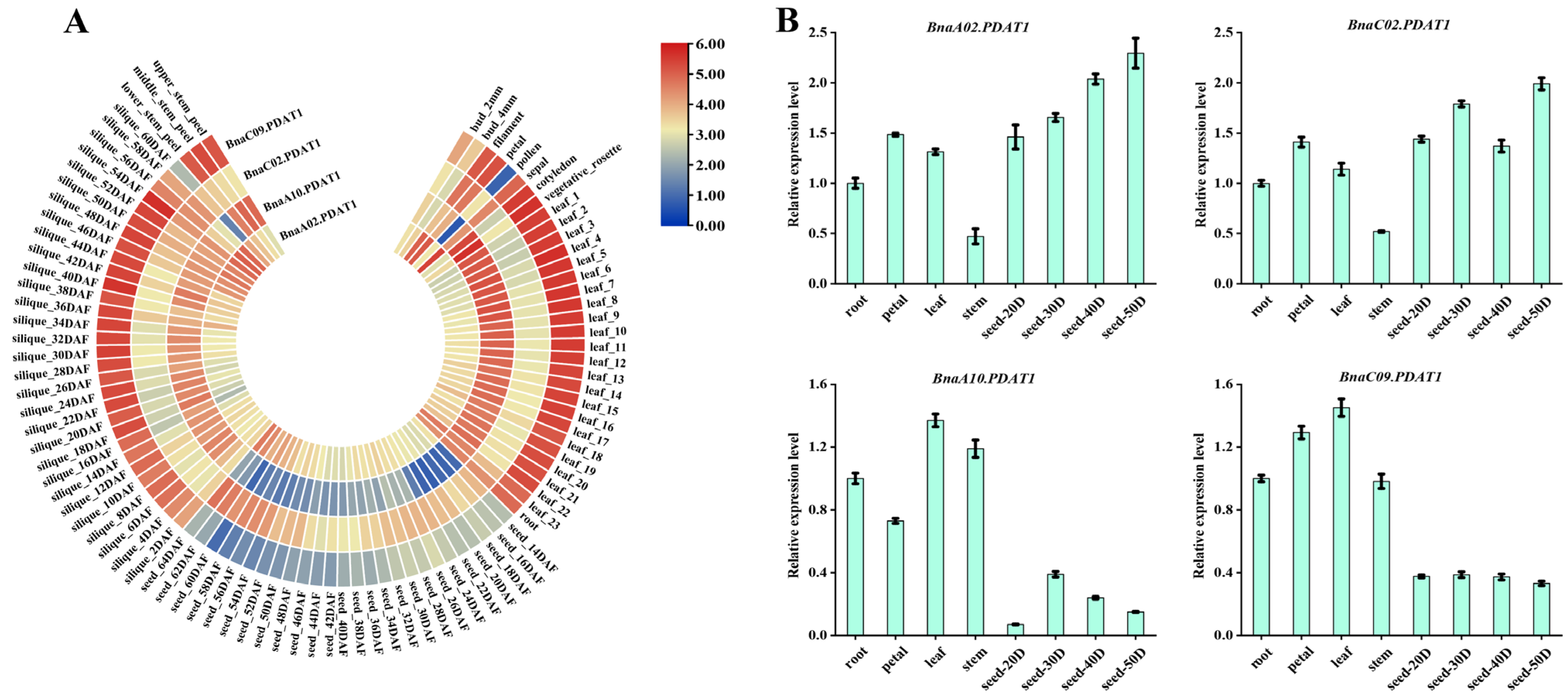
3.5. Analysis of Expression Patterns of BnaPDAT Genes in Different Tissues

To investigate the tissue-specific expression profiles of the BnaPDAT gene family in B. napus, this study utilized RNA sequencing (RNA-seq) data from the B. napus cultivar “ZS11” available in the BnTIR public database for tissue-specific expression analysis (Figure 5B, Supplementary Table S2). The results demonstrated that all four BnaPDAT genes were expressed across the plant, but their expression levels varied significantly among different tissues. Specifically, BnaA02.PDAT1 and BnaC02.PDAT1 exhibited similar expression patterns, with high expression levels throughout the plant, particularly during the late stages of seed development. In contrast, BnaA10.PDAT1 and BnaC09.PDAT1 showed the highest expression in leaves and siliques but lower expression in seeds (Figure 5A). To validate the transcriptome data, RNA samples were collected from various tissues of “ZS11”, including roots, stems, leaves, flowers, and seeds at 20, 30, 40, and 50 days after flowering, and subjected to RT-qPCR analysis. The results confirmed that BnaA02.PDAT1 and BnaC02.PDAT1 were more highly expressed in seeds than in other tissues. The expression of BnaA02.PDAT1 gradually increased during seed development, while BnaC02.PDAT1 displayed a dynamic pattern characterized by an initial increase, followed by a decline and then a subsequent rise. Meanwhile, BnaA10.PDAT1 and BnaC09.PDAT1 exhibited significantly lower expression levels in seeds compared to other tissues. These expression patterns were largely consistent with the transcriptome data, further supporting the reliability of the RNA-seq results. In summary, the PDAT gene family plays a crucial role in the growth and development of B. napus, and the divergent expression patterns of individual PDAT members across tissues may reflect their functional specialization in plant development and stress responses.

3.6. Analysis of BnaPDAT Gene Expression Patterns Under Different Abiotic Stress Treatments

To investigate the expression profiles of BnaPDAT genes under various abiotic stress conditions, this study analyzed expression data of the BnaPDAT gene family in leaves subjected to different stress treatments using the BnTIR database (Figure 6, Supplementary Table S3). The results revealed distinct expression patterns among the genes under specific stresses. Under salt, drought, and osmotic stress, BnaA02.PDAT1 and BnaC02.PDAT1 were significantly induced, while BnaA10.PDAT1 and BnaC09.PDAT1 were markedly suppressed. Under freezing stress, all PDAT family members exhibited pronounced induction at 6 h post treatment, followed by a gradual decline in induction over time. In response to low-temperature stress, three PDAT genes (BnaA02.PDAT1, BnaC02.PDAT1, and BnaC09.PDAT1) showed downregulated expression, whereas BnaA10.PDAT1 remained unaffected. Heat stress had relatively minor effects on the expression of PDAT family members.

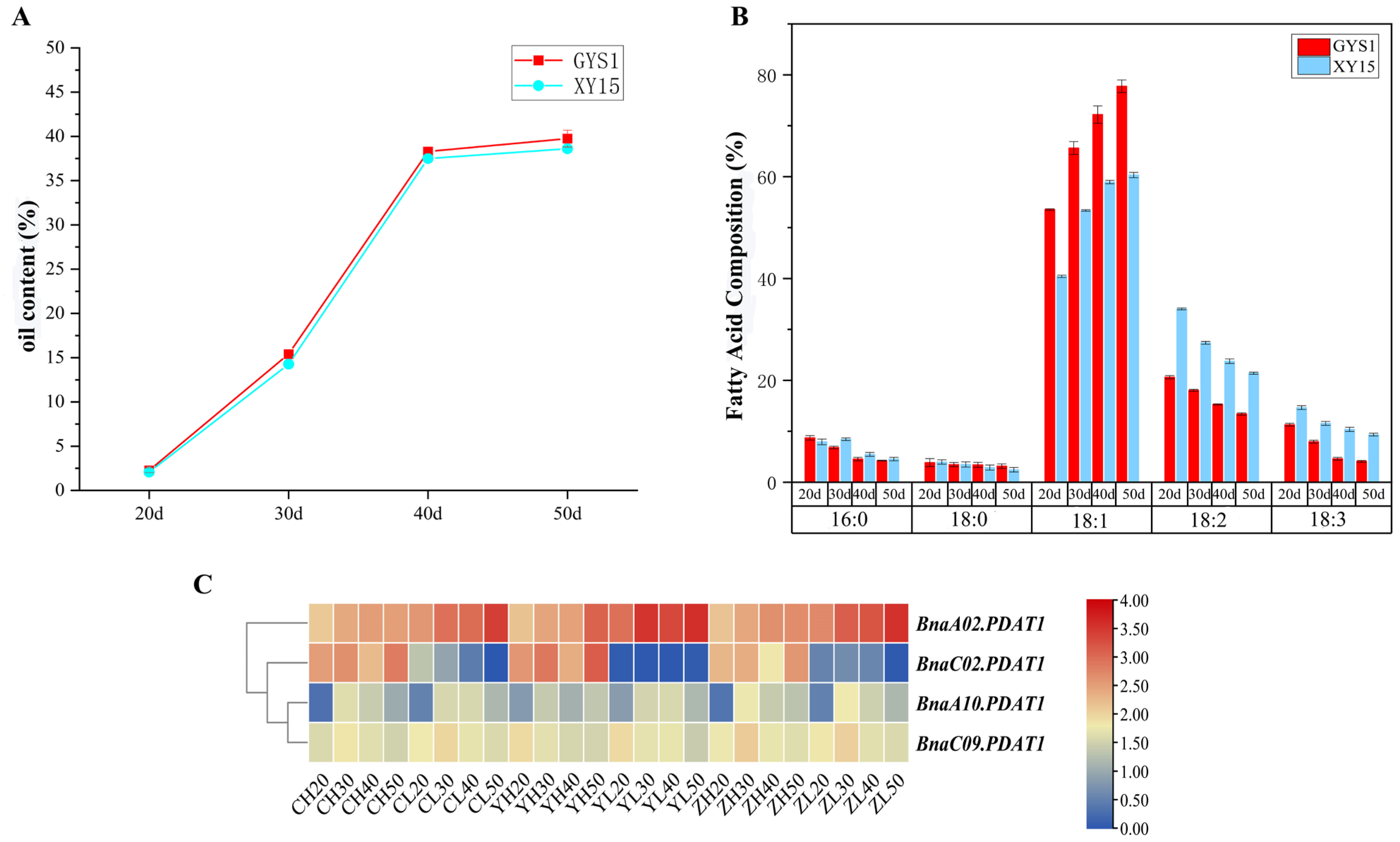
3.7. Expression Analysis of BnaPDAT Genes in High- and Low-Oleic Acid Materials

To further investigate the roles of the BnaPDAT gene family in lipid biosynthesis and seed development, this study analyzed oil content and fatty acid composition in two materials with distinct oleic acid levels: the high-oleic acid material “Gaoyousuan 1” and the low-oleic acid material “Xiangyou 15” (Figure 7A,B). The results showed that oil content in both materials followed an “S-shaped” curve during seed development, with “Gaoyousuan 1” exhibiting slightly higher oil content than “Xiangyou 15”, though the difference was not statistically significant. Fatty acid profiling revealed that oleic acid content increased consistently with oil accumulation, and “Gaoyousuan 1” had significantly higher oleic acid levels than “Xiangyou 15”. Conversely, linoleic and linolenic acid levels in “Gaoyousuan 1” were consistently lower than those in “Xiangyou 15”. Based on transcriptome data previously generated by our group, we further analyzed BnaPDAT gene expression in both materials across different geographical locations and developmental stages (Figure 7C, Supplementary Table S4). The results indicated no significant differences in the expression of BnaA10.PDAT1 and BnaC09.PDAT1 between the two materials. However, BnaA02.PDAT1 exhibited lower expression in the high-oleic acid material compared to the low-oleic acid material, while BnaC02.PDAT1 expression was significantly higher in the high-oleic acid material. These findings suggest that BnaA02.PDAT1 and BnaC02.PDAT1 may participate in fatty acid accumulation in rapeseed seeds and play critical roles in regulating oleic acid content.

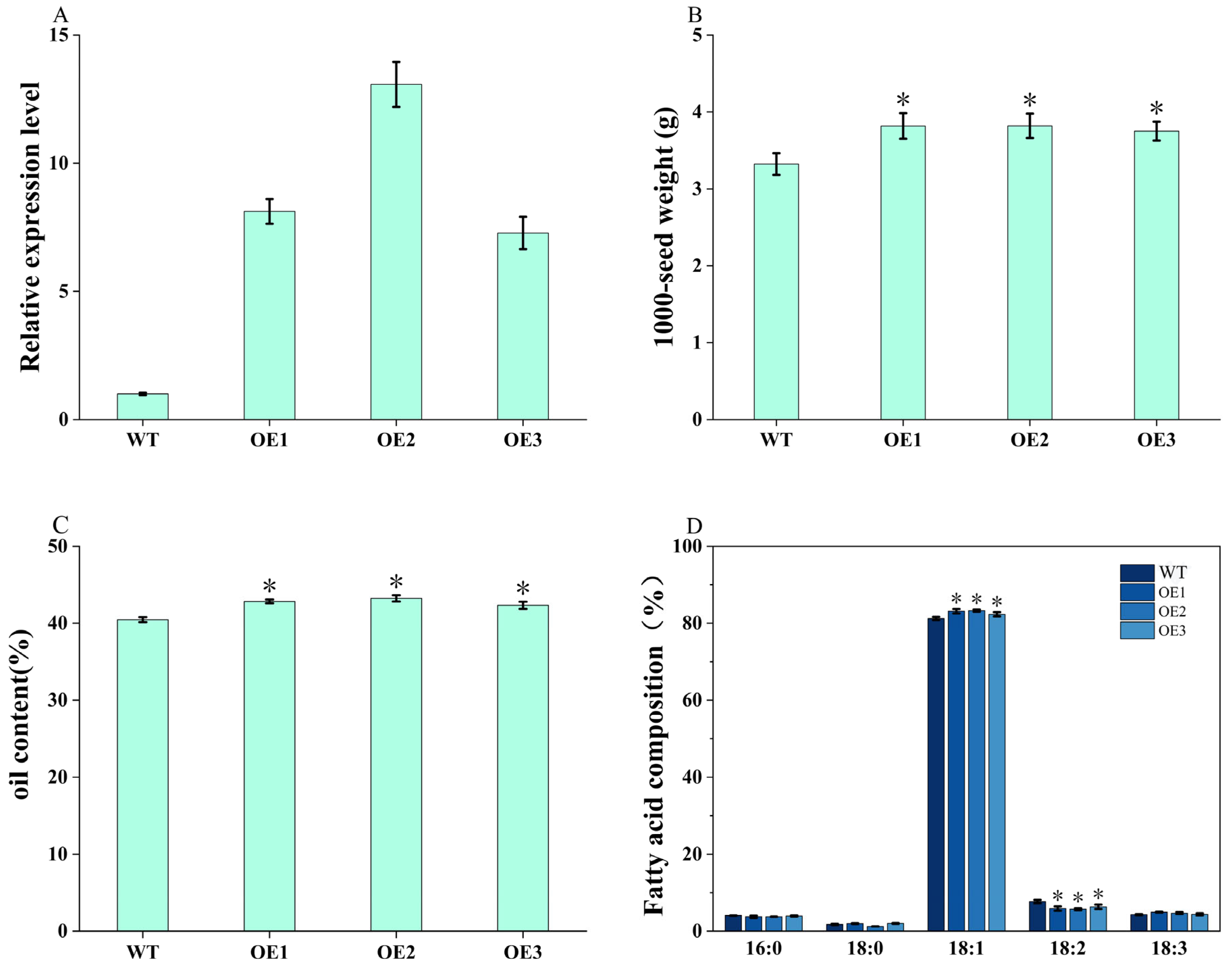
3.8. Overexpression of BnaA02.PDAT1 Enhances Seed Oil Accumulation

Based on transcriptome data, we selected BnaA02.PDAT1, which exhibits high expression in seeds, for functional validation through overexpression. To investigate its impact on lipid synthesis in the rapeseed cultivar “GYS1”, we constructed the overexpression vector PC1300S-BnaA02.PDAT1 and transformed it into B. napus, generating three transgenic lines (OE1, OE2, and OE3) of “GYS1”. qRT-PCR analysis revealed that BnaA02.PDAT1 expression was upregulated to varying degrees in the overexpression lines compared to the wild type (WT) (Figure 8A). To assess the effect of BnaA02.PDAT1 on seed oil accumulation, mature seeds were analyzed for thousand-seed weight, oil content, and fatty acid composition. The results demonstrated that thousand-seed weight in transgenic plants was significantly higher than in WT, increasing by 12.95–14.76% (Figure 8B). Total oil content in transgenic seeds also rose by 1.86–2.77% compared to WT (Figure 8C). Further analysis of fatty acid composition revealed that oleic acid content was significantly elevated in all transgenic lines, while linoleic acid content was markedly reduced relative to WT (Figure 8D). These findings indicate that BnaA02.PDAT1 enhances seed oil accumulation and alters fatty acid composition in rapeseed.

4. Discussion

The biosynthesis of triacylglycerol (TAG) in plant seeds primarily occurs through two pathways: the acyl-CoA-dependent de novo synthesis pathway (Kennedy pathway) and the acyl-CoA-independent synthesis pathway, where PDAT serves as a critical rate-limiting enzyme. Investigating these lipid synthesis pathways holds significant importance for enhancing seed oil content and improving oil composition. Compared to the Kennedy pathway, research on the PDAT-mediated lipid synthesis pathway in plants has lagged. In 2000, Dahlqvist et al. first identified the PDAT gene in Saccharomyces cerevisiae [36], followed by subsequent discoveries of PDAT genes in plants and algae [37]. In this study, we identified four, two, and two PDAT gene members in the genomes of B. napus, B. rapa, and B. olerace, respectively—far fewer than the number reported in cotton [38]. These results suggest evolutionary expansion or contraction within the PDAT gene family. Phylogenetic analysis integrating PDAT proteins from A. thaliana, B. napus, B. rapa, and B. oleracea classified them into two subfamilies: PDAT1 and PDAT2. The PDAT1 subfamily was widely present in B. napus and its progenitor species, while the PDAT2 subfamily was retained only in A. thaliana, indicating that PDAT2 may have been gradually lost during evolution. The occurrence of this phenomenon may be due to the fact that the BnaPDAT gene family experienced selective loss of PDAT2 after whole-genome duplication (WGD) events due to functional redundancy, unlike fiber crops such as cotton. As an oilseed crop, B. napus may rely more on the PDAT1-mediated acyl editing pathway to meet the demand for synthesizing high proportions of polyunsaturated fatty acids in seeds [39]. Gene structure and conserved motif analyses revealed that PDAT family members exhibit highly conserved gene architectures and functional domains, which may be closely associated with their critical functions in lipid synthesis.
The PDAT protein is a key rate-limiting enzyme in the acyl-CoA-independent TAG synthesis pathway. Previous studies have demonstrated that PDAT genes can partially compensate for the function of DGAT genes, participating in TAG synthesis during lipid accumulation in seed development [40]. For example, the PDAT gene in Camelina sativa is highly expressed during the early stages of seed development, coinciding with the most active phase of lipid accumulation [40]. Similarly, the OePDAT1-1 gene in olive exhibits significantly elevated expression levels during the later stages of seed development [41]. This study corroborates that most BnaPDAT genes in B. napus show high expression during the middle to late stages of seed development, suggesting their involvement in lipid accumulation. Notably, the expression of BnaA02.PDAT1 was lower in high-oleic acid materials compared to low-oleic acid materials, while BnaC02.PDAT1 displayed the opposite pattern. This divergence may reflect their functional specialization in regulating fatty acid composition during oil biosynthesis.
The PDAT genes are not only involved in triacylglycerol (TAG) synthesis but also play roles in plant responses to abiotic stress [42]. Under abiotic stress conditions, plants can modulate lipid metabolism through genetic regulation to enhance stress tolerance [43]. PDAT genes facilitate the conversion of membrane lipids to TAG, maintaining membrane lipid homeostasis [40] and thereby mitigating damage caused by environmental stress [44]. Additionally, studies in olive have shown that PDAT genes exhibit distinct physiological functions in lipid accumulation and stress responses [41]. The A. thaliana MYB96 transcription factor affects TAG synthesis by regulating the transcription of AtDGAT1 and AtPDAT1, thereby enhancing the drought tolerance of seedlings [45]. Cis-regulatory element analysis of B. napus BnaPDAT genes revealed that their promoter regions contain abundant stress-related regulatory elements as well as multiple hormone-responsive elements, suggesting their potential involvement in both abiotic stress adaptation and hormonal signaling. In this study, expression profiling under various stress conditions demonstrated that most BnaPDAT genes were upregulated to varying degrees. Specifically, BnaA02.PDAT1 and BnaC02.PDAT1 were strongly induced under salt stress, drought stress, and osmotic stress, whereas BnaA10.PDAT1 and BnaC09.PDAT1 were significantly suppressed. Under freezing stress, all PDAT family members showed marked induction at 6 h post treatment. More interestingly, studies have shown that PDAT seems to participate in stress responses in green algae [36], A. thaliana [42], and C. sativa [17]. For example, PDAT1-regulated TAG accumulation can improve the heat tolerance of A. thaliana plants [42]. These findings indicate that PDAT genes possess dual functions in lipid biosynthesis and stress adaptation.
Previous research has indicated that a T-DNA insertion mutation with an AtPDAT1 knockout in Arabidopsis had no impact on lipid content and/or fatty acid components. However, our results demonstrate that overexpression of BnaA02.PDAT1 significantly increased thousand-seed weight and oil content in B. napus seeds; the thousand-seed weight increased significantly by 12.95–14.76%. Additionally, the total oil content in transgenic seeds was 1.86–2.77% higher than that of WT. Furthermore, the fatty acid composition in the seeds was also altered. These results are similar to those from the overexpression of the Sapium sebiferum PDAT1 gene in B. napus, which successfully altered the fatty acid composition and increased the seed oil content [46]. Those results strongly support a role for PDAT1 in seed oil biosynthesis. With the rapid development of synthetic biology technologies such as genetic transformation, gene sequencing, CRISPR gene editing, DNA synthesis and assembly, and protein synthesis and assembly, future research can utilize PDAT to design and modify lipid metabolic pathways in different organisms. It will also become more efficient to modularly assemble PDAT with other key lipid metabolic genes and precisely insert them into the host genome. This will facilitate crop genetic improvement, large-scale commercial production of oils, and the construction and application of new lipid molecules.

5. Conclusions

In summary, this study identified four PDAT gene members in B. napus through an integrated analysis of its whole genome. Phylogenetic analysis revealed that B. napus PDAT members belong exclusively to the PDAT1 subfamily, with PDAT2 having been lost during evolution, highlighting the complex evolutionary history of the PDAT gene family. Gene expression profiling demonstrated that BnaPDAT genes participate in seed development and abiotic stress responses in B. napus. Furthermore, overexpression of BnaA02.PDAT1 significantly enhanced seed oil content and thousand-seed weight while altering fatty acid composition. Our findings confirm the critical role of BnaPDAT genes in B. napus seed development and their impact on oil accumulation, providing a foundation for further exploration of the mechanisms by which BnaPDAT regulates lipid biosynthesis and stress adaptation in rapeseed. These results suggest that BnaPDAT genes could serve as valuable targets for breeding programs aimed at improving seed oil content and stress tolerance in oilseed crops. Future research could focus on utilizing CRISPR-based gene editing to precisely modify PDAT genes. Offering valuable germplasm resources for developing novel high-oil-content B. napus varieties.

Supplementary Materials

The following supporting information can be downloaded at https://www.mdpi.com/article/10.3390/agronomy15051204/s1. Table S1: The qPCR primer sequence used in this experiment; Table S2: Transcriptome sequencing data of BnaPDAT gene in different organizations; Table S3: Transcriptome sequencing data of BnaPDAT gene under different abiotic stresses; Table S4: Transcriptome sequencing data of BnaPDAT gene in high- and low-oleic acid B. napus materials; Figure S1: Take ordinary cDNA and run qPCR directly to verify the availability of primers; Figure S2: The multiple sequence alignment of plant BnPDAT proteins. At, Arabidopsis thaliana; Bna, Brassica napus; Figure S3: Conserved domains of PDAT in A. thaliana, B. napus, B. oleracea, and B. rapa.

Author Contributions

Conceptualization, C.G., M.G. and H.C.; methodology, H.C.; software, H.C.; validation, H.C.; formal analysis, H.C.; investigation, H.C.; data curation, H.C.; writing—original draft preparation, H.C.; writing—review and editing, M.G.; supervision, C.G. and M.G.; project administration, M.G. All authors have read and agreed to the published version of the manuscript.

Funding

This research was funded by the Hunan Agriculture Research System (HARS-03).

Data Availability Statement

All the data included in this study are available upon request by contacting the corresponding author.

Conflicts of Interest

The authors declare no conflicts of interest.

References

  1. Woodfield, H.K.; Sturtevant, D.; Borisjuk, L.; Munz, E.; Guschina, I.A.; Chapman, K.; Harwood, J.L. Spatial and Temporal Mapping of Key Lipid Species in Brassica napus Seeds. Plant Physiol. 2017, 173, 1998–2009. [Google Scholar] [CrossRef] [PubMed]
  2. Carlsson, A.S.; Yilmaz, J.L.; Green, A.G.; Stymne, S.; Hofvander, P. Replacing fossil oil with fresh oil—With what and for what? Eur. J. Lipid Sci. Technol. 2011, 113, 812–831. [Google Scholar] [CrossRef]
  3. Biermann, U.; Bornscheuer, U.; Meier, M.A.R.; Metzger, J.O.; Schäfer, H.J. Oils and Fats as Renewable Raw Materials in Chemistry. Angew. Chem.-Int. Ed. 2011, 50, 3854–3871. [Google Scholar] [CrossRef] [PubMed]
  4. Bates, P.D.; Stymne, S.; Ohlrogge, J. Biochemical pathways in seed oil synthesis. Curr. Opin. Plant Biol. 2013, 16, 358–364. [Google Scholar] [CrossRef] [PubMed]
  5. Huang, A.H.C. Plant Lipid Droplets and Their Associated Proteins: Potential for Rapid Advances. Plant Physiol. 2017, 176, 1894–1918. [Google Scholar] [CrossRef]
  6. Cao, J.; Li, J.-L.; Li, D.; Tobin, J.F.; Gimeno, R.E. Molecular identification of microsomal acyl-CoA:glycerol-3-phosphate acyltransferase, a key enzyme in de novo triacylglycerol synthesis. Proc. Natl. Acad. Sci. USA 2006, 103, 19695–19700. [Google Scholar] [CrossRef]
  7. Shockey, J.; Regmi, A.; Cotton, K.; Adhikari, N.; Browse, J.; Bates, P.D. Identification of Arabidopsis GPAT9 (At5g60620) as an Essential Gene Involved in Triacylglycerol Biosynthesis. Plant Physiol. 2015, 170, 163–179. [Google Scholar] [CrossRef]
  8. Lewin, T.M.; Wang, P.; Coleman, R.A. Analysis of Amino Acid Motifs Diagnostic for the sn-Glycerol-3-phosphate Acyltransferase Reaction. Biochemistry 1999, 38, 5764–5771. [Google Scholar] [CrossRef]
  9. Weiss, S.B.; Kennedy, E.P. The Enzymatic Synthesis of Triglycerides. J. Am. Chem. Soc. 1956, 78, 3550. [Google Scholar] [CrossRef]
  10. Shockey, J.M.; Gidda, S.K.; Chapital, D.C.; Kuan, J.-C.; Dhanoa, P.K.; Bland, J.M.; Rothstein, S.J.; Mullen, R.T.; Dyer, J.M. Tung Tree DGAT1 and DGAT2 Have Nonredundant Functions in Triacylglycerol Biosynthesis and Are Localized to Different Subdomains of the Endoplasmic Reticulum. Plant Cell 2006, 18, 2294–2313. [Google Scholar] [CrossRef]
  11. Stahl, U.; Carlsson, A.S.; Lenman, M.; Dahlqvist, A.; Huang, B.; Banaś, W.; Banaś, A.; Stymne, S. Cloning and Functional Characterization of a Phospholipid:Diacylglycerol Acyltransferase from Arabidopsis. Plant Physiol. 2004, 135, 1324–1335. [Google Scholar] [CrossRef] [PubMed]
  12. Zhang, M.; Fan, J.; Taylor, D.C.; Ohlrogge, J.B. DGAT1 and PDAT1 Acyltransferases Have Overlapping Functions in Arabidopsis Triacylglycerol Biosynthesis and Are Essential for Normal Pollen and Seed Development. Plant Cell 2009, 21, 3885–3901. [Google Scholar] [CrossRef] [PubMed]
  13. Simpson, J.P.; Ohlrogge, J.B. A Novel Pathway for Triacylglycerol Biosynthesis Is Responsible for the Accumulation of Massive Quantities of Glycerolipids in the Surface Wax of Bayberry (Myrica pensylvanica) Fruit. Plant Cell 2016, 28, 248–264. [Google Scholar] [CrossRef]
  14. Fan, J.; Yan, C.; Zhang, X.; Xu, C. Dual Role for Phospholipid:Diacylglycerol Acyltransferase: Enhancing Fatty Acid Synthesis and Diverting Fatty Acids from Membrane Lipids to Triacylglycerol in Arabidopsis Leaves. Plant Cell 2013, 25, 3506–3518. [Google Scholar] [CrossRef]
  15. Pan, X.; Siloto, R.M.; Wickramarathna, A.D.; Mietkiewska, E.; Weselake, R.J. Identification of a Pair of Phospholipid:Diacylglycerol Acyltransferases from Developing Flax (Linum usitatissimum L.) Seed Catalyzing the Selective Production of Trilinolenin. J. Biol. Chem. 2013, 288, 24173–24188. [Google Scholar] [CrossRef]
  16. Kim, H.U.; Lee, K.-R.; Go, Y.S.; Jung, J.H.; Suh, M.-C.; Kim, J.B. Endoplasmic Reticulum-Located PDAT1-2 from Castor Bean Enhances Hydroxy Fatty Acid Accumulation in Transgenic Plants. Plant Cell Physiol. 2011, 52, 983–993. [Google Scholar] [CrossRef] [PubMed]
  17. Yuan, L.; Mao, X.; Zhao, K.; Ji, X.; Ji, C.; Xue, J.; Li, R. Characterisation of phospholipid: Diacylglycerol acyltransferases (PDATs) from Camelina sativa and their roles in stress responses. Biol. Open 2017, 6, 1024–1034. [Google Scholar] [CrossRef]
  18. Demski, K.; Łosiewska, A.; Jasieniecka-Gazarkiewicz, K.; Klińska, S.; Banaś, A. Phospholipid:Diacylglycerol Acyltransferase1 Overexpression Delays Senescence and Enhances Post-heat and Cold Exposure Fitness. Front. Plant Sci. 2020, 11, 611897. [Google Scholar] [CrossRef]
  19. Parkin, I.A.; Sharpe, A.G.; Keith, D.J.; Lydiate, D.J. Identification of the A and C genomes of amphidiploid Brassica napus (oilseed rape). Genome 1995, 38, 1122–1131. [Google Scholar] [CrossRef]
  20. Palmer, J.D.; Shields, C.R.; Cohen, D.B.; Orton, T.J. Chloroplast DNA evolution and the origin of amphidiploid Brassica species. Theor. Appl. Genet. 1983, 65, 181–189. [Google Scholar] [CrossRef]
  21. Yates, A.D.; Allen, J.; Amode, R.M.; Azov, A.G.; Barba, M.; Becerra, A.; Bhai, J.; Campbell, L.I.; Martinez, M.C.; Chakiachvili, M.; et al. Ensembl Genomes 2022: An expanding genome resource for non-vertebrates. Nucleic Acids Res. 2022, 50, D996–D1003. [Google Scholar] [CrossRef] [PubMed]
  22. Mistry, J.; Chuguransky, S.; Williams, L.; Qureshi, M.; Salazar, G.A.; Sonnhammer, E.L.L.; Tosatto, S.C.E.; Paladin, L.; Raj, S.; Richardson, L.J.; et al. Pfam: The protein families database in 2021. Nucleic Acids Res. 2020, 49, D412–D419. [Google Scholar] [CrossRef]
  23. Abbas, A.; Ye, F. Computational methods and key considerations for in silico design of proteolysis targeting chimera (PROTACs). Int. J. Biol. Macromol. 2024, 277, 134293. [Google Scholar] [CrossRef] [PubMed]
  24. Bernhofer, M.; Dallago, C.; Karl, T.; Satagopam, V.; Heinzinger, M.; Littmann, M.; Olenyi, T.; Qiu, J.; Schütze, K.; Yachdav, G.; et al. PredictProtein—Predicting Protein Structure and Function for 29 Years. Nucleic Acids Res. 2021, 49, W535–W540. [Google Scholar] [CrossRef]
  25. Minh, B.Q.; Schmidt, H.A.; Chernomor, O.; Schrempf, D.; Woodhams, M.D.; von Haeseler, A.; Lanfear, R. IQ-TREE 2: New Models and Efficient Methods for Phylogenetic Inference in the Genomic Era. Mol. Biol. Evol. 2020, 37, 1530–1534. [Google Scholar] [CrossRef]
  26. Nystrom, S.L.; McKay, D.J. Memes: A motif analysis environment in R using tools from the MEME Suite. PLoS Comput. Biol. 2021, 17, e1008991. [Google Scholar] [CrossRef]
  27. Chen, C.; Wu, Y.; Li, J.; Wang, X.; Zeng, Z.; Xu, J.; Liu, Y.; Feng, J.; Chen, H.; He, Y.; et al. TBtools-II: A “one for all, all for one” bioinformatics platform for biological big-data mining. Mol. Plant 2023, 16, 1733–1742. [Google Scholar] [CrossRef] [PubMed]
  28. Lescot, M.; Déhais, P.; Thijs, G.; Marchal, K.; Moreau, Y.; Van de Peer, Y.; Rouzé, P.; Rombauts, S. PlantCARE, a database of plant cis-acting regulatory elements and a portal to tools for in silico analysis of promoter sequences. Nucleic Acids Res. 2002, 30, 325–327. [Google Scholar] [CrossRef]
  29. Wang, X.; Zhao, D.; Li, X.; Zhou, B.; Chang, T.; Hong, B.; Guan, C.; Guan, M. Integrated Analysis of lncRNA–mRNA Regulatory Networks Related to Lipid Metabolism in High-Oleic-Acid Rapeseed. Int. J. Mol. Sci. 2023, 24, 6277. [Google Scholar] [CrossRef]
  30. Liu, D.; Yu, L.; Wei, L.; Yu, P.; Wang, J.; Zhao, H.; Zhang, Y.; Zhang, S.; Yang, Z.; Chen, G.; et al. BnTIR: An online transcriptome platform for exploring RNA-seq libraries for oil crop Brassica napus. Plant Biotechnol. J. 2021, 19, 1895–1897. [Google Scholar] [CrossRef]
  31. Livak, K.J.; Schmittgen, T.D. Analysis of Relative Gene Expression Data Using Real-Time Quantitative PCR and the 2−ΔΔCT Method. Methods 2001, 25, 402–408. [Google Scholar] [CrossRef] [PubMed]
  32. Yang, H.; Liu, J.; Huang, S.; Guo, T.; Deng, L.; Hua, W. Selection and evaluation of novel reference genes for quantitative reverse transcription PCR (qRT-PCR) based on genome and transcriptome data in Brassica napus L. Gene 2014, 538, 113–122. [Google Scholar] [CrossRef] [PubMed]
  33. Dai, C.; Li, Y.; Li, L.; Du, Z.; Lin, S.; Tian, X.; Li, S.; Yang, B.; Yao, W.; Wang, J.; et al. An efficient Agrobacterium-mediated transformation method using hypocotyl as explants for Brassica napus. Mol. Breed. 2020, 40, 96. [Google Scholar] [CrossRef]
  34. Li, F.; Wu, X.; Lam, P.; Bird, D.; Zheng, H.; Samuels, L.; Jetter, R.; Kunst, L. Identification of the Wax Ester Synthase/Acyl-Coenzyme A:Diacylglycerol Acyltransferase WSD1 Required for Stem Wax Ester Biosynthesis in Arabidopsis. Plant Physiol. 2008, 148, 97–107. [Google Scholar] [CrossRef] [PubMed]
  35. López-Bascón, M.A.; Luque de Castro, M.D. Chapter 11—Soxhlet Extraction. In Liquid-Phase Extraction; Poole, C.F., Ed.; Elsevier: Amsterdam, The Netherlands, 2020; pp. 327–354. [Google Scholar] [CrossRef]
  36. Dahlqvist, A.; Ståhl, U.; Lenman, M.; Banas, A.; Lee, M.; Sandager, K.; Ronne, H.; Stymne, S. Phospholipid:diacylglycerol acyltransferase: An enzyme that catalyzes the acyl-CoA-independent formation of triacylglycerol in yeast and plants. Proc. Natl. Acad. Sci. USA 2000, 97, 6487–6492. [Google Scholar] [CrossRef]
  37. Merchant, S.S.; Prochnik, S.E.; Vallon, O.; Harris, E.H.; Karpowicz, S.J.; Witman, G.B.; Terry, A.; Salamov, A.; Fritz-Laylin, L.K.; Maréchal-Drouard, L.; et al. The Chlamydomonas Genome Reveals the Evolution of Key Animal and Plant Functions. Science 2007, 318, 245–250. [Google Scholar] [CrossRef]
  38. Zang, X.; Geng, X.; Ma, L.; Wang, N.; Pei, W.; Wu, M.; Zhang, J.; Yu, J. A genome-wide analysis of the phospholipid: Diacylglycerol acyltransferase gene family in Gossypium. BMC Genom. 2019, 20, 402. [Google Scholar] [CrossRef]
  39. Cittadino, G.M.; Andrews, J.; Purewal, H.; Estanislao Acuña Avila, P.; Arnone, J.T. Functional Clustering of Metabolically Related Genes Is Conserved across Dikarya. J. Fungi 2023, 9, 523. [Google Scholar] [CrossRef]
  40. Fan, J.; Yan, C.; Xu, C. Phospholipid:diacylglycerol acyltransferase-mediated triacylglycerol biosynthesis is crucial for protection against fatty acid-induced cell death in growing tissues of Arabidopsis. Plant J. 2013, 76, 930–942. [Google Scholar] [CrossRef]
  41. Hernández, M.L.; Moretti, S.; Sicardo, M.D.; García, Ú.; Pérez, A.; Sebastiani, L.; Martínez-Rivas, J.M. Distinct Physiological Roles of Three Phospholipid: Diacylglycerol Acyltransferase Genes in Olive Fruit with Respect to Oil Accumulation and the Response to Abiotic Stress. Front. Plant Sci. 2021, 12, 751959. [Google Scholar] [CrossRef]
  42. Mueller, S.P.; Unger, M.; Guender, L.; Fekete, A.; Mueller, M.J. Phospholipid:Diacylglycerol Acyltransferase-Mediated Triacylglyerol Synthesis Augments Basal Thermotolerance. Plant Physiol. 2017, 175, 486–497. [Google Scholar] [CrossRef] [PubMed]
  43. Gasulla, F.; vom Dorp, K.; Dombrink, I.; Zähringer, U.; Gisch, N.; Dörmann, P.; Bartels, D. The role of lipid metabolism in the acquisition of desiccation tolerance in raterostigma plantagineum: A comparative approach. Plant J. 2013, 75, 726–741. [Google Scholar] [CrossRef] [PubMed]
  44. Yoon, K.; Han, D.; Li, Y.; Sommerfeld, M.; Hu, Q. Phospholipid:Diacylglycerol Acyltransferase Is a Multifunctional Enzyme Involved in Membrane Lipid Turnover and Degradation While Synthesizing Triacylglycerol in the Unicellular Green Microalga Chlamydomonas reinhardtii. Plant Cell 2012, 24, 3708–3724. [Google Scholar] [CrossRef] [PubMed]
  45. Lee, H.G.; Park, M.-E.; Park, B.Y.; Kim, H.U.; Seo, P.J. The Arabidopsis MYB96 Transcription Factor Mediates ABA-Dependent Triacylglycerol Accumulation in Vegetative Tissues under Drought Stress Conditions. Plants 2019, 8, 296. [Google Scholar] [CrossRef]
  46. Zhou, B.; Fei, W.; Yang, S.; Yang, F.; Qu, G.; Tang, W.; Ou, J.; Peng, D. Alteration of the fatty acid composition of Brassica napus L. via overexpression of phospholipid: Diacylglycerol acyltransferase 1 from Sapium sebiferum (L.) Roxb. Plant Sci. 2020, 298, 110562. [Google Scholar] [CrossRef]
Figure 1. Phylogenetic relationships, gene structure, and motifs in PDAT genes. (A) Phylogenetic analysis of the PDAT gene family classified 10 PDAT proteins into two subgroups: PDAT1 and PDAT2. In the phylogenetic tree, red pentagrams represent the two PDAT proteins in A. thaliana, blue triangles denote the four PDAT proteins in B. napus, green triangles indicate the two PDAT proteins in B. oleracea, and pink triangles signify the two PDAT proteins in B. rapa. (B) The motif composition of PDAT proteins. Motifs 1–10 are shown in different colored boxes. The length of each protein can be estimated using the scale at the bottom. (C) Introns, exons, and UTRs, where introns are marked with black horizontal lines. (D) The information for six conserved motifs.
Agronomy 15 01204 g001
Figure 2. Chromosomal distribution of PDAT genes in A. thaliana (A), B. napus (B), B. oleracea (C), and B. rapa (D). The length of each chromosome is shown in Mb, the chromosomal number is shown on the left side of the chromosome, and the PDAT genes are highlighted in red.
Agronomy 15 01204 g002
Figure 3. Intraspecific collinearity of PDAT genes in B. napus and their collinearity with PDAT genes in A. thaliana, B. oleracea, and B. rapa; the legend represents gene density, the lines represent gene pairs with collinearity (A). The red and blue lines represent the collinearity between B. napus and other species (B,C).
Agronomy 15 01204 g003
Figure 4. Cis elements detected in the promoter of the BnPDAT genes. (A) Promoter element distribution, where different colors correspond to different elements in the figure below; (B) the heat map shows the number of promoter elements, and the gray square indicates that the elements could not be detected.
Agronomy 15 01204 g004
Figure 5. Expression pattern analysis of BnaPDAT in different tissues. (A) Expression patterns of BnaPDAT from the BnIR; scale bar is Log2 (TPM)-normalized expression, blue denotes low expression, and red denotes strong expression. (B) RT-qPCR validation of BnaPDAT in different tissues. The BnActin gene was used as internal reference gene. Error bars represent the standard error. Student’s t-test (n = 3; data are means ± SD).
Agronomy 15 01204 g005
Figure 6. Expression patterns of PDAT gene in B. napus under different abiotic stresses. Scale bar is Log2 (TPM)-normalized expression.
Agronomy 15 01204 g006
Figure 7. Oil content, fatty acid composition, and PDAT gene expression patterns of high- and low-oleic acid B. napus seeds at different stages. (A) Oil content, (B) fatty acid composition, and (C) PDAT gene expression patterns. C—Changsha, Hunan, China; Z—Quzhou, Zhejiang, China; Y—Kunming, Yunnan, China; H—high-oleic acid rapeseed “Gaoyousuan No.1”; L—low-oleic acid rapeseed “Xiangyou No.15”.
Agronomy 15 01204 g007
Figure 8. Analysis of expression levels, thousand-seed weight, oil content, and fatty acid composition in BnaA02.PDAT1 transgenic rapeseed seeds. (A) qRT-PCR detection of BnaA02.PDAT1 expression in 20-day-old seeds of transgenic lines overexpressing BnaA02.PDAT1 gene. The BnActin gene was used as internal reference gene. (B) Thousand-seed weight; (C) oil content; (D) fatty acid composition. Error bars represent the standard error. Student’s t-test (n = 3; data are means ± SD). * denotes significance at p < 0.05.
Agronomy 15 01204 g008
Table 1. Physical and Chemical Characteristics of PDAT Gene Family.
Gene NameGene IDLengthPIMolecular Weight (kDa)Instability IndexPredicted Subcellular LocalizationHydrophobicity Index
BnaA10.PDAT1BnaA10T0220200ZS6666.0873.7536.18periplasm−0.32
BnaC09.PDAT1BnaC09T0524800ZS6675.9873.9435.89periplasm−0.323
BnaA02.PDAT1BnaA02T0049700ZS6656.6173.5336.55periplasm−0.302
BnaC02.PDAT1BnaC02T0057200ZS6607.0473.136.86periplasm−0.288
BraA02.PDAT1Bra023426.15976.3865.6935.83periplasm−0.251
BraA10.PDAT1Bra008812.16666.1173.7437.59periplasm−0.315
BoC09.PDAT1Bo9g166600.16676.0873.8235.51periplasm−0.304
BoC02.PDAT1Bo2g011450.16606.7173.1138.25periplasm−0.287
AtPDAT2AT3G44830.16658.6973.6549.02outer membrane−0.226
AtPDAT1AT5G13640.16716.574.1535.95periplasm−0.301
Disclaimer/Publisher’s Note: The statements, opinions and data contained in all publications are solely those of the individual author(s) and contributor(s) and not of MDPI and/or the editor(s). MDPI and/or the editor(s) disclaim responsibility for any injury to people or property resulting from any ideas, methods, instructions or products referred to in the content.

