Article

Heat Stress Downregulates Photosystem I Redox State on Leaf Photosynthesis in Grapevine

1
Department of Plant Science, School of Agriculture and Biology, Shanghai Jiao Tong University, Shanghai 200240, China
2
School of Agriculture, Food and Wine, The University of Adelaide, PMB 1, Glen Osmond, SA 5064, Australia
*
Authors to whom correspondence should be addressed.
Agronomy 2025, 15(4), 948; https://doi.org/10.3390/agronomy15040948
Submission received: 28 February 2025 / Revised: 7 April 2025 / Accepted: 11 April 2025 / Published: 14 April 2025

Abstract

:
Semi-arid viticultural regions globally are experiencing severe and frequent growing-season heat waves that negatively impact grapevine (Vitis vinifera L.) physiological performance and productivity. At the leaf level, heat stress can photodamage both Photosystem I (PSI) and Photosystem II (PSII). In order to study the self-protection mechanism of grapevine leaves, in this study, 3-year-old potted ‘Merlot’ and ‘Muscat Hamburg’ grapevines were exposed to a 5-day simulated heatwave (45/25 °C day/night) and compared to vines maintained at 25/18 °C. After heat exposure, ‘Merlot’ demonstrated superior thermotolerance and superior physiological performance as measured by gas exchange, oxidative parameters, chlorophyll loss, and photoinhibition of PSI and PSII. Additionally, non-photochemical quenching (NPQ) dissipated the excess light energy in the form of heat. Y(NPQ) progressively rose from 0 to 0.6, signaling the start of the grapevine leaves’ self-defense against temperature stress. Furthermore, the stimulation of cyclic electron flow (CEF) under high temperatures contributed to the energy balance of PSI. The CEF of ‘Muscat Hamburg’ under high light intensities increased dramatically from 1 to 4. NAD(P)H dehydrogenase-dependent CEF around PSI increased markedly, suggesting its role in self-protection. These results demonstrate that both NPQ and CEF play key photoprotective roles by generating a proton gradient under heat stress.

1. Introduction

The persistent tendency toward global warming has had a negative effect on agriculture, which worsens heat and drought stress, contributing to yield loss and poor quality of crops [1]. According to the IPCC report [2], the global surface temperature during the most recent decade (2011–2020) was 0.95 °C to 1.20 °C higher than the period 1850–1900. Extreme heat events have become more frequent and more intense across the world. In the southern part of China, protected cultivation is widely utilized for viticulture, where daily maximum temperatures during the growing season often exceed 45 °C [3]. Climate change has had a significant impact on vineyards worldwide [4]. Severe heat stress in grapevine manifests in negative physiological responses at different levels including photosynthesis, one of the most sensitive processes to temperature [5]. Photoinhibition, which refers to the rate of photosynthesis decreasing under too much light, occurs at a temperature just slightly higher than optimal growth conditions [6,7].
The photosynthetic apparatus has evolved various mechanisms to minimize photodamage due to heat stress [8]. Photosystem II (PSII) is considered to be the most heat-sensitive system of grapevines [9]. Following leaf exposure to heat, PSII can be severely damaged and cause the limitation of the CO2 fixation, and then decreases the consumption of ATP and NADPH, resulting in an excess of NADPH, which consequently reduces photosynthetic electron transport [10]. In higher plants, the core process of photoprotection of PSII is non-photochemical quenching (NPQ). NPQ is activated by a high trans-thylakoid proton gradient (∆pH). The formation of ∆pH leads to the protonation of PsbS protein, a protein sub-unit of PSII, which then activates violaxanthin deepoxidase (VDE) to synthesize zeaxanthin. The accumulation of zeaxanthin and protonation of PsbS stimulates the energy-dependent quenching (qE), which dissipates excess light energy in the form of heat in the center reaction of PSII [11]. Furthermore, the acidification of thylakoid membranes is regulated by proton circulation of photosynthesis via the influx into and efflux from the lumen, which is, respectively, modulated by the rate of linear electron flow (LEF), CEF, and catalytic activity of ATP synthase [12,13].
Photosystem I (PSI) is relatively stable under high temperatures, but heat stress leads to the oxidization of PSII and stroma, altering the electron transport from PSII to PSI [14]. Cyclic electron flow (CEF) around PSI has been shown to be an effective mechanism in preventing PSI acceptor side limitation. In Arabidopsis, electrons are recycled by two separate pathways: the first involves photosynthetic complex I (NDH), and the second involves the PGR5 and PGRL1 proteins [15]. Under stress, the stimulation of CEF promotes the energization of the thylakoid membrane—the rapid formation of a proton motive force (pmf), which comprises an electrical potential gradient (∆Ψ) and a pH gradient (∆pH), across the membrane as protons are pumped from the stroma to the lumen. The ∆pH downregulates plastoquinone (PQ) oxidation at the cyt b6/f complex, guides electron flow to PSI, and, due to CEF, increases the ATP/NADPH ratio to meet the energy budget for growth and metabolism [16,17]. In model plants like Arabidopsis and tobacco, CEF has been extensively researched their protective effect under high temperatures [18,19]. Under heat stress, the fixed amount of CO2 is reduced and carbon assimilation is blocked, causing ATP and NADPH to accumulate. This, in turn, causes electrons to produce active superoxide anion radicals with ferredoxin, which are unable to oxidize P700. In this case, CEF mitigate PSI damage by controlling the ATP-dependent reduction of O2 and CO2 [20]. However, little studies have been found in photoinhibition of PSI and CEF in grapevine leaves under heat stress. The goal of this work is to evaluate PSII and PSI thermostability and exploration the effect of short-term heat stress on photoinhibition of PSI in grapevine leaves.
Grapes are produced and consumed in large quantities due to their high market value in China, which has recently ranked first in the world in terms of production [21]. ‘Muscat Hamburg’ is original from Europe, prized for the late maturity, high yield, and Muscat flavor, and generally consumed as table grape or making wine [22,23]. ‘Merlot’ is widely cultivated around the world because of its good performance in wine-making [24]. Their sensibilities of those two cultivars are well documented in the environmental factors, particularly the elevated temperature [25,26,27]. However, the photosynthetic reactions are rarely studied.
In this study, we characterized the physiological responses of grapevine leaves to short-term heat stress. Leaf net photosynthesis, chlorophyll fluorescence, and P700 redox state were measured to evaluate the thermostability of PSII and PSI. The main objective was to explore the induction of CEF in PSI of grapevine, which was as a photoprotective mechanism during high temperature exposure, and to compare the heat stress tolerance of two grapevine cultivars Vitis vinifera cv. Merlot and cv. Muscat Hamburg.

2. Materials and Methods

2.1. Plant Materials, Growth Conditions and Treatments

Three-year-old ‘Merlot’ and ‘Muscat Hamburg’ grapevines (Vitis vinifera L.) were grown in pots (50 cm for diameter) with loam, perlite, and sand (1:1:1) in the greenhouse at Shanghai Jiao-tong University, Shanghai, China, in 2021. Both control and heat stress vines were cultivated with the same watering and fertilizer conditions. Sufficient water supply was provided via drip irrigation. The grapevines were acclimated for three days at 65–75% relative humidity and 25 °C/18 °C day and night cycle, with 400 μmol m−2 s−1 artificial light intensity in a phytotron. Air temperature and humidity were measured by HOBO U23-002 data loggers (Onset Computer Corporation, Cape Cod, MA, USA). The heat stress treatment was imposed on a subset of vines on the fourth day. The ambient temperature of the heat stress group was maintained at 45 °C from 9:00 to 18:00 h, and 28 °C during the night (18:00 to 9:00 h) for five consecutive days. For each treatment, six replications (vines) were used in a completely randomized design.

2.2. Gas-Exchange Measurements

Mature (fully expanded and 40–50 days post-unfolding) leaves located in the mid-shoot region (node positions 4–5) were selected for all leaf measurements. A CIRAS-3 Portable Photosynthesis System (PP Systems, Amesbury, MA, USA) with a PLC3 Universal Leaf Cuvette (6 cm2 area) was used to measure leaf gas exchange at 9:00 am on each of the five days of the experiment. The ambient CO2 concentration and air flow rate were 400 μmol·mol−1 and 420 μmol·mol−1, respectively. Leaf temperature, Net CO2 Assimilation (An), stomatal conductance (gs), transpiration rate (E), and leaf-to-air vapor pressure deficit (VPD) were recorded.

2.3. Measurements of PSI and PSII Parameters

The P700 redox state and chlorophyll fluorescence, which provide information on PSI and PSII, respectively, were measured with a Dual PAM-100 (Heinz Walz, Effeltrich, Germany). Leaves exposed to ambient and warm temperatures (25 °C and 45 °C) were dark-adapted for at least 30 min to determine PQ pool, P700+ reduction, and post-illumination fluorescence were measured as described by [28,29]. Light response curves were obtained for several photosynthetic parameters with the 60 s illumination periods under light intensities of 0, 15, 50, 101, 140, 190, 251, 322, 406, 646, 1011, and 1256 μmol photons m−2 s−1. PSI parameters were calculated based on [30]: the quantum yield of PSI, Y(I) = (Pm′ − P)/Pm; the quantum yield of PSI non-photochemical energy dissipation due to donor side limitation, Y(ND) = P/Pm; the quantum yield of the non-photochemical energy dissipation due to the acceptor side limitation, Y(NA) = (Pm − Pm′)/Pm; the electron flow through PSI, ETR(I) = PPFD × Y(I) × 0.84 × 0.5; P, the level of P700 signal under light; Pm, the maximum level of oxidizable P700; Pm′, the maximum level of oxidizable P700 under the light.
PSII parameters were calculated as per [31,32]: the effective quantum yield of PSII; Y(II) = (Fm′ − Fs)/Fm′; Quantum yield of regulated energy dissipation of PSII, Y(NPQ) = (Fm − Fm′)/Fm; the quantum yield of non-regulated energy dissipation of PSII, Y(NO) = Fs/Fm; the photochemical quenching coefficient, qP = (Fm′ − Fs)/(Fm′ − Fo′); the fraction of open PSII reaction centers qL = [(Fq′/Fv′)/(Fo′/F′)]; the electron flow through PSII, ETR(II) = PPFD × Y(II) × 0.84 × 0.5. Fo, the minimal fluorescence yield; Fm, the maximum fluorescence after dark; Fm′, the maximal fluorescence of light-adapted inflorescences; Fs, the level of steady-state fluorescence under the light.

2.4. Imaging-PAM Analysis

PAM fluorometry was used to measure the leaves of the middle nodes of each plant, at the 0, 3, and 5 days of heat treatment by Maxi-Imaging-PAM (Heinz Walz GmbH, Effeltrich, Germany) and ImagingWin software version 2.0. The leaves were dark-acclimated for more than 30 min to obtain the fluorescence according to Backes et al. [33].

2.5. Chlorophyll Extraction and Measurement

Total chlorophyll, chlorophyll a (chla), chlorophyll b (chlb), chlorophyll a + chlorophyll b (chla+b), and carotenoid were measured from mature leaves of ‘Muscat Hamburg’ and ‘Merlot’, which were sampled everyday morning during the experiment. Absolute ethanol was added to 100 mg of fresh leaf samples, the extracted solution was then washed with ethanol to a final volume of 50 mL. Samples were kept in the dark during the entire process of chlorophyll extraction. Light absorption values at 400, 645, and 663 nm of extract solution were measured with a spectrophotometer (Macylab, Shanghai, China) at room temperature. The formula to calculate the content is shown as follows:
Chlorophyll a = (12.7 × A663 − 2.69 × A645)/10 × v/(1000 × w)
Chlorophyll b = (22.9 × A645 − 4.86 × A663)/10 × v/(1000 × w)
Total Chlorophyll = (8.02 × A663 − 20.2 × A645)/10 × v/(1000 × w)
Carotenoid = [4.7 × A440 − 0.27 × (20.2 × A645 + 8.02 × A663)] × v/(1000 × w)

2.6. Measurement of H2O2, Malondialdehyde (MDA) and Antioxidant Enzyme Activities

Grapevine leaves treated by heat stress (45 °C) and room temperature (25 °C) were sampled. An amount of 0.1 g grapevine leave was prepared to measure the parameters. The H2O2 and MDA content, the activity of Catalase (CAT), Superoxide Dismutase (SOD) and Peroxidase (POD) were measured using the Hydrogen Peroxide (H2O2) Content Assay Kit (BC3590), Malondialdehyde (MDA) Content Assay Kit (BC0020), Catalase (CAT) Activity Assay Kit (BC0200), Superoxide Dismutase (SOD) Activity Assay Kit (BC0170) and Peroxidase (POD) Activity Assay Kit (BC0090) following the manufacturer protocol (Solarbio, Beijing, China), respectively. All analyses had three biological replicates.

2.7. Statistical Analyses

Data of physiological and photosynthetic parameters of two cultivars under different temperature treatments are reported as the means ± standard error (SE) and analyzed by one-way analysis of variance (ANOVA) and Duncan’s multiple range tests (p < 0.05) by SPSS version 13.0.

3. Results

3.1. Temperature Monitoring and Changes of Chlorophyll and Carotenoid Contents Under Heat Stress

The ambient air temperature in the heat stress chamber was approximately 45 °C, which was approx. the same as the temperature setpoint of the phytotron during the day (Figure 1a,b). The mean leaf temperature was 29 °C and 41 °C for the control and heat-treated vines, respectively, corresponding to a difference of 4 °C higher for the control and 4 °C lower for heat-treated grapevines between air and leaf temperature. Additionally, no significant differences were found in the leaf temperature measured of the two cultivars (Figure 1c).
The chlorophyll content of the two cultivars was both affected by heat stress. According to Figure 2, there were no significant changes in chlorophyll content under control conditions. Under heat stress, all cultivars showed a progressive loss of chlorophyll over time. The initial chlorophyll and carotenoid content of both cultivars were similar, and in both treatments, showed a declining pattern over time. The speed of decrease in catenoid for two cultivars was constant, while for chlorophyll content of ‘Muscat Hamburg’, the degradation of the first day was faster, which may be due to its lower sensitivity to heat stress.

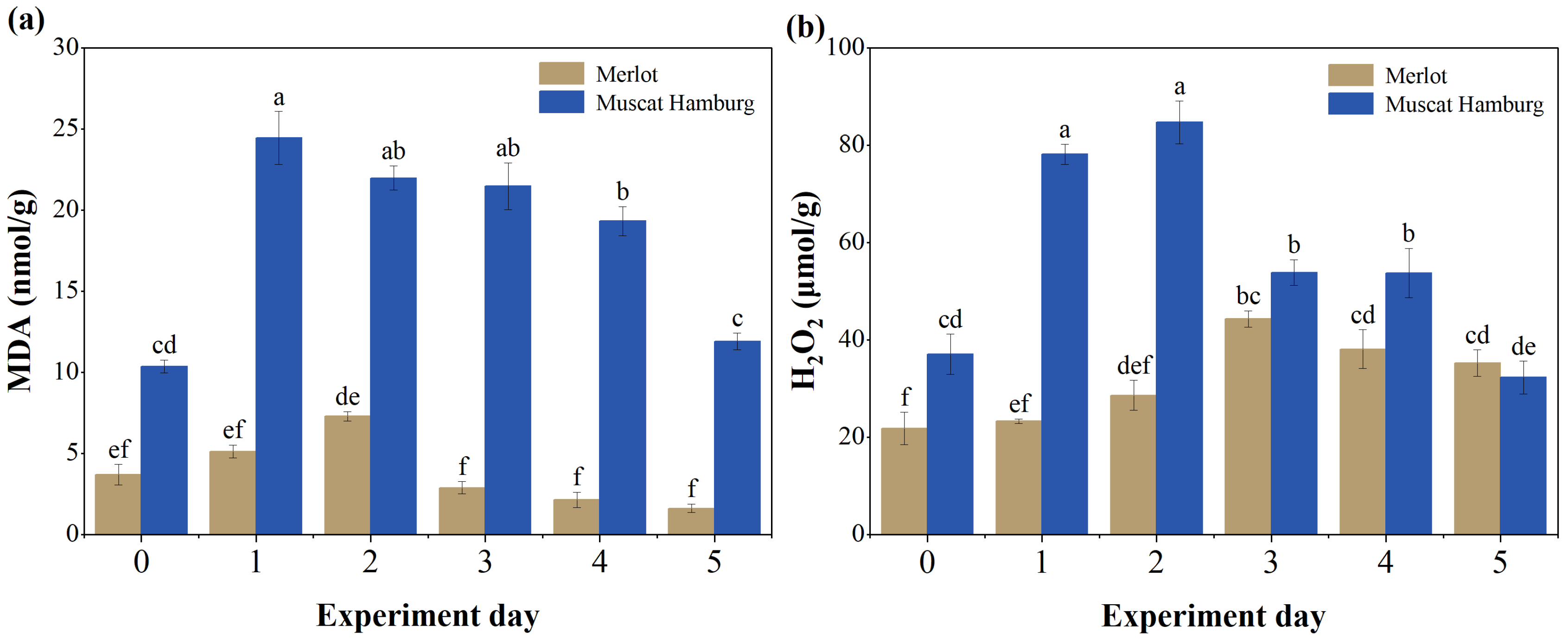
3.2. Changes in H2O2, MDA, and Antioxidant Enzyme Activities Under Heat Stress

Figure 3 showed the H2O2 and MDA content after heat stress treatment of ‘Merlot’ and ‘Muscat Hamburg’ leaves. It was evident that the MDA content of both two cultivars accumulated after high temperature, reached to peak on the first day for ‘Muscat Hamburg’ and the second day for ‘Merlot’, respectively, and then gradually decreased (Figure 3a). Additionally, changes in the H2O2 content for the two varieties had the same tendency, which both increased after heat stress and then decreased. However, the H2O2 content of ‘Muscat Hamburg’ leaves after stress increased rapidly on the first two days and reached the highest value then deceased, while the content of ‘Merlot’ leaves augmented until the 3rd day, and then reduced (Figure 3b).
The content of SOD, POD, and CAT after high temperatures are illustrated in Figure 4. The activities of these enzymes exhibited similar trends, which were enhanced initially after heat stress and then reduced. Specifically, for ‘Merlot’ grapevine leaves, the SOD and CAT activities reached the highest level on the 1st day, and POD activities maximized on the 2nd day, then rapidly weakened. Nevertheless, for ‘Muscat Hamburg’ grapevine leaves, the enzyme activities were constantly higher, and reached the peak on the 2nd (CAT) and 3rd (SOD and POD) day, then decreased. Comparing within the two cultivars, the H2O2 and MDA content of ‘Muscat Hamburg’ illustrated faster and higher augmentation, and the enzyme activities of (SOD, POD, and CAT) of ‘Merlot’ presented a larger increase and responded more rapidly to high temperature, indicating the better heat tolerance of ‘Merlot’.

3.3. Evolution of Leaf Gas Exchange Parameters Under Heat Stress

According to Figure 5, An was approx. 15 μmol·m2·s1 for ‘Muscat Hamburg’ and 20 μmol·m2·s1 for ‘Merlot’, which remained unchanged throughout the experiment days for plants of the control group. Under heat stress, the An of both cultivars rapidly decreased, but the rate of decline in ‘Muscat Hamburg’ was faster than in ‘Merlot’. The decline in An of ‘Merlot’ was greater on the third day, while An for ‘Muscat Hamburg’ was lowest on the second day. Gs and E followed a similar pattern as An, which remained stable for the control group, but decreased slightly under heat stress. However, the variation of VPD showed the opposite trend except for the control group. VPD for two cultivars was sustainedly elevated to 5.5 kPa for ‘Muscat Hamburg’ on the fourth day and 6.5 kPa for ‘Merlot’ at the end of the day during the whole treatment period.

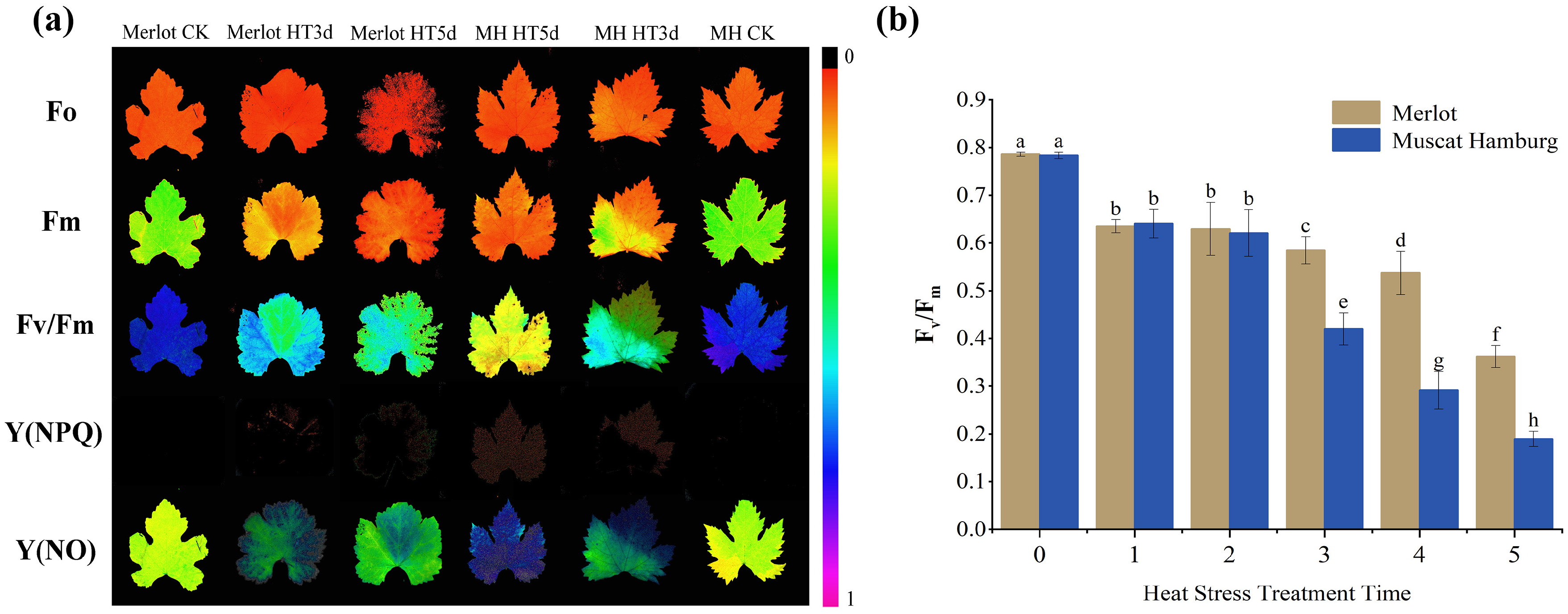
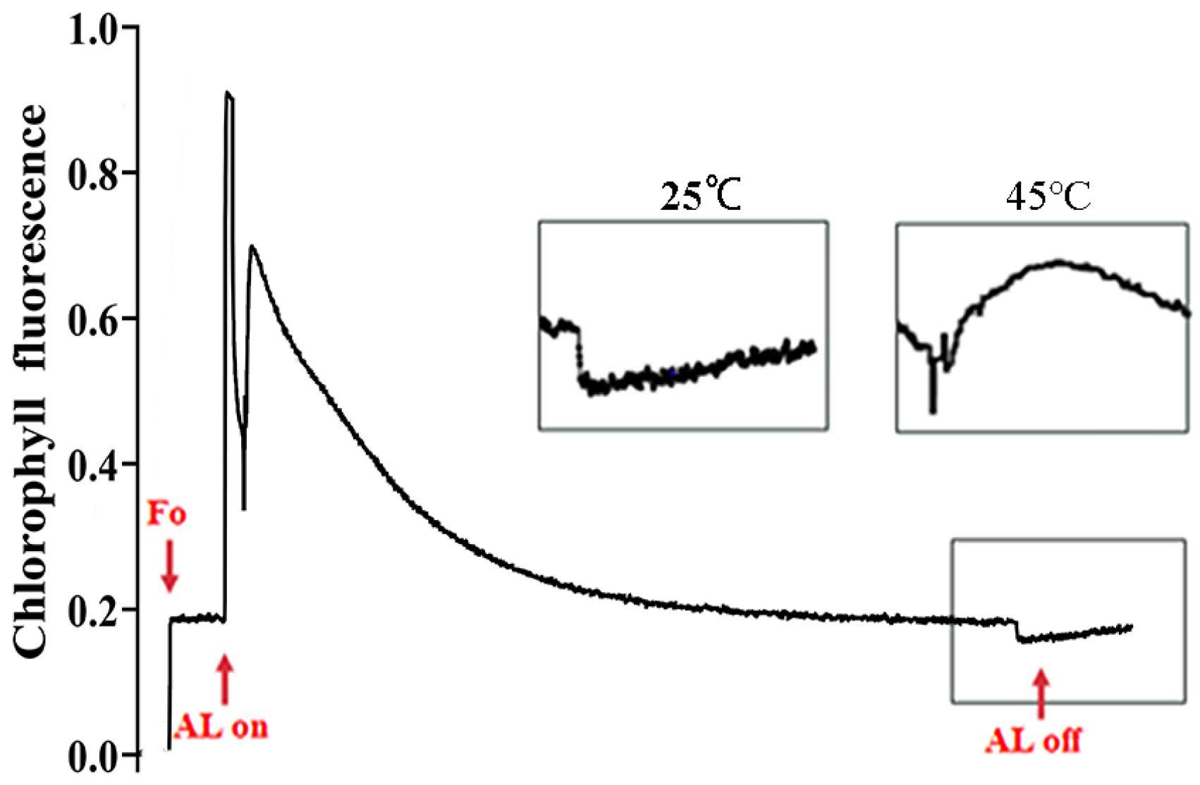
3.4. Impact of Heat Stress on Chlorophyll a Fluorescence in Grapevine Leaves

Based on Figure 6a, minimal chlorophyll fluorescence yield parameter F0 was measured under dark-adapted conditions, presented an orange color before treatment, and gradually the color turned darker (reddish tones), indicating a gradual decrease in F0 under heat stress. Fm was also measured after dark adaptation and indicated in green color for leaves before treatment. The color turned yellow at the outer edge, and then the color changed to orange which spread to whole leaves on the last day of treatment, showing the decrease in Fm under stress. F0 was more stable than Fm. The maximum quantum yield of PSII, Fv/Fm, is considered amongst the most significant parameters of photosynthesis due to its sensitivity to environmental change [34]. This parameter was observed as dark blue initially in healthy leaves, then the color changed to light blue and green on the third day after stress. After five days of treatment, a large area of ‘Merlot’ leaves turned green, while leaves of ‘Muscat Hamburg’ turned yellow with red spots, demonstrating a faster decrease in Fv/Fm from 0.8 to 0.2 (Figure 6b).
The quantum yield of regulated energy dissipation of PSII, Y(NPQ), increased during heat stress. For leaves of ‘Merlot’, green and yellow spots were found along leaf margins and spread to the entire outer edge. By the third day of heat stress, approx. one-third of the leaf blade was covered with red and yellow spots, and by the fifth and final day of heat exposure, all leaves had turned red, indicating the production of NPQ under heat stress. Similarly, the quantum yield of non-regulated energy dissipation of PSII, Y(NO), also increased in response to heat. The color of heat-treated leaves changed from yellow-green to green, and eventually to blue with heat stress. ‘Muscat Hamburg’ was more sensitive to heat than ‘Merlot’ based on the more rapid decrease in this parameter because their color was faster to turn blue and their blue color was darker.

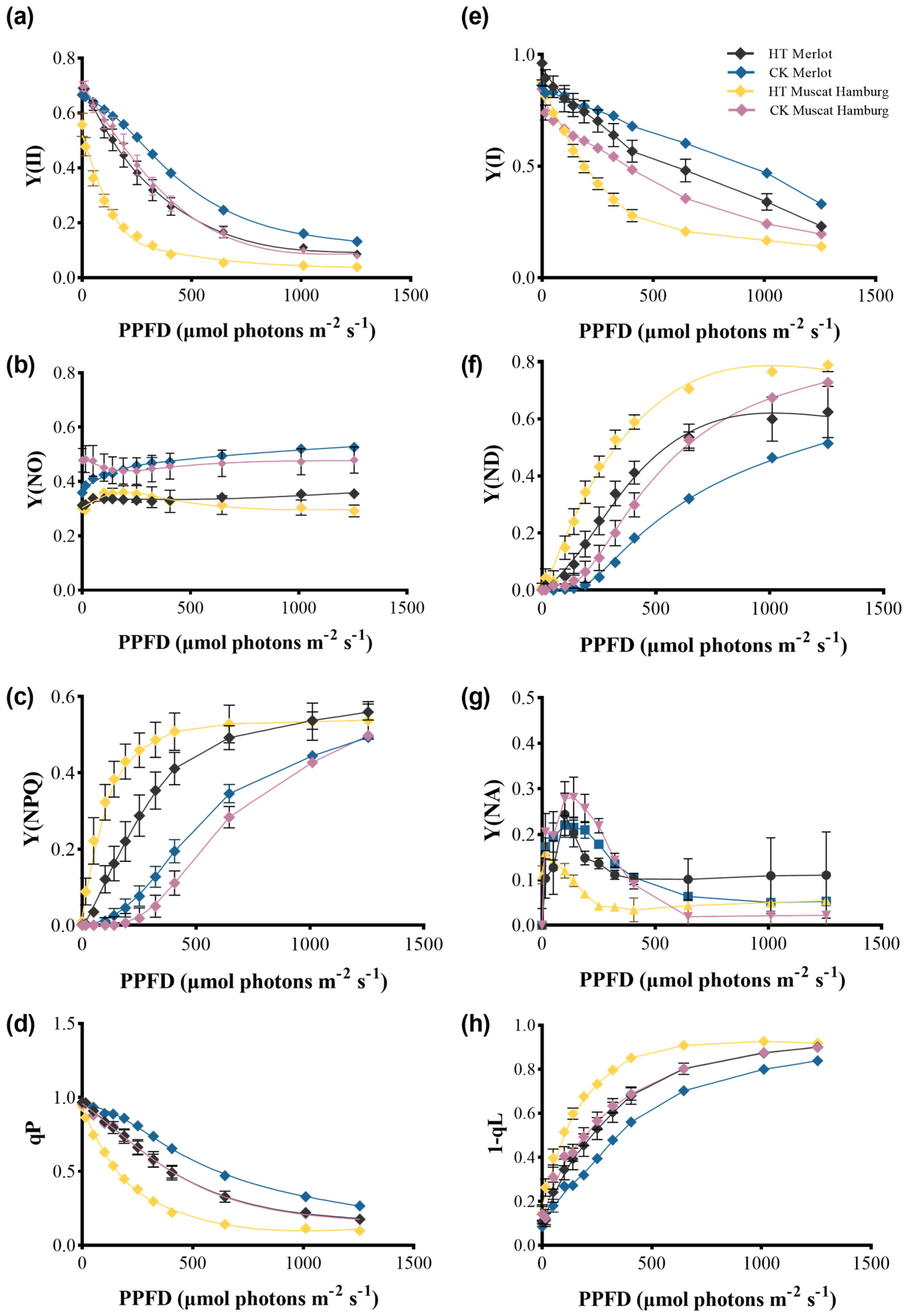
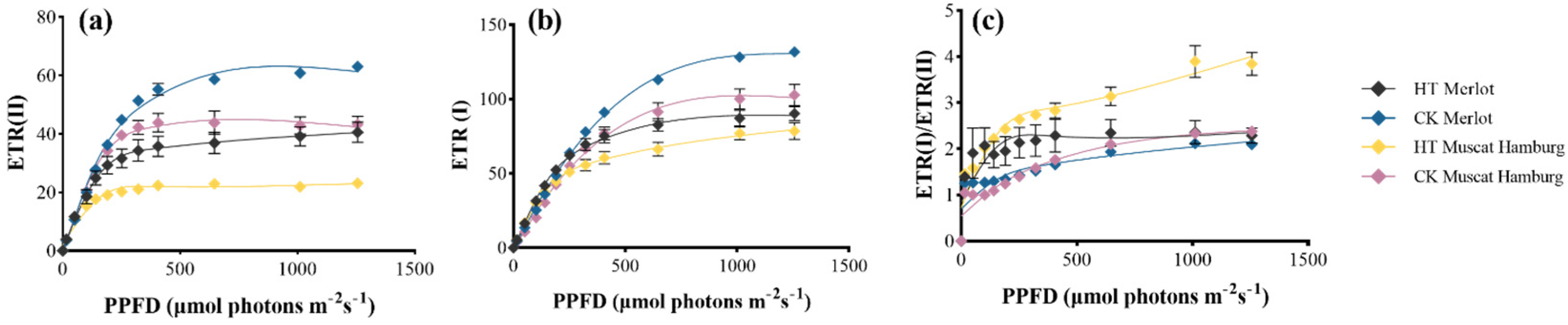
3.5. PSI and PSII Performance of Grapevine Leaves Under Heat Stress

The heat-induced changes in photosynthetic parameters of electron transport were evaluated using light response curves. PSII was sensitive to high temperature, as indicated by the steeper decline in Y(II) in HT leaves than in control ones, particularly at low light intensities. Photochemical quenching, qP, was one of the important factors in the effective quantum yield of PSII. Our results showed that qP decreased rapidly with increasing light intensity, which was considered to be the main reason for the decrease in Y(II) (Figure 7a,d). Meanwhile, Y(NPQ) increased gradually as excess light energy was dissipated as heat in PSII, marking the initiation of self-protection of grapevine leaves experiencing temperature stress. Moreover, the quantum yield of non-regulated energy dissipation of PSII Y(NO) was relatively stable but lower at high temperatures (Figure 7b,c).
PSI was relatively stable, so the decrease in Y(I) indicated irreversible damage under 45 °C in both cultivars (Figure 7e). PSI donor side limitation Y(ND) increased with the light intensity, and under heat stress, the increase was larger. PSI acceptor side limitation Y(NA) varied a little for ‘Merlot’, while it declined slightly at the low light intensity for ‘Muscat Hamburg’ (Figure 7f,g). Compared with ‘Muscat Hamburg’, parameters associated with PSII and PSI of ‘Merlot’ leaves presented smaller variation, indicating better thermo-resistance.
The 1-qL indicates the reduction state of the PQ pool, which increased along with the light intensity and increased with greater exposure to high temperature (Figure 7h). Relative to the control group, the PQ pool size significantly declined under heat stress (Figure 8). Therefore, the electrons transported to PSI via PQ were reduced, and the electron transport efficiency of PSII was diminished.

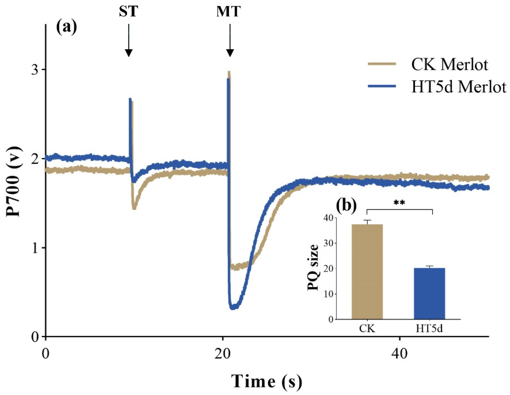
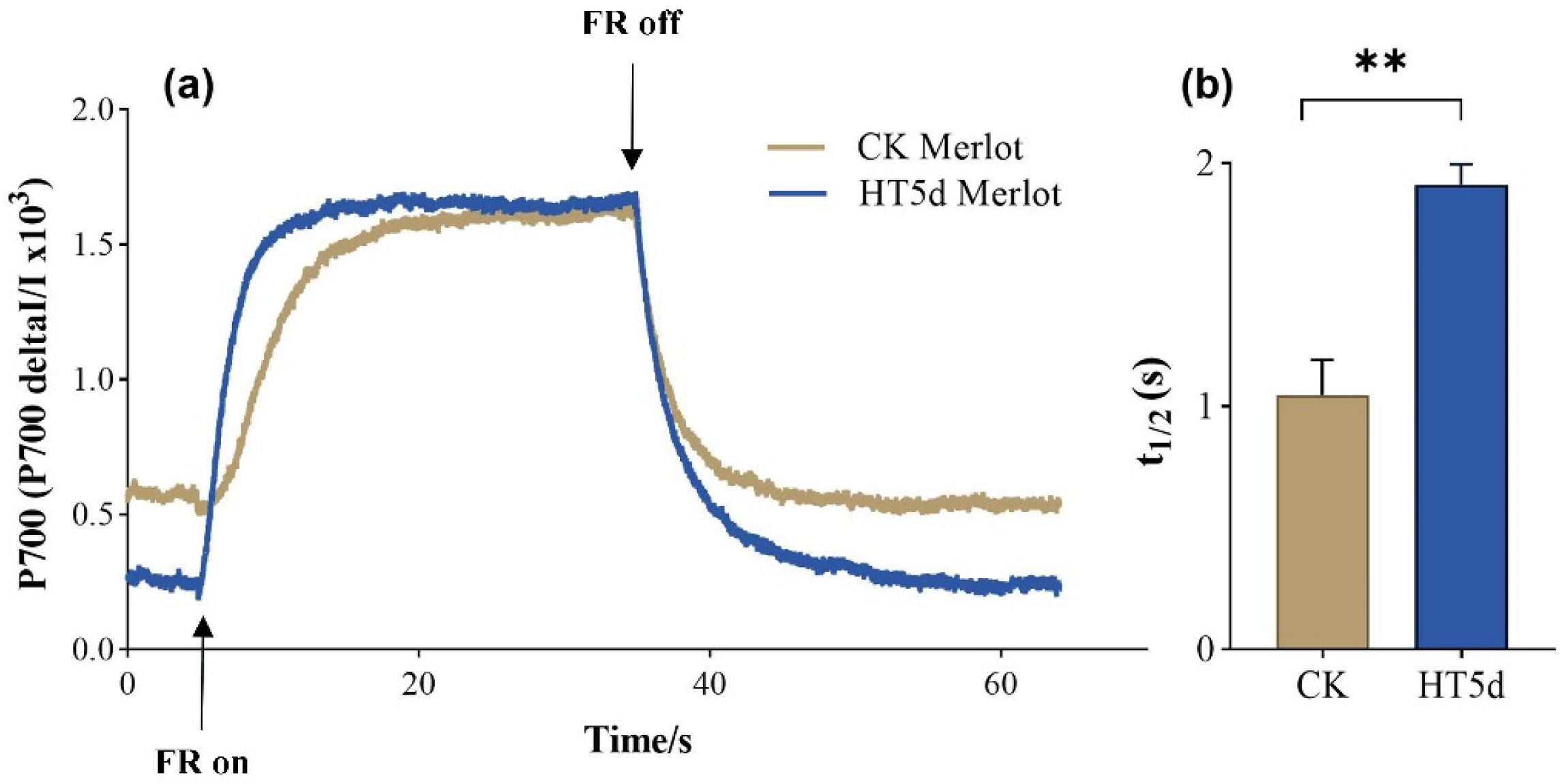
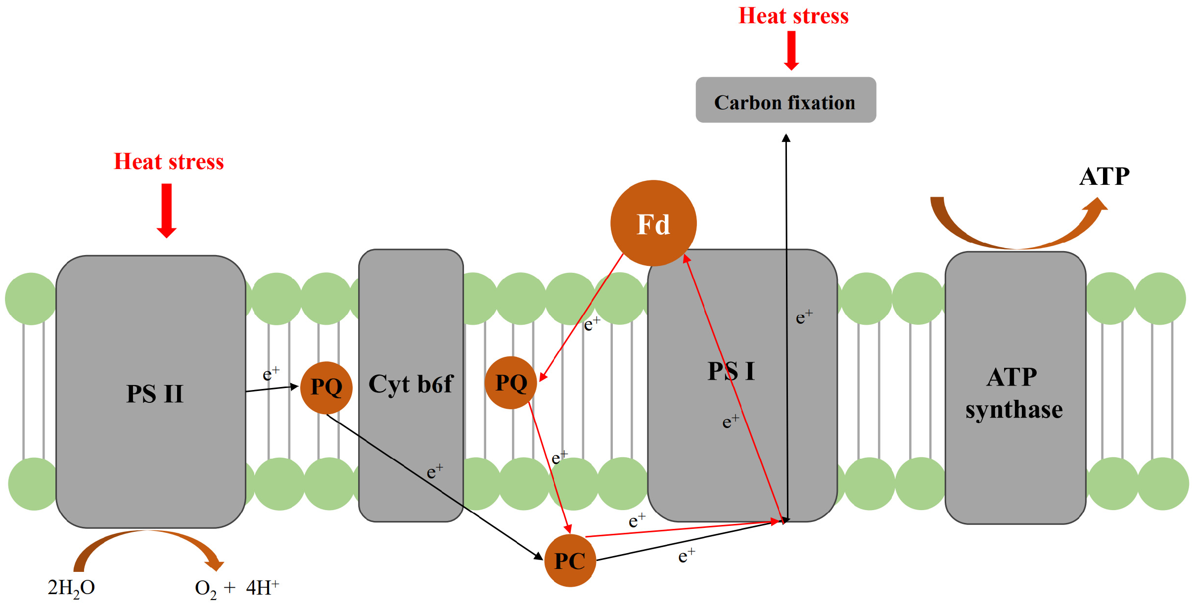
3.6. Impacts on Cyclic Electron Flow of PSI Under Heat Stress

Photosynthetic electron flow through ETR(I) and ETR(II) in the leaves of ‘Merlot’ and ‘Muscat Hamburg’ was significantly reduced at all light intensities under high temperatures (Figure 9a,b). CEF, indicated by the ratio of ETR(I)/ETR(II), increased sharply and the increment grew with increasing light intensity. These data indicated that CEF, relative to the linear electron transport rate, was stimulated by heat stress in the leaves of both cultivars. Additionally, the increased range of ‘Muscat Hamburg’, especially under high light intensities, was larger (Figure 9c), indicating the CEF was stronger activated. The CEF activity is reflected by the rate of P700+ reduction. Due to the sensitivity of ‘Merlot’ to heat, the reduction kinetics of P700+ of ‘Merlot’ was driven by high temperature (Figure 10a). The speed of CEF was indicated by calculating the half-time of the dark reduction curve; longer half-times under heat stress indicated slower CEF rates (Figure 10b).
Two pathways have been demonstrated for CEF: one is those mediated by the chloroplast NADH dehydrogenase-like complex (NDH). The transient increase in chlorophyll fluorescence after illumination was used to measure the NDH-dependent CEF. Figure 11 shows a visible transient post-illumination increase in chlorophyll fluorescence after AL off. Both the rate of the increasing phase and the amplitude of the post-illumination increase in chlorophyll fluorescence were enhanced with rising temperatures, indicating the activation of NDH-dependent CEF in the leaves of ‘Merlot’.

4. Discussion

In recent years, extreme weather events such as heatwaves have occurred frequently with significant economic losses to agriculture. In our experiment, based on the wildly utilization of protected cultivation of viticulture, 45 °C was set to perform heat stress in this study. Chlorophyll loss occurs prematurely in leaves of grapevine cultivars ‘Merlot’ and ‘Muscat Hamburg’ under heat stress, which naturally happens during senescence [35]. The leaves of control groups of both cultivars showed no significant changes in chlorophyll, demonstrating that the primary chlorophyll and catenoid loss was due to heat shock instead of natural senescence. The mitigation of chlorophyll loss could be regarded as an indicator of heat tolerance. In our study, the loss of chlorophyll and catenoid may be the consequence of damage to thylakoid membranes and PSII caused by heat shock. The fast degradation of chlorophyll b under heat stress demonstrated the decrease in light-harvesting chlorophyll a/b-binding proteins (LHC) in PSII, thus transferring the excitation energy of PSII and decreasing the photooxidative damage [36].
High temperature stress can result in plant cell membrane damage, produce ROS, then contribute to oxidative damage and protein degeneration [37]. Normally, reactive oxygen species (ROS) in plants is in a state of dynamic balance. However, when plants are under stress, their own antioxidant system is unable to remove excess ROS, resulting in their imbalance [38]. Changes in H2O2 content are frequently used to estimate the level of plant stress since it can indicate the accumulation of ROS in plant cells after stress treatment [39]. In this experiment, the H2O2 content was accumulated, indicating that grape leaves had been damaged by high temperature stress at this time, which is the same as the previous studies [40]. The accumulation of ROS leads to lipid peroxidation, which is harmful to membrane lipids, proteins, and DNA [41]. MDA serves as a marker for cellular lipid peroxidation, and after stress, it is measured to reflect the oxidative degree of the plasma membrane [42]. In this study, MDA content was accumulated under high temperature treatment which demonstrated that lipid membrane peroxidation and relative permeability of lipid membrane increased in grapevine leaves, then a large amount of MDA was produced and resulted in the lipid membrane damage.
Enzyme SOD, POD, and CAT are secreted when plants suffer from stress and are important enzymes metabolized to defend against oxidative stress [43]. The joint function of these enzymes could maintain the normal level of intracellular free radical content, thus reducing the toxicity of ROS [44]. In this experiment, the activities of the three enzymes were significantly increased, which may be due to oxidative damage of leaves caused by high temperature stress. With the increase in high temperature treatment period, the activities of enzymes decreased significantly, which probably means that the self-regulation ability of grape leaves has been weakened, the ability of endogenous antioxidant oxidase system to clear ROS has been reduced, the metabolism of ROS in vivo has been disrupted, and the plant membrane has been seriously damaged.
Photosynthesis is one of the most sensitive processes to heat stress. The photosynthetic responses of grapevines are variable under distinct heat stress conditions. In this experiment, gs and An decreased greatly in both two cultivars, showing a partial closure of stomata caused by heat stress, indicating that the reduction in assimilation was partly due to the stomatal limitation. Additionally, the decline in E was directly associated with gs due to the stomatal closure under high temperatures [45]. VPD was considered to be the major environmental factor influencing stomatal response and photosynthesis. The increase in VPD was likely related to the decrease in E [46]. Under high VPD, the insufficient water flux into the stem cannot meet the high rate of transpiration of leaves, then the E will decrease and the stomata will finally close [47]. The limitations to CO2 assimilation imposed by stomatal closure may promote an imbalance between photochemical activity at PSII and the electron requirement for photosynthesis, leading to overexcitation and subsequent photoinhibition damage of PSII reaction centers [48].
Severe photoinhibition under heat stress of PSII in both cultivars was observed based on decreased Fv/Fm. Our results indicate that the qP values at 45 °C were much lower than those at 25 °C, indicating that the fraction of PSII centers in the open state decreased and the primary acceptor of PSII (QA) became more reduced under heat stress [49]. Meanwhile, NPQ is the dominant pathway to dissipate excessive light energy in the format of heat energy encompassing several quenching mechanisms [50]. With the extension time of high temperature treatment, Y(NPQ), indicating the regulated thermal energy dissipation, increased to absorb the extra excitation energy in PSII, reducing ROS generation (not specifically measured in this study) and making PSII resistant to variable light, which is regarded as a key mechanism to protect PSII from photodamage. Y(NO) reflects the nonphotochemical quenching. The relatively stable but lower value under heat stress indicated no increased excitation pressures in PSII reaction centers [33].
Photoinhibition of PSI is caused by the oxygen of hydroxyl radicals during the transfer of excess excited electrons from PSII to PSI [51]. The electrons transferred from PSII to PSI can be used for downstream energy requirements, such as carbon dioxide fixation [52]. However, high temperatures reduce the rate of CO2 assimilation, leading to excessive reduction in PSI receptor side, and excessive accumulation of NADPH, causing the large production of hydroxyl radicals. The accumulation of hydroxyl radicals depends on the over-reduction of the PSI receptor side. As indicated by PSI photoinhibition, our results showed that Y(NA) decreased with increasing temperature, which is associated with the inhibition of Y(I) and the increase in Y(ND). Additionally, the redox state of the PQ pool, which serves as an electron transporter between PSII and PSI, is dependent on the LEF ratio of the two photosystems. The preferential excitation of PSII or PSI stimulates the reduction or oxidation of the PQ pool [53,54]. The increase in 1-qL and the decrease in PQ pool size indicated a reduced electron transfer rate from PSII to P700+ in LEF, which corresponded to the changes in Y(I) and Y(II).
Under heat stress, though ETR(I) and ETR(II) both decreased, the ratio of ETR(I)/ETR(II), which is a measure of CEF in PSI, was higher. The CEF is generally considered to be an effective pathway to protect PSI against photoinhibition under stress [55]. Rapid increases in temperature result in CEF being highly activated in mature leaves, stimulating the rapid generation of ΔpH and pmf [14]. In addition to photosynthesis control at the Cyt b6/f complex, ΔpH is also involved in the regulation of ATP/NADPH production ratio in primary metabolism [56]. Additionally, high ΔpH stimulates the induction of NPQ to excess light energy and acidification of the thylakoid lumen, which activates qE by proportionating PsbS and activating violaxanthin deepoxidase [57]. CEF has long been hypothesized to be facilitated by H2O2 produced when the electron transport chain in chloroplasts is over-reduced [58]. In Arabidopsis, a sharp rise in CEF was noted when plants expressing chloroplast glycolate oxidase were moved from high CO2 non-photorespiratory settings to ambient air. [59]. In our study, the CEF of ‘Muscat Hamburg’ was larger than ‘Merlot’, especially under high light conditions, while maybe triggered by the larger accumulation of H2O2 content. Additionally, variations in light levels can influence the effects of heat stress on CEF. During heat stress in pea leaves, CEF seems to be inhibited in the presence of low light [60]. This maybe the reason why the CEF between two cultivars was less induced under lower light condition but increased significantly under higher light.
The electron flow from ferredoxin (Fd) to PQ pool in CEF is measured by chlorophyll fluorescence. After illumination with actinic light, steady-state chlorophyll fluorescence drops to a minimum, then substantially rises, which is due to NDH-dependent CEF. A knockout of NDH in Arabidopsis showed no suppression of photosynthetic activity under fluctuating light, indicating that the NDH-dependent pathway in CEF is a minor pathway compared with the PGR-dependent pathway [61]. However, the sensitivity to high temperature stress of NDH-deficient mutants indicated the activation of the NDH-depended pathway under heat stress [62]. Grapevines may also function similarly to respond to and even adapt to high temperature events (Figure 12).

5. Conclusions

In conclusion, our study demonstrated that under heat stress, ‘Merlot’ had better physiological performance, as indicated by chlorophyll loss, oxidative parameters, gas exchange, and photoinhibition of PSI and PSII, and showed better thermotolerance, compared with ‘Muscat Hamburg’. In addition, the damage of the PSII center caused by severe high temperatures contributed to the oxidization of QA. The increase in NPQ showed its importance for the protection of ‘Merlot’ leaves by dissipating the excess absorbed light energy of the PSII center. Moreover, due to the reduced electron transfer rate in LEF, CEF around PSI was stimulated to generate the proton gradient around the photosystem, which is shown to be an effective pathway for heat dissipation. The NDH-dependent CEF was strongly activated, suggesting its importance in alleviating photoinhibition in ‘Merlot’ leaves under heat stress, while the performance of PGR5/PGRL1-dependent CEF under heat stress in grapevine leaves is still unclear. In future research, the molecular mechanism of function of NDH-dependent and PGR5/PGRL1-dependent CEF under heat stress will be studied. Additionally, more cultivars will be involved in this study to evaluate the thermotolerance of grape leaves, which provides valuable insights for selecting grape varieties under high-temperature conditions. Additionally, it offers a theoretical foundation for research on grape cultivation in high-temperature environments.

Author Contributions

Conceptualization, S.W. and Q.Q.; validation, L.W. and V.P.; formal analysis, Q.Q. and D.G.; data curation, Q.Q. and Y.S.; writing—original draft preparation, Q.Q.; writing—review and editing, S.W. and V.P.; supervision, S.W. and V.P.; project administration, S.W.; funding acquisition, L.W. All authors have read and agreed to the published version of the manuscript.

Funding

This research was funded by Fujian Province science and technology plan project under grant number 2024S0051, the Ningbo Science and Technology Development Special Fund under the grant number 2024S018, and China Agriculture Research System under grant number CARS-29-zp-7.

Data Availability Statement

All study data are detailed in the article; further inquiries can be directed to the corresponding author.

Conflicts of Interest

The authors declare no conflicts of interest.

References

  1. Lobell, D.B.; Field, C.B.; Cahill, K.N.; Bonfils, C. Impacts of future climate change on California perennial crop yields: Model projections with climate and crop uncertainties. Agric. For. Meteorol. 2006, 141, 208–218. [Google Scholar] [CrossRef]
  2. IPCC. Special Report-Climate Change on 1.5 °C; Cambridge University Press: Cambridge, UK; New York, NY, USA, 2018; 616p. [Google Scholar]
  3. Zha, Q.; Xi, X.; Jiang, A.; Wang, S.; Tian, Y. Changes in the protective mechanism of photosystem II and molecular regulation in response to high temperature stress in grapevines. Plant Physiol. Biochem. 2016, 101, 43–53. [Google Scholar] [CrossRef]
  4. Schultz, H. Climate change and viticulture: A European perspective on climatology, carbon dioxide and UV-B effects. Aust. J. Grape Wine Res. 2000, 6, 2–12. [Google Scholar] [CrossRef]
  5. Zha, Q.; Xi, X.; Jiang, A.; Tian, Y. High Temperature Affects Photosynthetic and Molecular Processes in Field-Cultivated Vitis vinifera L. × Vitis labrusca L. Photochem. Photobiol. 2016, 92, 446–454. [Google Scholar] [CrossRef]
  6. Allakhverdiev, S.I.; Kreslavski, V.D.; Klimov, V.V.; Los, D.A.; Carpentier, R.; Mohanty, P. Heat stress: An overview of molecular responses in photosynthesis. Photosynth. Res. 2008, 98, 541–550. [Google Scholar] [CrossRef] [PubMed]
  7. Yin, Y.; Li, S.; Liao, W.; Lu, Q.; Wen, X.; Lu, C. Photosystem II photochemistry, photoinhibition, and the xanthophyll cycle in heat-stressed rice leaves. J. Plant Physiol. 2010, 167, 959–966. [Google Scholar] [CrossRef] [PubMed]
  8. Kramer, D.M.; Evans, J.R. The importance of energy balance in improving photosynthetic productivity. Plant Physiol. 2011, 155, 70–78. [Google Scholar] [CrossRef]
  9. Venios, X.; Korkas, E.; Nisiotou, A.; Banilas, G. Grapevine Responses to Heat Stress and Global Warming. Plants 2020, 9, 1754. [Google Scholar] [CrossRef]
  10. Takahashi, S.; Murata, N. How do environmental stresses accelerate photoinhibition? Trends Plant Sci. 2008, 13, 178–182. [Google Scholar] [CrossRef]
  11. Nicol, L.; Croce, R. The PsbS protein and low pH are necessary and sufficient to induce quenching in the light-harvesting complex of plants LHCII. Sci. Rep. 2021, 11, 7415. [Google Scholar] [CrossRef]
  12. Munekage, Y.; Shikanai, T. Cyclic electron transport through photosystem I. Plant Biotechnol. 2005, 22, 361–369. [Google Scholar] [CrossRef]
  13. Takizawa, K.; Kanazawa, A.; Kramer, D.M. Depletion of stromal Pi induces high ‘energy–dependent’ antenna exciton quenching (qE) by decreasing proton conductivity at CFO–CF1 ATP synthase. Plant Cell Environ. 2008, 31, 235–243. [Google Scholar] [CrossRef] [PubMed]
  14. Zhang, R.; Sharkey, T.D. Photosynthetic electron transport and proton flux under moderate heat stress. Photosynth. Res. 2009, 100, 29–43. [Google Scholar] [CrossRef] [PubMed]
  15. Shikanai, T. Central role of cyclic electron transport around photosystem I in the regulation of photosynthesis. Curr. Opin. Biotechnol. 2014, 26, 25–30. [Google Scholar] [CrossRef]
  16. Yamamoto, H.; Shikanai, T. PGR5-Dependent Cyclic Electron Flow Protects Photosystem I under Fluctuating Light at Donor and Acceptor Sides. Plant Physiol. 2019, 179, 588–600. [Google Scholar] [CrossRef]
  17. Tan, S.L.; Yang, Y.J.; Huang, W. Moderate heat stress accelerates photoinhibition of photosystem I under fluctuating light in tobacco young leaves. Photosynth. Res. 2020, 144, 373–382. [Google Scholar] [CrossRef] [PubMed]
  18. Che, Y.; Wang, H.; Zhang, B.; Gao, S.; Wang, Z.; Wang, Y.; Zhang, H.; Sun, G. Elevated air temperature damage to photosynthetic apparatus alleviated by enhanced cyclic electron flow around photosystem I in tobacco leaves. Ecotoxicol. Environ. Saf. 2020, 204, 111136. [Google Scholar]
  19. Takagi, D.; Miyake, C. PROTON GRADIENT REGULATION 5 supports linear electron flow to oxidize photosystem I. Physiol. Plant. 2018, 164, 337–348. [Google Scholar] [CrossRef]
  20. Chaux, F.; Peltier, G.; Johnson, X. A security network in PSI photoprotection: Regulation of photosynthetic control, NPQ and O2 photoreduction by cyclic electron flow. Front. Plant Sci. 2015, 6, 875. [Google Scholar] [CrossRef]
  21. International Organization of Vine and Wine. Available online: https://www.oiv.int/what-we-do/country-report?oiv (accessed on 10 April 2025).
  22. Fenoll, J.; Manso, A.; Hellín, P.; Ruiz, L.; Flores, P. Changes in the aromatic composition of the Vitis vinifera grape Muscat Hamburg during ripening. Food Chem. 2009, 114, 420–428. [Google Scholar] [CrossRef]
  23. Tamizheezham, U.; Muthuvel, I.; Subbiah, A. Effect of Temperature on Shelf life of Muscat Hamburg Grapes under Storage. Madras Agric. J. 2018, 105, 176. [Google Scholar] [CrossRef]
  24. de Rosas, I.; Deis, L.; Baldo, Y.; Cavagnaro, J.B.; Cavagnaro, P.F. High Temperature Alters Anthocyanin Concentration and Composition in Grape Berries of Malbec, Merlot, and Pinot Noir in a Cultivar-Dependent Manner. Plants 2022, 11, 926. [Google Scholar] [CrossRef] [PubMed]
  25. Carbonell-Bejerano, P.; Santa María, E.; Torres-Pérez, R.; Royo, C.; Lijavetzky, D.; Bravo, G.; Aguirreolea, J.; Sánchez-Díaz, M.; Antolín, M.C.; Martínez-Zapater, J.M. Thermotolerance Responses in Ripening Berries of Vitis vinifera L. cv Muscat Hamburg. Plant Cell Physiol. 2013, 54, 1200–1216. [Google Scholar] [CrossRef] [PubMed]
  26. Zhang, X.; Huang, B. Drought priming-induced heat tolerance: Metabolic pathways and molecular mechanisms. In Priming-Mediated Stress and Cross-Stress Tolerance in Crop Plants; Academic Press: Cambridge, MA, USA, 2020; pp. 149–160. [Google Scholar]
  27. Lu, H.-C.; Chen, W.-K.; Wang, Y.; Bai, X.-J.; Cheng, G.; Duan, C.-Q.; Wang, J.; He, F. Effect of the seasonal climatic variations on the flavonoid accumulation in Vitis vinifera cv ‘Muscat hamburg’ and ‘Victoria’ grapes under the double cropping system. Foods 2021, 11, 48. [Google Scholar] [CrossRef] [PubMed]
  28. Li, P.; Weng, J.; Zhang, Q.; Yu, L.; Yao, Q.; Chang, L.; Niu, Q. Physiological and biochemical responses of Cucumis melo L. chloroplasts to low-phosphate stress. Front. Plant Sci. 2018, 9, 1525. [Google Scholar] [CrossRef]
  29. Iwai, M.; Yokono, M.; Kono, M.; Noguchi, K.; Akimoto, S.; Nakano, A. Light-harvesting complex Lhcb9 confers a green alga-type photosystem I supercomplex to the moss Physcomitrella patens. Nat. Plants 2015, 1, 14008. [Google Scholar] [CrossRef]
  30. Klughammer, C.; Schreiber, U. An improved method, using saturating light pulses, for the determination of photosystem I quantum yield via P700+-absorbance changes at 830 nm. Planta 1994, 192, 261–268. [Google Scholar] [CrossRef]
  31. Hendrickson, L.; Furbank, R.T.; Chow, W.S. A simple alternative approach to assessing the fate of absorbed light energy using chlorophyll fluorescence. Photosynth. Res. 2004, 82, 73–81. [Google Scholar] [CrossRef]
  32. Kramer, D.M.; Johnson, G.; Kiirats, O.; Edwards, G.E. New fluorescence parameters for the determination of QA redox state and excitation energy fluxes. Photosynth. Res. 2004, 79, 209–218. [Google Scholar] [CrossRef]
  33. Backes, A.; Vaillant-Gaveau, N.; Esmaeel, Q.; Ait Barka, E.; Jacquard, C. A biological agent modulates the physiology of barley infected with Drechslera teres. Sci. Rep. 2021, 11, 8330. [Google Scholar] [CrossRef]
  34. Guidi, L.; Lo Piccolo, E.; Landi, M. Chlorophyll fluorescence, photoinhibition and abiotic stress: Does it make any difference the fact to be a C3 or C4 species? Front. Plant Sci. 2019, 10, 174. [Google Scholar] [CrossRef] [PubMed]
  35. Thomson, W.W.; Nothnagel, E.A.; Huffaker, R.C. Plant Senescence: Its Biochemistry and Physiology; California University: Riverside, CA, USA, 1987. [Google Scholar]
  36. Xu, Q.; Paulsen, A.Q.; Guikema, J.A.; Paulsen, G.M. Functional and ultrastructural injury to photosynthesis in wheat by high temperature during maturation. Environ. Exp. Bot. 1995, 35, 43–54. [Google Scholar] [CrossRef]
  37. Kamatchi, K.M.; Anitha, K.; Kumar, K.A.; Senthil, A.; Kalarani, M.; Djanaguiraman, M. Impacts of combined drought and high-temperature stress on growth, physiology, and yield of crops. Plant Physiol. Rep. 2024, 29, 28–36. [Google Scholar] [CrossRef]
  38. Annadurai, M.K.K.; Alagarsamy, S.; Karuppasami, K.M.; Ramakrishnan, S.; Subramanian, M.; Venugopal, P.R.; Muthurajan, R.; Vellingiri, G.; Dhashnamurthi, V.; Veerasamy, R. Melatonin decreases negative effects of combined drought and high temperature stresses through enhanced antioxidant defense system in tomato leaves. Horticulturae 2023, 9, 673. [Google Scholar] [CrossRef]
  39. Zhang, M.; Smith, J.A.C.; Harberd, N.P.; Jiang, C. The regulatory roles of ethylene and reactive oxygen species (ROS) in plant salt stress responses. Plant Mol. Biol. 2016, 91, 651–659. [Google Scholar] [CrossRef]
  40. Su, Y.; Li, X.; Cao, Z.; Gao, Z.; Du, Y. Effects of Long-Term High Temperatures in the Root Zone on the Physiological Characteristics of Grapevine Leaves and Roots: Implications for Viticulture Practices. Horticulturae 2024, 10, 245. [Google Scholar] [CrossRef]
  41. Choudhary, S.; Bhat, T.M.; Alwutayd, K.M.; Abd El-Moneim, D.; Naaz, N. Salicylic acid enhances thermotolerance and antioxidant defense in Trigonella foenum graecum L. under heat stress. Heliyon 2024, 10, e27227. [Google Scholar] [CrossRef] [PubMed]
  42. Hao, F.; Wang, X.; Chen, J. Involvement of plasma-membrane NADPH oxidase in nickel-induced oxidative stress in roots of wheat seedlings. Plant Sci. 2006, 170, 151–158. [Google Scholar] [CrossRef]
  43. Zeng, C.-Q.; Liu, W.-X.; Hao, J.-Y.; Fan, D.-N.; Chen, L.-M.; Xu, H.-N.; Li, K.-Z. Measuring the expression and activity of the CAT enzyme to determine Al resistance in soybean. Plant Physiol. Biochem. 2019, 144, 254–263. [Google Scholar] [CrossRef]
  44. Mittler, R. Oxidative stress, antioxidants and stress tolerance. Trends Plant Sci. 2002, 7, 405–410. [Google Scholar] [CrossRef]
  45. Bassanezi, R.B.; Amorim, L.; Filho, A.B.; Berger, R.D. Gas Exchange and Emission of Chlorophyll Fluorescence during the Monocycle of Rust, Angular Leaf Spot and Anthracnose on Bean Leaves as a Function of their Trophic Characteristics. J. Phytopathol. 2002, 150, 37–47. [Google Scholar] [CrossRef]
  46. Zhang, D.; Zhang, Z.; Li, J.; Chang, Y.; Du, Q.; Pan, T. Regulation of vapor pressure deficit by greenhouse micro-fog systems improved growth and productivity of tomato via enhancing photosynthesis during summer season. PLoS ONE 2015, 10, e0133919. [Google Scholar] [CrossRef] [PubMed]
  47. Inoue, T.; Sunaga, M.; Ito, M.; Yuchen, Q.; Matsushima, Y.; Sakoda, K.; Yamori, W. Minimizing VPD Fluctuations Maintains Higher Stomatal Conductance and Photosynthesis, Resulting in Improvement of Plant Growth in Lettuce. Front. Plant Sci. 2021, 12, 646144. [Google Scholar] [CrossRef]
  48. Souza, R.P.; Machado, E.C.; Silva, J.A.B.; Lagôa, A.M.M.A.; Silveira, J.A.G. Photosynthetic gas exchange, chlorophyll fluorescence and some associated metabolic changes in cowpea (Vigna unguiculata) during water stress and recovery. Environ. Exp. Bot. 2004, 51, 45–56. [Google Scholar] [CrossRef]
  49. Maxwell, D.P.; Falk, S.; Trick, C.G.; Huner, N. Growth at Low Temperature Mimics High-Light Acclimation in Chlorella vulgaris. Plant Physiol. 1994, 105, 535–543. [Google Scholar] [CrossRef]
  50. Li, Q.; Yao, Z.-J.; Mi, H. Alleviation of Photoinhibition by Co-ordination of Chlororespiration and Cyclic Electron Flow Mediated by NDH under Heat Stressed Condition in Tobacco. Front. Plant Sci. 2016, 7, 285. [Google Scholar] [CrossRef]
  51. Havaux, M.; Davaud, A. Photoinhibition of photosynthesis in chilled potato leaves is not correlated with a loss of Photosystem-II activity. Photosynth. Res. 2004, 40, 75–92. [Google Scholar] [CrossRef]
  52. Suggett, D.J.; MacIntyre, H.L.; Kana, T.M.; Geider, R.J. Comparing electron transport with gas exchange: Parameterising exchange rates between alternative photosynthetic currencies for eukaryotic phytoplankton. Aquat. Microb. Ecol. 2009, 56, 147–162. [Google Scholar] [CrossRef]
  53. Laisk, A.; Eichelmann, H.; Oja, V.; Peterson, R.B. Control of cytochrome b6f at low and high light intensity and cyclic electron transport in leaves. Biochim. Biophys. Acta (BBA) Bioenerg. 2005, 1708, 79–90. [Google Scholar] [CrossRef]
  54. Mattila, H.; Khorobrykh, S.; Hakala-Yatkin, M.; Havurinne, V.; Kuusisto, I.; Antal, T.; Tyystjärvi, T.; Tyystjärvi, E. Action spectrum of the redox state of the plastoquinone pool defines its function in plant acclimation. Plant J. 2020, 104, 1088–1104. [Google Scholar] [CrossRef]
  55. Hahn, A.; Vonck, J.; Mills, D.J.; Meier, T.; Kühlbrandt, W. Structure, mechanism, and regulation of the chloroplast ATP synthase. Science 2018, 360, eaat4318. [Google Scholar] [CrossRef] [PubMed]
  56. Alboresi, A.; Storti, M.; Morosinotto, T. Balancing protection and efficiency in the regulation of photosynthetic electron transport across plant evolution. New Phytol. 2019, 221, 105–109. [Google Scholar] [CrossRef] [PubMed]
  57. Huang, W.; Zhang, S.-B.; Xu, J.-C.; Liu, T. Plasticity in roles of cyclic electron flow around photosystem I at contrasting temperatures in the chilling-sensitive plant Calotropis gigantea. Environ. Exp. Bot. 2017, 141, 145–153. [Google Scholar] [CrossRef]
  58. Abogadallah, G.M. Differential regulation of photorespiratory gene expression by moderate and severe salt and drought stress in relation to oxidative stress. Plant Sci 2011, 180, 540–547. [Google Scholar] [CrossRef] [PubMed]
  59. Strand, D.D.; Livingston, A.K.; Satoh-Cruz, M.; Froehlich, J.E.; Maurino, V.G.; Kramer, D.M. Activation of cyclic electron flow by hydrogen peroxide in vivo. Proc. Natl. Acad. Sci. USA 2015, 112, 5539–5544. [Google Scholar] [CrossRef]
  60. Havaux, M.; Greppin, H.; Strasser, R.J. Functioning of photosystems I and II in pea leaves exposed to heat stress in the presence or absence of light: Analysis using in-vivo fluorescence, absorbance, oxygen and photoacoustic measurements. Planta 1991, 186, 88–98. [Google Scholar] [CrossRef]
  61. Kono, M.; Noguchi, K.; Terashima, I. Roles of the Cyclic Electron Flow Around PSI (CEF-PSI) and O2-Dependent Alternative Pathways in Regulation of the Photosynthetic Electron Flow in Short-Term Fluctuating Light in Arabidopsis thaliana. Plant Cell Physiol. 2014, 55, 990–1004. [Google Scholar] [CrossRef]
  62. Wang, P.; Duan, W.; Takabayashi, A.; Endo, T.; Shikanai, T.; Ye, J.-Y.; Mi, H. Chloroplastic NAD(P)H Dehydrogenase in Tobacco Leaves Functions in Alleviation of Oxidative Damage Caused by Temperature Stress. Plant Physiol. 2006, 141, 465–474. [Google Scholar] [CrossRef]
Figure 1. Air and leaf temperature of grapevine during heat stress. (a) Air temperature measured in the control phytotron. (b) Air temperature measured in the heat stress cabinet. (c) Leaf temperature measured of ‘Merlot’ and ‘Muscat Hamburg’ (MH). CK: control; HT: heat stress treatment. Mean ± SE was calculated from 3 replications.
Agronomy 15 00948 g001
Figure 2. Effects of heat stress on (a) chlorophyll a content, (b) chlorophyll b content (c) chlorophyll content, and (d) carotenoid content of young and mature leaves of ‘Merlot’ and ‘Muscat Hamburg’ on the 0, 1, 2, 3, 4 and 5 days. The values are means ± SE of 3 independent replications. Different letters present statistically different means (p < 0.05) between points.
Agronomy 15 00948 g002
Figure 3. Effects of heat stress on (a) Malondialdehyde (MDA) content and (b) H2O2 content in grapevine leaves on the 0, 1, 2, 3, 4 and 5 days. The values are means ± SE of 3 independent replications. Different letters present statistically different means (p < 0.05) between points.
Agronomy 15 00948 g003
Figure 4. Effects of heat stress on (a) Superoxide Dismutase (SOD) activity, (b) Peroxidase (POD) activity, and (c) Catalase (CAT) activity in grapevine leaves on the 0, 1, 2, 3, 4 and 5 days. The values are means ± SE of 3 independent replications. Different letters present statistically different means (p < 0.05) between points.
Agronomy 15 00948 g004
Figure 5. Variation of (a) net CO2 Assimilation (An), (b) stomatal conductance (gs), (c) transpiration rate (E), and (d) leaf-to-air vapor pressure deficit (VPD) on ‘Merlot’ and ‘Muscat Hamburg’ grapevine leaves under heat stress on the 0, 1, 2, 3, 4 and 5 days. Bars represent the standard error of the means, calculated from three independent experiments for each experimental condition and for each time-point. Different letters present statistically different means (p < 0.05) between points.
Agronomy 15 00948 g005
Figure 6. Parameters of chlorophyll a fluorescence (F0), maximal fluorescence (Fm), maximal photosystem II quantum yield (Fv/Fm), the quantum yield of regulated energy dissipation of PSII Y(NPQ), and quantum yield of non-regulated energy dissipation of PSII Y(NO) (a) and data of Fv/Fm (b) on ‘Merlot’ and ‘Muscat Hamburg’ grapevine leaves under heat stress on the 0, 3 and 5 days. Three independent biological replicates were performed. Bars represent the standard error of the means, calculated from three independent experiments for each experimental condition and for each time-point. Different letters present statistically different means (p < 0.05) between points.
Agronomy 15 00948 g006
Figure 7. Light-response curves of (a) the effective quantum yield of PSII Y(II), (b) quantum yield of non-regulated energy dissipation of PSII Y(NO), (c) the quantum yield of regulated energy dissipation of PSII Y(NPQ), (d) the photochemical quenching coefficient qP, (e) the quantum yield of PSI photochemistry Y(I), (f) the quantum yield of PSI non-photochemical energy dissipation due to donor side limitation Y(ND), (g) the quantum yield of the non-photochemical energy dissipation due to the acceptor side limitation Y(NA) and (h) 1-qL in ‘Merlot’ and ‘Muscat Hamburg’ leaves under control (25 °C) and heat stress (45 °C). qL: the fraction of open PSII reaction centers. The means ± SE were calculated from 5 replications.
Agronomy 15 00948 g007
Figure 8. Effect of 5 days of heat stress on PQ pools of ‘Merlot’ leaves. The P700 signal was firstly stimulated by a single turnover flash (ST, 50 ms) and then by multiple turnover flashes (MT, 50 ms) of far-red (FR) light. (a) The complementary areas between the stationary level of P700+ and the oxidation curves of P700 under the background far-red light after the irradiation with MT and ST are referred to as MT-area and ST-area. The ratio of MT-area and ST-area is calculated to derive the size of functional PQ pools. (b) The difference is significant at p < 0.01 (**). Error bars represent means ± SD (n = 3).
Agronomy 15 00948 g008
Figure 9. Light-response curves of the electron transport rate through PSII, ETR(II) (a), the electron transport rate through PSI, ETR(I) (b), and ETR(I)/ETR(II) (c) in ‘Merlot’ and ‘Muscat Hamburg’ leaves under control (25 °C) and heat stress (45 °C). The means ± SE were calculated from 5 replications.
Agronomy 15 00948 g009
Figure 10. P700 reduction kinetic traces of ‘Merlot’ leaves under control and heat stress of 5 days. (a) P700 reduction kinetic traces. The P700 signal was opened, then the far-red light (FR) was turned on, and the traces of the P700 signal would rise to the top. FR was turned off after the P700 signal trace was stable, which will then quickly reduce in the dark until to the steady state. (b) The time required for the reduction to half of the dark reduction curve was calculated for control and heat stress leaves. The difference is significant at p < 0.01 (**). Error bars represent means ± SD (n = 3).
Agronomy 15 00948 g010
Figure 11. Post-illumination increases in chlorophyll fluorescence of ‘Merlot’ leaves under control and heat stress of 5 days. Grapevine leaves under control and heat stress were exposed to actinic light (AL) (50 µmol photons m−2 s−1) for 2 min. AL was turned off and the subsequent change in chlorophyll fluorescence was measured as NDH activity. Inset were magnified traces of light-to-dark transition of grapevine leaves under different temperatures. Fo, the Minimal chlorophyll fluorescence yield.
Agronomy 15 00948 g011
Figure 12. Schematic presentation of effects on the CEF (red arrows) in the thylakoid membranes under heat stress. High temperatures trigger a decrease in NPQ, an excessive buildup of light energy in PSII, photoinhibition of PSI, and an increase in ROS. Additionally, the LEF (black arrows) was changed into CEF via Fd, and NDH-dependent CEF was dramatically induced.
Agronomy 15 00948 g012
Disclaimer/Publisher’s Note: The statements, opinions and data contained in all publications are solely those of the individual author(s) and contributor(s) and not of MDPI and/or the editor(s). MDPI and/or the editor(s) disclaim responsibility for any injury to people or property resulting from any ideas, methods, instructions or products referred to in the content.

