Article

Rhamnolipid-Stabilized Essential Oils Nanoemulsions: Sustainable Biopesticides and Biostimulants with Potential for Crop Protection

by
Lucille T. Kourdova
1,2,†,
Milagro Mottola
1,2,†,
Micaela Peppino Margutti
1,2,3,
María Florencia Bogino
1,2,
Paula Maritano
1,2,
Raquel Viviana Vico
4,5,
Francisca Blanco-Herrera
3,
María Laura Fanani
1,2,* and
Georgina Fabro
1,2,*
1
Departamento de Química Biológica Ranwel Caputto (DQBRC), Facultad de Ciencias Químicas, Universidad Nacional de Córdoba, Córdoba X5000HUA, Argentina
2
Centro de Investigaciones en Química Biológica de Córdoba (CIQUIBIC), National Scientific and Technical Research Council (CONICET), Córdoba X5000HUA, Argentina
3
Centro de Biotecnología Vegetal, Facultad de Ciencias de la Vida, Universidad Andrés Bello, Santiago 8370146, Chile
4
Departamento de Química Orgánica, Facultad de Ciencias Químicas, Universidad Nacional de Córdoba, Córdoba X5000HUA, Argentina
5
Instituto de Investigaciones en Físicoquímica de Córdoba (INFIQC), National Scientific and Technical Research Council (CONICET), Córdoba X5000HUA, Argentina
*
Authors to whom correspondence should be addressed.
These authors contribute equally to this work.
Agronomy 2025, 15(4), 824; https://doi.org/10.3390/agronomy15040824
Submission received: 5 March 2025 / Revised: 24 March 2025 / Accepted: 25 March 2025 / Published: 27 March 2025

Abstract

:
The search for environmentally friendly solutions to effectively control crop pests while safeguarding human health has become a global priority. One promising strategy is to enhance plant defenses by pre-inducing their innate immune system. In this study, we developed rhamnolipid (RL)-stabilized nanoemulsions (NEs) encapsulating essential oils (EOs) as potential biopesticides and biostimulants for agroindustrial applications. These NEs were designed to improve the solubility and stability of EOs while effectively combining their insecticidal and/or repellent activities with the bioactive properties of RLs. In this regard, our interdisciplinary approach involved formulating and characterizing these NEs and evaluating their stability and wettability on plant leaf surfaces. We further evaluated their effects on bacterial growth in vitro and in the model plant Arabidopsis thaliana, along with their impact on beneficial soil microorganisms. We analyzed their ability to stimulate the plant’s immune system and their impact on the viability and reproduction of the aphid Myzus persicae. Additionally, we explored whether RLs stimulate plant defenses through alterations in the leaf cuticle. Our findings demonstrate that RL-stabilized EO-NEs are effective bioprotectants and biostimulants in the model plant, offering a sustainable alternative that could reduce reliance on chemical pesticides in agriculture.

1. Introduction

A major challenge in agriculture is the widespread use of chemical pesticides, which cause environmental contamination and lead to resistant pathogen strains [1]. Addressing this requires global efforts to develop pest control solutions that reduce environmental and health risks. Natural rhamnolipids (RLs) have gained attention due to their biotechnological applications across industries. Their ecological benefits, such as low toxicity and biodegradability [2,3], make them appealing for sustainable agribusiness practices [4]. Natural and synthetic RLs exhibit antibacterial and antifungal properties and can enhance plant defenses through Induced Resistance (IR) [5,6,7,8]. These features make RLs a promising eco-friendly alternative to traditional pesticides [9,10].
Natural RLs also function as effective emulsifiers [11], suitable for encapsulating hydrophobic bioactive compounds of agricultural interest. They stabilize oil-in-water nanoemulsions (NEs) with 50–500 nm droplet sizes, offering increased surface area, physical stability, and resistance to gravitational separation and droplet aggregation. This makes NEs ideal for applications requiring prolonged shelf life and reliability under diverse conditions. In prior research, we showed that RLs effectively stabilize n-hexadecane (HD) NEs while enhancing Arabidopsis thaliana’s immune response to the bacterial pathogen Pseudomonas syringae pv tomato DC3000 (Pst) [8]. These NEs remained stable for over 60 days without microbial contamination or phase separation, highlighting their potential for agricultural applications.
Essential oils (EOs) from aromatic plants are gaining popularity as eco-friendly alternatives to conventional pesticides due to their repellent and insecticidal properties [12,13,14,15]. However, their lipophilicity, volatility, and instability limit their broader application [16]. Encapsulation techniques, such as coacervation, nanoprecipitation, and spray drying, help to control EOs’ volatility and extend shelf life [17,18,19,20]. However, these methods often rely on synthetic stabilizers or complex processes, limiting scalability and bioactivity preservation [21,22,23].
In contrast, NEs offer enhanced EO bioavailability, solubility, and stability, enabling gradual release and prolonged efficacy against pests and pathogens [24,25,26]. This study investigated RL-stabilized NEs containing EOs like thyme, rue, tea tree, cinnamon, and lemon, exploring their potential for future crop protection [27,28,29,30,31]. These EOs, rich in bioactive compounds (e.g., terpenes, phenols, aldehydes), exhibit antimicrobial and insecticidal properties [20,32,33,34,35]. To develop eco-friendly, cost-effective crop protection tools, we created these RL-stabilized EO-NEs that combine the RLs’ bioactivity with EOs’ protective effects, extending their shelf life and agricultural applicability. We hypothesized that these formulations could induce plant immunity and provide sustainable pest control. We assessed their biophysical and biological properties, including particle stability, plant surface wettability, effects on leaf cuticle dynamics, and their impact on pathogenic bacteria growth, aphid viability, and beneficial soil bacteria. Our findings confirm that RL-stabilized EO-NEs are effective, sustainable bioprotectants and biostimulants in a plant model system. This highlights the potential impact these formulations may have on supporting the development of a pesticide-free agroindustry.

2. Materials and Methods

2.1. Materials

The natural RLs were purchased from Sigma-Aldrich (Burlington, VT, USA). The selected EOs, based on their promising antimicrobial and insecticidal properties, were lavender, rosemary, cinnamon, thyme, lemon, rue, and tea tree, all purchased from Pura Química (Córdoba, Argentina). All reagents were analytical grade (99% purity) and used without further purification. Deionized water, with a resistivity of ~18 MΩ cm, was obtained from a Milli-Q Gradient System (Millipore, Bedford, MA, USA).

2.2. Spectroscopic Characterization of Essential Oils

Essential oils (EOs) of cinnamon, lavender, lemon, thyme (T), rue (R), and tea tree (A) were chosen for their antimicrobial and insect-repellent properties [33,34] for the development of rhamnolipid (RL)-stabilized nanoemulsions (NEs). The essential oils (thyme, rue, and tea tree) were characterized spectroscopically by 1H NMR and 13C NMR (Supplementary Figures S1 and S2). The experiments were performed on a Bruker Advance II 400 high-resolution spectrometer, operating at 400.13 MHz for 1H and 100.03 Hz for 13C. The essential oils were dissolved in CCl3D (Sigma-Aldrich, Burlington, VT, USA) to prepare solutions for NMR analysis. Chemical shifts were recorded in parts per million (ppm).

2.3. Design and Production of RL-NEs

Oil/water (O/W) RL nanoemulsions were prepared using 2% w/v EOs (lavender, rosemary, cinnamon, lemon, thyme, rue, or tea tree) or n-hexadecane (HD) as the oil phase following the laboratory’s pre-established protocols [8].

2.4. Particle Analysis

Dynamic light scattering (DLS) and ζ potential measurements were performed to assess oil droplet size distribution and electrostatic properties in aqueous solutions using a NicompTM 380 Submicron Particle Sizer (PSS-NICOMP, Santa Barbara, CA, USA). Particle size was measured with a 530 nm laser at a 90° angle by fitting a correlation function, and the hydrodynamic diameter was calculated using the Stokes–Einstein equation. ζ potential measurements were conducted with a Zetasizer SZ-100-Z (Horiba, Ltd., Kyoto, Japan) using a 532 nm, 10 mW laser and laser-Doppler velocimetry. For the analysis, lipid particles were suspended in 5 mM potassium phosphate buffer at pH 7.17.

2.5. Contact Angle Analysis

We evaluated the wettability of RL-stabilized EO-NEs for their potential use in agroindustrial applications. The wettability, characterized by the contact angle between formulation droplets and plant leaves, influences the biological effectiveness of NEs. A high wettability would facilitate penetration through the cuticle layer of leaves, enhancing compound translocation. The contact angle of the NEs containing the different EOs was determined on the surface of freshly excised leaves of the model plant Arabidopsis thaliana. Leaf sections at both sides of the midrib were attached to a slide with double-sided adhesive tape [8], and small drops (4 μL in size) of NEs were deposited on its surface and photographed. Images were acquired with a telescopic zoom camera (Canon SX530 HS, Canon Inc., Tokyo, Japan) and processed by two independent operators using the “Drop analysis—LB-ADSA” Plug-in of the open-source Fiji software (http://fiji.sc/Fiji, Biomedical Imaging Group, EPFL, Lausanne, VD, Switzerland, accessed on 1 October 2022) [36,37].

2.6. Plant Growth and Treatment with NEs. A. thaliana

Col-0 plants were grown in commercial substrate (Grow Mix Multipro, Terrafertil SA, Buenos Aires, Argentina) at 20–22 °C with 60–70% relative humidity and a 12 h light/12 h dark cycle (light intensity 150 μE) for 6–8 weeks. Fully expanded leaves were sprayed with NEs-containing 0.2% w/v EOs at the RL concentration of 300 μM [8] diluted in 5 mM potassium phosphate buffer at pH 7.17 (negative control). NEs, formulated with HD as the oil phase, were also used as a control.

2.7. Effect of NEs on the Growth of Beneficial Soil Microorganisms and the Pathogenic Bacterial Strain Pseudomonas Syringae pv Tomato DC3000 (Pst) In Vitro

Agar diffusion tests were conducted to assess the effect of NEs on soil beneficial microorganisms (Pseudomonas fluorescens, Azospirillum argentinense, Trichoderma spp.) and the pathogenic bacterium Pst. For bacteria, Luria–Bertani agar (LBA) plates were inoculated with 100 μL of bacterial inoculum at 0.5 McFarland density (1.5 × 10⁸ CFU/mL). LBA with antibiotics (ampicillin, 100 µg/mL for A. argentinense; tetracycline, 7 µg/mL; chloramphenicol, 34 µg/mL for P. fluorescens; rifampicin, 50 µg/mL; kanamycin, 50 µg/mL for Pst) were used. For Trichoderma spp., an agar disc plug was placed on Potato Dextrose Agar (PDA) without antibiotics. Sterile 7 mm filter paper discs containing 10 μL of NEs (300 μM RLs) were added within 15 min. Plates were incubated for 24 h at 28 °C (P. fluorescens and Pst) or room temperature (A. argentinense and Trichoderma spp.), and inhibition zones were measured.
To study NEs’ effect on soil microbiota, pots with commercial substrate (Grow Mix Multipro, Terrafertil SA) were sprayed with 2 mL of NEs (300 μM RLs). After 6 days, 1 g soil samples were collected into 15 mL tubes with 9 mL of sterile physiological solution, vortexed for 1 min, and decanted for 1 h. Supernatants (100 μL) were seeded onto LBA and incubated for 24 h at 25 °C. The area of microbial growth on scanned plates was measured using Fiji software [37].

2.8. Analysis of NEs Effect on Bacterial Growth in Planta

Eight-week-old A. thaliana plants were sprayed with NEs at 300 μM of RLs containing either EOs or HD (3 mL/six plants) or buffer as control. Three days after treatment, plants were inoculated with the bacterial pathogen Pst. Bacterial growth curves were carried out following existing protocols [38]. Bacterial growth curves were performed in quadruplicate.

2.9. Evaluation of the Effect of NEs on Arabidopsis thaliana Defense Gene Expression

The expression pattern of PR1 and PDF1.2 genes associated with the Arabidopsis defense signaling pathways, dependent on salicylic acid (SA) or jasmonic acid (JA), respectively, was monitored [39,40]. Leaves pre-treated for 72 h with NEs or controls were collected on days 0 (before inoculation with Pst) and days 1 and 3 post-inoculation. RNA extraction was performed using hot phenol. Then, two micrograms of total RNA were used for reverse transcription with M-MLV (TransGenBiotech AE101, TransGen Biotech Co., Ltd., Beijing, China) according to the manufacturer’s instructions. The expression level of the transcripts was determined by real-time qRT-PCR using the CFX96 system (BIO-RAD, Hercules, CA, USA) and the SYBR Green Master Mix qPCR kit (GreenLight Master Mix, INBIO Highway, INBIO Highway, Wuhan, China) as previously described [6,8]. The relative expression of each gene was determined considering Ubiquitin 5 (UBI5) as a housekeeping gene and using the 2 ( ( C _ t ) ) method [41].

2.10. Effects of NEs Against the Aphid Myzus Persicae

Parthenogenetic colonies of the green peach aphid M. persicae were maintained on Brassica oleracea var. capitata plants grown in peat moss substrate. Growth chambers were to 25 °C, with 12 h/12 h dark/light cycle, and 50% relative humidity. B. oleracea plants were regularly changed to ensure optimal aphid growth. For a choice assay, A. thaliana plants were sprayed with NEs or controls. One plant was treated with buffer or control NEs with HD and the other with EO-containing NEs. After 24 h, a choice experiment was conducted with 30 aphids, and their distribution on the plants was recorded after 24 h. To assess the effect of NEs on aphid survival, detached A. thaliana leaves were immersed in EO-containing NEs (300 μM RLs), dried on Petri dishes, and then ten aphids were placed on the adaxial side. Survival rates were analyzed after 24 h. Besides, to study the indirect effect of NEs on aphid reproduction, two surviving aphids were transferred to untreated Col-0 plants, and their reproductive capacity was measured by counting eggs and nymphs under a stereoscopic binocular microscope at 200× magnification [42]. For direct effects on reproduction, 6–8-week-old Arabidopsis plants were sprayed with 3 mL of NEs (300 μM RLs) or controls. Two aphids per condition were placed on wet leaves, and offspring (eggs, nymphs) were counted. All experiments were replicated three times.

2.11. Regulation of Plant Leaf Cuticle’s Molecular Environment Dynamics by NE

The fluorescence probe LAURDAN was used to analyze molecular environment dynamics in freshly excised A. thaliana leaves. Solvent dipolar relaxation processes, related to solvent penetration and dynamics on hydrophobic structures [43], occur when LAURDAN is in excited state. The Generalized Polarization (GP) parameter was calculated using the formula:
G P = ( I B I R ) ( I B + I R )  
where IB and IR represent fluorescence emission intensities at ≈434 nm (gel phase) and ≈490 nm (liquid crystalline phase), respectively. Freshly cut A. thaliana leaves were sprayed with RL suspensions or NEs, dried, and submerged in 50 µM LAURDAN solution for 1.5 h. LAURDAN solution was prepared by diluting a 5 mM ethanol-concentrated stock into 5 mM potassium phosphate buffer (pH 7.17). The final ethanol content did not exceed 1% v/v. Confocal microscopy images were acquired (Z-stacks) with FV1200 (Olympus corp., Tokyo, Japan) microscope using λEx = 405 nm; λEm = 430–450 nm or 485–505 nm. LAURDAN GP values were calculated using the Fiji software. Additionally, the LAURDAN GP of aqueous NE or RL suspension was analyzed with a Varian Cary Eclipse spectrophotometer. Emission spectra were recorded at λEx = 340 nm, with 5 nm slits and a 3 nm optical pass.

2.12. Statistical Analysis

A one-way analysis of variance (ANOVA) and Tukey’s or Dunnett’s post-hoc test were used for multiple comparisons of samples. Comparisons between two groups were performed by using the Student’s t-test. Statistical analyses were performed using the GraphPad Prism 5.0 software. A p < 0.05 was considered statistically significant.

3. Results

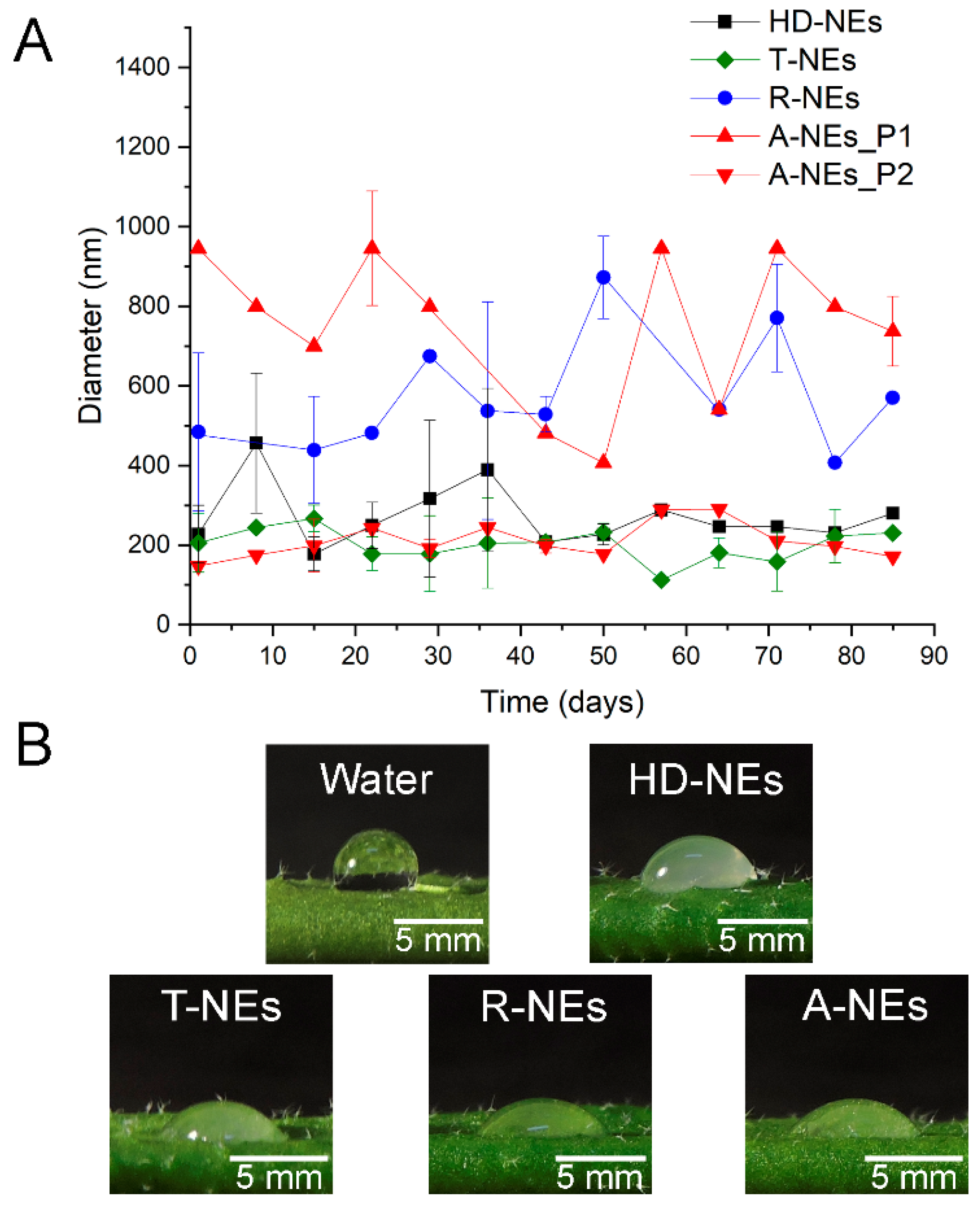
3.1. Development and Stability of RL-Stabilized EO-NEs

The stability of the formulated NEs containing EOs was evaluated over 85 days using dynamic light scattering (DLS) and ζ-potential measurements. Among the tested formulations, only thyme (T-NEs), rue (R-NEs), and tea tree (A-NEs) NEs remained stable at room temperature for over 80 days (Figure 1A). In contrast, lavender and lemon EO-NEs exhibited phase separation after one week, while cinnamon oil showed oil deposition due to its higher density (>1 g/mL) compared to other EOs.
Particle size distribution and ζ-potential values were recorded to assess colloidal stability. Table 1 presents the nanosized droplets and negative ζ potential values for all analyzed NEs. Figure 1A illustrates that particle sizes remained stable over 85 days without agitation. The negative ζ potential values suggest strong electrostatic repulsion, promoting colloidal stability. T-NEs maintained an average particle size of approximately 200 nm, while R-NEs exhibited a size range of 400–600 nm. A-NEs displayed two distinct populations (P1 and P2 showing ≈190 and ≈700 nm, respectively) with a ζ potential less negative (Table 1). These particle size distributions remained consistent over time, confirming the stability of the formulations. Furthermore, no visible microbial contamination was detected in stable NEs despite the non-sterile preparation, supporting the known antimicrobial properties of RLs. Since NE stability was tested for 85 days, all experiments were carried out within this period to ensure the steadiness of the formulations.

3.2. Wettability of NEs on the Surface of Plant Leaves

A. thaliana leaves, known for their hydrophobicity, exhibited a contact angle of 106° on freshly detached samples (Figure 1B). RL suspensions significantly reduced this angle to 38°, while HD-NEs showed a contact angle of 84°, which is close to the threshold between wettable and non-wettable surfaces. Testing of EO-NEs revealed even lower contact angles than HD-NEs, with T-NEs exhibiting the lowest contact angle among all tested formulations, indicating superior wettability and suggesting the potential for increased biological activity (Figure 1B and Table 1).

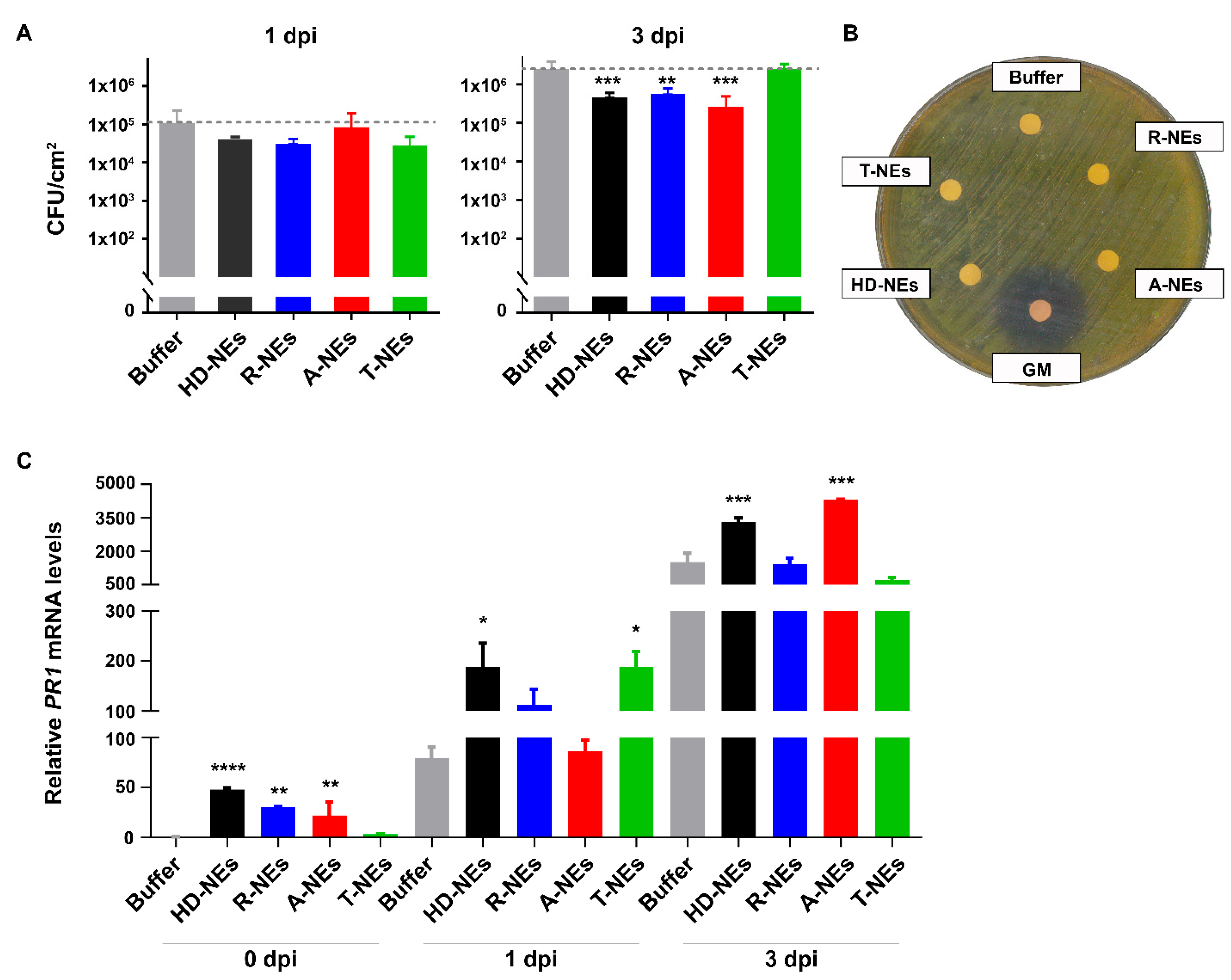
3.3. Evaluating the Efficacy of NEs in Protecting Plants Against Bacterial Pathogens

This study evaluated the effects of EO-NEs on Pst growth in planta, in vitro, and on plant immune responses. To assess the impact of pre-treatment on Pst growth in leaf tissues, A. thaliana plants were sprayed with A-NEs, T-NEs, R-NEs, HD-NEs (control without EOs), or buffer (spraying control). No visible leaf damage was observed, confirming the suitability of EO-NEs as biopesticides. Three days post-treatment, plants were inoculated with Pst, and the bacterial growth was measured at 1 and 3 days post-inoculation (dpi). The results shown in Figure 2 demonstrate that the pre-treatment of foliar tissues with all the tested NEs was effective in reducing bacterial growth in planta at the early (1 dpi) and/or late (3 dpi) stages of infection, with varying efficacy depending on the oil phase used (Figure 2A). While the observed reductions in Pst growth were statistically significant only at 3 dpi, a trend toward reduction of bacterial growth was already evident at 1 dpi. Notably, the use of EOs did not further enhance the efficacy of HD-NEs in reducing Pst growth in planta (Figure 2A), indicating that the observed effect is primarily attributable to the presence of RLs in the NEs.
In vitro agar diffusion assays showed no direct impact of NEs on Pst growth (Figure 2B). Since we observed a decrease of Pst in planta without affecting its growth in vitro, we wonder if this could be related to the induction of the plant immune system triggered by NEs. Gene expression analysis of key plant defense markers was conducted to confirm this hypothesis. PR1 (pathogenesis-related 1), a marker of the salicylic acid (SA) pathway, and PDF1.2 (plant defensin 1.2), a marker of the jasmonic acid (JA) pathway, were monitored. PR1 expression was moderately induced by HD-NEs, R-NEs, and A-NEs 72 h post-treatment (0 dpi), before Pst inoculation (Figure 2C). Increased PR1 expression inversely correlated with bacterial levels, suggesting SA-dependent immune activation contributed to pathogen reduction. Conversely, PDF1.2 expression decreased when PR1 was induced, confirming antagonistic signaling (see Supplementary Figure S3).

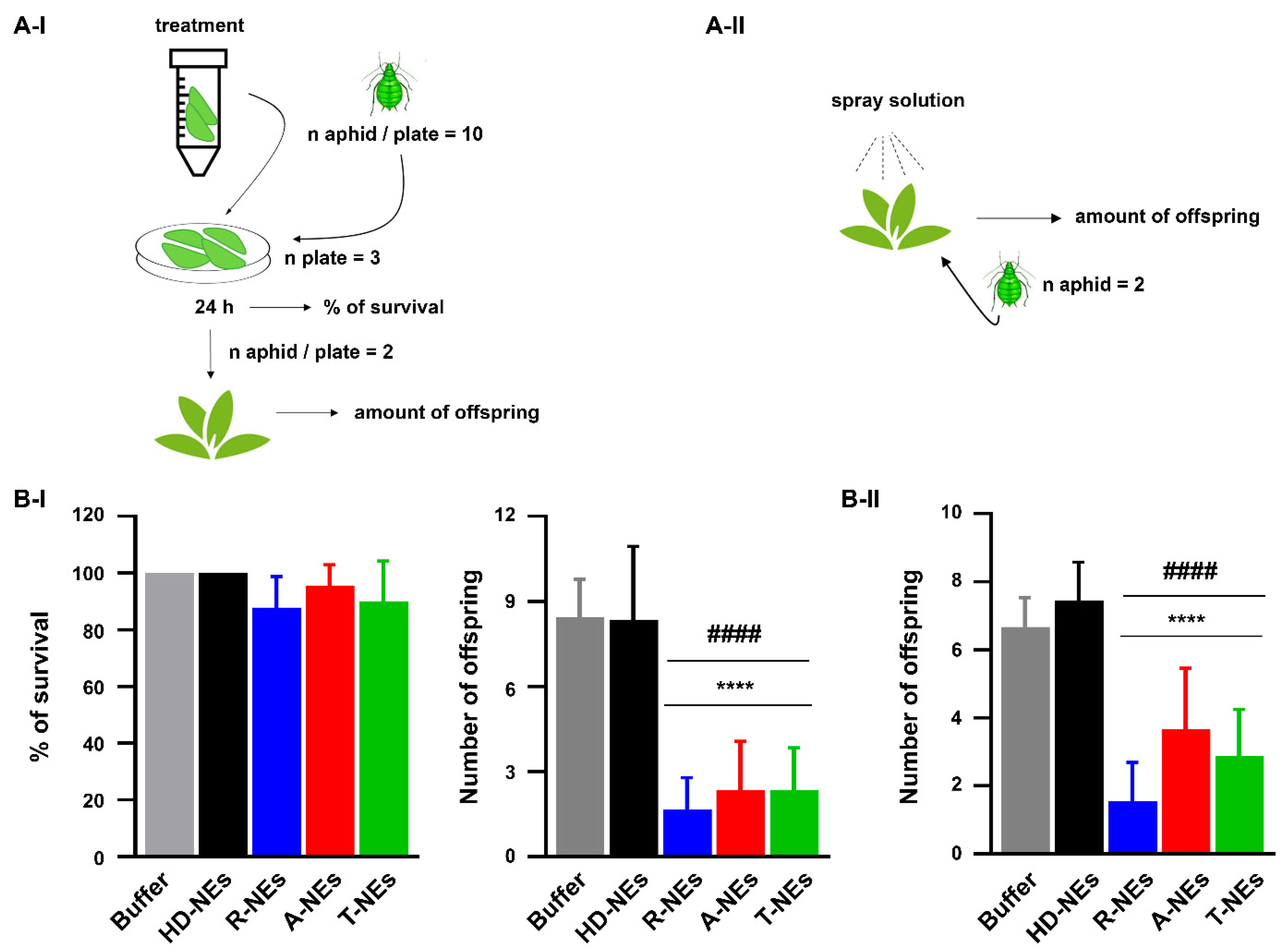
3.4. Assessing the Efficacy of NEs in Protecting Plants from the Aphid Myzus Persicae

The impact of NEs on the survival and reproduction of Myzus persicae, a polyphagous aphid pest, was assessed. A choice assay evaluated the repellent effects of NEs, revealing that only R-NEs significantly reduced aphid preference compared to the buffer control, although the effect was limited (see Supplementary Figure S4).
To investigate aphid survival, excised Arabidopsis leaves were immersed in NEs, dried, and exposed to aphids (Figure 3A-I). Aphids remained on treated leaves, indicating no immediate repellent effect. After 24 h, survival rates were comparable across all treatments, suggesting NEs did not directly impact aphid mortality (Figure 3B-I, left panel).
The reproduction was analyzed by transferring surviving aphids from pre-treated leaves onto untreated plants and counting offspring after seven days. EO-containing NEs (T-, A-, and R-NEs) significantly reduced aphid offspring compared to buffer and HD-NEs (Figure 3B-I, right panel). An experimental setup was developed to evaluate the effect of NEs sprayed directly onto the plant rather than over detached leaves (Figure 3A-II). We sprayed adult Arabidopsis plants and immediately deposited two adult aphids on each plant. NEs did not possess insecticidal activity, as all the aphids survived direct contact with them. Notably, the NEs containing EOs (T-, A-, and R-NEs) again demonstrated a significant reduction in the number of aphid offspring compared to the treatment with buffer or HD-NEs (Figure 3B-II). These findings suggest that EOs influence aphid reproductive behavior rather than directly affecting survival or inducing plant defenses against aphids. The reduction in offspring persisted when aphids were transferred to untreated plants, indicating a potential residual effect interfering with feeding, mating, or both.

3.5. Evaluation of NE Activity on Natural Soil Microbiota and Bioinoculants of Interest for Agroindustry

The environmental impact of NEs was assessed through in vitro microbial growth assays to determine potential ecotoxicity on soil microbiota and bioinoculant microorganisms. The toxicity of NEs on soil microbiota was evaluated by applying them to the plant growth substrate and monitoring the growth of microorganisms extracted from the soil. Results demonstrated that NEs, under the conditions used in planta trials, did not inhibit the growth of naturally occurring soil organisms (see Supplementary Figures S5 and S6).
Additionally, the impact of NEs on beneficial fungi, such as Trichoderma spp., and bacteria, including Pseudomonas fluorescens and Azospirillum argentinense [44], was analyzed. Disc diffusion assays showed that NEs did not negatively affect the growth of these beneficial microorganisms (see Supplementary Figures S5 and S6). Known antibiotics and fungicides were included as positive controls. These findings indicate that RL-EO-NEs are non-toxic to soil microbial communities.

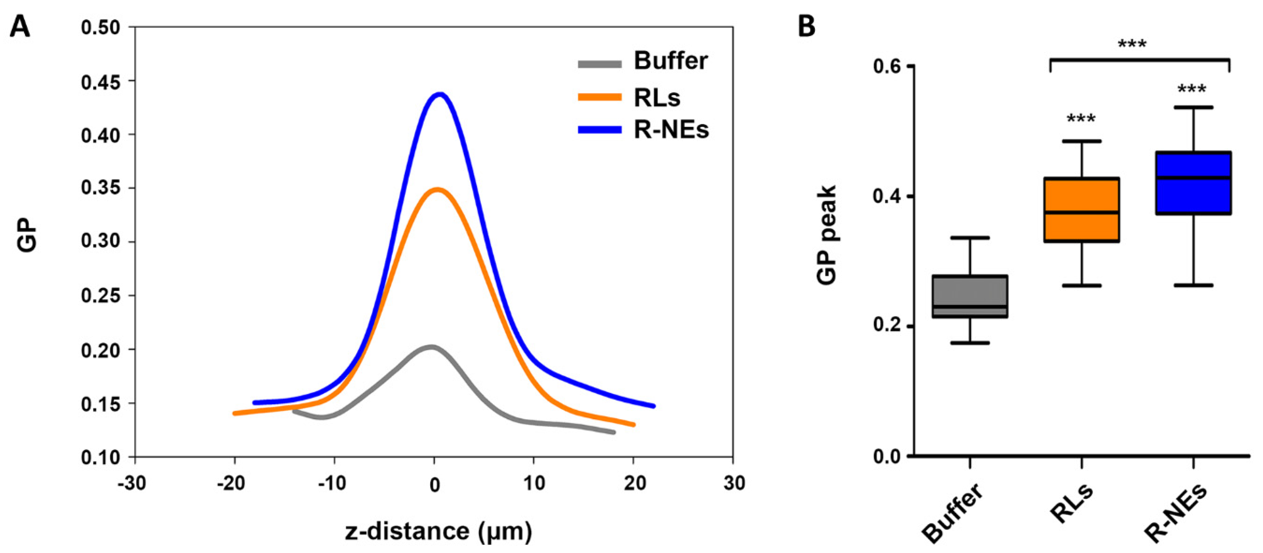
3.6. Regulation of Environmental Dipolar Relaxation of Plant Leaf Cuticle by RLs and RL-NEs

LAURDAN-treated Arabidopsis leaves were examined by fluorescence microscopy (see Supplementary File, Section 5). Z-stack images from the leaf exterior to the interior revealed a primary GP peak corresponding to the cuticle and cell wall/plasma membrane regions (Figure 4, Supplementary Figure S7A and Video S1). Pre-treatment with RL suspension or R-NEs containing 300 µM RLs increased GP values in the cuticle region (Figure 4). Notably, RL suspensions with LAURDAN did not show high GP values (Supplementary Figure S7B), suggesting that the increased GP in the cuticle is due to RLs’ interaction with the cuticle and/or cell membrane components rather than to the RLs themselves. The increase in GP values suggests interactions between RLs and plant waxes, possibly tightening the cuticle structure.

4. Discussion

The thermodynamic instability of colloidal systems often leads to destabilization mechanisms such as coalescence or Ostwald ripening [45]. However, using charged amphiphiles for electrostatic and steric stabilization has proven effective in enhancing the stability of NEs [46]. The results of this study confirmed that the stability of NEs is strongly dependent on droplet size and surface charge. The nanosized droplets observed in T-NEs, R-NEs, and A-NEs contributed to their prolonged kinetic stability, consistent with previous findings reporting that smaller droplets improve long-term stability [47]. The negative ζ potential values further reinforced this stability by preventing droplet aggregation through electrostatic repulsion [45].
The phase separation of NEs with lavender or lemon EOs after one week of storage suggests that oil density and composition play a significant role in NEs’ stability. The higher density of cinnamon oil (>1 g/mL) resulted in sedimentation, unlike the other EOs, which have lower densities than water. These findings align with previous reports on droplet size and oil type influencing NE stability [48,49]. The literature reported droplet sizes of ≈110 nm for medium-chain triglycerides and ≈500 nm for lemon oil [48], while long-chain glycerides yield sizes between 21 and 336 nm [49]. Comparatively, our previous work with HD-NEs resulted in droplet sizes of ≈240 nm with a ζ potential of −55 mV [8]. The observed stability of T-NEs, R-NEs, and A-NEs aligns with these references, demonstrating the robustness of RL-stabilized NEs over prolonged storage. The dual population of A-NEs (≈190 nm and ≈700 nm) suggests a complex stabilization mechanism, possibly influenced by the molecular composition of tea tree oil and its interaction with RLs.
RLs have been documented for their effectiveness in preventing microbial contamination [50], making them suitable for formulations requiring extended shelf life without preservatives. The long-term stability and the absence of microbial growth in T-NEs, R-NEs, and A-NEs reinforce their potential use in sustainable crop protection strategies.
The effectiveness of NEs in agricultural applications depends on their ability to adhere to plant surfaces and facilitate the absorption of active compounds. The plant cuticle, a hydrophobic barrier, affects the interaction between formulation droplets and leaf surfaces [51,52]. Surfaces with a contact angle below 90° are classified as hydrophilic, whereas those above 90° are considered hydrophobic [53]. The 106° contact angle measured for water droplets on A. thaliana leaves confirms their hydrophobic nature, which can limit the effectiveness of aqueous formulations. Our findings demonstrate that EO-NEs significantly decreased the contact angle, enhancing the adhesion and penetration of NEs, thereby maximizing their biological activity and agroindustrial potential. The lower contact angles observed for EO-NEs compared to HD-NEs suggest that specific EOs low-polarity components partitioning in the aqueous phase reduce surface tension and improve wettability [54].
T-NEs exhibited the most favorable characteristics, including the lowest contact angle, small particle size, and the most negative ζ potential. However, all three stable EO-NEs displayed comparable properties, reinforcing their potential for widespread use in crop protection strategies. Our studies confirmed that NEs prime the plant immune system, enhancing resistance against Pst infection. RL-stabilized NEs activate SA-mediated defense pathways before pathogen detection and reduce Pst populations during the infection. This pre-activation of plant defenses provides an advantage by enabling a faster response upon pathogen challenge.
The absence of direct effects on bacterial growth in vitro suggests that NEs function through immune priming rather than direct antimicrobial activity. SA-mediated immune responses are particularly effective against biotrophic pathogens like Pst, whereas JA-mediated responses target necrotrophic pathogens and insect pests [39,40]. The observed inverse correlation between PR1 and PDF1.2 expression aligns with known antagonistic signaling between these pathways, further supporting the role of SA activation in Pst growth suppression.
In addition, EO-NEs exhibited comparable protection to HD-NEs. Their ability to induce PR1 expression and suppress bacterial proliferation suggests they could be integrated into sustainable plant protection strategies. Future studies should explore their efficacy against a broader range of pathogens and evaluate their long-term effects on plant health.
The polyphagous aphid M. persicae is a pest that feeds on phloem sap and transmits viral and phytoplasma diseases, reducing crop yields [42,55]. EO-containing NEs significantly affect aphid reproduction despite showing limited repellence and no direct impact on survival. While R-NEs exhibited mild repellent properties, aphids did not actively avoid treated leaves, remaining on them even after exposure. However, the observed decrease in offspring suggests that EOs disrupt aphid reproductive processes.
The long-lasting reduction in reproduction when aphids were moved to untreated plants implies a residual effect, possibly affecting feeding behaviors, energy allocation for oviposition, or mating success. Since HD-NEs did not influence aphid reproduction, the observed effects are attributed to bioactive compounds in the EOs, like oligogalacturonides, which are known to modulate insect behavior [42]. Thus, EO-containing NEs may interfere with aphid recognition or feeding patterns, reducing their reproductive capacity.
These findings highlight the potential of EO-containing NEs as plant protection compounds that regulate aphid populations. It is worth noting that only the EO of pennyroyal (Mentha pulegium) has been previously documented to exhibit insecticidal activity against M. persicae [35,56]. Further studies are needed to elucidate the mechanisms underlying these effects and assess their applicability in integrated pest management strategies.
RL-stabilized EO-NEs do not pose a risk to soil microbiota or beneficial microorganisms, reinforcing their potential as environmentally friendly agricultural formulations. The ability of these NEs to preserve microbial communities is crucial for maintaining soil health and ecosystem balance, particularly in sustainable agriculture. In contrast, conventional agrochemicals often have broader-spectrum effects, potentially disrupting soil microbiota and compromising agricultural sustainability [1].
The role of natural RLs in boosting plant immune responses [6,57] is still debatable. Schellenberger and co-workers [7] demonstrated that 3-hydroxyalkanoate, a precursor of RLs, is recognized by the LORE receptor on the plant plasma membrane, although a specific RL receptor has not yet been identified. Alternatively, a non-canonical immune response influenced by the plasma membrane’s sphingolipid composition has been proposed. The plant cuticle, which serves as a barrier against external stresses, can be altered by biotic or abiotic factors, playing a key role in disease resistance. Mutants of A. thaliana with altered cutin content in showed enhanced defense due to increased cuticular permeability [58,59], suggesting a connection between cuticle properties and plant defense. This raises the question of whether modifications in cuticle characteristics, such as wax structure and hydration, influence pathogen resistance.
To investigate potential interactions with the plant cuticle, we examined the biophysical properties of the leaf surface using fluorescence microscopy. The amphiphilic probe LAURDAN partitions into hydrophobic structures, where water dipolar relaxation affects its fluorescence, allowing the assessment of solvent dynamics in lipid environments. The degree of relaxation is quantified using the LAURDAN Generalized Polarization (GP) function (see Section 2).
The observed increase in LAURDAN GP values in RL suspensions, R-NEs, and on treated leaf surfaces suggests that RLs interact with the plant cuticle and epidermal cell membranes. Plant waxes, long acylated (20–34C) compounds with keto groups [60], may interact with the polar acyl groups of rhamnose and their hydrophobic tails, tightening the cuticle structure, which is detectable by LAURDAN.
Unlike traditional receptor-based immune activation, our findings indicate that the leaf surface is sensitive to amphiphilic bioactive compounds, suggesting a non-canonical mechanism involving the cuticle’s structural properties. The interaction between RLs and plant waxes may strengthen the cuticle, influencing pathogen resistance. Additionally, changes in cuticle hydration could modulate innate immune responses, as cuticle integrity is linked to plant defense signaling.
This study introduces a novel method for assessing leaf surface biophysical properties, highlighting the leaf surface’s potential as a target for activating non-canonical defense mechanisms through amphiphilic bioactive compounds. Further research is needed to explore how these structural modifications influence long-term plant immunity and their implications for sustainable agricultural practices.

5. Conclusions

Our results demonstrated the excellent emulsifying properties of natural RLs. We successfully developed three EO-containing NEs that exhibited good stability and wettability on the leaves’ surface. These NEs effectively induced the plant immune system and reduced the infection by the bacterial pathogen Pst in Arabidopsis thaliana, which is also an important threat to tomato plants.
The capacity of NEs to enhance the plant’s innate immune system against bacteria was attributed to the presence of RLs, with neither HD nor EOs hindering its effect. By investigating the biophysical properties of the external layer of the leaf surface, we demonstrated that RLs alter the cuticle and cell wall/plasma membrane. Those results opened a new perspective for exploring alternative/non-canonical mechanisms that may trigger plant immune responses.
Notably, none of the tested stable NEs exhibited deleterious effects on plants, soil’s natural microbiota, or the beneficial microbes typically included in bioinoculants. Furthermore, the three stable NEs showed remarkable efficacy in inhibiting aphid reproduction, an effect attributed to the EOs content in the NEs. Nevertheless, its impact on oviposition still requires further investigation.
We believe that the results of this work will significantly impact future research on phytosanitary products, offering safer, more sustainable, and more effective solutions for controlling pests and diseases in crops. Furthermore, we hope to contribute to developing more responsible and environmentally friendly agricultural practices, thereby improving food security and the sustainability of agricultural production globally.

Supplementary Materials

The following supporting information can be downloaded at: https://www.mdpi.com/article/10.3390/agronomy15040824/s1. Figure S1. 1H NMR spectra of selected essential oils; Figure S2. 13C NMR spectra of selected essential oils; Figure S3. Effect of NEs pre-treatment on the induction of plant defenses; Figure S4. Effect of NEs pre-treatment on aphids settling choice; Figure S5. Effect of RLs-EOs-NEs on substrate microbiota and beneficial soil bacteria; Figure S6. RLs-NEs do not interfere with the growth of the substrate microbiota or the development of the beneficial soil fungus Trichoderma spp.; Figure S7. Environmental dipolar relaxation of plant leaf cuticle by RLs and RLs-NEs and LAURDAN Generalized Polarization (GP) parameter of aqueous suspensions containing RLs or EOs-Nes; Video S1. LAURDAN-stained leaf surface microscopy, https://drive.google.com/file/d/1HjphhT5S6GZvYdlwugXH69sMw8kqsXIS/view, accessed on 1 October 2022. The following references corresponds to supplementary file [61,62,63,64,65].

Author Contributions

Conceptualization, project administration, supervision, writing, and editing: M.L.F. and G.F. Funding acquisition: M.L.F., G.F., M.P.M., L.T.K. and M.F.B. Writing—original draft: L.T.K., M.M., M.P.M., M.F.B. and F.B.-H. Data curation, investigation, methodology, visualization, and validation: L.T.K., M.M., R.V.V., M.P.M., M.F.B. and P.M. L.T.K. is a postdoctoral fellow of ANPCyT associated with the project FONCyT PICT 2019-02331; M.M., R.V.V., G.F. and M.L.F. are career researchers of CONICET-UNC. M.P.M. is a postdoctoral fellow in the laboratory of F.B-H., who is the main research director at Facultad de Ciencias de la Vida, Universidad de Andres Bello. M.F.B. is a CONICET PhD fellow at the G.F. laboratory. P.M. is an undergrad student at DQBRC-FCQ-UNC. All authors have read and agreed to the published version of the manuscript.

Funding

This research was funded by the Consejo Nacional de Investigaciones Científicas y Técnicas (CONICET), Agencia Nacional de Promoción Científica y Tecnológica (ANPCyT, FONCyT PICT 2019-02331 and PICT 2021-0766 to M.L.F. and PICT-2020-0763 to G.F.), and the Secretary of Science and Technology of Universidad Nacional de Córdoba (SECyT-UNC), Argentina to M.L.F. and G.F. Agencia Nacional de Investigacón y Desarrollo (ANID) through the Fondo Nacional de Desarrollo Científico y Tecnológico (ANID) FONDECYT Regular 1210320, ANID PIA/BASAL AFB240003, ANID—Programa Iniciativa Científica Milenio—(IBio) ICN17_022, to F.B.-H. Additional support was provided by Programa Jovenes en Ciencia 2023, funded by Provincia de Córdoba and Universidad Nacional de Córdoba (UNC). Microscopy images have been acquired at the Centro de Micro y Nanoscopía de Córdoba (CEMINCO), part of the National Microscopy System (Innovación, Ciencia y Tecnología, Argentina).

Data Availability Statement

The data supporting the findings of this study are available from the corresponding authors upon reasonable request. Due to intellectual property considerations, some data cannot be made publicly available. For further inquiries, please contact the corresponding author.

Acknowledgments

We thank Ernesto Ambroggio (CIQUIBIC), Manuel Cabadas and. Ramiro Lascano from Unidad de Estudios Agropecuarios, UDEA-CONICET- INTA-CIAP, for their generous discussion about LAURDAN fluorescence in plant cells. We thank all the members of Nicolás Cecchini and María Elena Alvarez’s groups for their input and fruitful discussion of our results. We also thank the staff of Centro de Micro y Nanoscopía de Córdoba (CEMINCO) for their valuable assistance and support. We would like to state that we used ImageJ, Software version 2.16.0 (NIH-USA) for Image analysis and BioRender (https://www.biorender.com/) and Canva free software (https://www.canva.com/) for the graphical abstract.

Conflicts of Interest

The authors declare no conflicts of interest. The funders had no role in the design of the study; in the collection, analyses, or interpretation of data; in the writing of the manuscript; or in the decision to publish the results.

Abbreviations

The following abbreviations are used in this manuscript:
RLsRhamnolipid
NEsNanoemulsions
EOsEssential oils
HDn-hexadecane
PstPseudomonas syringae pv tomato DC3000
LAURDAN 1-[6-(Dimethylamino)naphthalen-2-yl]dodecan-1-one
GPGeneralized Polarization of LAURDAN
T-NEsNEs containing thyme EO
R-NEsNEs containing rue EO
A-NEsNEs containing tea tree EO
HD-NEsNEs containing n-hexadecane
PR1Pathogenesis-related 1 gene
PDF1.2Plant defensin 1.2 gene

References

  1. Tudi, M.; Ruan, H.D.; Wang, L.; Lyu, J.; Sadler, R.; Connell, D.; Chu, C.; Phung, D.T. Agriculture Development, Pesticide Application and Its Impact on the Environment. Int. J. Environ. Res. Public Health 2021, 18, 1–24. [Google Scholar] [CrossRef] [PubMed]
  2. Kashif, A.; Rehman, R.; Fuwad, A.; Shahid, M.K.; Dayarathne, H.N.P.; Jamal, A.; Aftab, M.N.; Mainali, B.; Choi, Y. Current Advances in the Classification, Production, Properties and Applications of Microbial Biosurfactants—A Critical Review. Adv. Colloid Interface Sci. 2022, 306, 102718. [Google Scholar] [CrossRef] [PubMed]
  3. Guzmán, E.; Ortega, F.; Rubio, R.G. Exploring the World of Rhamnolipids: A Critical Review of Their Production, Interfacial Properties, and Potential Application. Curr. Opin. Colloid Interface Sci. 2024, 69, 101780. [Google Scholar] [CrossRef]
  4. Suh, S.-J.; Invally, K.; Ju, L.-K. Rhamnolipids: Pathways, Productivities, and Potential. In Biobased Surfactants; Hayes, D.G., Solaiman, D.K.Y., Ashby, R.D., Eds.; AOCS Press: Champaign, IL, USA, 2019; pp. 169–203. ISBN 9780128127056. [Google Scholar]
  5. De Kesel, J.; Conrath, U.; Flors, V.; Luna, E.; Mageroy, M.H.; Mauch-Mani, B.; Pastor, V.; Pozo, M.J.; Pieterse, C.M.J.; Ton, J.; et al. The Induced Resistance Lexicon: Do’s and Don’ts. Trends Plant Sci. 2021, 26, 685–691. [Google Scholar] [CrossRef]
  6. Sanchez, L.; Courteaux, B.; Hubert, J.; Kauffmann, S.; Renault, J.-H.; Clément, C.; Baillieul, F.; Dorey, S. Rhamnolipids Elicit Defense Responses and Induce Disease Resistance against Biotrophic, Hemibiotrophic, and Necrotrophic Pathogens That Require Different Signaling Pathways in Arabidopsis and Highlight a Central Role for Salicylic Acid. Plant Physiol. 2012, 160, 1630–1641. [Google Scholar] [CrossRef]
  7. Schellenberger, R.; Crouzet, J.; Nickzad, A.; Shu, L.-J.; Kutschera, A.; Gerster, T.; Borie, N.; Dawid, C.; Cloutier, M.; Villaume, S.; et al. Bacterial Rhamnolipids and Their 3-Hydroxyalkanoate Precursors Activate Arabidopsis Innate Immunity through Two Independent Mechanisms. Proc. Natl. Acad. Sci. USA 2021, 118, e2101366118. [Google Scholar] [CrossRef]
  8. Mottola, M.; Bertolino, M.C.; Kourdova, L.T.; Valdivia Pérez, J.A.; Bogino, M.F.; Nocelli, N.E.; Chaveriat, L.; Martin, P.; Vico, R.V.; Fabro, G.; et al. Nanoemulsions of Synthetic Rhamnolipids Act as Plant Resistance Inducers without Damaging Plant Tissues or Affecting Soil Microbiota. Front. Plant Sci. 2023, 14, 1195718. [Google Scholar] [CrossRef]
  9. Spoel, S.H.; Dong, X. How Do Plants Achieve Immunity? Defence without Specialized Immune Cells. Nat. Rev. Immunol. 2012, 12, 89–100. [Google Scholar] [CrossRef]
  10. Grandellis, C.; Garavaglia, B.S.; Gottig, N.; Lonez, C.; Ruysschaert, J.M.; Ottado, J. DOTAP, a Lipidic Transfection Reagent, Triggers Arabidopsis Plant Defense Responses. Planta 2019, 249, 469–480. [Google Scholar] [CrossRef]
  11. Lovaglio, R.B.; dos Santos, F.J.; Jafelicci, M.; Contiero, J. Rhamnolipid Emulsifying Activity and Emulsion Stability: PH Rules. Colloids Surfaces B Biointerfaces 2011, 85, 301–305. [Google Scholar] [CrossRef]
  12. Fierascu, R.C.; Fierascu, I.C.I.; Dinu-Pirvu, C.E.; Fierascu, I.C.I.; Paunescu, A. The Application of Essential Oils as a Next-Generation of Pesticides: Recent Developments and Future Perspectives. Z. Naturforsch. C 2020, 75, 183–204. [Google Scholar] [CrossRef] [PubMed]
  13. Regnault-Roger, C.; Vincent, C.; Arnason, J.T. Essential Oils in Insect Control: Low-Risk Products in a High-Stakes World. Annu. Rev. Entomol. 2012, 57, 405–424. [Google Scholar] [CrossRef]
  14. Sharma, N.; Sharma, K.; Gupta, S.S.; Ranjan, K.R.; Mishra, V.; Mukherjee, M. Das Essential Oil-Based Biopesticides. In Essential Oils; Wiley: Hoboken, NJ, USA, 2023; pp. 443–463. [Google Scholar]
  15. Sharma, A.; Gumber, K.; Gohain, A.; Bhatia, T.; Sohal, H.S.; Mutreja, V.; Bhardwaj, G. Importance of Essential Oils and Current Trends in Use of Essential Oils (Aroma Therapy, Agrofood, and Medicinal Usage). In Essential Oils; Elsevier: Amsterdam, The Netherlands, 2023; pp. 53–83. [Google Scholar]
  16. Napoli, E.; Di Vito, M. Toward a New Future for Essential Oils. Antibiotics 2021, 10, 207. [Google Scholar] [CrossRef]
  17. Parashar, P.; Pathak, K. Augmented Stability and Efficacy of Essential Oils Through Encapsulation Approach. In Essential Oils; Wiley: Hoboken, NJ, USA, 2023; pp. 269–289. [Google Scholar]
  18. Bezerra, F.W.F.; Freitas, L.C.; Cunha, V.M.B.; Aires, G.C.M.; Pinto, R.H.H.; de Carvalho Junior, R.N. Encapsulated Essential Oils. In Essential Oils; Wiley: Hoboken, NJ, USA, 2023; pp. 619–634. [Google Scholar]
  19. Kha, T.C.; Le, P.H. Encapsulation Technologies of Essential Oils for Various Industrial Applications. In Essential Oils; Wiley: Hoboken, NJ, USA, 2023; pp. 635–670. [Google Scholar]
  20. Haba, E.; Bouhdid, S.; Torrego-Solana, N.; Marqués, A.M.; Espuny, M.J.; García-Celma, M.J.; Manresa, A. Rhamnolipids as Emulsifying Agents for Essential Oil Formulations: Antimicrobial Effect against Candida albicans and Methicillin-Resistant Staphylococcus aureus. Int. J. Pharm. 2014, 476, 134–141. [Google Scholar] [CrossRef]
  21. Napiórkowska, A.; Kurek, M. Coacervation as a Novel Method of Microencapsulation of Essential Oils—A Review. Molecules 2022, 27, 5142. [Google Scholar] [CrossRef] [PubMed]
  22. Yammine, J.; Chihib, N.-E.; Gharsallaoui, A.; Ismail, A.; Karam, L. Advances in Essential Oils Encapsulation: Development, Characterization and Release Mechanisms. Polym. Bull. 2024, 81, 3837–3882. [Google Scholar] [CrossRef]
  23. Sousa, V.I.; Parente, J.F.; Marques, J.F.; Forte, M.A.; Tavares, C.J. Microencapsulation of Essential Oils: A Review. Polymers 2022, 14, 1730. [Google Scholar] [CrossRef]
  24. Dunan, L.; Malanga, T.; Benhamou, S.; Papaiconomou, N.; Desneux, N.; Lavoir, A.-V.; Michel, T. Effects of Essential Oil-Based Formulation on Biopesticide Activity. Ind. Crops Prod. 2023, 202, 117006. [Google Scholar] [CrossRef]
  25. Hebishy, E.; Collette, L.; Iheozor-Ejiofor, P.; Onarinde, B.A. Stability and Antimicrobial Activity of Lemongrass Essential Oil in Nanoemulsions Produced by High-intensity Ultrasounds and Stabilized by Soy Lecithin, Hydrolyzed Whey Proteins, Gum Arabic, or Their Ternary Admixture. J. Food Process. Preserv. 2022, 46, e16840. [Google Scholar] [CrossRef]
  26. Salvia-Trujillo, L.; Rojas-Graü, A.; Soliva-Fortuny, R.; Martín-Belloso, O. Physicochemical Characterization and Antimicrobial Activity of Food-Grade Emulsions and Nanoemulsions Incorporating Essential Oils. Food Hydrocoll. 2015, 43, 547–556. [Google Scholar] [CrossRef]
  27. Iahtisham-Ul-Haq; Khan, S.; Sohail, M.; Iqbal, M.J.; Awan, K.A.; Nayik, G.A. Tea Tree Essential Oil. In Essential Oils; Elsevier: Amsterdam, The Netherlands, 2023; pp. 479–500. [Google Scholar]
  28. Saeed, F.; Afzaal, M.; Raza, M.A.; Rasheed, A.; Hussain, M.; Nayik, G.A.; Ansari, M.J. Lavender Essential Oil: Nutritional, Compositional, and Therapeutic Insights. In Essential Oils; Elsevier: Amsterdam, The Netherlands, 2023; pp. 85–101. [Google Scholar]
  29. Kaur, G.; Kaur, K.; Saluja, P. Citrus Essential Oil (Grapefruit, Orange, Lemon). In Essential Oils; Elsevier: Amsterdam, The Netherlands, 2023; pp. 179–215. [Google Scholar]
  30. Inanoglu, S.; Goksen, G.; Nayik, G.A.; Mozaffari Nejad, A.S. Essential Oils from Lamiaceae Family (Rosemary, Thyme, Mint, Basil). In Essential Oils; Elsevier: Amsterdam, The Netherlands, 2023; pp. 309–324. [Google Scholar]
  31. Liaqat, A.; Ahsan, S.; Fayyaz, M.S.; Ali, A.; Ashfaq, S.A.; Khan, S.; Khan, M.A.; Mehmood, T.; Khaliq, A.; Chughtai, M.F.J.; et al. Cinnamon Essential Oil. In Essential Oils; Elsevier: Amsterdam, The Netherlands, 2023; pp. 377–390. [Google Scholar]
  32. Said-, H.A.H.; Ahl, A.; Hikal, W.; Tkachenko, K.; Said-Al, H.A.H.; Said-Al Ahl, H.A.H.; Hikal, W.M.; Tkachenko, K.G. Essential Oils with Potential as Insecticidal Agents: A Review. Int. J. Environ. Plan. Manag. 2017, 3, 23–33. [Google Scholar]
  33. Raveau, R.; Fontaine, J.; Lounès-Hadj Sahraoui, A. Essential Oils as Potential Alternative Biocontrol Products against Plant Pathogens and Weeds: A Review. Foods 2020, 9, 365. [Google Scholar] [CrossRef] [PubMed]
  34. Nerio, L.S.; Olivero-Verbel, J.; Stashenko, E. Repellent Activity of Essential Oils: A Review. Bioresour. Technol. 2010, 101, 372–378. [Google Scholar] [CrossRef]
  35. Altalhi, T.; Neves Cruz, J.; Inamuddin, I. Essential Oils. Extraction Methods and Applications; John Wiley & Sons, Inc.: Hoboken, NJ, USA, 2023; ISBN 9781119829355. [Google Scholar]
  36. Stalder, A.F.; Melchior, T.; Müller, M.; Sage, D.; Blu, T.; Unser, M. Low-Bond Axisymmetric Drop Shape Analysis for Surface Tension and Contact Angle Measurements of Sessile Drops. Colloids Surfaces A Physicochem. Eng. Asp. 2010, 364, 72–81. [Google Scholar] [CrossRef]
  37. Schindelin, J.; Arganda-Carreras, I.; Frise, E.; Kaynig, V.; Longair, M.; Pietzsch, T.; Preibisch, S.; Rueden, C.; Saalfeld, S.; Schmid, B.; et al. Fiji: An Open-Source Platform for Biological-Image Analysis. Nat. Methods 2012, 9, 676–682. [Google Scholar] [CrossRef] [PubMed]
  38. Fabro, G.; Rizzi, Y.S.; Alvarez, M.E. Arabidopsis Proline Dehydrogenase Contributes to Flagellin-Mediated PAMP-Triggered Immunity by Affecting RBOHD. Mol. Plant-Microbe Interact. 2016, 29, 620–628. [Google Scholar] [CrossRef] [PubMed]
  39. Vos, I.A.; Moritz, L.; Pieterse, C.M.J.; Van Wees, S.C.M. Impact of Hormonal Crosstalk on Plant Resistance and Fitness under Multi-Attacker Conditions. Front. Plant Sci. 2015, 6, 639. [Google Scholar] [CrossRef]
  40. Van der Does, D.; Leon-Reyes, A.; Koornneef, A.; Van Verk, M.C.; Rodenburg, N.; Pauwels, L.; Goossens, A.; Körbes, A.P.; Memelink, J.; Ritsema, T.; et al. Salicylic Acid Suppresses Jasmonic Acid Signaling Downstream of SCFCOI1-JAZ by Targeting GCC Promoter Motifs via Transcription Factor ORA59. Plant Cell 2013, 25, 744–761. [Google Scholar] [CrossRef]
  41. Pfaffl, M.W. A New Mathematical Model for Relative Quantification in Real-Time RT-PCR. Nucleic Acids Res. 2001, 29, e45. [Google Scholar] [CrossRef]
  42. Silva-Sanzana, C.; Zavala, D.; Moraga, F.; Herrera-Vásquez, A.; Blanco-Herrera, F. Oligogalacturonides Enhance Resistance against Aphids through Pattern-Triggered Immunity and Activation of Salicylic Acid Signaling. Int. J. Mol. Sci. 2022, 23, 9753. [Google Scholar] [CrossRef]
  43. Socas, L.B.P.; Valdivia-Pérez, J.A.; Fanani, M.L.; Ambroggio, E.E. Multidimensional Spectral Phasors of LAURDAN’s Excitation–Emission Matrices: The Ultimate Sensor for Lipid Phases? J. Am. Chem. Soc. 2024, 146, 17230–17239. [Google Scholar] [CrossRef]
  44. Kumar, M.; Poonam; Ahmad, S.; Singh, R.P. Plant Growth Promoting Microbes: Diverse Roles for Sustainable and Ecofriendly Agriculture. Energy Nexus 2022, 7, 100133. [Google Scholar] [CrossRef]
  45. Evans, F.D.; Wennerstrom, H. The Colloidal Domain. Where Physics, Chemistry, Biology an Thechnology Meet, 2nd ed.; Wiley-VCH: New York, NY, USA, 1999; ISBN 0-471-24247-0. [Google Scholar]
  46. Landfester, K. Synthesis of Colloidal Particles in Miniemulsions. Annu. Rev. Mater. Res. 2006, 36, 231–279. [Google Scholar] [CrossRef]
  47. McClements, D.J. Nanoemulsions versus Microemulsions: Terminology, Differences, and Similarities. Soft Matter 2012, 8, 1719–1729. [Google Scholar] [CrossRef]
  48. Bai, L.; McClements, D.J. Formation and Stabilization of Nanoemulsions Using Biosurfactants: Rhamnolipids. J. Colloid Interface Sci. 2016, 479, 71–79. [Google Scholar] [CrossRef]
  49. Khan, M.A.; Ullah, K.; Rahman, N.u.; Mahmood, A.; Müllertz, A.; Mannan, A.; Murtaza, G.; Khan, S.A. Formulation, Characterization and in-Vitro Evaluation of Self-Nanoemulsifying Drug Delivery System Containing Rhamnolipid Biosurfactant. J. Drug Deliv. Sci. Technol. 2022, 75, 103673. [Google Scholar] [CrossRef]
  50. Haba, E.; Pinazo, A.; Jauregui, O.; Espuny, M.J.; Infante, M.R.; Manresa, A. Physicochemical Characterization and Antimicrobial Properties of Rhamnolipids Produced by Pseudomonas Aeruginosa 47T2 NCBIM 40044. Biotechnol. Bioeng. 2003, 81, 316–322. [Google Scholar] [CrossRef]
  51. Liu, H.; Shao, B.; Long, X.; Yao, Y.; Meng, Q. Foliar Penetration Enhanced by Biosurfactant Rhamnolipid. Colloids Surfaces B Biointerfaces 2016, 145, 548–554. [Google Scholar] [CrossRef]
  52. Soffe, R.; Bernach, M.; Remus-Emsermann, M.N.P.; Nock, V. Replicating Arabidopsis Model Leaf Surfaces for Phyllosphere Microbiology. Sci. Rep. 2019, 9, 14420. [Google Scholar] [CrossRef]
  53. Davies, J.T.; Rideal, E.K. Interfacial Fenomena, 2nd ed.; Academic Press: London, UK, 1963. [Google Scholar]
  54. Álvarez-Martínez, F.J.; Barrajón-Catalán, E.; Herranz-López, M.; Micol, V. Antibacterial Plant Compounds, Extracts and Essential Oils: An Updated Review on Their Effects and Putative Mechanisms of Action. Phytomedicine 2021, 90, 153626. [Google Scholar] [CrossRef]
  55. Valenzuela, I.; Hoffmann, A.A. Effects of Aphid Feeding and Associated Virus Injury on Grain Crops in Australia. Austral Entomol. 2015, 54, 292–305. [Google Scholar] [CrossRef]
  56. Kavetsou, E.; Koutsoukos, S.; Daferera, D.; Polissiou, M.G.; Karagiannis, D.; Perdikis, D.C.; Detsi, A. Encapsulation of Mentha Pulegium Essential Oil in Yeast Cell Microcarriers: An Approach to Environmentally Friendly Pesticides. J. Agric. Food Chem. 2019, 67, 4746–4753. [Google Scholar] [CrossRef]
  57. Vatsa, P.; Sanchez, L.; Clement, C.; Baillieul, F.; Dorey, S. Rhamnolipid Biosurfactants as New Players in Animal and Plant Defense against Microbes. Int. J. Mol. Sci. 2010, 11, 5095–5108. [Google Scholar] [CrossRef]
  58. Aragón, W.; Formey, D.; Aviles-Baltazar, N.Y.; Torres, M.; Serrano, M. Arabidopsis thaliana Cuticle Composition Contributes to Differential Defense Response to Botrytis Cinerea. Front. Plant Sci. 2021, 12, 738949. [Google Scholar] [CrossRef]
  59. Ju, S.; Go, Y.S.; Choi, H.J.; Park, J.M.; Suh, M.C. DEWAX Transcription Factor Is Involved in Resistance to Botrytis cinerea in Arabidopsis thaliana and Camelina sativa. Front. Plant Sci. 2017, 8, 1210. [Google Scholar] [CrossRef] [PubMed]
  60. von Wettstein-Knowles, P. Plant Waxes. In Encyclopedia of Life Sciences; Wiley: Chichester, UK, 2016; pp. 1–13. ISBN 9780470015902. [Google Scholar]
  61. Bagatolli, L.A. To see or not to see: Lateral organization of biological membranes and fluorescence microscopy. Biochim. Biophys. Acta-Biomembr. 2006, 1758, 1541–1556. [Google Scholar] [CrossRef]
  62. Balci, M. (Ed.) Chemical Shift. In Basic 1H- and 13C-NMR Spectroscopy; Elsevier: Amsterdam, The Netherlands, 2005; pp. 25–85. [Google Scholar]
  63. Gunther, G.; Malacrida, L.; Jameson, D.M.; Gratton, E.; Sánchez, S.A. LAURDAN since Weber: The Quest for Visualizing Membrane Heterogeneity. Acc. Chem. Res. 2021, 54, 976–987. [Google Scholar] [CrossRef]
  64. Otaiza-González, S.; Cabadas, M.; Robert, G.; Stock, R.P.; Malacrida, L.; Lascano, R.; Bagatolli, L.A. The innards of the cell: Studies of water dipolar relaxation using the ACDAN fluorescent probe. Methods Appl. Fluoresc. 2022, 10, 044010. [Google Scholar] [CrossRef]
  65. Pinto, S.N.; Fernandes, F.; Fedorov, A.; Futerman, A.H.; Silva, L.C.; Prieto, M. A combined fluorescence spectroscopy, confocal and 2-photon microscopy approach to re-evaluate the properties of sphingolipid domains. Biochim. Biophys. Acta-Biomembr. 2013, 1828, 2099–2110. [Google Scholar] [CrossRef]
Figure 1. Particle size distribution and wettability of NEs containing the selected EOs: thyme (T-NEs), rue (R-NEs), and tea tree (A-NEs). (A) Dynamic light scattering (DLS) measurements of NEs upon storage at room temperature. The values are the mean ± SD from three independent samples. P1 and P2 are the populations 1 and 2 for the A-NEs sample, respectively. (B) Representative images of 4 μL-size water and HD-NEs droplets used as references, as well as droplets of NEs containing EOs (300 μM of RL) deposited onto A. thaliana leaves.
Agronomy 15 00824 g001
Figure 2. Effect of pre-treatment with NEs on the development of Pst and the induction of plant defenses. (A) Bacterial growth curves were performed on days 1 and 3 post-infection (dpi), showing Pst growth in leaf tissues of plants pre-treated with NEs. The y-axis shows colony-forming units (CFU) per leaf square centimeter (cm2). The x-axis indicates the pre-treatment applied to the plants (HD-NEs: hexadecane NEs, A-NEs: tea tree EO-NEs, T-NEs: thyme EO-NEs, R-NEs: rue EO-NEs). NEs were applied by spraying solutions at 300 μM of RLs. Values shown are the mean ± SD (n = 12 leaf discs per point) of one representative experiment of four performed. (B) Effect of NEs on Pst growth in vitro. The presence/absence of growth inhibition halos formed around NE- or buffer-containing paper discs is shown. Positive control: 15 μM gentamycin (GM). (C) Expression pattern of the SA-marker gene PR1 in plants pre-treated with NEs 3 days before infecting with Pst (day 0) and 1- or 3- dpi, monitored by RT-qPCR. Data is the mean ± SD of 3 technical replicates. PR1 expression was relativized to UBI5. One-way analysis of variance with Dunnett’s post hoc test was applied in (A,C). Asterisks indicate significant differences compared with buffer (*: p < 0.05, **: p < 0.01, ***: p < 0.001, **** p < 0.0001).
Agronomy 15 00824 g002
Figure 3. Effect of pre-treatment of A. thaliana leaves with NEs on the survival of the polyphagous aphid Myzus persicae and its reproductive capability. (A-I) Leaves were immersed in the NEs of RLs and EOs at RL concentration of 300 μM and allowed to dry in plates. Then, 10 aphids per condition were deposited on each plate (aphids/plate: 10), and 24 h later, the percentage of survivors was quantified ((B-I) left panel). Two surviving aphids from each plate (aphids/plate: 2) were transferred with a brush to one untreated plant (aphids/plant: 2) for 7 days to analyze their reproductive capacity ((B-I) right panel). The assay was performed in triplicate. (A-II) Whole plants were sprayed with NEs or controls, and immediately, two aphids were deposited onto them. After 7 days, the number of survivors and their offspring (eggs) were counted (B-II). Values shown are the mean ± SD. A one-way analysis of variance with Dunnett’s post hoc test was applied to (B-I,B-II). (****) and (####) indicate significant differences (p < 0.0001) comparing all samples vs. the buffer or HD-NEs, respectively.
Agronomy 15 00824 g003
Figure 4. Environmental dipolar relaxation of plant leaf cuticle by RL suspension and RL-NEs. (A) Representative z-axis profiles of GP values are shown along the leaves’ cuticular and epidermal cell layers. The selected ROI for z-plot analysis was 17.7 × 17.7 μm size. The zero distance was set at the GP peak. The pre-treatment applied to the leaves was buffer, RL suspension, or R-NEs containing 300 μM of RLs. (B) Statistical analysis of the GP values at the z-plot peaks (corresponding to zero z-distance in plot (A). One-way analysis of variance with Dunnett’s post hoc test was applied. *** p < 0.001 vs. buffer.
Agronomy 15 00824 g004
Table 1. Physicochemical characterization of the selected RL-stabilized NEs.
Nanoemulsions (NEs)Oil PhaseDiameter (nm) *PDI **ζ-Potential ** (mV)Contact Angle (°) ***
P1P2
HD-NEsHexadecane241 ± 80-0.4 ± 0.1−55 ± 284 ± 11
T-NEsThyme EO184 ± 61-0.7 ± 0.3−65 ± 257 ± 4
R-NEsRue EO553 ± 149-1.1 ± 0.1−51 ± 360 ± 8
A-NEsTea Tree EO196 ± 49684 ± 2031.6 ± 0.5−25 ± 461 ± 5
* P1 and P2 are populations of different diameters representing the two main peaks of a more complex distribution. The appearance frequencies for P1 and P2 were 39% and 35%, respectively. These values were obtained by averaging all measurements (34 in total) taken over 85 days across three replicate samples. ** ζ-potential values are the average of n = 20 and diameter of n = 36 samples ± SD. PDI was calculated as the square of the standard deviation divided by the mean particle diameter. *** The water contact angle of 106 ± 5 was taken as a reference. The values are mean ± SD from two independent experiments (with triplicate droplets), analyzed by two independent operators.
Disclaimer/Publisher’s Note: The statements, opinions and data contained in all publications are solely those of the individual author(s) and contributor(s) and not of MDPI and/or the editor(s). MDPI and/or the editor(s) disclaim responsibility for any injury to people or property resulting from any ideas, methods, instructions or products referred to in the content.

