Article

Biocontrol Potential of Bacillus subtilis A3 Against Corn Stalk Rot and Its Impact on Root-Associated Microbial Communities

1
State Key Laboratory of North China Crop Improvement and Regulation, Hebei Agricultural University, Baoding 071000, China
2
Hebei Key Laboratory of Plant Physiology and Molecular Pathology, Hebei Agricultural University, Baoding 071000, China
3
College of Plant Protection, Hebei Agricultural University, Baoding 071000, China
4
College of Life Sciences, Hebei Agricultural University, Baoding 071000, China
*
Authors to whom correspondence should be addressed.
These authors contributed equally to this work.
Agronomy 2025, 15(3), 706; https://doi.org/10.3390/agronomy15030706
Submission received: 15 February 2025 / Revised: 10 March 2025 / Accepted: 12 March 2025 / Published: 14 March 2025
(This article belongs to the Special Issue Environmentally Friendly Ways to Control Plant Disease)

Abstract

:
Fusarium stalk rot (FSR), a devastating soil-borne disease caused by Fusarium species, severely threatens global maize production through yield losses and mycotoxin contamination. Bacillus subtilis, a plant growth-promoting rhizobacterium (PGPR), has shown potential as a biocontrol agent against soil-borne pathogens, but its efficacy and mechanisms against maize FSR remain poorly understood. In this study, an identified strain of B. subtilis A3 was introduced to study its biological control potential against corn stalk rot. The bacteriostatic stability of the biocontrol strain was assessed, revealing that its inhibitory activity against F. graminearum remained consistent over five consecutive generations, indicating robust bacteriostatic stability. The strain also exhibited inhibitory effects on F. verticilliodes, F. proliferalum, and other pathogenic fungi, demonstrating it has broad-spectrum antibacterial activity. Indoor experiments showed that treatment with the biocontrol strain significantly increased plant height, stem diameter, and fresh weight, indicating a positive impact on corn growth. Additionally, the biocontrol strain A3 markedly reduced the lesion length of corn stalk rot, confirming its efficacy in controlling the disease. Field trials demonstrated that the growth of the A3-coated corn seeds was better than the control seeds, the control effect of FSR disease was 45.75%, and the yield increase was 3.6%. Microscopic observations revealed that the biocontrol strain A3 caused the hyphal tips of F. graminearum to swell and exhibit a beaded morphology, inhibiting normal growth. The volatile substances produced by A3 also showed significant antibacterial activity, with the antibacterial spectrum aligning with that of the biocontrol strain. Using headspace solid-phase microextraction and GC-MS, various antibacterial compounds were identified in the volatile substances. Analysis of root-associated microorganisms indicated that A3 significantly changed the microbial community composition. Co-occurrence network analysis revealed that A3-treated plants had fewer edges and lower negative correlations among bacterial communities. This study establishes the strong biocontrol potential of B. subtilis A3 against Fusarium stalk rot in corn, demonstrating its robust bacteriostatic stability, broad-spectrum antibacterial activity, positive impact on plant growth, and significant disease control efficacy, while also revealing its ability to alter root-associated microbial communities. These findings provide a foundation for further research into the mechanism of B. subtilis and its application in field biological control.

1. Introduction

Bacillus subtilis is a kind of Gram-positive bacterium that exists in soil and the rhizosphere of plants [1,2]. It has a certain control effect on a variety of fungal and bacterial diseases [3,4,5,6]. B. subtilis can enhance the ability of plant resistance to biological stress through antagonism [7], inducing plant resistance [8] and promoting plant growth [2,9]. The secondary metabolites produced by B. subtilis, such as antibacterial proteins and lipopeptides, can inhibit the growth of a pathogen or cause cell lysis and death. Research has demonstrated that B. subtilis possesses the ability to infiltrate and disrupt the mycelial structure of F. graminearum through the biosynthesis of fengycins, chitinase, and surfactins [10,11]. Additionally, B. subtilis also promotes plant growth and improves plant resistance by producing enzymes or carriers of carbon assimilation and iron assimilation. Resistance can also be achieved by inducing disease or insect resistance in plants, such as by activating keratin, cork, and wax metabolites. Therefore, B. subtilis has significant potential for biological control in plant disease prevention and growth promotion. Moreover, B. subtilis can also produce a variety of active metabolites, especially volatile organic compounds (VOCs), which positions it as a promising candidate as a safe and efficient biological agent. VOCs exhibit diverse direct and indirect interactions with plants, demonstrating multifaceted roles in plant–microbe interactions. These compounds have been shown to inhibit pathogenic microorganisms [12,13,14,15], regulate populations of insects and nematodes [16], enhance plant growth and development [17], and induce systemic resistance in plants [18]. The biological effects of VOCs on plants are highly variable, with their impact being determined by the specific molecular composition and concentration of the volatile compounds [19]. The beneficial interactions between VOCs and plants are mediated through multiple biochemical mechanisms. Substantial evidence indicates that VOCs can modulate the biosynthesis and signaling pathways of key plant hormones, including auxin, cytokinin, ethylene, and abscisic acid, thereby influencing plant growth and stress responses [20]. Furthermore, research has demonstrated that VOCs can enhance plant nutrient acquisition efficiency by improving the uptake of essential elements such as iron and sulfur from the soil environment. Additionally, certain VOCs possess the capacity to metabolize sulfur-containing volatile compounds, potentially contributing to the nutrient cycling processes in the rhizosphere [21].
Corn stalk rot, also known as bacterial wilt or stem base rot, is a prevalent and destructive disease in corn-producing regions worldwide [22]. First reported in the Americas in 1914 [23], the disease has since spread to Africa, Europe, Australia, and other places [24]. It occurred in China in the 1920s and has been prevalent in the country since the 1970s, becoming a major disease of corn. Stalk rot is a soil-borne disease caused by complex pathogens [24]. In general years, its incidence rate ranges from 10% to 20%, but in serious years, it can reach 50% to 60%, leading to yield reductions of about 25% or even total crop loss. The disease typically develops during the late filling stage of corn, with the peak occurring from the end of milk ripening to wax ripening. Symptoms progress from the first infected leaves to the entire plant within 5 to 8 days. The pathogen mainly infects the root and stalk about 10 cm above the root, causing plants to fall in the later stage, which affects mechanized harvesting. Leaf symptoms include a grayish color and dryness, often accompanied by corn ear sagging, ear stalk flexibility and softness, and difficulty in breaking them off. The disease causes small ears, dry cob shrinkage, a dry and not full kernel, a significant decrease in hundred-grain weight, and threshing difficulties, resulting in serious yield reduction [25]. Additionally, toxins are produced in the stalks of corn plants with stalk rot, which brings a great danger to the roughage with corn stalks as raw materials [26]. Therefore, it is of great significance to control the occurrence and harm of corn stem rot. Given the complexity of the pathogens, the influence of climate on the disease onset, and the late stage of infection, effective chemical control measures for stalk rot remain elusive.
A plant’s microbiome is a major determinant of its health and yield [27]. The assembly and composition of a plant rhizosphere microbiome are closely related to the interaction between the host plant and its soil microbial community [28,29]. Host plants can regulate the structure of root-associated microorganisms, and this structural change in turn affects the growth and development of the host plant. Studies have shown that multiple factors can influence the composition of a plant microbiome, which can mainly be summarized as rhizosphere effects (the effects of root metabolic products on the microbial community), immune systems, nutrient transfer, and host genotype-determined stress responses [30,31]. Plants can recruit beneficial microorganisms by altering root exudates [32,33,34], and these beneficial microorganisms can suppress the harm of pathogenic bacteria through direct or indirect means [35,36,37]. These modes of action mainly include the secretion of antibacterial compounds [38,39], hyperparasitism [40], and competition with pathogens for resources such as nutrients and space [41]. It can be seen that host plants also rely on microorganisms to resist pathogens. Considering the influence of rhizosphere microecology, rationally, designing and utilizing such synthetic microbial communities that are universal, long-lasting, and scalable for plant protection [42] will be one of the important strategies for plant protection in future sustainable agriculture. Therefore, it is of great significance to reveal the mechanism by which plant root-associated microorganisms affect the composition and assembly of a rhizosphere microbial community.
The primary objective of this study is to investigate the potential of B. subtilis-based biocontrol strategies for managing corn stalk rot, a devastating disease affecting global corn production. Specifically, we aim to (1) isolate and identify B. subtilis strains with enhanced antagonistic activity against F. graminearum; (2) evaluate their efficacy in promoting corn growth and suppressing stalk rot under both controlled and field conditions; and (3) elucidate the mechanisms underlying plant–microbe interactions through a comprehensive analysis of rhizosphere microbiome dynamics. By integrating molecular characterization, physiological assessments, and advanced microbiome analysis, this research seeks to develop sustainable biocontrol solutions while advancing our understanding of microbial-mediated plant protection mechanisms. The findings are expected to contribute significantly to the development of effective, environmentally friendly strategies for corn stalk rot management and provide insights into the design of synthetic microbial communities for plant protection.

2. Methods and Materials

2.1. Isolation and Identification of Bacillus subtilis A3

All samples were collected from the experimental site of Hebei Agricultural University, Baoding City, Hebei Province, China, at the R6 stage of the corn. A 1 g sample of rhizosphere soil from maize plants affected by stalk rot was aseptically transferred into a test tube containing 9 mL of sterile distilled water, followed by vigorous vortexing to achieve a homogeneous suspension, designated as the 10−1 dilution. Subsequently, 1 mL of the 10−1 suspension was serially transferred into a 10 mL centrifuge tube supplemented with 9 mL of sterile distilled water, mixed thoroughly, and labeled as the 10−2 dilution. This serial dilution procedure was iteratively performed to obtain a dilution series ranging from 10−1 to 10−8. Aliquots of 100 μL from each dilution were aseptically spread onto LB agar plates in triplicate. The inoculated plates were then incubated at 37 °C for 24 h in a bacterial incubator to facilitate microbial growth. The antagonistic activity of biocontrol bacteria against Fusarium graminearum was determined by using PDA plate double culture technique. A total of 8 mm F. graminearum was inoculated in the center of the medium, and the bacteria were inoculated in 4 locations 2 cm away from the PDA center. The medium was placed in an incubator at 25 °C for 5 days to observe the growth, measure the width of the inhibition zone, and select the bacterial strain with the largest inhibition zone width as the biocontrol strain. Single colonies of biocontrol strain were picked and identified based on morphological characteristics, physiological–iochemical tests, and 16S rRNA gene sequencing. Physiological and biochemical identification of isolates was performed according to Bergey’s Manual of Systematic Bacteriology [43]. DNA was extracted using the BMamp Rapid Bacterial DNA Kit DL111-01. A 16S rRNA universal primer (27 F (5′-AGAGTTTGATCCTGGCTCAG-3′) 1492 R (5′-GGTTACCTTGTTACGACTT-3′) was used for PCR amplification. The amplified product was sent to Beijing Biomed Gene technology Co., Ltd., for DNA sequencing. The 16S sequences were retrieved using the Basic Local Assignment Search Tool (BLAST 2.14.0+) and compared with the GenBank database. The species classification of biocontrol bacteria was determined according to sequence consistency. The phylogenetic tree was constructed by the neighbor joining method in MEGA 7.0 software.

2.2. Antibacterial Stability and Spectrum Tests of A3

The stability of B. subtilis A3 was evaluated through serial passage experiments. The strain was subcultured for 5 passages in nutrient broth at 37 °C, with samples collected every passage. Antagonistic activity was assessed using a dual-culture assay against F. graminearum.
The pathogenic fungi of common corn diseases, including Setosphaeria turcica, F. proliferatum, Bipolaris zeicola (Stout) Shoem., F. verticillioides, Curvularia lunata, Helminthosporium maydis, and F. oxysporum, were selected for the antibacterial spectrum determination test [44]. Pure culture plates of the above pathogenic fungi were cultivated. Plugs (8 mm in diameter) were collected from the edges of the colonies using a sterile perforator and inoculated at the centers of the PDA plates. A total of 10 μL of bacterial culture liquid was inoculated at four positions around the inoculated fungal disc, 20 mm away from the edge of the plate. PDA plates inoculated with S. turcica and other pathogenic fungi but not A3 were used as controls. The experiment was repeated three times. The inhibitory effect was calculated as the following:
Inhibitory rate (%) = (colony diameter of control group − colony diameter of treated group)/colony diameter of control group × 100

2.3. Antibacterial Activity Test of the Antibacterial Substances from Strain A3

2.3.1. Isolation and Identification of Antibacterial Substances

A3 was inoculated into a Landy medium and cultured at 28 °C and 180 r/min for 48 h. The cultured broth was then transferred to a 50 mL sterilized centrifuge tube and centrifuged at 4 °C and 5000 r/min for 10 min. The supernatant was retained after discarding the precipitate. The pH of the supernatant was adjusted to 2.0 with 6 mol/L HCl and left to stand overnight at 4 °C. The liquid was then transferred to a 50 mL sterilized centrifuge tube and centrifuged at 4 °C and 10,000 r/min for 15 min. The supernatant was discarded and the precipitate retained. A total of 2 mL of chromatographic-grade methanol was added to the precipitate and left to extract for 8 h. After standing, the mixture was centrifuged at 4 °C and 10,000 r/min for 15 min. The precipitate was discarded and the crude extract of lipopeptides was obtained. The crude lipopeptides from the bacteria were detected using an Agilent 6460 Triple Quad LC/MS (Agilent Technologies, Santa Clara, CA, USA).
The nutrient agar (NA) cultures of A3 after 1 day, 2 days, 3 days, and 5 days of cultivation were used for GC-MS analysis. The VOCs produced by A3 were adsorbed using the headspace solid-phase microextraction (HS-SPME) method. The specific operation steps were as follows. The fiber extraction head was placed in the gas chromatography–mass spectrometry (GC–MS) injection port and aged at 250 °C for 20 min. Meanwhile, the triangular flask containing the NA culture was placed in a 60 °C water bath for 20–30 min. The aged fiber extraction head was punctured through the sealing film and inserted into the triangular flask containing the NA culture. The fiber extraction head was pushed out and adsorbed in the water bath at 60 °C for 15–20 min. After adsorption, the fiber extraction head was retrieved and the headspace extraction device was inserted into the GC-MS injection port. The fiber extraction head was pushed out and the VOC components were determined. The injection time was 5–10 min. The NA medium without inoculation of A3 was used as a blank control.
GC conditions: Injection port temperature 250 °C, carrier gas helium, flow rate 1.2 mL/min, splitless injection, injection time 5 min. Programmed temperature rise: initial temperature 40 °C, held for 3 min, then increased at 3 °C/min to 160 °C and held for 5 min, then increased at 8 °C/min to 220 °C and held for 5 min. The chromatographic column was an Agilent HP-5MS capillary column (30 m × 0.25 mm × 0.25 μm). MS conditions: ion source temperature 230 °C, quadrupole temperature 150 °C, transfer line temperature 200 °C, electron energy 70 eV. Full scan mode was used for detection, with a detection range of 50–600 amu. After detection, database (National Institute of Standards and Technology, NIST) matching was automatically performed. Components with a relative peak area greater than 1.0% and both Reverse Similarity Index (RSI) and Similarity Index (SI) greater than 800 were selected for dynamic component analysis.

2.3.2. Antibacterial Activity of Antibacterial Substances

The antibacterial activity of lipopeptide substances was determined by the plate confrontation method. An 8 mm diameter F. graminearum colony was inoculated at the center of the plate. Sterilized filter paper discs with a diameter of 5 mm were placed on both sides at a distance of 3 cm from the center. A total of 50 μL of sterile methanol was dropped on the left filter paper disc, and 50 μL of the crude lipopeptide substance was dropped on the right filter paper disc. The Petri dish was sealed and incubated at 28 °C for 5 days. The antibacterial effect was observed.
The antibacterial activity of volatile compounds was determined by the double-plate method. A total of 200 µL of the A3 strain at a concentration of 1 × 10⁸ CFU/mL was spread on an LB solid plate. Different fungal colonies (0.8 cm in diameter) provided by the laboratory were inoculated in the center of the solid fungal culture plates. The control group was treated by inoculating the same amount of LB liquid medium in the solid LB plate and then the plates were placed together. Three replicate experiments were conducted. The two plates were placed together and sealed with double layers of sealing film. They were incubated at 27 °C for 7 days. The diameters of the colonies in both groups were measured and the antibacterial rate was calculated.

2.4. Indoor Efficacy Test of A3

2.4.1. Efficacy of A3 Against Corn Stalk Rot

The experiment comprised a control group and an A3 treatment group, each consisting of 30 maize plants. At the four-leaf stage of the maize seedlings, the prepared A3 fermentation broth was applied by irrigating each pot with 200 mL of the solution. The control group received an equivalent volume of LB liquid medium. Upon stem development, the maize plants were inoculated with a pre-prepared F. graminearum conidial suspension using a needle puncture technique. Specifically, a disposable syringe needle was used to penetrate the second stem node above the root, and 2 mL of the pathogen suspension was introduced at each inoculation site. Each treatment was replicated three times. Following the inoculation, the plants were regularly irrigated with tap water to maintain adequate moisture levels. After a 7-day incubation period, the stems were longitudinally dissected to evaluate the incidence of disease symptoms across different treatment groups. The extent of lesion development was quantitatively assessed by measuring the lesion length.

2.4.2. Effects of A3 on Corn Growth

When the corn seedlings grew to the four-leaf stage, the prepared fermentation liquid was used for irrigation treatment. A suspension of 1 × 10⁸ CFU/mL was added, and 200 mL of the suspension was poured into each pot of corn. The same amount of LB liquid medium was used as a blank control. Three replicates were set up. After 30 days of root irrigation treatment, the corn seedlings in the pots were gently removed from the soil in an open area, and the soil adhering to the roots was gently removed. The soil adhering to the roots was washed off with clean water to ensure that the entire corn plant was not damaged. The height and stem diameter of the plants were accurately measured with a ruler, the fresh weight of the whole plant was weighed, and the chlorophyll content was calculated.

2.5. Field Efficacy Test of Biocontrol Agent for Corn Stalk Rot

2.5.1. Seed Coating of Corn with A3

B. subtilis A3 was cultured in LB medium at 37 °C with shaking at 220 r/min until the bacterial concentration reached 108 CFU/mL. The fermentation broth was then centrifuged to obtain a concentrated biocontrol bacterial slurry. This slurry was subsequently mixed with a dispersant (3% CMC-Na) and an ultraviolet protectant (1.5% sorbitol). The mixture was then combined with a film-forming agent and uniformly coated onto Zhengdan 958 corn seeds at a ratio of 1:50 (slurry: seeds). After shade drying, the coated seeds were ready for use. The biocontrol bacterial content on the seed surface was quantified to be in the range of 5.0 × 10⁸ to 7.0 × 10⁸ CFU/mL.

2.5.2. Field Experiment Design

The field experiment was conducted in the experimental field of Hebei Agricultural University in Baoding City, Hebei Province, China. The seeds used were Zhengdan 958, sown at equal intervals. The row spacing was 0.6 m and the plant spacing was 0.3 m, with a planting area of 100 m2. The test group was corn seeds coated with biological seed dressing, and the control group was white corn seeds. Each group had 60 corn plants, planted in five rows. All maize seeds were sown and grown in the experimental field during the natural growing season at the Hebei Agricultural University (Baoding City, Hebei Province, China).

2.5.3. Field Observation

When the corn reached the tenth-Leaf stage (V10), the needle puncture and stem injection method was used. A horizontal needle hole was made through the second stem section from the ground, and 2 mL of F. graminearum spore suspension was injected. Both the experimental group and the control group were injected with the pathogen spore suspension. At 17, 24, and 31 days after inoculation, 3 corn plants were randomly selected from each group and the stems were horizontally cut along the needle hole to observe and detect the lesions. At the same time, a statistical survey was conducted in both experimental areas to calculate the disease severity grade of the corn.
Disease index (%) = [∑(number of diseased stem segments at each grade × disease grade)/(total number of stems × the highest grade)] × 100
Control effect (%) = [(disease index of the control—disease index of the treatment)/disease index of the control] × 100
After the corn was fully grown and mature, all the corn from the experimental group and the control group was collected, and 9 indicators including the number of rows per ear, number of grains per row, ear diameter, ear length, tip blank length, moisture content, hundred-grain weight, seed setting rate, and yield were statistically analyzed.

2.6. Detection of Microbial Diversity in the Rhizosphere Soil and Root of Corn

The sampling time was the maturity stage (R6, approximately 50–60 days after the first silks emerged) of the maize. The five-point sampling method was used, and three plants were selected from each sample point. Treatment and control were repeated three times. Litter and soil around the root were removed with a shovel, and the soil attached to the root was collected and placed in a 50 mL sterilized centrifuge tube [45]. Samples from different sample points were mixed and numbered and brought back to the laboratory in an ice box. For the root sample collection, the root was vigorously shaken to remove the adhesive soil, and the root was shaken in a 500 mL sterile triangle bottle containing 50 mL PBS for 20 min. The root was repeatedly washed twice in PBS, and the remaining adhesive microorganisms were removed by ultrasound [46]. The 16S/ITS amplicon sequencing and analysis were conducted by Beijing Youji Co. Ltd. (Shanghai, China). Soil DNA was extracted using the E.Z.N.A.® Soil DNA Kit (OMEGA Bio-Tek, Norcross, GA, USA) and root material was extracted using the FastDNA Spin Kit (MP Biomedicals, Solon, OH, USA), measured with a Nanodrop 2000 (Thermo Scientific, Waltham, MA, USA), and assessed by agarose gel electrophoresis. DNA was stored at −80 °C until further analysis. For each sample, the hypervariable region V5–V7 of the bacterial 16S rRNA gene was amplified with primer pairs 799F (5′-AACMGGATTAGATACCCKG-3′) and 1193R (5′-ACGTCATCCCCACCTTCC-3′) [47], and the ITS region of the fungal rRNA gene was amplified using the fungal-specific primer pair ITS1F (5′-CTTGGTCATTTAGAGGAAGTAA-3′)/ITS2R (5′-GCTGCGTTCTTCATCGATGC-3′) [48]. Polymerase chain reactions (PCRs) were performed as the following: 3 min initial denaturation at 95 °C, 27 cycles of denaturation at 98 °C (15 s), annealing at 55 °C (30 s), elongation at 72 °C (30 s), and final extension at 72 °C for 5 min. After purifying the amplicons and quantification, an Illumina Miseq platform was used for sequencing. The paired-end sequence reads were processed by merging, quality trimming, and filtering using USEARCH v. 5.2.236. Subsequently, the sequences were aligned and clustered into operational taxonomic units (OTUs) at a 97% similarity threshold using the UPARSE-OTU algorithm implemented in QIIME.

2.7. Statistical Analysis

One-way ANOVA and Duncan’s test were used to analyze the difference in alpha index values (p < 0.05). The ADONIS analysis method based on Bray–Curtis dissimilarity was used to analyze beta diversity, and principal coordination analysis (PCoA) was used for visualization. The ggClusterNet package [49] of R (version 4.4.2) was used to build the co-occurrence network, and layout_net adopted the model_Gephi.2. The r threshold was 0.6, and the p threshold was 0.05. Linear discriminant analysis effect size (LEfSe) was used to identify biomarkers.

3. Results

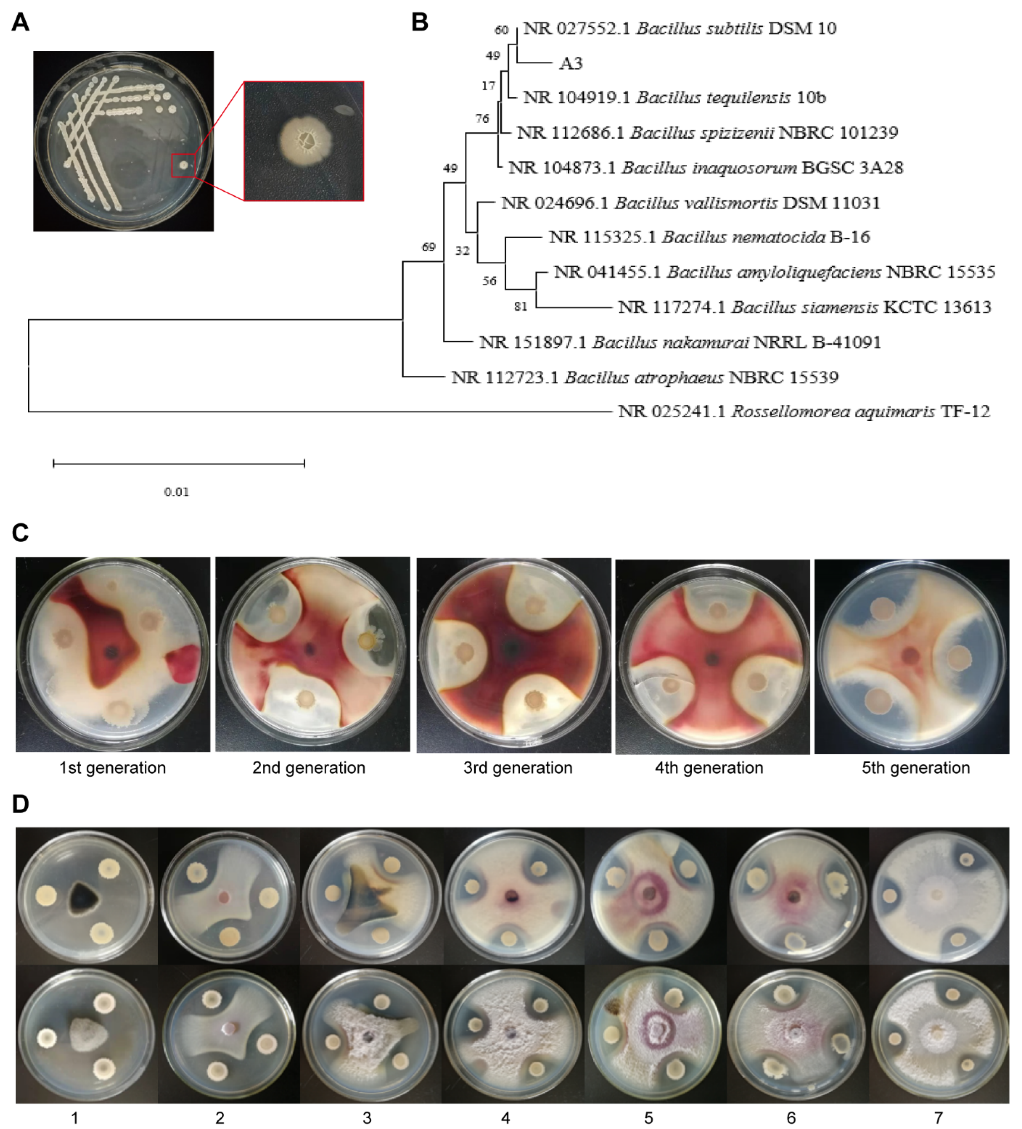
3.1. Identification of the Biocontrol Strain A3 for Corn Stalk Rot

Culturable bacteria were isolated from the corn rhizosphere soil employing the dilution spread plate technique on LB agar. Following the purification process, these bacterial isolates were subjected to antagonistic culture against Fusarium graminearum on PDA medium, incubated at 27 °C for a duration of 7 days. Through this screening, a bacterial strain demonstrating significant inhibitory activity against F. graminearum was identified and designated as A3. The morphological and molecular characteristics of isolate A3 are consistent with those of Bacillus subtilis. This isolate was deposited in the China General Microbiological Culture Collection Center (CGMCC No. 23953). The colony morphology of the A3 was observed in the experiment. The results showed that the colony of this strain on LB solid medium was grayish white, opaque, dry, and shriveled on the surface, with slightly yellowish and wrinkled edges (Figure 1A). The physiological and biochemical identification of the biocontrol strain was conducted. The results indicated that there were no obvious phenomena in the malate test, phenylalanine deaminase test, and H2S production test, etc. This strain showed negative results in five tests, while the remaining reactions were positive (Table S1). The sequencing data of the biocontrol strain were subjected to a homological comparison in the NCBI database. Subsequently, a phylogenetic tree was constructed using MEGA7.0 software. The results clearly demonstrated that the biocontrol strain obtained from the experiment was Bacillus subtilis (Figure 1B).

3.2. Determination of the Stability and Antibacterial Spectrum of B. subtilis A3

By subculturing, we could assess whether the inhibitory efficacy of the bacterial strains was maintained or declined over time. The results showed that strain A3 still had an obvious inhibitory effect on F. graminearum after five successive generations of culture, indicating the antimicrobial activity of A3 was stable (Figure 1C). Using the plate confrontation method, it was found that the biocontrol strain had certain antibacterial effects on Setosphaeria turcica, F. proliferatum, Bipolaris zeicola (Stout) Shoem., F. verticillioides, Curvularia lunata, Helminthosporium maydis, and F. oxysporum, indicating that A3 had a broad antibacterial spectrum (Figure 1D).

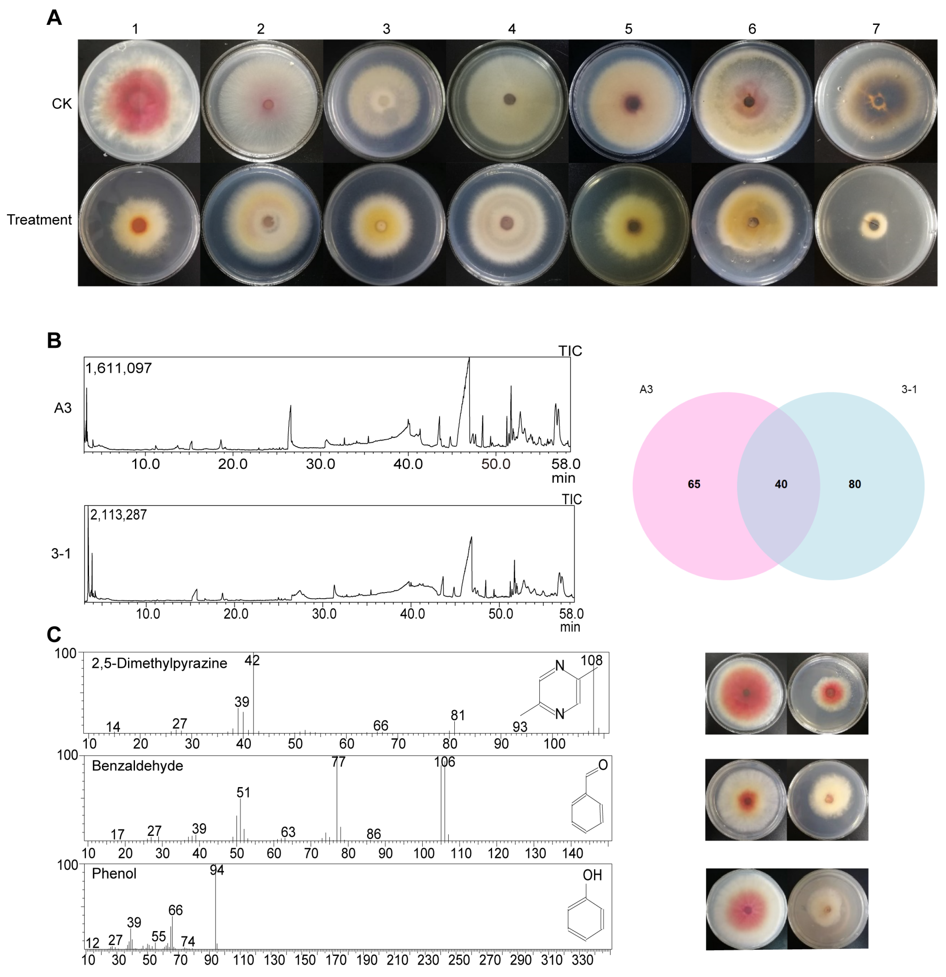
3.3. Inhibitory Effect of B. subtilis A3 on F. graminearum

The fermentation broth of the biocontrol strain A3 had a certain impact on the normal growth of F. graminearum mycelia. The mycelia of F. graminearum in the control group were uniform and could extend normally, while in the experimental group, the mycelia of F. graminearum at the tips were swollen and presented a distinct bead-like arrangement with obvious deformity (Figure S1A). Different concentrations of A3 fermentation broth were applied, and it was found that 50% of the fermentation supernatant could significantly inhibit the growth of F. graminearum (Figure S1B). As the supernatant concentration increased, the inhibition rate gradually increased, and the inhibition rate of 50% of the fermentation supernatant could reach over 60% (Figure S1C), indicating that the substances secreted by A3 during growth could inhibit the growth of F. graminearum.
By employing the double-plate method, it was discovered that the volatile gas produced by A3 was capable of inhibiting the growth of pathogenic fungi including F. graminearum, F. oxysporum, F. solani, F. verticillioides, Curvularia lunata, Helminthosporium carbonum, and F. acuminatum (Figure 2A). The volatile substances produced by the sample were detected and identified by GC-MS. Through gas chromatography–mass spectrometry, it was found that there were 65 unique components in A3 compared with the non-volatile inhibitory strain B. siamensis 3-1 (Figure 2B). Among them, C6H8N2, C7H6O, and C6H6O were selected for the inhibition test, and it was found that all three substances could inhibit the growth of F. graminearum. The three compounds were 2,5-dimethylpyrazine, benzaldehyde, and phenol (Figure 2C).

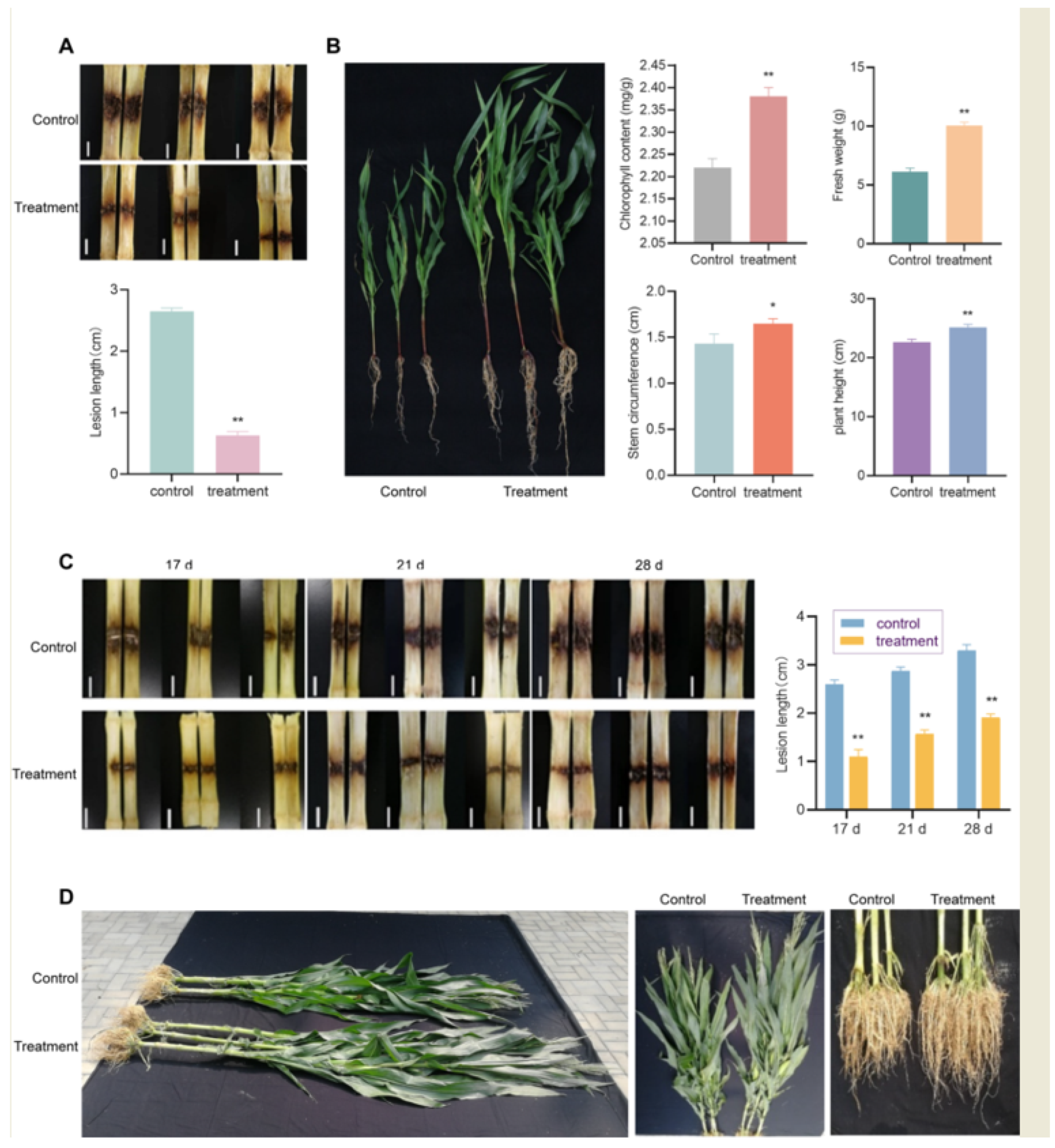
3.4. Indoor Control Effect of B. subtilis A3 Fermentation Solution Irrigation on Corn Stalk Rot and Plant Growth

Using the acupuncture method, the suspension of F. graminearum conidia was injected into the corn that had been treated with root irrigation and the untreated corn. After 30 days of injection, observation was conducted and the control effect was calculated. It was found that the lesion area was significantly reduced after the root irrigation treatment with B. subtilis A3. Through measurement, the lesion area of the experimental group was approximately 2.62 cm2, and that of the control group was approximately 7.48 cm2. According to the formula, the inhibition rate was approximately 65.21% (Figure 3A).
The corn seedlings were treated with root irrigation, and the data were statistically analyzed after 30 days. By comparing the plant height, stem circumference, fresh weight, and chlorophyll content of the corn seedlings, it was found that the treatment group had a certain promoting effect on the growth of the corn. Compared with the control group, the plant height of the treatment group increased by 11.45%, the stem circumference increased by 16.08%, the fresh weight increased by 62.62%, and the chlorophyll content increased by 7.21% (Figure 3B).

3.5. Field Control Effect of Seed Coating with B. subtilis A3 on Corn Stalk Rot

The field control efficacy of the biocontrol agent was analyzed. The results showed that after inoculation with F. graminearum, both the test group and the control group of corn stalks had varying degrees of disease, with the control group being more severe, showing that the corn stalks began to turn black and rot at the base and spread to the surrounding areas. The disease incidence in the test group was significantly lower than that in the control group, with only the area around the needle holes turning brown (Figure 3C). The lesion area of the corn stalks in the test group was smaller than that in the blank control group at 17, 24, and 31 days after inoculation, and the inhibitory effect was statistically calculated (Figure 3D). Through analysis, it was found that the biological seed coating agent could effectively inhibit the infection of F. graminearum.
The disease conditions of the two treatments in the field experiment were investigated and the data were statistically analyzed (Table 1). It was found that after coating the corn seeds with B. subtilis A3, the treatment group was greater than the control group in terms of ear length, hundred-grain weight, seed yield rate, and production, and the treatment group had a shorter barren ear tip length. The disease index of stalk rot in the seed-coated group was lower than that in the control group. Among them, the control efficacy of the biocontrol agent was 45.75%, indicating that the coating seeds could effectively prevent corn stalk rot in the field experiment.

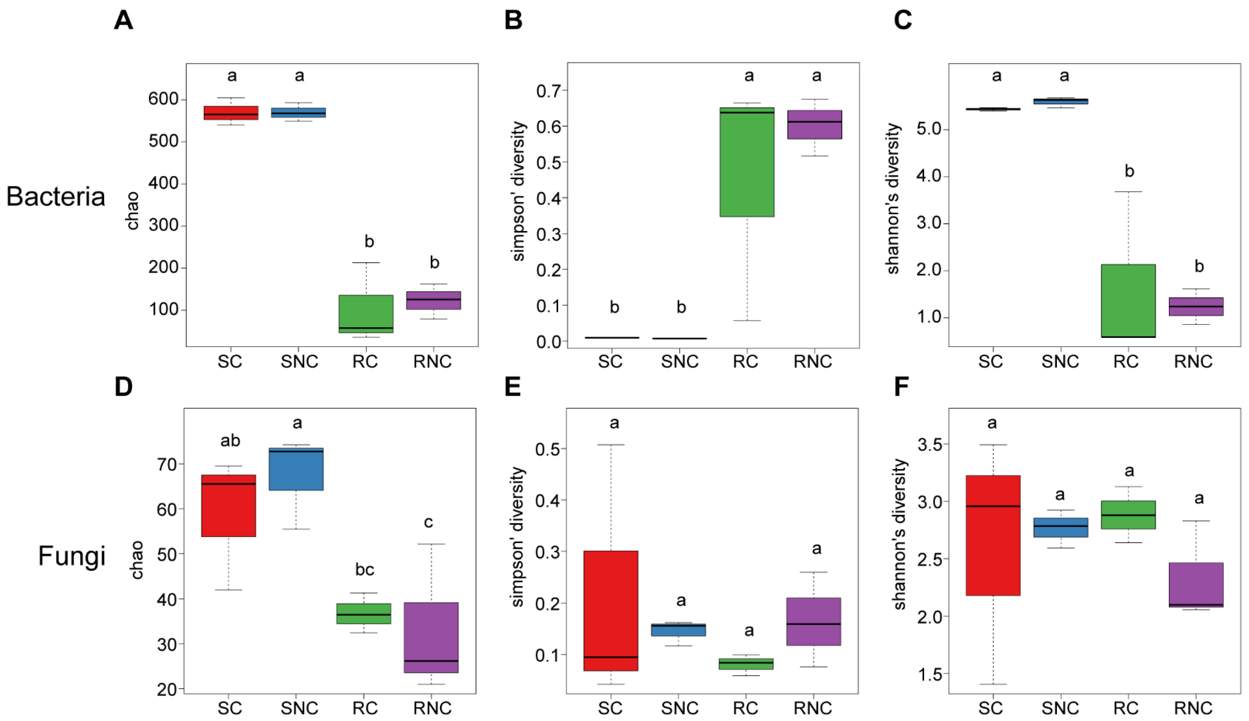
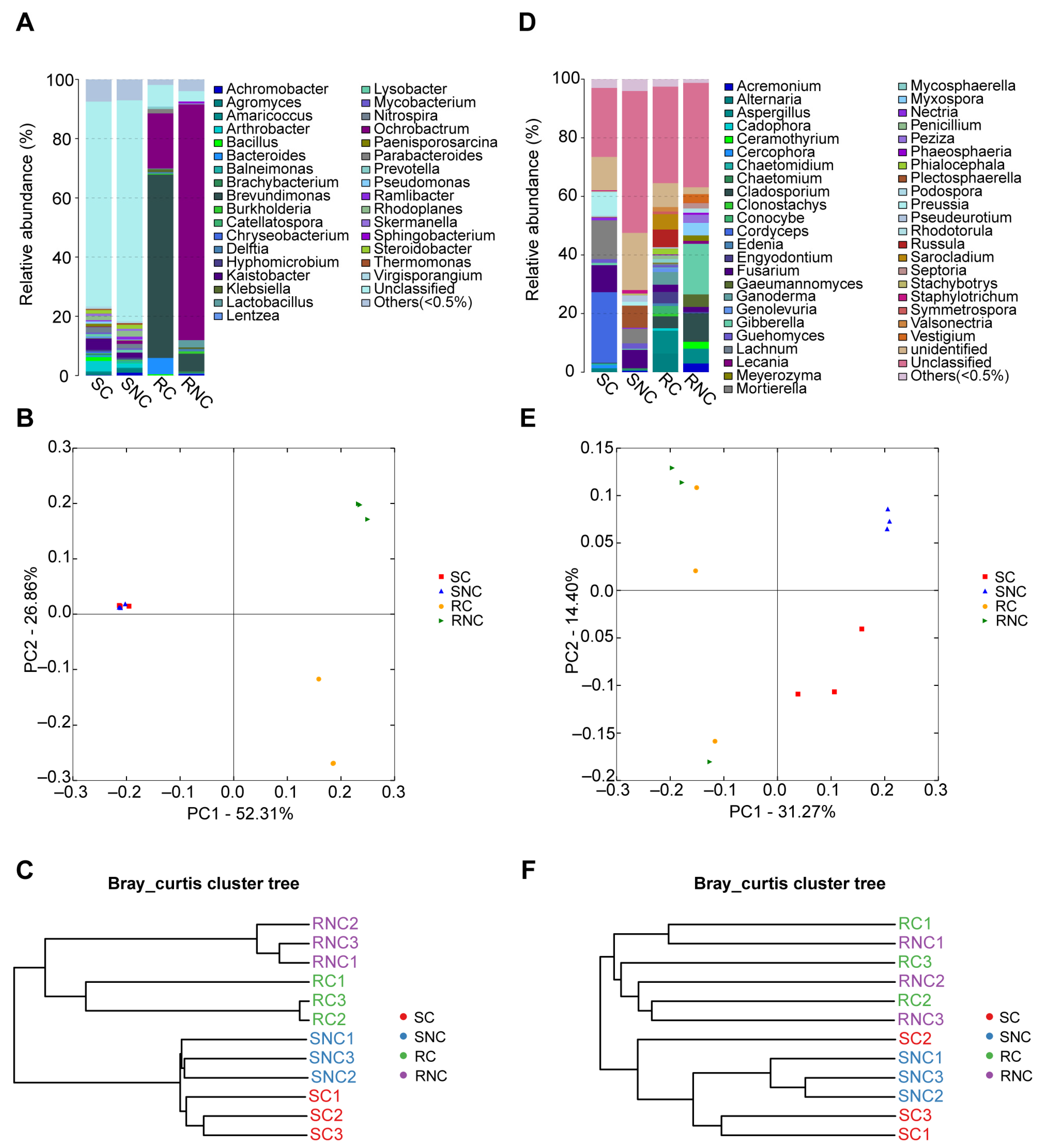
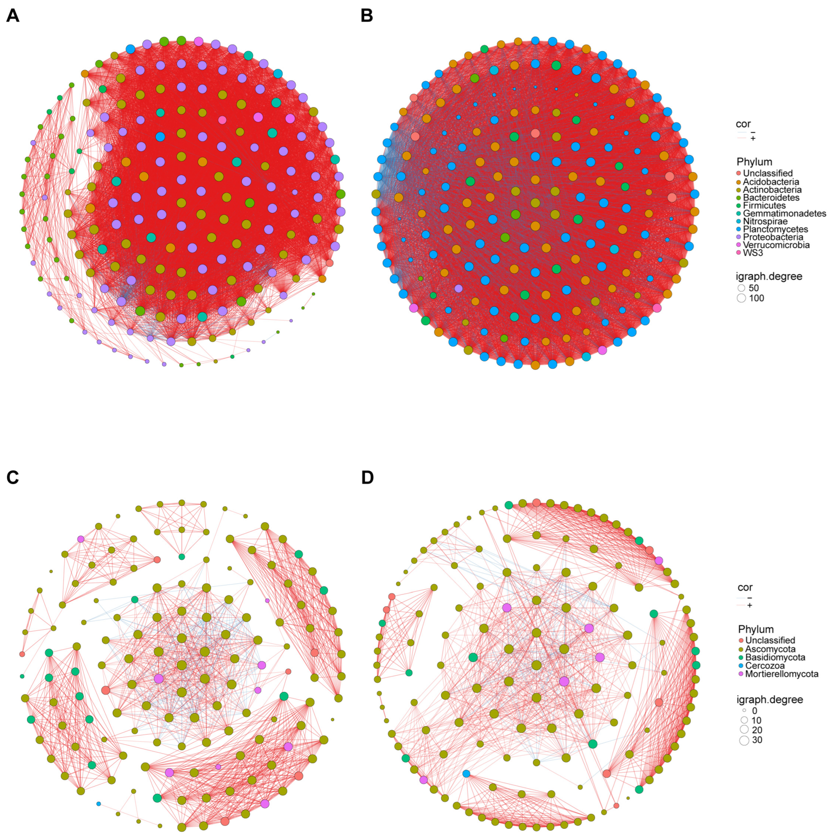
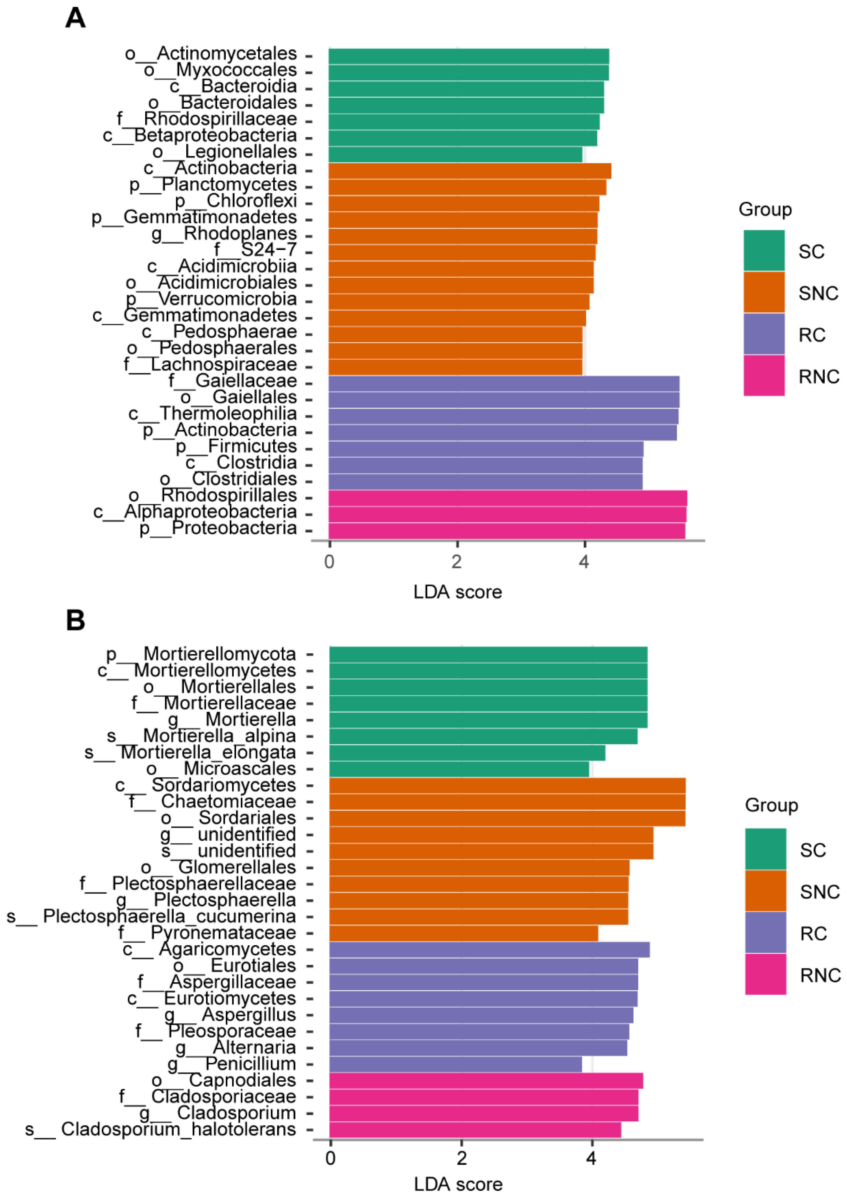
3.6. Effect of Seed Coating with A3 on Microbial Structure and Diversity of Corn Rhizosphere Soil and Root Endophyte

The number of valid bacterial tags in all samples ranged from 15,959 to 30,829, and the number of valid fungal tags ranged from 847 to 147,956. The Shannon index rarefaction curves of each group were gentle (Figure S2), indicating that the sequencing depth had covered most of the species. The data volume fully reflected the diversity of species in each sample. The richness (Chao 1 index) and species diversity (Shannon and Simpson indices) of the rhizosphere and endophytic microorganisms were calculated and subjected to significance tests (Figure 4). The Chao 1, Shannon, and Simpson indices of the rhizosphere bacterial community were significantly different from those of the endophytic bacterial community, while there were no significant differences between the seed coating agent treatment and the control in both ecological environments. This indicates that the species diversity and distribution uniformity of the rhizosphere bacterial community were higher than those of the endophytic bacterial community, and the influence of dominant species on the community structure was greater in the endophyte The A3 treatment had no significant effect on the diversity of the rhizosphere bacterial community in the field. The Chao 1 index of the rhizosphere fungal community was significantly different from that of the endophytic fungal community, while the Shannon and Simpson indices of the rhizosphere fungal community were not significantly different from those of the endophytic fungal community. There were no significant differences between the seed coating agent treatment and the control in the same compartments, indicating that there were no significant differences in the species diversity and distribution uniformity of the rhizosphere and endophytic fungal communities. The A3 treatment had no significant effect on the diversity of the rhizosphere fungal community in the field.
The top 10 genera with the highest abundance in the bacterial community were Ochrobactrum, Brevundimonas, Bacteroides, Kaistobacter, Arthrobacter, Rhodoplanes, Nitrospira, Lactobacillus, Steroidobacter, and Bacillus (Figure 5A). PCoA indicated that in the rhizosphere soil, the bacterial communities of the coated treatment and the control treatment were not significantly separated, while in the root endophytic, the bacterial communities of the coated treatment and the control treatment were significantly separated, and there was a clear separation between the rhizosphere and the root endophytic. The bacterial communities could explain 52.31% (PC1) and 26.86% (PC2) of the total bacterial variation (Figure 5B). A hierarchical clustering tree was constructed to describe and compare the similarities among samples (Figure 5C). According to the similarity of community composition, the rhizosphere communities and the root endophytic communities were separated in the clusters, and the treatments and the control were also separated. The top 10 genera with the highest abundance in fungal communities were Cordyceps, Fusarium, Gibberella, Mortierella, Cladosporium, Aspergillus, Preussia, Plectosphaerella, Alternaria, and Russula (Figure 5D). PCoA indicated that in the rhizosphere soil, the fungal communities of the coated treatment and the control treatment were significantly separated, while in the root endophytic, the fungal communities of the coated treatment and the control treatment were not significantly separated, and there was a clear separation between the rhizosphere and the root endophytic. The fungal communities could explain 31.27% (PC1) and 14.40% (PC2) of the total fungal variation (Figure 5E). A hierarchical clustering tree was constructed to describe and compare the similarities among samples (Figure 5F). According to the similarity of community composition, the rhizosphere communities and the root endophytic communities were separated in the clusters, but the treatments and the control were not separated. We can infer that the differences in the rhizosphere and root endophytic environments had a significant impact on the composition of microorganisms, while the application of a single biocontrol agent had a relatively small impact on the composition of bacteria and an even smaller impact on the composition of fungi.
Microbial networks were established based on significant correlations (Spearman’s correlation, p < 0.05, absolute r > 0.6) among different treatments. Our results showed that the bacterial networks had more nodes and edges than the fungal networks in the same compartment (Figure 6). In addition, the results showed that the complexity of the CK bacterial network (determined by the number of edges and connectance) was more complex than that of the treatment. Moreover, no matter if it was bacteria or fungi, the network had a large proportion of positive correlation (Table 2). The topological property analysis of network nodes showed that neither fungi nor bacteria had a central point in the network (Figure S3).
To further explore the impact of B. subtilis-coated seeds on microbial communities, we used LEfSe to analyze the species differences among the groups. The results indicated that the abundances of Actinomycetales, Myxococcales, Bacteroidia, Bacteroidales, Rhodospirillaceae, Betaproteobacteria, and Legionellales in the rhizosphere soil of the coated seeds were significantly higher than those in other treatments. In the control rhizosphere soil, the abundances of Actinobacteria, Planctomycetes, Chloroflexi, Gemmatimonadetes, Rhodoplanes, S24-7, Acidimicrobiia, Acidimicrobiales, Verrucomicropia, Gemmatimonadetes, Pedosphaerae, Pedosphaerales, and Achnospiraceae were the highest. In the coated seeds’ root endophytic, the abundances of Gaiellales, Thermoleophilia, Actinobacteria, Firmicutes, Clostridia, and Clostridiales were the highest, while in the control root interior, the abundances of Rhodospirillales, Alphaproteobacteria, and Proteobacteria were the highest (Figure 7A). In the rhizosphere soil of the coated seeds, the abundances of Mortierellomycota, Mortierelomycetes, Mortierellales, Mortierellaceae, Mortierella, Mortierella_alpina, Mortierella_elongatao, and Microascales were significantly higher than those in other treatments. In the control rhizosphere soil, the abundances of Sordariomycetes, Chaetomiaceae, Sordariales, Glomerellales, Plectosphaerellaceae, Plectosphaerella, Plectosphaerella_cucumerina, and Pyronemataceae were the highest. In the coated seeds’ root interior, the abundances of Agaricomycetes, Eurotiales, Aspergillaceae, Eurotiomycetes, Aspergillus, Pleosporaceae, Alternaria, and Penicillium were the highest, while in the control root endophytic, the abundances of Capnodiales, Cladosporiaceae, Cladosporium, and Cladosporium_halotolerans were the highest (Figure 7B).

4. Discussion

This study identified Bacillus subtilis A3 and investigated its biocontrol effect on corn stalk rot. The results showed that the isolate A3 exhibited broad-spectrum inhibitory effects on various corn pathogens through competitive action, including Rhizoctonia solani, Fusarium oxysporum, Phytophthora sojae, Sclerotinia sclerotiorum, and F. graminearum. Through in vitro confrontation assays, it was found that the inhibition rate of A3 against F. graminearum was 65.9%, suggesting that there might have been ecological niche and nutrient competition between B. subtilis and F. graminearum. Besides competitive action, the active substances produced by B. subtilis also play an important role in the biological control of plant pathogens. It has been reported that these substances can inhibit the spore germination and mycelial growth of Verticillium dahliae [50], Colletotrichum gloeosporioides [51], Magnaporthe oryzae [52], Botrytis cinerea [53], and Phytophthora capsici [54]. In this study, the inhibition rates of the volatile organic compounds secreted by A3 against pathogenic fungi, including F. graminearum, were all above 60%. Through gas chromatography analysis, it was found that the three volatile organic compounds with good inhibitory effects specific to A3 were 2,5-dimethylpyrazine (2,5-DMP), benzaldehyde, and phenol. The results indicated that 2,5-DMP had the best inhibitory effect on F. graminearum, suggesting that this substance has broad application prospects in the prevention and control of corn stalk rot. Studies have shown that B. velezensis [55], B. amyloliquefaciens [56], Pseudomonas protegens [12], and B. subtilis [57] can secrete 2,5-DMP and have inhibitory effects on fungi. Therefore, this study once again experimentally proved that B. subtilis can inhibit F. graminearum by secreting 2,5-DMP and has an inhibitory effect on corn stalk rot, but this antibacterial mechanism needs further analysis.
After 30 days of indoor inoculation of F. graminearum with A3 fermentation liquid, the inhibition rate of the corn plants was found to be 65.21%, and it also had a promoting effect on plant growth. This study showed that the incidence of stalk rot after seed coating was lower than that of the control group, and the control effect of the bacterial agent treatment was over 40%, indicating that seed coating can effectively prevent and control corn stalk rot in field experiments. The growth-promoting effect of Bacillus subtilis has been confirmed in many studies, for example, B. subtilis rhizosphere promotes plant root development [58], and two specific components of B. subtilis extracellular matrix, amyloid TasA and phenol subtilin, can increase the concentration of reactive oxygen species in sweet melon seeds and stimulate the development of a radicle, which, to a certain extent, affects growth and development [9]. B. subtilis harbors an array of both direct and indirect modalities to foster plant growth and, in turn, augment crop yield [2]. This encompasses enhancing the accessibility of nutrients [59], modulating the equilibrium of plant growth hormones [60], and mitigating the intensity of abiotic stresses [61]. The growth-promoting mechanism of A3 on corn plants needs to be further explored. In the present study, the results showed that the incidence of stalk rot after seed coating was lower than that of control group, and the control effect of bactericide treatment was more than 40%, indicating that seed coating could effectively control corn stalk rot in field trials.
Plant roots inhabit a large number of microbial communities, which are mainly divided into rhizosphere microbial communities and root endophytic microbial communities according to their different habitats, which colonize around and inside the roots of plants, respectively [31]. Seed microbial coating is an important means of biological control of microbial inoculants. Studies have shown that a seed coating agent can significantly increase the yield and improve the microbial community network of corn [62]. In this study, by planting corn seeds coated with biological agents, it was found that they not only reduced the occurrence of corn stalk rot but also affected the composition and structure of the soil and corn root microbiota. Through microbial component analysis, in the bacterial microbiota composition of soil and corn roots, the relative abundance of Bacteroidetes increased, while Proteobacteria and Acidobacteria, which are indicators of soil nutrient conditions, decreased. The decrease in the relative abundance of Acidobacteria in the soil reduced the competitive pressure on other bacterial phyla, helping to increase the relative abundance of other bacterial phyla, such as Bacteroidetes. Proteobacteria and Bacteroidetes are nutrient-rich bacteria and are commonly found in nutrient-rich soil. In the fungal microbiota composition of soil and corn roots, the relative abundance of Ascomycota decreased, while the relative abundance of Mortierella increased. It has been found that Ascomycota and Basidiomycota are pathogenic fungi that can cause varying degrees of plant diseases [63,64], while Mortierella is a beneficial fungus that can promote the absorption of nutrients by plants and help plant growth [65]. In conclusion, after the seeds were coated with biological seed coating, the species richness of microorganisms in the corn roots and soil showed inconsistent trends. However, in general, the increase in the abundance of beneficial bacteria and fungi and the decrease in the abundance of pathogenic fungi are one of the ways that biological seed coatings promote plant growth and control diseases.

5. Conclusions

In this study, we identified and characterized Bacillus subtilis strain A3 as a potent biocontrol agent against Fusarium graminearum, the causative agent of corn stalk rot. Strain A3 demonstrated significant antifungal activity, broad-spectrum antibacterial properties, and stable inhibitory effects across multiple generations. Furthermore, volatile organic compounds (VOCs) produced by A3, including 2,5-dimethylpyrazine, benzaldehyde, and phenol, were identified as key contributors to its antifungal activity. In both indoor and field experiments, A3 exhibited remarkable efficacy in controlling corn stalk rot. Root irrigation with A3 fermentation broth reduced lesion areas by 65.21% and promoted corn growth, as evidenced by significant increases in plant height, stem circumference, fresh weight, and chlorophyll content. Field trials further confirmed that seed coating with A3 significantly lowered disease incidence and severity, while enhancing crop yield parameters such as the ear length, hundred-grain weight, and seed yield rate. Microbial community analysis revealed that A3 treatment did not significantly alter the diversity of the rhizosphere or endophytic bacterial and fungal communities. However, it influenced the relative abundances of specific taxa, such as Mortierella in the rhizosphere and Aspergillus in root endophytes, suggesting a targeted modulation of microbial dynamics. Network analysis highlighted the complexity of bacterial interactions compared to fungi, with a predominance of positive correlations in both communities. These findings underscore the potential of B. subtilis A3 as a sustainable and effective biocontrol agent for managing corn stalk rot. Its dual role in pathogen suppression and plant growth promotion, coupled with its minimal impact on soil microbial diversity, positions A3 as a promising candidate for integrated pest management strategies in agriculture. Future research should focus on optimizing formulation and application methods, as well as exploring the mechanistic basis of A3’s interactions with plant and microbial communities to further enhance its efficacy and stability.

Supplementary Materials

The following supporting information can be downloaded at: https://www.mdpi.com/article/10.3390/agronomy15030706/s1, Supplemental Figure S1: Inhibitory effect of B. subtilis A3 fermentation broth on Fusarium graminearum. (A) Microscopic observations of hyphae edge from the control and fermentation broth-treated groups. (B) Inhibitory effect of B. subtilis A3 fermentation broth on spore germination of F. graminearum from the control and fermentation broth-treated groups. (C) Inhibition rate of different fermentation broth concentration; Supplemental Figure S2: Bacterial (A) and fungal (B) rarefaction curves for all samples at a 97% OTU sequence similarity threshold; Supplemental Figure S3: Zi-Pi diagram of the molecular ecological network. (A,B) Zi-Pi diagram of the bacterial communities of the A3 (A) and control (B) treatments. (C,D) Zi-Pi diagram of the fungal communities of the A3 (C) and control (D) treatments; Supplemental Table S1: Physiological and biochemical identification.

Author Contributions

Conceptualization, K.Z., J.X. and J.D.; Methodology, K.Z.; Formal analysis, S.J.; Investigation, S.J. and Y.D.; Resources, Y.D. and H.C.; Data curation, Y.D.; Writing—original draft, L.W.; Writing—review & editing, L.W.; Visualization, L.W., S.J. and H.C.; Supervision, K.Z., J.X. and J.D.; Project administration, J.X. and J.D.; Funding acquisition, J.X. and J.D. All authors have read and agreed to the published version of the manuscript.

Funding

This work was supported by the Science and Technology Planning Project of Baoding (2494N032), Central Government Guides Local Science and Technology Development Projects (216Z6502G), China Agriculture Research System (CARS-02), and Research Project of Basic Scientific Research in Provincial Universities of Hebei Province (KY2023063).

Data Availability Statement

The data presented in this study are openly available in the China National Center for Bioinformation databases under BioProject ID PRJCA036063 [China National Center for Bioinformation] [https://www.cncb.ac.cn/?lang=en] [BioProject ID PRJCA036063] [Accessed on 11 March 2025].

Conflicts of Interest

The authors declare no conflicts of interest.

References

  1. Kunst, F.; Ogasawara, N.; Moszer, I.; Albertini, A.M.; Alloni, G.; Azevedo, V.; Bertero, M.G.; Bessieres, P.; Bolotin, A.; Borchert, S.; et al. The complete genome sequence of the Gram-positive bacterium Bacillus subtilis. Nature 1997, 390, 249–256. [Google Scholar] [CrossRef]
  2. Blake, C.; Christensen, M.N.; Kovács, Á.T. Molecular aspects of plant growth promotion and protection by Bacillus subtilis. Mol. Plant Microbe Interact. 2021, 34, 15–25. [Google Scholar] [CrossRef]
  3. Almasoudi, N.M.; Sallam, N.M.A.; Ali, E.F.; Alqurashi, A.S.; Issa, A.A.; Althobaiti, F.; Housny, M.; Alomari, H.; Abo-Elyousr, K.A.M. Development of Bacillus spp for controlling wilt disease and improving the growth of tomato. Eur. J. Plant Pathol. 2025. [Google Scholar] [CrossRef]
  4. Xu, L.H.; Shang, Q.H.; Nicolaisen, M.; Zeng, R.; Gao, S.G.; Gao, P.; Song, Z.W.; Dai, F.M.; Zhang, J.Z. Biocontrol potential of rhizospheric Bacillus strains against Sclerotinia minor jagger causing lettuce drop. Microorganisms 2025, 13, 68. [Google Scholar] [CrossRef]
  5. Legrifi, I.; Al Figuigui, J.; Lahmamsi, H.; Taoussi, M.; Radi, M.; Belabess, Z.; Lazraq, A.; Barka, E.A.; Lahlali, R. Unlocking olive rhizobacteria: Harnessing biocontrol power to combat olive root rot and promote plant growth. Int. Microbiol. 2025. [Google Scholar] [CrossRef]
  6. Zhao, Y.; Liu, F.; Lan, X.; Xu, W.X.; Dong, W.B.; Ke, S.Y.; Wu, H.Q. Discovery, characterization, and application of broad-spectrum antimicrobial peptide AtR905 from Aspergillus terreus as a biocontrol agent. J. Agric. Food Chem. 2024, 12, 2793–2804. [Google Scholar] [CrossRef]
  7. Zhou, Y.; Li, Q.; Peng, Z.; Zhang, J.; Li, J. Biocontrol effect of Bacillus subtilis YPS-32 on potato common scab and its complete genome sequence analysis. J. Agric. Food Chem. 2022, 70, 5339–5348. [Google Scholar] [CrossRef]
  8. Wang, X.J.; Xie, S.Y.; Mu, X.Y.; Guan, B.; Hu, Y.Z.; Ni, Y.Q. Investigating the resistance responses to Alternaria brassicicola in ‘Korla’ fragrant pear fruit induced by a biocontrol strain Bacillus subtilis Y2. Postharvest Biol. Technol. 2023, 199, 112293. [Google Scholar] [CrossRef]
  9. Berlanga-Clavero, M.V.; Molina-Santiago, C.; Caraballo-Rodríguez, A.M.; Petras, D.; Díaz-Martínez, L.; Pérez-García, A.; de Vicente, A.; Carrión, V.J.; Dorrestein, P.C.; Romero, D. Bacillus subtilis biofilm matrix components target seed oil bodies to promote growth and anti-fungal resistance in melon. Nat. Microbiol. 2022, 7, 1001–1015. [Google Scholar] [CrossRef]
  10. Wang, J.; Liu, J.; Chen, H.; Yao, J. Characterization of Fusarium graminearum inhibitory lipopeptide from Bacillus subtilis IB. Appl. Microbiol. Biotechnol. 2007, 76, 889–894. [Google Scholar] [CrossRef]
  11. Zhao, Y.J.; Selvaraj, J.N.; Xing, F.G.; Zhou, L.; Wang, Y.; Song, H.M.; Tan, X.X.; Sun, L.C.; Sangare, L.; Folly, Y.M.E.; et al. Antagonistic Action of Bacillus subtilis Strain SG6 on Fusarium graminearum. PLoS ONE 2014, 9, e92486. [Google Scholar] [CrossRef]
  12. Zhao, Q.; Cao, J.; Cai, X.; Wang, J.; Kong, F.; Wang, D.; Wang, J. Antagonistic activity of volatile organic compounds produced by acid-tolerant Pseudomonas protegens CLP-6 as biological fumigants to control tobacco bacterial wilt caused by Ralstonia solanacearum. Appl. Environ. Microbiol. 2023, 89, e01892-22. [Google Scholar] [CrossRef]
  13. Raio, A. Diverse roles played by “Pseudomonas fluorescens complex” volatile compounds in their interaction with phytopathogenic microrganims, pests and plants. World J. Microb. Biot. 2024, 40, 80. [Google Scholar] [CrossRef]
  14. Zaid, D.S.; Li, W.; Yang, S.; Li, Y. Identification of bioactive compounds of Bacillus velezensis HNA3 that contribute to its dual effects as plant growth promoter and biocontrol against post-harvested fungi. Microbiol. Spectr. 2023, 11, e0051923. [Google Scholar] [CrossRef]
  15. Ulloa, P.A.; Valencia, A.L.; Olivares, D.; Poblete-Morales, M.; Silva-Moreno, E.; Defilippi, B.G. Antifungal effect of volatile organic compounds (VOCs) release from Antarctic bacteria under postharvest conditions. Food Packag. Shelf 2023, 39, 101160. [Google Scholar] [CrossRef]
  16. Song, W.; Dai, M.; Gao, S.; Mi, Y.; Zhang, S.; Wei, J.; Zhao, H.; Duan, F.; Liang, C.; Shi, Q. Volatile organic compounds produced by Paenibacillus polymyxa J2-4 exhibit toxic activity against Meloidogyne incognita. Pest Manag. Sci. 2024, 80, 1289–1299. [Google Scholar] [CrossRef]
  17. Poveda, J. Beneficial effects of microbial volatile organic compounds (MVOCs) in plants. Appl. Soil Ecol. 2021, 168, 104118. [Google Scholar] [CrossRef]
  18. Song, G.C.; Ryu, C.M. Two volatile organic compounds trigger plant self-defense against a bacterial pathogen and a sucking insect in cucumber under open field conditions. Int. J. Mol. Sci. 2013, 14, 9803–9819. [Google Scholar] [CrossRef]
  19. Giorgio, A.; De Stradis, A.; Lo Cantore, P.; Iacobellis, N.S. Biocide effects of volatile organic compounds produced by potential biocontrol rhizobacteria on Sclerotinia sclerotiorum. Front. Microbiol. 2015, 6, 1056. [Google Scholar] [CrossRef]
  20. Bailly, A.; Groenhagen, U.; Schulz, S.; Geisler, M.; Eberl, L.; Weisskopf, L. The inter-kingdom volatile signal indole promotes root development by interfering with auxin signalling. Plant J. 2014, 80, 758–771. [Google Scholar] [CrossRef]
  21. Weisskopf, L.; Schulz, S.; Garbeva, P. Microbial volatile organic compounds in intra-kingdom and inter-kingdom interactions. Nat. Rev. Microbiol. 2021, 19, 391–404. [Google Scholar] [CrossRef]
  22. Xue, J.; Gao, S.; Hou, L.Y.; Li, L.L.; Ming, B.; Xie, R.Z.; Wang, K.R.; Hou, P.; Li, S.K. Physiological influence of stalk rot on maize lodging after physiological maturity. Agronomy 2021, 11, 2271. [Google Scholar] [CrossRef]
  23. Pammel, L.H. Serious root and stalk diseases of corn. IOWA Agric. 1914, 15, 156–158. [Google Scholar]
  24. Khokhar, M.K.; Hooda, K.S.; Sharma, S.S.; Singh, V. Post flowering stalk rot complex of maize—Present status and future prospects. Maydica 2014, 59, 226–242. [Google Scholar]
  25. Ledečan, T.; Šumić, D.; Brkić, I.; Jambrović, A.; Zdunić, Z. Resistance of maize inbreds and their hybrids to Fusarium stalk rot. Czech J. Genet. Plant Breed. 2003, 39, 15–20. [Google Scholar] [CrossRef]
  26. Sumarah, M.W. The deoxynivalenol challenge. J. Agric. Food Chem. 2022, 70, 9619–9624. [Google Scholar] [CrossRef]
  27. Berendsen, R.L.; Pieterse, C.M.; Bakker, P.A. The rhizosphere microbiome and plant health. Trends Plant Sci. 2012, 17, 478–486. [Google Scholar] [CrossRef]
  28. Badri, D.V.; Chaparro, J.M.; Zhang, R.; Shen, Q.; Vivanco, J.M. Application of natural blends of phytochemicals derived from the root exudates of Arabidopsis to the soil reveal that phenolic-related compounds predominantly modulate the soil microbiome. J. Biol. Chem. 2013, 288, 4502–4512. [Google Scholar] [CrossRef]
  29. Bonito, G.; Reynolds, H.; Robeson, M.S., 2nd; Nelson, J.; Hodkinson, B.P.; Tuskan, G.; Schadt, C.W.; Vilgalys, R. Plant host and soil origin influence fungal and bacterial assemblages in the roots of woody plants. Mol. Ecol. 2014, 23, 3356–3370. [Google Scholar] [CrossRef]
  30. Müller, D.B.; Vogel, C.; Bai, Y.; Vorholt, J.A. The plant microbiota: Systems-level insights and perspectives. Annu. Rev. Genet. 2016, 50, 211–234. [Google Scholar] [CrossRef]
  31. Bai, B.; Liu, W.; Qiu, X.; Zhang, J.; Zhang, J.; Bai, Y. The root microbiome: Community assembly and its contributions to plant fitness. J. Integr. Plant Biol. 2022, 64, 230–243. [Google Scholar] [CrossRef] [PubMed]
  32. Busby, P.E.; Soman, C.; Wagner, M.R.; Friesen, M.L.; Kremer, J.; Bennett, A.; Morsy, M.; Eisen, J.A.; Leach, J.E.; Dangl, J.L. Research priorities for harnessing plant microbiomes in sustainable agriculture. PLoS Biol. 2017, 15, e2001793. [Google Scholar] [CrossRef]
  33. Pang, Z.; Chen, J.; Wang, T.; Gao, C.; Li, Z.; Guo, L.; Xu, J.; Cheng, Y. Linking plant secondary metabolites and plant microbiomes: A review. Front. Plant Sci. 2021, 12, 621276. [Google Scholar] [CrossRef] [PubMed]
  34. Wagner, M.R.; Tang, C.; Salvato, F.; Clouse, K.M.; Bartlett, A.; Vintila, S.; Phillips, L.; Sermons, S.; Hoffmann, M.; Balint-Kurti, P.J.; et al. Microbe-dependent heterosis in maize. Proc. Natl. Acad. Sci. USA 2021, 118, e2021965118. [Google Scholar] [CrossRef] [PubMed]
  35. Mendes, R.; Kruijt, M.; de Bruijn, I.; Dekkers, E.; van der Voort, M.; Schneider, J.H.; Piceno, Y.M.; DeSantis, T.Z.; Andersen, G.L.; Bakker, P.A.; et al. Deciphering the rhizosphere microbiome for disease-suppressive bacteria. Science 2011, 332, 1097–1100. [Google Scholar] [CrossRef]
  36. Santhanam, R.; Luu, V.T.; Weinhold, A.; Goldberg, J.; Oh, Y.; Baldwin, I.T. Native root-associated bacteria rescue a plant from a sudden-wilt disease that emerged during continuous cropping. Proc. Natl. Acad. Sci. USA 2015, 112, E5013–E5020. [Google Scholar] [CrossRef]
  37. Cha, J.Y.; Han, S.; Hong, H.J.; Cho, H.; Kim, D.; Kwon, Y.; Kwon, S.K.; Crüsemann, M.; Bok Lee, Y.; Kim, J.F.; et al. Microbial and biochemical basis of a Fusarium wilt-suppressive soil. ISME J. 2016, 10, 119–129. [Google Scholar] [CrossRef]
  38. Chen, Y.; Wang, J.; Yang, N.; Wen, Z.; Sun, X.; Chai, Y.; Ma, Z. Wheat microbiome bacteria can reduce virulence of a plant pathogenic fungus by altering histone acetylation. Nat. Commun. 2018, 9, 3429. [Google Scholar] [CrossRef]
  39. Helfrich, E.J.N.; Vogel, C.M.; Ueoka, R.; Schäfer, M.; Ryffel, F.; Müller, D.B.; Probst, S.; Kreuzer, M.; Piel, J.; Vorholt, J.A. Bipartite interactions, antibiotic production and biosynthetic potential of the Arabidopsis leaf microbiome. Nat. Microbiol. 2018, 3, 909–919. [Google Scholar] [CrossRef]
  40. Parratt, S.R.; Laine, A.L. Pathogen dynamics under both bottom-up host resistance and top-down hyperparasite attack. J. Appl. Ecol. 2018, 55, 2976–2985. [Google Scholar] [CrossRef]
  41. Zelezniak, A.; Andrejev, S.; Ponomarova, O.; Mende, D.R.; Bork, P.; Patil, K.R. Metabolic dependencies drive species co-occurrence in diverse microbial communities. Proc. Natl. Acad. Sci. USA 2015, 112, 6449–6454. [Google Scholar] [CrossRef]
  42. Vorholt, J.A.; Vogel, C.; Carlström, C.I.; Müller, D.B. Establishing causality: Opportunities of synthetic communities for plant microbiome research. Cell Host Microbe 2017, 22, 142–155. [Google Scholar] [CrossRef] [PubMed]
  43. Vos, P.; Garrity, G.M.; Jones, D.; Krieg, N.R.; Ludwig, W.; Rainey, F.A.; Schleifer, K.-H.; Whitman, W.B. Bergey’s Manual of Systematic Bacteriology; Springer: New York, NY, USA, 2009; Volume 3. [Google Scholar]
  44. Geng, S.; Zhang, Z.; Zhao, Y.; Zhao, R.; Li, J.; Liu, Y.; Cao, Z.; Zhao, B.; Dong, J. Identification of novel Bacillus velezensis zm026 in corn diseases control and fumonisin inhibition. J. Integr. Agric. 2025; in press. [Google Scholar] [CrossRef]
  45. Edwards, J.; Johnson, C.; Santos-Medellín, C.; Lurie, E.; Podishetty, N.K.; Bhatnagar, S.; Eisen, J.A.; Sundaresan, V. Structure, variation, and assembly of the root-associated microbiomes of rice. Proc. Natl. Acad. Sci. USA 2015, 112, E911–E920. [Google Scholar] [CrossRef]
  46. Beirinckx, S.; Viaene, T.; Haegeman, A.; Debode, J.; Amery, F.; Vandenabeele, S.; Nelissen, H.; Inzé, D.; Tito, R.; Raes, J.; et al. Tapping into the maize root microbiome to identify bacteria that promote growth under chilling conditions. Microbiome 2020, 8, 54. [Google Scholar] [CrossRef] [PubMed]
  47. Bulgarelli, D.; Garrido-Oter, R.; Münch, P.C.; Weiman, A.; Dröge, J.; Pan, Y.; McHardy, A.C.; Schulze-Lefert, P. Structure and function of the bacterial root microbiota in wild and domesticated barley. Cell Host Microbe 2015, 17, 392–403. [Google Scholar] [CrossRef] [PubMed]
  48. Adams, R.I.; Miletto, M.; Taylor, J.W.; Bruns, T.D. Dispersal in microbes: Fungi in indoor air are dominated by outdoor air and show dispersal limitation at short distances. ISME J. 2013, 7, 1262–1273. [Google Scholar] [CrossRef]
  49. Wen, T.; Xie, P.; Yang, S.; Niu, G.; Liu, X.; Ding, Z.; Xue, C.; Liu, Y.-X.; Shen, Q.; Yuan, J. ggClusterNet: An R package for microbiome network analysis and modularity-based multiple network layouts. iMeta 2022, 1, e32. [Google Scholar] [CrossRef]
  50. Abuduaini, X.; Aili, A.; Lin, R.R.; Song, G.G.; Huang, Y.; Chen, Z.Y.; Zhao, H.P.; Luo, Q.; Zhao, H.X. The lethal effect of Bacillus subtilis Z15 secondary metabolites on Verticillium dahliae. Nat. Prod. Commun. 2021, 16, 1934578X20986728. [Google Scholar] [CrossRef]
  51. Wang, K.; Qin, Z.; Wu, S.Y.; Zhao, P.Y.; Zhen, C.Y.; Gao, H.Y. Antifungal mechanism of volatile organic compounds produced by Bacillus subtilis CF-3 on Colletotrichum gloeosporioides assessed using omics technology. J. Agric. Food Chem. 2021, 69, 5267–5278. [Google Scholar] [CrossRef]
  52. Zhu, H.J.; Wu, S.L.; Tang, S.J.; Xu, J.; He, Y.L.; Ren, Z.H.; Liu, E.R. Isolation, identification and characterization of biopotential cyclic lipopeptides from Bacillus subtilis strain JN005 and its antifungal activity against rice pathogen Magnaporthe oryzae. Biol. Control 2023, 182, 105241. [Google Scholar] [CrossRef]
  53. Chen, H.; Xiao, X.; Wang, J.; Wu, L.J.; Zheng, Z.M.; Yu, Z.L. Antagonistic effects of volatiles generated by Bacillus subtilis on spore germination and hyphal growth of the plant pathogen, Botrytis cinerea. Biotechnol. Lett. 2008, 30, 919–923. [Google Scholar] [CrossRef] [PubMed]
  54. Chung, S.; Kong, H.; Buyer, J.S.; Lakshman, D.K.; Lydon, J.; Kim, S.D.; Roberts, D.P. Isolation and partial characterization of Bacillus subtilis ME488 for suppression of soilborne pathogens of cucumber and pepper. Appl. Microbiol. Biotechnol. 2008, 80, 115–123. [Google Scholar] [CrossRef] [PubMed]
  55. Gao, Z.; Zhang, B.; Liu, H.; Han, J.; Zhang, Y. Identification of endophytic Bacillus velezensis ZSY-1 strain and antifungal activity of its volatile compounds against Alternaria solani and Botrytis cinerea. Biol. Control 2017, 105, 27–39. [Google Scholar] [CrossRef]
  56. Guevara-Avendaño, E.; Bejarano-Bolívar, A.A.; Kiel-Martínez, A.-L.; Ramírez-Vázquez, M.; Méndez-Bravo, A.; von Wobeser, E.A.; Sánchez-Rangel, D.; Guerrero-Analco, J.A.; Eskalen, A.; Reverchon, F. Avocado rhizobacteria emit volatile organic compounds with antifungal activity against Fusarium solani, Fusarium sp. associated with Kuroshio shot hole borer, and Colletotrichum gloeosporioides. Microbiol. Res. 2019, 219, 74–83. [Google Scholar] [CrossRef]
  57. Zhang, L.; Cao, Y.; Tong, J.; Xu, Y. An alkylpyrazine synthesis mechanism involving l-Threonine-3-Dehydrogenase describes the production of 2,5-Dimethylpyrazine and 2,3,5-Trimethylpyrazine by Bacillus subtilis. Appl. Environ. Microbiol. 2019, 85, e01807-19. [Google Scholar] [CrossRef]
  58. He, A.-L.; Zhao, L.-Y.; Ren, W.; Li, H.-R.; Paré, P.W.; Zhao, Q.; Zhang, J.-L. A volatile producing Bacillus subtilis strain from the rhizosphere of Haloxylon ammodendron promotes plant root development. Plant Soil 2023, 486, 661–680. [Google Scholar] [CrossRef]
  59. Saeid, A.; Prochownik, E.; Dobrowolska-Iwanek, J. Phosphorus solubilization by Bacillus species. Molecules 2018, 23, 2897. [Google Scholar] [CrossRef]
  60. Xie, S.-S.; Wu, H.-J.; Zang, H.-Y.; Wu, L.-M.; Zhu, Q.-Q.; Gao, X.-W. Plant growth promotion by spermidine-producing Bacillus subtilis OKB105. Mol. Plant Microbe Interact. 2014, 27, 655–663. [Google Scholar] [CrossRef]
  61. Woo, O.-G.; Kim, H.; Kim, J.-S.; Keum, H.L.; Lee, K.-C.; Sul, W.J.; Lee, J.-H. Bacillus subtilis strain GOT9 confers enhanced tolerance to drought and salt stresses in Arabidopsis thaliana and Brassica campestris. Plant Physiol. Biochem. 2020, 148, 359–367. [Google Scholar] [CrossRef]
  62. Dai, Z.; Ahmed, W.; Yang, J.; Yao, X.; Zhang, J.; Wei, L.; Ji, G. Seed coat treatment by plant-growth-promoting rhizobacteria Lysobacter antibioticus 13–6 enhances maize yield and changes rhizosphere bacterial communities. Biol. Fert. Soils 2023, 59, 317–331. [Google Scholar] [CrossRef]
  63. Berbee, M.L. The phylogeny of plant and animal pathogens in the Ascomycota. Physiol. Mol. Plant Pathol. 2001, 59, 165–187. [Google Scholar] [CrossRef]
  64. Morrow, C.A.; Fraser, J.A. Sexual reproduction and dimorphism in the pathogenic basidiomycetes. FEMS Yeast Res. 2009, 9, 161–177. [Google Scholar] [CrossRef] [PubMed]
  65. Zhu, R.; Jin, L.; Sang, Y.; Hu, S.; Wang, B.T.; Jin, F.J. Characterization of potassium-solubilizing fungi, Mortierella spp., isolated from a poplar plantation rhizosphere soil. Arch. Microbiol. 2024, 206, 157. [Google Scholar] [CrossRef]
Figure 1. Screening and antibacterial properties of Bacillus subtilis A3. (A) Colony morphology. (B) Phylogenetic tree. (C) Bacteriostatic stability. (D) Antibacterial spectrum (1: Setosphaeria turcica, 2: Fusarium proliferatum, 3: Bipolaris zeicola (Stout) Shoem, 4: F. verticillioides, 5: Curvularia lunata, 6: Helminthosporium maydis, 7: F. oxysporum).
Agronomy 15 00706 g001
Figure 2. Volatile substances produced by B. subtilis A3. (A) Antibacterial spectrum (1: Fusarium graminearum, 2: F. oxysporum, 3: F. solani, 4: F. verticillioides, 5: Curvularia lunata, 6: Helminthosporium carbonum, 7: F. acuminatum). (B) GC-MS results for A3 and B. vallismortis 3-1 (left panel) and their common and unique volatile substances (right panel). (C) Structures of three volatile substances (left panel) and their inhibitory effect on F. graminearum (right panel).
Agronomy 15 00706 g002
Figure 3. Indoor and field control effect of B. subtilis A3 on corn stalk rot. (A) Effect of indoor control. (B) Indoor growth-promoting effect. (C) Effect of field control. (D) Field growth-promoting effect. Data were presented as mean with standard error (SE) and analyzed using Student’s t test. “**” means significant difference at p < 0.01. “*” means significant difference at p < 0.05.
Agronomy 15 00706 g003
Figure 4. Alpha diversity (Chao 1, Shannon, and Simpson) of the 16S rRNA gene-based bacterial communities and ITS gene-based fungal communities. (AC) Bacterial community. (DF) Fungal community. Group SC represents rhizosphere soil treated with A3. Group SNC represents rhizosphere soil treated without A3. Group RC represents root endophyte treated with A3. Group RNC represents root endophytic treated without A3. One-way ANOVA and multiple pairwise comparison (Duncan) were used with p  <  0.05, n = 3. Lower case letters at each group (“a” and “b”) indicate differences among the four treatments, and the same letters mean that they are not significantly different at p < 0.05.
Agronomy 15 00706 g004
Figure 5. Differences in bacterial (left panel) and fungal (right panel) community structures of different samples. (A,D) Relative abundances of bacterial (A) and fungal (D) genera in different samples. (B,E) PCoA of bacterial (B) and fungal (E) communities. (C,F) Cluster tree of soil bacterial (C) and fungal (F) communities from different rhizosphere soil samples based on the Bray–Curtis index. Group SC represents rhizosphere soil treated with A3. Group SNC represents rhizosphere soil treated without A3. Group RC represents root endophytic treated with A3. Group RNC represents root endophytic treated without A3.
Agronomy 15 00706 g005
Figure 6. Co-occurrence network analysis of bacterial and fungal communities. (A,B) Co-occurrence network of the bacterial communities of the A3 (A) and control (B) treatments. (C,D) Co-occurrence network of the fungal communities of the A3 (C) and control (D) treatments.
Agronomy 15 00706 g006
Figure 7. Linear discriminant analysis effect size (LEfSe) for different groups. (A) Bacteria. (B) Fungi. Group SC represents rhizosphere soil treated with A3. Group SNC represents rhizosphere soil treated without A3. Group RC represents root endophytic treated with A3. Group RNC represents root endophytic treated without A3.
Agronomy 15 00706 g007
Table 1. Field control effect of seed coating with B. subtilis A3 on corn stalk rot and its effect on yield.
TreatmentBarren Ear Tip Length (cm)Ear Length
(cm)
Ear Diameter
(cm)
Kernel RowsKernel Number per RowSeed Yield Rate (%)Hundred-Grain Weight (g)Production
(kg)
Disease Index (%)Control Effect (%)
Control1.11 ± 0.2116.57 ± 0.124.34 ± 0.1815.37 ± 0.0831.04 ± 0.6184.46 ± 05136.95 ± 1.71636 ± 0.2656.71
A30.55 ± 0.2418.39 ± 0.234.13 ± 0.3315.31 ± 0.0735.55 ± 0.3185.37 ± 0.0537.77 ± 02.02659 ± 0.2130.7645.75
Table 2. Properties of the microbial co-occurrence networks.
PropertyBacteriaFungi
TRTCKTRTCK
Number of nodes200196145138
Number of edges920112,06611681067
Number of positive correlations880897001050963
Number of negative correlations393236615104
Mean degree92.01123.1216.1115.46
Connectance0.460.630.110.11
Network diameter6.685.135.255.04
Disclaimer/Publisher’s Note: The statements, opinions and data contained in all publications are solely those of the individual author(s) and contributor(s) and not of MDPI and/or the editor(s). MDPI and/or the editor(s) disclaim responsibility for any injury to people or property resulting from any ideas, methods, instructions or products referred to in the content.

