Article

Fly High: Volatile Organic Compounds for the Early Detection of the Seed-Borne Pathogen Curtobacterium flaccumfaciens pv. flaccumfaciens

Laboratorio di Patologia Vegetale Molecolare, Dipartimento di Scienze e Tecnologie Agrarie, Alimentari Ambientali e Forestali, Università degli Studi di Firenze, Via della Lastruccia 10, Sesto Fiorentino, 50019 Firenze, Italy
*
Authors to whom correspondence should be addressed.
Agronomy 2025, 15(2), 497; https://doi.org/10.3390/agronomy15020497
Submission received: 31 December 2024 / Revised: 7 February 2025 / Accepted: 14 February 2025 / Published: 19 February 2025

Abstract

:
The global demand for legumes has grown significantly since the 1960s, due to their high protein content and environmental benefits. However, this growth could also facilitate the spread of seed-borne pathogens like Curtobacterium flaccumfaciens pv. flaccumfaciens (Cff). Cff is a Gram-positive bacterium causing bacterial wilt in common beans and poses substantial challenges in regard to its detection and management, due to its long latent period and xylemic nature. Traditional diagnostic methods have proven insufficient, highlighting the need for innovative approaches. This study explores the potential of volatile organic compounds (VOCs) produced by Cff to be used as diagnostic markers to prevent the spread of seed-borne pathogens. First, we analyzed the VOCs emitted by different Cff strains in vitro, identifying a unique blend of five major VOCs. Subsequently, we verified the presence of these VOCs in vivo in artificially infected Cannellino beans. Phenylmethanol and 2-methoxy-4-vinylphenol emerged as key diagnostic markers, differentiating Cff from other bacterial pathogens of beans, such as Pseudomonas savastanoi pv. phaseolicola and Xanthomonas phaseoli pv. phaseoli. Our findings suggest that VOC fingerprinting offers a non-invasive, effective method for the early detection of Cff, even in asymptomatic seeds. This innovative approach holds significant promise for improving seed-borne disease management and supporting the development of practical diagnostic tools for field applications. Further research should aim to enhance the sensitivity and specificity of VOC-based diagnostics, facilitating the rapid and accurate screening of plant materials at ports of entry. This would contribute to the sustainability and health of leguminous crop production.

1. Introduction

The global market for legumes has been growing steadily since the 1960s, driven by their high protein content and the increasing demand for sustainable diets [1,2,3]. Legumes play a crucial role in providing essential nutrients and are a key component of environmentally friendly agricultural practices. The globalization of the market in recent decades has facilitated the unregulated movement of plant materials and goods across continents, including the international trade of legumes. However, this has inevitably contributed to the increased spread of plant pathogens into geographical regions where they were previously absent and where they can find new hosts, thus boosting the risk of epidemic outbreaks.
Among the major emerging seed-borne pathogens is Curtobacterium flaccumfaciens pv. flaccumfaciens (Cff), a Gram-positive bacterium, known for causing bacterial wilt in leguminous crops. Initially observed in the USA in the 1920s, Cff has since spread globally, impacting crops such as common bean, mung bean, cowpea, and soybean [4,5,6]. Cff is a vascular pathogen that systemically colonizes its host plants, surviving in plant residues and entering plants through contaminated seeds or wounds [5,7,8,9,10]. Disease severity intensifies during hot and dry periods, due to the in planta localization of Cff and its impact on the functionality of the xylem [11].
Cff-infected seeds can appear discolored, exhibiting yellow, orange, pink, red, or purple hues, particularly in white-seeded bean cultivars, depending on the pigmentation of the different Cff strains [6,11,12,13,14]. The percentage of discolored, symptomatic bean seeds can range from 0 to 25%, as observed in field experiments in Canada and the USA [10,15].
In North America, outbreaks were sporadic until the late 20th century, when a resurgence occurred, impacting crops such as mung bean and cowpea in Australia, and soybean in South America. The pathogen’s presence has also been documented in Asia and Africa, with recent reports in Europe requiring urgent and stringent quarantine measures to prevent its further spread [7,16,17,18,19,20,21,22,23]. In fact, Cff is included in the list of A1 quarantine pests as part of Commission Implementing Regulation (EU) 2019/2072, Annex II A, by the European Union [24], and in the A2 list of quarantine plant pathogens drawn up by the European and Mediterranean Plant Protection Organization (EPPO), due to its high phytosanitary significance [25]. Recent reports highlight the adaptability of Cff to diverse environmental conditions, emphasizing the need for robust surveillance and diagnostic tools to mitigate economic losses.
Detecting Cff is challenging due to its long latent period in regard to infected plant materials, slow growth, and endophytic nature [26]. Various methods, including visual inspection, numerical taxonomy, serology, and PCR fingerprinting, have been employed with varying success [27,28]. Despite these diagnostic advancements, early detection remains challenging, impacting quarantine measures and seed certification protocols. Recent advancements in PCR-based methods offer improved sensitivity and specificity, but challenges remain in accurately identifying and controlling the pathogen [29,30]. Emerging non-destructive techniques, such as photoacoustic detection, hold promise for addressing these challenges by offering rapid, high-throughput screening, and sensitive diagnostics for seed-borne pathogens, including Cff [31].
On the other hand, volatile organic compounds (VOCs) produced by phytopathogenic bacteria represent an emerging area of research, with highly significant diagnostic potential. These low molecular weight organic molecules, with high vapor pressures at room temperature, play a crucial role in plant–microbe interactions, influencing plant physiology and their natural defenses [32]. For at least four decades, VOCs have been well-known as part of plant defense strategies, both direct and indirect, against herbivores [33,34]. More recently, VOCs have also been used as biomarkers for the early detection of bacterial plant infections, as occurs, for example, with Xanthomonas vesicatoria [35,36]. The application of gas chromatography–mass spectrometry (GC–MS) has enhanced the characterization and quantification of VOCs, further supporting their diagnostic potential [37].
In this work, we first investigated, in vitro, the possibility of differentiating the strains of Cff from among other bacterial pathogens affecting legumes, based on their VOC profile. Then, we verified, in vivo, whether these “targeted” VOCs could be detected in Cff-infected beans. This paper explores for the first time the potential of VOCs produced by Cff to be used as diagnostic markers for this seed-borne quarantine plant pathogen to prevent its spread.

2. Materials and Methods

2.1. Bacterial Strains and Culture Conditions

The bacterial plant pathogens that infect common beans, and used in this study, are several Curtobacterium flaccumfaciens pv. flaccumfaciens (Cff), Pseudomonas savastanoi pv. phaseolicola (Psp), and Xanthomonas phaseoli pv. phaseoli (Xpp) strains (Table 1).

2.2. Bacterial Inoculation and Seed Infection

Legume-based naturalized media have been used [31] to mimic the seed-borne conditions found in regard to Cff in nature. In addition to common bean (Phaseolus vulgaris), which is considered to be the major host for Cff, soybean (Glycine max), cowpea (Vigna unguiculata), pea (Pisum sativum), giant white bean (Phaseolus coccineus), and lentil (Lens culinaris) have also been used to produce legume-flour. Moreover, durum wheat flour has also been tested as a non-legume comparison (Table 2). Three different concentrations (9 g/L, 18 g/L, 36 g/L) of legume and wheat flours were used as naturalized media. As a control, agarized Luria–Bertani (LB) broth [38], nutrient broth yeast (NBY) [39], and tryptone broth yeast (TBY) [40] were also used. A 5 µL suspension (OD600 0.5) of each bacterial strain (Table 1) was centrally inoculated onto a sterilized agar slant (3 mL of medium placed in 10 mL glass vials (Agilent Technologies Inc., Santa Clara, CA, USA) (Figure S1). The origin and main features of the legume and wheat flours used in this study are reported in Table 2. The bacterial cultures were incubated at 26 °C for 48 or 60 h.
The same bacterial suspensions were also used to artificially infect bean seeds (Cannellino cultivar) in a vacuum chamber and under reduced pressure for 15 min, and were analyzed under the same conditions. Both symptomatic and asymptomatic Cannellino bean seeds, artificially infected with Cff, Psp, or Xpp, were used for the VOC analysis. The presence of Cff in each tested bean seed was assessed at the end of the VOC analysis, by using highly specific PCR-based tests for Cff [28,29].

2.3. Analysis of Volatile Organic Compounds (VOCs)

The VOCs were analyzed by HS-SPME–GC–MS (headspace solid-phase microextraction sampling coupled with gas chromatography with mass spectrometry). Each sample was incubated for 10 min at 60 °C.
Following incubation, the VOCs were sampled by exposing the SPME fiber to the headspace for 30 min. The SPME fiber used was a 2 cm PDMS/DVB/Carboxen fiber (code 57348-U) (Supelco, Bellefonte, PA, USA). The coating thickness was 50 µm (DVB layer), 30 µm (CAR/PDMS layer), and the fiber was preconditioned according to the manufacturer’s protocol before use. This fiber was selected as the most suitable for the specific experimental design used in the study, as it provides a larger sorbing phase and the least selectivity, as established in the preliminary experiments (data not reported), and as also reported in the literature on VOCs in terms of food matrices and plant emissions [41,42]. The procedure was automated using a Gerstel MPS2 XL auto-sampler (Gerstel, Mülheim an der Ruhr, Germany), ensuring consistent HS-SPME conditions.
The chromatographic profiles were obtained using a HP-Innowax polar chromatographic column (60 m × 0.25 mm I.D. × 0.5 µm DF) from Agilent USA, with helium as the carrier gas, at a flow rate of 1.2 mL·min−1. An Agilent 5977 mass spectrometer (Agilent Technologies Inc., Palo Alto, CA, USA)with EI ionization, operating at 70 eV, was used in scan mode, with 4 scans·s−1 in the m/z range 29-350. The identification of the VOCs was performed by matching the mass spectra of each peak after deconvolution with those reported in the NIST mass spectral database 2014, and though a comparison of the Kovats retention index of each compound with those reported in the literature [43]. Agilent Mass Hunter Qual (version 6.0.0) was used for data mining, while Agilent Mass Hunter Quant (version 7.5.1) was used for processing the raw data files.

2.4. Experimental Design and Statistics

The experiments reported here have been carried out since June 2020. Each sample was tested with nine biological replicates, across three independent experiments. Both for the in vitro and in vivo analysis, the results are expressed as the percentage of a single VOC in terms of the total amount of the five target VOCs. Analysis of variance (ANOVA) was used to identify statistically significant differences. The one-way ANOVA was performed using PRISM software (version 9.0.1 for Windows), and then Tukey’s HSD post hoc test was performed (p < 0.05). In addition, for each single VOC, uncertainty was calculated as a percentage of the standard error based on the averaged value of the measurements that had been replicated three times.

3. Results

3.1. Analysis of Cff VOCs Produced During In Vitro Culture

More than 100 VOCs were identified as being produced in vitro by Cff strains on both naturalized and synthetic culture media. Five major VOCs were evaluated as being exclusively produced in Cannellino bean flour by Cff strains, i.e., 2-methyl-1-butanol/3-methyl-1-butanol, phenylmethanol, 6,10-dimethyl-5,9-undecadien-2-one, and 2-methoxy-4-vinylphenol (Figure 1) (Table S1). These compounds were chosen because they showed statistically significant differences in terms of production or degradation between the Cff-inoculated and non-inoculated samples. The only exception was the Cff 50R strain, which was unable to produce 2-methoxy-4-vinylphenol. Another statistically significant and interesting finding was that benzaldehyde, which is naturally produced by legumes, was degraded by the Cff strains in the inoculated samples, but not in the uninoculated samples.
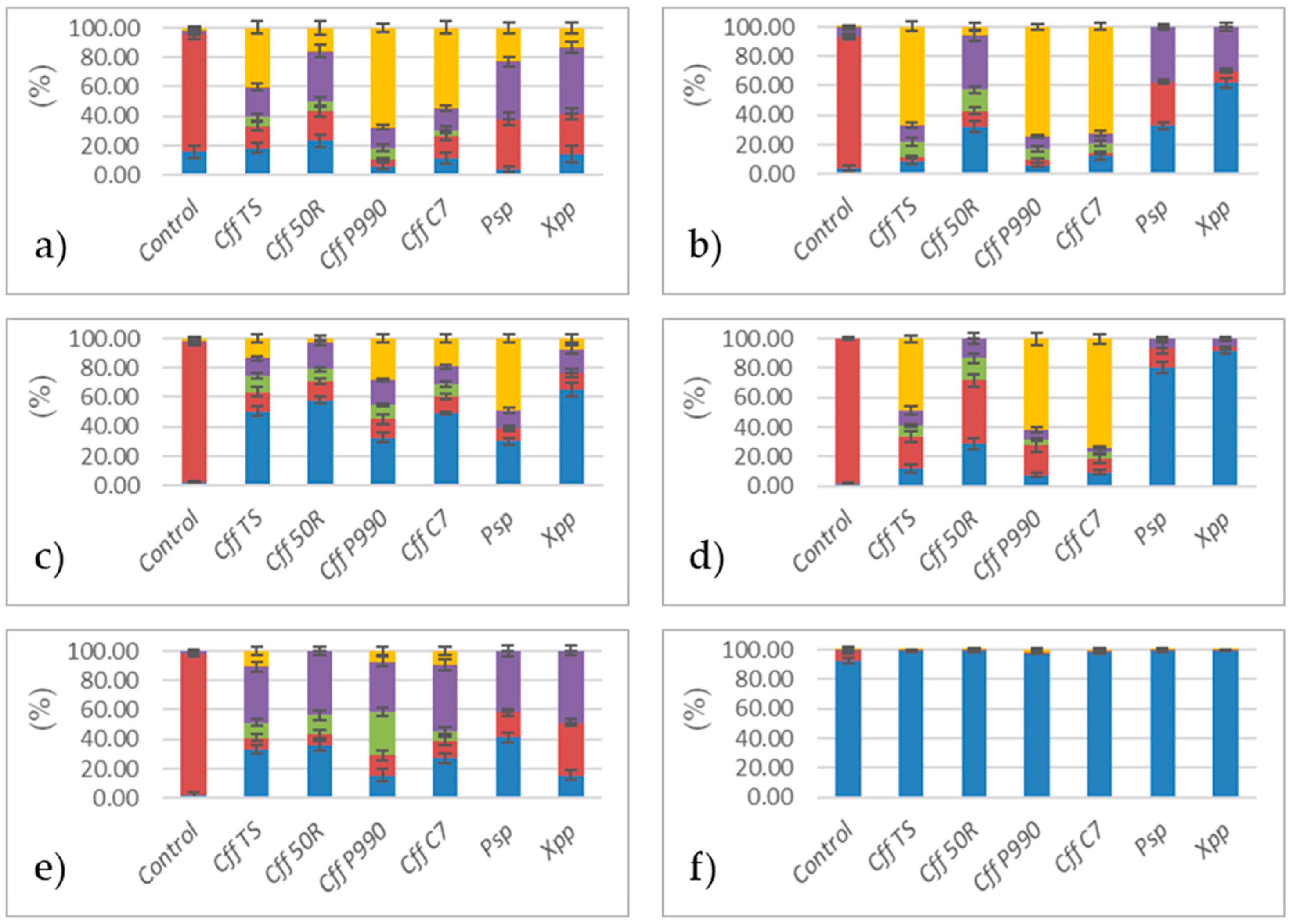
Interestingly, the VOC profiles produced by Cff strains grown on naturalized media based on Cannellino or Borlotto bean flour were significantly different from those produced by Cff grown on synthetic media (Figure 2), as well as from those produced by Psp and Xpp under the same cultural conditions (Table S2).
Intriguingly, the same VOC composition was found in Cff strains grown on naturalized media based on soybean, cowpea, pea, giant white bean, and lentil (Figure 3) flour, while a completely different VOC profile was produced by Cff when grown in a durum wheat flour-based medium; wheat is not listed among Cff plant hosts [44] (Figure 3). It is worth noting that the concentration of legume flour in the naturalized media tested did not affect the composition of the VOCs produced by Cff (Figure 4).
According to the data obtained, phenylmethanol should be considered a potential key diagnostic marker for Cff, as it can differentiate Cff from other seed-borne phytopathogenic bacteria affecting beans, such as Psp and Xpp (Figure 2 and Figure 3).

3.2. Analysis of VOCs Produced by Artificially Cff-Infected Bean Seeds

Based on the five major VOCs emitted by Cff strains in vitro, the analysis was focused on artificially Cff-infected Cannellino bean seeds. Figure 5 shows the VOCs produced by artificially Cff-infected bean seeds ten days post-inoculation. The representative Cff strains, Cff TS, Cff 50R, Cff P990, and Cff C7, were used. For comparison, bean seeds were also artificially inoculated with Psp and Xpp. As a negative control, a sterile physiological solution was used for the inoculation. The data obtained in vivo fully confirmed the specificity of the VOC profile for Cff obtained in vitro, in both symptomatic and asymptomatic seeds. No similar VOC profiles were detected in the uninoculated or mock-inoculated Cannellino bean seeds, as well as in Psp- and Xpp-infected bean seeds. The presence of Cff in the VOC positive samples was further confirmed using PCR-based tests for Cff [28,29] (Figure 5).

4. Discussion

This study highlights two major findings. Firstly, it demonstrates that Cff grown in vitro emits a distinct VOC profile that can effectively differentiate this bacterium from other legume bacterial pathogens. Notably, phenylmethanol could serve as a diagnostic marker, even when considered on its own. Additionally, 2-methoxy-4-vinylphenol is produced by all the representative Cff strains tested, except for the Cff 50R strain. This suggests that some VOCs produced by Cff strains could also be useful for mapping the evolution of these bacteria. This finding highlights the variability in metabolic pathways among different strains and such differences in VOC production could reflect genetic divergence or adaptation processes. Therefore, identifying and analyzing these variations may provide insights into the evolutionary trajectories and ecological niches of Cff strains.
Furthermore, our results showed that specific VOCs emitted in vitro by pure culture (i.e., phenylmethanol, 2-methoxy-4-vinylphenol) were also released from Cff-infected symptomatic and asymptomatic Cannellino bean seeds.
Since VOC production from microbial species may strongly depend on the culture conditions [45], our results confirm that Cff emits a similar blend of VOCs when grown in legume flour-based media and in Cff-infected Cannellino beans. Conversely, these VOCs are absent when Cff is grown on naturalized media made using flour from plants reported as non-hosts for this bacterium (i.e., durum wheat) or on synthetic media (i.e., LB, NBY, TBY). The analysis focused exclusively on those VOCs associated with the Cff–legume interaction, without considering VOCs that are constitutively and exclusively released by legumes and, thus, are not potential markers of Cff infection [46,47].
The remarkable similarity in the mass spectrum of the 2-methyl-1-butanol + 3-methyl-1-butanol compounds, as well as their similar retention indexes, did not allow an unbiased individual quantification of these two compounds, therefore the peak at 17.6 min was integrated and considered to be the sum of the two compounds. This sum is significant in comparison to the control for Cff, as well as for Xpp and Psp.
To address the practicality of VOC-based detection, it is important to recognize both its potential and limitations. The challenge in identifying VOCs as specific markers lies in the fact that, during plant–pathogen interactions, a complex combination of VOCs is generated and released by both the plant and the pathogen, as well as a consequence of their interaction. This makes it difficult to isolate VOCs that are specific to a single pathogen. As a result, a more reliable diagnostic framework would be a specific complex VOC profile, which is a broader VOC signature, that can consistently associate the presence of a pathogen with the host context, i.e., the host commodity subject to official testing.
Benzaldehyde is an aromatic flavor compound, which is responsible for the taste and odor of bitter almonds [48]. All the tested bacteria are capable of metabolizing benzaldehyde, either by degrading it or by producing it from precursor compounds, such as phenylalanine, an amino acid involved in the biosynthesis of aromatic derivatives [49]. The use of benzaldehyde as a marker for the identification of dried beans is well-documented in the literature [37].
Phenylmethanol is highlighted as a promising compound for use as a diagnostic marker for Cff, based on the results presented. Neither Xpp nor Psp produces phenylmethanol in naturalized media. As an antiseptic, it physiologically prevents secondary infections due to its bacteriostatic effects, which enhance membrane fluidity and destabilize its structural integrity [50,51]. The data supporting this compound’s diagnostic potential appears robust in regard to all naturalized media and comparatively weaker in regard to synthetic media.
In addition, 6,10-dimethyl-5,9-undecadien-2-one exhibits a broad spectrum of bioactivity, including germicidal and antimicrobial properties [52]. This terpenoid demonstrates potential in regard to legume-based naturalized media, but lacks diagnostic significance as an individual compound for Cff detection.
Moreover, 2-methoxy-4-vinylphenol is an aromatic substance used as a flavoring agent and its antimicrobial activities have been reported [53,54]. The production of this VOC by some microorganisms, such as Saccharomyces cerevisiae and Pseudomonas fluorescens, is known to involve trans-ferulic acid as a precursor [55,56]. It is interesting to note that 2-methoxy-4-vinylphenol has strain specificity within Cff. In fact, it is not substantially produced by Cff 50R, as well as by Xpp and Psp. The greatest variations occur in naturalized media, which mimic the actual host–pathogen molecular interaction.
Overall, four representative Cff isolates were tested here, and a specific VOC signature was demonstrated for bean seeds infected by Cff, as well as for Cff strains growing in vitro on legume-based media, but not for the other two bacterial seed-borne pathogens of beans. Therefore, this study provides initial evidence of a specific VOC fingerprint associated with the presence of Cff in its major hosts. However, further validation with a broader range of isolates and in diverse environmental conditions is necessary to ensure reliability across different contexts and confirm the reproducibility of this approach. Further investigations will establish the sensitivity of VOCs as biomarkers for Cff, particularly in distinguishing infected seeds in natural and artificial conditions, by comparing and combining VOC detection with other methodologies, such as molecular approaches. Ongoing experiments aim to compare VOC flux rates with symptom progression and Cff growth in planta, assessed using PCR analysis [28,29,30], to investigate the applicability of VOC profiling across different infection stages.
Additionally, exploring the integration of VOC detection with portable analytical tools, such as electronic noses, could significantly enhance the feasibility of using these biomarkers in agricultural diagnostics. Such advancements could provide rapid, non-invasive tools for monitoring asymptomatic infections directly in the field.

5. Conclusions

This is the first report in which it has been demonstrated that VOCs produced by a seed-borne quarantine plant pathogen hold considerable promise for improving the diagnostic monitoring of these plant materials, where pathogen infections are often asymptomatic and the sampling consistency for PCR-based tests is difficult to establish as statistically significant. The blend of VOCs identified here for Cff has strong potential to be exploited as a non-invasive early diagnostic marker of Cff infection, particularly in asymptomatic infected seeds, as a contribution to mitigating the spread of this harmful pathogen.
Nevertheless, further experiments are ongoing to thoroughly estimate the sensitivity of VOCs as biomarkers by comparing the flux rates of VOCs emitted from Cff-infected seeds, both naturally and artificially, with the development of the symptoms assessed using molecular methods. Future research will also focus on developing practical tools for field applications in terms of these results, contributing to the sustainability and health of leguminous crop production. This will involve the design and testing of portable diagnostic devices, such as electronic noses or miniaturized GC–MS instruments, capable of detecting and analyzing VOC emissions directly in the field. Additionally, large-scale validation trials across diverse geographic regions and environmental conditions will be essential to assess the robustness and reproducibility of VOC-based diagnostics.
Overall, these findings suggest that VOC fingerprinting could be an innovative method to assist phytosanitary services in rapidly surveying plant materials at ports of entry, by merely sampling the air within the containers where plants and seeds have been stored. This approach has the potential to revolutionize quarantine inspections by providing a non-invasive, rapid, and cost-effective preliminary diagnostic tool, thereby reducing the reliance on time-consuming and labor-intensive laboratory testing. The ability to detect asymptomatic infections without the need for destructive sampling could significantly enhance the efficiency of monitoring programs, particularly for high-risk, seed-borne pathogens like Cff. Moreover, the application of this method extends beyond quarantine scenarios, offering opportunities for its adoption in integrated pest management systems, where the early detection of pathogens is critical for preventing outbreaks and ensuring sustainable crop production.

Supplementary Materials

The following supporting information can be downloaded at: https://www.mdpi.com/article/10.3390/agronomy15020497/s1, Figure S1: Vials with naturalized Cannellino Phaseolus vulgaris medium inoculated with Cff strains for VOC analysis; Table S1. Identification of volatile organic compounds (VOCs) detected in this study. The table includes the IUPAC name, the corresponding NIST name, and the CAS Registry Number for each compound. The dual listing for 2-methyl-1-butanol/3-methyl-1-butanol reflects the structural similarity and overlapping detection of these isomers; Table S2: Percentage of single VOC out of the total sum of the most abundant VOCs measured in the headspace of vials where pathogenic bacteria were grown (values are the mean of nine replicated measurements ± percentage uncertainty).

Author Contributions

Conceptualization, D.G., S.T. and L.C.; methodology, D.G. and L.C.; software, D.G. and L.C.; validation, D.G., L.C. and S.T.; formal analysis, D.G., L.C., and S.T.; investigation, D.G., L.C. and S.T.; resources, S.T.; data curation, D.G., L.C. and S.T.; writing—original draft preparation, D.G. and S.T.; writing—review and editing, D.G., S.T. and L.C.; supervision, S.T.; project administration, S.T.; funding acquisition, S.T. All authors have read and agreed to the published version of the manuscript.

Funding

This research was funded by Ministero della Difesa, project SFINGE (ref. 2018/075-274/2019), and by Fondazione Cassa di Risparmio di Firenze, project SMART (2020/1629).

Data Availability Statement

The raw data supporting the conclusions of this article will be made available by the authors on request.

Acknowledgments

We would like to thank Ebrahim Osdaghi (University of Tehran, Iran) and Robert M. Harveson (University of Nebraska, USA) for generously providing some of their Cff isolates, in full compliance with current international and national legislation. Their support reflects the principle that scientific progress relies on the sharing of essential resources. Thanks are due to Doron Teper (Dept Plant Pathology, Agricultural Research Organization, Rishon, Israel), for the support given by such a brilliant colleague and for the stimulating discussions on Gram-positive bacterial phytopathogens.

Conflicts of Interest

The authors declare that there are no conflicts of interest.

References

  1. O’Rourke, J.A.; Bolon, Y.-T.; Bucciarelli, B.; Vance, C.P. Legume Genomics: Understanding Biology through DNA and RNA Sequencing. Ann. Bot. 2014, 113, 1107–1120. [Google Scholar] [CrossRef] [PubMed]
  2. Powers, S.E.; Thavarajah, D. Checking Agriculture’s Pulse: Field Pea (Pisum sativum L.), Sustainability, and Phosphorus Use Efficiency. Front. Plant Sci. 2019, 10, 1489. [Google Scholar] [CrossRef]
  3. FAOSTAT. Available online: https://www.fao.org/faostat/en/#data/QCL (accessed on 29 December 2024).
  4. Osdaghi, E.; Pakdaman Sardrood, B.; Bavi, M.; Akbari Oghaz, N.; Kimiaei, S.; Hadian, S. First Report of Curtobacterium flaccumfaciens pv. flaccumfaciens Causing Cowpea Bacterial Wilt in Iran. J. Phytopathol. 2015, 163, 653–656. [Google Scholar] [CrossRef]
  5. Osdaghi, E.; Young, A.J.; Harveson, R.M. Bacterial Wilt of Dry Beans Caused by Curtobacterium flaccumfaciens pv. flaccumfaciens: A New Threat from an Old Enemy. Mol. Plant Pathol. 2020, 21, 605–621. [Google Scholar] [CrossRef]
  6. EFSA Panel on Plant Health (EFSA PLH Panel); Jeger, M.; Bragard, C.; Caffier, D.; Candresse, T.; Chatzivassiliou, E.; Dehnen-Schmutz, K.; Gilioli, G.; Grégoire, J.-C.; Jaques Miret, J.A.; et al. Pest Categorisation of Curtobacterium flaccumfaciens pv. flaccumfaciens. EFSA J. 2018, 16, e05299. [Google Scholar] [CrossRef]
  7. Hedges, F. A Bacterial Wilt of the Bean Caused by Bacterium flaccumfaciens Nov. Sp. Science 1922, 55, 433–434. [Google Scholar] [CrossRef] [PubMed]
  8. Hedges, F. Bacterial Wilt of Beans (Bacterium flaccumfaciens Hedges), Including Comparisons with Bacterium phaseoli. Phytopathology 1926, 16, 1–22. [Google Scholar]
  9. Zaumeter, W.J. Comparative Pathological Histology of Three Bacterial Diseases of Bean. J. Agric. Res. 1932, 44, 605–632. [Google Scholar]
  10. Harveson, R.M.; Schwartz, H.F.; Urrea, C.A.; Yonts, C.D. Bacterial Wilt of Dry-Edible Beans in the Central High Plains of the U.S.: Past, Present, and Future. Plant Dis. 2015, 99, 1665–1677. [Google Scholar] [CrossRef] [PubMed]
  11. Huang, H.C.; Erickson, R.S.; Balasubramanian, P.M.; Hsieh, T.F.; Conner, R.L. Resurgence of Bacterial Wilt of Common Bean in North America. Can. J. Plant Pathol. 2009, 31, 290–300. [Google Scholar] [CrossRef]
  12. Schuster, M.L.; Sayre, R.M. A Coryneform Bacterium Induces Purple-Colored Seed and Leaf Hypertrophy of Phaseolus vulgaris and Other Leguminosae. Phytopathology 1967, 57, 1064–1066. [Google Scholar]
  13. Harveson, R.M. The Multicolored Bacterium. Available online: https://www.apsnet.org/edcenter/apsnetfeatures/pages/bacterium.aspx (accessed on 29 December 2024).
  14. Osdaghi, E.; Taghavi, S.M.; Hamzehzarghani, H.; Fazliarab, A.; Harveson, R.M.; Lamichhane, J.R. Occurrence and Characterization of a New Red-Pigmented Variant of Curtobacterium flaccumfaciens, the Causal Agent of Bacterial Wilt of Edible Dry Beans in Iran. Eur. J. Plant Pathol. 2016, 146, 129–145. [Google Scholar] [CrossRef]
  15. Harveson, R.M.; Schwartz, H.F.; Vidaver, A.K.; Lambrecht, P.A.; Otto, K.L. New Outbreaks of Bacterial Wilt of Dry Bean in Nebraska Observed from Field Infections. Plant Dis. 2006, 90, 681. [Google Scholar] [CrossRef]
  16. Wood, B.A.; Easdown, W.J. A New Bacterial Disease of Mung Bean and Cowpea for Australia. Australas. Plant Pathol. 1990, 19, 16–21. [Google Scholar] [CrossRef]
  17. Sammer, U.F.; Reiher, K. Curtobacterium flaccumfaciens pv. flaccumfaciens on Soybean in Germany—A Threat for Farming. J. Phytopathol. 2012, 160, 314–316. [Google Scholar] [CrossRef]
  18. Pilik, R.; Tesic, S.; Ignatov, A.N.; Tarakanov, R.I.; Dorofeeva, L.V.; Lukyanova, A.A.; Evseev, P.V.; Dzhalilov, F.S.-U.; Miroshnikov, K.A. First Report of Curtobacterium flaccumfaciens pv. flaccumfaciens Causing Bacterial Wilt and Blight on Sunflower in Russia. Plant Dis. 2023, 107, 1621. [Google Scholar] [CrossRef]
  19. Curtobacterium flaccumfaciens pv. flaccumfaciens (CORBFL) [World Distribution]|EPPO Global Database. Available online: https://gd.eppo.int/taxon/CORBFL/distribution (accessed on 29 December 2024).
  20. EPPO Global Database. Available online: https://gd.eppo.int/reporting/article-7341 (accessed on 29 December 2024).
  21. Curtobacterium flaccumfaciens pv. flaccumfaciens (CORBFL) [Netherlands]|EPPO Global Database. Available online: https://gd.eppo.int/taxon/CORBFL/distribution/NL (accessed on 29 December 2024).
  22. Curtobacterium flaccumfaciens pv. flaccumfaciens (CORBFL) [Switzerland]|EPPO Global Database. Available online: https://gd.eppo.int/taxon/CORBFL/distribution/CH (accessed on 29 December 2024).
  23. Kizheva, Y.; Pandova, M.; Dimitrova, M.; Gladicheva, Y.; Garkova, M.; Pirnareva, D.; Donchev, D.; Moncheva, P.; Hristova, P. First Report of Curtobacterium flaccumfaciens in Bulgaria. Pathogens 2024, 13, 483. [Google Scholar] [CrossRef]
  24. Implementing Regulation—2019/2072—EN—EUR-Lex. Available online: https://eur-lex.europa.eu/eli/reg_impl/2019/2072/oj/eng (accessed on 30 December 2024).
  25. Curtobacterium flaccumfaciens pv. flaccumfaciens (CORBFL) [Categorization]|EPPO Global Database. Available online: https://gd.eppo.int/taxon/CORBFL/categorization (accessed on 29 December 2024).
  26. Van Vuurde, J.W.L.; van den, B.G.W.; Birnbaum, Y. Immunofluorescence Microscopy and Enzyme-Linked Immunosorbent Assay as Potential Routine Tests for the Detection of Pseudomonas syringae phaseolicola and Xanthomonas campestris pv. phaseoli in Bean Seed. Seed Sci. Technol. 1983, 11, 547–559. [Google Scholar]
  27. Messenberg Guimaraés, P.; Palmano, S.; Smith, J.J.; Grossi de Sá, M.F.; Saddler, G.S. Development of a PCR Test for the Detection of Curtobacterium flaccumfaciens pv. flaccumfaciens. Antonie Van Leeuwenhoek 2001, 80, 1–10. [Google Scholar] [CrossRef] [PubMed]
  28. Tegli, S.; Sereni, A.; Surico, G. PCR-Based Assay for the Detection of Curtobacterium flaccumfaciens pv. flaccumfaciens in Bean Seeds. Lett. Appl. Microbiol. 2002, 35, 331–337. [Google Scholar] [CrossRef]
  29. Tegli, S.; Biancalani, C.; Ignatov, A.N.; Osdaghi, E. A Powerful LAMP Weapon against the Threat of the Quarantine Plant Pathogen Curtobacterium flaccumfaciens pv. flaccumfaciens. Microorganisms 2020, 8, 1705. [Google Scholar] [CrossRef]
  30. Tegli, S.; Gaudioso, D.; Stefanucci, D. Innovative Detection of the Quarantine Plant Pathogen Curtobacterium flaccumfaciens pv. flaccumfaciens, Causal Agent of Bacterial Wilt of Leguminous Plants. In Plant Pathology: Method and Protocols; Luchi, N., Ed.; Springer: New York, NY, USA, 2022; pp. 251–261. ISBN 978-1-07-162517-0. [Google Scholar]
  31. Cavigli, L.; Gaudioso, D.; Faraloni, C.; Agati, G.; Tegli, S. Exploiting Bacterial Pigmentation for Non-Destructive Detection of Seed-Borne Pathogens by Using Photoacoustic Techniques. Sensors 2024, 24, 7616. [Google Scholar] [CrossRef] [PubMed]
  32. Kai, M.; Haustein, M.; Molina, F.; Petri, A.; Scholz, B.; Piechulla, B. Bacterial Volatiles and Their Action Potential. Appl. Microbiol. Biotechnol. 2009, 81, 1001–1012. [Google Scholar] [CrossRef] [PubMed]
  33. Arimura, G.; Ozawa, R.; Nishioka, T.; Boland, W.; Koch, T.; Kühnemann, F.; Takabayashi, J. Herbivore-Induced Volatiles Induce the Emission of Ethylene in Neighboring Lima Bean Plants. Plant J. 2002, 29, 87–98. [Google Scholar] [CrossRef]
  34. Bouwmeester, H.; Schuurink, R.C.; Bleeker, P.M.; Schiestl, F. The Role of Volatiles in Plant Communication. Plant J. 2019, 100, 892–907. [Google Scholar] [CrossRef]
  35. Ninkovic, V.; Markovic, D.; Rensing, M. Plant Volatiles as Cues and Signals in Plant Communication. Plant Cell Environ. 2021, 44, 1030–1043. [Google Scholar] [CrossRef]
  36. Weise, T.; Kai, M.; Gummesson, A.; Troeger, A.; von Reuß, S.; Piepenborn, S.; Kosterka, F.; Sklorz, M.; Zimmermann, R.; Francke, W.; et al. Volatile Organic Compounds Produced by the Phytopathogenic Bacterium Xanthomonas campestris pv. vesicatoria 85-10. Beilstein J. Org. Chem. 2012, 8, 579–596. [Google Scholar] [CrossRef]
  37. Boots, A.W.; Smolinska, A.; van Berkel, J.J.B.N.; Fijten, R.R.R.; Stobberingh, E.E.; Boumans, M.L.L.; Moonen, E.J.; Wouters, E.F.M.; Dallinga, J.W.; Schooten, F.J.V. Identification of Microorganisms Based on Headspace Analysis of Volatile Organic Compounds by Gas Chromatography–Mass Spectrometry. J. Breath. Res. 2014, 8, 027106. [Google Scholar] [CrossRef] [PubMed]
  38. Miller, J.H. Experiments in Molecular Genetics; Cold Spring Harbor Laboratory: Cold Spring Harbor, NY, USA, 1972. [Google Scholar]
  39. Vidaver, A.K. Synthetic and Complex Media for the Rapid Detection of Fluorescence of Phytopathogenic Pseudomonads: Effect of the Carbon Source. Appl. Microbiol. 1967, 15, 1523–1524. [Google Scholar] [CrossRef]
  40. Dreiseikelmann, B.; Bunk, B.; Spröer, C.; Rohde, M.; Nimtz, M.; Wittmann, J. Characterization and Genome Comparisons of Three Achromobacter Phages of the Family Siphoviridae. Arch. Virol. 2017, 162, 2191–2201. [Google Scholar] [CrossRef]
  41. Pugliese, C.; Sirtori, F.; Škrlep, M.; Piasentier, E.; Calamai, L.; Franci, O.; Čandek-Potokar, M. The Effect of Ripening Time on the Chemical, Textural, Volatile and Sensorial Traits of Bicep Femoris and Semimembranosus Muscles of the Slovenian Dry-Cured Ham Kraški Pršut. Meat Sci. 2015, 100, 58–68. [Google Scholar] [CrossRef]
  42. Catola, S.; Kaidala Ganesha, S.D.; Calamai, L.; Loreto, F.; Ranieri, A.; Centritto, M. Headspace-Solid Phase Microextraction Approach for Dimethylsulfoniopropionate Quantification in Solanum lycopersicum Plants Subjected to Water Stress. Front. Plant Sci. 2016, 7, 1257. [Google Scholar] [CrossRef] [PubMed]
  43. Informatics, N.O. of D. and NIST Chemistry WebBook. Available online: https://webbook.nist.gov/chemistry/ (accessed on 30 December 2024).
  44. Curtobacterium flaccumfaciens pv. flaccumfaciens (CORBFL), EPPO Global Database. Available online: https://gd.eppo.int/taxon/CORBFL/hosts (accessed on 29 December 2024).
  45. Blom, D.; Fabbri, C.; Connor, E.C.; Schiestl, F.P.; Klauser, D.R.; Boller, T.; Eberl, L.; Weisskopf, L. Production of Plant Growth Modulating Volatiles Is Widespread among Rhizosphere Bacteria and Strongly Depends on Culture Conditions. Environ. Microbiol. 2011, 13, 3047–3058. [Google Scholar] [CrossRef] [PubMed]
  46. Roland, W.S.U.; Pouvreau, L.; Curran, J.; van de Velde, F.; de Kok, P.M.T. Flavor Aspects of Pulse Ingredients. Cereal Chem. 2017, 94, 58–65. [Google Scholar] [CrossRef]
  47. Rajhi, I.; Baccouri, B.; Rajhi, F.; Mhadhbi, H.; Flamini, G. Monitoring the Volatile Compounds Status of Whole Seeds and Flours of Legume Cultivars. Food Biosci. 2021, 41, 101105. [Google Scholar] [CrossRef]
  48. Scott, H.R.; Scott, L.E. Process of Treating Nut Kernels to Produce Food Ingredients 1922. U.S. Patent 1,416,128, 16 May 1922. [Google Scholar]
  49. Nierop Groot, M.N.; de Bont, J.A.M. Conversion of Phenylalanine to Benzaldehyde Initiated by an Aminotransferase in Lactobacillus plantarum. Appl. Environ. Microbiol. 1998, 64, 3009–3013. [Google Scholar] [CrossRef]
  50. Yano, T.; Miyahara, Y.; Morii, N.; Okano, T.; Kubota, H. Pentanol and Benzyl Alcohol Attack Bacterial Surface Structures Differently. Appl. Environ. Microbiol. 2016, 82, 402–408. [Google Scholar] [CrossRef]
  51. Garrido, A.; Atencio, L.A.; Bethancourt, R.; Bethancourt, A.; Guzmán, H.; Gutiérrez, M.; Durant-Archibold, A.A. Antibacterial Activity of Volatile Organic Compounds Produced by the Octocoral-Associated Bacteria Bacillus sp. BO53 and Pseudoalteromonas Sp. GA327. Antibiotics 2020, 9, 923. [Google Scholar] [CrossRef] [PubMed]
  52. Bonikowski, R.; Świtakowska, P.; Kula, J. Synthesis, Odour Evaluation and Antimicrobial Activity of Some Geranyl Acetone and Nerolidol Analogues. Flavour Fragr. J. 2015, 30, 238–244. [Google Scholar] [CrossRef]
  53. Rubab, M.; Chelliah, R.; Saravanakumar, K.; Barathikannan, K.; Wei, S.; Kim, J.-R.; Yoo, D.; Wang, M.-H.; Oh, D.-H. Bioactive Potential of 2-Methoxy-4-Vinylphenol and Benzofuran from Brassica oleracea L. Var. Capitate f, Rubra (Red Cabbage) on Oxidative and Microbiological Stability of Beef Meat. Foods 2020, 9, 568. [Google Scholar] [CrossRef]
  54. Lone, A.S.; Ravindran, K.C.; Jeandet, P. Evaluation of Antimicrobial Activity and Bioactive Compound Analysis of Verbascum thapsus L. A Folklore Medicinal Plant. Phytomed. Plus 2024, 4, 100560. [Google Scholar] [CrossRef]
  55. Huang, Z.; Dostal, L.; Rosazza, J.P. Microbial Transformations of Ferulic Acid by Saccharomyces cerevisiae and Pseudomonas fluorescens. Appl. Environ. Microbiol. 1993, 59, 2244–2250. [Google Scholar] [CrossRef]
  56. Mishra, S.; Sachan, A.; Vidyarthi, A.S.; Sachan, S.G. Transformation of Ferulic Acid to 4-Vinyl Guaiacol as a Major Metabolite: A Microbial Approach. Rev. Environ. Sci. Biotechnol. 2014, 13, 377–385. [Google Scholar] [CrossRef]
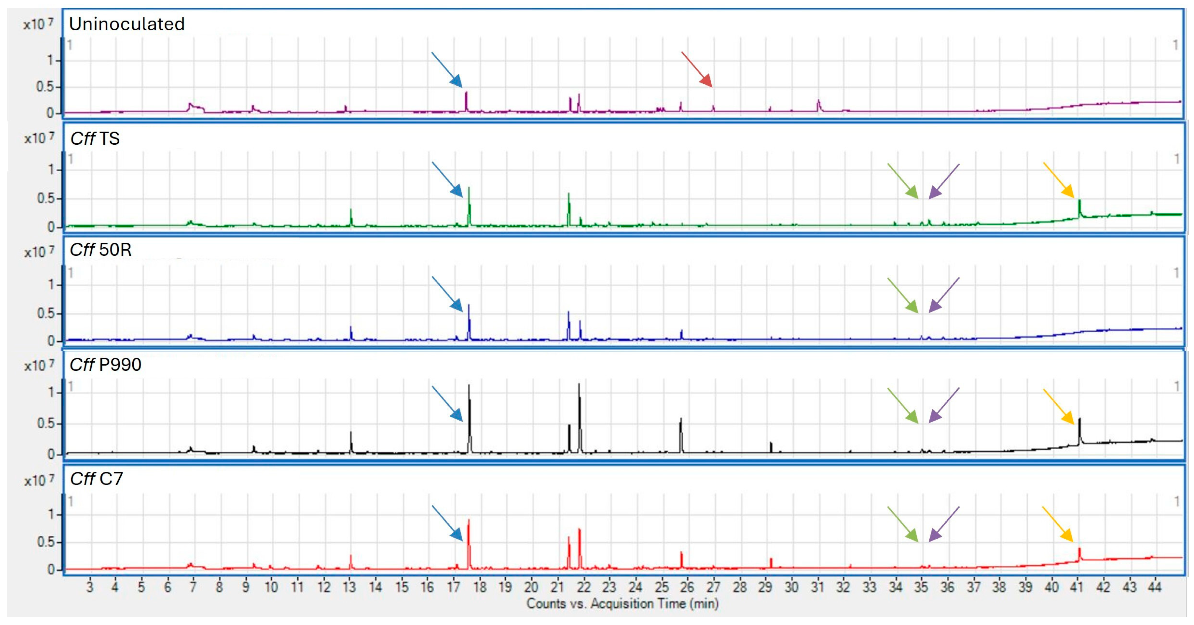
Figure 1. Chromatograms representing the VOCs emitted in the headspace of the vials containing a Cannellino naturalized medium uninoculated or inoculated with Cff TS, Cff 50R, Cff P990, Cff C7. Arrows point to the most significant VOCs, in color code mode: (blue) 2-methyl-1-butanol + 3-methyl-1-butanol; (red) benzaldehyde; (green) phenylmethanol; (violet) 6,10-dimethyl-5,9-undecadien-2-one; (orange) 2-methoxy-4-vinylphenol.
Agronomy 15 00497 g001
Figure 2. Percentage of single VOC out of the total sum of the most abundant VOCs produced in vitro by Cff strains (TS, 50R, P990, C7), Psp and Xpp, in (a) P. vulgaris Cannellino and (b) Borlotto flour naturalized media, and in (c) LB, (d) NBY, (e) TBY synthetic media. Compounds: (blue) 2-methyl-1-butanol + 3-methyl-1-butanol; (red) benzaldehyde; (green) phenylmethanol; (violet) 6,10-dimethyl-5,9-undecadien-2-one; (orange) 2-methoxy-4-vinylphenol. Values are the mean of nine replicates ± percentage uncertainty.
Agronomy 15 00497 g002
Figure 3. Percentage of single VOC out of the total sum of the most abundant VOCs produced in vitro by Cff strains (TS, 50R, P990, C7), Psp and Xpp, on naturalized media made with flours of (a) G. max; (b) V. unguiculata; (c) P. sativum; (d) P. coccineus; (e) L. culinaris; (f) T. durum. Compounds: (blue) 2-methyl-1-butanol + 3-methyl-1-butanol; (red) benzaldehyde; (green) phenylmethanol; (violet) 6,10-dimethyl-5,9-undecadien-2-one; (orange) 2-methoxy-4-vinylphenol. Values are the mean of nine replicates ± percentage uncertainty.
Agronomy 15 00497 g003
Figure 4. Percentage of single VOC out of the total sum of the most abundant VOCs produced in vitro by Cff strains (TS, 50R, P990, C7), Psp and Xpp, in a P. vulgaris Cannellino flour naturalized medium at different concentrations: (a) 9 g/L, (b) 18 g/L, (c) 36 g/L. Compounds: (blue) 2-methyl-1-butanol + 3-methyl-1-butanol; (red) benzaldehyde; (green) phenylmethanol; (violet) 6,10-dimethyl-5,9-undecadien-2-one; (orange) 2-methoxy-4-vinylphenol. Values are the mean of nine replicates ± percentage uncertainty.
Agronomy 15 00497 g004
Figure 5. VOCs produced by Cannellino bean seeds artificially infected with Cff, Psp, and Xpp. (a) GC–MS vials with Cannellino bean seeds (1) mock-inoculated, or inoculated with (2) Cff TS, (3) Cff 50R. (b) Specific PCR test [28] on Cannellino bean seeds infected with Cff TS and Cff P990 (lanes 2-4 and 5-7, respectively), C+ positive control Cff pure DNA (20 ng/reaction), (C-) negative control, Marker GeneRuler 100 bp DNA Ladder (Thermo Fisher Scientific, Waltham, MA, USA). (c) Percentage of single VOC out of the total sum of the most abundant VOCs produced by Cannellino bean seeds artificially infected with Cff strains (TS, 50R, P990, C7), Psp and Xpp. Compounds: (blue) 2-methyl-1-butanol + 3-methyl-1-butanol; (red) benzaldehyde; (green) phenylmethanol; (violet) 6,10-dimethyl-5,9-undecadien-2-one; (orange) 2-methoxy-4-vinylphenol. Values are the mean of nine replicates ± percentage uncertainty.
Agronomy 15 00497 g005
Table 1. Bacterial plant pathogens of legumes used in this study and their main features.
StrainInternational
Code 1
Colony
Color and Type
OriginYear
HostCountry
Cff TSICMP 2584
CFBP 3418
yellow–fluidalPhaseolus vulgarisHungary1957
Cff 50RICMP 22071
CFBP 8819
red–fluidalPhaseolus vulgarisIran2014
Cff P990ICMP 22053
CFBP 8820
yellow–fluidalCapsicum annumIran2015
Cff C7J24orange–fluidalPhaseolus vulgarisUSA2004
PspIPV-BO 2325creamy darkPhaseolus vulgarisItalynot available
Xpp95-61yellow–fluidalPhaseolus vulgarisUSAnot available
1 Abbreviations: ICMP, International Collection of Microorganisms from Plants; CFBP, Collection Française de Bactéries Phytopathogènes; J24 and 95-61, Dept. Plant Pathology, University of Nebraska, USA; IPV-BO, ex-Istituto per la Protezione delle Piante, University of Bologna, Italy.
Table 2. Legume- and wheat-based flours used in this study.
Plant GrainsVarietyOriginLatitudeLongitude
Phaseolus vulgarisCannellinoArgentina−31.4200−64.1887
Phaseolus vulgarisBorlottoArgentina−31.6406−60.6917
Glycine maxSandokanItaly44.727111.2892
Vigna unguiculataOcchio del ValdarnoItaly43.516611.5666
Pisum sativumPrimaveraItaly41.505515.3391
Phaseolus coccineusSpagna BiancoItaly40.915114.7954
Lens culinarisDelle CreteItaly43.335111.3158
Triticum durumSenatore CappelliItaly43.467611.0434
Disclaimer/Publisher’s Note: The statements, opinions and data contained in all publications are solely those of the individual author(s) and contributor(s) and not of MDPI and/or the editor(s). MDPI and/or the editor(s) disclaim responsibility for any injury to people or property resulting from any ideas, methods, instructions or products referred to in the content.