Article

High-Throughput Sequence Analysis of Microbial Communities of Soybean in Northeast China

1
College of Bioscience and Biotechnology, Shenyang Agricultural University, Shenyang 110866, China
2
Nematology Institute of Northern China, Shenyang Agricultural University, Shenyang 110866, China
*
Author to whom correspondence should be addressed.
Agronomy 2025, 15(2), 436; https://doi.org/10.3390/agronomy15020436
Submission received: 24 December 2024 / Revised: 8 February 2025 / Accepted: 9 February 2025 / Published: 10 February 2025
(This article belongs to the Special Issue Recent Advances in Legume Crop Protection)

Abstract

:
Soybean, an essential oil crop in China, has witnessed accelerated seed transfer domestically and abroad in recent years. Seed carriage has emerged as a major route for the dissemination of soybean diseases. In this study, 14 soybean cultivars from three northeastern provinces were collected and examined for seed-borne microorganisms using traditional detection technology and high-throughput sequencing technology. Through traditional detection techniques, a total of six genera of bacteria and seventeen genera of fungi were isolated from the test varieties. The quantity and types of microorganisms on the seed surface were greater than those on the seed coat and within the seed, while the seed coat and internal seed contained fewer microorganisms. The dominant fungal genera were Cladosporium, Fusarium, Aspergillus, and Alternaria, accounting for 21.23%, 17.45%, 15.57%, and 11.56% of the genera, respectively. The dominant bacterial genera were Pseudomonas, Sphingomonas, and Pantoea, accounting for 37.46%, 17.29%, and 15.27% of the genera, respectively. The dominant genera obtained through traditional seed-carrying assay techniques were also dominant in high-throughput sequencing. However, some dominant genera obtained through high-throughput sequencing were not isolated by traditional methods. High-throughput sequencing analysis revealed that soybean seeds from Jilin Province had the highest abundance of seed-borne fungi, followed by seeds from Liaoning Province and Heilongjiang Province. Jilin Province also had the highest abundance of seed-borne bacteria, followed by Heilongjiang Province and Liaoning Province. The isolation and identification of microorganisms on soybean seeds provide a scientific basis for seed quarantine treatment and disease control, which is of great significance for soybean production in China.

1. Introduction

Soybean (Glycine max (L.) Merr), a significant grain and oil crop globally, supplies humans with high-quality plant protein and oil, playing a crucial role in ensuring food production safety [1]. As an agricultural production means, high-quality seeds directly impact production [2]. Seeds are associated with diverse microorganisms during their development, harvesting, and storage stages [3]. Some of these microorganisms can cause seed discoloration and decay, and some can adhere to the seed surface or invade seed tissues, facilitating the spread of diseases across growing seasons and locations, having a significant negative impact on germination, seedling growth, and adult plant health [4,5].
The traditional identification of fungi in seed-carrying detection mainly classifies fungi based on their morphological, growth, physiological, and biochemical characteristics. However, numerous fungal species may not be easily detected, and their development, physiological, and biochemical characteristics are susceptible to environmental changes [6]. Due to the limitations of the culture medium used for isolation, it is difficult to isolate all types of bacteria, and identification is also challenging [7]. Consequently, it is challenging to fully identify seed-borne bacteria using traditional methods [8]. With the advancement of molecular biology technology, nucleic acid sequence analysis has become prevalent in microbial classification and identification. Currently, commonly used techniques include 18S rDNA, Internal Transcribed Spacer (ITS), and 16S rDNA [9,10]. 18S rDNA and ITS are suitable for fungal identification, and 16S rDNA can reveal the characteristic nucleic acid sequences of biological species and is considered the most suitable indicator for bacterial phylogeny and classification identification [11]. In recent years, powerful databases and user-friendly software for high-throughput sequencing (NGS) platforms (including SOLiD, Illumina, and 454 sequencing) have contributed to a deeper and more comprehensive analysis of complex microbial communities [12]. To date, DNA-NGS technology has been successfully applied to the analysis of microbial communities in various samples, such as soil, air, water, intestines, leaves, roots, and fruits, and is widely used in agricultural research on plant rhizosphere microorganisms [13]. The plants involved include Arabidopsis thaliana, rice, potato, tobacco, soybean, etc. [14]. However, few studies have been conducted to understand the changes in microbial communities in seeds.
This experiment is based on the use of high-throughput sequencing technology and traditional seed-carrying detection technology to study the diversity of microbial communities on soybean seeds and aims to clarify the seed-carrying situation in different regions and provide references for soybean seed production, safe transportation, and disease control.

2. Materials and Methods

2.1. Soybean Cultivars

A total of 14 test soybean seed varieties collected from different locations were provided by the Comprehensive Soybean Test Station in Northeast China in Nov 2023. Detailed information about the soybean seeds is presented in Table 1. All samples were sealed in Ziplock bags and stored under cold storage at 4 °C.

2.2. Traditional Detection of Seed Carrier Microbes

In the present study, seed carrier microbes included fungi and bacteria on the seed surface, seed coat, and seed interior.
Isolation of seed surface microbes: Twenty grains of different soybean varieties were randomly selected. The grains were soaked in 20 mL of sterile distilled water for 30 min and centrifuged at 12,000 rpm for 10 min. A 100 μL volume of liquid from the bottom of the centrifuge tube was transferred and spread uniformly on a Petri dish containing potato glucose AGAR (PDA) and beef extract peptone AGAR (NA); each treatment was repeated 4 times. Sterile distilled water was used as a control under the same conditions. The Petri dishes were incubated at 28 °C.
Isolation of seed coat and interior microbes: The seed coat and seed embryo were separated and soaked in 0.5% NaClO for 10 min, washed 3 times with sterile distilled water, and then air-dried [15]. The dried seed coat and seed embryo were placed on PDA and NA media, with 5 grains or 5 tissue blocks per dish, and each treatment was repeated 4 times [16]. The Petri dishes were incubated at 28 °C.

2.3. Identification of Fungi

After culturing for 3–5 d, the colonies were isolated according to morphology, color, and other apparent characteristics and then cultured at 28 °C until most of the colonies produced spores. Single spores were isolated from the colonies and cultured for final identification. Isolates were identified according to their phenotypic characteristics, including the culture-based characteristics of fungi. Fungal DNA isolation was carried out using the TIANamp Genomic DNA Kit (TIANGEN Biotech Beijing Co. Ltd., Beijing, China). Fungal rDNA ITS fragments were sequenced and compared to assist with identification [17]. The fungal ITS universal primer pairs ITS1 (5′-TCCGTAGGTGAACCTGCGG-3′) and ITS4 (5′-TCCTCCGCTTATTGATATGC-3′) were utilized [18].

2.4. Identification of Bacteria

After culturing for 2–3 days, morphologically unique bacterial colonies were selected from each plate, streaked on fresh plates for purification, and then cultured in NA medium for identification. Physiological and biochemical tests were performed to identify bacteria, according to Gerhardt and Murray [19]. Isolates were further identified via phylogenetic analysis of 16S rRNA gene sequences. To extract genomic DNA, standard techniques were used [10,20,21]. The 16S rRNA sequences were amplified with the primer pair 27F (5′-AGAGTTTGATCATGGCTCAG-3′) and 1492R (5′-ACGGTTACCTTGTTACGACTT-3′) [22,23]. The PCR products were inserted into the PGEM-T vector (Promega, Madison, WI, USA). The plasmid was extracted and sequenced using the Genewiz Biotechnology Co., Ltd. (Suzhou, China). The 16S rRNA gene sequence was identified using the GenBank database.

2.5. Microbial DNA Extraction and Polymerase Chain Reaction

Seeds from 14 different soybean varieties were soaked in a 5% NaClO solution for 5 min and then rinsed with autoclaved distilled water after the bleach was drained. Once seed surface sterility was confirmed, 150 seeds were ground gently in an autoclaved mortar using 0.5 mL of 50 mM Na2HPO4 buffer per gram of dry seed weight [24]. DNeasy PowerSoil DNA Kit (Qiagen, Germany) was utilized to extract total DNA from the ground seed suspension (100 μL). PCR was performed using the specific primers 341F (5′-CCTAYGGGRBGCASCA) and 806R (5′-GGACTACNNGGGTATCTAAT) and ITS5-1737 (5′-GGAAGTAAAAGTCGTAACAAGG-3′) and /ITS2-2043 (5′-GCTGCGTTCTTCAT CGATGC-3′) to amplify the 16S rRNA and ITS genes [25]. Amplification was carried out using the following program: an initial denaturation at 98 °C for 1 min, followed by 30 cycles of amplification at 98 °C for 10 s, 50 °C for 30 s, and 72 °C for 30 s, and a final extension at 72 °C for 5 min. Amplicons were gel-purified using the gel recovery kit (Gene JET, Thermo Scientific, Waltham, MA, USA) and sequenced using Ion S5TMXL at the Beijing Novo Gene Technology Co., Ltd., Beijing, China.

2.6. Bioinformatics Analysis

Bacterial species annotation was conducted using the Mothur method and the SSUrRNA database [26] of SILVA132 (with a set threshold of 0.8–1) [27]. Species annotation analysis of fungi was performed using the blast method in Qiime software [28] and the Unit (Version 7.2) database [29] for fungi, which were analyzed at each taxonomic level. The MUSCLE software (Version 3.8.31) was used for fast multi-sequence alignment to obtain the phylogenetic relationships of all OTU sequences [30]. Finally, data for each sample were homogenized.
The OTU abundance information was normalized using the sequence number. The alpha diversity was subsequently analyzed using the normalized data.
Alpha diversity was employed to analyze species diversity in a sample using five indices: observed species, Chao1, Shannon, Simpson, and ACE. All of these indices were calculated with QIIME (Version 1.7.0).
A Petal map was created according to the OTU analysis results to visually display the species distinctions of the microbes in the sample. The top 10 species with the highest abundance at each classification level were selected based on the species annotation results, and a cumulative columnar plot of relative species abundance was generated. Rarefaction and rank abundance curves were generated, and NMDS Analysis was conducted using R software (Version 3.2.2).

3. Results

3.1. Analysis of the Diversity of Microorganisms Carried by Soybean Seeds Using Traditional Detection Technique

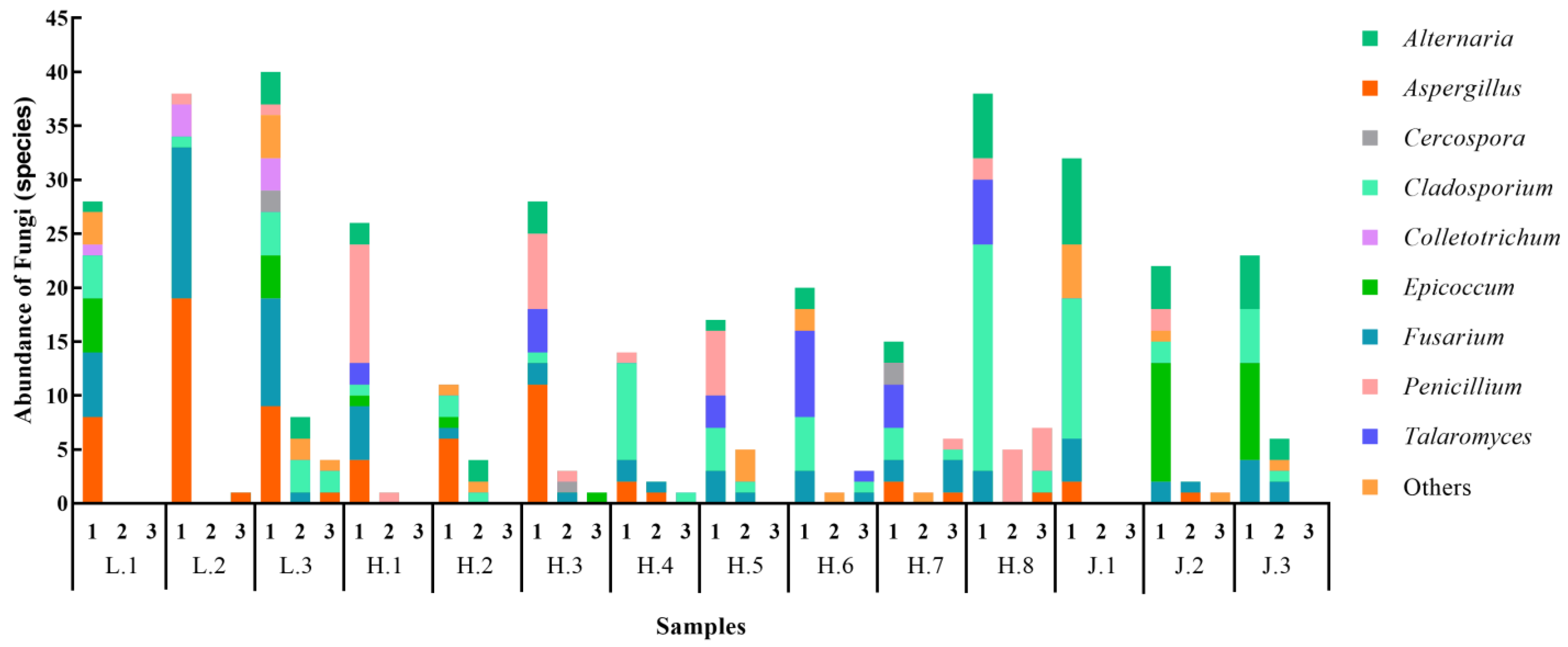
3.1.1. Fungal Diversity on Soybean Seeds

A total of 17 genera of fungi were isolated from the tested soybean seeds. More fungal species were carried on the surface of the seeds than in the interior of the seeds and on the seed coat. The L.3 sample carried the highest number of fungi, and the H.2 sample carried the least number of fungi (Figure 1). The number of fungi carried by seeds in Heilongjiang Province was relatively lower.

3.1.2. Bacterial Diversity on Soybean Seeds

A total of six genera of bacteria were identified in soybean seeds; the dominant genera were Pseudomonas, Sphingomonas, Pantoea, and Bacillus, with frequencies of 37.46%, 17.29%, 15.28%, and 7.49%, respectively. The distribution of bacteria on soybean seeds was basically the same as that of fungi. The number of bacteria on the seed surface was higher than on the seed coat and in the seed embryo. Soybean seeds from Jilin had a greater diversity of bacteria (Figure 2).

3.2. Microbial Diversity of Soybean Seeds Based on High-Throughput Sequencing Technology

3.2.1. Analysis of Fungi Using ITS Sequencing

The Alpha indices and ITS sequencing information for the 14 soybean seed varieties are shown in Table 2. A total of 1,179,748 original data were obtained from the ITS region. The average number of original data from soybean seeds sampled from Liaoning, Jilin, and Heilongjiang Provinces were 83,088, 84,972, and 84,446, respectively. Analyzing the relationship between Operational Taxonomic Units (OTUs) obtained from different samples and represented using the OTU Flower figure (Figure 3A) showed that the number of OTUs shared by the 14 soybean seed samples was 140.

3.2.2. Analysis of Bacteria Based on 16S rDNA Sequences

Alpha indices based on 16S rDNA sequencing of the 14 soybean seed samples are detailed in Table 3. A total of 1,166,335 original data were obtained from the 16S rDNA region. The average number of original data for soybean seed samples from Liaoning, Jilin, and Heilongjiang Provinces were 83,732, 87,194, and 81,645, respectively. The results from the OTU flower figure (Figure 3B) showed that 160 OTUs were shared by the 14 soybean seed samples. Sample H.7 had the highest number of unique OTUs (34), while samples L.1 and H.6 had the lowest number of unique OTUs (2 each).

3.3. Alpha Diversity of Microorganisms Carried by Soybean Seeds

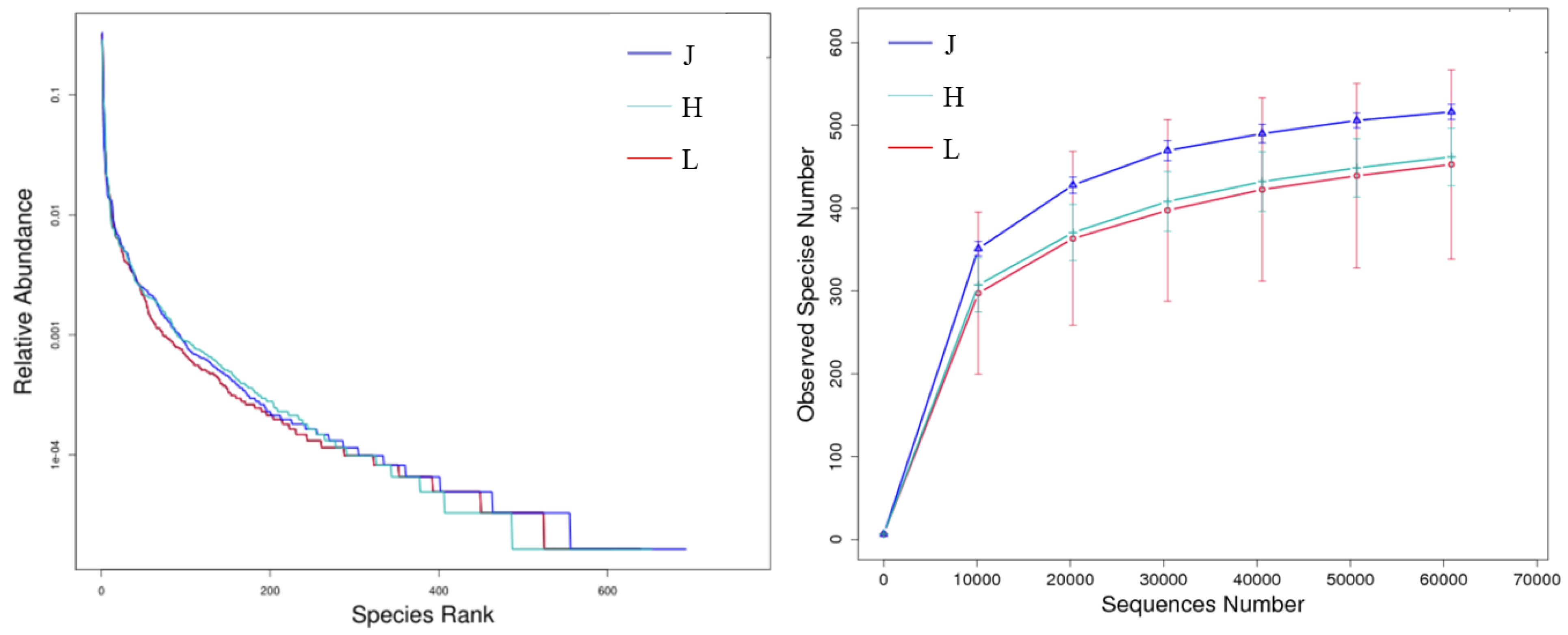
3.3.1. Alpha Diversity of Fungi

To analyze the richness of the fungal community structure of the 14 soybean seeds from northeast China, the microbial diversity index was analyzed at a 97% similarity threshold using the chao1 index, observed species index, Shannon index, ACE index, and Simpson index (Table 2). Among the 14 soybean seed samples, sample L.2 had the largest Chao1 and Shannon index values and contained the most observed species, indicating a more abundant fungal community. Conversely, sample L.3 had the smallest Chao1 and Shannon index values and contained the fewest observed species, suggesting a relatively harmonious fungal community.
Rarefaction and rank abundance curves were also employed for alpha diversity analysis. The rank abundance curve (Figure 4) indicated a more even distribution and higher abundance of fungi associated with soybean seeds from Jilin Province, followed by Liaoning Province and Heilongjiang Province. In the rarefaction curve in Figure 4, the number of observed OTUs in soybean samples from Jilin Province was more than 500, while Liaoning Province had the lowest OTU count, at 400.

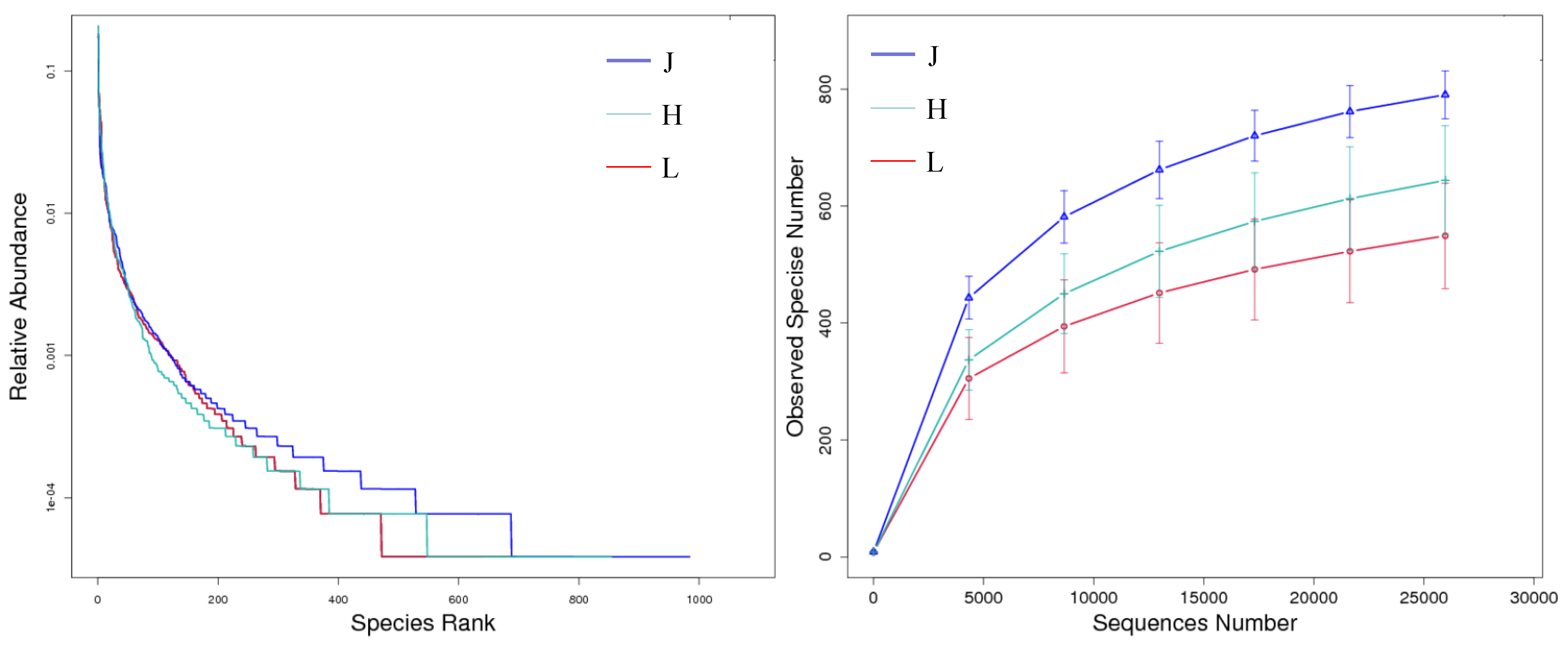
3.3.2. Alpha Diversity of Bacteria

Among the 14 soybean seed samples, samples J.2 and J.3 had relatively large Chao1 and Shannon index values (Table 3) and contained more observed species, suggesting a more abundant bacterial community. From the rank abundance curve, it can be seen that the distribution and abundance of bacteria in the three provinces were essentially the same as those of fungi (Figure 5). The number of observed OTUs in soybean samples from Jilin Province was about 800, while the number of OTUs in samples from Liaoning Province was about 500. Overall, soybean seeds from Jilin Province were more abundant in bacteria, followed by seeds from Heilongjiang Province and Liaoning Province.

3.4. Characteristics of Microbial Communities Carried by Soybean Seeds

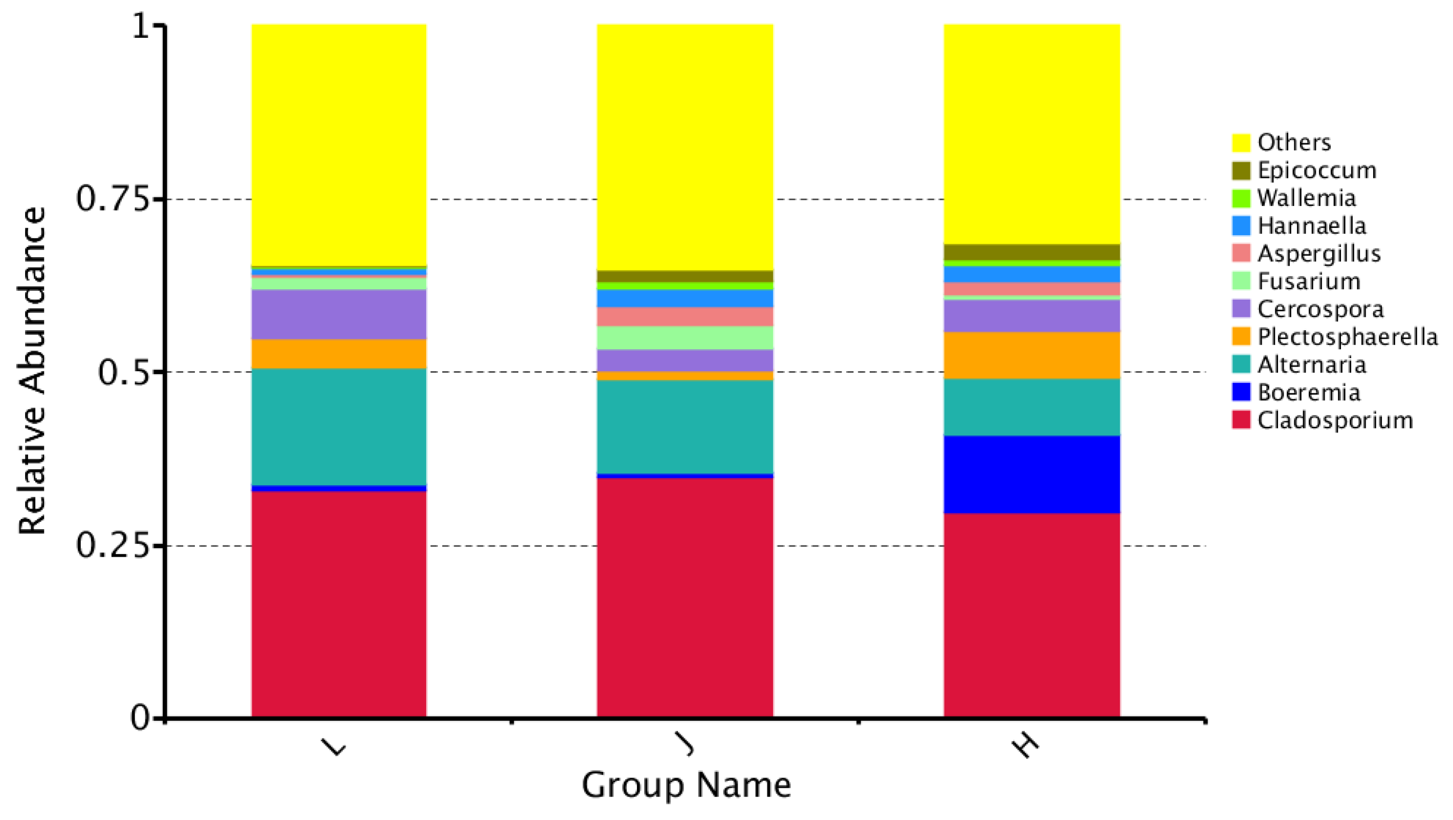
3.4.1. Characteristics of Fungal Communities

A total of 185 species of fungi were detected and identified in soybean seed samples from northeast China. The distribution and proportions of the 10 dominant genera with maximum abundance at each taxonomic level are shown in Figure 6. Cladosporium and Boeremia had the greatest relative abundances among the top ten genera in this study.

3.4.2. Characteristics of Bacterial Communities

A total of 273 species of bacteria were detected and identified in soybean seed samples from northeast China. The distribution and proportions of the top 10 dominant genera with maximum abundance are shown in Figure 7. Methylobacterium and Sphingomonas had the greatest relative abundances among the top ten genera in this study.

3.5. Changes in Related Microbial Communities Carried by Soybean Seeds in Northeast China

The results of Non-Metric Multi-Dimensional Scaling (NMDS) showed that there were inter-group and intra-group differences between microbial species associated with soybean seeds from Liaoning, Jilin, and Heilongjiang Provinces (Figure 8).

3.5.1. Changes in Fungal Communities

Analysis of similarities (ANOSIM) was used to further detect changes in fungal composition. The statistical analysis score for Liaoning and Jilin samples was r = 0.07407, p = 0.4 (p < 0.05, significant level). At the genus level (Figure 9), there were significant differences in Penicillium and Hannaella between the Jilin and Liaoning groups, in Plectosphaerella between Heilongjiang and Jilin groups, and in Epicoccum and Symmetrospora between Heilongjiang and Liaoning groups.

3.5.2. Changes in Related Bacterial Communities

The statistical analysis score for the Liaoning and Jilin samples was r = 0, p = 0.5 (p < 0.05, significant level). However, at the genus level (Figure 10), there were significant differences in Roseomonas, Nakamurella, and Segetibacter between the Heilongjiang and Jilin groups and in Spirosoma and Kineococcus between the Heilongjiang and Liaoning groups.

4. Discussion

Plants are colonized by numerous microorganisms, forming intricate plant microbial communities [31]. Plant-associated microorganisms, mainly comprising bacteria and fungi, have the potential to influence the primary functions of the host [32]. A wide variety of microorganisms directly or indirectly affect seeds during their development, harvesting, and storage [33]. Some microbes can induce seed coloration and decomposition, while others can adhere to the seed surface or penetrate internal tissues, facilitating the transmission of diseases across consecutive growing seasons and different locations [4,34]. Therefore, it is imperative to investigate the diversity of the microbial composition of soybean seeds to clarify the soybean seed-carrying situation in different regions and provide references for soybean seed production, safe transportation, and disease control. In the present study, traditional seed bacteria detection technology and high-throughput sequencing technology were compared to analyze bacteria on soybean seeds in northeast China.
Comparison of two isolation methods: Traditional fungal isolation and identification revealed that dominant genera such as Cladosporium (21.23%), Fusarium (17.45%), Aspergillus (15.57%), and Alternaria (11.56%) were also dominant in high-throughput sequencing identification, accounting for 31.40%, 1.25%, 1.61%, and 11.15% of the fungi, respectively. Other dominant genera, Cercospora (1.42%) and Epicoccum (1.89%), accounted for 4.77% and 1.62% of the fungi, respectively, in high-throughput sequencing. Boeremia (4.20%) and Hannaella (1.23%) were not found in traditional fungal isolates. The dominant genera identified through traditional bacterial isolation, namely Pseudomonas (37.46%), Sphingomonas (17.29%), and Pantoea (15.27%), accounted for 3.06%, 9.97%, and 2.81% of the bacteria, respectively, in high-throughput sequencing identification. Other dominant genera identified by high-throughput sequencing were not found in traditional bacterial isolation. Different separation methods lead to different separation ratios.
The analysis of microbial communities by high-throughput sequencing technology showed that there were differences in the composition of microbial communities between groups and within groups. In pairwise comparisons of three regions, there was no significant difference in community structure between groups; however, at the genus level, the differences between some genera between the groups were significant or even extremely significant. The results indicate that there were significant differences in soybean seed bacteria carriage in northeast China.
The detection of seed diseases is a crucial part of crop disease control and prevention throughout the introduction, reproduction, and acquisition stages [35]. Seed health testing is used to assess and determine the specific varieties and quantities of pathogens present in the seeds [36]. The quality of seeds has a significant impact on the full realization of their yield and crop value potential [37]. The traditional seed carrier detection technology is suitable for detecting the carrier site and each part of the seed and is suitable for most seed carrier detections. This method can be used for the study of seed health, species-borne diseases, and target pathogens, and it can identify and preserve all isolated microorganisms [38]. However, one drawback is that the cultivated colonies may exhibit interconnectivity or even overlap, thus posing challenges in terms of their separation. Identifying certain isolated bacteria might be challenging due to the susceptibility of a small number of slow-growing bacteria to competition and the influence of dominant bacteria. Moreover, these bacteria may exhibit minimal or invisible colony growth [39]. During this study, due to the limitations of the medium, it was not possible to detect all microorganisms in the tested seed samples. High-throughput sequencing technology offers the advantage of directly obtaining genomic DNA from the sample, which contains both cultured and non-cultured microbial genes. The absence of separation significantly reduces expenses. Using a large amount of data, it is possible to obtain the entire sample’s microbiological information in order to streamline the taxonomic examination of microorganisms. Without a strain, we cannot perform further tests; hence, in the context of seed bacterial identification, it is possible to employ both of these detection approaches concurrently, thereby ensuring that the test outcomes are complementary.

5. Conclusions

Whether the dominant genera of Fusarium, Alternaria, Urospora, and Pseudomonas can cause soybean diseases still needs to be verified using Koch’s rule. In this experiment, traditional seed bacteria detection technology and high-throughput sequencing technology were combined to detect the bacteria-carrying status and health status of soybean seeds in northeast China to provide a theoretical basis for safe transportation and disease control in soybean seed production areas and to achieve accurate inspection purposes.

Author Contributions

Conceptualization, Y.W., L.C. and Y.D.; methodology, F.M. and W.D.; software, Q.B. and X.Z.; validation, F.M. and Y.W.; formal analysis, F.M.; investigation, W.D. and Q.B.; resources, Y.W., Q.B. and H.F.; data curation, W.D. and Y.D.; writing—original draft preparation, Y.W. and H.F.; writing—review and editing, L.C.; supervision, Y.D.; project administration, L.C.; funding acquisition, Y.W. and X.Z. All authors have read and agreed to the published version of the manuscript.

Funding

This work was supported by the National Key R&D Program of China (2023YFD1400400) and the Central Guidance for Local Scientific and Technological Development Funds (2024ZY0018).

Data Availability Statement

The original contributions presented in the study are included in the article; further inquiries can be directed to the corresponding author.

Conflicts of Interest

The authors declare no conflicts of interest.

References

  1. Colletti, A.; Attrovio, A.; Boffa, L.; Mantegna, S.; Cravotto, G. Valorisation of by-products from soybean (Glycine max (L.) Merr.) processing. Molecules 2020, 25, 2129. [Google Scholar] [CrossRef]
  2. Kockelmann, A.; Tilcher, R.; Fischer, U. Seed production and processing. SugarTech 2010, 12, 267–275. [Google Scholar] [CrossRef]
  3. Martín, I.; Gálvez, L.; Guasch, L.; Palmero, D. Fungal Pathogens and Seed Storage in the Dry State. Plants 2022, 11, 3167. [Google Scholar] [CrossRef] [PubMed]
  4. Barret, M.; Guimbaud, J.F.; Darrasse, A.; Jacques, M.A. Plant microbiota affects seed transmission of phytopathogenic microorganisms. Mol. Plant Pathol. 2016, 17, 791–795. [Google Scholar] [CrossRef] [PubMed]
  5. Javaid, M.M.; Mahmood, A.; Alshaya, D.S.; AlKahtani, M.D.F.; Waheed, H.; Wasaya, A.; Khan, S.A.; Naqve, M.; Haider, I.; Shahid, M.A.; et al. Influence of environmental factors on seed germination and seedling characteristics of perennial ryegrass (Lolium perenne L.). Sci. Rep. 2022, 12, 9522. [Google Scholar] [CrossRef] [PubMed]
  6. Long, R.L.; Gorecki, M.J.; Renton, M.; Scott, J.K.; Colville, L.; Goggin, D.E.; Commander, L.E.; Westcott, D.A.; Cherry, H.; Finch-Savage, W.E. The ecophysiology of seed persistence: A mechanistic view of the journey to germination or demise. Biol. Rev. 2015, 90, 31–59. [Google Scholar] [CrossRef] [PubMed]
  7. Gitaitis, R.; Walcott, R. The epidemiology and management of seedborne bacterial diseases. Annu. Rev. Phytopathol. 2007, 45, 371–397. [Google Scholar] [CrossRef] [PubMed]
  8. Berg, T.; Tesoriero, L.; Hailstones, D. PCR-based detection of Xanthomonas campestris pathovars in Brassica seed. Plant Pathol. 2005, 54, 416–427. [Google Scholar] [CrossRef]
  9. Franco-Duarte, R.; Černáková, L.; Kadam, S.; Kaushik, K.S.; Salehi, B.; Bevilacqua, A.; Corbo, M.R.; Antolak, H.; Dybka-Stępień, K.; Leszczewicz, M.; et al. Advances in chemical and biological methods to identify microorganisms—From past to present. Microorganisms 2019, 7, 130. [Google Scholar] [CrossRef] [PubMed]
  10. Simonin, M.; Briand, M.; Chesneau, G.; Rochefort, A.; Marais, C.; Sarniguet, A.; Barret, M. Seed microbiota revealed by a large-scale meta-analysis including 50 plant species. New Phytol. 2022, 234, 1448–1463. [Google Scholar] [CrossRef] [PubMed]
  11. Jiang, F.; Ruan, Y.; Chen, X.-H.; Yu, H.-L.; Cheng, T.; Duan, X.-Y.; Liu, Y.-G.; Zhang, H.-Y.; Zhang, Q.-Y. Metabolites of pathogenic microorganisms database (MPMdb) and its seed metabolite applications. Microbiol. Spectr. 2024, 12, e02342-23. [Google Scholar] [CrossRef] [PubMed]
  12. Xie, Y. Developmental Paths Differentiation and Genome Analysis of the Basidiomycota, Coprinopsis cinerea. ProQuest Dissertations & Theses, Hong Kong University of Science and Technology, Hong Kong, 2020. [Google Scholar]
  13. Olimi, E.; Kusstatscher, P.; Wicaksono, W.A.; Abdelfattah, A.; Cernava, T.; Berg, G. Insights into the microbiome assembly during different growth stages and storage of strawberry plants. Environ. Microbiome 2022, 17, 21. [Google Scholar] [CrossRef] [PubMed]
  14. Razzaq, A.; Saleem, F.; Kanwal, M.; Mustafa, G.; Yousaf, S.; Arshad, H.M.I.; Hameed, M.K.; Khan, M.S.; Joyia, F.A. Modern Trends in Plant Genome Editing: An Inclusive Review of the CRISPR/Cas9 Toolbox. Int. J. Mol. Sci. 2019, 20, 4045. [Google Scholar] [CrossRef]
  15. Hosseini, P.; Matthews, B.F. Regulatory interplay between soybean root and soybean cyst nematode during a resistant and susceptible reaction. BMC Plant Biol. 2014, 14, 300. [Google Scholar] [CrossRef] [PubMed]
  16. Roy, K.W.; Baird, R.E.; Abney, T.S. A review of soybean (Glycine max) seed, pod, and flower mycofloras in North America, with methods and a key for identification of selected fungi. Mycopathologia 2001, 150, 15–27. [Google Scholar] [CrossRef]
  17. Azerang, P.; Khalaj, V.; Kobarfard, F.; Owlia, P.; Sardari, S.; Shahidi, S. Molecular characterization of a fungus producing membrane active metabolite and analysis of the produced secondary metabolite. Iran. Biomed. J. 2019, 23, 121–128. [Google Scholar] [CrossRef]
  18. Manter, D.K.; Vivanco, J.M. Use of the ITS primers, ITS1F and ITS4, to characterize fungal abundance and diversity in mixed-template samples by qPCR and length heterogeneity analysis. J. Microbiol. Methods 2007, 71, 7–14. [Google Scholar] [CrossRef]
  19. Gerhardt, P.; Murray, R.G.E.; Costilow, R.; Nester, E.; Wood, W.; Krieg, N.; Phillips, G. Manual of Methods for General Bacteriology; American Society for Microbiology: Washington, DC, USA, 1981. [Google Scholar]
  20. Sambrook, J.; Fritsch, E.F.; Maniatis, T. Molecular Cloning: A Laboratory Manual; Cold Spring Harbor Laboratory Press: Cold Spring Harbor, NY, USA, 1989. [Google Scholar]
  21. Zhao, J.; Liu, D.; Wang, Y.; Zhu, X.; Xuan, Y.; Liu, X.; Fan, H.; Chen, L.; Duan, Y. Biocontrol potential of Microbacterium maritypicum Sneb159 against Heterodera glycines. Pest Manag. Sci. 2019, 75, 3381–3391. [Google Scholar] [CrossRef] [PubMed]
  22. Edwards, U.; Rogall, T.; Blöcker, H.; Emde, M.; Böttger, E.C. Isolation and direct complete nucleotide determination of entire genes. Characterization of a gene coding for 16S ribosomal RNA. Nucleic Acids Res. 1989, 17, 7843–7853. [Google Scholar] [CrossRef]
  23. Lane, D. 16S/23S rRNA sequencing. In Nucleic Acid Techniques in Bacterial Systematics; Modern Microbiological Methods; John Wiley & Sons: New York, NY, USA, 1991; pp. 115–175. [Google Scholar]
  24. Johnston Monje, D.; Raizada, M.N. Conservation and diversity of seed associated endophytes in Zea across boundaries of evolution, ethnography and ecology. PLoS ONE 2011, 6, e20396. [Google Scholar] [CrossRef] [PubMed]
  25. Lima, H.S.; Mancine, N.; Peruchi, G.B.; Francisco, C.S.; Wang, N.; de Souza, R.S.C.; Armanhi, J.S.L.; Della Coletta-Filho, H. Microbial community of cultivated and uncultivated citrus rhizosphere microbiota in Brazil. Sci. Data 2024, 11, 1294. [Google Scholar] [CrossRef] [PubMed]
  26. Klindworth, A.; Pruesse, E.; Schweer, T.; Peplies, J.; Quast, C.; Horn, M.; Glöckner, F.O. Evaluation of general 16S ribosomal RNA gene PCR primers for classical and next-generation sequencing-based diversity studies. Nucleic Acids Res. 2013, 41, e1. [Google Scholar] [CrossRef]
  27. Wang, Q.; Garrity, G.M.; Tiedje, J.M.; Cole, J.R. Naive Bayesian classifier for rapid assignment of rRNA sequences into the new bacterial taxonomy. Appl. Environ. Microbiol. 2007, 73, 5261–5267. [Google Scholar] [CrossRef] [PubMed]
  28. Altschul, S.F.; Gish, W.; Miller, W.; Myers, E.W.; Lipman, D.J. Basic local alignment search tool. J. Mol. Biol. 1990, 215, 403–410. [Google Scholar] [CrossRef]
  29. Kõljalg, U.; Nilsson, R.H.; Abarenkov, K.; Tedersoo, L.; Taylor, A.F.S.; Bahram, M.; Bates, S.T.; Bruns, T.D.; Bengtsson-Palme, J.; Callaghan, T.M.; et al. Towards a unified paradigm for sequence-based identification of fungi. Mol. Ecol. 2013, 22, 5271–5277. [Google Scholar] [CrossRef]
  30. Edgar, R.C. UPARSE: Highly accurate OTU sequences from microbial amplicon reads. Nat. Methods 2013, 10, 996–998. [Google Scholar] [CrossRef]
  31. Bever, J.D.; Platt, T.G.; Morton, E.R. Microbial population and community dynamics on plant roots and their feedbacks on plant communities. Annu. Rev. Microbiol. 2012, 66, 265–283. [Google Scholar] [CrossRef] [PubMed]
  32. Carpentieri-Pipolo, V.; de Almeida Lopes, K.B.; Degrassi, G. Phenotypic and genotypic characterization of endophytic bacteria associated with transgenic and non-transgenic soybean plants. Arch. Microbiol. 2019, 201, 1029–1045. [Google Scholar] [CrossRef]
  33. Abdulmumeen, H.A.; Risikat, A.N.; Sururah, A.R. Food: Its preservatives, additives and applications. Int. J. Chem. Biochem. Sci. 2012, 1, 36–47. [Google Scholar]
  34. Barret, M.; Briand, M.; Bonneau, S.; Préveaux, A.; Valière, S.; Bouchez, O.; Hunault, G.; Simoneau, P.; Jacques, M.-A. Emergence shapes the structure of the seed microbiota. Appl. Environ. Microbiol. 2015, 81, 1257–1266. [Google Scholar] [CrossRef]
  35. Munkvold, G.P. Seed pathology progress in academia and industry. Annu. Rev. Phytopathol. 2009, 47, 285–311. [Google Scholar] [CrossRef] [PubMed]
  36. Mancini, V.; Murolo, S.; Romanazzi, G. Diagnostic methods for detecting fungal pathogens on vegetable seeds. Plant Pathol. 2016, 65, 691–703. [Google Scholar] [CrossRef]
  37. Sevik, M.A.; Arli-Sokmen, M. Estimation of the effect of Tomato spotted wilt virus (TSWV) infection on some yield components of tomato. Phytoparasitica 2012, 40, 87–93. [Google Scholar] [CrossRef]
  38. ElMasry, G.; ElGamal, R.; Mandour, N.; Gou, P.; Al-Rejaie, S.; Belin, E.; Rousseau, D. Emerging thermal imaging techniques for seed quality evaluation: Principles and applications. Food Res. Int. 2020, 131, 109025. [Google Scholar] [CrossRef] [PubMed]
  39. Li, X.; Gu, A.; Zhang, X.; Xu, Y.; Luo, L.; Li, J. Comparative analysis of three kinds of testing methods for rice seed borne fungi. Food Res. Int. 2011, 11, 43–51. [Google Scholar]
Figure 1. Distribution of seed-borne fungi on 14 soybean varieties in northeast China. 1 represents the fungi detected on the seed surface, 2 represents the fundi detected on the seed coat, and 3 represents the fungi detected on the interior of the seeds.
Agronomy 15 00436 g001
Figure 2. Distribution of seed-borne bacteria on 14 soybean varieties in northeast China. 1 represents bacteria detected on the seed surface, 2 represents bacteria detected on seed coats, and 3 represents bacteria detected in the interior of seeds.
Agronomy 15 00436 g002
Figure 3. OTU flower figures of soybean seed microbial carriers. (A) Fungal flower figure; (B) bacterial flower figure.
Agronomy 15 00436 g003
Figure 4. Rank abundance and rarefaction curves of fungi on soybean seeds. (Left) Rank abundance curve; (Right) rarefaction curve.
Agronomy 15 00436 g004
Figure 5. Rank abundance and rarefaction curves of bacteria on soybean seeds. (Left) Rank abundance curve; (Right) rarefaction curve.
Agronomy 15 00436 g005
Figure 6. Relative abundance of the top ten genera of fungi on soybean seeds.
Agronomy 15 00436 g006
Figure 7. Relative abundance of the top ten genera of bacteria on soybean seeds.
Agronomy 15 00436 g007
Figure 8. NMDS analysis of soybean seed carrier microbes (left: fungi; right: bacteria).
Agronomy 15 00436 g008
Figure 9. Heatmap of species annotations showing differences between groups of fungi on soybean seeds.
Agronomy 15 00436 g009
Figure 10. Heatmap of species annotations showing differences between groups of bacteria on soybean seeds.
Agronomy 15 00436 g010
Table 1. The locations and codes of different soybean varieties.
LocationSoybean CultivarsAbbreviation
LiaoningTiefeng 31L.1
Liaodou 21L.2
Liaodou 15L.3
JilinHuamidou 30J.1
Jinong 38J.2
Dedou 10J.3
HeilongjiangNongqing Bean 20H.1
Heinong 64H.2
Kangxian No. 9H.3
Beidou 17H.4
Dongsheng 9H.5
Heihe 43H.6
Qinong 10H.7
Longken 336 H.8
Table 2. Alpha indices and sequence information for fungi on soybean seeds.
Sample NameRaw ReadsObserved SpeciesShannonSimpsonChao1ACE
L.181,9184214.4640.852458.558459.703
L.284,8856065.7930.934800.130669.691
L.382,4603323.0470.732367.646370.855
H.185,4744844.9020.875505.121512.919
H.284,2554935.3390.888522.647537.702
H.383,8434764.7150.860511.019514.682
H.485,2624924.8630.882538.250543.746
H.581,9244854.7550.832515.611519.778
H.684,1033904.1250.838447.468444.756
H.782,8064504.5930.887497.788493.661
H.887,9014264.9170.913462.167468.367
J.184,3775254.5640.839554.500555.380
J.285,5145205.3900.905535.769537.281
J.385,0265044.4590.831534.000539.290
Table 3. Alpha indices and sequence information for bacteria on soybean seeds.
Sample NameRaw ReadsObserved SpeciesShannonSimpsonChao1ACE
L.176,6715495.9890.960710.803672.319
L.286,2466606.3100.953811.076795.754
L.388,2794394.7670.906566.147565.411
H.187,6457515.9340.941918.632918.984
H.286,2437286.0500.954790.569827.661
H.381,8376725.3360.911861.000842.736
H.488,1306655.7380.939829.478853.429
H.587,4176295.9690.960853.438814.259
H.681,8225675.4360.932721.670758.503
H.781,9536996.4580.965814.636832.801
H.858,5104424.4080.831457.000477.162
J.188,7937426.2450.955919.300919.734
J.286,3398426.2590.939929.345943.371
J.386,4507886.8030.965833.200854.305
Disclaimer/Publisher’s Note: The statements, opinions and data contained in all publications are solely those of the individual author(s) and contributor(s) and not of MDPI and/or the editor(s). MDPI and/or the editor(s) disclaim responsibility for any injury to people or property resulting from any ideas, methods, instructions or products referred to in the content.

