Article

Multifunctional Endophytic Fungi from Ginger (Zingiber officinale) with Antimicrobial, Enzymatic, and Antioxidant Potential

by
Rogelio Borrego
1,
Alejandro Bódalo
1,
Inmaculada Izquierdo-Bueno
1,
Javier Moraga
1,
María Carbú
1,
Hernando José Bolivar-Anillo
2,
María Dolores Vela-Delgado
3,
Jesús M. Cantoral
1,*,
Carlos Garrido
1,* and
Victoria E. González-Rodríguez
1,*
1
Laboratorio de Microbiología, Departamento de Biomedicina, Biotecnología y Salud Pública, Facultad de Ciencias del Mar y Ambientales, Universidad de Cádiz, 11510 Puerto Real, Cádiz, Spain
2
Faculty of Basic and Biomedical Sciences, Center for Research on Biodiversity and Climate Change—ADAPTIA, Simon Bolivar University, Barranquilla 080002, Colombia
3
IFAPA Rancho de la Merced, Sede CHIPIONA, Camino Esparragosa s/n, 11550 Chipiona, Spain
*
Authors to whom correspondence should be addressed.
Agronomy 2025, 15(12), 2711; https://doi.org/10.3390/agronomy15122711
Submission received: 29 September 2025 / Revised: 18 November 2025 / Accepted: 24 November 2025 / Published: 25 November 2025

Abstract

Endophytic fungi were isolated from ginger (Zingiber officinale) tubers and identified through molecular characterization of ITS and 28S rRNA regions. Nine species were obtained, belonging to the genera Aspergillus, Penicillium, Plectosphaerella, and Pseudogymnoascus. Several isolates, particularly Penicillium melinii, Aspergillus ustus, and Plectosphaerella cucumerina, exhibited strong antagonistic activity against Botrytis cinerea (up to 98.6% growth inhibition), while moderate effects were observed against Colletotrichum acutatum, Staphylococcus aureus and Klebsiella pneumoniae. All isolates produced at least one extracellular enzyme, with lipolytic and cellulolytic enzymes being the most frequently observed, and showed measurable antioxidant activity (EC50 values ranging from 21.7 to 673.6 µg/mL). P. melinii and P. cucumerina demonstrated the highest radical scavenging capacities. These findings reveal the multifunctional potential of ginger-associated endophytic fungi as sustainable sources of bioactive compounds, with promising applications in biocontrol, food preservation, and industrial biotechnology.

1. Introduction

Endophytic microorganisms are defined as microorganisms, typically fungi and bacteria, that inhabit plant tissues [1,2] either inter- or intracellularly, without causing any apparent harm or disease symptoms to their host plants [3,4,5]. These microorganisms establish a wide range of beneficial interactions with their hosts, including mutualism, symbiosis, and predominantly commensalism [6,7,8]. Moreover, many endophytes play important roles in plant growth promotion, especially under adverse environmental conditions such as drought, salinity, or nutrient-poor soils, thereby contributing to plant adaptation and resilience.
Endophytes were first described in 1809 by the German botanist Heinrich Friedrich Link, who used the term “endophytae” to refer to partially parasitic fungi in plants [5], and it is estimated that nearly all of the 300,000 higher plants on Earth harbor at least one endophytic microorganism. Despite their ubiquity, only a small fraction (approximately 1–2%) has been studied in depth, largely due to the difficulties in isolation, identification, and taxonomic classification arising from their cryptic nature [6,8,9].
Endophytic microorganisms associated with medicinal plants have received particular attention because of their dual ecological and biotechnological significance. These microbes not only benefit from the nutrients and protection provided by their host plants but also enhance the host’s tolerance to stress conditions, including high salinity, drought, and pathogen attack [10]. They are capable of producing a wide variety of bioactive metabolites, such as alkaloids, terpenoids, and phenolic compounds, that are often synthesized in response to stress, offering potential applications in the development of natural pesticides, drugs, and other bio-based products.
Endophytic microorganisms contribute to plant health through several mechanisms. They produce secondary metabolites that inhibit the growth of phytopathogens, compete for nutrients and space, and activate plant immune responses [1,2]. Some endophytes also form biofilms that protect roots from pathogenic invasion, further reinforcing plant defense strategies [4]. Several genera, including Trichoderma, Bacillus, and Fusarium, have been extensively studied for their ability to promote plant growth through the production of phytohormones, phosphate solubilization, and the induction of systemic resistance [3,11].
In the agri-food industry, studying endophytes is crucial for exploiting their capacity to combat phytopathogenic microorganisms responsible for major crop diseases such as anthracnose and gray mold. These properties not only enhance agricultural productivity but also have potential applications in human and veterinary medicine. Moreover, endophytic microorganisms represent a valuable source of bioactive compounds with broad biotechnological relevance. Their ability to synthesize antimicrobial, anticancer, and immunomodulatory molecules makes them promising candidates for the discovery of new drugs and natural therapeutic agents [2,9].
Microorganisms from diverse ecological niches, including soils, aquatic environments, and plant tissues, constitute an inexhaustible source of bioactive metabolites with promising biotechnological and pharmaceutical applications. Recent studies have demonstrated their ability to produce compounds with antimicrobial, antioxidant, degradative, and even neuroactive properties, highlighting their ecological versatility and metabolic potential [12,13,14]. These findings reinforce the importance of exploring endophytic fungi as alternative producers of valuable metabolites that can contribute to sustainable solutions in agriculture, environmental remediation, and medicine.
For instance, ginger (Zingiber officinale), a medicinal plant belonging to the family Zingiberaceae and native to South Asia, has been traditionally used worldwide as both a culinary spice and a medicinal plant in Indian and Chinese traditional medicine for more than two centuries [15,16,17,18]. Its medicinal properties and characteristic flavor are mainly attributed to its phytochemical profile, which includes volatile and non-volatile compounds such as sesquiterpenes, monoterpenes, paradol, gingerol, and gingerdiol, together with essential elements like carbohydrates, lipids, iron, magnesium, calcium, and vitamin C [18,19,20]. Furthermore, the increasing cultivation of ginger in non-native regions enhances its importance as a model species for studying endophytic microorganisms in medicinal plants.
Interestingly, many of ginger’s bioactive properties are believed to be mediated by its associated endophytic microorganisms [21]. These microorganisms have demonstrated the ability to synthesize a wide variety of biologically active metabolites, including antioxidant compounds, novel antibiotics, antifungals, immunosuppressants, anticancer agents, and other substances of agricultural and industrial interest. Endophytes thus represent an emerging and sustainable resource for biotechnology, offering environmentally friendly alternatives to chemical fertilizers and pesticides while contributing to the development of biologically based strategies for crop protection [10].
Within the fungal kingdom, numerous endophytic fungi have been identified in ginger tissues, showing potential application across both the agri-food and medical sectors, particularly as antifungal and antibacterial agents [11,22,23]. Although several studies have examined the endophytic microbiota of Zingiber officinale, the composition and diversity of these fungal communities are strongly shaped by environmental and geographical factors. Since endophytes can originate not only from the host plant itself but also from soil-associated microorganisms capable of forming symbiotic interactions, studying ginger plants from different regions of the world remains crucial. Each new isolation effort contributes to broadening our understanding of the ecological variability of ginger-associated fungi and enhances the likelihood of identifying strains with distinctive metabolic profiles and valuable biotechnological potential.
In the present study, we employed modern molecular techniques, including DNA sequencing and phylogenetic analysis, to identify the endophytic fungi present in ginger tubers. We aimed to evaluate their antimicrobial potential against phytopathogenic fungi and clinically relevant bacteria, thereby exploring their usefulness as biocontrol agents. Additionally, we investigated whether extracts derived from their fermentation exhibited antioxidant and antibacterial activities. The outcomes of this research are expected to contribute to the development of novel biotechnological applications, such as the use of endophytes as biofertilizers and biocontrol agents.

2. Results

2.1. Isolation and Identification of Endophytic Fungi from Ginger

A total of nine endophytic fungal strains were isolated from ginger tubers, as described in Section 4.2. After surface sterilization, macerated tuber tissues were plated on Potato Dextrose Agar (PDA; Condalab S.A.) medium, and fungal colonies began to grow within a few days. Colonies were subcultured onto fresh PDA medium based on their morphological characteristics until axenic cultures were obtained.
Microscopic observations, under stereoscopic and optical microscopes, revealed the hyphae and conidia of each isolate. Additionally, DNA extraction and sequencing were performed to identify the isolates at the molecular level. A comparison of the obtained sequences with the Basic Local Alignment Search Tool (BLAST, from the National Centre for Biotechnology Information. Nucleotide sequences were deposited in GenBank (http://www.ncbi.nlm.nih.gov/Genbank/; accessed on 20 July 2025;) database confirmed the genus and species of most isolates, providing complementary evidence for the morphological characterization.
The identified fungal strains included Aspergillus ustus, Aspergillus keveii, Penicillium chrysogenum, Penicillium steckii, Penicillium melinii, Penicillium citreonigrum, Pseudogymnoascus pannorum and Plectosphaerella cucumerina (Table 1).
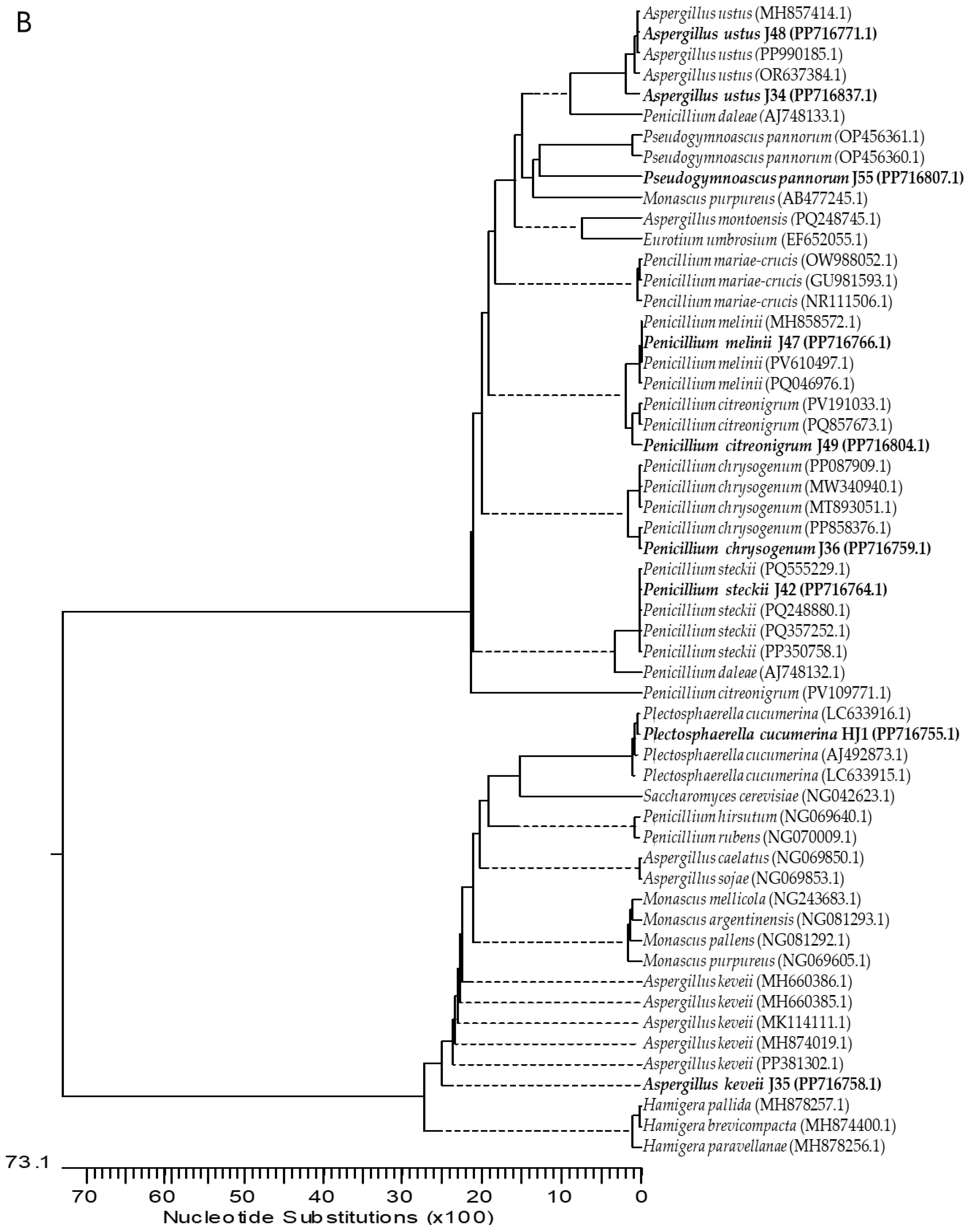
Neighbor-joining phylogenetic trees were constructed based on ITS and 28S rRNA sequences using MegAlign (Lasergene v.7.1, DNASTAR Inc., Madison, MI, USA). Evolutionary distances were computed using the Kimura 2-parameter method. The length of each pair of branches represents the evolutionary distance between the sequence pairs. The sequences of related fungal species/genera were downloaded from the GenBank database, including representatives of the families Aspergillaceae, Trichocomaceae, Pseudeurotiaceae, and Trichosphaeriaceae, to which the studied genera belong. The phylogenetic trees shown in Figure 1 were constructed using (A) sixty-nine sequences, including eight genera and thirty-six species, for the ribosomal DNA region comprising the intergenic spacers ITS1 and ITS2, including the 5.8S rRNA (Figure 1A); (B) fifty-six sequences, including nine genera and twenty-seven species, for the 28S rRNA gene (Figure 1B). Based on all these studies, isolates were determined as indicated in Table 1.

2.2. Antagonistic Activity Assays

2.2.1. Antifungal Activity of Endophytes Against Botrytis Cinerea B05.10 and Colletotrichum Acutatum IMI348489

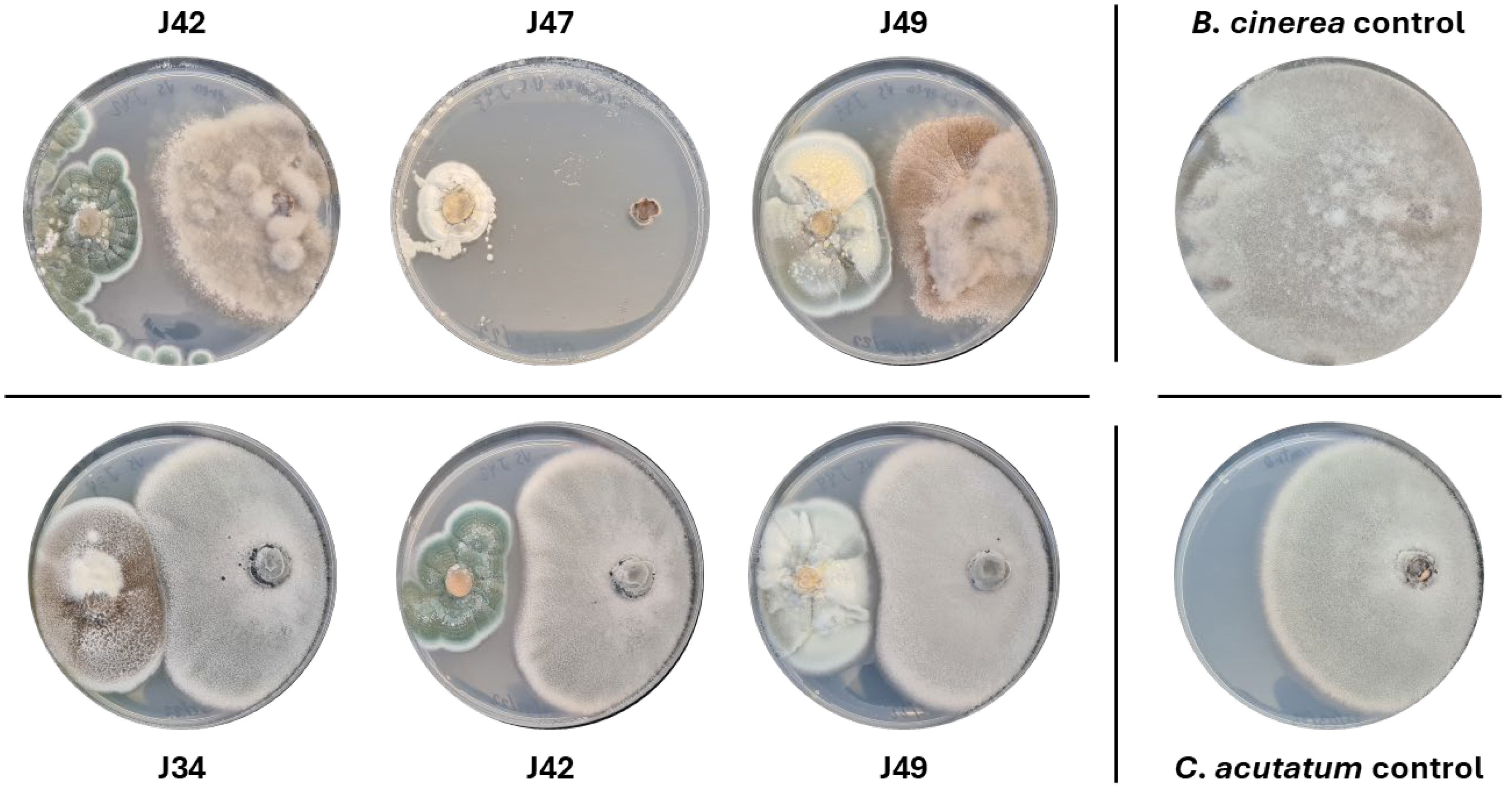
The antifungal activity of endophytic fungi was evaluated in vitro against B. cinerea B05.10 and C. acutatum IMI348489. Areas of fungal growth in co-culture with the phytopathogens were measured and compared with those of the phytopathogens in pure culture (Figure 2). The percentage of inhibition was calculated following the method described by Tenorio-Salgado et al. (2013) [24] with minor modifications and expressed as inhibition percentage (Table 2).
Most of the endophytic fungi exhibited antifungal activity (Table 2). Inhibition of B. cinerea ranged from 32.21% (Aspergillus keveii, J35) to 98.56% (Penicillium melinii, J47), whereas inhibition of C. acutatum was markedly lower, ranging from 7.91% to 38.76%. Penicillium melinii (J47) showed the highest inhibition against B. cinerea, followed by Aspergillus ustus (J34) with 80.06%, while several other Penicillium isolates (P. chrysogenum, P. steckii and P. citreonigrum) also demonstrated moderate inhibition levels. Interestingly, the two isolates identified as Aspergillus ustus displayed markedly different activities, with J34 being highly active and J48 showing minimal inhibition. Notably, all endophytes displayed stronger inhibitory effects against B. cinerea B05.10 than against C. acutatum IMI348489, highlighting the strain-dependent nature of antifungal activity and identifying certain isolates as promising candidates for biocontrol applications.

2.2.2. Antibacterial Activity Against Staphylococcus aureus and Klebsiella pneumoniae

The antibacterial activity during co-culture was evaluated against two bacterial strains, Staphylococcus aureus (Gram-positive) and Klebsiella pneumoniae (Gram-negative), as part of the screening for potential antibacterial activity.
All fungal isolates inhibited the growth of S. aureus to varying degrees, with inhibition values ranging from 17.69 ± 7.32% (Penicillium steckii, J42) to 82.07 ± 3.93% (Penicillium chrysogenum, J36) (Table 2). These results demonstrate a consistent antibacterial effect among the tested endophytic fungi against S. aureus, although the intensity of inhibition varied among isolates. High inhibition values were observed for A. ustus (J34, 69.84 ± 5.20%) and P. chrysogenum (J36, 82.07 ± 3.93%), confirming their strong activity against Gram-positive bacteria. Conversely, isolates such as P. steckii (J42) and Pseudogymnoascus pannorum (J55) showed comparatively lower inhibition percentages, reflecting strain-dependent variability in metabolite production or activity.
In contrast, none of the fungal isolates exhibited detectable antibacterial activity against K. pneumoniae under the co-culture conditions tested. This lack of inhibition is consistent with the intrinsic resistance mechanisms of Gram-negative bacteria, particularly due to the presence of an outer membrane that limits the penetration of many bioactive compounds.
These results confirm that endophytic fungi associated with Zingiber officinale exhibit selective antibacterial activity, primarily effective against Gram-positive bacteria such as S. aureus, and highlight their potential as sources of bioactive metabolites with targeted antimicrobial properties.

2.3. Enzymatic Activity Assays

After five days of incubation at 25 °C, enzymatic activity was assessed based on the presence of clear halos around the fungal colonies. Depending on the fungal species, enzymatic activity was observed for specific enzyme types, as summarized in Table 3. The enzymatic activity of each isolate was quantified using the Enzyme Index (EI), calculated as previously described by Abe et al. (2015) [25].
In general, most of the evaluated endophytes exhibited activity for at least one of the tested enzymes. The average enzymatic activity profile followed a decreasing order: lipolytic, cellulolytic, esterase, proteolytic and amylolytic. This pattern was consistent with the frequency of active isolates, with lipolytic and cellulolytic activities detected in 8 of 9 fungi, esterase activity in 6 of 9, and proteolytic and amylolytic activities in 3 of 9.
Penicillium chrysogenum displayed a more specialized enzymatic profile, showing activity exclusively in the cellulolytic assay, which may reflect adaptation to specific substrates or ecological niches. Similarly, Plectosphaerella cucumerina exhibited esterase and lipolytic activities, suggesting a focused enzymatic strategy rather than a broad-spectrum enzyme production. This diversity of enzymatic patterns among isolates highlights the functional specialization that may exist within endophytic fungi and underscores the potential to identify strains tailored for specific biotechnological applications. Under the tested conditions, neither of these two fungi produced amylolytic or proteolytic enzymes beyond the activities described above. In contrast, Penicillium steckii demonstrated the highest enzymatic activity, being the only fungus capable of producing all the enzymes tested in this experiment. Furthermore, this fungus showed the highest proteolytic activity (EI = 2.03 ± 0.05), whereas Penicillium citreonigrum was the main producer of lipolytic enzymes (EI = 2.01 ± 0.08).
As described by Abe et al. (2015) [25] we considered a significant activity when EI was higher than 2. According to this criterion, Penicillium steckii and Penicillium citreonigrum showed significant proteolytic and lipolytic activity, respectively.

2.4. Antioxidant Activity Assays

The antioxidant activity of the crude extracts was evaluated using the 2,2-diphenyl-1-picrylhydrazyl (DPPH) radical scavenging assay. After measuring the absorbance of the crude extracts dissolved in DPPH solutions at different concentrations, the remaining DPPH percentage was calculated from the initial and final absorbance values [26]. The results are summarized in Table 4.
The Half Maximal Effective Concentration (EC50) value represents the concentration of the extract required to scavenge 50% of the DPPH radicals. A lower EC50 value indicates higher antioxidant activity, as less extract is needed to achieve this effect [27].
As detailed in Table 4, the fungal extracts exhibited a considerable and variable range of antioxidant activities. Among the tested isolates, Penicillium melinii (J47) demonstrated the most potent antioxidant activity, achieving an EC50 value of 23.31 µg/mL. In stark contrast, Pseudogymnoascus pannorum (J55) showed the lowest activity, with an EC50 of 515.95 µg/mL.
Intermediate levels of activity were observed for other strains, notably Plectosphaerella cucumerina (HJ1) and Aspergillus keveii (J35), which exhibited EC50 values of 26.50 µg/mL and 52.12 µg/mL, respectively. While none of the fungal extracts matched the antioxidant efficacy of ascorbic acid, the significant activity recorded for several isolates, particularly P. melinii, highlights their potential as valuable sources of natural antioxidant compounds.

2.5. Antibacterial Activity of Endophytic Fungal Extracts

The antibacterial potential of fungal crude extracts was evaluated against S. aureus (Gram-positive) and K. pneumoniae (Gram-negative). After 48 h of incubation, bacterial growth was monitored hourly by measuring the absorbance. The percentage of inhibition was calculated relative to a positive control where bacteria were allowed to grow without any inhibition. The results are summarized in Table 5.
Chloramphenicol, used as a reference antibiotic, demonstrated complete inhibition of K. pneumoniae growth at 16 mg/L. However, its efficacy against S. aureus was lower, reducing the growth rate even at 32 mg/L, but without achieving complete inhibition under the tested conditions.
The crude extracts of the endophytic fungi showed strong antibacterial activity against S. aureus. Eight out of nine fungal isolates (J34, J35, J42, J47, J48, J49, J55, HJ1) achieved 100% inhibition of S. aureus growth (Table 5). The remaining isolate, P. chrysogenum (J36), also demonstrated substantial activity with 76.86% inhibition. These findings indicate a widespread presence of potent anti-S. aureus compounds among the tested endophytic fungi. In contrast, the activity against K. pneumoniae was lower and more variable. Two isolates, Penicillium chrysogenum (J36) and Pseudogymnoascus pannorum (J55), showed no detectable inhibition (0.00%). The highest inhibition was observed for Plectosphaerella cucumerina (HJ1) at 59.92%, followed by Aspergillus ustus (J34) with 48.22%. Other isolates displayed moderate to low activity, ranging from 16.30% to 32.51%. These results highlight a broader and more consistent antibacterial potential of the extracts against Gram-positive bacteria compared with Gram-negative bacteria.

3. Discussion

3.1. Importance of Endophytic Microorganisms

It is estimated that endophytic microorganisms inhabit all the 300,000 plant species on Earth [28], making them an essential component of plant life [6,8,29,30]. While many of these microorganisms establish commensal relationships with their host, deriving benefits without causing harm or providing direct advantages, a significant number form mutualistic or symbiotic associations. These interactions play a crucial role in plant growth and survival, particularly under adverse environmental conditions [6]. Therefore, endophytes are recognized as important contributors to plant resilience and ecosystem functioning.
Beyond their ecological significance, endophytes are a rich source of bioactive compounds with biotechnological potential. They produce diverse metabolites with anticancer, anti-inflammatory, and antioxidant properties [8,30]. In recent years, several studies have demonstrated that ginger-associated endophytes also synthesize antimicrobial and antioxidant compounds, confirming the metabolic versatility of this microbiome [31,32,33].
Our results revealed a high capacity of endophytic fungi to produce antioxidants and antimicrobial substances, as well as several extracellular enzymes, as evidenced by the positive outcomes in each assay. These findings are consistent with previous reports on Zingiber spp. endophytes showing strain-dependent inhibition of phytopathogenic fungi and clinically relevant bacteria [32,34]. Altogether, this supports the potential of these isolates as promising candidates for biotechnological applications in agriculture, industry, and medicine.
The isolation of nine endophytic fungal strains from Zingiber officinale tubers revealed a taxonomically diverse set of in vitro cultivated fungi, primarily belonging to the genera Penicillium and Aspergillus, along with less commonly reported genera, namely Plectosphaerella cucumerina and Pseudogymnoascus pannorum (synonym Geomyces pannorum). The predominance of Penicillium and Aspergillus aligns with numerous studies of medicinal plants, where these genera frequently appear as endophytes and are recognized for their repertoire of bioactive secondary metabolites and enzymes [28,35]. In ginger, the diversity of endophytic communities has also been shown to vary according to cultivar and geographic origin [32], supporting the idea that host and environment jointly shape endophytic assemblages.
The presence of Plectosphaerella cucumerina is particularly interesting. Traditionally considered a weak pathogen in some hosts, recent work has shown that P. cucumerina can act as a beneficial endophyte. For example, Ding et al. (2022) isolated a strain of P. cucumerina from Rumex gmelinii which significantly promoted host growth; transcriptome analyses revealed upregulation of host genes related to amino acid and carbohydrate metabolism, suggesting that P. cucumerina contributes to resource allocation and stress responses in the host [36]. In addition, P. cucumerina has been observed to produce compounds with antibiofilm and antivirulence activities in endophytic contexts [37,38]. Its ability to colonize healthy tissues without causing symptoms reinforces its potential mutualistic or at least commensal role.
Pseudogymnoascus pannorum is less well-documented as an endophyte in warm-climate plants, but evidence suggests it can adapt to various ecological niches. It has been isolated as an endophyte from Brassica species under controlled conditions, where it formed stable, nonpathogenic associations and even exhibited disease-suppressive effects [39]. Although commonly studied in cold environments as a psychrotolerant fungus, its presence in ginger implies notable physiological flexibility and the potential to produce bioactive metabolites under different temperature regimes [40,41].
The identification approach used in our work combined morphological and molecular methods, including sequencing of ITS and 28S rRNA regions, which produced high-quality matches with GenBank reference sequences. This provides confidence in species-level assignments. While P. cucumerina and P. pannorum have been rarely reported as endophytes of ginger, their recovery here broadens the known diversity associated with this host. Rather than claiming taxonomic novelty, our study contributes to expanding the catalog of ginger-associated endophytes by identifying isolates with distinct biochemical profiles [31,32].
The diversity observed among our isolates suggests that ginger tubers harbor a wider range of endophytic fungi beyond commonly reported genera. This underlines the potential of less-explored taxa as sources of unique functional traits and supports the role of medicinal plants as reservoirs of underexplored fungal diversity. Continued isolation efforts across different cultivars and growing regions may reveal additional strains with specialized metabolic repertoire and biotechnological potential [33].
Taken together, the taxonomic diversity observed, and the multifunctional capacities detected indicate that ginger-associated endophytes can play complementary ecological and biotechnological roles, as further supported by their antagonistic activities described below.

3.2. Antagonistic Activity

Building upon the diversity patterns described above, the ability of endophytic microorganisms to inhibit or slow the growth of phytopathogenic fungi has been widely studied [42,43,44,45,46,47]. This property represents a valuable ecological strategy and a promising tool for sustainable plant protection. The use of fungal endophytes as biological control agents can reduce dependence on chemical pesticides, thereby mitigating environmental contamination and aligning with increasingly strict regulations in agricultural markets. Comparable findings in Zingiber officinale have shown that endophytes from its rhizosphere and tissues possess antifungal metabolites capable of suppressing major phytopathogens [31,32,33].
Our results revealed that all isolated endophytic fungi displayed antagonistic activity against at least one of the two tested phytopathogens. In general, inhibition of Botrytis cinerea was greater than that of Colletotrichum acutatum, suggesting differential pathogen susceptibility. For example, the lowest inhibition against B. cinerea was 32.21%, whereas the maximum against C. acutatum reached only 38.76%, both by Aspergillus keveii. Several isolates, notably Penicillium melinii (98.56%), Aspergillus ustus (80.06%), and Penicillium chrysogenum (70.72%), demonstrated high inhibition of B. cinerea, positioning them as promising candidates for future biocontrol testing under greenhouse or field conditions. Similar high inhibition levels have been reported in endophytic Penicillium and Aspergillus species from ginger and other medicinal plants, where metabolites such as alkaloids and phenolics were linked to pathogen suppression [33,34].
Given the increasing global challenge of antibiotic resistance, endophytic fungi have emerged as promising natural sources of novel antibacterial agents [48,49,50]. Parallel antibacterial assays confirmed the potential of these isolates to inhibit bacterial growth. On bacterial lawns, all endophytes grew over Staphylococcus aureus, but none over Klebsiella pneumoniae. When crude extracts were tested, nearly all isolates completely inhibited S. aureus, while inhibition of K. pneumoniae was less than 60%. This pattern is consistent with previous findings in ginger endophytes, where Gram-positive bacteria showed higher susceptibility to fungal metabolites [32,33]. Our isolate of Plectosphaerella cucumerina exhibited strong inhibition of S. aureus, contrasting with the lack of activity reported for other P. cucumerina strains [38], reinforcing that antimicrobial potential is highly strain dependent [38].
It is important to note that the concentrations of crude extracts used in this study are not directly comparable to those of purified antibiotics. Crude fungal extracts are complex mixtures in which the active compound(s) constitute only a small fraction of the total material, and higher effective concentrations are therefore expected at this stage. This does not diminish the relevance of the findings. The inhibition observed in these assays reflects genuine biological activity and indicates that the isolates can produce metabolites with antibacterial potential. As is common in early bioprospecting work, these results should be viewed as strong preliminary indicators of bioactivity that justify future purification and characterization efforts, rather than as direct evidence of the final potency of individual compounds.
The differential inhibition observed between Gram-positive and Gram-negative bacteria may be attributed to structural differences in their cell walls. Gram-negative bacteria, such as K. pneumoniae, possess a lipopolysaccharide-rich outer membrane that restricts compound diffusion and confers intrinsic resistance [49,51]. Similar observations have been noted in recent studies, where endophytic metabolites from Arthrinium sp. and Penicillium spp. displayed potent activity against S. aureus but limited effects on E. coli and Klebsiella [33,52]. These results support the idea that crude extracts from endophytic fungi can contain diverse molecules with selective targets, and that further purification and fractionation could reveal specific agents effective against Gram-negative pathogens [53].
Among the most active isolates in this study, Plectosphaerella cucumerina, Aspergillus ustus, and Penicillium steckii completely inhibited S. aureus, highlighting their preliminary potential for pharmaceutical exploration. The inhibition observed in crude extracts indicates that these fungi may produce metabolites with antibacterial activity, although the use of crude, non-purified extracts at relatively high concentrations prevents drawing conclusions about the potency of individual compounds. Further purification and characterization would be needed to determine whether truly potent antibiotic molecules are present. Previous studies have shown that endophytic fungi from ginger, such as Fusarium solani and Penicillium commune, also produce potent antibacterial compounds including diketopiperazines, flavonoids, and phenolic derivatives [31,33].
From an agricultural perspective, these dual antifungal and antibacterial activities are particularly valuable. The ability of several isolates to inhibit both B. cinerea and S. aureus suggests broad-spectrum antimicrobial potential that could be harnessed for integrated pest and disease management. However, because the assays were performed using crude extracts applied at relatively high concentrations, these observations should be interpreted with appropriate caution. The activity detected indicates clear biological potential, but it does not yet allow direct conclusions about efficacy in plants or agricultural systems without further studies involving purification, metabolite identification, and functional validation. This is consistent with previous work showing that moderate or even partial in vitro inhibition can translate into meaningful reductions of disease severity under greenhouse conditions, as demonstrated for endophytic fungi controlling B. cinerea in bean plants [54]. More broadly, reviews on microbial antagonists of B. cinerea emphasize that the relationship between in vitro inhibition and in planta performance often depends on multiple factors, including metabolite stability, colonization ability, and environmental interactions [9]. Such dual functionality has also been reported in other plant-associated fungi under certain experimental conditions, including Trichoderma harzianum and Aureobasidium pullulans, which suppress multiple plant pathogens through enzymatic degradation and volatile organic compounds [55,56,57,58]. Previous studies have reported similar inhibition patterns in other fungal species. Trichoderma harzianum and T. asperellum have shown strong inhibition of B. cinerea (close to 100% on Petri dishes) and significantly reduced disease incidence (up to 80%) under greenhouse conditions. Moreover, volatile organic compounds (VOCs) produced by T. asperellum inhibited C. acutatum by 56.86 [55,56,57]. Another fungus from the genus Aureobasidium, A. pullulans, has been reported to reduce the growth of B. cinerea by 90% and C. acutatum by 72% [58], while Clonostachys rosea has also shown a high inhibition capacity against B. cinerea [59].
Similarly, the inhibition capacity of some endophytes against S. aureus and K. pneumoniae has also been documented. For instance, Trichoderma harzianum produces crude extracts that inhibit these and other bacterial species [55]. Other notable examples include Phomopsis sp., Fusarium oxysporum, and Cladosporium cladosporioides, which exhibit comparable antibacterial potential [60,61,62].
Most of the endophytes studied in this research showed remarkable antimicrobial activity, particularly against B. cinerea and S. aureus, highlighting their potential relevance for future applications in medicine and agriculture. Activity against C. acutatum and K. pneumoniae was more variable, suggesting differences in pathogen susceptibility and opening opportunities to further explore the mechanisms underlying these selective effects. This variability underlines the value of screening diverse endophytic strains to identify those with the most promising profiles for biocontrol and antimicrobial applications.
In addition to their antimicrobial properties, many of these endophytes exhibited enzymatic and antioxidant activities, further expanding their potential biotechnological applications, as discussed in the following section. Altogether, these findings underscore the versatility of ginger-associated endophytes as promising biocontrol agents and reservoirs of bioactive metabolites with potential applications in sustainable agriculture and drug discovery.

3.3. Enzymatic and Antioxidant Activity

Beyond their antagonistic capacity, the enzymatic and antioxidant activities observed in the fungal isolates highlight their potential applications across multiple industries. Fungal enzymes represent around 60% of the enzymes used industrially, reflecting their importance in biotechnology [63]. Similar results have been described for endophytic fungi from Zingiber officinale, where extracellular enzymes contribute to host adaptation and the production of industrially relevant metabolites [31,32].
All fungi demonstrated the ability to produce at least one extracellular enzyme (Table 3), with lipolytic and cellulolytic activities being the most frequent. These results highlight their potential as promising candidates for industrial enzyme production [63]. These findings agree with previous reports describing the strong enzymatic capacity of Penicillium and Aspergillus species, widely exploited in several industrial sectors [64,65,66]. Therefore, it was expected that most of the isolates from this study, which belong to these genera, would exhibit enzymatic potential. In particular, P. steckii stood out as the only isolate capable of producing all tested enzymes, with proteolytic activity (EI > 2). Such a profile reinforces its potential for industrial enzyme production, especially in sectors where proteases are in high demand [47].
Interestingly, the less-studied genera identified in this work also demonstrated relevant enzymatic profiles. P. cucumerina exhibited esterase and lipolytic activities, consistent with previous studies reporting enzymes involved in amino acid metabolism [48,49,50], carbohydrate synthesis, and xylanase production [36,67,68]. Similarly, Pseudogymnoascus pannorum exhibited cellulolytic, lipolytic, and proteolytic activities. This observation is consistent with studies reporting the production of all the enzymes observed in this study, as well as additional xylanases and laccases, although this activity can vary between strains [69]. Comparable enzyme profiles have also been found in other ginger-associated endophytes, particularly Aspergillus and Penicillium species, which secrete cellulases, amylases, and esterases with potential biotechnological relevance [33,34].
The extracellular enzyme profiles observed are consistent with the functional traits typically associated with endophytic adaptation. Cellulolytic and pectinolytic activities likely facilitate entry and asymptomatic colonization of host tissues by locally remodeling plant cell-walls, as previously described [70]. Lipolytic and esterase activities may support surface colonization and nutrient acquisition, such as cuticular lipid turnover, reflecting common strategies among plant-associated microbes [71]. In parallel, proteolytic and other lytic enzymes (e.g., chitinases, β-1,3-glucanases) can contribute to antagonism against phytopathogenic fungi by degrading cell-wall components [72]. Altogether, these enzymatic traits may also interact with plant defense mechanisms and induced systemic resistance, reinforcing the ecological and biotechnological potential of ginger-associated endophytes [33,73].
In addition to enzymatic activity, all fungal isolates displayed antioxidant potential, which could be harnessed to produce substances that prevent cellular damage by scavenging free radicals [74]. The antioxidant assays demonstrated a wide variability among the fungal isolates obtained from Z. officinale. Specifically, Penicillium melinii (J47, EC50 = 23.31 µg/mL) and Plectosphaerella cucumerina (HJ1, EC50 = 26.50 µg/mL) exhibited the strongest radical scavenging capacities, whereas Pseudogymnoascus pannorum (J55) showed the weakest effect (EC50 = 515.95 µg/mL). These results indicate that the antioxidant potential of endophytic fungi from ginger is highly strain-dependent, even among isolates of the same genus. While DPPH-based assays provide a well-established first indication of radical scavenging capacity, further analyses would allow a deeper understanding of the specific metabolites responsible for this activity.
Comparable antioxidant activities have been reported for other endophytic Penicillium species, which produced bioactive extracts with radical scavenging capacity under different culture conditions [75,76,77]. The activity of P. melinii observed in our study is consistent with these findings, reinforcing the relevance of Penicillium endophytes as promising sources of natural antioxidant metabolites. Similar antioxidant patterns have been observed in Arthrinium and Penicillium endophytes producing polyphenolic or flavonoid-like metabolites with high DPPH-scavenging efficiency [33,34].
The notable activity of P. cucumerina is particularly noteworthy, since to the best of our knowledge, no previous studies have reported quantitative antioxidant values for this genus. Although P. cucumerina has been linked to plant growth promotion and antimicrobial traits [36,67], our findings provide the first evidence that it can also produce metabolites with substantial antioxidant capacity. This expands the functional profile of P. cucumerina and reinforces the importance of studying less-explored endophytic taxa.
Pseudogymnoascus pannorum exhibited an EC50 markedly higher than the other isolates, indicating a distinct antioxidant profile that may be linked to its psychrotolerant ecology. This distinct profile may reflect alternative metabolic pathways, opening new perspectives on the ecological and biotechnological roles of this genus. Previous studies have mainly associated Pseudogymnoascus species with enzymatic activities or adaptation to extreme environments rather than antioxidant potential [69].
Overall, these findings place P. melinii and P. cucumerina among the most active antioxidant producers of these endophytic fungi, emphasizing their potential for biotechnological applications. The activities observed also highlight the value of exploring their metabolite profiles in future work to better understand the mechanisms underlying their antioxidant performance. Their activity, comparable or superior to many reported Penicillium isolates, suggests that ginger-associated endophytes could serve as promising candidates for the discovery of natural antioxidants with possible applications in food preservation, cosmetics, and pharmaceuticals, where safer alternatives to synthetic additives are in demand.
Taken together, the antimicrobial, enzymatic, and antioxidant profiles described throughout this discussion highlight the multifunctional potential of these endophytic fungi and their relevance as a sustainable source of bioactive compounds. These combined attributes reinforce the relevance of ginger-associated endophytes as promising candidates for sustainable agricultural practices, food preservation, and possible future biotechnological applications, which could be further expanded as the biochemical nature of their active metabolites becomes better understood.

4. Materials and Methods

4.1. Isolation and Purification of Endophytic Fungi from Ginger Tubers

Endophytic fungi were isolated from healthy adult ginger plants (Zingiber officinale), provided by the Instituto de Investigación y Formación Agraria y Pesquera (IFAPA), located in Chipiona (Cádiz), Spain. Samples were taken to the laboratory for immediate processing. Only tubers were processed to isolate endophytic fungi, as they were the focus of this study. Surface sterilization was performed following Potshangbam et al. (2017) [29] with modifications: tubers were immersed in 80% ethanol for 3 min, followed by immersion in 4% sodium hypochlorite for 5 min. After sterilization, they were rinsed three times with sterile distilled water. Sterilization efficiency was confirmed by plating aliquots of the final rinse water on Yeast Extract Glucose Agar (YGA) prepared with yeast extract 5 g/L, glucose 20 g/L and agar 12 g/L, and PDA plates, which were incubated at 25 °C for 7 days to ensure no microbial growth [29]. The outer peel of the tubers was removed using a sterile scalpel. The inner tissue was chopped into small pieces and macerated with 1 mL of sterile 0.9% NaCl solution in a sterile mortar. The resulting macerate was plated onto PDA and incubated at 25 °C for 7 days [29,78]. Fungal colonies were selected based on their morphological appearance and repeatedly subcultured on fresh PDA plates until pure isolates were obtained. Each isolate was assigned an alphanumeric code (e.g., J34, J35, J47) corresponding to its order of isolation and maintenance in the internal fungal collection of the University of Cádiz. These codes were used throughout the study to ensure traceability of the isolates and consistency in data presentation. Pure fungal isolates were maintained on PDA plates at 25 °C and glycerol stocks (30% v/v) were stored at −25 °C for further studies [79].

4.2. Morphological Characterization of Fungi

Plates with fungi were visualized under a stereoscopic microscope (ZEISS Stemi 305, Carl Zeiss AG, Oberkochen, Germany) to determine their three-dimensional morphology. Two samples of mycelium were taken from each plate. One sample was directly observed under an optical microscope (DM750, Leica Microsystem S.L.U., L’Hospitalet de Llobregat, Spain), while the other was stained with methylene blue prior to observation. The microscopic examination allowed for the distinction of hyphae, conidiophores, and spores, as well as the identification of fungal genera. Additionally, in some cases it was possible to determine whether the hyphae were septate or not.

4.3. Molecular Characterization of Fungi

Fungal genomic DNA was extracted following two different protocols: Bolívar Anillo, (2018) [80] and Garrido et al. (2009) [81]. The purity and concentration of DNA were measured using a Nanodrop 2000 spectrophotometer (Thermo Scientific, Madrid, Spain).
Three pairs of primers were used for gene sequencing: ITS1–ITS4 [82], LROR-LR7 [83] and Bt2a–Bt2b [84] (Table 6). PCR reactions were carried out in a SimpliAmp Thermal Cycler (Applied Biosystems) in a total volume of 50 μL containing 1× reaction buffer, 2 mM MgCl2, 0.2 mM dNTP mix, 0.2 μM of each primer, 1.5 U of Go-Taq® DNA Polymerase (Promega) and 100 ng of genomic DNA. The cycling conditions varied depending on the primer pair: For ITS1–ITS4, 95 °C for 5 min, followed by 35 cycles of 95 °C for 45 s, 52 °C for 45 s, and 72 °C for 1 min 20 s, with a final extension at 72 °C for 10 min [82]. For LROR-LR7, 95 °C for 5 min, followed by 30 cycles of 95 °C for 1 min, 50 °C for 45 s, and 72 °C for 1 min, with a final extension at 72 °C for 10 min [83]. For Bt2a–Bt2b, 95 °C for 5 min, followed by 35 cycles of 95 °C for 1 min, 59 °C for 1 min, and 72 °C for 45 s, and ended with a final extension at 72 °C for 10 min [84].
PCR products were analyzed by gel electrophoresis following standard protocols [85]. Amplification products were purified using the GeneJET PCR Purification Kit (Thermo Scientific, Waltham, MA, USA) and quantified with a NanoDrop 2000c spectrophotometer (Thermo Scientific, USA) to confirm their quality before being sent to Macrogen for sequencing. Sequences were assembled using the DNASTAR® Lasergene package (DNASTAR, Inc., Madison, WI, USA), and complementary strands were compared using the Basic Local Alignment Search Tool (BLAST) with the nucleotide database from the National Center for Biotechnology Information (NCBI). Nucleotide sequences were deposited in GenBank (http://www.ncbi.nlm.nih.gov/Genbank/, accessed on 20 July 2025; accession numbers are shown in Table 1). Sequences were aligned, and a neighbor-joining phylogenetic analysis was conducted using MegAlign v.7.1.0 from the DNASTAR® Lasergene package (DNASTAR, Inc., Madison, MI, USA). To study the phylogenetic relationship of our isolates, eighty-one sequences of related genera and species were downloaded from the GenBank database and included in the phylogenetic tree (Figure 1)

4.4. Antagonistic Activity Assays

4.4.1. Antagonistic Assays Against Botrytis Cinerea B05.10 and Colletotrichum Acutatum IMI348489

The antagonistic effect of the fungal isolates against B. cinerea B05.10 and C. acutatum IMI348489 was evaluated. Nine-millimeter mycelial discs from 14-day-old cultures of both pathogens were inoculated on PDA plates along with the fungal isolates. Plates were incubated at 25 °C for 14 days and results were recorded at the end of the incubation period.
The antagonistic effect was calculated following the method described by Tenorio-Salgado et al. (2013) [24] with modifications. Instead of measuring the colony radius, the area of each fungal colony was determined using the computer program JMicroVision (Roduit, N. Version 1.3.4.) and compared with a positive control (pathogens grown alone on PDA). The percentage of inhibition was determined using the following formula:
M y c e l i a l   G r o w t h   I n h i b i t i o n % = A c A 1 A c · 100
where Ac represents the mean area of B. cinerea B05.10 or C. acutatum IMI348489 grown alone on PDA and A1 represents the area of the same pathogen in the presence of endophytic fungus. Each treatment was tested in triplicate plates, where the pathogen and the endophytic fungus were inoculated on opposite sides of the same Petri dish to allow direct interaction and observation of the inhibition zone. Colony growth inhibition was quantified by measuring the reduction in the pathogen’s mycelial area in co-culture compared with its growth in control plates (pathogen grown alone), using JMicroVision software. v1.3.1

4.4.2. Antagonistic Assays Against Staphylococcus aureus and Klebsiella pneumoniae

The antagonistic activity of the endophytic fungal isolates was also tested against the Gram-positive bacterium S. aureus (CECT469) and the Gram-negative bacterium K. pneumoniae (CECT517) from the UCA’s own collection. The assay was performed as described by Sahani et al. (2017) [86].
The bacteria were first grown in LB agar plates at 37 °C for 48 h. A single colony of each bacterium was then resuspended in 0.9% NaCl solution, and the bacterial suspension was adjusted to 0.5 McFarland standard. Subsequently, each bacterial suspension was evenly spread over a Mueller Hinton Agar (MHA) plate using sterile swabs to ensure complete coverage of the medium [87]. Once the surface of the plates dried, 14-day-old mycelial discs (9 mm in diameter) of each endophytic fungal isolate were placed onto the bacterial lawn, with three fungal discs per plate. Plates were incubated at 25 °C, and results were recorded after 10 days [86] with JMicroVision software to determine the area of each fungal mycelia. We selected this incubation temperature as it is the average temperature which their host plant (ginger) needs to grow.
The antagonistic activity assays, shown in Section 4, were performed in triplicate, and the inhibition values are expressed as the mean ± standard deviation of three independent experiments to ensure statistical reliability and reproducibility of the results.

4.5. Enzymatic Activity Assay of Endophytic Fungi

4.5.1. Amylolytic Activity Assay

The capacity of the fungal isolates to hydrolyze starch into sugars was assessed as follows: the medium consisted of 5% (w/v) tryptone soya broth (TSB) (Scharlau), supplemented with 2.5% (w/v) agar and 1% (w/v) soluble starch. Plates were incubated for 120 h at room temperature. Subsequently, 5 mL of a 1% iodine solution was added to each plate to reveal the result. The presence of clear halos around the colonies was considered a positive result [88,89].

4.5.2. Cellulolytic Activity Assay

Cellulolytic activity was evaluated as described by Kasana et al. [90]. Each fungal isolate was inoculated onto CMC agar containing carboxymethyl cellulose (CMC) sodium salt as the primary substrate. Plates were incubated for five days at room temperature. After incubation, each plate was flooded with Gram’s iodine solution for 5 min, and the excess solution was removed. After 30 min a clearance zone around the colonies, in contrast to the purple coloration of the rest of the plate, indicated positive cellulolytic activity.

4.5.3. Esterase Activity Assay

Esterase activity was evaluated using the method described by Sierra (1957) [91]. After medium sterilization, 1% (v/v) sterile Tween 80 was added to the medium. Plates were inoculated with fungal isolates and incubated for 5 days at room temperature. Degradation zones around the fungal colonies, visible as microcrystals, indicated esterase activity [88,91].

4.5.4. Lipolytic Activity Assay

Lipolytic activity was evaluated following a protocol like that used for esterase activity, but with Tween 20 as the substrate instead of Tween 80. Degradation zones around the fungal colonies, observed as microcrystals, indicated positive lipolytic activity [88,91].

4.5.5. Proteolytic Activity Assay

Proteolytic activity was assessed using a 10% skimmed milk medium, following the protocol optimized by Castro et al. (2014) [80,92]. The medium contained (g/L): tryptone 5; yeast extract 2.5; glucose 1; NaCl 2.5 and agar 18. pH 7.0; After sterilization, 100 mL of skimmed milk was added to the medium. Single fungal colonies were inoculated onto the solid medium and incubated at 25 °C for 72 h. The presence of halos around the fungal colonies indicated positive proteolytic activity [80,92].
All enzymatic assays described in this section were carried out in triplicate to ensure reproducibility. Furthermore, the area of each halo was determined with JMicroVision software.

4.6. Fungal Fermentation, Extraction, and Assay of Crude Extracts

4.6.1. Fungal Cultivation and Liquid Phase Extraction of Metabolites

Fungal metabolites were extracted after liquid fermentation. Once sufficient fungal growth was achieved, nine mycelial plugs (9 mm in diameter) were inoculated into three 250 mL Erlenmeyer flasks, each containing 250 mL of 2% (w/v) malt extract medium (Condalab). The cultures were incubated at 25 °C under agitation at 110 rpm for 14 days. After 14 days of fermentation, the fungal mycelia were separated from the culture medium by filtration. The metabolites were then extracted from the aqueous phase using ethyl acetate (1:1, v/v). This extraction process was repeated three times to maximize recovery. The resulting organic phases were pooled, dried over anhydrous sodium sulfate, and concentrated to dryness using a rotary evaporator under reduced pressure at 30 °C, as described by Mtibaà et al. [93].

4.6.2. Antioxidant Activity Assay

The antioxidant activity of the crude extracts was evaluated using the DPPH radical scavenging method as described by Brand-Williams et al. [26], with modifications. A DPPH solution (6 × 10⁻⁵ M in ethanol) was prepared, and 150 µL of this solution was used as a control in triplicate on 96-well plates. For the samples, 3.75 µL of crude extracts at different concentrations was mixed with 146.25 µL of the DPPH solution, also in triplicate [26].
The reactions were monitored using a MultiskanTM Go photometer (Thermo Scientific, USA) by measuring the absorbance of solutions at wavelength of 515 nm every 15 min over a period of 3 h. After testing different concentrations of each crude extract, the percentage of remaining DPPH was calculated using this formula:
%   r e m a n e n t   D P P H = A b s t A b s 0 · 100
where Abs0 is the absorbance at the initial time and Abst is the absorbance at the final time.
The results were plotted as DPPH remaining (%) versus crude extract concentration using Microsoft Excel to generate a polynomial regression curve. From these plots, the EC50 for each extract was determined. The regression equation was then used to determine the concentration (X) at which the % DPPH remaining (Y) equaled 50%, thereby calculating the EC50 for each fungal extract. The values of the extracts were compared with that of ascorbic acid, which was used as a reference antioxidant due to its high scavenging capacity.

4.6.3. Antibacterial Activity Assay of Crude Extracts

The antibacterial activity of the crude extracts was evaluated against S. aureus and K. pneumoniae from the bacterial strain collection of the University of Cádiz (UCA), following the guidelines of the Clinical and Laboratory Standards Institute (CLSI) [94]. Prior to the assay, all crude extracts were adjusted to a final concentration of 1 mg/mL in DMSO to ensure comparability among samples.
Each well of a 96-well microplate was prepared with 178 µL of Mueller Hinton Broth (MHB), 0,2 mg of crude extract dissolved in 2 µL of DMSO, and 20 µL of a bacterial suspension adjusted to 5 × 10⁶ CFU/mL, for a total reaction volume of 200 µL. The plates were incubated at 37 °C under static conditions for 48 h. Bacterial growth was monitored hourly by measuring the absorbance at 625 nm using a Multiskan™ GO microplate spectrophotometer (Thermo Scientific, USA). The percentage of inhibition was calculated using the equation described by Gutiérrez-Escobar et al. [95]:
%   I n h i b i t i o n = ( 1 T F S a m p l e T O S a m p l e ( T F B l a n k T O B l a n k ) T F G r o w t h T O G r o w t h ( T F B l a n k T O B l a n k ) ) · 100
where TOSample and TFSample correspond to the initial and final absorbance values of the wells containing the bacterial culture and crude extract. TOGrowth and TFGrowth correspond to the initial and final absorbance values of the wells containing the bacterial culture without the crude extract. TOBlank and TFBlank correspond to the initial and final absorbance values of the wells containing only the growth medium and crude extract.
All experimental assays were performed in triplicate, and quantitative values are reported as mean ± standard deviation. To ensure full transparency and reproducibility, the complete raw datasets from all antifungals, antibacterial, enzymatic, and antioxidant assays have been compiled and are available in the Supplementary Excel files (Supplementary Materials S1–S3).

5. Conclusions

This study demonstrated that ginger tubers (Zingiber officinale) harbor a diverse community of endophytic fungi, mainly belonging to the genera Penicillium and Aspergillus, but also including less frequently reported taxa such as Plectosphaerella cucumerina and Pseudogymnoascus pannorum. Several isolates exhibited strong antagonistic effects against Botrytis cinerea and moderate inhibition of Colletotrichum acutatum, highlighting their potential role as biocontrol agents against important plant pathogens. In addition, most fungal extracts showed antibacterial activity, particularly against Staphylococcus aureus, suggesting that ginger-associated endophytes may also represent a valuable source of novel antimicrobial compounds.
Beyond their antimicrobial properties, the isolates displayed broad enzymatic capabilities and measurable antioxidant activity. In particular, Penicillium melinii and Plectosphaerella cucumerina demonstrated remarkable radical scavenging capacities, expanding the biotechnological relevance of these species. Taken together, these findings emphasize the multifunctional potential of ginger-derived endophytic fungi for applications in sustainable agriculture, food preservation, pharmaceuticals, and other industrial sectors.
In future research, it will be essential to isolate and characterize the active metabolites responsible for the observed biological activities, elucidate their molecular mechanisms of action, and evaluate their safety and effectiveness under in vivo conditions. Furthermore, scaling up fermentation processes and optimizing metabolite production could help translate these findings into practical biotechnological and agricultural applications.

Supplementary Materials

The following supporting information can be downloaded at: https://www.mdpi.com/article/10.3390/agronomy15122711/s1, Supplementary material S1 file: data for 2.2. Antagonistic Activity Assays and 2.3 enzymatic activities; Supplementary material S2 file: data for Antioxidant activity of crude extracts from endophytes; Supplementary material S3 file: data for Antibacterial Activity Assay of Crude Extracts.

Author Contributions

All the authors collaborated in the experiments described in this article. R.B. personally completed all of the experiments and was supported by A.B., I.I.-B., J.M., C.G., V.E.G.-R. and M.C. Data curation, A.B. and H.J.B.-A.; funding acquisition, J.M.C., C.G. and M.C.; investigation, R.B., I.I.-B., A.B., J.M., C.G., H.J.B.-A., J.M.C., M.D.V.-D., V.E.G.-R. and M.C.; methodology, R.B., C.G., H.J.B.-A., V.E.G.-R. and M.C.; writing—original draft, A.B., I.I.-B., J.M., C.G., H.J.B.-A., J.M.C., M.D.V.-D., V.E.G.-R. and M.C.; writing—review and editing, C.G., M.C., V.E.G.-R. and J.M.C. All authors have read and agreed to the published version of the manuscript.

Funding

This research was supported by a grant from PID-2021-1228990-B-C22 funded by MCIN/AEI/10.13039/501100011033 and by FEDER/UE; The authors thank the financial support from Programa Operativo FEDER Andalucía 2021–2027 and Consejería de Universidad, Investigación e Innovación, Junta de Andalucía (Project FEDER-UCA-2024-A2-52); and the University of Cádiz PhD grant by the “Programa de Fomento e Impulso de la actividad de Investigación y Transferencia de la Universidad de Cádiz”.

Data Availability Statement

The original contributions presented in this study are included in the article. Further inquiries can be directed to the corresponding authors.

Conflicts of Interest

The authors declare no conflicts of interest.

References

  1. Harrison, J.G.; Griffin, E.A. The Diversity and Distribution of Endophytes across Biomes, Plant Phylogeny and Host Tissues: How Far Have We Come and Where Do We Go from Here? Environ. Microbiol. 2020, 22, 2107–2123. [Google Scholar] [CrossRef] [PubMed]
  2. Asad, S.; Priyashantha, A.K.H.; Tibpromma, S.; Luo, Y.; Zhang, J.; Fan, Z.; Zhao, L.; Shen, K.; Niu, C.; Lu, L.; et al. Coffee-Associated Endophytes: Plant Growth Promotion and Crop Protection. Biology 2023, 12, 911. [Google Scholar] [CrossRef] [PubMed]
  3. Chandra, H.; Yadav, A.; Prasad, R.; Kalra, S.J.S.; Singh, A.; Bhardwaj, N.; Gupta, K.K. Fungal Endophytes from Medicinal Plants Acting as Natural Therapeutic Reservoir. Microbe 2024, 3, 100073. [Google Scholar] [CrossRef]
  4. Fadiji, A.E.; Galeemelwe, O.; Babalola, O.O. Unravelling the Endophytic Virome Inhabiting Maize Plant. Agronomy 2022, 12, 1867. [Google Scholar] [CrossRef]
  5. Liao, C.; Doilom, M.; Jeewon, R.; Hyde, K.D.; Manawasinghe, I.S.; Chethana, K.W.T.; Balasuriya, A.; Thakshila, S.A.D.; Luo, M.; Mapook, A.; et al. Challenges and Update on Fungal Endophytes: Classification, Definition, Diversity, Ecology, Evolution and Functions. Fungal Divers. 2025, 131, 301–367. [Google Scholar] [CrossRef]
  6. Bolivar-Anillo, H.J.; González-Rodríguez, V.E.; Almeida, G.R.; Izquierdo-Bueno, I.; Moraga, J.; Carbú, M.; Cantoral, J.M.; Garrido, C. Endophytic Microorganisms as an Alternative for the Biocontrol of Phytophthora spp. In Agro-Economic Risks of Phytophthora and an Effective Biocontrol Approach; Intechopen: London, UK, 2021. [Google Scholar] [CrossRef]
  7. Gouda, S.; Das, G.; Sen, S.K.; Shin, H.S.; Patra, J.K. Endophytes: A Treasure House of Bioactive Compounds of Medicinal Importance. Front. Microbiol. 2016, 7, 219261. [Google Scholar] [CrossRef]
  8. Sharma, M.; Kansal, R.; Singh, D. Endophytic Microorganisms: Their Role in Plant Growth and Crop Improvement. In New and Future Developments in Microbial Biotechnology and Bioengineering: Crop Improvement Through Microbial Biotechnology; Elsevier: Amsterdam, The Netherlands, 2018; pp. 391–413. [Google Scholar] [CrossRef]
  9. Bolívar-Anillo, H.J.; Garrido, C.; Collado, I.G. Endophytic Microorganisms for Biocontrol of the Phytopathogenic Fungus Botrytis cinerea. Phytochem. Rev. 2020, 19, 721–740. [Google Scholar] [CrossRef]
  10. Nongalleima, K.; Dey, A.; Deb, L.; Singh, C.B.; Thongam, B.; Devi, H.S.; Devi, S.I. Endophytic Fungus Isolated From Zingiber zerumbet (L.) Sm. Inhibits Free Radicals And Cyclooxygenase Activity. Int. J. Pharmtech Res. 2013, 5, 301–307. [Google Scholar]
  11. Andriambeloson, H.O.; Rasolomampianina, R.; Ralambondrahety, R.; Andrianantenaina, R.; Raherimandimby, M.; Randriamiharisoa, F. Biological Potentials of Ginger Associated Streptomyces Compared with Ginger Essential Oil. Am. J. Life Sci. 2016, 4, 152–163. [Google Scholar] [CrossRef]
  12. Sudhakaran, G.; Chakraborty, S.; Kumar, A.; Bharti, S.A.K.; Csaba, V.; Valan Arasu, M.; Namasivayam, S.K.R.; Arockiaraj, J. Biochemical Insights into Diverse Psilocybe Mushrooms and Their Metabolites as Sources of Neuroactive Agents: A Review. Curr. Microbiol. 2025, 82, 386. [Google Scholar] [CrossRef]
  13. Avinash, G.P.; Namasivayam, S.K.R.; Pattukumar, V.; Priyanka, S. Microbial Biodegradation of Polyethylene Terephthalate Microplastics by an Indigenous Candida Tropicalis Strain and Biocompatibility Evaluation of Microplastics-Degraded Metabolites in GIFT Tilapia. 3 Biotech 2025, 15, 227. [Google Scholar] [CrossRef]
  14. Karthick Raja Namasivayam, S.; Arvind Bharani, R.S.; Samrat, K. Allochthonous Rhizobacterial Inoculation of Vigna radiata Promotes Plant Growth and Anti-Bacterial Metabolite Production. Biocatal. Agric. Biotechnol. 2024, 56, 103034. [Google Scholar] [CrossRef]
  15. Gupta, J.; Sharma, B.; Sorout, R.; Singh, R.G.; Ittishree; Sharma, M.C. Ginger (Zingiber officinale) in Traditional Chinese Medicine: A Comprehensive Review of Its Anti-Inflammatory Properties and Clinical Applications. Pharmacol. Res. Mod. Chin. Med. 2025, 14, 100561. [Google Scholar] [CrossRef]
  16. Oladosu, Y.; Rafii, M.Y.; Arolu, F.; Murugesu, S.; Chukwu, S.C.; Salisu, M.A.; Fagbohun, I.K.; Muftaudeen, T.K.; Kadar, A.I.; Oladosu, Y.; et al. Genetic Diversity and Utilization of Ginger (Zingiber Officinale) for Varietal Improvement: A Review. AIMS Agric. Food 2024, 9, 183–208. [Google Scholar] [CrossRef]
  17. Shahrajabian, M.H.; Sun, W.; Cheng, Q.; Shahrajabian, M.H.; Sun, W.; Cheng, Q. Pharmacological Uses and Health Benefits of Ginger (Zingiber officinale) in Traditional Asian and Ancient Chinese Medicine, and Modern Practice. Not. Sci. Biol. 2019, 11, 309–319. [Google Scholar] [CrossRef]
  18. Shahrajabian, M.H.; Sun, W.; Cheng, Q. Clinical Aspects and Health Benefits of Ginger (Zingiber officinale) in Both Traditional Chinese Medicine and Modern Industry. Acta Agric. Scand. B Soil. Plant Sci. 2019, 69, 546–556. [Google Scholar] [CrossRef]
  19. Mashhadi, N.S.; Ghiasvand, R.; Askari, G.; Hariri, M.; Darvishi, L.; Mofid, M.R. Anti-Oxidative and Anti-Inflammatory Effects of Ginger in Health and Physical Activity: Review of Current Evidence. Int. J. Prev. Med. 2013, 4, S36. [Google Scholar]
  20. Al-Awwadi, N.A.J. Potential Health Benefits and Scientific Review of Ginger. J. Pharmacogn. Phytother. 2017, 9, 111–116. [Google Scholar] [CrossRef]
  21. Owen, N.L.; Hundley, N. Endophytes—The Chemical Synthesizers inside Plants. Sci. Prog. 2004, 87, 79–99. [Google Scholar] [CrossRef]
  22. Ginting, R.C.B.; Sukarno, N.; Widyastuti, U.T.U.T.; Darusman, L.K.; Kanaya, S. Diversity of Endophytic Fungi from Red Ginger (Zingiber officinale Rosc.) Plant and Their Inhibitory Effect to Fusarium oxysporum Plant Pathogenic Fungi. Hayati 2013, 20, 127. [Google Scholar] [CrossRef]
  23. Suebrasri, T.; Somteds, A.; Harada, H.; Kanokmedhakul, S.; Jogloy, S.; Ekprasert, J.; Lumyong, S.; Boonlue, S. Novel Endophytic Fungi with Fungicidal Metabolites Suppress Sclerotium Disease. Rhizosphere 2020, 16, 100250. [Google Scholar] [CrossRef]
  24. Tenorio-Salgado, S.; Tinoco, R.; Vazquez-Duhalt, R.; Caballero-Mellado, J.; Perez-Rueda, E. Identification of Volatile Compounds Produced by the Bacterium Burkholderia tropica That Inhibit the Growth of Fungal Pathogens. Bioengineered 2013, 4, 236–243. [Google Scholar] [CrossRef] [PubMed]
  25. Abe, C.A.L.; Faria, C.B.; de Castro, F.F.; de Souza, S.R.; dos Santos, F.C.; da Silva, C.N.; Tessmann, D.J.; Barbosa-Tessmann, I.P. Fungi Isolated from Maize (Zea mays L.) Grains and Production of Associated Enzyme Activities. Int. J. Mol. Sci. 2015, 16, 15328. [Google Scholar] [CrossRef] [PubMed]
  26. Brand-Williams, W.; Cuvelier, M.E.; Berset, C. Use of a Free Radical Method to Evaluate Antioxidant Activity. LWT Food Sci. Technol. 1995, 28, 25–30. [Google Scholar] [CrossRef]
  27. Sridhar, K.; Charles, A.L. In Vitro Antioxidant Activity of Kyoho Grape Extracts in DPPH and ABTS Assays: Estimation Methods for EC50 Using Advanced Statistical Programs. Food Chem. 2019, 275, 41–49. [Google Scholar] [CrossRef]
  28. Mamangkey, J.; Mendes, L.W.; Mustopa, A.Z.; Hartanto, A. Endophytic Aspergillii and Penicillii from Medicinal Plants: A Focus on Antimicrobial and Multidrug Resistant Pathogens Inhibitory Activity. BioTechnologia 2024, 105, 83. [Google Scholar] [CrossRef]
  29. Potshangbam, M.; Devi, I.; Sahoo, D.; Strobel, G. Functional Characterization of Endophytic Fungal Community Associated with Oryza sativa L. and Zea mays L. Front. Microbiol. 2017, 8, 325. [Google Scholar] [CrossRef]
  30. Nair, D.N.; Padmavathy, S. Impact of Endophytic Microorganisms on Plants, Environment and Humans. Sci. World J. 2014, 2014, 250693. [Google Scholar] [CrossRef]
  31. Sabu, R.; Soumya, K.R.; Radhakrishnan, E.K. Endophytic Nocardiopsis sp. from Zingiber officinale with Both Antiphytopathogenic Mechanisms and Antibiofilm Activity against Clinical Isolates. 3 Biotech 2017, 7, 115. [Google Scholar] [CrossRef]
  32. Anisha, C.; Radhakrishnan, E.K. Metabolite Analysis of Endophytic Fungi from Cultivars of Zingiber officinale Rosc. Identifies Myriad of Bioactive Compounds Including Tyrosol. 3 Biotech 2017, 7, 146. [Google Scholar] [CrossRef]
  33. Pansanit, A.; Pripdeevech, P. Antibacterial Secondary Metabolites from an Endophytic Fungus, Arthrinium sp. MFLUCC16-1053 Isolated from Zingiber cassumunar. Mycology 2018, 9, 264–272. [Google Scholar] [CrossRef]
  34. Danagoudar, A.; Joshi, C.G.; Sunil Kumar, R.; Poyya, J.; Nivya, T.; Hulikere, M.M.; Anu Appaiah, K. Molecular Profiling and Antioxidant as Well as Anti-Bacterial Potential of Polyphenol Producing Endophytic Fungus-Aspergillus austroafricanus CGJ-B3. Mycology 2017, 8, 28–38. [Google Scholar] [CrossRef]
  35. Gupta, S.; Choudhary, M.; Singh, B.; Singh, R.; Dhar, M.K.; Kaul, S. Diversity and Biological Activity of Fungal Endophytes of Zingiber officinale Rosc. with Emphasis on Aspergillus terreus as a Biocontrol Agent of Its Leaf Spot. Biocatal. Agric. Biotechnol. 2022, 39, 102234. [Google Scholar] [CrossRef]
  36. Ding, C.; Wang, S.; Li, J.; Wang, Z. Transcriptomic Analysis Reveals the Mechanism of Host Growth Promotion by Endophytic Fungus of Rumex gmelinii Turcz. Arch. Microbiol. 2022, 204, 443. [Google Scholar] [CrossRef]
  37. Ap, A.; Wahyudi, A.; Ramadhan, F.; Widjajanti, H. Bioprospecting of Non-Mycorrhizal Endophytic Fungi Associated with Ferns and Mosses. Curr. Res. Environ. Appl. Mycol. J. Fungal Biol. 2021, 11, 416–437. [Google Scholar] [CrossRef]
  38. Barolo, M.I.; Castelli, M.V.; López, S.N. Antimicrobial Activity of Fungal Endophytes Associated with Peperomia argyreia (Piperaceae). Appl. Microbiol. 2024, 4, 753–770. [Google Scholar] [CrossRef]
  39. Roodi, D.; Millner, J.P.; McGill, C.R.; Johnson, R.D.; Hea, S.Y.; Brookes, J.J.; Glare, T.R.; Card, S.D. Development of Plant–Fungal Endophyte Associations to Suppress Phoma Stem Canker in Brassica. Microorganisms 2021, 9, 2387. [Google Scholar] [CrossRef]
  40. Chaturvedi, V.; DeFiglio, H.; Chaturvedi, S. Phenotype Profiling of White-Nose Syndrome Pathogen Pseudogymnoascus destructans and Closely-Related Pseudogymnoascus pannorum Reveals Metabolic Differences Underlying Fungal Lifestyles. F1000Research 2018, 7, 665. [Google Scholar] [CrossRef]
  41. Gomes, E.C.Q.; Gonçalves, V.N.; da Costa, M.C.; de Freitas, G.J.C.; Santos, D.A.; Johann, S.; Oliveira, J.B.S.; da Paixão, T.A.; Convey, P.; Rosa, L.H. Pathogenicity of Psychrotolerant Strains of Antarctic Pseudogmynoascus Fungi Reveals Potential Opportunistic Profiles. Microbe 2024, 5, 100186. [Google Scholar] [CrossRef]
  42. Luu, T.A.; Phi, Q.T.; Nguyen, T.T.H.; Van Dinh, M.; Pham, B.N.; Do, Q.T. Antagonistic Activity of Endophytic Bacteria Isolated from Weed Plant against Stem End Rot Pathogen of Pitaya in Vietnam. Egypt. J. Biol. Pest. Control 2021, 31, 14. [Google Scholar] [CrossRef]
  43. Campanile, G.; Ruscelli, A.; Luisi, N. Antagonistic Activity of Endophytic Fungi towards Diplodia corticola Assessed by In Vitro and in Planta Tests. Eur. J. Plant Pathol. 2007, 117, 237–246. [Google Scholar] [CrossRef]
  44. Zanudin, N.A.B.M.; Hasan, N.; Mansor, P.B. Antagonistic Activity of Fungal Endophytes Isolated from Garcinia atroviridis against Colletotrichum gloeosporioides. Hayati 2020, 27, 209–214. [Google Scholar] [CrossRef]
  45. Bustamante, M.I.; Elfar, K.; Eskalen, A. Evaluation of the Antifungal Activity of Endophytic and Rhizospheric Bacteria against Grapevine Trunk Pathogens. Microorganisms 2022, 10, 2035. [Google Scholar] [CrossRef] [PubMed]
  46. Al-Badi, R.S.; Karunasinghe, T.G.; Al-Sadi, A.M.; Al-Mahmooli, I.H.; Velazhahan, R. In Vitro Antagonistic Activity of Endophytic Fungi Isolated from Shirazi Thyme (Zataria multiflora Boiss.) against Monosporascus cannonballus. Pol. J. Microbiol. 2020, 69, 379–383. [Google Scholar] [CrossRef] [PubMed]
  47. Kushwaha, P.; Kashyap, P.L.; Srivastava, A.K.; Tiwari, R.K. Plant Growth Promoting and Antifungal Activity in Endophytic Bacillus Strains from Pearl Millet (Pennisetum glaucum). Braz. J. Microbiol. 2020, 51, 229–241. [Google Scholar] [CrossRef]
  48. Nia, R.; Mia, M.; Oktapiana, K. Antibacterial Activity Test of Endophytic Fungus from Mangrove Plant (Rhizophora apiculata L.) and (Bruguiera gymnorrizha (L.) Lamk.) Against Klebsiella pneumoniae ATCC 700603. KnE Life Sci. 2017, 2, 146–157. [Google Scholar] [CrossRef]
  49. Silva, D.P.D.; Cardoso, M.S.; Macedo, A.J. Endophytic Fungi as a Source of Antibacterial Compounds—A Focus on Gram-Negative Bacteria. Antibiotics 2022, 11, 1509. [Google Scholar] [CrossRef]
  50. Elmaidomy, A.H.; Shady, N.H.; Abdeljawad, K.M.; Elzamkan, M.B.; Helmy, H.H.; Tarshan, E.A.; Adly, A.N.; Hussien, Y.H.; Sayed, N.G.; Zayed, A.; et al. Antimicrobial Potentials of Natural Products against Multidrug Resistance Pathogens: A Comprehensive Review. RSC Adv. 2022, 12, 29078–29102. [Google Scholar] [CrossRef]
  51. Rosdee, S.; Wisessombat, S.; Tayeh, M.; Malakul, R.; Phanaksri, T.; Sianglum, W. Antibacterial Activity of the Endophytic Fungal Extracts and Synergistic Effects of Combinations of Ethylenediaminetetraacetic Acid (EDTA) against Pseudomonas aeruginosa and Escherichia coli. PeerJ 2025, 13, e19074. [Google Scholar] [CrossRef]
  52. Li, Y.; Kumar, S.; Zhang, L. Mechanisms of Antibiotic Resistance and Developments in Therapeutic Strategies to Combat Klebsiella pneumoniae Infection. Infect. Drug Resist. 2024, 17, 1107–1119. [Google Scholar] [CrossRef]
  53. Eshboev, F.; Mamadalieva, N.; Nazarov, P.A.; Hussain, H.; Katanaev, V.; Egamberdieva, D.; Azimova, S. Antimicrobial Action Mechanisms of Natural Compounds Isolated from Endophytic Microorganisms. Antibiotics 2024, 13, 271. [Google Scholar] [CrossRef] [PubMed]
  54. Bolívar-Anillo, H.J.; González-Rodríguez, V.E.; Cantoral, J.M.; García-Sánchez, D.; Collado, I.G.; Garrido, C. Endophytic Bacteria Bacillus subtilis, Isolated from Zea mays, as Potential Biocontrol Agent against Botrytis cinerea. Biology 2021, 10, 492. [Google Scholar] [CrossRef]
  55. Chávez-Avilés, M.N.; García-Álvarez, M.; Ávila-Oviedo, J.L.; Hernández-Hernández, I.; Bautista-Ortega, P.I.; Macías-Rodríguez, L.I. Volatile Organic Compounds Produced by Trichoderma asperellum with Antifungal Properties against Colletotrichum acutatum. Microorganisms 2024, 12, 2007. [Google Scholar] [CrossRef]
  56. Saravanakumar, K.; Lu, Z.; Xia, H.; Wang, M.; Sun, J.; Wang, S.; Wang, Q.Q.; Li, Y.; Chen, J. Triggering the Biocontrol of Botrytis cinerea by Trichoderma harzianum through Inhibition of Pathogenicity and Virulence Related Proteins. Front. Agric. Sci. Eng. 2018, 5, 271–279. [Google Scholar] [CrossRef]
  57. Kuzmanovska, B.; Rusevski, R.; Jankulovska, M.; Oreshkovikj, K.B. Antagonistic Activity of Trichoderma asperellum and Trichoderma harzianum against Genetically Diverse Botrytis cinerea Isolates. Chil. J. Agric. Res. 2018, 78, 391–399. [Google Scholar] [CrossRef]
  58. Iqbal, M.; Broberg, A.; Andreasson, E.; Stenberg, J.A. Biocontrol Potential of Beneficial Fungus Aureobasidium pullulans Against Botrytis cinerea and Colletotrichum acutatum. Phytopathology 2023, 113, 1428–1438. [Google Scholar] [CrossRef]
  59. Li, F.; Ghanizadeh, H.; Song, W.; Miao, S.; Wang, H.; Chen, X.; Liu, J.; Wang, A. Combined Use of Trichoderma harzianum and Clonostachys rosea to Manage Botrytis cinerea Infection in Tomato Plants. Eur. J. Plant Pathol. 2023, 167, 637–650. [Google Scholar] [CrossRef]
  60. Srinivas, C. Antimicrobial Activity and Phytochemical Analysis of Crude Extracts of Endophytic Fungi Isolated from Plumeria acuminata L. and Plumeria obtusifolia L. Pelagia Res. Libr. Eur. J. Exp. Biol. 2014, 4, 35–43. [Google Scholar]
  61. Desale, M.G.; Bodhankar, M.G. Antimicrobial Activity of Endophytic Fungi Isolated from Vitex negundo Linn. Int. J. Curr. Microbiol. App. Sci. 2013, 2, 389–395. [Google Scholar]
  62. Nwakanma, C.; Njoku, E.N.; T, P. Antimicrobial Activity of Secondary Metabolites of Fungi Isolated from Leaves of Bush Mango. J. Next Gener. Seq. Appl. 2016, 3, 1–6. [Google Scholar] [CrossRef]
  63. Mishra, R.; Kushveer, J.S.; Revanthbabu, P.; Sarma, V.V. Endophytic Fungi and Their Enzymatic Potential. In Advances in Endophytic Fungal Research; Springer: Cham, Switzerland, 2019; pp. 283–337. [Google Scholar] [CrossRef]
  64. Ashtekar, N.; Anand, G.; Thulasiram, H.V.; Rajeshkumar, K.C. Genus Penicillium: Advances and Application in the Modern Era. In New and Future Developments in Microbial Biotechnology and Bioengineering: Recent Advances in Application of Fungi and Fungal Metabolites: Current Aspects; Elsevier: Amsterdam, The Netherlands, 2021; pp. 201–213. [Google Scholar] [CrossRef]
  65. Abdel-Azeem, A.M.; Abo Nahas, H.H.; Abdel-Azeem, M.A.; Tariq, F.J.; Yadav, A.N. Biodiversity and Ecological Perspective of Industrially Important Fungi An Introduction. In Industrially Important Fungi for Sustainable Development; Springer: Cham, Switzerland, 2021; pp. 1–34. [Google Scholar] [CrossRef]
  66. Abdel-Azeem, A.M.; Abdel-Azeem, M.A.; Abdul-Hadi, S.Y.; Darwish, A.G. Aspergillus: Biodiversity, Ecological Significances, and Industrial Applications. In Recent Advancement in White Biotechnology Through Fungi. Fungal Biology; Springer: Cham, Switzerland, 2019; pp. 121–179. [Google Scholar] [CrossRef]
  67. Muñoz-Barrios, A.; Sopeña-Torres, S.; Ramos, B.; López, G.; Del Hierro, I.; Díaz-González, S.; González-Melendi, P.; Mélida, H.; Fernández-Calleja, V.; Mixão, V.; et al. Differential Expression of Fungal Genes Determines the Lifestyle of Plectosphaerella Strains During Arabidopsis thaliana Colonization. Mol. Plant Microbe Interact. 2020, 33, 1299–1314. [Google Scholar] [CrossRef]
  68. Zhan, F.X.; Wang, Q.H.; Jiang, S.J.; Zhou, Y.L.; Zhang, G.M.; Ma, Y.H. Developing a Xylanase XYNZG from Plectosphaerella cucumerina for Baking by Heterologously Expressed in Kluyveromyces lactis. BMC Biotechnol. 2014, 14, 107. [Google Scholar] [CrossRef] [PubMed]
  69. Martorell, M.M.; Ruberto, L.A.M.; Fernández, P.M.; De Figueroa, L.I.C.; Mac Cormack, W.P. Biodiversity and Enzymes Bioprospection of Antarctic Filamentous Fungi. Antarct. Sci. 2019, 31, 3–12. [Google Scholar] [CrossRef]
  70. Mengistu, A.A. Endophytes: Colonization, Behaviour, and Their Role in Defense Mechanism. Int. J. Microbiol. 2020, 2020, 6927219. [Google Scholar] [CrossRef]
  71. Bhadra, F.; Gupta, A.; Vasundhara, M.; Reddy, M.S. Endophytic Fungi: A Potential Source of Industrial Enzyme Producers. 3 Biotech 2022, 12, 86. [Google Scholar] [CrossRef]
  72. Jha, P.; Kaur, T.; Chhabra, I.; Panja, A.; Paul, S.; Kumar, V.; Malik, T. Endophytic Fungi: Hidden Treasure Chest of Antimicrobial Metabolites Interrelationship of Endophytes and Metabolites. Front. Microbiol. 2023, 14, 1227830. [Google Scholar] [CrossRef]
  73. Redkar, A.; Sabale, M.; Zuccaro, A.; Di Pietro, A. Determinants of Endophytic and Pathogenic Lifestyle in Root Colonizing Fungi. Curr. Opin. Plant Biol. 2022, 67, 102226. [Google Scholar] [CrossRef]
  74. Ibrahim, M.; Oyebanji, E.; Fowora, M.; Aiyeolemi, A.; Orabuchi, C.; Akinnawo, B.; Adekunle, A.A. Extracts of Endophytic Fungi from Leaves of Selected Nigerian Ethnomedicinal Plants Exhibited Antioxidant Activity. BMC Complement. Med. Ther. 2021, 21, 98. [Google Scholar] [CrossRef] [PubMed]
  75. Osman, M.E.; Abou-zeid, A.M.; Abu-Saied, M.A.; Ayid, M.M.; El-Zawawy, N.A. Diversity and Bioprospecting Activities of Endophytic Fungi Associated with Different Egyptian Medicinal Plants. Sci. Rep. 2025, 15, 19494. [Google Scholar] [CrossRef]
  76. Naveen, K.V.; Saravanakumar, K.; Sathiyaseelan, A.; Wang, M.H. Comparative Analysis of the Antioxidant, Antidiabetic, Antibacterial, Cytoprotective Potential and Metabolite Profile of Two Endophytic Penicillium spp. Antioxidants 2023, 12, 248. [Google Scholar] [CrossRef]
  77. Toghueo, R.M.K.; Boyom, F.F. Endophytic Penicillium Species and Their Agricultural, Biotechnological, and Pharmaceutical Applications. 3 Biotech 2020, 10, 107. [Google Scholar] [CrossRef]
  78. Strobel, G.; Daisy, B. Bioprospecting for Microbial Endophytes and Their Natural Products. Microbiol. Mol. Biol. Rev. 2003, 67, 491–502. [Google Scholar] [CrossRef]
  79. Shahid, M.; Hameed, S.; Tariq, M.; Zafar, M.; Ali, A.; Ahmad, N. Characterization of Mineral Phosphate-Solubilizing Bacteria for Enhanced Sunflower Growth and Yield-Attributing Traits. Ann. Microbiol. 2015, 65, 1525–1536. [Google Scholar] [CrossRef]
  80. Bolívar Anillo, H.J. Evaluación de La Capacidad Antifúngica y de Promoción de Crecimiento Vegetal de La Microbiota Endófita Aisladas de Plantas de Maíz (Zea mays) Así Como de Hongos Antagonistas Aislados de Otras Fuentes; Universidad de Cádiz: Cadiz, Spain, 2018. [Google Scholar]
  81. Garrido, C.; Carbú, M.; Fernández-Acero, F.J.; Vallejo, I.; Manuel Cantoral, J. Phylogenetic Relationships and Genome Organisation of Colletotrichum acutatum Causing Anthracnose in Strawberry. Eur. J. Plant Pathol. 2009, 125, 397–411. [Google Scholar] [CrossRef]
  82. White, T.J.; Bruns, T.; Lee, S.; Taylor, J. Amplification and Direct Sequencing of Fungal Ribosomal RNA Genes for Phylogenetics. In PCR Protocols: A Guide to Methods and Applications; Academic Press: New York, NY, USA, 1990; pp. 315–322. [Google Scholar]
  83. Cubeta, M.A. Characterization of Anastomosis Groups of Binucleate Rhizoctonia Species Using Restriction Analysis of an Amplified Ribosomal RNA Gene. Phytopathology 1991, 81, 1395. [Google Scholar] [CrossRef]
  84. Glass, N.L.; Donaldson, G.C. Development of Primer Sets Designed for Use with the PCR to Amplify Conserved Genes from Filamentous Ascomycetes. Appl. Environ. Microbiol. 1995, 61, 1323–1330. [Google Scholar] [CrossRef]
  85. Aly, A.H.; Debbab, A.; Kjer, J.; Proksch, P. Fungal Endophytes from Higher Plants: A Prolific Source of Phytochemicals and Other Bioactive Natural Products. Fungal Divers. 2010, 41, 1–16. [Google Scholar] [CrossRef]
  86. Sahani, K.; Thakur, D.; Hemalatha, K.P.J.; Ganguly, A. Antibacterial Activity Of Endophytes From Selected Medicinal Plants. Int. J. Adv. Res. 2017, 5, 2076–2086. [Google Scholar] [CrossRef]
  87. Powthong, P.; Jantrapanukorn, B.; Thongmee, A.; Suntornthiticharoen, P. Screening of Antimicrobial Activities of the Endophytic Fungi Isolated from Sesbania grandiflora (L.) Pers. J. Agric. Sci. Technol. 2013, 15, 1513–1522. [Google Scholar]
  88. Dias, A.C.F.; Andreote, F.D.; Dini-Andreote, F.; Lacava, P.T.; Sá, A.L.B.; Melo, I.S.; Azevedo, J.L.; Araújo, W.L. Diversity and Biotechnological Potential of Culturable Bacteria from Brazilian Mangrove Sediment. World J. Microbiol. Biotechnol. 2009, 25, 1305–1311. [Google Scholar] [CrossRef]
  89. Hankin, L.; Anagnostakis, S.L. The Use of Solid Media for Detection of Enzyme Production by Fungi. Oecologia 1975, 67, 597. [Google Scholar] [CrossRef]
  90. Kasana, R.C.; Salwan, R.; Dhar, H.; Dutt, S.; Gulati, A. A Rapid and Easy Method for the Detection of Microbial Cellulases on Agar Plates Using Gram’s Iodine. Curr. Microbiol. 2008, 57, 503–507. [Google Scholar] [CrossRef]
  91. Sierra, G. A Simple Method for the Detection of Lipolytic Activity of Micro-Organisms and Some Observations on the Influence of the Contact between Cells and Fatty Substrates. Antonie Van. Leeuwenhoek 1957, 23, 15–22. [Google Scholar] [CrossRef]
  92. Castro, R.A.; Quecine, M.C.; Lacava, P.T.; Batista, B.D.; Luvizotto, D.M.; Marcon, J.; Ferreira, A.; Melo, I.S.; Azevedo, J.L. Isolation and Enzyme Bioprospection of Endophytic Bacteria Associated with Plants of Brazilian Mangrove Ecosystem. Springerplus 2014, 3, 382. [Google Scholar] [CrossRef]
  93. Mtibaà, R.; Ezzanad, A.; Aranda, E.; Pozo, C.; Ghariani, B.; Moraga, J.; Nasri, M.; Manuel Cantoral, J.; Garrido, C.; Mechichi, T. Biodegradation and Toxicity Reduction of Nonylphenol, 4-Tert-Octylphenol and 2,4-Dichlorophenol by the Ascomycetous Fungus Thielavia sp HJ22: Identification of Fungal Metabolites and Proposal of a Putative Pathway. Sci. Total Environ. 2020, 708, 135129. [Google Scholar] [CrossRef]
  94. Cockerill, F.R.C.; Clinical and Laboratory Standards Institute. Methods for Dilution Antimicrobial Susceptibility Tests for Bacteria That Grow Aerobically: Approved Standard, 9th ed.; CSLI: Wayne, PA, USA, 2012; Available online: https://search.worldcat.org/es/title/894734842 (accessed on 25 July 2025).
  95. Gutiérrez-Escobar, R.; Fernández-Marín, M.I.; Richard, T.; Fernández-Morales, A.; Carbú, M.; Cebrian-Tarancón, C.; Torija, M.J.; Puertas, B.; Cantos-Villar, E. Development and Characterization of a Pure Stilbene Extract from Grapevine Shoots for Use as a Preservative in Wine. Food Control 2021, 121, 107684. [Google Scholar] [CrossRef]
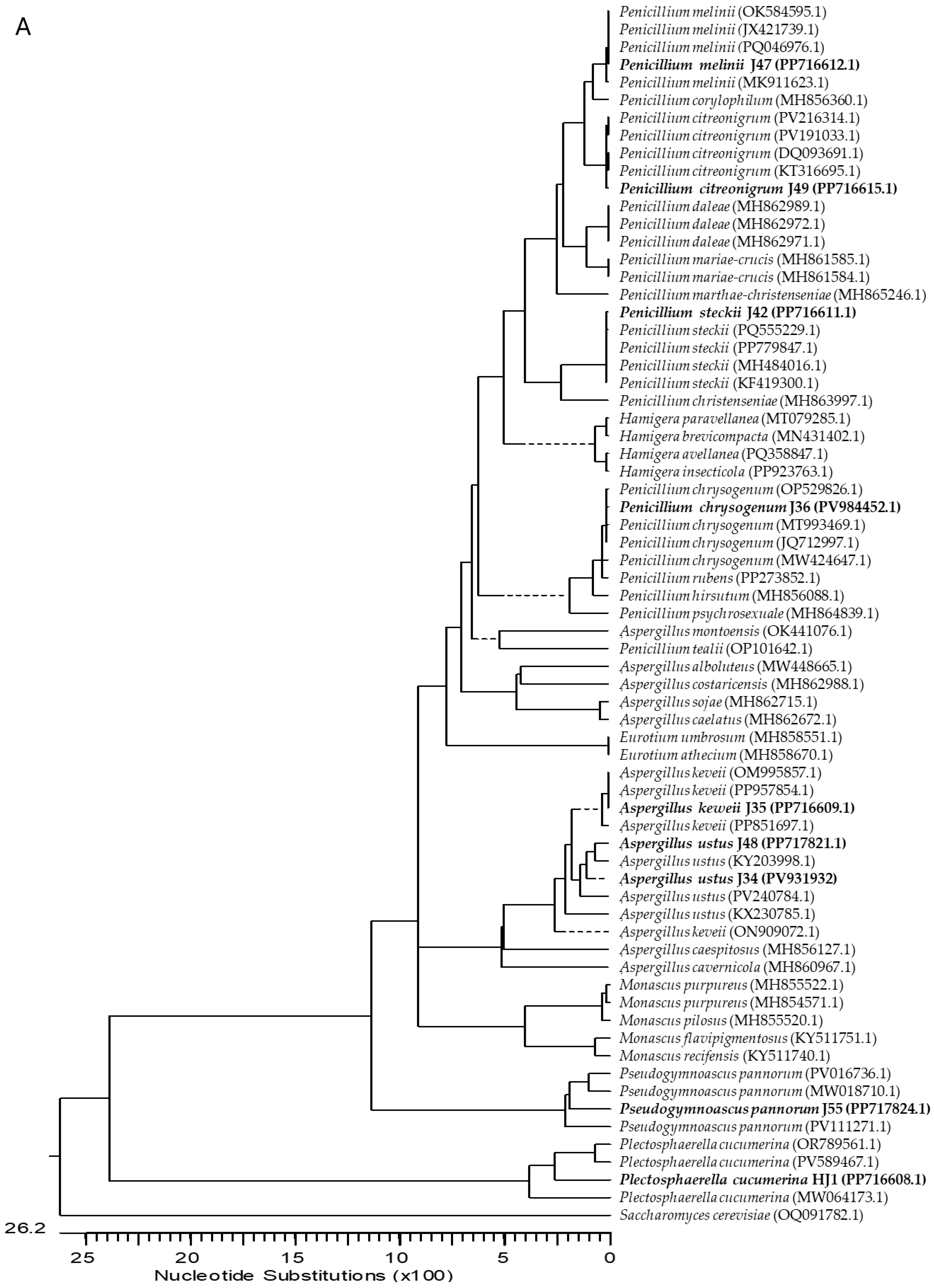
Figure 1. Neighbor-joining phylogenetic trees constructed using (A) the intergenic spacers ITS1 and ITS2, including the 5.8S rRNA region, and (B) the 28S rRNA gene sequences. The trees were generated with the Neighbor-Joining method using the Kimura 2-parameter model implemented in MegAlign (Lasergene v.7.1, DNASTAR Inc., Madison, MI, USA), with bootstrap analysis based on 1000 replicates. Sequences obtained in this study are shown in bold, while reference sequences were retrieved from the GenBank database, with accession numbers indicated in parentheses. Branch lengths represent genetic distances between sequences. The dotted line denotes a negative branch length, and the scale bar indicates the number of nucleotide substitutions per site.
Agronomy 15 02711 g001aAgronomy 15 02711 g001b
Figure 2. Dual culture assays showing the antagonistic interactions between endophytic fungal isolates and phytopathogenic fungi. Top row: confrontation assays of isolates J42, J47, and J49 against Botrytis cinerea B05.10, with the endophytic fungus placed on the left and B. cinerea on the right. The control plate shows B. cinerea growing alone under identical conditions. Bottom row: confrontation assays of isolates J34, J42, and J49 against Colletotrichum acutatum IMI348489, maintaining the same plate arrangement (endophyte on the left, pathogen on the right) and including the corresponding control plate. Antifungal activity is evidenced by the inhibition zones and reduced mycelial growth of the pathogens compared with the controls.
Agronomy 15 02711 g002
Table 1. Molecular identification of endophytic fungal isolates obtained from ginger tubers.
IsolateIdentificationGenBank Acc. N.
ITS-5.8S rRNA28S rRNA
J34Aspergillus ustusPV931932PP716837
J35Aspergillus keveiiPP716609PP716758
J36Penicillium chrysogenumPV984452PP716759
J42Penicillium steckiiPP716611PP716764
J47Penicillium meliniiPP716612PP716766
J48Aspergillus ustusPP717821PP716771
J49Penicillium citreonigrumPP716615PP716804
J55Pseudogymnoascus pannorumPP717824PP716807
HJ1Plectosphaerella cucumerinaPP716608PP716755
Table 2. Antimicrobial activity of endophytic fungi against Botrytis cinerea B05.10, Colletotrichum acutatum IMI348489, and Staphylococcus aureus CECT469.
Endophytes% Inhibition of B. cinerea% Inhibition of C. acutatum% Inhibition of S. aureus
J34Aspergillus ustus80.06 ± 6.229.45 ± 0.8769.84 ± 5.20
J35Aspergillus keveii32.21 ± 3.0538.76 ± 1.4727.26 ± 11.61
J36Penicillium chrysogenum70.72 ± 1.4217.17 ± 0.5282.07 ± 3.93
J42Penicillium steckii50.66 ± 5.9511.09 ± 0.7917.69 ± 7.32
J47Penicillium melinii98.56 ± 0.1115.51 ± 0.4932.27 ± 6.72
J48Aspergillus ustus0.00 ± 0.007.91 ± 0.1876.84 ± 2.77
J49Penicillium citreonigrum51.94 ± 5.1410.36 ± 1.6726.80 ± 2.41
J55Pseudogymnoascus pannorum33.66 ± 8.258.32 ± 0.3269.24± 6.21
HJ1Plectosphaerella cucumerina49.20 ± 1.2113.49 ± 1.0274.14 ± 4.04
Data represent mean values ± standard deviation from three independent replicates (n = 3).
Table 3. Enzymatic activity of endophytic fungi expressed as the Enzyme Index (EI).
EndophytesAmylolytic ActivityCellulolytic ActivityEsterase
Activity
Lipolytic
Activity
Proteolytic
Activity
J34Aspergillus ustus0.00 ± 0.001.41 ± 0.031.25 ± 0.081.11 ± 0.020.00 ± 0.00
J35Aspergillus keveii1.31 ± 0.111.54 ± 0.011.14 ± 0.041.52 ± 0.050.00 ± 0.00
J36Penicillium chrysogenum0.00 ± 0.001.34 ± 0.040.00 ± 0.000.00 ± 0.000.00 ± 0.00
J42Penicillium steckii1.21 ± 0.041.60 ± 0.031.33 ± 0.071.45 ± 0.052.03 ± 0.05
J47Penicillium melinii0.00 ± 0.001.20 ± 0.031.19 ± 0.041.63 ± 0.020.00 ± 0.00
J48Aspergillus ustus1.13 ± 0.031.45 ± 0.050.00 ± 0.001.19 ± 0.051.15 ± 0.03
J49Penicillium citreonigrum0.00 ± 0.001.24 ± 0.041.51 ± 0.092.01 ± 0.080.00 ± 0.00
J55Pseudogymnoascus pannorum0.00 ± 0.001.61 ± 0.040.00 ± 0.001.30 ± 0.021.12 ± 0.03
HJ1Plectosphaerella cucumerina0.00 ± 0.000.00 ± 0.001.17 ± 0.031.47 ± 0.060.00 ± 0.00
Note: The EI (R/r) represents the ratio of the diameter of the degradation halo (R) to the diameter of the fungal colony (r), both measured in cm. Data represent mean values ± standard deviation from three independent replicates (n = 3).
Table 4. Antioxidant activity of crude extracts from endophytes.
Endophytes ExtractsHalf Maximal Effective Concentration (EC50) (µg/mL)
J34Aspergillus ustus133.59 ± 3.87
J35Aspergillus keveii52.12 ± 1.10
J36Penicillium chrysogenum38.53 ± 2.37
J42Penicillium steckii53.16 ± 1.34
J47Penicillium melinii23.31 ± 0.61
J48Aspergillus ustus68.59 ± 2.65
J49Penicillium citreonigrum54.92 ± 1.25
J55Pseudogymnoascus pannorum515.95 ± 10.57
HJ1Plectosphaerella cucumerina26.50 ± 0.80
Note: Ascorbic acid was used as a reference antioxidant, with an EC50 of 2.31 ± 0.03 µg/mL. Data represent mean values ± standard deviation from three independent replicates (n = 3).
Table 5. Antibacterial activity of crude extracts from endophytes (1 mg/mL).
Endophytes Extracts% Inhibition of S. aureus% Inhibition of K. pneumoniae
J34Aspergillus ustus100 ± 0.4048.22 ± 2.15
J35Aspergillus keveii100 ± 0.2932.51 ± 2.82
J36Penicillium chrysogenum76.86 ± 2.490.00
J42Penicillium steckii100 ± 1.6725.70 ± 4.20
J47Penicillium melinii100 ± 1.1216.30 ± 5.10
J48Aspergillus ustus100 ± 0.3927.17 ± 2.26
J49Penicillium citreonigrum100 ± 0.3628.34 ± 2.04
J55Pseudogymnoascus pannorum100 ± 0.170.00
HJ1Plectosphaerella cucumerina100 ± 0.5959.92 ± 4.32
Data represent mean values ± standard deviation from three independent replicates (n = 3).
Table 6. Primers used in this study.
PrimerSequence (5′ → 3′)Product Size (bp)ReferenceAmplified Region
ITS1TCC GTA GGT GAA CCT GCG G600[80]ITS region
ITS4TCC TCC GCT TAT TGA TAT GC
LRORACC CGC TGA ACT TAA GC1500[83]28S ribosomal RNA
LR7TAC TAC CAC CAA GAT CT
Bt2aGGT AAC CAA ATC GGT GCT GCT TTC500[84]b-tubulin
Bt2bACC CTC AGT GTA GTG ACC CTT GGC
Disclaimer/Publisher’s Note: The statements, opinions and data contained in all publications are solely those of the individual author(s) and contributor(s) and not of MDPI and/or the editor(s). MDPI and/or the editor(s) disclaim responsibility for any injury to people or property resulting from any ideas, methods, instructions or products referred to in the content.