Article

Phytochemical and Gene Network Analysis Elucidating the Key Genes Involved in the Biosynthesis of Gomisin J in Schisandra sphenanthera

1
College of Forestry, Central South University of Forestry & Technology, 498 South Shaoshan Road, Changsha 410004, China
2
Yuelushan Laboratory, Qiushi Building, Hunan Agricultural University, Furong District, Changsha 410128, China
3
The Belt and Road International Union Research Center for Tropical Arid Non-Wood Forest in Hunan Province, 498 South Shaoshan Road, Changsha 410004, China
*
Authors to whom correspondence should be addressed.
These authors contributed equally to this work.
Agronomy 2024, 14(3), 576; https://doi.org/10.3390/agronomy14030576
Submission received: 31 January 2024 / Revised: 5 March 2024 / Accepted: 11 March 2024 / Published: 14 March 2024
(This article belongs to the Section Crop Breeding and Genetics)

Abstract

:
The biosynthesis and distribution of lignans in medicinal plants, particularly in Schisandra sphenanthera, hold significant pharmacological importance. This study bridges the knowledge gap in understanding the tissue-specific biosynthesis and distribution of these compounds, with a focus on Gomisin J. Our phytochemical analysis revealed a distinct accumulation pattern of Gomisin J, predominantly in the roots, contrasting with the distribution of Pregomisin and Dihydroguaiaretic acid. This finding highlights the roots’ unique role in lignan storage and biosynthesis. Further, differential gene expression analysis across various tissues illuminated the biosynthetic pathways and regulatory mechanisms of these lignans. Utilizing Weighted Gene Co-expression Network Analysis (WGCNA), we identified the MEtan module as a key player, strongly correlated with Gomisin J levels. This module’s in-depth examination revealed the crucial involvement of four cytochrome P450 (CYP) enzymes and eight transcription factors. Notably, the CYP genes DN6828 and DN2874-i3 exhibited up-regulation in roots across both male and female plants, while DN51746 was specifically up-regulated in male roots, indicating a potential gender-specific aspect in Gomisin J biosynthesis. Comparative analysis with functionally characterized CYP71A homologs suggests these CYP genes might be involved in distinct biosynthetic pathways, including terpenoids, alkaloids, and phenylpropanoids, and potentially in lignan biosynthesis. This hypothesis, supported by their more than 55% identity with CYP71As and strong correlation with Gomisin J concentration, opens avenues for novel discoveries in lignan biosynthesis, pending further functional characterization. Our research provides a comprehensive understanding of the genetic and metabolic mechanisms underlying the tissue-specific distribution of lignans in Schisandra sphenanthera, offering valuable insights for their pharmacological exploitation.

1. Introduction

Schisandra sphenanthera, a traditional Chinese medicinal plant, is distinguished by its unique composition of bioactive lignans, which contribute to its diverse pharmacological activities. Among these lignans, Gomisin J emerges as a compound of significant medicinal interest due to its potent pharmacological properties. This plant’s lignan profile, particularly rich in Gomisin J, sets it apart not only from other plants but also from other species within the Schisandra genus, underscoring the importance of species-specific research in understanding the full pharmacological potential of these compounds. In general, Gomisin J, found in various Schisandra species including Schisandra rubriflora, exhibits notable anti-HIV activity, highlighting its potential as a medicinal agent [1]. In Schisandra sphenanthera, Gomisin J is a key contributor to the plant’s pharmacological profile, which includes anti-inflammatory, antioxidant, anticancer, liver protection, and immunomodulatory activities [2,3,4,5,6]. This lignan’s presence in Schisandra sphenanthera, along with other lignans, differentiates it from other species, such as Schisandra chinensis, where Pregomisin has been studied for its effects on drug metabolism, particularly its inhibition of UGT1A9 activity, a process crucial for the glucuronidation of various endogenous and xenobiotic substances [7].
In recent years, the focus on lignan biosynthesis, particularly Gomisin J, in plants has intensified due to its widespread medicinal use and the advancements in synthetic biology technologies. This surge in interest is driven by the increasing demand for these compounds in various industries and the desire to understand their biosynthetic pathways more comprehensively. Studies have revealed multiple novel lignan-biosynthetic pathways, leading to efficient, sustainable, and stable production of lignans in plants [8], and efforts are being made to understand and manipulate their biosynthetic pathways for improved foodstuffs [9].
Cytochrome P450 enzymes are central to the biosynthesis of lignans, playing a complex role in phenolic metabolism and contributing to the production of numerous phenolic compounds with medicinal and defensive applications in plants [10,11,12]. The engineering of lignan biosynthesis in cell cultures, as demonstrated in Forsythia, shows promise for the efficient production of lignans with antitumor and antioxidant properties [13]. Similarly, Linum nodiflorum cell cultures have been shown to produce significant amounts of cytotoxic lignans, highlighting the key role of enzymes in their biosynthesis [14]. Moreover, the role of specific cytochrome P450 enzymes, such as CYP81Q1 in Sesamum, in lignan biosynthesis has been elucidated, providing insight into unique modes of cytochrome P450-mediated lignan biosynthesis [15]. Plant P450 proteins and transcripts serve as downstream reporters for various biochemical pathways, responding to chemical, developmental, and environmental cues [16]. Additionally, the diverse roles of plant cytochrome P450 monooxygenases in biosynthetic and detoxification pathways underscore their complex relationships with substrates [17]. For instance, the inhibitory effects of Schisandra lignans, including Gomisin J, on various cytochrome P450 enzymes suggest potential anti-cancer and hepatoprotective effects [18]. And the discovery of new cytochrome P450 genes, such as CYP71BJ1 in Catharanthus roseus, has advanced our understanding of alkaloid biosynthesis control points [19]. Gomisin J and N from Schisandra chinensis have been found to inhibit Wnt/β-catenin signaling, indicating potential for colorectal cancer prevention and treatment [20]. Furthermore, the development of new biosynthetic routes in E. coli for converting lignan precursors to valuable compounds demonstrates the potential of harnessing plant enzymes for health-promoting properties [21]. Plant cytochrome P450s play crucial roles in the biotransformation of xenobiotics and endogenous substrates, influencing secondary metabolite biosynthesis and detoxification [22].
In summary, the study of Gomisin J biosynthesis in plants like Schisandra chinensis is of paramount importance due to its wide-ranging pharmacological effects. A deeper understanding of the specific cytochrome P450 enzymes involved in its biosynthesis could pave the way for enhanced utilization of Gomisin J in various therapeutic contexts. The advancements in synthetic biology and the increasing demand for lignans in the pharmaceutical industry underscore the need for comprehensive studies on their biosynthesis, regulation, and potential applications. However, several knowledge gaps persist in this area, which are crucial for understanding and manipulating these pathways for therapeutic applications. One of the primary challenges is the incomplete understanding of the regulation of lignin, suberin, and cutin precursor biosynthesis, which are closely related to lignan biosynthesis [23]. This gap hinders efforts to engineer the formation of these compounds in plants for producing customized biopolymers. Additionally, the localization of lignans within plant tissues, such as in the secondary wall of sclerite cells in flaxseed, is not fully understood, with about 90% of metabolites found in specific areas [24]. Moreover, the role of lignans in plant defense, such as their inhibitory effects on growth and trichothecene biosynthesis in Fusarium graminearum, is another area that requires further exploration [25]. Metabolic engineering of lignan-biosynthesizing plants is considered promising for efficient, sustainable, and stable lignan production [26]. However, despite their potential applications, the detailed biosynthetic pathways and mechanisms of these compounds remain only partially understood [25,27]. Central to this biosynthesis are cytochrome P450 enzymes, such as CYP81Q1 in Sesamum and CYP81Qs in Schisandra chinensis, which play a crucial role, yet a comprehensive understanding of these enzymes and their functions in lignan synthesis is still evolving [15,28].
Advances in metabolic engineering, exemplified by studies in Forsythia cell cultures and Linum album cells, have shown promising potential for enhancing lignan production, with significant implications for antitumor and antioxidant activities [13,29]. This progress is paralleled by investigations into dietary lignans like enterolactone, which are being studied for their potential to reduce cancer risk, particularly breast cancer [30]. Furthermore, the integration of modern RNA-seq technology and bioinformatics tools has become instrumental in unraveling the complex gene networks involved in lignan biosynthesis. This approach is exemplified by studies highlighting Gomisin J’s role in inhibiting Wnt/β-catenin signaling and hepatic lipogenesis, which not only underscores the metabolic significance of this lignan but also suggests a complex interplay of biosynthetic pathways [5,20]. The methodological synergy of combining phytochemical analysis with RNA-seq, as outlined by Ji and Sadreyev [31], provides a comprehensive method for quantitating RNA-seq data, crucial for understanding the molecular mechanisms underlying Gomisin J biosynthesis. This multifaceted research approach is pivotal in identifying key genes and regulatory elements in the biosynthesis of Gomisin J, offering insights into its pharmacological applications and contributing to the broader understanding of secondary metabolism in medicinal plants.
This study addresses critical gaps in understanding the biosynthesis of Gomisin J in Schisandra sphenanthera, a key step in advancing plant secondary metabolism knowledge and developing novel therapeutics. Our comprehensive analysis reveals distinct lignan concentration patterns across various tissues, highlighting tissue-specific accumulation, with Gomisin J and Pregomisin predominantly found in roots and fruits, and Dihydroguaiaretic acid mainly in leaves. These findings suggest differences in biosynthesis, storage, and physiological roles. Differential gene expression analysis and Weighted Gene Co-expression Network Analysis (WGCNA) identify the tan module, strongly correlated with Gomisin J, as containing crucial genes for its biosynthesis and regulation, underscored by the presence of transcription factors and cytochrome P450 enzymes. This module’s diverse biological functions, including disease resistance and enzymatic activities, indicate its role in the plant’s response to environmental stresses and metabolic processes. The brown module, rich in transcription factors and cytochrome P450 enzymes, further implicates stress responses, developmental processes, and metabolic pathways. Collectively, these insights into tissue-specific gene expression and regulatory networks enhance our understanding of Schisandra sphenanthera’s secondary metabolism, guiding future pharmacological research and the potential development of active compounds.

2. Materials and Methods

2.1. Plant Material and Growth Condition

Samples of Schisandra sphenanthera were gathered from the Experimental Base in Tongtong County, Huaihua City, Hunan Province, China. These included four components (root, stem, leaf, and ripe fruits) from healthy, four-year-old female plants and three components (root, stem, and leaf) from male plants, each with three biological replicates. The collected samples were then split into two groups for conducting both metabolic and transcriptomic analyses.

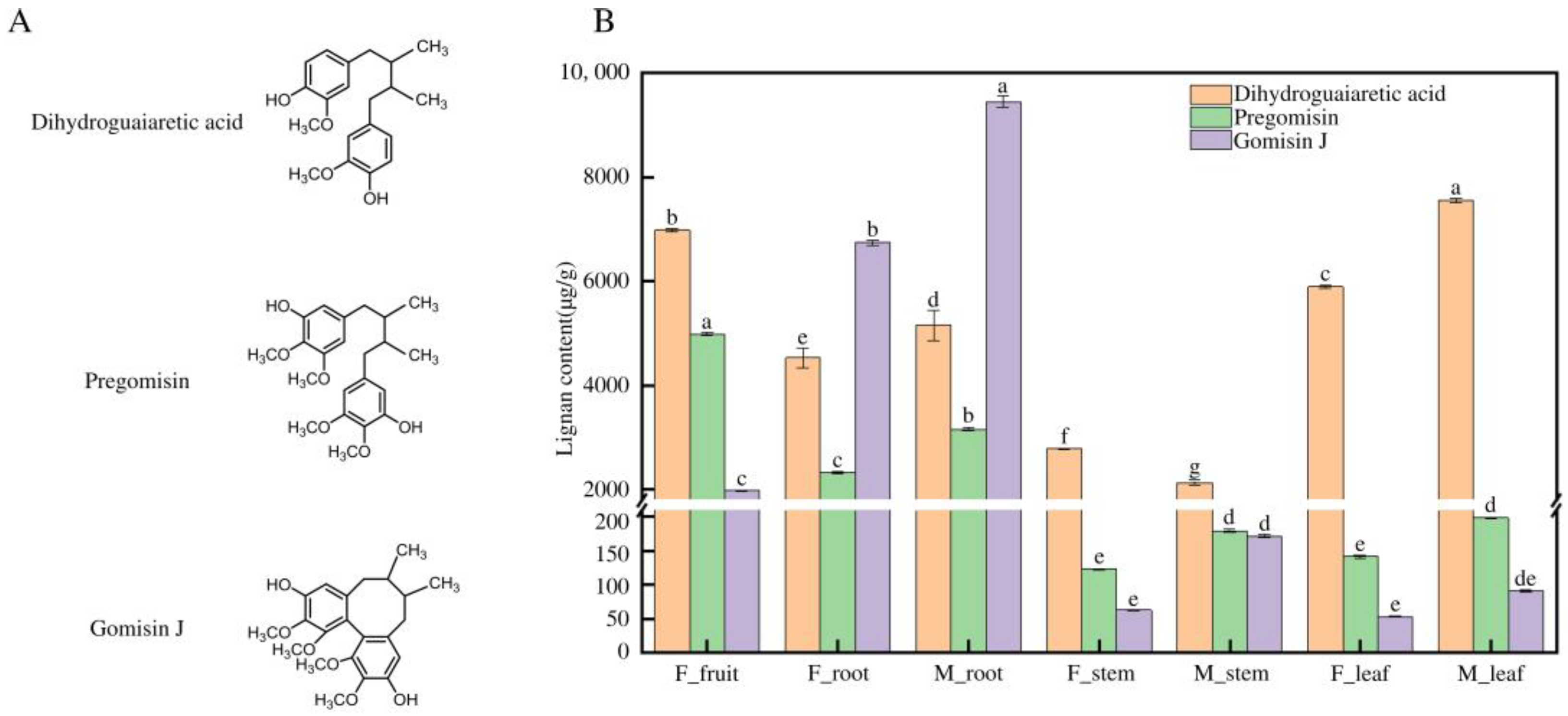
2.2. HPLC Analysis of Lignan Content in Different Tissues of Schisandra sphenanthera

Weigh approximately 0.1 g of the sample, add 1 mL of methanol, grind it into a paste, and then transfer it into an EP tube. Extract with ultrasonication for 30 min, centrifuge, and collect the supernatant. Filter the extracted sample through a 0.22 μm syringe filter. Perform HPLC analysis using the Thermo U3000 high-performance liquid chromatography system. Use a Thermo C18 reverse-phase chromatography column (250 mm × 4.6 mm, 5 μm) for separation. Prepare the mobile phase as follows: A: acetonitrile; B: 0.1% phosphoric acid in water. The injection volume is 10 μL, with a flow rate of 1 mL/min, column temperature at 30 °C, and a run time of 45 min. Use a UV detector at a wavelength of 220 nm. Standards of Dihydroguaiaretic acid (batch number: BBP00124), Pregomisin (batch number: BBP04182), and Gomisin J (batch number: BBP04154), were procured from BioBioPha Co., Ltd. (Kunming, China).

2.3. RNA Extraction and RNA-Seq Workflow in Schisandra sphenanthera

In this study, a detailed RNA-seq workflow was employed to conduct a comprehensive analysis of the Schisandra sphenanthera transcriptome. RNA was extracted from seven different tissues of Schisandra sphenanthera, each with three biological replicates, and then transcribed into cDNA. After the cDNA fragments were purified, a Poly(A) tail was added, and Illumina sequencing adapters were attached. These sequences were then sequenced using the Illumina NovaSeq 6000 system. For data refinement, Trimmomatic (version 0.39) was used to filter the raw data. After removing low-quality reads, those with over 50% of bases having a Q value of 20 or less, clean reads were assembled using Trinity (version 2.15.1) set to default parameters. Finally, RapClust (version 0.1.2) was applied to cluster the transcripts, eliminating redundant sequences, thus as long non-redundant unigenes.
For this study, TransDecoder (version 5.7.0) was employed for the prediction of coding sequences (CDS) of unigenes. For sequence annotation, DIAMOND (version 2.1.8) was utilized to align the predicted CDS with local Nr, KEGG, KOG, TrEMBL, and SwissProt databases, selecting optimal sequences for final annotation. Furthermore, unigenes were annotated against the Pfam database (version 35.0) using PfamScan (version 1.6), applying an E-value threshold of less than 1 × 10⁻⁵. Gene Ontology (GO) and KEGG Orthology (KO) annotations were conducted using InterProScan (version 5.63-95.0) and KofamScan (version 1.3.0), respectively. Gene expression levels were quantified employing Salmon and TPM metrics. This comprehensive methodology was pivotal in identifying crucial genes and pathways in lignan biosynthesis within Schisandra sphenanthera, thereby augmenting the understanding of its genetic composition and pharmacological characteristics.

2.4. WGCNA Methodology and Clustering Analysis in Schisandra sphenanthera

In this research, Weighted Gene Co-expression Network Analysis (WGCNA) was utilized, employing the R package to construct co-expression networks and identify gene modules. The analysis was executed with specific parameters: a soft threshold power of 10, a maximum block size of 25,000, a minimum module size of 30 genes, and a merge cut height of 0.25. The relationship between the modules and phenotypes was depicted using the “ggplot2” package in R to create a heatmap. Further, to elucidate the function of differentially expressed genes (DEGs) in various tissues of Schisandra sphenanthera, clustering analysis was conducted. This analysis incorporated a hierarchical approach based on Euclidean distances and Ward’s clustering method.

2.5. Basic Bioinformatics Analysis of Cytochrome P450

The ORF Finder (https://www.ncbi.nlm.nih.gov/orffinder/, accessed on 3 January 2024) was used to identify the open reading frames of candidate genes of Schisandra sphenanthera. The physicochemical properties were predicted using the free online tool Protparam of ExPASy Proteomics Server (https://web.expasy.org/protparam/, accessed on 3 January 2024). Subcellular localization of cytochrome P450 proteins was predicted using the free online tool Cell-PLoc 2.0 (http://www.csbio.sjtu.edu.cn/bioinf/Cell-PLoc-2/, accessed on 3 January 2024). Conservative domains were predicted by the conserved domain database (https://www.ncbi.nlm.nih.gov/Structure/cdd/cdd.shtml, accessed on 3 January 2024). Phylogenetic trees were constructed using MEGA 11.0 software, employing the maximum likelihood (ML) method with a bootstrap value set to 1000.

3. Results

3.1. Tissue-Specific Distribution of Lignans in Schisandra sphenanthera

The measurement of lignan concentrations in various tissues of Schisandra sphenanthera is crucial for understanding their distribution and potential therapeutic applications. This comprehensive analysis reveals distinct patterns in the accumulation of three lignans—Gomisin J, Pregomisin, and Dihydroguaiaretic acid—across different tissues in both male and female plants (Figure 1). In female (FM) tissues, Gomisin J is notably concentrated in the roots, with an average concentration of approximately 6748.70 ± 49.79 µg/g, significantly surpassing its presence in other tissues. This high concentration in roots is a remarkable finding, indicating a tissue-specific accumulation pattern where roots serve as the primary storage location for Gomisin J. The fruit also shows a notable concentration of Gomisin J, averaging around 1977.52 ± 9.11 µg/g, but its levels in the leaf and stem are considerably lower, averaging around 52.68 ± 0.77 µg/g and 62.52 ± 0.87 µg/g, respectively. This distribution suggests a selective accumulation of Gomisin J in specific tissues, with roots and fruits being the predominant sites. Pregomisin exhibits a similar trend, with the highest concentrations found in the roots (average 2334.54 ± 18.63 µg/g) and fruits (average 4988.79 ± 22.40 µg/g), while its levels in leaves and stems are significantly lower. This pattern reinforces the observation that both Gomisin J and Pregomisin are preferentially accumulated in the roots and fruits of Schisandra sphenanthera. Conversely, Dihydroguaiaretic acid displays a distinct distribution, being predominantly concentrated in the leaves and fruits, with an average concentration of 5896.50 ± 26.37 µg/g and 6983.53 ± 33.35 µg/g, which is substantially higher than in other tissues. The root tissues show moderate levels of this lignan, while the stem contains the least amount. This contrasting distribution pattern of Dihydroguaiaretic acid, compared to Gomisin J and Pregomisin, suggests different biosynthetic and storage mechanisms. In male (M) plant tissues, the concentration of Gomisin J in the roots is even more pronounced, averaging around 9452.51 ± 108.10 µg/g, which is significantly higher than in other mature tissues. This finding underscores the roots’ role as the primary site for Gomisin J accumulation in both FM and M states. Pregomisin and Dihydroguaiaretic acid also show tissue-specific distributions in mature plants, with Pregomisin being most abundant in the fruits and Dihydroguaiaretic acid in the leaves. In summary, the data indicate a clear tissue-specific pattern in the accumulation of lignans in Schisandra sphenanthera, with roots (especially in the mature state) and fruits predominantly accumulating Gomisin J and Pregomisin, while Dihydroguaiaretic acid is primarily concentrated in the leaves. These variations in lignan distribution across different tissues suggest potential differences in their biosynthesis, storage, and physiological roles, contributing significantly to our understanding of their distribution and potential pharmacological uses.

3.2. Elucidating Tissue-Specific Gene Expression Patterns in Schisandra sphenanthera with DEG Analysis

The differentially expressed genes (DEGs) across various tissues of Schisandra sphenanthera provide a comprehensive overview of the expression differences, crucial for identifying key genes for subsequent Weighted Gene Co-expression Network Analysis (WGCNA) (Table 1). In FM tissues, the fruit exhibits a notably higher number of DEGs compared to other tissues, with 2987 up-regulated and 3961 down-regulated genes in comparison to the leaf, and even more pronounced differences when compared to the root and stem. This significant variation in gene expression in the fruit suggests a unique profile that could be linked to its distinct biological functions and developmental processes. The root also shows considerable differential expression, particularly when compared to the leaf and stem, indicating specific gene regulation patterns in these tissues. Comparisons between FM and M tissues reveal interesting patterns. Notably, the FM leaf vs. M leaf and FM root vs. M root comparisons show relatively fewer DEGs, suggesting a stabilization of gene expression patterns as the plant matures. This could imply a reduction in developmental and physiological changes in these tissues during the maturation process. However, the M leaf vs. M root and M leaf vs. M stem comparisons still show a significant number of DEGs, indicating ongoing differential gene expression even in mature tissues.
The overall trend of the analysis indicates that while there are differences in gene expression between male and female plants across various tissues, these differences are less pronounced compared to the significant variations observed in the fruit. The fruit’s distinct gene expression profile underscores its potential role in lignan biosynthesis and accumulation, which is a key area of interest for pharmacological applications. In conclusion, the differential gene expression patterns in Schisandra sphenanthera reveal significant tissue-specific variations, with the most notable differences observed in the fruit tissue. These findings provide valuable insights into the distinct biological roles and developmental processes of each tissue. Understanding these patterns is crucial for guiding further research into the pharmacological applications of Schisandra sphenanthera, particularly in the context of its traditional medicinal uses and the potential for developing novel therapeutic agents.

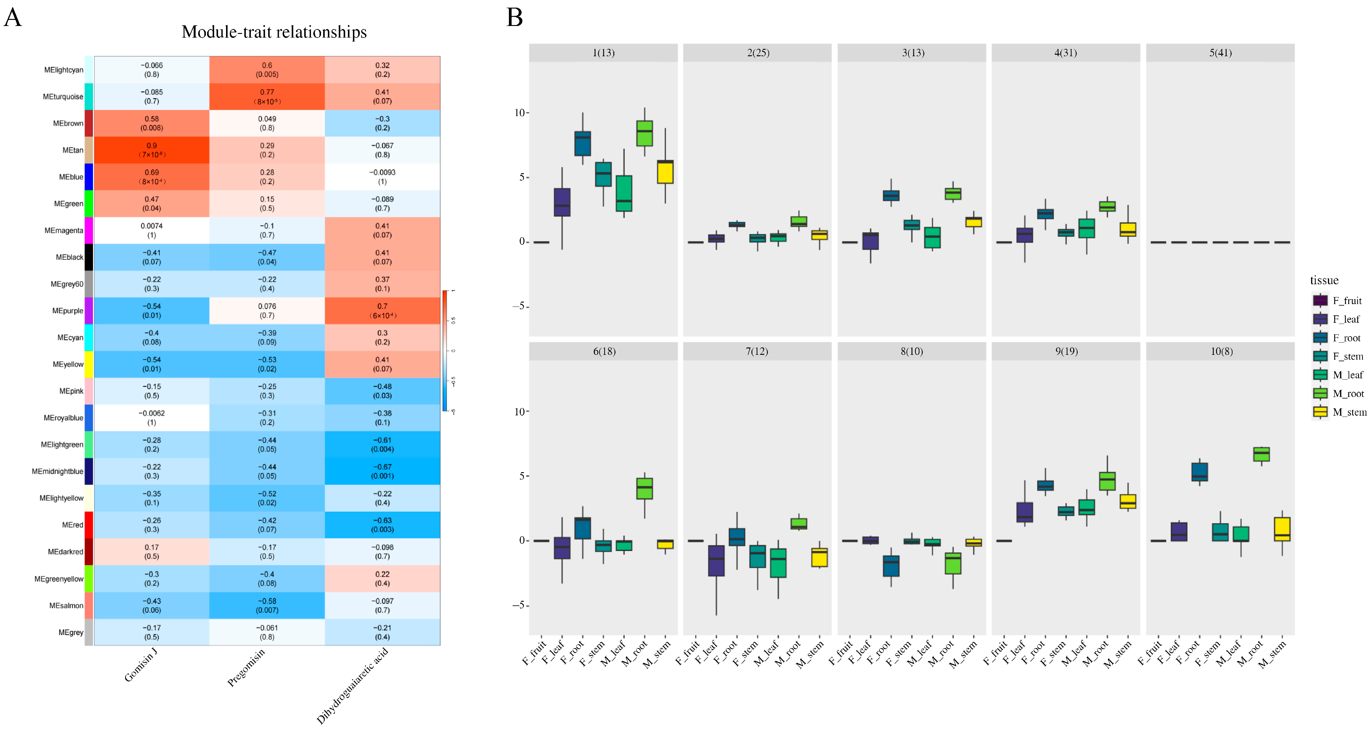
3.3. WGCNA Analysis Deciphering the Gene Expression Patterns in Lignan Biosynthesis

The application of Weighted Gene Co-expression Network Analysis (WGCNA) in the study of Schisandra sphenanthera provides critical insights into the complex gene expression patterns associated with lignan biosynthesis. The module–trait heatmap of the WGCNA reveals the correlation between various gene modules, each represented by a distinct color, and specific lignans (Figure 2A). The MEtan module, in particular, stands out with a strong positive correlation with Gomisin J (0.9), suggesting that it contains genes crucially involved in the synthesis or regulation of this lignan. The MEtan module, contrasted by the MEbrown’s varied effects on Gomisin J and Dihydroguaiaretic acid and the MEturquoise’s link to Pregomisin (0.77), emerges as a central hub. The differential gene expression across modules, such as the 190 genes in MEtan against the 2885 in MEturquoise, points to distinct contributions to lignan metabolism. These insights present a network of gene modules with multifaceted impacts on lignan production, positioning the MEtan module as a primary candidate for further research and metabolic engineering to enhance lignan yields. The varying degrees of positive and negative correlations observed across other modules with the three lignans underscore a multifaceted network of gene interactions influencing lignan production. The presence of correlation coefficients and associated ‘p’ values within the heatmap cells further substantiates the statistical significance of these relationships.
Complementing the WGCNA heatmap, the clustering algorithm delineates ten groups of module genes, revealing a spectrum of expression patterns across Schisandra sphenanthera tissues (Figure 2B). Groups 1–5, with their larger gene pools, appear to embody core expression patterns reflective of general plant functions. Meanwhile, the smaller, latter half, groups 6–10, seems to capture more specialized or tissue-centric expressions, hinting at niche roles in plant physiology. Notably, most genes within these clusters are highly up-regulated in the root, except for group 8, suggesting a concentrated activity in this tissue, which may be critical for Gomisin J accumulation. This up-regulation in roots could signify an adaptive concentration of metabolic pathways pertinent to Gomisin J biosynthesis, as roots often serve as storage sites for bioactive compounds in plants. The outliers identified in the box plots, deviating from the median expression levels, warrant special attention. They could represent unique regulatory genes or enzymes directly involved in the biosynthetic pathway of Gomisin J or stress response elements pivotal to the plant’s survival strategy. These outlier genes might thus offer a focal point for understanding the intricacies of lignan biosynthesis, particularly the enhanced synthesis or sequestration of Gomisin J in the roots of Schisandra sphenanthera.

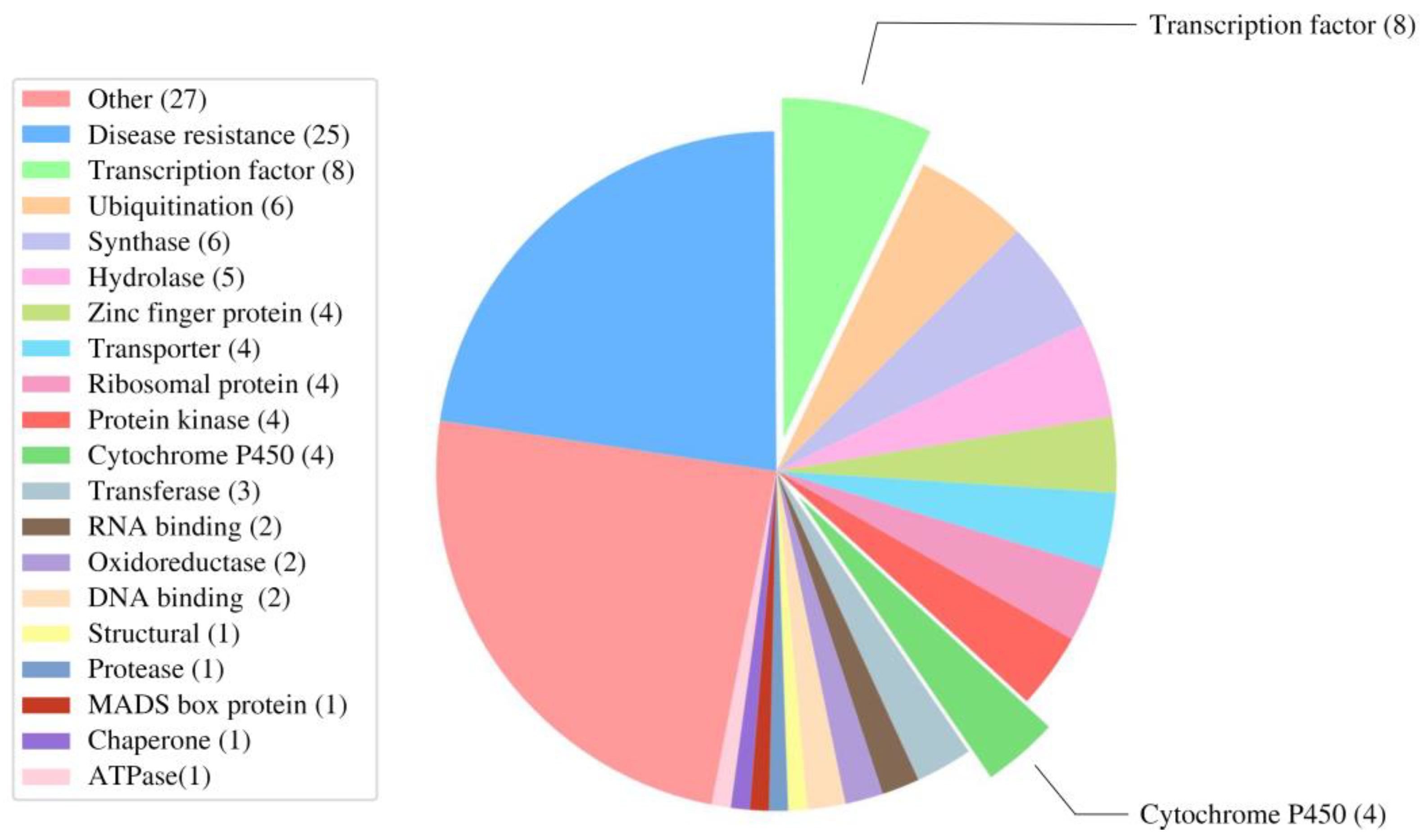
3.4. Deciphering the Tan Module Genes to Uncover Key Genes in Gomisin J Biosynthesis

Weighted Gene Co-expression Network Analysis (WGCNA) serves as a powerful tool to identify gene clusters with coordinated expression patterns, providing insights into the molecular mechanisms underlying complex biological processes. In the case of Schisandra sphenanthera, WGCNA has been instrumental in identifying the tan module, a cluster of genes highly correlated with the biosynthesis of Gomisin J, a pharmacologically significant lignan. Deciphering the tan module has pinpointed a multifunctional gene cluster integral to Gomisin J biosynthesis in Schisandra sphenanthera (Figure 3). Out of 190 genes, 111 annotated genes display a spectrum of activities, from disease resistance (25 genes), suggesting involvement in defensive pathways, to a suite of transcription factors (8 genes) that likely orchestrate the regulation of Gomisin J synthesis. The tan module’s diversity is further exemplified by genes implicated in ubiquitination and synthase activity, each category with six genes, pointing to roles in protein turnover and molecule synthesis. Cytochrome P450 enzymes (4 genes) within the module are particularly noteworthy for their potential direct engagement in Gomisin J’s biosynthetic oxidation–reduction reactions, aligning with their established roles in plant secondary metabolism. This module also encompasses genes linked to transport, protein synthesis, and signal transduction, reflecting its broad involvement in plant physiology. The aggregation of these genes suggests a complex regulatory network orchestrating Gomisin J production, with implications for understanding the modulation of secondary metabolites and advancing the cultivation of lignan-rich Schisandra plants.

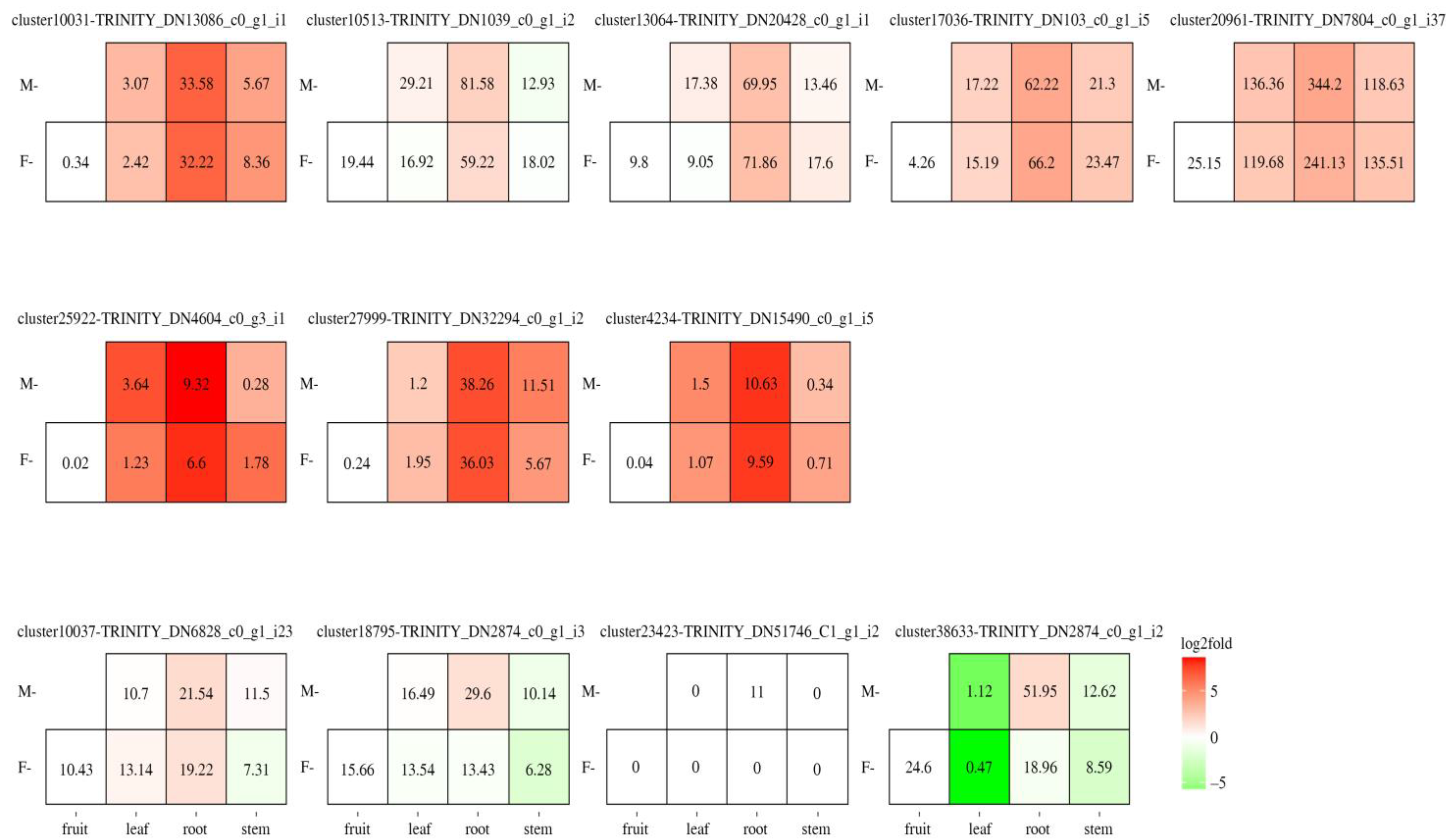
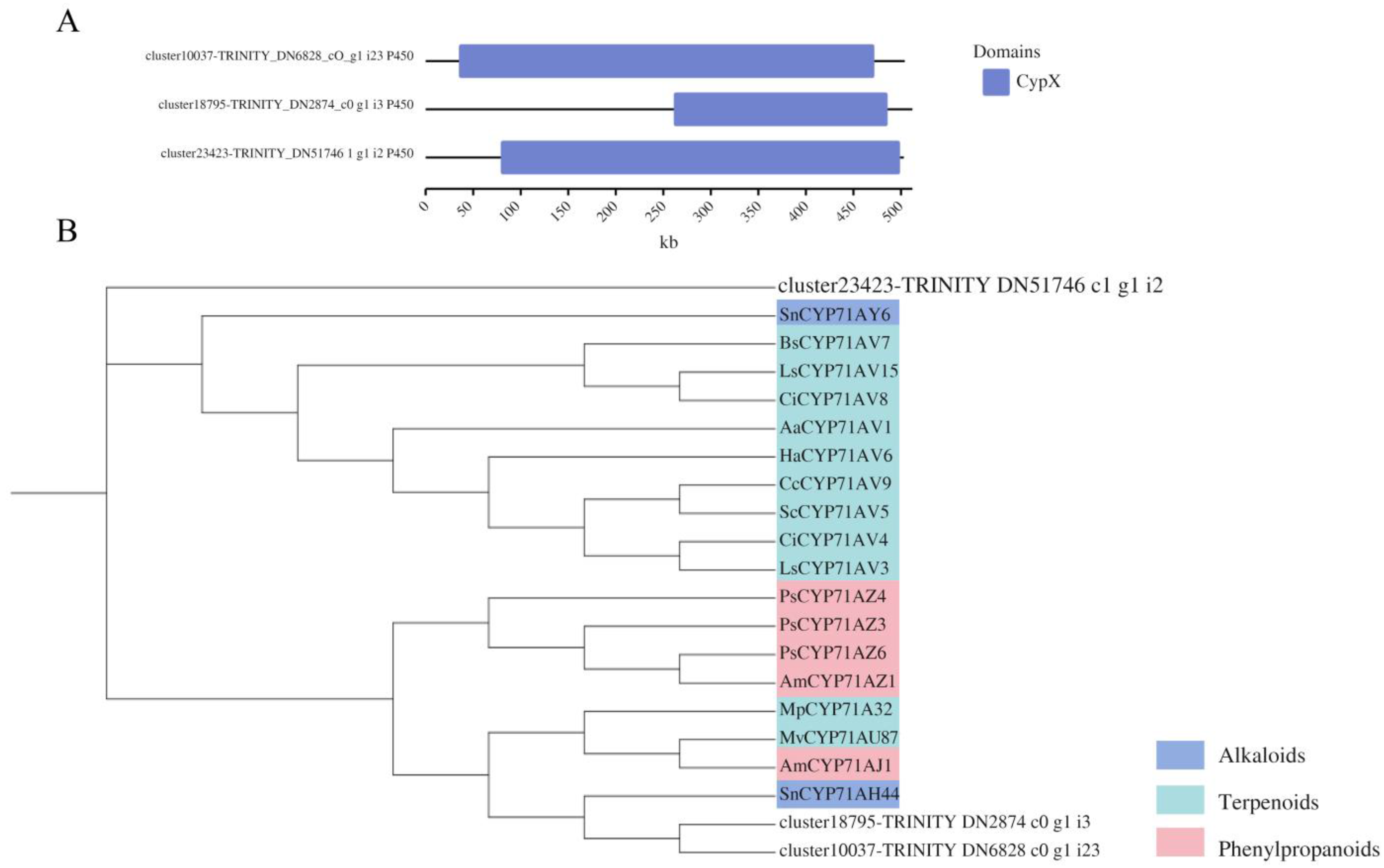
3.5. Investigating Transcription Factors and Cytochrome P450 Roles in Gomisin J Biosynthesis in Schisandra sphenanthera

In the quest to understand the biosynthesis of Gomisin J in Schisandra sphenanthera, a focused analysis of the tan module from a Weighted Gene Co-expression Network Analysis (WGCNA) reveals critical insights. This module, comprising eight transcription factors and four cytochrome P450 enzymes, plays a pivotal role in the plant’s developmental processes. Significantly, the transcription factors are notably up-regulated in both male and female roots, aligning with the observed higher concentrations of Gomisin J in these tissues (Figure 4). For the cytochrome P450 genes, DN6828 and DN2874 are up-regulated in the roots of both male and female plants, while DN51746 and three other cytochrome P450 genes show up-regulation exclusively in male plant roots (Figure 4). This differential expression pattern correlates with the higher concentrations of Gomisin J found in male roots, ranging from 9000 to 10,000 µg/g, compared to 6000–7000 µg/g in female roots (Figure 1). This suggests that the increased expression of certain cytochrome P450 genes in male roots may contribute to the higher Gomisin J content, highlighting a potential gender-specific aspect in the biosynthesis of this lignan.
On the other hand, the transcription factors, including NAC-domain-containing proteins, basic leucine zipper (bZIP), ethylene-responsive factors (AP2), Myb DNA binding, helix-loop-helix (HLH), and WRKY, are instrumental in regulating gene expression (Table S1). They are potentially involved in various biological processes, such as stress response, fruit ripening, flower development, and pathogen defense, indicating their potential role in the biosynthesis and regulation of Gomisin J. In general, all TFs were significantly up-regulated in both male and female plants’ root tissues; this also matches our previous results indicating that Gomisin J is higher in root, and we proposed that TFs may play a regulatory role in lignan synthesis. In addition, we conducted a comparative analysis of three CYPs with functionally characterized homologs, noting that DN2874-i2 is not full-length due to assembly limitations without a reference genome for Schisandra sphenanthera (Table S2). Physicochemical property analysis using Protparam revealed variations in amino acid composition, molecular weight, and isoelectric points of the CYP genes. The amino acid counts for DN6828, DN2874-i3, and DN51746 are 503, 511, and 502, respectively, with molecular weights of 56.72, 58.02, and 57.02 kDa. Their instability coefficients (>40) classify them as unstable proteins, and their hydrophilic nature is indicated by negative hydrophilicity values. Subcellular localization predictions suggest their presence in the endoplasmic reticulum (Table 2), and domain predictions confirm their belonging to the CYP family. Blast analysis revealed high similarity (>60%) of two CYP sequences with CYP71A in various species, classifying them within the CYP71A subfamily. However, the family classification of the third CYP sequence remains undetermined (Figure 5A). Phylogenetic analysis with reported CYP71A family genes confirmed that our two CYP450s cluster with other CYP71As (Figure 5B), suggesting their involvement in the biosynthesis of terpenoids, alkaloids, and phenylpropanoids.

4. Discussion

The comprehensive analysis of lignan concentrations in various tissues of Schisandra sphenanthera has highlighted the pivotal role of cytochrome P450 enzymes in lignan biosynthesis. These enzymes are central to the phenylpropanoid pathway, catalyzing rate-limiting hydroxylation steps leading to the biosynthesis of lignin and numerous other phenolic compounds in plants [10]. Their diverse roles in synthesizing lignin intermediates, sterols, terpenoids, flavonoids, and other secondary plant products [17] are crucial for understanding the tissue-specific distribution of lignans observed in Schisandra sphenanthera. This understanding is further enriched by recognizing that cytochrome P450 enzymes catalyze diverse reactions in the biosynthesis or catabolism of plant bioactive molecules, including lignans [32]. Their involvement in the functional modification of plant natural products is key to the diversification of lignans [33]. For instance, CYP81Q1 in Sesamum catalyzes (+)-sesamin biosynthesis from (+)-pinoresinol, providing insight into unique modes of cytochrome-P450-mediated lignan biosynthesis [15].
The diversification within P450 gene superfamilies has led to the emergence of new metabolic pathways throughout land plant evolution, including lignan biosynthesis [34]. However, despite being the largest family of enzymes in secondary metabolism, the characterized biochemical functions of cytochrome P450s are very small [10]. In this study, the tissue-specific distribution of lignans in Schisandra sphenanthera, particularly the predominance of Gomisin J in roots, can be linked to the differential expression and activity of these cytochrome P450 enzymes (Figure 1). The high concentration of Gomisin J in roots suggests an active biosynthetic pathway mediated by specific P450 enzymes in this tissue. Understanding the regulation and function of these enzymes is crucial for elucidating the biosynthetic pathways of lignans and their accumulation in different plant tissues. In the context of Schisandra sphenanthera, the involvement of cytochrome P450 enzymes in lignan biosynthesis is further substantiated by their essential role in plant steroid hormone biosynthesis and inactivation, including the hydroxylation/oxidation of lignans [35]. This is particularly relevant for Gomisin J, a lignan whose biosynthesis could be significantly influenced by the activity of these enzymes. For instance, CYP3A5 has been shown to metabolize lignan 12 (schisantherin E) to generate major metabolites [36].
The role of transcription factors in lignan biosynthesis, particularly in Schisandra sphenanthera, is a critical aspect of understanding the regulatory mechanisms underlying this complex metabolic pathway. Transcription factors such as MYB, WRKY, AP2/ERF, and others play a pivotal role in controlling the biosynthesis and accumulation of secondary metabolites, including lignans, by integrating internal and external signals and binding to cis-elements [37,38]. For instance, MYB transcription factors are known to be crucial in the biosynthesis of secondary metabolites like lignin, a compound closely related to lignans [38]. Similarly, WRKY transcription factors have been identified as essential regulators of specialized metabolism, including lignan biosynthesis [39]. These transcription factors modulate various signal transduction pathways during biotic and abiotic stresses, contributing to diverse plant processes and defense-related genes [40]. The AP2/ERF transcription factor family, including Ii049, positively regulates lignan biosynthesis in Isatis indigotica by activating salicylic acid signaling and lignan/lignin pathway genes [41]. This highlights the intricate network of transcriptional regulation in lignan biosynthesis. Additionally, the involvement of microRNAs in regulating genes, enzymes, or transcription factors in lignan biosynthesis points to a complex post-transcriptional regulatory layer [42]. In our study, the identification of transcription factors such as NAC, bZIP, AP2, Myb, HLH, and WRKY within the tan module of the WGCNA analysis suggests their significant role in regulating the expression of cytochrome P450 enzymes and other genes involved in lignan biosynthesis (Table 2). These transcription factors are likely responsible for the differential expression patterns observed in various tissues, correlating with the tissue-specific accumulation of lignans like Gomisin J. And the comparative analysis of these transcription factors with other studies reveals a common theme in plant secondary metabolism, where transcription factors orchestrate the biosynthesis of complex compounds like lignans. For example, the overexpression of MYB genes AmMYB308 and AmMYB330 in tobacco plants represses phenolic acid metabolism and lignin biosynthesis [43].
Besides the CYP and transcription factors identified in the tan module, the clustering analysis further divides the module genes into ten distinct groups, revealing diverse expression patterns across different tissue types (Figure 2B). This variation in gene expression is indicative of the complex regulatory mechanisms that control lignan production in Schisandra sphenanthera. The presence of outliers in several groups suggests that some genes have expression levels that deviate significantly from the median of the group, potentially playing unique roles in lignan biosynthesis. This finding from Schisandra sphenanthera is supported by various studies on other plant species. For instance, the complete mitochondrial genome of Schisandra sphenanthera contains 58 genes, including 41 protein-coding genes and 14 transfer RNA genes [44], which could be involved in the biosynthesis of lignans. Additionally, the fruits of Schisandra sphenanthera contain various lignans, including two new dibenzocyclooctadiene lignans, benzoylgomisin U and tigloylgomisin O [45], indicating the diversity of lignan compounds in the plant. Furthermore, the roots of Schisandra sphenanthera contain nine new lignans with noteworthy antioxidant activity [46], highlighting the significance of roots in lignan accumulation. The variation in lignans in Schisandra sphenanthera fruits is also related to the geographical distribution of the plant [47], suggesting environmental factors play a role in lignan biosynthesis. The tissue-specific distribution of lignans in Schisandra sphenanthera, particularly the predominance of Gomisin J in roots, mirrors the diverse localization patterns observed in other plants. The role of cytochrome P450 enzymes and transcription factors in regulating lignan biosynthesis in Schisandra sphenanthera aligns with their crucial functions in other species, as seen in the biosynthesis of sesamin in Sesamum [15]. Moreover, cytochrome P450s are key players in plant defense, secondary metabolite production, and pharmacology, offering new alternatives to modern medicines [48]. They are crucial in herbicide metabolism in plants, potentially leading to crop improvement and weed resistance mitigation [49].
Expanding upon our insights into the regulatory intricacies of lignan biosynthesis in Schisandra sphenanthera, we now turn our attention to the predictive functional analysis and forthcoming characterization of identified cytochrome P450 (CYP) genes: DN6828, DN2874-i3, and DN51746. These genes, posited as central to the biosynthesis of Gomisin J and a spectrum of secondary metabolites, delineate a crucial segment of the plant’s metabolic architecture. Plant cytochrome P450 monooxygenases, renowned for their versatile roles across biosynthetic and detoxification pathways, engage in intricate interactions with a myriad of substrates, highlighting their pivotal role in the realms of pharmacogenomics, drug metabolism, and xenobiotic processing [17]. A comparative analysis with functionally delineated homologs from the CYP71A subfamily, implicated in the biosynthesis of terpenoids, alkaloids, and phenylpropanoids, suggests these enzymes’ extended involvement in plant secondary metabolism. Nevertheless, the protein identity of these CYPs to CYP71As surpassing 55% intimates shared functionalities yet hints at their participation in unique biosynthetic routes. The pronounced correlation between these CYP genes and Gomisin J concentrations, juxtaposed with their modest similarity to established CYP71As, proposes an uncharted facet of CYP functionality in lignan biosynthesis. This inferred novel contribution to lignan biosynthesis, especially concerning Gomisin J, accentuates the imperative for their further functional elucidation. Given the current lack of a high-efficiency transformation system in Schisandra sphenanthera, our strategy encompasses characterizing these CYP genes within surrogate model systems such as Arabidopsis, tobacco, or yeast [13,34]. This methodology aligns with the conventions of functional genomics, where model organisms elucidate gene functionalities, notably in secondary metabolite synthesis. Yeast, with its comprehensive genetic characterization, presents an effective medium for dissecting plant gene functionalities within complex metabolic frameworks [21]. The anticipated functional dissection of DN6828, DN2874-i3, and DN51746 in these models aims to illuminate their specific roles in Schisandra sphenanthera’s lignan biosynthesis. This venture is expected not only to deepen our comprehension of the plant’s secondary metabolism but also to pioneer novel avenues for augmenting the yield of pharmacologically potent compounds. Anticipated outcomes from this research endeavor promise substantial contributions to plant biochemistry and pharmacognosy, enriching our grasp of the molecular underpinnings governing the biosynthesis of plant-derived medicinal agents.

5. Conclusions

This investigation into the lignan biosynthesis within mature Schisandra sphenanthera plants has markedly deepened our comprehension of the specific roles that cytochrome P450 enzymes and transcription factors play in the nuanced distribution and storage of these pharmacologically active compounds. Central to our findings is the significant concentration of Gomisin J within the root tissues, underscoring the specialized function of cytochrome P450 enzymes in the plant’s secondary metabolic pathways. This pivotal discovery, alongside detailed differential gene expression analyses, illuminates the sophisticated regulatory mechanisms that underpin lignan biosynthesis, particularly highlighting the unique gene expression profiles observed in fruit tissues. Our application of Weighted Gene Co-expression Network Analysis (WGCNA) has been instrumental in identifying the MEtan module, which exhibits a strong correlation with Gomisin J levels, thereby pinpointing key genes for lignan biosynthesis. Notably, the cytochrome P450 genes DN6828 and DN2874-i3 demonstrated up-regulation in the roots across genders, with DN51746 showing a unique up-regulation in male roots, suggesting a gender-specific aspect to Gomisin J biosynthesis. The comparative analysis with CYP71A homologs, despite less than 50% identity, suggests these CYP genes may partake in unique biosynthetic pathways, potentially including lignan biosynthesis. This hypothesis opens new avenues for research, particularly in understanding the novel roles these enzymes may play in the biosynthesis of Gomisin J and other lignans, pending further functional characterization. The insights garnered from this comprehensive study not only propel forward our understanding of plant secondary metabolism but also pave the way for future pharmacological exploration of lignans, especially towards developing plant-based therapeutics. These findings lay a solid foundation for subsequent research in plant secondary metabolism, aiming at the enhancement of lignan production for medicinal use. Moreover, the identification of a potential novel aspect of CYP function in lignan biosynthesis highlights the necessity for further functional characterization of these enzymes to elucidate their specific contributions to the biosynthesis of Gomisin J and other lignans in Schisandra sphenanthera.

Supplementary Materials

The following supporting information can be downloaded at: https://www.mdpi.com/article/10.3390/agronomy14030576/s1, Table S1: Annotation of group 12 genes; Table S2: Sequence information of DN2874-i2 gene.

Author Contributions

B.W. and J.P. contributed equally to this manuscript. Conceptualization, S.S. and S.W.; methodology, B.W., J.P. and H.F.; software, B.W., J.P., S.S. and F.S.; validation, B.W., J.P., H.F. and F.S.; data curation, B.W. and J.P.; writing—original draft preparation, B.W. and J.P.; writing—review and editing, S.S. and S.W.; funding acquisition, S.W. All authors have read and agreed to the published version of the manuscript.

Funding

This research was funded by Key R&D Project of Hunan Province (2022WK2021) and the Hunan Policy Project’s Science and Technology Aid to Xinjiang and Science and Technology Aid to Tibet (2023WK4008).

Data Availability Statement

The datasets presented in this study can be found in online repositories. The names of the repository/repositories and accession number(s) can be found below: https://www.ncbi.nlm.nih.gov/, accessed on 20 January 2024, PRJNA1066962.

Conflicts of Interest

The authors declare no conflicts of interest.

References

  1. Chen, M.; Kilgore, N.; Lee, K.; Chen, D.F. Rubrisandrins A and B, lignans and related anti-HIV compounds from Schisandra rubriflora. J. Nat. Prod. 2006, 69, 1697–1701. [Google Scholar] [CrossRef] [PubMed]
  2. Ren, R.; Ci, X.; Li, H.; Luo, G.; Li, R.T.; Deng, X.M. New Dibenzocyclooctadiene Lignans from Schisandra sphenanthera and Their Proinflammatory Cytokine Inhibitory Activities. Z. Naturforsch. B 2010, 65, 1–8. [Google Scholar] [CrossRef]
  3. Li, M.; Zhao, L.; Shi, Y.; Wang, J.; Lv, H.; Song, X.; Zhao, Q.; Zhao, Q.; Jing, R.; Hu, J. Gomisin J attenuates cerebral ischemia/reperfusion injury by inducing anti-apoptotic, anti-inflammatory, and antioxidant effects in rats. Bioengineered 2022, 13, 6908–6918. [Google Scholar]
  4. Li, R.; Yang, W. Gomisin J inhibits the glioma progression by inducing apoptosis and reducing HKII-regulated glycolysis. Biochem. Biophys. Res. Commun. 2020, 529, 15–22. [Google Scholar] [CrossRef]
  5. Kim, M.; Lim, S.J.; Lee, H.J.; Kim, S.Y.; Nho, C.W. Gomisin J inhibits oleic acid-induced hepatic lipogenesis by activation of the AMPK-dependent pathway and inhibition of the hepatokine fetuin-A in HepG2 cells. J. Agric. Food Chem. 2015, 63, 9729–9739. [Google Scholar] [CrossRef]
  6. Samil, J.; Moon, H.; Kim, S.; Quỳnh, N.T.; Yu, J.; Sandag, Z.; Le, D.T.; Lee, H.; Lee, H.; Lee, M. Anticancer of gomisin J from Schisandra chinensis fruit. Oncol. Rep. 2018, 41, 711–717. [Google Scholar]
  7. Song, J.H.; Cui, L.; An, L.B.; Li, W.T.; Fang, Z.; Zhang, Y.Y.; Dong, P.; Wu, X.; Wang, L.X.; Gonzalez, F.; et al. Inhibition of UDP-Glucuronosyltransferases (UGTs) Activity by constituents of Schisandra chinensis. Phytother. Res. 2015, 29, 1658–1664. [Google Scholar] [CrossRef]
  8. Satake, H.; Shiraishi, A.; Koyama, T.; Matsumoto, E.; Morimoto, K.; Bahabadi, S.E.; Ono, E.; Murata, J. Lignan Biosynthesis for Food Bioengineering. In Food Biosynthesis; Academic Press: Cambridge, MA, USA, 2017; pp. 351–379. [Google Scholar]
  9. Costa, M.A.; Xia, Z.; Davin, L.; Lewis, N. Toward Engineering the Metabolic Pathways of Cancer-Preventing Lignans in Cereal Grains and Other Crops. In Phytochemicals in Human Health Protection, Nutrition, and Plant Defense; Springer: Boston, MA, USA, 1999; pp. 67–87. [Google Scholar]
  10. Ehlting, J.; Hamberger, B.; Million-Rousseau, R.; Werck-Reichhart, D. Cytochromes P450 in phenolic metabolism. Phytoch. Rev. 2006, 5, 239–270. [Google Scholar] [CrossRef]
  11. Zhao, X.; Zhao, Y.; Gou, M.; Liu, C. Tissue-preferential recruitment of electron transfer chains for cytochrome P450-catalyzed phenolic biosynthesis. Sci. Adv. 2023, 9, 438. [Google Scholar] [CrossRef] [PubMed]
  12. Liu, L.; Cui, H.; Huang, Y.; Yan, H.; Zhou, Y.; Wan, Y. Molecular docking and in vitro evaluations reveal the role of human cytochrome P450 3A4 in the cross-coupling metabolism of phenolic xenobiotics. Environ. Res. 2023, 10, 115256. [Google Scholar] [CrossRef] [PubMed]
  13. Kim, H.J.; Ono, E.; Morimoto, K.; Yamagaki, T.; Okazawa, A.; Kobayashi, A.; Satake, H. Metabolic engineering of lignan biosynthesis in Forsythia cell culture. Plant Cell Phys. 2009, 50, 2200–2209. [Google Scholar] [CrossRef]
  14. Kuhlmann, S.; Kranz, K.; Lücking, B.; Alfermann, A.; Petersen, M. Aspects of cytotoxic lignan biosynthesis in suspension cultures of Linum nodiflorum. Phytoch. Rev. 2002, 1, 37–43. [Google Scholar] [CrossRef]
  15. Ono, E.; Nakai, M.; Fukui, Y.; Tomimori, N.; Fukuchi-Mizutani, M.; Saito, M.; Satake, H.; Tanaka, T.; Katsuta, M.; Umezawa, T.; et al. Formation of two methylenedioxy bridges by a Sesamum CYP81Q protein yielding a furofuran lignan, (+)-sesamin. Proc. Natl. Acad. Sci. USA 2006, 103, 10116–10121. [Google Scholar] [CrossRef] [PubMed]
  16. Schuler, M.; Werck-Reichhart, D. Functional genomics of P450s. Annu. Rev. Plant Biol. 2003, 54, 629–667. [Google Scholar] [CrossRef]
  17. Schuler, M. Plant Cytochrome P450 Monooxygenases. Crit. Rev. Plant Sci. 1996, 15, 235–284. [Google Scholar] [CrossRef]
  18. Seo, H.J.; Ji, S.B.; Kim, S.E.; Lee, G.M.; Park, S.Y.; Wu, Z.; Jang, D.; Liu, K.H. Inhibitory Effects of Schisandra Lignans on Cytochrome P450s and Uridine 5′-Diphospho-Glucuronosyl Transferases in Human Liver Microsomes. Pharmaceutics 2021, 13, 371. [Google Scholar] [CrossRef] [PubMed]
  19. Giddings, L.; Liscombe, D.; Hamilton, J.; Childs, K.; DellaPenna, D.; Buell, C.; O’Connor, S. A Stereoselective Hydroxylation Step of Alkaloid Biosynthesis by a Unique Cytochrome P450 in Catharanthus roseus. J. Biol. Chem. 2011, 286, 16751–16757. [Google Scholar] [CrossRef]
  20. Kang, K.; Lee, K.M.; Yoo, J.H.; Lee, H.J.; Kim, C.; Nho, C. Dibenzocyclooctadiene lignans, gomisins J and N inhibit the Wnt/β-catenin signaling pathway in HCT116 cells. Biochem. Biophys. Res. Commun. 2012, 428, 285–291. [Google Scholar] [CrossRef]
  21. Decembrino, D.; Ricklefs, E.; Wohlgemuth, S.; Girhard, M.; Schullehner, K.; Jach, G.; Urlacher, V. Assembly of Plant Enzymes in E. coli for the Production of the Valuable (-)-Podophyllotoxin Precursor (-)-Pluviatolide. ACS Synth. Biol. 2020, 9, 3091–3103. [Google Scholar] [CrossRef]
  22. Gorinova, N.; Nedkovska, M.; Bakalova, E.; Atanassov, A.; Ohkawa, H. Biotransformation of Xenobiotics and Endogenous Substrates by Plant Cytochrome P450s. Biotechnol. Biotechnol. Equip. 1999, 13, 13–19. [Google Scholar] [CrossRef]
  23. Xin, A.; Herburger, K. Precursor biosynthesis regulation of lignin, suberin and cutin. Protoplasma 2021, 258, 1171–1178. [Google Scholar] [CrossRef] [PubMed]
  24. Attoumbré, J.; Bienaimé, C.; Dubois, F.; Fliniaux, M.; Chabbert, B.; Baltora-Rosset, S. Development of antibodies against secoisolariciresinol—Application to the immunolocalization of lignans in Linum usitatissimum seeds. Phytochemistry 2010, 71, 1979–1987. [Google Scholar] [CrossRef] [PubMed]
  25. Kulik, T.; Buśko, M.; Pszczółkowska, A.; Perkowski, J.; Okorski, A. Plant lignans inhibit growth and trichothecene biosynthesis in Fusarium graminearum. Lett. Appl. Microbiol. 2014, 59, 99–107. [Google Scholar] [CrossRef] [PubMed]
  26. Satake, H.; Ono, E.; Murata, J. Recent advances in the metabolic engineering of lignan biosynthesis pathways for the production of transgenic plant-based foods and supplements. J. Agric. Food Chem. 2013, 61, 11721–11729. [Google Scholar] [CrossRef]
  27. Köse, L.P.; Gulcin, I. Inhibition effects of some lignans on carbonic anhydrase, acetylcholinesterase and butyrylcholinesterase enzymes. Rec. Nat. Prod. 2017, 11, 558–561. [Google Scholar] [CrossRef]
  28. Fu, H.; Guo, C.; Peng, J.; Shao, F.; Sheng, S.; Wang, S. Transcriptomic insights and cytochrome P450 gene analysis in Kadsura coccinea for lignan biosynthesis. Genes 2024, 15, 270. [Google Scholar] [CrossRef]
  29. Baldi, A.; Farkya, S.; Jain, A.; Gupta, N.; Mehra, R.; Datta, V.; Srivastava, A.; Bisaria, V. Enhanced production of podophyllotoxins by co-culture of transformed Linum album cells with plant growth-promoting fungi. Pure Appl. Chem. 2010, 82, 227–241. [Google Scholar] [CrossRef]
  30. Saarinen, N.; Wärri, A.; Airio, M.; Smeds, A.; Mäkelä, S. Role of dietary lignans in the reduction of breast cancer risk. Mol. Nutr. Food Res. 2007, 51, 857–866. [Google Scholar] [CrossRef]
  31. Ji, F.; Sadreyev, R.I. RNA-seq: Basic bioinformatics analysis. Curr. Protoc. Mol. Biol. 2018, 124, e68. [Google Scholar] [CrossRef]
  32. Morant, M.; Bak, S.; Møller, B.L.; Werck-Reichhart, D. Plant cytochromes P450: Tools for pharmacology, plant protection and phytoremediation. Curr. Opin. Biotechnol. 2003, 14, 151–162. [Google Scholar] [CrossRef]
  33. Liu, X.; Zhu, X.; Wang, H.; Liu, T.; Cheng, J.; Jiang, H. Discovery and modification of cytochrome P450 for plant natural products biosynthesis. Synth. Syst. Biotechnol. 2020, 5, 187–199. [Google Scholar] [CrossRef]
  34. Mizutani, M.; Ohta, D. Diversification of P450 genes during land plant evolution. Annu. Rev. Plant Biol. 2010, 61, 291–315. [Google Scholar] [CrossRef]
  35. Nomura, T.; Bishop, G. Cytochrome P450s in plant steroid hormone synthesis and metabolism. Phytochem. Rev. 2006, 5, 421–432. [Google Scholar] [CrossRef]
  36. Wu, J.J.; Cao, Y.F.; Feng, L.; He, Y.Q.; Hong, J.Y.; Dou, T.; Wang, P.; Hao, D.; Ge, G.; Yang, L. A Naturally Occurring Isoform-Specific Probe for Highly Selective and Sensitive Detection of Human Cytochrome P450 3A5. J. Med. Chem. 2017, 60, 3804–3813. [Google Scholar] [CrossRef]
  37. Yang, C.Q.; Fang, X.; Wu, X.M.; Mao, Y.B.; Wang, L.J.; Chen, X.Y. Transcriptional regulation of plant secondary metabolism F. J. Integr. Plant Biol. 2012, 54, 703–712. [Google Scholar] [CrossRef]
  38. Wang, X.; Niu, Y.; Zheng, Y. Multiple functions of MYB transcription factors in abiotic stress responses. Int. J. Mol. Sci. 2021, 22, 6125. [Google Scholar] [CrossRef] [PubMed]
  39. Schluttenhofer, C.M.; Yuan, L. Regulation of Specialized Metabolism by WRKY Transcription Factors. Plant Physiol. 2015, 167, 295–306. [Google Scholar] [CrossRef]
  40. Wani, S.H.; Anand, S.; Singh, B.; Bohra, A.; Joshi, R. WRKY transcription factors and plant defense responses: Latest discoveries and future prospects. Plant Cell Rep. 2021, 40, 1071–1085. [Google Scholar] [CrossRef] [PubMed]
  41. Ma, R.; Xiao, Y.; Lv, Z.; Tan, H.; Chen, R.; Li, Q.; Chen, J.; Wang, Y.; Yin, J.; Zhang, L.; et al. AP2/ERF transcription factor, Ii049, positively regulates lignan biosynthesis in Isatis indigotica through activating salicylic acid signaling and lignan/lignin pathway genes. Front. Dev. Plant Sci. 2017, 8, 1361. [Google Scholar] [CrossRef] [PubMed]
  42. Ražná, K.; Harenčár, Ľ.; Kučka, M. The Involvement of microRNAs in Plant Lignan Biosynthesis—Current View. Cells 2022, 11, 2151. [Google Scholar] [CrossRef] [PubMed]
  43. Tamagnone, L.; Merida, A.; Parr, A.; Mackay, S.; Culianez-Macia, F.A.; Roberts, K.; Martin, C. The AmMYB308 and AmMYB330 transcription factors from Antirrhinum regulate phenylpropanoid and lignin biosynthesis in transgenic tobacco. Plant Cell 1998, 10, 135–154. [Google Scholar] [CrossRef]
  44. Yu, X.; Feng, Y.; Zhai, W.; Chen, M.; Wu, G. The complete mitochondrial genome of Schisandra sphenanthera (Schisandraceae). Mitochondrial DNA B Resour. 2018, 3, 1246–1247. [Google Scholar] [CrossRef]
  45. Ikeya, Y.; Sugama, K.; Okada, M.; Mitsuhashi, H. Two lignans from Schisandra sphenanthera. Phytochemistry 1991, 30, 975–980. [Google Scholar] [CrossRef]
  46. Jiang, K.; Song, Q.; Peng, S.J.; Zhao, Q.Q.; Li, G.; Li, Y.; Gao, K. New lignans from the roots of Schisandra sphenanthera. Fitoterapia 2015, 103, 63–70. [Google Scholar] [CrossRef]
  47. Gao, J.P.; Wang, Y.H.; Yu, Y.Q.; Chen, D.F. Determination by HPLC and Variation Regularity of Lignan Constituents in Chinese Crude Drug Fructus Schisandrae sphenantherae. Chin. J. Nat. Med. 2003, 2, 28–32. [Google Scholar]
  48. Rasool, S.; Mohamed, R. Plant cytochrome P450s: Nomenclature and involvement in natural product biosynthesis. Protoplasma 2016, 253, 1197–1209. [Google Scholar] [CrossRef] [PubMed]
  49. Dimaano, N.G.; Iwakami, S. Cytochrome P450 enzymes involved in herbicide metabolism in plants. Pest Manag. Sci. 2020, 76, 3346–3355. [Google Scholar]
Figure 1. Lignan concentrations in different plant tissues of Schisandra sphenanthera. (A) Chemical structure of Dihydroguaiaretic acid, Pregomisin, and Gomisin J. (B) Lignan concentration of Dihydroguaiaretic acid, Pregomisin, and Gomisin J in different plant tissues. The columns of the same color show the concentration of the same substance detected in different tissues and organs, and the value is expressed as the mean ± standard deviation (n = 3). Different letters on the columns of the same color indicate significant differences in the concentration of the same substance in different tissues and organs (Duncan’s test, p < 0.05).
Agronomy 14 00576 g001
Figure 2. WGCNA to identify hub genes correlated lignans synthesis and analysis of brown module (390 DEGs) genes using a hierarchical clustering algorithm. (A) WGCNA module–phenotype correlation analysis. (B) The module–trait heatmap of the WGCNA. By calculating Euclidean distances for measuring similarity between gene expression profiles and using Ward’s method for clustering, 10 distinct groups were delineated based on minimal within-cluster variance.
Agronomy 14 00576 g002
Figure 3. Multifunctional gene cluster integral to Gomisin J biosynthesis in Schisandra sphenanthera.
Agronomy 14 00576 g003
Figure 4. Heatmaps of hub genes in group 12. Heatmaps illustrating expression patterns of pivotal genes, with log2-fold change metrics relative to control conditions, where red signifies up-regulation and green down-regulation, with explicit expression values displayed.
Agronomy 14 00576 g004
Figure 5. Conserved domain and phylogenetic tree of CYPs in Schisandra sphenanthera. (A) Conserved domain prediction of three CYPs in Schisandra sphenanthera. (B) Phylogenetic tree of CYPs in Schisandra sphenanthera and other reported CYP71A family genes. The sequences were obtained from NCBI GenBank with the following accession numbers: SnCYP71AY6 (UQZ09621.1); BsCYP71AV7 (ADF43083); LsCYP71AV15 (AIX97103.1); CiCYP71AV8 (ADM86719); AaCYP71AV1 (ABB82944.1); HaCYP71AV6 (ADF43082); CcCYP71AV9 (AIA09035.1); ScCYP71AV5 (ADF43081); CiCYP71AV4 (ADF43080.1); LsCYP71AV3 (ADF32078); PsCYP71AZ4 (AXB38861.1); PsCYP71AZ3 (AXB38860.1); PsCYP71AZ6 (AXB38863.1); AmCYP71AZ1 (ABO32529.1); MpCYP71A32 (AAL06397.1); MvCYP71AU87 (QBS13810); AmCYP71AJ1 (AAT06911.1); SnCYP71AH44 (UQZ09629.1).
Agronomy 14 00576 g005
Table 1. Matrix of differentially expressed genes (DEGs) across various tissues and genders in Schisandra sphenanthera.
FMFruitFMLeafFMRootFMStemMLeafMRootMStem
FMFruit02987/3961
6948
3728/5124
8852
3196/4375
7571
2337/3291
5628
3162/6037
9199
2424/3085
5509
FMLeaf 03065/3601
6666
1521/1181
2702
12/15
27
2131/3261
5392
1320/685
2005
FMRoot 01852/1159
3011
2085/1944
4029
111/135
246
1524/522
2046
FMStem 0832/1125
1957
1486/3902
5388
44/63
107
MLeaf 01696/2704
4400
1116/433
1549
MRoot 02937/524
3461
MStem 0
Note: The numbers before the slash represent up-regulated DEGs, while those after the slash indicate down-regulated DEGs. The final number denotes the total DEGs identified between each pair of tissues.
Table 2. Analysis of physical and chemical properties of 3 cytochrome P450 genes.
Gene IDCDS (bp)Number of Amino AcidsMw (kDa)pICYP450 Subfamily Prediction (NCBI)Instability IndexGRAVYSubcellular Localization
cluster10037-TRINITY_DN6828_c0_g1_i23151250356.726.38CYP71A1-like44.96−0.063Endoplasmic reticulum
cluster18795-TRINITY_DN2874_c0_g1_i3153651158.026.59CYP71A149.75−0.122Endoplasmic reticulum
cluster23423-TRINITY_DN51746_c1_g1_i2150950257.028.4CYP48.26−0.169Endoplasmic reticulum
Disclaimer/Publisher’s Note: The statements, opinions and data contained in all publications are solely those of the individual author(s) and contributor(s) and not of MDPI and/or the editor(s). MDPI and/or the editor(s) disclaim responsibility for any injury to people or property resulting from any ideas, methods, instructions or products referred to in the content.

