Review

Research Progress of Single-Cell Transcriptome Sequencing Technology in Plants

1
College of Agronomy, Gansu Agricultural University, Lanzhou 730070, China
2
Gansu Provincial Key Laboratory of Aridland Crop Science, Gansu Agricultural University, Lanzhou 730070, China
3
Gansu Key Laboratory of Crop Improvement & Germplasm Enhancement, Gansu Agricultural University, Lanzhou 730070, China
*
Author to whom correspondence should be addressed.
Agronomy 2024, 14(11), 2530; https://doi.org/10.3390/agronomy14112530
Submission received: 20 August 2024 / Revised: 15 October 2024 / Accepted: 15 October 2024 / Published: 28 October 2024
(This article belongs to the Section Crop Breeding and Genetics)

Abstract

:
Multicellular organisms exhibit inherent cellular heterogeneity that cannot be captured by traditional high-throughput sequencing techniques, resulting in the unique cellular characteristics of individual cells being neglected. Single-cell transcriptome sequencing (scRNA-seq) technology can be used to determine the gene expression levels of each individual cell, facilitating the study of intercellular expression heterogeneity. This review provides a comprehensive overview of the development and applications of scRNA-seq technology in plant research. We highlight the significance of integrating single-cell multi-omics approaches to achieve a holistic understanding of plant systems. Additionally, we discuss the current challenges and future research directions for scRNA-seq technology in plant studies, aiming to offer valuable insights for its application across various plant species.

1. Introduction

Cells are the basic structural unit of an organism, with their division, differentiation, and functions regulated by gene expression [1]. Although different cells within the same organism share the same genetic material, the expression levels of these genes can vary significantly between cell types, leading to cellular differentiation and specialized functions [2,3]. Bulk RNA-seq, a method for high-throughput sequencing of mRNA and non-coding RNA from specific tissues, provides a comprehensive view of gene expression levels within a cell population [4]. However, this method may overlook individual cellular differences, potentially skewing the interpretation of gene regulation [5,6]. ScRNA-seq addresses this limitation by amplifying and sequencing mRNA from individual cells, allowing for the detailed assessment of gene expression levels at single-cell resolution [7]. This approach enhances our understanding of genetic mechanisms underlying cellular development (Figure 1) [8]. Currently, scRNA-seq technology is widely applied in analyzing human and animal organ tissues, such as the heart, liver, brain, etc., to explore cell types, molecular mechanisms, and gene expression patterns in various diseases, providing a novel theoretical foundation for disease prevention [9,10]. However, its application in plant studies has been slower due to the challenges posed by cell walls, which complicate protoplast isolation [11].
In plant research, scRNA-seq technology is utilized to construct a cell development atlas, identify cell types, map cell development trajectories, and analyze differential gene expression among subgroups [12]. The first successful application of plant single-cell sequencing in 2015 identified root tip cells in A. thaliana [13]. Since then, numerous studies have reported the application of scRNA-seq in A. thaliana, exploring diverse applications such as the characterization of common and rare cell types, identifying novel cell types, analyzing cell dynamic development trajectories, constructing gene regulatory networks, and studying cross-species conservatism [14,15,16]. Recent advancements in molecular biotechnology and protoplast preparation have facilitated the increased use of scRNA-seq in non-model plants. Studies have now been conducted on various plants, including rice [17], maize [18], Chinese cabbage [19], tea [20], peanuts [21], cotton [22,23], tomatoes [24], and poplar [25]. These studies confirm the feasibility of scRNA-seq in plants and offer new insights into plant cell types, functions, and evolution.
Recent reviews have provided in-depth insights into the experimental procedures and analytical methods of scRNA-seq, highlighting the advantages and limitations of this technique [26,27,28,29,30]. However, there remains a lack of comprehensive work addressing the application of scRNA-seq in plant research. This review aims to fill this gap by detailing the development of single-cell technologies, discussing their advantages and disadvantages, and systematically analyzing their applications across various aspects of plant research. We also emphasize the importance of integrating single-cell multi-omics approaches to achieve a holistic understanding of plant systems.

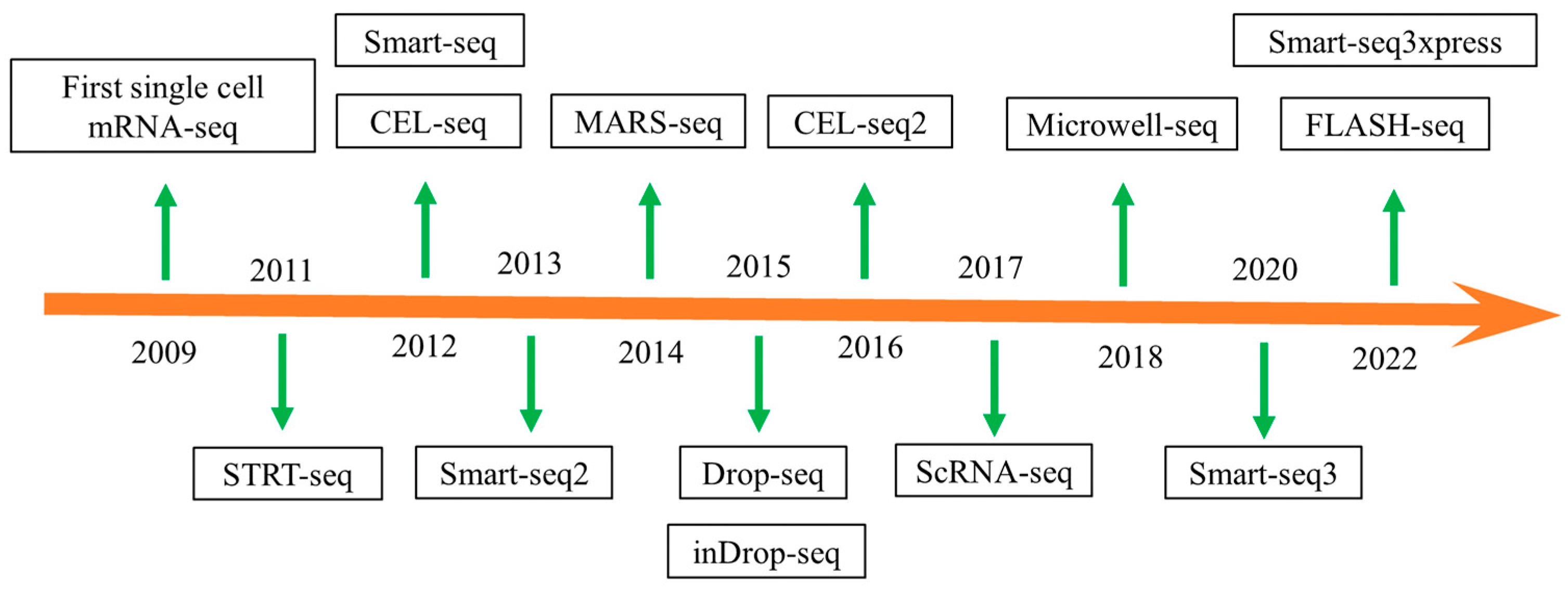
2. Development of Single-Cell Sequencing Technology

The deciphering of the human genome has significantly expanded our understanding of eukaryotic genomes and expression profiles, highlighting the complexity of the genome [31,32]. Gene structure analysis has shown that a single gene can generate different transcripts through different splicing patterns [33]. Genes can also exhibit random monoallelic expression, where only one parental allele is expressed at a time, leading to cellular heterogeneity within the same organ [34,35]. Consequently, there is a critical need for a precise understanding of cell expression patterns, which has led to the development of new sequencing techniques that enable the analysis of single-cell expression. This necessity has given rise to the emergence of single-cell sequencing technology. Figure 2 shows the chronological development of single-cell RNA-seq technologies.
The origins of single-cell technology can be traced back to the 1980s when the development of cell separation techniques laid the foundation for single-cell analysis [36]. In the 1990s, the introduction of single-cell PCR enabled the amplification of RNA from individual cells. This advancement facilitated researchers’ ability to study gene expression at the cellular level [37]. Subsequently, the advent of next-generation sequencing (NGS) technology significantly enhanced the power and efficiency of genomic and transcriptomic analyses [38]. This technological progress provided the basis for the development of single-cell transcriptomics. The early 21st century was a breakthrough for single-cell sequencing technology. In 2009, Tang et al. [39] presented a method for amplifying the entire transcriptome of a single cell, efficiently and unbiasedly amplifying 3 kb cDNA sequences. This technique not only detects previously overlooked transcripts in conventional RNA-seq analyses, but also provides quantitative data on their abundance based on mRNA-seq read frequencies. This breakthrough enabled the study of rare cell types, such as germ cells and early embryonic stages, significantly advancing our understanding of transcriptome dynamics in mammalian development. The method pioneered single-cell transcriptome sequencing technology, making the detection of single-cell transcriptomes a reality. In 2011, Islam et al. [40] developed STRT-seq (Single-cell Tagged Reverse Transcription Sequencing) based on the Illumina platform. This method involves placing a single cell in a 96-well plate with a lysis buffer, adding reverse transcriptase to synthesize the first strand, and incorporating 3–6 C-bases at the cDNA end. A helper oligo is then integrated into the cDNA using template conversion techniques [41], which is amplified by PCR and sequenced with Illumina. This approach does not require known marker genes and enables cell type differentiation based on gene expression patterns, enhancing the detection of mixed cell samples. However, PCR amplification can generate numerous secondary products. In 2012, Hashimshony et al. [42] introduced CEL-seq (Cell Expression by Linear Amplification and Sequencing), which utilizes linear amplification instead of PCR. Compared to other methods, the CEL-seq technique utilizes linear amplification in vitro, offering multiplexing and repeatability while significantly reducing amplification and downstream processing time, allowing for the preparation of dozens of samples for sequencing within 2–3 days. CEL-seq is superior to STRT-seq in terms of stability, sensitivity, and reproducibility. Hashimshony et al. [43] later improved this technology with CEL-seq 2, which reduced the length of the reverse transcription primers and optimized database construction, enhancing detection sensitivity. However, CEL-seq 2 is limited to short fragments of single-cell libraries and does not provide full-length transcriptome information. The same year, Ramskold et al. [44] introduced Smart-seq (Switching Mechanism at the 5′ end of the RNA Transcript Sequencing), which enables full-length transcriptome analysis. Smart-seq improves the coverage of transcripts larger than 1 kb, better analyzes transcript isoforms, and facilitates single nucleotide polymorphism detection. Comparing Smart-seq data and RNA-seq data from individual mouse egg cells showed that Smart-seq detects a broader range of transcripts [45]. In 2013, Picelli et al. [46] refined Smart-seq with the introduction of Smart-seq 2, which improved cDNA library yield, length, detection, coverage, and accuracy while reducing sequencing costs. In 2014, Jaitin et al. [47] developed MARS-seq (massively parallel RNA single-cell sequencing), which enables the simultaneous analysis of thousands of cells through RNA-seq multiplexing. This technique avoids amplification bias and labeling errors by using fluorescence-activated cell sorting to sort individual cells into 384-well plates, thereby increasing throughput and reproducibility.
Advances in single-cell technology have highlighted the importance of studying cell heterogeneity alongside obtaining single-cell transcriptome data. To address the need for improved single-cell flux capture, Macosko et al. [48] introduced Drop-seq (droplet-based RNA-seq), which utilizes droplet microfluidic technology to encapsulate single cells with microbeads and oil-containing molecular barcodes, enabling RNA hybridization within nanoscale oil-in-water droplets followed by reverse transcription. Drop-seq allows for the simultaneous transcriptome analysis of thousands of single cells through high-throughput sequencing. Similarly, Klein et al. [49] developed inDrop-seq (indexing droplet-based RNA-seq), incorporating a T7 promoter sequence and enabling the linear in vitro amplification of RNA after cDNA synthesis. Droplet microfluidic methods offer ease of use, higher throughput, and cost efficiency compared to other techniques. In 2017, 10X Genomics introduced scRNA-seq technology, which uses microfluidics to sort individual cells. This method involves mixing single cells and gel beads with oil to create small water-in-oil droplets. The gel microbeads contain specific DNA fragments of barcodes, a UMI (Unique Molecular Identifier), PolyT, and a reaction reagent mixture (gel bead in emulsion, GEM). The GEM is then dissolved in gel microbeads so that individual cells can be lysed in the microdroplets. The lysed mRNA is then reverse-transcribed into cDNA, amplified, purified, and used to construct a single-cell cDNA library for sequencing. The microbeads’ barcodes confirm that the transcripts originate from the same cell, with each transcript having a unique UMI recognition sequence [50,51]. Due to its cost effectiveness in library construction, standardized operation, and analysis processes, scRNA-seq technology has become a leading method for single-cell sequencing in both animals and plants [52,53]. In 2018, Han et al. [54] introduced Microwell-seq technology, which uses agarose micropores to capture individual cells and magnetic beads for mRNA capture. This technology allows for large-scale parallel single-cell transcriptome analysis, offering high maneuverability and compatibility with minimal reliance on import reagents.
The large-scale RNA sequencing of individual cells can reveal the expression patterns of genes, isoforms, and alleles across various cell types and states [55,56]. In 2020, Hagemann-Jensen et al. [55] introduced the Smart-seq 3 technology as an advancement of Smart-seq 2. By integrating Smart-seq 3 and Smart-seq technology, researchers can capture the complete transcriptome and append distinct molecular tags to the 5′ end for RNA identification and quantification. This approach enables the computational reconstruction of numerous RNA molecules per cell. Smart-seq 3 has a higher sensitivity compared to Smart-seq 2 and enables the detection of thousands of transcripts in individual cells. In 2022, Hagemann-Jensen et al. [57] introduced the Smart-seq 3xpress technology as an optimized version of Smart-seq 3. This innovation enhances cell throughput and minimizes reagent consumption by simplifying the original Smart-seq 3 method. Compared to droplet microfluidics-based single-cell transcriptome sequencing techniques, Smart-seq 3xpress offers comprehensive transcriptional coverage and uncovers variations in cell subtypes associated with specific cell types. This advance not only addresses the resource-intensive nature of the original approach but also challenges conventional wisdom regarding the optimal number of cells needed for precise cell clustering. Subsequently, Hahaut et al. [58] introduced FLASH-seq, a rapid full-length single-cell sequencing method with high sensitivity. Compared to Smart-seq 3, FLASH-seq offers improved sensitivity, shorter processing time, easy automation, and the potential for resource reduction through miniaturization. Contrasted with 10x Genomics (Chromium X, Pleasanton, CA, USA), FLASH-seq not only identifies more genes and gene variations but also enhances subtype comprehension with comprehensive genome coverage. Moreover, its full-length data enable the analysis of genomic single nucleotide polymorphisms (SNPs) at the single-cell level, capturing a large number of SNPs. Splice alterations and epigenetic changes are two main sources of SNPs. By capturing transcriptomic and epigenomic information from individual cells, researchers can more precisely analyze cell type, state, and fate-determination processes. These technological advancements enhance our understanding of how cells generate SNPs through mechanisms such as epigenetics and splicing, which, in turn, influence cell function and the occurrence of diseases.
To date, advancements in single-cell transcriptome technology have made significant progress. Initially, only a few single cells could be captured; however, today, thousands of cells can be captured for transcriptome sequencing. With improvements in sequencing technology and reductions in costs, this technology has expanded its applications across various fields, including medicine [59], animals [60], plants [29], and microorganisms [61]. These developments indicate that single-cell sequencing technology holds great potential for future advancements. Table 1 shows the advantages and disadvantages of some representative single-cell technologies [62].

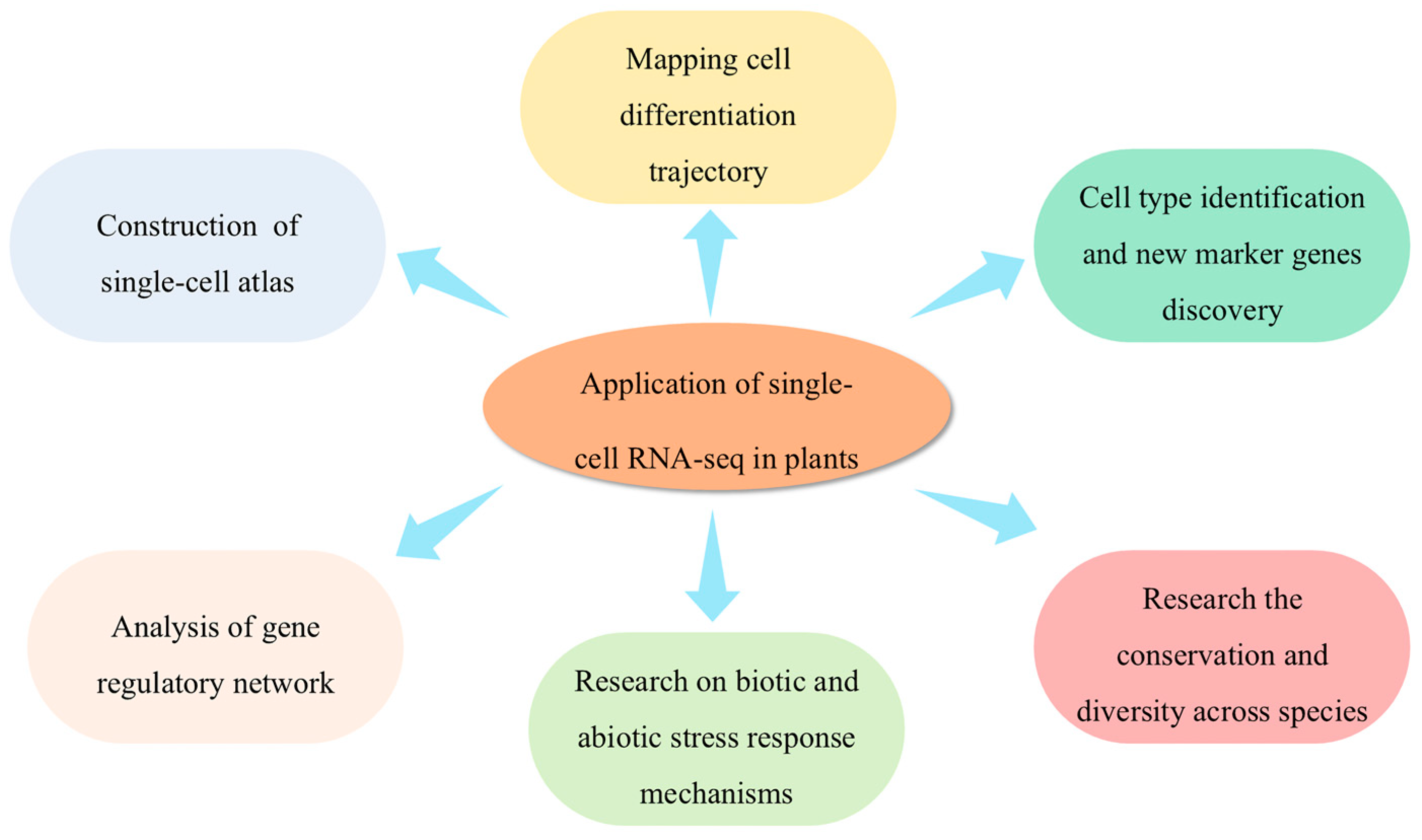
3. Applications of scRNA-seq in Plants

Plant tissue is composed of diverse cell types that collaborate to facilitate normal growth. Since these cells share nearly identical genetic information, functional differences arise from the programmed regulation of transcriptional activity during cell differentiation [63]. Therefore, investigating the cell heterogeneity using scRNA-seq can provide valuable insights into plant physiology and development. ScRNA-seq technology facilitates a comprehensive examination of gene expression at the individual cell level, making it ideal for exploring gene expression variability among cells and understanding how individual cell functions influence overall organism development (Figure 3) [53,64,65].

3.1. Construction of a Single-Cell Transcriptome Atlas and Cell Type Identification

Plant tissue consists of cells with diverse morphologies and specialized functions, each displaying distinct gene expression patterns. By utilizing scRNA-seq to construct a plant cell atlas, researchers can dissect cellular composition within plant tissues and uncover individual cells’ transcriptomic profiles. This process aids in identifying cell types and their functions [66]. For instance, scRNA-seq analysis of rice radicle root tips resulted in the creation of a comprehensive single-cell transcriptional atlas. Cell clustering and uniform manifold approximation and projection (UMAP) visualization identified 21 clusters, including various cell types such as the epidermis, exodermis, sclerenchyma, cortex, endodermis, pericycle, meristem, and vascular tissue, highlighting the heterogeneity of rice root tip cells [67]. Similarly, the scRNA-seq of Chinese cabbage leaves at the rosette stage constructed a single-cell transcriptome atlas with 17 distinct clusters. Using the orthologs of marker genes in A. thaliana, eight cell types were identified, namely mesophyll cells, epidermis, vasculature cells, bundle sheath cells, guard cells, proliferating cells, phloem cells, and xylem cells, revealing the cellular heterogeneity in Chinese cabbage leaves [19]. Constructing a plant single-cell transcription atlas enables us to identify not only the common cell types in plant tissue but also discover rare cell groups, furthering the study of their vital functions in plant development and differentiation. For instance, sequencing rice root tips cultured for 3 days using scRNA-seq resulted in a root tip cell atlas with eight cell types: epidermal cells, endodermal cells, cortex cells, stele cells, root hair cells, root caps, metaxylem cells, and epidermis (near root hair). Notably, the epidermis (near root hair) was identified as a transitional cell type between the root tip epidermal cells and root hair cells in rice [17]. Additionally, the scRNA-seq of A. thaliana root tips cultured for 6 days identified quiescent center (QC) cells based on specific QC marker genes [68].

3.2. Discovery of New Marker Genes

Cell markers are genes that are highly expressed in a specific cell type but not in other types [69]. In multicellular organisms such as plants, the development of cell types and specific functions relies on the unique gene expression patterns within each cell [70]. Single-cell transcriptome analysis utilizes differential analysis to pinpoint characteristic genes in specific cell subsets, which can then be combined with marker genes for accurately identifying different cell types. Discovering new marker genes is essential for uncovering cell heterogeneity and identifying unknown cell populations.
In a study, the scRNA-seq of A. thaliana cotyledons cultured for 5 days revealed the expression of known marker genes involved in stomata lineage development. The specific expression of FAMA, too many mouths(TMM), high carbon dioxide (HIC), and scream (SCRM) was noted, while other marker genes showed no specific expression. By examining gene expression profiles across different cell clusters, new marker genes highly expressed in each cluster were identified, some of which may play a role in regulating stomatal lineage cell development [71]. Similarly, Denyer et al. [68] performed the scRNA-seq of A. thaliana seedlings’ root tissues and identified hundreds of new marker genes by comparing differential gene expression among cell clusters. Ten cluster-specific marker genes with high specificity previously unreported in relation to root development were selected for experimental verification. Through the construction of a promoter—3xVenus-NLS (nuclear localization sequence)—reporter gene line, it was found that the expression patterns of eight genes were consistent with the prediction, showing cell or tissue specificity. The study suggests that in the absence of a reference gene set, this method can effectively classify cell groups and predict gene expression patterns in plants based on the expression profiles of genes in each cell cluster.

3.3. Mapping Cell Differentiation Trajectories

Cell differentiation is the process by which cells of the same origin selectively express the genome in a temporal and spatial manner, leading to the generation of distinct cell types with unique morphological and functional features [72]. This complicated process involves the regulation of gene expression, which ultimately gives rise to characteristic proteins. Pseudo-time analysis is based on the similarity of gene expression patterns between cells, using a tool like Monocle 2, which orders individual cells along a trajectory to infer the continuous differentiation trajectory or subtype evolution [15,73]. Mapping the differentiation trajectory helps reconstruct the temporal changes in cell state and delve into the relationship between cell type and state, providing insights into the differentiation pathways of plant cells and the dynamic developmental processes they undergo [68].
The cell types of root tips undergo a dynamic developmental process at the seedling stage, which consists of the gradual differentiation of a group of undifferentiated apical meristem cells. Li et al. [74] used Monocle 3 to analyze maize root tip cells, revealing a continuous pseudo-time trajectory originating from meristem cells. This trajectory diverged into two paths: one leading to cortex development (cortex and endodermis) and another to stele development (stele, protophloem sieve, companion cells, and pericycle). The trajectory for epidermal cell development showed the progression from epidermal cells, culminating in root hair cells and mature epidermal cells. GO enrichment analysis indicated that genes expressed in these branches were associated with trichoblast differentiation and plant-type cell wall organization. These results support the differentiation of root hair cells from epidermal cells and highlight transcriptional changes in root epidermal cell differentiation, elucidating the developmental process of root hair formation. Similarly, Liu et al. [17] investigated the differentiation trajectory of cell types in the outermost layers of rice roots, finding that epidermal differentiation at the root tip initiates from epidermal cells and bifurcates into two directions: one forming the root cap and mature epidermal cells, the other leading to transitional epidermal cells and ultimately differentiation into root hair cells. Stomata are unique structures in the plant epidermis that participate in gas exchange between plants and the environment, playing a crucial role in photosynthesis, respiration, and transpiration [75]. Liu et al. [71] performed scRNA-seq on A. thaliana cotyledon cells, identifying 11 cell types at different stages of stomatal development. Pseudo-time analysis revealed significant changes in gene expression during stomatal cell development, leading to the identification of marker genes and their crucial roles in stomatal development.

3.4. Analysis of Gene Regulatory Networks

To understand plant development regulatory mechanisms, analyzing gene or transcription factor regulatory networks in different cell types is essential [70]. Plant genomes produce numerous long noncoding RNAs (lncRNAs) expressed in specific cells or developmental stages. These lncRNAs play crucial roles in regulating various biological processes, such as stress responses, morphological development, stem elongation, tillering, and flowering. He et al. [76] integrated 28 scRNA-seq datasets to create a single-cell transcriptome atlas of Arabidopsis seedlings, revealing a co-expression regulatory network of lncRNA and mRNA. Their analysis of xylem cells showed that many gene modules were co-expressed by lncRNAs and protein-coding genes under the influence of the same transcription factors. In another study, Liu et al. [71] investigated transcription factors in different cell types, identifying that basic pentacysteine 1 (BPC1), basic pentacysteine 2 (BPC2), basic pentacysteine 4 (BPC4), basic pentacysteine 6 (BPC6), and WRKY33 were highly expressed in meristematic mother cells (MMC) and guard mother cells (GMC). BPC proteins and WRKY33 are crucial for seedling growth, development, and stress responses. Further analysis revealed that BPC1, BPC6, and WRKY33 were core transcription factors regulating target gene expression in MMC, EM (early stage meristemoid), LM (late stage meristemoid), and GMC. Notably, the co-expression of BPC1 and BPC6 positively influences the majority of known transcription factors, suggesting their role in orchestrating early stomatal lineage cell development. Xu et al. [18] explored the co-expression relationship between knotted 1 (KN1) and its direct regulatory targets using scRNA-seq data, revealing significantly co-expressed genes at the single-cell expression level (p < 0.01). Additionally, combined with chip-seq data, they identified 79 and 55 candidate regulatory targets for homeodomain leucine zipper IV6 (ZmHDZIV6) and mads 16 (ZmM16), respectively.

3.5. Studying the Conservatism and the Diversity Among Cell Types Across Species

Comparing the morphology and biological traits of different subspecies within the same species or between different species is crucial for germplasm resources. At the single-cell level, constructing a single-cell transcriptome atlas for different subspecies or species can uncover both differentiation and conservatism during development. This analysis is valuable for understanding how different subspecies or species respond to external environmental stimuli [77].
Liu et al. [17] performed scRNA-seq on the Japonica group cultivar Nipponbare (Nip) and the Indica group cultivar 93-11, which are prominent cultivated varieties used in Asian rice breeding. They constructed a single-cell transcriptome atlas for the root tips of both cultivars. Pseudo-time analysis revealed that the differentiation of the root tip epidermis progressed from epidermal cells to mature epidermal cells and root hair cells along the pseudo-time axis. The developmental trajectories of these two rice subspecies followed a highly consistent pseudo-time order, demonstrating conserved developmental trajectories across subspecies. Li et al. [74] examined the diversity and conservatism of root cell types in maize and rice. By integrating scRNA-seq data from the root tips of both species, they found that root hair, endodermis, phloem, and exodermis cell types were highly similar between maize and rice. Notably, the root hair cells of both species showed a high correlation and clustered together. Further analysis identified 58 conserved genes expressed in the root hair cells of both maize and rice. GO enrichment analysis indicated that homologous genes in maize were involved in the development of root hair and epidermal cells. These findings highlight the functional similarities of homologous conserved genes in maize and rice, offering a novel approach to identifying cell types or tissue-specific genes. To explore the conservatism and differences in the evolution of fiber initiation cells in cotton and kapok, Ding et al. [78] integrated scRNA-seq data from both species for a comprehensive analysis. They identified 277 homologous genes related to fiber initiation, with 74 genes shared between cotton and kapok. GO enrichment analysis revealed that most specific expression genes (SEGs) were involved in similar biological processes, with only a part of the SEGs in kapok enriched during cell division, suggesting a similarity in the fiber initiation mechanism between cotton and kapok.

3.6. Research on Biotic and Abiotic Stress Response Mechanisms

Biotic and abiotic stresses have detrimental impacts on plant growth and development, physiological metabolism, and substance transport [79]. While bulk RNA-seq provides information on gene expression levels across entire tissues, it does not capture the expression heterogeneity among different cell types. In contrast, scRNA-seq offers a high-resolution view, enabling the identification of transcriptional differences specific to cell types. By utilizing scRNA-seq, researchers can delve into the molecular mechanisms underlying plant stress responses and the discovery of potential stress resistance genes, thereby gaining a more comprehensive understanding of plant survival strategies [80].

3.6.1. Research on Biotic Stress Response Mechanisms

Plant diseases significantly threaten food security and sustainable development, highlighting the importance of understanding the mechanisms underlying plant disease occurrence to develop effective pathogen defense strategies [81]. Host–pathogen interactions are dynamic processes, yet the precise immune responses of plant tissues and cells during infection are not fully understood. ScRNA-seq provides a high-resolution approach to elucidate the main cell types and molecular regulatory mechanisms involved in plant disease responses [82].
In a study, Tang et al. [82] utilized scRNA-seq to explore the transcriptional heterogeneity in A. thaliana leaves infected with Colletotrichum higginsianum at 24 h and 40 h. They observed significant differences in cell proportions among clusters under different infection durations, with the most notable variation occurring at 40 h. The immune receptor nucleotide-binding domain and leucine-rich repeats (NLRs) play an important role in plant defense; thus, the expression patterns of NLRs in different cell types were studied. The results showed that some toll/interleukin-1 receptor nucleotide-binding site leucine-rich repeat (TNL) genes, such as recognition of peronospora parasitica 1 (RPP1), peronospora parasitica 1 (RPP5), suppressor of mpr1-1 (SNC1), and ribosomal protein S6 (RPS6), were predominantly expressed in procambium cells. Conversely, ribosome synthesis regulator 1B (RRS1B), constitutive shade avoidance 1 (CSA1), and chitin synthase 1 (CHS1) were highly induced in phloem companion cells at 24 h but not at 40 h. The study also highlighted the crucial role of RNL in immune signaling, with different RNL family members showing variable expression levels across cell types. For instance, ADR-1 was highly expressed in procambium cells after 40 h of infection, while ADR 1-L1 showed high expression in vascular sheath cells at both time points. ADR 1-L2 was highly expressed in procambium cells and induced in various cell types. These findings provide new insights into the regulation and deployment of immune receptor families across different cell types, paving the way for further exploration into the functional mechanisms of plant immune receptors. Stalk rot is a prevalent disease in maize production, caused by Fusarium verticillioides (Fv), leading to yield loss and decreased quality [83]. To explore the resistance mechanism and immune regulatory network of maize against Fv invasion, Cao et al. [84] investigated the resistance mechanisms and immune regulatory networks in maize using scRNA-seq on root tips of both resistant and susceptible maize inbred lines infected with Fv. They identified seven major cell types and revealed that maize root tips respond to Fv invasion through multiple disease resistance signaling pathways. Additionally, 12 key regulatory modules for Fv response were identified. Notably, the differential expression of wuschel homeobox transcription factor 5b (ZmWOX5b) and pin-formed 1a (ZmPIN1a) genes in the apical meristem was linked to the disease resistance molecular signaling pathway, through regulating indoleacetic acid synthesis protecting the apical meristem from Fv infection.
When plants are infected by a pathogen, it triggers its innate defense system to combat the invasion. However, the impact of the pathogen on different cell types and the intercellular signaling mechanisms remains unclear. Botrytis cinerea is a fungal disease that severely affects the growth of woodland strawberry leaves, flowers, and fruits, posing a significant threat to woodland strawberry production. Bai et al. [85] conducted a study where they sequenced woodland strawberry leaves infected with Botrytis cinerea at 0 h, 6 h, and 12 h time points, creating a detailed single-cell transcriptome atlas of woodland strawberry leaves in response to early Botrytis cinerea infection. The study revealed that the proportions of different cell types changed upon Botrytis cinerea infection. The proportion of hydathode cells increased to 16.6% and 16.3%, respectively, representing a 5% rise compared to the control at 6 and 12 h post-infection (hpi). Conversely, the proportion of mesophyll cells decreased from 13% to 9% at 6 hpi and further dropped to 3.5% at 12 hpi. The proportions of cells and UMI values showed a significant increase in hydathode, upper epidermal, and mesophyll cells during infection, suggesting that these cell types may serve as the initial responders to Botrytis cinerea infection. In addition, various genes associated with the early recognition and signal transduction of Botrytis cinerea, such as leucine-rich repeat (LRR) family proteins and cysteine-rich receptor-like proteins, were identified. Notably, not all cell types express the same resistance genes in leaf tissues. For instance, calmodulin-related genes like CML42 were highly expressed in hydathode cells at 6 hpi, while WRKY75 was up-regulated in the upper epidermal cells, playing a role in the jasmonic acid signaling pathway for plant defense regulation. These discoveries provide a basis for further research on the dynamic process of Botrytis cinerea infection, the functional analysis of candidate genes, and the development of resistant plant varieties.

3.6.2. Research on Abiotic Stress Response Mechanisms

Abiotic stress is a significant environmental factor impacting plant growth and development. Previous research has largely focused on plant physiology under abiotic stress [86], but there is a lack of studies specifically investigating alterations in plant microscopic cell composition and cell types. However, identifying cell types and variations in cell population composition under abiotic stress can enhance the understanding of plant cell mechanisms and developmental biology [87].
Global climate change has led to extreme temperatures, with plant leaves being the first responders to environmental shifts. Chinese cabbage is a cold season leafy vegetable that experiences significant impacts on leaf development due to temperature variations. A recent study by Sun et al. [88] analyzed the single-cell transcriptome of Chinese cabbage leaves subjected to heat stress. The results indicated that heat treatment did not alter cell types but notable differences in cell proportions were observed. After heat treatment, mesophyll cells, epidermal cells, guard cells, and vascular cells showed a decrease in the UMI, whereas proliferating cells did not. Specific gene expressions in response to heat stress were noted among various cell types. For instance, BAA03g00780, encoding a metal-binding protein active after NaCl and heavy metal stress, was up-regulated in mesophyll cells post-heat treatment. Similarly, genes like BAA03g43520, BAA07g19420, BAA03g14340, and BAA10g23280 showed differential regulation in epidermal cells, vascular bundle cells, guard cells, and proliferating cells, respectively. Variations in gene expression patterns under heat stress were observed, with MSS3 (a calcium-binding EF-hand family protein) up-regulated in mesophyll cells but down-regulated in epidermal cells, and WRKY8 (a DNA-binding protein) up-regulated in vascular cells but down-regulated in mesophyll cells. Furthermore, several ribosomal protein family genes also exhibited cell type-specific expression changes. These findings provide insights into gene expression patterns and the impact of heat stress on cell type-specific genes.
Phosphate deficiency in the soil impacts plant growth as roots rely on soil phosphate, which can be obtained by increasing root hair density. Wendrich et al. [89] utilized scRNA-seq on A. thaliana root tips to discover that target of monopteros 5/lonesome highway (TMO5/LHW) target genes were notably present in root hair cells. The TMO5/LHW heterodimer stimulates cytokinin synthesis in vascular cells, enhancing root hair density under low phosphorus conditions by altering epidermal cell length and fate. The response of root hairs to low phosphorus conditions is influenced by TMO5 and cytokinins, indicating a correlation between cytokinin signals, root hair response in the epidermis, and phosphate consumption in vascular cells. Wang et al. [90] investigated the impact of high-salt and low-nitrogen stress on rice seedlings using the scRNA-seq technique, identifying five distinct cell types in both the treatment and control groups. Differential gene expression across samples highlighted that transcriptome changes in response to abiotic stress were cell type-specific, altering the cell population composition and impeding mesophyll cell differentiation. This research contributes to a deeper understanding of plant cell biology and development, with implications for breeding stress-tolerant crops in the future.
The root system is fundamental to maize growth and yield. Li et al. [74] focused on characterizing nitrate response at the single-cell level in maize root tips cultured for 4 days. The results indicated that nitrate did not significantly alter cell types or numbers but revealed notable differences in nitrate response genes among the eight cell types, particularly in cortex and companion cells. Nitrate induced the expression of ZmGS2 (glutamine synthetase 2) and ZmNAR2.1 (nitrate transporter 2.1) genes in root epidermal cells, with ZmGS 6 (glutamine synthetase 6), ZmNIR 1 (nitrite reductase 1), and genes related to NADPH synthesis being highly expressed in the roots for NO3 assimilation.
Boron is an essential trace element for plant growth, and its deficiency will lead to reduced crop yield [91]. Chen et al. [92] explored the impact of boron deficiency on stem tip development in peas using scRNA-seq. They found that while boron deficiency did not alter cell classification, it increased the proportion of shoot apical meristem and mesophyll cells, and decreased epidermal and vascular cells. This deficiency affected signal transduction related to shoot apical development, resulting in reduced photosynthetic rates and stomatal density in mesophyll cells. The study also identified key regulatory factors in this process, offering valuable insights for enhancing plant stress resistance and developing genotypes resilient to boron deficiency.

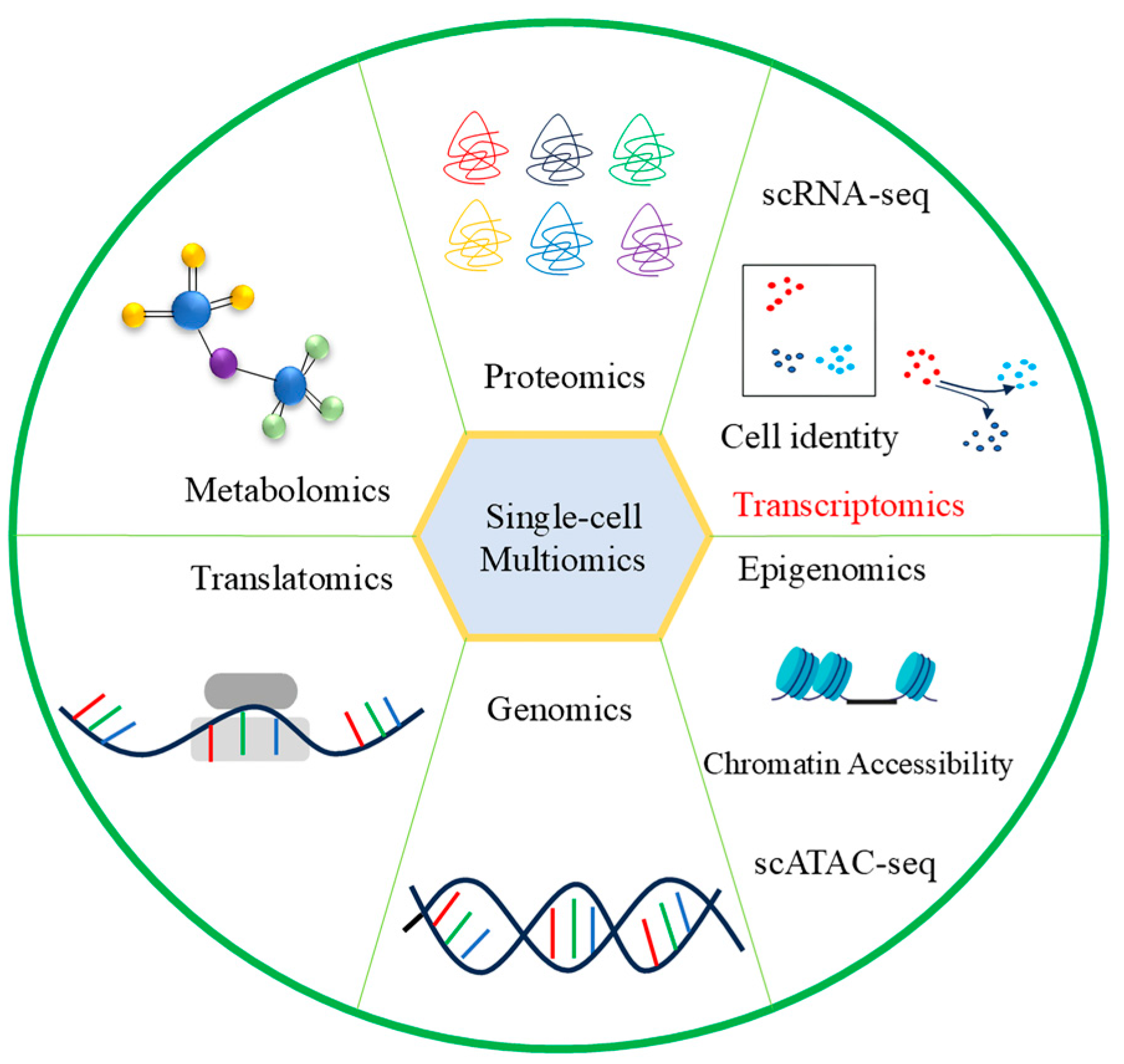
3.7. Combined Analysis of Single-Cell Transcriptome and Other Omics

Recent advancements in multi-omics technologies have enabled single-cell multi-omics analyses (Figure 4). Integrating biological data from various levels, including genomics, transcriptomics, proteomics, and spatial omics, can offer a novel perspective and a deeper understanding of the underlying plant growth and development [28,93,94].

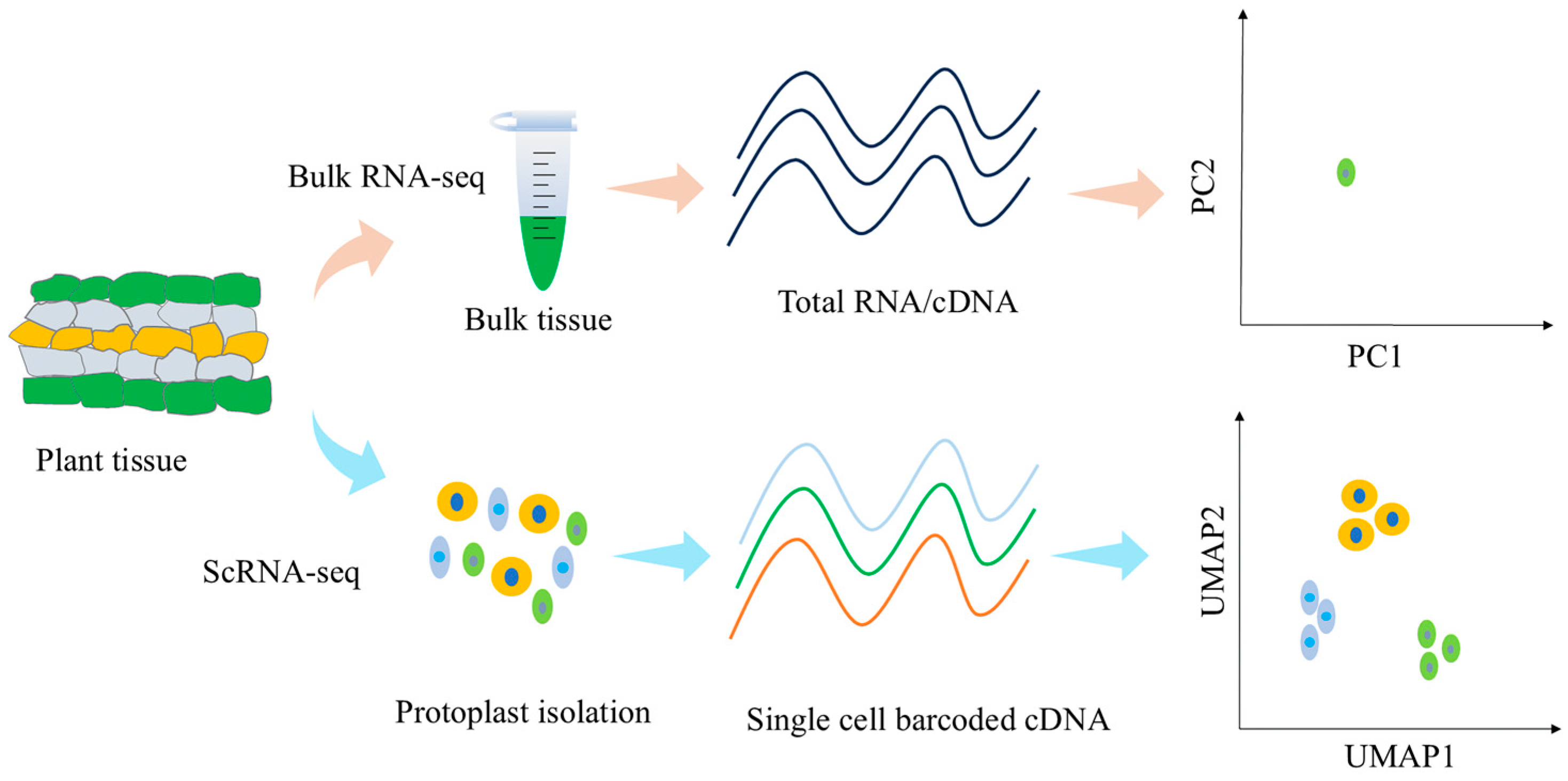
3.7.1. Single-Cell Transcriptional Sequencing and Bulk RNA-seq

ScRNA-seq offers high resolution but may have lower gene coverage compared to bulk RNA-seq. Integrating bulk RNA-seq data with scRNA-seq can enhance the imputation of missing data points and provide prior knowledge about biological pathways, regulatory networks, and gene interactions to guide the interpretation of single-cell results [95]. A high correlation between the two datasets indicates the reliability of scRNA-seq data in reflecting cell characteristics. Cross-referencing can further validate the accuracy of scRNA-seq annotations. This approach highlights the potential for gene discovery and result verification at a single-cell resolution, leveraging the complementary strengths of both omics methods [74].
In a recent study, Sun et al. [23] investigated the biological processes underlying cotton pigment glands’ morphogenesis and the molecular mechanisms influencing the delayed morphological development of these glands. By correlating scRNA-seq and bulk RNA-seq data, they identified the top 10 marker genes in pigment gland cells with significant expression differences between samples with and without glands in bulk RNA-seq. Notably, the most marker genes down-regulate in non-gland cells compared to bulk samples containing glands, suggesting the higher quality and accuracy of cell annotation in scRNA-seq. Furthermore, they identified differential genes involved in the developmental trajectory of pigment gland cells. The expression patterns of the top five subgroup marker genes increased during germination in bulk RNA-seq, while marker genes associated with secretory cells and apoptotic cells were not expressed at 0 and 12 h, aligning with the development of cotyledon pigment glands. These findings confirmed the precision of re-clustering and pseudo-time analysis. Similarly, Li et al. [87] utilized both bulk RNA-seq and scRNA-seq analyses on cotton root tips. They observed a pearson correlation coefficient (PCC) of approximately 98% between isolated protoplasts and unisolated bulk RNA-seq data, and around 80% between the two biological replicates of dissociated protoplasts from scRNA-seq data. Notably, the PCC between bulk RNA-seq and scRNA-seq data following protoplast isolation was higher than that without protoplast isolation, underscoring the high quality and reliability of scRNA-seq data.

3.7.2. Single-Cell Transcriptome and Spatial Transcriptome

Understanding the spatial expression patterns of genes in plant tissue cells is crucial for comprehending plant development, evolution, plant–environment interactions, and other biological processes [96]. Traditional methods such as histochemical staining, in situ hybridization, DNA microarray, and bulk RNA-seq have been used to analyze the spatial expression characteristics of target genes at the tissue level and identify key genes associated with growth and development [97,98,99]. Moreover, cell sorting techniques like laser capture microdissection (LCM), fluorescence-activated cell sorting (FACS), and cell type-specific tagged nuclear separation techniques (INTACT) are combined with bulk RNA-seq to explore the spatial expression of target genes in specific cell types [100,101,102,103]. However, these approaches suffer from low throughput and low resolution, which limits the investigation of regulatory mechanisms in tissues with high cellular heterogeneity. ScRNA-seq has overcome these limitations by enabling the identification of gene expression patterns at the single-cell level, thus facilitating the analysis of cell heterogeneity, cell type identification, and cell lineage construction [28,53,104]. Despite this, scRNA-seq requires cell separation from tissues, leading to a loss of spatial information crucial for understanding cell development and function [96,105]. Spatial transcriptome sequencing (ST-seq) analyzes gene expression at a spatial level by retaining sample location information on a gene chip for mRNA analysis within a single tissue section [106]. This technique enables the identification of functional gene expression in specific tissue regions and provides comprehensive spatial location information of the tissue [107]. This is useful for studying plant regulatory networks; ST-seq maintains spatial information during transcriptome analysis, offering precise results for studying plant regulatory networks. However, it does not achieve single-cell resolution [93]. Integrating scRNA-seq and ST-seq techniques enables the spatial localization of distinct single-cell subsets and the annotation of cell types within spatial transcriptome regions, enhancing the understanding of cell subsets and their interactions in development, homeostasis, and disease incidence [108].
Xia et al. [109] mapped the single-cell spatial transcriptome atlas of Arabidopsis leaves. Previous studies on plant single cells faced the challenge of distinguishing very similar cell subsets due to overlapping molecular characteristics, such as upper and lower epidermal cells within epidermal cells, palisade cells, and sponge cells within mesophyll cells. However, by integrating spatial information with single-cell transcriptome data, the researchers successfully differentiated similarly expressed upper and lower epidermal cells, palisade cells, and sponge cells, shedding light on the distinctions and functions of these cell types. In a separate study, Li et al. [110] constructed a single-cell transcriptional atlas of primary and secondary growth tissues in poplar stems using scRNA-seq and analyzed stem cross-section cells using a spatial transcriptome approach. This integration allowed for the identification of cell clusters and marker gene localization, providing a systematic view of the molecular networks involved in wood formation at the cellular level. Moreover, Liu et al. [111] combined single-nuclear transcriptome sequencing (snRNA-seq) and Stereo-seq techniques to create a single-nuclear transcriptome atlas of soybean root nodules and roots. Their study revealed dynamic gene expression changes during root nodule maturation and identified key transitional cell subtypes crucial for nodule maturation and nitrogen fixation, offering new insights into rhizobia–legume symbiosis.

3.7.3. Single-Cell Transcriptome and ATAC-seq

Gene expression in eukaryotes is closely linked to chromatin accessibility, reflecting chromatin’s transcriptional activity and serving as a key area of gene expression regulation research [112]. Assay for transposase-accessible chromatin using sequencing (ATAC-seq) is a rapid and sensitive epigenetic technique that utilizes the Tn5 enzyme to cleave open chromatin regions for amplification and sequencing [112]. Coupled with motif analysis, ATAC-seq can identify specific transcription factors involved in gene regulation [113]. This method allows for the assessment of chromatin accessibility, the identification of active regulatory sequences, the exploration of gene expression regulation, and the examination of cell heterogeneity through chromatin accessibility discrepancies to elucidate the underlying reasons for gene expression changes at the epigenetic level [114,115,116]. Integrating scRNA-seq and ATAC-seq techniques can pinpoint cis-regulatory elements and trans-acting factors relevant to cellular activity, elucidating the collaborative impact of regulatory elements on cellular regulation [117].
Zhang et al. [67] performed scRNA-seq and ATAC-seq on meristem and extension regions of the rice radicle, revealing root heterogeneity and constructing an epidermal cell differentiation trajectory. They also elucidated the correlation between gene expression and chromatin accessibility during root stem cell differentiation and the evolutionary conservatism of root tip cell types between monocotyledonous rice and dicotyledonous A. thaliana. Similarly, Wang et al. [118] integrated scRNA-seq and scATAC-seq to systematically characterize cells of the outer integument of ovules from wild-type cotton and fuzzless/lintless (fl) cotton. Combining analyses of the scRNAseq data from wild-type and fl cotton, they identified five cell populations, including fiber cells, and constructed the fiber lineage development trajectory. Combining scRNA-seq and scATAC-seq data, they identified two cardinal cis-regulatory elements (CREs): TCP (Teosinte branched 1/Cycloidea/Proliferating cell factor) motif and TCP-like motif. These elements are bound by the trans factors GhTCP14s to modulate the circadian rhythmic metabolism of mitochondria and protein translation by regulating approximately one-third of genes highly expressed in fiber cells. This study unprecedentedly reveals a new route to improving fiber traits by engineering the circadian clock of fiber cells. Additionally, Xu et al. [18] constructed a developmental atlas of maize ears using scRNA-seq and combining ATAC-seq data to explore gene expression regulatory networks, identifying yield trait-related genes through genome-wide association studies (GWAS), thus advancing the application of single-cell sequencing in crop research.

4. Challenges and Prospects

ScRNA-seq has been extensively used in animal research, but it remains a nascent field in plant research that requires further exploration and development. Several challenges remain. Firstly, enhancing sequencing efficiency and reducing costs are important development directions for the future. Different species exhibit significant differences, even within the same tissues. Plant tissues exhibit a greater complexity and diversity of cell types compared to animal tissues, leading to the cost of scRNA-seq being relatively high compared to bulk RNA-seq, mainly due to the additional steps required to isolate individual cells and library preparation, which limits its widespread application in plant research [119]. However, with advancements in technology, a reduction in the cost of single-cell transcriptome sequencing is anticipated in the future. For example, SPLit-seq (split pool ligation-based transcriptome sequencing) technology reduces the cost of single-cell transcriptome sequencing by utilizing low-cost combinatorial barcodes. This technological innovation has significantly enhanced the accessibility and applications of single-cell sequencing [120]. Furthermore, Paired-seq technology integrates a highly efficient single-cell capture manipulation microfluidic chip with DNA-encoding microbead technology, allowing for the simultaneous analysis of hundreds or thousands of single cells, thereby improving sequencing efficiency and reducing costs [121].
In addition to the cost and complexity challenges, experimental protocols for scRNA-seq need continuous refinement. Currently, this technology is predominantly applied to the model plant A. thaliana. However, the commonly used method of protoplast isolation in model plants may not be suitable for non-model plants [27]. This discrepancy arises from significant differences in tissue structure, cellular characteristics, and experimental conditions between non-model and model plants, which limits the applicability of traditional methods for non-model species. For instance, non-model plants like peanuts exhibit highly differentiated tissues, complicating the preparation of protoplasts. Furthermore, a major challenge during the preparation process is the loss of transient transcriptional information within cells [122]. Therefore, optimizing protoplast preparation for non-model plants is crucial for increasing both the quantity and activity of protoplasts. In single-cell sequencing studies, the number of cells significantly impacts the results. Research indicates that the number of cells captured ranges from a few hundred to several thousand in different study categories. Most studies show that a minimum of 1000 cells is necessary for reliable results. In addition, cell viability and fragmentation rates are important factors affecting results. During protoplast preparation, the viability should be 98 percent and the fragmentation rate should be less than 10 percent [48,51,123].
Fresh and young plant tissues are the basis for the preparation of high-quality protoplasts. The plant cell wall’s main components are cellulose and pectin; enzymatic digestion of the plant cell wall is necessary to obtain free single cells. Unfortunately, this treatment alters the expression levels of many genes, particularly those involved in defense and stress responses, rendering protoplasts undesirable for the study of genes that participate in early symbiotic interactions [124]. However, snRNA-seq has emerged as a viable alternative to protoplasts for single-cell sequencing, demonstrating significant potential. The snRNA-seq technology directly sequences RNA molecules without requiring mRNA processing steps, offering several significant advantages. First, it captures the full-length sequence of RNA, including complex transcript isoforms and RNA modifications, which are often overlooked in conventional sequencing methods. Second, direct RNA sequencing can be performed without PCR bias, enhancing accuracy and reliability. Furthermore, this technology is capable of detecting the chemical modifications of RNA, such as methylation (e.g., m6A), which is crucial for understanding gene expression regulation and disease mechanisms [125,126,127]. In animal studies, snRNA-seq addresses challenges in studying cell types that are difficult to dissociate into individual cells or sensitive to enzymatic treatment [125,128]. Similarly, snRNA-seq can be beneficial for plants that cannot be easily converted into a single-cell suspension or those with oversized cells [129]. It does not require a high-activity single-cell suspension or tissue dissociation, isolating only the cell nucleus from the cytoplasmic lysate, thereby avoiding potential biases that arise during tissue processing and accurately representing the different cell types within the tissue [130]. The study of snRNA-seq holds significant importance for analyzing specific samples.
Cell type annotation is a crucial aspect of scRNA-seq data analysis, with cell marker genes playing a key role in identifying and classifying cell types [69]. Currently, the CellMarker database provides comprehensive cell marker resources for both humans and mice [131]. However, plant research lacks a similarly extensive resource. Marker genes for Arabidopsis thaliana roots have been extensively studied and validated, but marker genes for other organs and species are sparse and dispersed across numerous publications, making it challenging for researchers to collect them for specific cells [69]. Therefore, there is a pressing need for a comprehensive, user-friendly single-cell database for plants. In recent years, several online-based databases have been established, such as the PscB (Plant scRNA-Seq Browser, https://www.zmbp-resources.uni-tuebingen.de/timmermans/plant-single-cell-browser, accessed on 1 June 2020) [132], PlantscRNAdb (Plant single-cell transcriptome database, http://ibi.zju.edu.cn/plantscrnadb/, accessed on 4 May 2021) [133], PCMDB (Plant Cell Marker DataBase, http://www.tobaccodb.org/pcmdb/, accessed on 28 October 2021) [69], PsctH (Plant Single Cell Transcriptome Hub, http://jinlab.hzau.edu.cn/PsctH/, accessed on 29 September 2021) [134], and scPlantDB (https://biobigdata.nju.edu.cn/scplantdb, accessed on 15 August 2023) [135]. Although these databases significantly aid cell type annotation, they provide limited information for non-model plants, highlighting the need for more comprehensive databases in the future.
Secondly, exploring additional application areas is another key focus for future development. Integrating scRNA-seq with other omics data can offer the potential for deeper insights into cellular biology. Single-cell and bulk genomics technologies each have strengths and limitations, and neither alone fully captures the regulatory elements across diverse cells in complex tissues [136]. For example, Przytycki et al. (2021) developed CellWalker, a network model that integrates scRNA data with bulk data, annotating unique cell types for bulk data using scRNA-seq information [137]. Additionally, jointly semi-orthogonal nonnegative matrix factorization (JSNMF) is a versatile toolkit that enables data visualization and the clustering of cells and facilitates downstream analysis. The application of JSNMF to single-cell multi-omics datasets from different tissues and different technologies results in superior performance in the clustering and data visualization of cells [138]. These approaches significantly improve the accuracy of cell labeling in noisy scRNA-seq data. Tissue dissociation in scRNA-seq results in a loss of spatial information about cell location. To address this, combining ST-seq with scRNA-seq or snRNA-seq could provide a more comprehensive view of spatial and single-cell gene expression [111,139]. Genome-wide association studies (GWAS) are effective for uncovering genotype–phenotype associations across species [140]. In animal research, integrating scRNA-seq with GWAS data has identified crucial tissues, cell types, and cell populations through which genetic variants influence traits or diseases [141,142]. However, this approach has not been widely applied in plant studies. Only Xu et al. (2021) has integrated scRNA-seq and GWAS to identify maize yield-associated genes [18]. In the future, it is essential to expand the use of these techniques in plant research to enhance our understanding of gene expression regulation, plant pathology, and crop improvement strategies.
Finally, the challenges include the inherent limitations of the technology itself and issues related to bioinformatics processing. Analyzing scRNA-seq data requires advanced computational techniques and tools. Ongoing development in data analysis pipelines, statistical algorithms, and visualization methods is crucial [143]. Improvements in these techniques and the creation of standardized, user-friendly analysis pipelines will enhance the interpretation of scRNA-seq data and promote research reproducibility. Overcoming these technical challenges will advance the field of scRNA-seq and provide deeper insights into cellular biology and disease mechanisms [125]. Future research must address these challenges to enhance the applicability of single-cell sequencing technology in plant research.

5. Conclusions

Single-cell sequencing technology is continually evolving, significantly advancing research across various fields, including animal and medical sciences, and contributing to breakthroughs in cell differentiation, cell reprogramming, and cancer diagnosis. In contrast, research utilizing scRNA-seq in plants remains in its early stages; however, scRNA-seq technology offers substantial advantages in resolving plant tissue heterogeneity, identifying rare cell types, constructing transcriptional regulatory networks for continuous tissue development, and uncovering functional genes. With ongoing technological advancements, scRNA-seq is anticipated to become increasingly applicable in plant research, providing valuable insights into plant cell differentiation and development, genetic improvement, and pest and disease resistance. Through single-cell sequencing, the heterogeneity of plant cells can be better understood, and the functions and interactions of different cell types in plants can be revealed. This understanding can help improve crop yield and quality while promoting sustainable agricultural development.

Author Contributions

Conceptualization, J.B.; validation, Z.Z., X.J. and R.T.; resources, Y.P.; data curation, J.L., J.C. and Z.L.; writing—original draft preparation, J.B.; writing—review and editing, J.B.; supervision, Y.P.; project administration, Y.P.; funding acquisition, Y.P. All authors have read and agreed to the published version of the manuscript.

Funding

This research was funded by the Gansu Province Science and Technology Plan—Major Project (22ZD6NA009), National Key Research and Development Project (2022YFD1201804), Central Guide Local Science and Technology Development Fund Project (23ZYQA0322), Gansu Province Science and Technology Plan—Major Project (21ZD10NF003), Gansu Province Higher Education Industry Support Plan (2022CYZC-46), The College Students’ Innovation and Entrepreneurship Training Program of Gansu Agricultural University (202401036, 202401046, 202401035), Innovation Star Project for Excellent Postgraduates of Gansu Province, China (2023CXZX-646), and Science and Technology Program of Gansu Province (22JR5RA848).

Data Availability Statement

Data sharing is not applicable to this article as no datasets were generated or analyzed in the study.

Conflicts of Interest

The authors declare no conflicts of interest.

References

  1. Tang, F.; Barbacioru, C.; Nordman, E.; Li, B.; Xu, N.; Bashkirov, V.I.; Lao, K.; Surani, M.A. Rna-Seq Analysis to Capture the Transcriptome Landscape of a Single Cell. Nat. Protoc. 2010, 5, 516–535. [Google Scholar] [CrossRef]
  2. Gough, A.; Stern, A.M.; Maier, J.; Lezon, T.; Shun, T.Y.; Chennubhotla, C.; Schurdak, M.E.; Haney, S.A.; Taylor, D.L. Biologically Relevant Heterogeneity: Metrics and Practical Insights. SLAS Discov. 2017, 22, 213–237. [Google Scholar] [CrossRef]
  3. Zhang, S.T.; Zhang, X.Y.; Zhu, C.; Li, Z.Y.; Fu, Z.R.; Zhang, Z.; Lai, Z.X.; Lin, Y.L. Single Cell Transcriptome Sequencing Technology and Its Application in Plants. Acta Hortic. Sin. 2022, 49, 2163–2173. [Google Scholar]
  4. Cui, K.; Wu, W.W.; Diao, Q.Y. Application and Research Progress on Transcriptomics. Biotechnol. Bull. 2019, 35, 1–9. [Google Scholar]
  5. Wen, L.; Tang, F.C. Recent Progress in Single-Cell Rna-Seq Analysis. Hereditas 2014, 36, 1069–1076. [Google Scholar]
  6. Zhu, Z.X.; Chen, X. Single Cell Sequencing Technology and Its Application Progress. Genom. Appl. Biol. 2015, 34, 902–908. [Google Scholar]
  7. Tanay, A.; Regev, A. Scaling Single-Cell Genomics from Phenomenology to Mechanism. Nature 2017, 541, 331–338. [Google Scholar] [CrossRef]
  8. Giacomello, S. A New Era for Plant Science: Spatial Single-Cell Transcriptomics. Curr. Opin. Plant Biol. 2021, 60, 102041. [Google Scholar] [CrossRef]
  9. Aldridge, S.; Teichmann, S.A. Single Cell Transcriptomics Comes of Age. Nat. Commun. 2020, 11, 4307. [Google Scholar] [CrossRef]
  10. Hedlund, E.; Deng, Q. Single-Cell Rna Sequencing: Technical Advancements and Biological Applications. Mol. Asp. Med. 2018, 59, 36–46. [Google Scholar] [CrossRef]
  11. Kadam, U.S.; Schulz, B.; Irudayaraj, J.M. Multiplex Single-Cell Quantification of Rare Rna Transcripts from Protoplasts in a Model Plant System. Plant J. 2017, 90, 1187–1195. [Google Scholar] [CrossRef]
  12. Zheng, H.-X.; Wu, F.-H.; Li, S.-M.; Zhang, X.S.; Sui, N. Single-Cell Profiling Lights Different Cell Trajectories in Plants. aBIOTECH 2021, 2, 64–78. [Google Scholar] [CrossRef]
  13. Efroni, I.; Ip, P.L.; Nawy, T.; Mello, A.; Birnbaum, K.D. Quantification of Cell Identity from Single-Cell Gene Expression Profiles. Genome Biol. 2015, 16, 9. [Google Scholar] [CrossRef]
  14. Apelt, F.; Mavrothalassiti, E.; Gupta, S.; Machin, F.; Olas, J.J.; Annunziata, M.G.; Schindelasch, D.; Kragler, F. Shoot and Root Single Cell Sequencing Reveals Tissue- and Daytime-Specific Transcriptome Profiles. Plant Physiol. 2022, 188, 861–878. [Google Scholar] [CrossRef]
  15. Jean-Baptiste, K.; McFaline-Figueroa, J.L.; Alexandre, C.M.; Dorrity, M.W.; Saunders, L.; Bubb, K.L.; Trapnell, C.; Fields, S.; Queitsch, C.; Cuperus, J.T. Dynamics of Gene Expression in Single Root Cells of Arabidopsis thaliana. Plant Cell 2019, 31, 993–1011. [Google Scholar] [CrossRef]
  16. Serrano-Ron, L.; Perez-Garcia, P.; Sanchez-Corrionero, A.; Gude, I.; Cabrera, J.; Ip, P.L.; Birnbaum, K.D.; Moreno-Risueno, M.A. Reconstruction of Lateral Root Formation through Single-Cell Rna Sequencing Reveals Order of Tissue Initiation. Mol. Plant 2021, 14, 1362–1378. [Google Scholar] [CrossRef]
  17. Liu, Q.; Liang, Z.; Feng, D.; Jiang, S.J.; Wang, Y.F.; Du, Z.Y.; Li, R.X.; Hu, G.H.; Zhang, P.X.; Ma, Y.F.; et al. Transcriptional Landscape of Rice Roots at the Single-Cell Resolution. Mol. Plant 2021, 14, 384–394. [Google Scholar] [CrossRef]
  18. Xu, X.; Crow, M.; Rice, B.R.; Li, F.; Harris, B.; Liu, L.; Demesa-Arevalo, E.; Lu, Z.; Wang, L.; Fox, N.; et al. Single-Cell Rna Sequencing of Developing Maize Ears Facilitates Functional Analysis and Trait Candidate Gene Discovery. Dev. Cell 2021, 56, 557–568.e6. [Google Scholar] [CrossRef]
  19. Guo, X.L.; Liang, J.L.; Lin, R.M.; Zhang, L.P.; Zhang, Z.C.; Wu, J.; Wang, X.W. Single-Cell Transcriptome Reveals Differentiation between Adaxial and Abaxial Mesophyll Cells in Brassica rapa. Plant Biotechnol. J. 2022, 20, 2233. [Google Scholar] [CrossRef]
  20. Wang, Q.; Wu, Y.; Peng, A.Q.; Cui, J.L.; Zhao, M.Y.; Pan, Y.T.; Zhang, M.T.; Tian, K.; Schwab, W.; Song, C.K. Single-Cell Transcriptome Atlas Reveals Developmental Trajectories and a Novel Metabolic Pathway of Catechin Esters in Tea Leaves. Plant Biotechnol. J. 2022, 20, 2089–2106. [Google Scholar] [CrossRef]
  21. Liu, H.; Hu, D.X.; Du, P.X.; Wang, L.P.; Liang, X.Q.; Li, H.F.; Lu, Q.; Li, S.X.; Liu, H.Y.; Chen, X.P. Single-Cell Rna-Seq Describes the Transcriptome Landscape and Identifies Critical Transcription Factors in the Leaf Blade of the Allotetraploid Peanut (Arachis hypogaea L.). Plant Biotechnol. J. 2021, 19, 2261–2276. [Google Scholar] [CrossRef]
  22. Qin, Y.; Sun, M.L.; Li, W.W.; Xu, M.Q.; Shao, L.; Liu, Y.Q.; Zhao, G.N.; Liu, Z.P.; Xu, Z.P.; You, J.Q. Single-Cell Rna-Seq Reveals Fate Determination Control of an Individual Fibre Cell Initiation in Cotton (Gossypium hirsutum). Plant Biotechnol. J. 2022, 20, 2372–2388. [Google Scholar] [CrossRef]
  23. Sun, Y.; Han, Y.; Sheng, K.; Yang, P.; Cao, Y.; Li, H.; Zhu, Q.H.; Chen, J.; Zhu, S.; Zhao, T. Single-Cell Transcriptomic Analysis Reveals the Developmental Trajectory and Transcriptional Regulatory Networks of Pigment Glands in Gossypium bickii. Mol. Plant 2023, 16, 694–708. [Google Scholar] [CrossRef]
  24. Tian, C.H.; Du, Q.W.; Xu, M.X.; Du, F.; Jiao, Y.L. Single-Nucleus Rna-Seq Resolves Spatiotemporal Developmental Trajectories in the Tomato Shoot Apex. BioRxiv 2020. [Google Scholar] [CrossRef]
  25. Xie, J.B.; Li, M.; Zeng, J.Y.; Li, X.; Zhang, D.Q. Single-Cell Rna Sequencing Profiles of Stem-Differentiating Xylem in Poplar. Plant Biotechnol. J. 2022, 20, 417–419. [Google Scholar] [CrossRef]
  26. Cuperus, J.T. Single-Cell Genomics in Plants: Current State, Future Directions, and Hurdles to Overcome. Plant Physiol. 2021, 188, 749–755. [Google Scholar] [CrossRef]
  27. Denyer, T.; Timmermans, M.C. Crafting a Blueprint for Single-Cell Rna Sequencing. Trends Plant Sci. 2022, 27, 92–103. [Google Scholar] [CrossRef]
  28. Mo, Y.J.; Jiao, Y.L. Advances and Applications of Single-Cell Omics Technologies in Plant Research. Plant J. 2022, 110, 1551–1563. [Google Scholar] [CrossRef]
  29. Seyfferth, C.; Renema, J.; Wendrich, J.R.; Eekhout, T.; Seurinck, R.; Vandamme, N.; Blob, B.; Saeys, Y.; Helariutta, Y.; Birnbaum, K.D.; et al. Advances and Opportunities in Single-Cell Transcriptomics for Plant Research. Annu. Rev. Plant Biol. 2021, 72, 847–866. [Google Scholar] [CrossRef]
  30. Xu, X.; Jackson, D. Single-Cell Analysis Opens a Goldmine for Plant Functional Studies. Curr. Opin. Biotechnol. 2023, 79, 102858. [Google Scholar] [CrossRef]
  31. Das, M.; Burge, C.B.; Park, E.; Colinas, J.; Pelletier, J. Assessment of the Total Number of Human Transcription Units. Genomics 2001, 77, 71–78. [Google Scholar] [CrossRef]
  32. Feingold, E.; Good, P.; Guyer, M.; Kamholz, S.; Liefer, L.; Wetterstrand, K.; Collins, F.; Gingeras, T.; Kampa, D.; Sekinger, E. The Encode (Encyclopedia of DNA Elements) Project. Science 2004, 306, 636–640. [Google Scholar]
  33. Shalek, A.K.; Satija, R.; Adiconis, X.; Gertner, R.S.; Gaublomme, J.T.; Raychowdhury, R.; Schwartz, S.; Yosef, N.; Malboeuf, C.; Lu, D.; et al. Single-Cell Transcriptomics Reveals Bimodality in Expression and Splicing in Immune Cells. Nature 2013, 498, 236–240. [Google Scholar] [CrossRef]
  34. Bengtsson, M.; Stahlberg, A.; Rorsman, P.; Kubista, M. Gene Expression Profiling in Single Cells from the Pancreatic Islets of Langerhans Reveals Lognormal Distribution of Mrna Levels. Genome Res. 2005, 15, 1388–1392. [Google Scholar] [CrossRef]
  35. Deng, Q.; Ramskold, D.; Reinius, B.; Sandberg, R. Single-Cell Rna-Seq Reveals Dynamic, Random Monoallelic Gene Expression in Mammalian Cells. Science 2014, 343, 193–196. [Google Scholar] [CrossRef]
  36. Eberwine, J.; Yeh, H.; Miyashiro, K.; Cao, Y.; Nair, S.; Finnell, R.; Zettel, M.; Coleman, P. Analysis of Gene Expression in Single Live Neurons. Proc. Natl. Acad. Sci. USA 1992, 89, 3010–3014. [Google Scholar] [CrossRef]
  37. Zou, J.-W.; Sun, M.; Yang, H.-Y. Single-Embryo Rt-Pcr Assay to Study Gene Expression Dynamics During Embryogenesis in Arabidopsis thaliana. Plant Mol. Biol. Rep. 2002, 20, 19–26. [Google Scholar] [CrossRef]
  38. Goodwin, S.; McPherson, J.D.; McCombie, W.R. Coming of Age: Ten Years of Next-Generation Sequencing Technologies. Nat. Rev. Genet. 2016, 17, 333–351. [Google Scholar] [CrossRef]
  39. Tang, F.; Barbacioru, C.; Wang, Y.; Nordman, E.; Lee, C.; Xu, N.; Wang, X.; Bodeau, J.; Tuch, B.B.; Siddiqui, A.; et al. Mrna-Seq Whole-Transcriptome Analysis of a Single Cell. Nat. Methods 2009, 6, 377–382. [Google Scholar] [CrossRef]
  40. Islam, S.; Kjallquist, U.; Moliner, A.; Zajac, P.; Fan, J.B.; Lonnerberg, P.; Linnarsson, S. Characterization of the Single-Cell Transcriptional Landscape by Highly Multiplex Rna-Seq. Genome Res. 2011, 21, 1160–1167. [Google Scholar] [CrossRef]
  41. Schmidt, W.M.; Mueller, M.W. Capselect: A Highly Sensitive Method for 5′ Cap-Dependent Enrichment of Full-Length Cdna in Pcr-Mediated Analysis of Mrnas. Nucleic Acids Res. 1999, 27, e31. [Google Scholar] [CrossRef]
  42. Hashimshony, T.; Wagner, F.; Sher, N.; Yanai, I. Cel-Seq: Single-Cell Rna-Seq by Multiplexed Linear Amplification. Cell Rep. 2012, 2, 666–673. [Google Scholar] [CrossRef]
  43. Hashimshony, T.; Senderovich, N.; Avital, G.; Klochendler, A.; de Leeuw, Y.; Anavy, L.; Gennert, D.; Li, S.; Livak, K.J.; Rozenblatt-Rosen, O.; et al. Cel-Seq2: Sensitive Highly-Multiplexed Single-Cell Rna-Seq. Genome Biol. 2016, 17, 77. [Google Scholar] [CrossRef]
  44. Ramsköld, D.; Luo, S.J.; Wang, Y.C.; Li, R.; Deng, Q.L.; Faridani, O.R.; Daniels, G.A.; Khrebtukova, I.; Loring, J.F.; Laurent, L.C. Full-Length Mrna-Seq from Single-Cell Levels of Rna and Individual Circulating Tumor Cells. Nat. Biotechnol. 2012, 30, 777–782. [Google Scholar] [CrossRef]
  45. Goetz, J.J.; Trimarchi, J.M. Transcriptome Sequencing of Single Cells with Smart-Seq. Nat. Biotechnol. 2012, 30, 763–765. [Google Scholar] [CrossRef]
  46. Picelli, S.; Bjorklund, A.K.; Faridani, O.R.; Sagasser, S.; Winberg, G.; Sandberg, R. Smart-Seq2 for Sensitive Full-Length Transcriptome Profiling in Single Cells. Nat. Methods 2013, 10, 1096–1098. [Google Scholar] [CrossRef]
  47. Jaitin, D.A.; Kenigsberg, E.; Keren-Shaul, H.; Elefant, N.; Paul, F.; Zaretsky, I.; Mildner, A.; Cohen, N.; Jung, S.; Tanay, A.; et al. Massively Parallel Single-Cell Rna-Seq for Marker-Free Decomposition of Tissues into Cell Types. Science 2014, 343, 776–779. [Google Scholar] [CrossRef]
  48. Macosko, E.Z.; Basu, A.; Satija, R.; Nemesh, J.; Shekhar, K.; Goldman, M.; Tirosh, I.; Bialas, A.R.; Kamitaki, N.; Martersteck, E.M.; et al. Highly Parallel Genome-Wide Expression Profiling of Individual Cells Using Nanoliter Droplets. Cell 2015, 161, 1202–1214. [Google Scholar] [CrossRef]
  49. Klein, A.M.; Mazutis, L.; Akartuna, I.; Tallapragada, N.; Veres, A.; Li, V.; Peshkin, L.; Weitz, D.A.; Kirschner, M.W. Droplet Barcoding for Single-Cell Transcriptomics Applied to Embryonic Stem Cells. Cell 2015, 161, 1187–1201. [Google Scholar] [CrossRef]
  50. Fan, H.C.; Fu, G.K.; Fodor, S.P. Expression Profiling: Combinatorial Labeling of Single Cells for Gene Expression Cytometry. Science 2015, 347, 1258367. [Google Scholar] [CrossRef]
  51. Zheng, G.X.; Terry, J.M.; Belgrader, P.; Ryvkin, P.; Bent, Z.W.; Wilson, R.; Ziraldo, S.B.; Wheeler, T.D.; McDermott, G.P.; Zhu, J.; et al. Massively Parallel Digital Transcriptional Profiling of Single Cells. Nat. Commun. 2017, 8, 14049. [Google Scholar] [CrossRef]
  52. Gawad, C.; Koh, W.; Quake, S.R. Single-Cell Genome Sequencing: Current State of the Science. Nat. Rev. Genet. 2016, 17, 175–188. [Google Scholar] [CrossRef]
  53. Shaw, R.; Tian, X.; Xu, J. Single-Cell Transcriptome Analysis in Plants: Advances and Challenges. Mol. Plant 2021, 14, 115–126. [Google Scholar] [CrossRef]
  54. Han, X.P.; Wang, R.Y.; Zhou, Y.C.; Fei, L.J.; Sun, H.Y.; Lai, S.J.; Saadatpour, A.; Zhou, Z.M.; Chen, H.D.; Ye, F. Mapping the Mouse Cell Atlas by Microwell-Seq. Cell 2018, 172, 1091–1107.e17. [Google Scholar] [CrossRef]
  55. Hagemann-Jensen, M.; Ziegenhain, C.; Chen, P.; Ramsköld, D.; Hendriks, G.J.; Larsson, A.J.M.; Faridani, O.R.; Sandberg, R. Single-Cell Rna Counting at Allele and Isoform Resolution Using Smart-Seq3. Nat. Biotechnol. 2020, 38, 708–714. [Google Scholar] [CrossRef]
  56. Sandberg, R. Entering the Era of Single-Cell Transcriptomics in Biology and Medicine. Nat. Methods 2014, 11, 22–24. [Google Scholar] [CrossRef]
  57. Hagemann-Jensen, M.; Ziegenhain, C.; Sandberg, R. Scalable Full-Transcript Coverage Single Cell Rna Sequencing with Smart-Seq3xpress. Nat. Biotechnol. 2022, 40, 1452–1457. [Google Scholar] [CrossRef]
  58. Hahaut, V.; Pavlinic, D.; Carbone, W.; Schuierer, S.; Balmer, P.; Quinodoz, M.; Renner, M.; Roma, G.; Cowan, C.S.; Picelli, S. Fast and Highly Sensitive Full-Length Single-Cell Rna Sequencing Using Flash-Seq. Nat. Biotechnol. 2022, 40, 1447–1451. [Google Scholar] [CrossRef]
  59. Chang, X.; Zheng, Y.; Xu, K. Single-Cell Rna Sequencing: Technological Progress and Biomedical Application in Cancer Research. Mol. Biotechnol. 2023, 66, 1497–1519. [Google Scholar] [CrossRef]
  60. Liu, Y.; Liang, S.; Wang, B.; Zhao, J.; Zi, X.; Yan, S.; Dou, T.; Jia, J.; Wang, K.; Ge, C. Advances in Single-Cell Sequencing Technology and Its Application in Poultry Science. Genes 2022, 13, 2211. [Google Scholar] [CrossRef]
  61. Wu, Y.H.; Zhuang, J.; Song, Y.F.; Gao, X.Y.; Chu, J.; Han, S.W. Advances in Single-Cell Sequencing Technology in Microbiome Research. Genes Dis. 2023, 11, 101129. [Google Scholar] [CrossRef]
  62. Natarajan, K.N.; Miao, Z.; Jiang, M.; Huang, X.; Zhou, H.; Xie, J.; Wang, C.; Qin, S.; Zhao, Z.; Wu, L. Comparative Analysis of Sequencing Technologies for Single-Cell Transcriptomics. Genome Bio. 2019, 20, 70. [Google Scholar] [CrossRef]
  63. Wang, Y.; Huan, Q.; Li, K.; Qian, W. Single-Cell Transcriptome Atlas of the Leaf and Root of Rice Seedlings. J. Genet. Genom. 2021, 48, 881–898. [Google Scholar] [CrossRef]
  64. Bawa, G.; Liu, Z.; Yu, X.; Qin, A.; Sun, X. Single-Cell Rna Sequencing for Plant Research: Insights and Possible Benefits. Int. J. Mol. Sci. 2022, 23, 4497. [Google Scholar] [CrossRef]
  65. Rich-Griffin, C.; Stechemesser, A.; Finch, J.; Lucas, E.; Ott, S.; Schafer, P. Single-Cell Transcriptomics: A High-Resolution Avenue for Plant Functional Genomics. Trends Plant Sci. 2020, 25, 186–197. [Google Scholar] [CrossRef]
  66. Birnbaum, K.D.; Otegui, M.S.; Bailey-Serres, J.; Rhee, S.Y. The Plant Cell Atlas: Focusing New Technologies on the Kingdom That Nourishes the Planet. Plant Physiol. 2022, 188, 675–679. [Google Scholar] [CrossRef]
  67. Zhang, T.Q.; Chen, Y.; Liu, Y.; Lin, W.H.; Wang, J.W. Single-Cell Transcriptome Atlas and Chromatin Accessibility Landscape Reveal Differentiation Trajectories in the Rice Root. Nat. Commun. 2021, 12, 2053. [Google Scholar] [CrossRef]
  68. Denyer, T.; Ma, X.; Klesen, S.; Scacchi, E.; Nieselt, K.; Timmermans, M.C.P. Spatiotemporal Developmental Trajectories in the Arabidopsis Root Revealed Using High-Throughput Single-Cell Rna Sequencing. Dev. Cell 2019, 48, 840–852.e5. [Google Scholar] [CrossRef]
  69. Jin, J.; Lu, P.; Xu, Y.; Tao, J.; Li, Z.; Wang, S.; Yu, S.; Wang, C.; Xie, X.; Gao, J. Pcmdb: A Curated and Comprehensive Resource of Plant Cell Markers. Nucleic Acids Res. 2022, 50, 1448–1455. [Google Scholar] [CrossRef]
  70. Tang, W.; Tang, A.Y. Transcriptional Mechanisms Regulating Gene Expression and Determining Cell Fates in Plant Development. J. For. Res. 2017, 28, 863–880. [Google Scholar] [CrossRef]
  71. Liu, Z.X.; Zhou, Y.P.; Guo, J.G.; Li, J.A.; Tian, Z.X.; Zhu, Z.N.; Wang, J.J.; Wu, R.; Zhang, B.; Hu, Y.J.; et al. Global Dynamic Molecular Profiling of Stomatal Lineage Cell Development by Single-Cell Rna Sequencing. Mol. Plant 2020, 13, 1178–1193. [Google Scholar] [CrossRef]
  72. Pierre-Jerome, E.; Drapek, C.; Benfey, P.N. Regulation of Division and Differentiation of Plant Stem Cells. Annu. Rev. Cell Dev. Bi. 2018, 34, 289–310. [Google Scholar] [CrossRef]
  73. LI, Y.; Sun, C. Research Progress in Single-Cell Rna-Seq of Plant. Biotechnol. Bull. 2021, 37, 60–66. [Google Scholar]
  74. Li, X.H.; Zhang, X.B.; Gao, S.; Cui, F.Q.; Chen, W.W.; Fan, L.A.; Qi, Y.W. Single-Cell Rna Sequencing Reveals the Landscape of Maize Root Tips and Assists in Identification of Cell Type-Specific Nitrate-Response Genes. Crop J. 2022, 10, 1589–1600. [Google Scholar] [CrossRef]
  75. Han, X.; Hu, Y.; Zhang, G.; Jiang, Y.; Chen, X.; Yu, D. Jasmonate Negatively Regulates Stomatal Development in Arabidopsis Cotyledons. Plant Physiol. 2018, 176, 2871–2885. [Google Scholar] [CrossRef]
  76. He, Z.; Lan, Y.; Zhou, X.; Yu, B.; Zhu, T.; Yang, F.; Fu, L.Y.; Chao, H.; Wang, J.; Feng, R.X.; et al. Single-Cell Transcriptome Analysis Dissects Lncrna-Associated Gene Networks in Arabidopsis. Plant Commun. 2024, 5, 100717. [Google Scholar] [CrossRef]
  77. Wang, R.Y.; Zhang, P.J.; Wang, J.J.; Ma, L.F.; E, W.; Suo, S.B.; Jiang, M.M.; Li, J.Q.; Chen, H.D.; Sun, H.Y. Construction of a Cross-Species Cell Landscape at Single-Cell Level. Nucleic Acids Res. 2023, 51, 501–516. [Google Scholar] [CrossRef]
  78. Ding, Y.; Gao, W.; Qin, Y.; Li, X.; Zhang, Z.; Lai, W.; Yang, Y.; Guo, K.; Li, P.; Zhou, S.; et al. Single-Cell Rna Landscape of the Special Fiber Initiation Process in Bombax ceiba. Plant Commun. 2023, 4, 100554. [Google Scholar] [CrossRef]
  79. Atkinson, N.J.; Urwin, P.E. The Interaction of Plant Biotic and Abiotic Stresses: From Genes to the Field. J. Exp. Bot. 2012, 63, 3523–3543. [Google Scholar] [CrossRef]
  80. Tenorio Berrío, R.; Dubois, M. Single-Cell Transcriptomics Reveal Heterogeneity in Plant Responses to the Environment: A Focus on Biotic and Abiotic Interactions. J. Exp. Bot. 2024, 75, erae107. [Google Scholar] [CrossRef]
  81. Carezzano, M.E.; Paletti Rovey, M.F.; Cappellari, L.d.R.; Gallarato, L.A.; Bogino, P.; Oliva, M.d.l.M.; Giordano, W. Biofilm-Forming Ability of Phytopathogenic Bacteria: A Review of Its Involvement in Plant Stress. Plants 2023, 12, 2207. [Google Scholar] [CrossRef]
  82. Tang, B.; Feng, L.; Hulin, M.T.; Ding, P.; Ma, W. Cell-Type-Specific Responses to Fungal Infection in Plants Revealed by Single-Cell Transcriptomics. Cell Host Microbe 2023, 31, 1732–1747.e5. [Google Scholar] [CrossRef]
  83. Ye, J.R.; Zhong, T.; Zhang, D.F.; Ma, C.Y.; Wang, L.N.; Yao, L.S.; Zhang, Q.Q.; Zhu, M.; Xu, M.L. The Auxin-Regulated Protein Zmauxrp1 Coordinates the Balance between Root Growth and Stalk Rot Disease Resistance in Maize. Mol. Plant 2019, 12, 360–373. [Google Scholar] [CrossRef]
  84. Cao, Y.Y.; Ma, J.; Han, S.B.; Hou, M.W.; Wei, X.; Zhang, X.R.; Zhang, Z.Y.; Sun, S.L.; Ku, L.X.; Tang, J.H. Single-Cell Rna Sequencing Profiles Reveal Cell Type-Specific Transcriptional Regulation Networks Conditioning Fungal Invasion in Maize Roots. Plant Biotechnol. J. 2023, 21, 1839–1859. [Google Scholar] [CrossRef]
  85. Bai, Y.; Liu, H.; Lyu, H.; Su, L.; Xiong, J.; Cheng, Z.M. Development of a Single-Cell Atlas for Woodland Strawberry (Fragaria vesca) Leaves During Early Botrytis cinerea Infection Using Single Cell Rna-Seq. Hortic. Res. 2022, 9, uhab055. [Google Scholar] [CrossRef]
  86. Boscaiu, M.; Fita, A. Physiological and Molecular Characterization of Crop Resistance to Abiotic Stresses. Agronomy 2020, 10, 1308. [Google Scholar] [CrossRef]
  87. Li, P.T.; Liu, Q.K.; Wei, Y.Y.; Xing, C.Z.; Xu, Z.P.; Ding, F.; Liu, Y.L.; Lu, Q.W.; Hu, N.; Wang, T.; et al. Transcriptional Landscape of Cotton Roots in Response to Salt Stress at Single-Cell Resolution. Plant Commun. 2024, 5, 100740. [Google Scholar] [CrossRef]
  88. Sun, X.; Feng, D.; Liu, M.; Qin, R.; Li, Y.; Lu, Y.; Zhang, X.; Wang, Y.; Shen, S.; Ma, W.; et al. Single-Cell Transcriptome Reveals Dominant Subgenome Expression and Transcriptional Response to Heat Stress in Chinese Cabbage. Genome Biol. 2022, 23, 262. [Google Scholar] [CrossRef]
  89. Wendrich, J.R.; Yang, B.; Vandamme, N.; Verstaen, K.; Smet, W.; Van de Velde, C.; Minne, M.; Wybouw, B.; Mor, E.; Arents, H.E.; et al. Vascular Transcription Factors Guide Plant Epidermal Responses to Limiting Phosphate Conditions. Science 2020, 370, eaay4970. [Google Scholar] [CrossRef]
  90. Wang, Y.; Huan, Q.; Chu, X.; Li, K.; Qian, W.F. Single-Cell Transcriptome Analyses Recapitulate the Cellular and Developmental Responses to Abiotic Stresses in Rice. BioRxiv 2020. [Google Scholar] [CrossRef]
  91. Goldbach, H.E.; Wimmer, M.A. Boron in Plants and Animals: Is There a Role Beyond Cell-Wall Structure? J. Plant Nutr. Soil Sci. 2007, 170, 39–48. [Google Scholar] [CrossRef]
  92. Chen, X.; Ru, Y.Q.; Takahashi, H.; Nakazono, M.; Shabala, S.; Smith, S.M.; Yu, M. Single-Cell Transcriptomic Analysis of Pea Shoot Development and Cell-Type-Specific Responses to Boron Deficiency. Plant J. 2024, 117, 302–322. [Google Scholar] [CrossRef]
  93. Chen, C.; Ge, Y.; Lu, L. Opportunities and Challenges in the Application of Single-Cell and Spatial Transcriptomics in Plants. Front. Plant Sci. 2023, 14, 1185377. [Google Scholar] [CrossRef]
  94. Zhang, J.; Ahmad, M.; Gao, H. Application of Single-Cell Multi-Omics Approaches in Horticulture Research. Mol. Hortic. 2023, 3, 18. [Google Scholar] [CrossRef]
  95. Peng, T.; Zhu, Q.; Yin, P.; Tan, K. Scrabble: Single-Cell Rna-Seq Imputation Constrained by Bulk Rna-Seq Data. Genome Biol. 2019, 20, 88. [Google Scholar] [CrossRef]
  96. Yin, R.; Xia, K.; Xu, X. Spatial Transcriptomics Drives a New Era in Plant Research. Plant J. 2023, 116, 1571–1581. [Google Scholar] [CrossRef]
  97. Ding, Y.; Chen, Z.; Zhu, C. Microarray-Based Analysis of Cadmium-Responsive Micrornas in Rice (Oryza sativa). J. Exp. Bot. 2011, 62, 3563–3573. [Google Scholar] [CrossRef]
  98. Lister, R.; O’Malley, R.C.; Tonti-Filippini, J.; Gregory, B.D.; Berry, C.C.; Millar, A.H.; Ecker, J.R. Highly Integrated Single-Base Resolution Maps of the Epigenome in Arabidopsis. Cell 2008, 133, 523–536. [Google Scholar] [CrossRef]
  99. Rozier, F.; Mirabet, V.; Vernoux, T.; Das, P. Analysis of 3d Gene Expression Patterns in Plants Using Whole-Mount Rna in Situ Hybridization. Nat. Protoc. 2014, 9, 2464–2475. [Google Scholar] [CrossRef]
  100. Blokhina, O.; Valerio, C.; Sokolowska, K.; Zhao, L.; Karkonen, A.; Niittyla, T.; Fagerstedt, K. Laser Capture Microdissection Protocol for Xylem Tissues of Woody Plants. Front. Plant Sci. 2016, 7, 1965. [Google Scholar] [CrossRef]
  101. Brady, S.M.; Orlando, D.A.; Lee, J.Y.; Wang, J.Y.; Koch, J.; Dinneny, J.R.; Mace, D.; Ohler, U.; Benfey, P.N. A High-Resolution Root Spatiotemporal Map Reveals Dominant Expression Patterns. Science 2007, 318, 801–806. [Google Scholar] [CrossRef]
  102. Moreno-Romero, J.; Santos-Gonzalez, J.; Hennig, L.; Kohler, C. Applying the Intact Method to Purify Endosperm Nuclei and to Generate Parental-Specific Epigenome Profiles. Nat. Protoc. 2017, 12, 238–254. [Google Scholar] [CrossRef]
  103. Shinozaki, Y.; Nicolas, P.; Fernandez-Pozo, N.; Ma, Q.; Evanich, D.J.; Shi, Y.; Xu, Y.; Zheng, Y.; Snyder, S.I.; Martin, L.B.B.; et al. High-Resolution Spatiotemporal Transcriptome Mapping of Tomato Fruit Development and Ripening. Nat. Commun. 2018, 9, 364. [Google Scholar] [CrossRef]
  104. Shahan, R.; Nolan, T.M.; Benfey, P.N. Single-Cell Analysis of Cell Identity in the Arabidopsis Root Apical Meristem: Insights and Opportunities. J. Exp. Bot. 2021, 72, 6679–6686. [Google Scholar] [CrossRef]
  105. Gurazada, S.G.R.; Cox Jr, K.L.; Czymmek, K.J.; Meyers, B.C. Space: The Final Frontier—Achieving Single-Cell, Spatially Resolved Transcriptomics in Plants. Emerg. Top. Life Sci. 2021, 5, 179–188. [Google Scholar]
  106. Shi, J.; Pan, Y.; Liu, X.; Cao, W.; Mu, Y.; Zhu, Q. Spatial Omics Sequencing Based on Microfluidic Array Chips. Biosensors 2023, 13, 712. [Google Scholar] [CrossRef]
  107. Ståhl, P.L.; Salmén, F.; Vickovic, S.; Lundmark, A.; Navarro, J.F.; Magnusson, J.; Giacomello, S.; Asp, M.; Westholm, J.O.; Huss, M. Visualization and Analysis of Gene Expression in Tissue Sections by Spatial Transcriptomics. Science 2016, 353, 78–82. [Google Scholar] [CrossRef]
  108. Stuart, T.; Butler, A.; Hoffman, P.; Hafemeister, C.; Papalexi, E.; Mauck, W.M.; Hao, Y.; Stoeckius, M.; Smibert, P.; Satija, R. Comprehensive Integration of Single-Cell Data. Cell 2019, 177, 1888–1902.e21. [Google Scholar] [CrossRef]
  109. Xia, K.; Sun, H.X.; Li, J.; Li, J.; Zhao, Y.; Chen, L.; Qin, C.; Chen, R.; Chen, Z.; Liu, G.; et al. The Single-Cell Stereo-Seq Reveals Region-Specific Cell Subtypes and Transcriptome Profiling in Arabidopsis Leaves. Dev. Cell 2022, 57, 1299–1310.e4. [Google Scholar] [CrossRef]
  110. Li, R.; Wang, Z.; Wang, J.W.; Li, L. Combining Single-Cell Rna Sequencing with Spatial Transcriptome Analysis Reveals Dynamic Molecular Maps of Cambium Differentiation in the Primary and Secondary Growth of Trees. Plant Commun. 2023, 4, 100665. [Google Scholar] [CrossRef]
  111. Liu, Z.; Kong, X.; Long, Y.; Liu, S.; Zhang, H.; Jia, J.; Cui, W.; Zhang, Z.; Song, X.; Qiu, L.; et al. Integrated Single-Nucleus and Spatial Transcriptomics Captures Transitional States in Soybean Nodule Maturation. Nat. Plants 2023, 9, 515–524. [Google Scholar] [CrossRef]
  112. Buenrostro, J.D.; Giresi, P.G.; Zaba, L.C.; Chang, H.Y.; Greenleaf, W.J. Transposition of Native Chromatin for Fast and Sensitive Epigenomic Profiling of Open Chromatin, DNA-Binding Proteins and Nucleosome Position. Nat. Methods 2013, 10, 1213–1218. [Google Scholar] [CrossRef]
  113. Li, Z.; Schulz, M.H.; Look, T.; Begemann, M.; Zenke, M.; Costa, I.G. Identification of Transcription Factor Binding Sites Using Atac-Seq. Genome Biol. 2019, 20, 45. [Google Scholar] [CrossRef]
  114. Bajic, M.; Maher, K.A.; Deal, R.B. Identification of Open Chromatin Regions in Plant Genomes Using Atac-Seq. Methods Mol. Biol. 2018, 1675, 183–201. [Google Scholar]
  115. Feng, D.; Liang, Z.; Wang, Y.; Yao, J.; Yuan, Z.; Hu, G.; Qu, R.; Xie, S.; Li, D.; Yang, L.; et al. Chromatin Accessibility Illuminates Single-Cell Regulatory Dynamics of Rice Root Tips. BMC Biol. 2022, 20, 274. [Google Scholar] [CrossRef]
  116. Wang, F.X.; Shang, G.D.; Wu, L.Y.; Mai, Y.X.; Gao, J.; Xu, Z.G.; Wang, J.W. Protocol for Assaying Chromatin Accessibility Using Atac-Seq in Plants. STAR Protoc. 2021, 2, 100289. [Google Scholar] [CrossRef]
  117. Farmer, A.; Thibivilliers, S.; Ryu, K.H.; Schiefelbein, J.; Libault, M. Single-Nucleus Rna and Atac Sequencing Reveals the Impact of Chromatin Accessibility on Gene Expression in Arabidopsis Roots at the Single-Cell Level. Mol. Plant 2021, 14, 372–383. [Google Scholar] [CrossRef]
  118. Wang, D.; Hu, X.; Ye, H.; Wang, Y.; Yang, Q.; Liang, X.; Wang, Z.; Zhou, Y.; Wen, M.; Yuan, X.; et al. Cell-Specific Clock-Controlled Gene Expression Program Regulates Rhythmic Fiber Cell Growth in Cotton. Genome Biol. 2023, 24, 49. [Google Scholar] [CrossRef]
  119. Netla, V.R.; Shinde, H.; Kumar, G.; Dudhate, A.; Hong, J.C.; Kadam, U.S. A Comparative Analysis of Single-Cell Transcriptomic Technologies in Plants and Animals. Curr. Plant Biol. 2023, 35, 100289. [Google Scholar] [CrossRef]
  120. Rosenberg, A.B.; Roco, C.M.; Muscat, R.A.; Kuchina, A.; Sample, P.; Yao, Z.; Graybuck, L.T.; Peeler, D.J.; Mukherjee, S.; Chen, W. Single-Cell Profiling of the Developing Mouse Brain and Spinal Cord with Split-Pool Barcoding. Science 2018, 360, 176–182. [Google Scholar] [CrossRef]
  121. Xu, X.; Zhang, M.; Zhang, X.; Liu, Y.; Cai, L.; Zhang, Q.; Chen, Q.; Lin, L.; Lin, S.; Song, Y. Decoding Expression Dynamics of Protein and Transcriptome at the Single-Cell Level in Paired Picoliter Chambers. Anal. Chem. 2022, 94, 8164–8173. [Google Scholar] [CrossRef]
  122. Liu, Y.; Li, C.; Han, Y.; Li, R.; Cui, F.; Zhang, H.; Su, X.; Liu, X.; Xu, G.; Wan, S. Spatial Transcriptome Analysis on Peanut Tissues Shed Light on Cell Heterogeneity of the Peg. Plant Biotechnol. J. 2022, 20, 1648. [Google Scholar] [CrossRef]
  123. Picelli, S.; Faridani, O.R.; Björklund, Å.K.; Winberg, G.; Sagasser, S.; Sandberg, R. Full-Length Rna-Seq from Single Cells Using Smart-Seq2. Nat. Protoc. 2014, 9, 171–181. [Google Scholar] [CrossRef]
  124. Liu, Z.; Yang, J.; Long, Y.; Zhang, C.; Wang, D.; Zhang, X.; Dong, W.; Zhao, L.; Liu, C.; Zhai, J. Single-Nucleus Transcriptomes Reveal Spatiotemporal Symbiotic Perception and Early Response in Medicago. Nat. Plants 2023, 9, 1734–1748. [Google Scholar] [CrossRef]
  125. Qu, H.-Q.; Kao, C.; Hakonarson, H. Single-Cell Rna Sequencing Technology Landscape in 2023. Stem Cells 2024, 42, 1–12. [Google Scholar] [CrossRef]
  126. Ding, J.; Adiconis, X.; Simmons, S.K.; Kowalczyk, M.S.; Hession, C.C.; Marjanovic, N.D.; Hughes, T.K.; Wadsworth, M.H.; Burks, T.; Nguyen, L.T. Systematic Comparison of Single-Cell and Single-Nucleus Rna-Sequencing Methods. Nat. Biotechnol. 2020, 38, 737–746. [Google Scholar] [CrossRef]
  127. Lake, B.B.; Codeluppi, S.; Yung, Y.C.; Gao, D.; Chun, J.; Kharchenko, P.V.; Linnarsson, S.; Zhang, K. A Comparative Strategy for Single-Nucleus and Single-Cell Transcriptomes Confirms Accuracy in Predicted Cell-Type Expression from Nuclear Rna. Sci. Rep. 2017, 7, 6031. [Google Scholar] [CrossRef]
  128. Blum, J.A.; Klemm, S.; Shadrach, J.L.; Guttenplan, K.A.; Nakayama, L.; Kathiria, A.; Hoang, P.T.; Gautier, O.; Kaltschmidt, J.A.; Greenleaf, W.J. Single-Cell Transcriptomic Analysis of the Adult Mouse Spinal Cord Reveals Molecular Diversity of Autonomic and Skeletal Motor Neurons. Nat. Neurosci. 2021, 24, 572–583. [Google Scholar] [CrossRef]
  129. Conde, D.; Triozzi, P.M.; Balmant, K.M.; Doty, A.L.; Miranda, M.; Boullosa, A.; Schmidt, H.W.; Pereira, W.J.; Dervinis, C.; Kirst, M. A Robust Method of Nuclei Isolation for Single-Cell Rna Sequencing of Solid Tissues from the Plant Genus Populus. PLoS ONE 2021, 16, e0251149. [Google Scholar] [CrossRef]
  130. Sunaga-Franze, D.Y.; Muino, J.M.; Braeuning, C.; Xu, X.; Zong, M.; Smaczniak, C.; Yan, W.; Fischer, C.; Vidal, R.; Kliem, M. Single-Nuclei Rna-Sequencing of Plant Tissues. BioRxiv 2020. [Google Scholar] [CrossRef]
  131. Zhang, X.; Lan, Y.; Xu, J.; Quan, F.; Zhao, E.; Deng, C.; Luo, T.; Xu, L.; Liao, G.; Yan, M. Cellmarker: A Manually Curated Resource of Cell Markers in Human and Mouse. Nucleic Acids Res. 2019, 47, 721–728. [Google Scholar] [CrossRef]
  132. Ma, X.; Denyer, T.; Timmermans, M.C. Pscb: A Browser to Explore Plant Single Cell Rna-Sequencing Data Sets. Plant Physiol. 2020, 183, 464–467. [Google Scholar] [CrossRef]
  133. Chen, H.; Yin, X.; Guo, L.; Yao, J.; Ding, Y.; Xu, X.; Liu, L.; Zhu, Q.-H.; Chu, Q.; Fan, L. Plantscrnadb: A Database for Plant Single-Cell Rna Analysis. Mol. Plant 2021, 14, 855–857. [Google Scholar] [CrossRef]
  134. Xu, Z.; Wang, Q.; Zhu, X.; Wang, G.; Qin, Y.; Ding, F.; Tu, L.; Daniell, H.; Zhang, X.; Jin, S. Plant Single Cell Transcriptome Hub (Pscth): An Integrated Online Tool to Explore the Plant Single-Cell Transcriptome Landscape. Plant Biotechnol. J. 2022, 20, 10–12. [Google Scholar] [CrossRef]
  135. He, Z.; Luo, Y.; Zhou, X.; Zhu, T.; Lan, Y.; Chen, D. Scplantdb: A Comprehensive Database for Exploring Cell Types and Markers of Plant Cell Atlases. Nucleic Acids Res. 2024, 52, 1629–1638. [Google Scholar] [CrossRef]
  136. Zhang, Q.; Jin, S.; Zou, X. Scab Detects Multiresolution Cell States with Clinical Significance by Integrating Single-Cell Genomics and Bulk Sequencing Data. Nucleic Acids Res. 2022, 50, 12112–12130. [Google Scholar] [CrossRef]
  137. Przytycki, P.F.; Pollard, K.S. Cellwalker Integrates Single-Cell and Bulk Data to Resolve Regulatory Elements across Cell Types in Complex Tissues. Genome Biol. 2021, 22, 61. [Google Scholar] [CrossRef]
  138. Ma, Y.; Sun, Z.; Zeng, P.; Zhang, W.; Lin, Z. Jsnmf Enables Effective and Accurate Integrative Analysis of Single-Cell Multiomics Data. Brief. Bioinform. 2022, 23, bbac105. [Google Scholar] [CrossRef]
  139. Yan, C.; Zhu, Y.; Chen, M.; Yang, K.; Cui, F.; Zou, Q.; Zhang, Z. Integration Tools for Scrna-Seq Data and Spatial Transcriptomics Sequencing Data. Brief. Funct. Genom. 2024, 23, elae002. [Google Scholar] [CrossRef]
  140. Sahito, J.H.; Zhang, H.; Gishkori, Z.G.N.; Ma, C.; Wang, Z.; Ding, D.; Zhang, X.; Tang, J. Advancements and Prospects of Genome-Wide Association Studies (Gwas) in Maize. Int. J. Mol. Sci. 2024, 25, 1918. [Google Scholar] [CrossRef]
  141. Calderon, D.; Bhaskar, A.; Knowles, D.A.; Golan, D.; Raj, T.; Fu, A.Q.; Pritchard, J.K. Inferring Relevant Cell Types for Complex Traits by Using Single-Cell Gene Expression. Am. J. Hum. Genet. 2017, 101, 686–699. [Google Scholar] [CrossRef]
  142. Zhang, M.J.; Hou, K.; Dey, K.K.; Sakaue, S.; Jagadeesh, K.A.; Weinand, K.; Taychameekiatchai, A.; Rao, P.; Pisco, A.O.; Zou, J. Polygenic Enrichment Distinguishes Disease Associations of Individual Cells in Single-Cell Rna-Seq Data. Nat. Genet. 2022, 54, 1572–1580. [Google Scholar] [CrossRef]
  143. Chen, G.; Ning, B.; Shi, T. Single-Cell Rna-Seq Technologies and Related Computational Data Analysis. Front. Genet. 2019, 10, 441123. [Google Scholar] [CrossRef]
Figure 1. Differences between bulk RNA-seq and scRNA-seq.
Agronomy 14 02530 g001
Figure 2. Chronological development of single-cell RNA-seq technologies.
Agronomy 14 02530 g002
Figure 3. Applications of scRNA-seq in plants.
Agronomy 14 02530 g003
Figure 4. Combined analysis of single-cell multi-omics techniques.
Agronomy 14 02530 g004
Table 1. Comparison of common scRNA-seq technologies.
TechniqueYearCell Sorting MethodAdvantageDisadvantageReference
Smart-seq2012Fluidigm C1
High sensitivity and repeatability;
Low cell count requirement;
Genetic testing is extensive;
Low abundance transcript amplification efficiency is poor;
[44]
CEL-seq2012FACS
Low error rate;
In vitro transcription, amplification byproducts reduced;
Strong 3′ preference;
Complex operation;
[42]
Smart-seq22013FACS
Full-length transcript sequencing, complete transcript information of each gene in cells can be obtained;
High sensitivity;
Wide applicability;
High single-cell resolution;
High transcript coverage;
Higher cost;
Complex operation;
The processing time is relatively long;
[46]
MARS-seq2014FACS
High throughput;
Suitable for large-scale single-cell RNA sequencing studies;
High accuracy and sensitivity;
Slow convergence rate;
Parameter adjustment is difficult;
High consumption of computing resources;
[47]
Drop-seq2015Droplet
High throughput, high sensitivity, high specificity, and low cost;
High flexibility and scalability;
Support for user-defined parameters;
High throughput;
mRNA capture efficiency is low;
Primers cannot be released from beads;
[48]
inDrop-seq2015Droplet
High sequencing efficiency and low reagent consumption;
High cell capture ratio;
Precise barcode marking;
Fewer barcodes;
Amplification by in vitro transcription requires a long time;
Distinguishing between libraries from real cells and background noise is complicated;
[49]
CEL-seq22016Fluidigm C1
Linear amplification;
High sensitivity, low cost, short operation time;
Low error rate;
Sequence preference;
Dependent on specific devices;
[43]
scRNA-seq2017Droplet
High single-cell resolution;
Applicable to multiple sample types;
Facilitates the identification of novel cell classes;
Tissue type restriction;
Technical complexity and high cost;
Data processing and analysis are difficult;
[51]
Smart-seq32020FACS
Efficient library building;
Comprehensive and in-depth sequencing analysis;
Low abundance transcripts are amplified with low efficiency;
Higher requirements for cell count;
[55]
FLASH-seq2022FACS
High sensitivity and fast operation;
Easy to automate and miniaturize.
High cost.
[58]
Note: FACS: fluorescence-activated cell sorting.
Disclaimer/Publisher’s Note: The statements, opinions and data contained in all publications are solely those of the individual author(s) and contributor(s) and not of MDPI and/or the editor(s). MDPI and/or the editor(s) disclaim responsibility for any injury to people or property resulting from any ideas, methods, instructions or products referred to in the content.

