Abstract
Red-skinned pears with a bright red color and abundant health benefits are favored by consumers. However, fruit coloration and inner quality are usually affected by adverse factors, which lead to a decline in fruit quality and commerciality. Methyl jasmonate (MeJA) has been reported to be involved in many plant processes, including anthocyanin accumulation, while the value of MeJA application for fruit coloration and quality improvement in red-skinned pears is still largely unclear. The application of 0, 0.5, 1.0, or 2.0 mM MeJA at different fruit development stages significantly promoted red coloration in ‘Danxiahong’ pears. Moreover, MeJA treatment increased the fruit soluble solids, improved the total sugar content, decreased the fruit acid content, and significantly increased the total sugar/total acid ratio. However, no significant effect was observed on the fruit’s shape or longitudinal or transverse diameters. RT-qPCR analysis indicated that the expression of anthocyanin biosynthetic regulatory and structural genes, including PbrMYB10, PbrbHLH3, PbrWD40, PbrPAL, PbrCHI, PbrDFR, and other genes, was induced by MeJA treatments. Overall, our findings demonstrate that the application of MeJA plays a significant role in promoting anthocyanin accumulation in pear peels, leading to enhanced fruit coloration. Furthermore, MeJA treatment also positively impacts the improvement of the inner fruit quality. These results not only provide valuable insights into the mechanism of MeJA-mediated coloration but also contribute to a better understanding of the overall role of MeJA in pear fruit development and quality enhancement.
1. Introduction
Pear is a major fruit tree species around the world and ranks as the third largest fruit tree in China [1]. It has been cultivated for more than 3000 years in China, which boasts abundant pear resource species and cultivars [2]. Mature pear fruits are mostly green-, yellow-, or brown-skinned; compared with traditional pears, red-skinned pears are more attractive to consumers due to their bright color and abundant anthocyanins [3,4]. In addition to contributing to fruit coloration, anthocyanins in red-skinned pears have multiple beneficial effects, such as antioxidative activities, which help to reduce free radicals and inhibit cardiovascular diseases and heart disease [5]. However, the coloration and quality of some red-skinned pears, especially blushed pears, are affected by inappropriate agronomic management, extreme climates, and other negative factors [6,7]. Additionally, poor fruit coloration and quality adversely affect consumers’ purchasing preferences and growers’ profits. Therefore, it is of great significance to investigate the proper ways to promote fruit coloration and pear quality, thus facilitating healthy and sustainable development for pear production.
Fruit skin color, which determines the quality of red-skinned pears, is the result of anthocyanin accumulation in the pear skin [3,8]. Many factors affect the coloration of red-skinned pears, including light, temperature, mineral elements, water deficit, and other external factors [7]. Bagging and debagging treatments increase the fruit’s sensitivity to light, which stimulates anthocyanin accumulation and fruit coloration in pears and apples [3,9,10]. A previous study revealed that the preharvest application of light-impermeable double-layer bags on the pear cultivars ‘Meirensu’ and ‘Yunhongli No. 1’ exhibited no redness on fruit skin, while pear fruits rapidly turned red when the bags were removed [9]. There is also evidence that factors that contribute to sugar accumulation help promote anthocyanin synthesis in fruit [11,12]. Sugars, including sucrose and glucose, act as signal substances in inducing the transcription of anthocyanin synthesis genes or promoting the activity of anthocyanin synthetic enzymes [13,14]. In the red Chinese sand pear cultivars ‘Meirensu’ and ‘Yunhongli No. 1’, anthocyanin accumulation patterns are consistent with total sugar content [15]. The application of 2% fructose significantly promotes anthocyanin accumulation in the skin of ‘Hongtaiyang’ pears, and a positive correlation has been observed between fructose and anthocyanin content [11].
Additionally, there is growing evidence indicating that certain plant signal molecules, such as jasmonate, play a role in enhancing fruit quality and coloration [16]. Methyl jasmonate (MeJA) is an endogenous plant hormone that has been identified as a crucial regulator mediating various developmental processes and defense responses against biotic and abiotic stress [17,18]. MeJA is generally considered to be involved in responses to fruit ripening [19,20,21], injury response [22], and defense response [23]. MeJA promotes fruit coloration in various plant species, such as apple [24], mango [25], peach [21], grape [26], strawberry [27], and pomegranate [28]. Furthermore, MeJA is involved in regulating the inner fruit quality. Preharvest MeJA application increases the total soluble solid content in raspberries [29] and grapes [26]. In addition, raspberries subjected to MeJA treatment have a higher total sugar content and lower contents of malic acid, citric acid, and titratable acid. The application of 5 mM MeJA in pomegranates significantly increased the contents of fructose and glucose, as well as the contents of oxalic acid, ascorbic acid, and malic acid [28]. In addition, MeJA treatment in mango increased the fruit’s glucose, fructose, and sucrose content during ripening [25].
The molecular mechanism underlying MeJA-mediated anthocyanin accumulation has been explored in Arabidopsis [30,31]. As a negative regulator in the jasmonate response, the jasmonate-ZIM domain (JAZ) protein reduced the activity of MYB and bHLH transcription factors, thereby inhibiting anthocyanin biosynthesis [31]. In Arabidopsis, when the jasmonate signal is perceived, the coronatine insensitive (COI) protein recruits the JAZ protein for ubiquitination and degradation, releasing the MBW complex and inducing the expression of anthocyanin biosynthesis genes, such as DFR and UFGT [30,31]. In peaches, MeJA treatment increases the expression of anthocyanin-biosynthesis-related genes, including PpMYB10, PpPAL, PpCHS, PpCHI, PpF3H, PpDFR, and PpUFGT [21]. In strawberries, the application of MeJA upregulates the expression of FaCHS, FaCHI, FaF3H, and FaUFGT, resulting in the accumulation of anthocyanin. Furthermore, MeJA treatment induces the transcription of most sugar-metabolism-related genes, including sucrose synthetic genes, while downregulating FaSS expression; this promotes sugar accumulation and reduces sugar degradation [27].
However, few studies have focused on the regulation of fruit coloration and quality in red-skinned pears by MeJA. In this study, the red-blushed pear cultivar ‘Danxiahong’ was subjected to different MeJA concentrations to investigate the effect of MeJA on pear fruit, including its appearance and inner quality. Additionally, the transcripts of genes associated with anthocyanin were investigated to reveal the potential molecular mechanism of MeJA-mediated fruit coloration. This study aims to investigate the effects of MeJA application on pear coloration and quality in production. Experimental evidence and technical guidance are provided to assist growers in improving their pear crops.
2. Materials and Methods
2.1. Fruit Material and Treatments
The red-blushed pear cultivar ‘Danxiahong’ was planted in the orchard (34.71° N, 113.70° E) of the Zhengzhou Fruit Research Institute, Chinese Academy of Agricultural Sciences, which is located in a temperate continental monsoon climate region, with an average annual precipitation of 638 mm. The soil type of the orchard is sandy loam. The soil physicochemical properties and mineral element contents are supplied in Table S1. Seven-year-old pear trees with similar growth performance and fruit loads, under normal agricultural management, were selected as the experimental trees.
For the MeJA treatment, three different concentration gradients (0.5, 1.0, and 2.0 mmol/L) were applied to the indicated pear trees 90, 100, and 110 days after full blossom (DAF). Pear trees treated with distilled water at the same indicated time points were considered the control group. Three replicates were established for each treatment, separated by interval trees, and the blocks were arranged randomly.
Pear fruit in the tree periphery was sampled at 95, 105, 115, 135, and 145 DAF (the fully mature stage) and then transported to the laboratory immediately for further investigation. The color of the fruit skin was determined, and the peels were subsequently sampled. Moreover, mature fruits from different treatments and the control were photographed, the fruits’ firmness and soluble solid content were determined, and the flesh was collected. The peels and flesh were frozen in liquid nitrogen immediately and transferred to a −80 °C refrigerator for subsequent experiments.
2.2. Fruit Skin Color Determination
The fruit skin color was determined using a CR-400 handheld chroma meter (Konica Minolta, Tokyo, Japan) [32]. Parameter values of L*, a*, and b* were recorded. L* represents the brightness of the fruit skin; the larger the L* value, the brighter the fruit surface. a* represents the greenness (a* < 0) or redness (a* > 0) of the skin color. b* represents blueness (b* < 0) or yellowness (b* > 0) of the skin color; the larger the absolute value, the darker the color. In addition, the h0 value (hue angle) was calculated using the following equation: h0 = tan−1 (b*/a*) (a* > 0, b* > 0). Furthermore, the C value, chroma, was calculated according to the following equation: C = [(a*)2 + (b*)2]1/2. Three biological repetitions were performed for each treatment, and each repetition was conducted with 10 pear fruits; a total of 30 pear fruits were used to determine the fruit skin color.
2.3. Anthocyanin Content Determination
The anthocyanin content was determined as described previously [33]. Briefly, approximately 0.2 g pear peel was ground into powder, treated with a 1 mL 1% hydrochloric acid–methanol solution (1:99, v/v) at 4 °C in the dark for 2 h, and centrifuged at 9500× g for 10 min. The supernatant was subsequently transferred to a new tube for anthocyanin measurements. Three biological repetitions were performed for each treatment, and each repetition was conducted with ten pear fruits. The absorbance was measured at 530 and 600 nm with a SpectraMax i3x Multi-Mode Detection Platform (Molecular Devices, Sunnyvale, CA, USA). The anthocyanin content was calculated using the following equation:
where “445.2” is the molecular weight of cyaniding-3-galactoside, “3.02 × 10⁴” is the molar extinction coefficient of cyaniding-3-galactoside, and “m” represents the peel weight.
Anthocyanin content (mg/100 g FW) = (A530 − A600) × 445.2/(3.02 × 104 × m),
2.4. Extraction and Determination of Chlorophyll and Carotenoid Contents
Chlorophyll and carotenoids were extracted and analyzed according to the method described by Cheng et al. [34]. Approximately 0.1 g of pear peel was ground into powder in liquid nitrogen, and then the powder was treated with an extraction solution of 1 mL 80% acetone–water (80:20, v/v) at room temperature in the dark. After incubation for 30 min, the mixture was centrifuged at 9500× g for 10 min, and the supernatant was transferred to a new tube for chlorophyll and carotenoid measurements. The absorbance was measured at 470, 645, and 663 nm with a SpectraMax i3x Multi-Mode Detection Platform (Molecular Devices, Sunnyvale, CA, USA). Three biological repetitions were performed for each treatment, and each repetition was conducted with ten pear fruits. The chlorophyll and carotenoid contents were calculated using the following equations:
where “m” indicates the peel weight.
Chlorophyll content (μg/g FW) = (20.21 × A645+ 8.02 × A663)/m;
Carotenoid content (μg/g FW) = (4.37 × A470+ 1.94 × A663 − 10.36 × A645)/m.
2.5. Measurement of Soluble Solid Content and Fruit Firmness
To measure the soluble solid content, pear flesh around the equatorial position was sampled, and the juice was squeezed out for measurement. Three different points that were equidistant from each other were sampled for each pear fruit. Three biological repetitions were performed for each treatment, and each repetition was conducted with ten pear fruits. The soluble solid content was measured with a PAL-1 digital hand-held “Pocket” Refractometer (ATAGO, Tokyo, Japan). Fruit firmness was measured following the method of Yang et al. [35]. The equatorial regions of the pear fruit were subjected to a GY-4 fruit firmness tester (Aiwoshi, Shenzhen, China), which was equipped with a probe 8 mm in diameter, and three different points with equal distances were assayed for each pear fruit. The penetration depth was set to 1.0 cm, and the results are expressed in kg/cm2.
2.6. Determination of Sugar and Acid Content
The sugar and acid contents were determined as described by Zhang et al. [11] and Liu et al. [36], respectively. Homogenized pear flesh (approximately 1.0 g) after fully grinding was treated with 10 mL of 82% acetonitrile–water (82:18, v/v) or double-distilled water at 40 °C for 1 h to extract sugars and acids, respectively. The mixture was centrifuged at 1000× g for 10 min. The supernatant was transferred to a new tube and then filtered with a 0.22 μm micron microporous membrane for the high-performance liquid chromatography (HPLC) analysis. Three biological repetitions were performed for each treatment, and each repetition was conducted with ten pear fruits.
The chromatographic system was composed of an HPLC system (model 1525, Waters, Milford, MA, USA), which was equipped with an autosampler (model 2707, Waters, Milford, MA, USA). A refractive index detector (model 2414, Waters, Milford, MA, USA) and a UV/visible light detector (model 2498, Waters, Milford, MA, USA) were used to detect sugars and acids, respectively. In addition, an NH2 column (4.6 mm × 250 mm, Agela, Wilmington, DE, USA) maintained at 37 °C was used for sugar separation. The acetonitrile–water (v/v = 82:18) solution was used as the mobile phase at a flow rate of 1.0 mL per minute. The HPLC analysis of acids was performed with an XSelect HSS T3 column (4.6 mm × 250 mm, Waters, Milford, MA, USA), which was maintained at 40 °C. A 0.22 mol L−1 aqueous potassium dihydrogen phosphate solution was used as the mobile phase, with a flow rate of 0.8 mL per minute. The injection volume was set to 10.0 μL, and the run times for the sugar and acid samples were 15 and 20 min, respectively.
The total sugar content was calculated as the sum of the fructose, sucrose, glucose, and sorbitol content, as described by Fan et al. [37] The total acid content, as described by Yao et al. [38], is represented as the sum of malic acid, citric acid, shikimic acid, quinic acid, and succinic acid.
2.7. RNA Isolation and RT-qPCR
Total RNA was extracted from pear peels using an RNA extraction kit (Zoman, Beijing, China) according to the manufacturer’s instructions. First-strand cDNA was synthesized via TransScript One-Step gDNA Removal and cDNA Synthesis SuperMix (TransGen Biotech, Beijing, China). RT-qPCR was performed with a TransStart Top Green qPCR SuperMix (TransGen Biotech, Beijing, China) using the Roche LightCycler 480 system (Roche, Basel, Switzerland), following the manufacturer’s instructions. The pear PbrTubulin gene was used as the internal control for the RT-qPCR analysis. Gene-transcript-level analysis was performed using the 2−∆∆Ct method [39]. Three biological replicates and three technical replicates were used in the experiment. The primer sequences used in this study are listed in Table S2.
2.8. Statistical Analysis
Statistical analyses were performed using SPSS 26 (SPSS Inc., Chicago, IL, USA). Data from three replicates were subjected to a one-way analysis of variance, and significant differences were analyzed using Duncan’s test (different letters indicate significant differences, p < 0.05) or the Student’s t-test (*, p < 0.05; and **, p < 0.01). The figures were generated using GraphPad Prism software (version 9.0.0; San Diego, CA, USA).
3. Results
3.1. Effects of MeJA on the Coloration of ‘Danxiahong’ Pears
To investigate the effect of MeJA on pear coloration, different MeJA concentrations were applied to the red-blushed pear cultivar ‘Danxiahong’. The results showed that the MeJA treatment enhanced the colored area and improved the redness of the pear skin compared with the control (Figure 1A). Furthermore, the fruits in the control group exhibited brighter skin and lower redness and yellowness than those in the MeJA treatment groups. Measurement of the color parameters showed that the L* and h0 values were higher in the control fruit than in the MeJA-treated fruit (Figure 1B,F). In contrast, the a* value was lower in the control fruit than in the MeJA-treated fruit (Figure 1C), which was consistent with the visual observations shown in Figure 1A. Furthermore, the L* value tended to decrease, indicating that the fruit brightness of the MeJA treatment decreased, but the redness of the fruit increased, resulting in a darker red fruit. Interestingly, there was no significant difference in the C value between the MeJA treatment and the control (Figure 1E). Additionally, the skin color parameters and coloration of the fruit were different when treated with different MeJA concentrations. For example, compared with 0.5 mM MeJA, the L* values of fruit treated with 1.0 and 2.0 mM MeJA increased, indicating that fruit brightness increased under the 1.0 and 2.0 mM MeJA treatments. Overall, the values of L*, b*, and h0 decreased, and a* increased in the skin of ‘Danxiahong’ pears when treated with MeJA. Combined with fruit phenotype (Figure 1A) and color parameters (Figure 1B–F), it was concluded that MeJA application enhanced pear skin coloration, and 2.0 mM MeJA treatment had the best effect on fruit coloration.
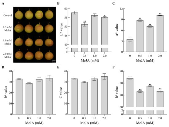
Figure 1.
Coloration of pear fruits under MeJA treatment. The skin coloration (A) and the color parameters, including the L* value (B), a* value (C), b* value (D), C value (E), and h0 value (F), of pear fruits under MeJA treatment. All the data represent the means ± SD of three biological replicates. Asterisks indicate statistical significance (*, p < 0.05; and **, p < 0.01), determined by the Student’s t-test, compared with corresponding controls.
3.2. Effects of MeJA on the Pigment Content of ‘Danxiahong’ Pear Peels
The concentrations of different pigments, namely, anthocyanin, chlorophyll, and carotenoid, were also analyzed. The anthocyanin content fluctuated within a narrow range during the early stages of the MeJA treatment, while it increased rapidly before full maturity (135 to 145 DAF) and reached its highest level during harvest (Figure 2A). Compared with the control, the anthocyanin content in pear peel significantly increased under MeJA treatment at the mature stage, thus resulting in a higher anthocyanin content and better coloration, which was consistent with the measurement of the fruit coloration parameters in Figure 1. Furthermore, a slight difference was observed in the anthocyanin content among different concentrations of MeJA treatments during the mature stage. Pear peels treated with 2.0 mM MeJA showed a significantly lower anthocyanin content than those treated with 0.5 and 1.0 mM MeJA, and no significant difference was detected between the 0.5 and 1.0 mM MeJA treatments. Overall, the anthocyanin content under the 1.0 mM MeJA treatment was the highest, 2.37 times that of the control, followed by the 0.5 mM MeJA treatment.

Figure 2.
The anthocyanin (A), chlorophyll (B), and carotenoid (C) contents of pear fruits under the MeJA treatment. All the data represent the means ± SD of three biological replicates. Different letters indicate significant differences determined by Duncan’s test (p < 0.05).
The effect of MeJA on chlorophyll content was investigated. The chlorophyll content in the control fruit gradually decreased with fruit ripening (115 to 145 DAF). The MeJA-treated fruit exhibited a significantly higher chlorophyll content at most time points than the non-treated fruit (Figure 2B), and, with the increase in the MeJA concentration, the chlorophyll content gradually decreased. Overall, the chlorophyll content in ‘Danxiahong’ pears was improved under the MeJA treatment, and fruits subjected to 0.5 mM MeJA exhibited the highest chlorophyll content.
The carotenoid content in the pear peels was also analyzed. The carotenoid content in pear peels showed a downward tendency during fruit ripening and reached the lowest level at the fully mature stage (145 DAF) (Figure 2C). Pear peels subjected to the MeJA treatment exhibited a sharp decrease in carotenoid content, in contrast to the control fruit, especially during the fully mature stage (135 to 145 DAF). Additionally, MeJA-treated peels had lower carotenoid content than the non-treated peels at the fully mature stage. The carotenoid content in mature fruit decreased with an increase in the MeJA concentration, and treatment with 2.0 mM MeJA had the lowest carotenoid content. Overall, the results indicate that MeJA treatment promotes carotenoid degradation in pear peels.
3.3. Effects of MeJA on the Fruit Size of ‘Danxiahong’ Pears
To investigate the effect of MeJA on fruit size, fruit weight, length, and width were measured, and the fruit shape index was calculated. The fruit size did not significantly change under the MeJA treatment. The fruit weight was higher in the 0.5 mM MeJA treatment group than in the other groups, but there was no significant difference (Figure 3A). Similarly, fruit length and width were not affected by the application of MeJA, although the fruit subjected to 0.5 mM MeJA exhibited a slight increase in fruit width (Figure 3B,C). Further investigations showed no significant difference in the fruit shape index between the different treatments and the control group (Figure 3D). Overall, the MeJA treatment tended to increase the fruit weight of the pears, and there was no significant change in the pear fruits’ shape among the different groups.
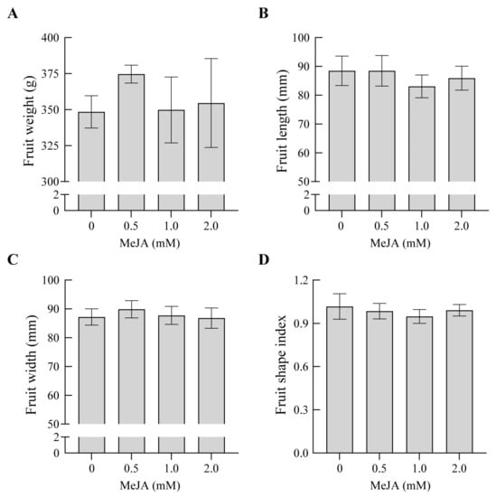
Figure 3.
Effects of MeJA on the fruit weight (A), length (B), width (C), and shape index (D) of ‘Danxiahong’ pears. All the data represent the means ± SD of three biological replicates.
3.4. Effects of MeJA on the Soluble Solid Content and Firmness of ‘Danxiahong’ Pears
The soluble solid content under the MeJA treatment was analyzed. With the increase in MeJA concentrations, the soluble solid content increased accordingly. Compared with non-treated fruit, the application of 2.0 mM MeJA significantly increased the fruit soluble solid content, whereas the 0.5 and 1.0 mM MeJA treatments did not reach a significant difference compared to the control (Figure 4A). Further analysis revealed that the soluble solid content under the application of 2.0 mM MeJA was 4.12% higher than that in the non-treated fruit, which was the highest among the different groups.
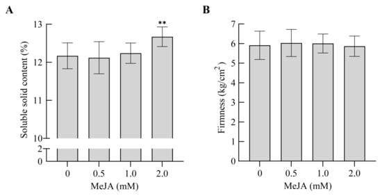
Figure 4.
Effects of MeJA on the soluble solid content (A) and firmness (B) of ‘Danxiahong’ pears. All the data represent the means ± SD of three biological replicates. Asterisks indicate statistical significance (**, p < 0.01), determined by the Student’s t-test, compared with corresponding controls.
Fruit firmness was also investigated at the fully mature stage. Fruit firmness fluctuated between 5.86 and 6.03 kg/cm2. Compared with the control, fruit firmness was not significantly affected under different MeJA concentrations (Figure 4B).
3.5. Effects of MeJA on Acid Content in ‘Danxiahong’ Pears
The effect of MeJA on acid content was also analyzed. As indicated in Figure 5, the total acid content was significantly reduced when subjected to the MeJA treatment (Figure 5A). Furthermore, the main acids in the pear fruits were analyzed, and the application of MeJA significantly decreased the contents of malic acid, shikimic acid, citric acid, and succinic acid (Figure 5B–E). The contents of quinic acid, malic acid, shikimic acid, citric acid, and succinic acid were significantly lower under the 0.5 and 1.0 mM MeJA treatments than in the control. However, the quinic acid content was significantly higher under the 2.0 mM MeJA treatment than in the control (Figure 5F). Within the range of MeJA concentrations, a lower MeJA concentration achieved a better reduction effect on total acid accumulation, and the application of 0.5 mM MeJA resulted in the lowest total acid content. Overall, the MeJA treatments decreased the total acid content and most of the main acids, indicating the role of MeJA in the improvement of pear taste.
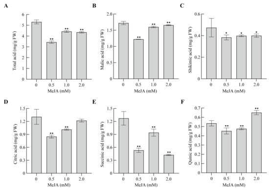
Figure 5.
Effects of MeJA on the acid content of ‘Danxiahong’ pears. The total acid (A), malic acid (B), shikimic acid (C), citric acid (D), succinic acid (E), and quinic acid (F) contents of pear fruits under the MeJA treatment. All the data represent the means ± SD of three biological replicates. Asterisks indicate statistical significance (*, p < 0.05; and **, p < 0.01), determined by the Student’s t-test, compared with corresponding controls.
3.6. Effects of MeJA on Sugar Content in ‘Danxiahong’ Pears
To characterize the effects of MeJA on the inner fruit quality, the flesh was subjected to HPLC analysis. Compared with the control, the MeJA treatment improved the total sugar content; with the increase in the MeJA concentration, the total sugar content increased correspondingly, reaching a significant difference compared to the control when subjected to the 2.0 mM MeJA treatment (Figure 6A). Additionally, the major sugars in the pear fruits were analyzed, and the proportion of sugar components was affected by the MeJA treatments. The fructose content significantly decreased under the 0.5 and 1.0 mM MeJA treatments, while it significantly increased under the 2.0 mM MeJA treatment (Figure 6B). With an increase in the MeJA concentration, the fructose content increased. The glucose and sucrose contents showed no significant differences between the control and different MeJA concentration treatments (Figure 6C,D). In contrast, the sorbitol content was significantly increased when subjected to the MeJA treatment (Figure 6E). In the experimental concentration range, the higher the MeJA concentration, the more improved the total sugar content (Figure 6A). The high concentration (2.0 mM) of the MeJA treatment led to the highest total sugar content, which was significantly higher than that of the other treatments. The MeJA treatments significantly increased the total sugar/total acid ratio (Figure 6F). The 0.5 mM MeJA treatment group had the highest total sugar/total acid ratio, which was increased by 54.50% compared to the control group. Overall, MeJA treatments increased the total sugar content and total sugar/total acid ratio, implying that MeJA contributed to the improvement of the inner quality of pears.
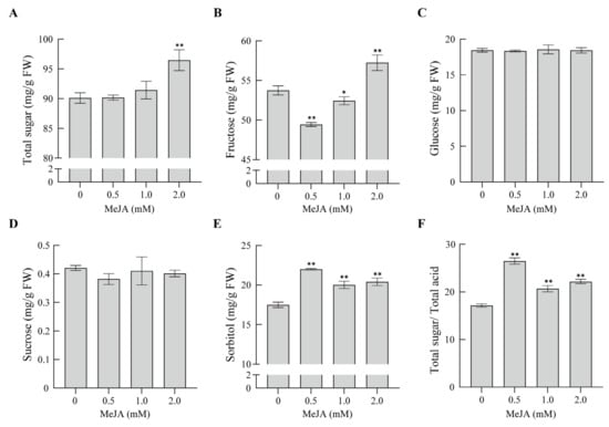
Figure 6.
Effects of MeJA on the sugar content and total sugar/total acid of ‘Danxiahong’ pears. The total sugar (A), fructose (B), glucose (C), sucrose (D), and sorbitol (E) contents and total sugar/total acid (F) of pear fruits under the MeJA treatment. All the data represent the means ± SD of three biological replicates. Asterisks indicate statistical significance (*, p < 0.05; and **, p < 0.01), determined by the Student’s t-test, compared with corresponding controls.
3.7. Effect of MeJA on the Expression of Genes Related to Anthocyanin Biosynthesis
It is well known that anthocyanin accumulation in fruit is mediated by a series of biosynthetic genes, especially the structural genes in the phenylpropanoid pathway. Furthermore, a set of transcription factors is involved in the regulation of anthocyanin biosynthesis. To clarify the molecular mechanism of anthocyanin accumulation mediated by MeJA in pear skin, an expression analysis of genes related to anthocyanin biosynthesis was performed under the application of 1.0 mM MeJA at different time points (Figure 7). Compared with the control, the expression of some structural anthocyanin biosynthesis-related genes was upregulated, such as that of PbrPAL, PbrCHI, PbrCHS, PbrF3H, PbrANS, PbrDFR, and PbrUFGT. Among these genes, the expression of PbrUGFT was notably upregulated, with the transcript level inducing a more than 15-fold increase compared to the corresponding control after a two-day treatment with MeJA. (Figure 7). Additionally, the expression of regulatory genes related to anthocyanin biosynthesis was investigated; after treatment with MeJA, the transcript levels were significantly induced, for example, those of PbrMYB10, PbrMYB114, PbrbHLH3, PbrbHLH33, and PbrWD40. Despite PbrbHLH33 exhibiting a low expression level, it still showed a significant difference compared to the corresponding control at different time points. Thus, these genes promoted anthocyanin biosynthesis, which was consistent with anthocyanin accumulation before fruit ripening. Taken together, the results indicate that MeJA-mediated fruit coloration in red-blushed pears occurs due to the expression of some structural genes and regulatory genes related to anthocyanin biosynthesis.
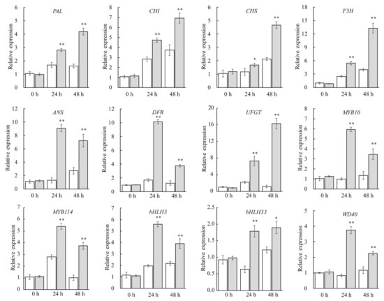
Figure 7.
Relative expression of anthocyanin-biosynthesis-related genes in pear peels under 1.0 mM MeJA and in the control group at different time points. All the data represent the means ± SD of three biological replicates. The white column indicates the control (pears sprayed with distilled water), and the gray column indicates pears sprayed with 1.0 mM MeJA. Asterisks indicate statistical significance (*, p < 0.05; and **, p < 0.01), determined by the Student’s t-test, compared with corresponding controls.
4. Discussion
The coloration of fruit skin is the result of the combined effects of various pigments, such as chlorophyll, carotenoids, and anthocyanins [40]. The formation of red coloration in pear peels mainly depends on the composition and anthocyanin concentration of the peels [3,8]. Previous reports have indicated no obvious correlation between chlorophyll and anthocyanin contents during fruit development in blush-skinned pears [3,41]. In the pear cultivars ‘Meirensu’ and ‘Yunhongli No. 1’, the chlorophyll and anthocyanin contents increased continuously in the early fruit development stage, while the chlorophyll content decreased rapidly in the middle and late stages of fruit development, and anthocyanin content increased continuously and reached the highest level at the maturity stage [15]. A previous study revealed that steam treatment with MeJA promoted chlorophyll degradation in apple peels [42]. However, Rudell et al. [24] reported that apples immersed in MeJA emulsion and then exposed to light showed an increase in the chlorophyll content in apple peels, primarily due to the increase in chlorophyll b content. Furthermore, grape fruit also exhibited an increase in chlorophyll content when treated with 10 mM MeJA [43]. In our research, the application of MeJA improved the anthocyanin and chlorophyll contents, while reducing the carotenoid content in pear peels. Different conclusions have been drawn regarding the effects of MeJA on chlorophyll and carotenoid accumulation in various plant species. These discrepancies may be attributed to factors such as the concentration of MeJA applied and the developmental stage of the plants. Further research is needed to better understand the specific conditions under which MeJA influences chlorophyll and carotenoid levels in different plants.
A growing body of evidence indicates that MeJA is involved in the regulation of anthocyanin accumulation in virous plant species [18,20,30,44,45]. The application of 100 μM MeJA steam to raspberries improved fruit redness during storage [46]. Shafiq et al. [44] demonstrated that spraying MeJA at 169 or 186 DAF (days after flowering) enhanced the coloration of red blush apples and significantly elevated the anthocyanin content in the peel. MeJA treatment from 147 to 175 DAF significantly improved peel coloration in apples, which reduced the L* and h0 values and increased the anthocyanin content in ‘Fuji’ peel [47]. Our results showed that spraying with various concentrations of MeJA significantly increased the anthocyanin content in pear peels and enhanced their redness. Further measurement of the color parameters also supports this conclusion. The enhanced coloration resulting from preharvest MeJA application may be attributed to ethylene production in plums [20] and apples [44]. A previous study indicated that ethylene inhibits anthocyanin biosynthesis in red pears [16]. Therefore, it seems that MeJA-mediated coloration in pears, particularly in sand pears, is not correlated with ethylene production. Further studies are needed to explore the potential reasons for the different mechanisms underlying MeJA-mediated coloration in pears and other fruit species.
It has been reported that jasmonate promotes the accumulation of anthocyanin and induces the expression of genes involved in anthocyanin biosynthesis. In Arabidopsis, genes associated with anthocyanin biosynthesis, such as AtPAP1/2, AtGL3, AtDFR, and AtUFGT, are induced by jasmonate [30]. In pears, PcMYB10 [8] and PcMYB114 [48] are positive regulators of anthocyanin accumulation. Furthermore, the application of exogenous MeJA significantly increased the anthocyanin content in pear callus. It also significantly promoted the transcription of genes associated with anthocyanin biosynthesis, including PcMYB10, PcMYC2, PcJAZ, PcCHS, PcCHI, PcF3H, PcDFR, and PcANS, where it was significantly promoted [45]. Similarly, in apple callus, the transcription of anthocyanin biosynthesis genes, such as MdMYB9/10/11, MdCHS, MdF3H, and MdUFGT, was upregulated when the callus was treated with MeJA [49,50]. In addition, the MYB transcription factor MdMYB24L has been reported to be involved in the jasmonate response, contributing to anthocyanin accumulation mediated by MeJA in apples [51]. Our results showed that the expression levels of anthocyanin biosynthesis-associated regulatory genes, including PbrMYB10, PbrMYB114, PbrbHLH3, PbrbHLH33, and PbrWD40, were significantly upregulated when subjected to MeJA treatment, which was consistent with previous reports [52] and verified the role of MeJA in fruit coloration. The MYB transcription factors involved in anthocyanin accumulation and their association with MeJA need to be further researched. Our results showed that the expression of structural genes, especially PbrUFGT, was upregulated by MeJA application. The combined action of regulatory genes and structural genes possibly leads to anthocyanin accumulation before fruit ripening.
The JAZ protein is a critical regulator in MeJA-mediated biological processes, and it is also involved in the regulation of anthocyanin accumulation. Previous reports have demonstrated that JAZ proteins interact with some transcription factors, thereby inhibiting their activity, including the transcription factors associated with anthocyanin biosynthesis in Arabidopsis [31,53,54]. In apples, MdJAZ8/11 interacted with MdMYB24L and formed a complex, which reduced the activity of the MYB-MYC2 complex and thus inhibited anthocyanin accumulation, while the application of MeJA promoted MdJAZ8/11 degradation and restored the activity of the MdMYB24L-MdMYC2 complex to promote anthocyanin biosynthesis [51]. In red Chinese sand pears, MeJA treatment reduced the expression of PpCOI1 while downregulating the transcriptional level of PpJAZ1 [55]. However, Premathilake et al. [45] demonstrated that the transcriptional abundances of JAZ genes were upregulated by MeJA treatments in pear callus. JAZ genes exhibited different transcription patterns when subjected to MeJA treatments. This variation in transcription patterns may be due to the tissue-specific expression of JAZ genes in different pear tissues. Our results indicate that the expression of regulatory genes, such as PbrMYB10 and PbrMYB114, was induced under the MeJA treatment (Figure 7). Since MeJA-mediated coloration in pear callus has been shown to be regulated by JAZ proteins, it raises the question of whether the underlying mechanism of coloration is the same or different in pear peel. Although treatment with MeJA has been found to enhance coloration in pear peel, the specific regulatory pathway involved remains unclear. Therefore, further research is necessary to investigate the roles of the JAZ gene and the regulation of its cascade in MeJA-induced anthocyanin accumulation.
Jasmonate can stimulate the production of numerous secondary metabolites in various plant species, playing crucial roles in biotic and abiotic stress responses, as well as defense responses [17,18,22]. In addition, there is ample evidence suggests that environmental stress, such as nitrogen deficiency, water deficiency, and low-temperature stress, typically triggers the accumulation of anthocyanin in many plant species [10,56,57]. In our research, the application of various concentrations of MeJA enhanced anthocyanin synthesis (Figure 2A). Given the well-documented involvement of MeJA in various defense responses and our research findings demonstrating its promotion of anthocyanin accumulation in pear peels, there is a strong suggestion that MeJA plays a dual role. Not only does it enhance the biosynthesis of anthocyanins, but it also appears to regulate plant defense responses during this process. However, in order to fully comprehend and unveil the regulatory mechanism underlying this overlap, further investigations are necessary.
Within a certain range, larger fruits are more appreciated by consumers and would obtain higher prices [58]. Cell expansion or elongation is promoted by an appropriate concentration of MeJA during fruit development, which contributes to the increase in fruit size and weight [59]. Different conclusions have been reached regarding the effect of MeJA on fruit size in different plant species. The single-fruit size and weight of the apples were reduced when subjected to 5, 10, and 20 mM MeJA at 48 DAF rather than at 119 DAF [59]. Peach trees treated with 0.8 mM MeJA at 56 DAF exhibited no significant difference from the control fruit in terms of fruit diameter and weight when harvested at the mature stage [60]. The postharvest application of 0.5 and 1.0 mM MeJA significantly increased the fruit weight in the plum cultivar ‘Black Splendor’, while the concentrations that proved effective in terms of fruit weight promotion were 0.5 and 2.0 mM MeJA in ‘Royal Rosa’ [58]. Thus, the effects of MeJA on fruit size and weight vary depending on the plant species, variety, MeJA concentration, and application stage. Our research revealed that the application of 0.5 mM MeJA had a positive effect on fruit size; furthermore, the vertical and transverse diameters of the fruit were increased. Correspondingly, the fruit weight increased (Figure 3). Given the diverse effects of various concentrations of MeJA on pear size and weight, it is speculated that the response may depend on sensitivity of the cells when exposed to different MeJA concentrations [61].
The effects of MeJA on fruit development and quality vary depending on the fruit species, application concentration, and stage [58]. Treatment with 0.44 mM MeJA at 102 DAF delayed peach ripening and decreased soluble solid accumulation [62]. The application of 8 mM MeJA promoted fruit ripening and leaf senescence in peaches [63]. The preharvest application of 1 and 5 mM MeJA treatments accelerated the ripening process, while 10 mM MeJA delayed fruit ripening in pomegranates [28]. Our study showed that pear firmness did not significantly change during fruit ripening when subjected to different MeJA concentrations, indicating that MeJA may have an insufficient ability to accelerate or delay pear ripening; the question of whether higher MeJA concentrations have a similar effect on fruit ripening as on peaches needs to be further explored.
As a common product in the terminal branch of the phenylpropane pathway, anthocyanins contribute to a broader range of colors in most vegetables and fruits; the final formation of anthocyanins usually undergoes a series of glycosylation reactions, which involve anthocyanidins, glycosyl derived from different sugars, and the catalysis enzymes of UFGT [64]. Sugars that participate in anthocyanin synthesis usually serve as energy sources, synthetic precursors, or signaling molecules [13,65,66]. Generally, unstable anthocyanidins undergo a glycosylation reaction and then transfer the sugar group to the 3-O position to generate stable anthocyanins [67]. In our research, the MeJA treatments promoted the accumulation of the main sugars in pear flesh, as well as the total sugar content (Figure 6A,E), implying the positive role of MeJA in sugar accumulation, which produced more sugars in pear fruit. The increase in sugar content contributes to anthocyanin synthesis, which has a positive effect on the pear skin’s blush. The matter of whether the MeJA-mediated regulator genes exhibit crosstalk in the accumulation of sugar and anthocyanins needs to be further investigated.
Our research indicates that MeJA application improved the contents of soluble solids, sorbitol, and total sugar in mature pear fruit and decreased the contents of shikimic acid and total acid. Meanwhile, the ratio of sugar to acid was significantly increased, resulting in an improved fruit taste. However, there are few studies on the effects of jasmonate on fruit development and quality, particularly regarding the molecular mechanisms of fruit development and quality formation [26,27]. The fine mechanism underlying the improvement of fruit quality mediated by MeJA application needs to be further explored.
5. Conclusions
In the present study, we investigated the effect of methyl jasmonate (MeJA) on fruit coloration and quality improvement in red-blushed pears. The findings support our hypothesis that MeJA application enhances fruit coloration by promoting anthocyanin accumulation in the pear peels. Furthermore, the MeJA treatment had positive effects on various quality parameters. It increased the fruit’s soluble solids, improving the total sugar content and resulting in a sweeter taste. Additionally, MeJA treatment decreased the fruit’s acid content, leading to a more balanced flavor profile. This was evident from the significant increase in the total sugar/total acid ratio. However, it is important to note that the MeJA treatment did not have a significant effect on the fruit shape or longitudinal or transverse diameters. The results of our real-time quantitative PCR (RT-qPCR) analysis revealed that the MeJA treatments induced the expression of anthocyanin biosynthetic regulatory and structural genes, providing insights into the underlying mechanisms of MeJA-induced changes in fruit coloration. Overall, our study demonstrates that spraying MeJA is a potential practical method for improving the fruit coloration and quality of red-blushed pears. These findings have important implications for the pear industry, offering a promising approach for growers to optimize fruit appearance and quality.
Supplementary Materials
The following supporting information can be downloaded at: https://www.mdpi.com/article/10.3390/agronomy13092409/s1, Table S1: The soil physicochemical properties and mineral element contents in the orchard; Table S2: The primers used in the experiments.
Author Contributions
Conceptualization and supervision: H.X.; methodology: B.L., R.D. and C.H.; formal analysis, writing—original draft preparation: B.L. and X.Z.; writing—review and editing: X.Z.; resources and funding acquisition: H.X. and J.Y. All authors have read and agreed to the published version of the manuscript.
Funding
The research was funded by the Major Scientific and Technological Project of Henan Province (221100110400), the National Natural Science Foundation of China (31971691), the Earmarked Fund for China Agriculture Research System (CARS-28), the Agricultural Science and Technology Innovation Program (CAAS-ASTIP), and the Collaborative Innovation Project of ZFRI, CAAS (ZGS202107).
Data Availability Statement
No new data were created or analyzed in this study. Data sharing is not applicable to this article.
Conflicts of Interest
The authors declare no conflict of interest.
References
- Xue, H.; Wang, S.; Yao, J.L.; Zhang, X.; Yang, J.; Wang, L.; Su, Y.; Chen, L.; Zhang, H.; Li, X. The genetic locus underlying red foliage and fruit skin traits is mapped to the same location in the two pear bud mutants ‘Red Zaosu’ and ‘Max Red Bartlett’. Hereditas 2018, 155, 25. [Google Scholar] [CrossRef]
- Wu, J.; Wang, Y.; Xu, J.; Korban, S.S.; Fei, Z.; Tao, S.; Ming, R.; Tai, S.; Khan, A.M.; Postman, J.D.; et al. Diversification and independent domestication of Asian and European pears. Genome Biol. 2018, 19, 77. [Google Scholar] [CrossRef] [PubMed]
- Huang, C.; Yu, B.; Teng, Y.; Su, J.; Shu, Q.; Cheng, Z.; Zeng, L. Effects of fruit bagging on coloring and related physiology, and qualities of red Chinese sand pears during fruit maturation. Sci. Hortic. 2009, 121, 149–158. [Google Scholar] [CrossRef]
- Xue, H.; Shi, T.; Wang, F.; Zhou, H.; Yang, J.; Wang, L.; Wang, S.; Su, Y.; Zhang, Z.; Qiao, Y.; et al. Interval mapping for red/green skin color in Asian pears using a modified QTL-seq method. Hortic. Res. 2017, 4, 17053. [Google Scholar] [CrossRef] [PubMed]
- He, J.; Giusti, M.M. Anthocyanins: Natural Colorants with Health-Promoting Properties. Annu. Rev. Food Sci. Technol. 2010, 1, 163–187. [Google Scholar] [CrossRef] [PubMed]
- Zhang, D.; Yu, B.; Bai, J.; Qian, M.; Shu, Q.; Su, J.; Teng, Y. Effects of high temperatures on UV-B/visible irradiation induced postharvest anthocyanin accumulation in ‘Yunhongli No. 1′ (Pyrus pyrifolia Nakai) pears. Sci. Hortic. 2012, 134, 53–59. [Google Scholar] [CrossRef]
- Thomson, G.E.; Turpin, S.; Goodwin, I. A review of preharvest anthocyanin development in full red and blush cultivars of European pear. N. Z. J. Crop Hortic. Sci. 2018, 46, 81–100. [Google Scholar] [CrossRef]
- Feng, S.; Wang, Y.; Yang, S.; Xu, Y.; Chen, X. Anthocyanin biosynthesis in pears is regulated by a R2R3-MYB transcription factor PyMYB10. Planta 2010, 232, 245–255. [Google Scholar] [CrossRef] [PubMed]
- Yu, B.; Zhang, D.; Huang, C.; Qian, M.; Zheng, X.; Teng, Y.; Su, J.; Shu, Q. Isolation of anthocyanin biosynthetic genes in red Chinese sand pear (Pyrus pyrifolia Nakai) and their expression as affected by organ/tissue, cultivar, bagging and fruit side. Sci. Hortic. 2012, 136, 29–37. [Google Scholar] [CrossRef]
- Steyn, W.J.; Wand, S.J.E.; Jacobs, G.; Rosecrance, R.C.; Roberts, S.C. Evidence for a photoprotective function of low-temperature-induced anthocyanin accumulation in apple and pear peel. Physiol. Plant. 2009, 136, 461–472. [Google Scholar] [CrossRef] [PubMed]
- Zhang, Q.; Yang, J.; Wang, L.; Wang, S.; Li, X.; Zhang, S. Correlation between soluble sugar and anthocyanin and effect of exogenous sugar on coloring of ‘Hongtaiyang’ pear. J. Fruit Sci. 2013, 30, 248–253. [Google Scholar]
- Awad, M.A.; de Jager, A.; van Westing, L.M. Flavonoid and chlorogenic acid levels in apple fruit: Characterisation of variation. Sci. Hortic. 2000, 83, 249–263. [Google Scholar] [CrossRef]
- Das, P.K.; Shin, D.H.; Choi, S.-B.; Park, Y.-I. Sugar-hormone cross-talk in anthocyanin biosynthesis. Mol. Cells 2012, 34, 501–507. [Google Scholar] [CrossRef]
- Solfanelli, C.; Poggi, A.; Loreti, E.; Alpi, A.; Perata, P. Sucrose-specific induction of the anthocyanin biosynthetic pathway in Arabidopsis. Plant Physiol. 2005, 140, 637–646. [Google Scholar] [CrossRef] [PubMed]
- Huang, C.; Yu, B.; Su, J.; Shu, Q.; Teng, Y. A study on coloration physiology of fruit in two red chinese sand pear cultivars ‘Meirensu’ and ‘Yunhongli No.1′. Sci. Agric. Sin. 2010, 43, 1433–1440. [Google Scholar] [CrossRef]
- Ni, J.; Zhao, Y.; Tao, R.; Yin, L.; Gao, L.; Strid, Å.; Qian, M.; Li, J.; Li, Y.; Shen, J.; et al. Ethylene mediates the branching of the jasmonate-induced flavonoid biosynthesis pathway by suppressing anthocyanin biosynthesis in red Chinese pear fruits. Plant Biotechnol. J. 2020, 18, 1223–1240. [Google Scholar] [CrossRef] [PubMed]
- Wasternack, C. Jasmonates: An update on biosynthesis, signal transduction and action in plant stress response, growth and development. Ann. Bot. 2007, 100, 681–697. [Google Scholar] [CrossRef] [PubMed]
- Wang, S.-Y.; Shi, X.-C.; Liu, F.-Q.; Laborda, P. Effects of exogenous methyl jasmonate on quality and preservation of postharvest fruits: A review. Food Chem. 2021, 353, 129482. [Google Scholar] [CrossRef] [PubMed]
- Fan, X.; Mattheis, J.P.; Fellman, J.K. A role for jasmonates in climacteric fruit ripening. Planta 1998, 204, 444–449. [Google Scholar] [CrossRef]
- Khan, A.S.; Singh, Z. Methyl jasmonate promotes fruit ripening and improves fruit quality in Japanese plum. J. Hortic. Sci. Biotechnol. 2007, 82, 695–706. [Google Scholar] [CrossRef]
- Wei, J.; Wen, X.; Tang, L. Effect of methyl jasmonic acid on peach fruit ripening progress. Sci. Hortic. 2017, 220, 206–213. [Google Scholar] [CrossRef]
- Howe, G.A. Jasmonates as signals in the wound response. J. Plant Growth Regul. 2004, 23, 223–237. [Google Scholar] [CrossRef]
- McConn, M.; Creelman, R.A.; Bell, E.; Mullet, J.E.; Browse, J. Jasmonate is essential for insect defense in Arabidopsis. Proc. Natl. Acad. Sci. USA 1997, 94, 5473–5477. [Google Scholar] [CrossRef]
- Rudell, D.R.; Mattheis, J.P.; Fan, X.; Fellman, J.K. Methyl jasmonate enhances anthocyanin accumulation and modifies production of phenolics and pigments in ‘Fuji’ apples. J. Am. Soc. Hortic. Sci. 2002, 127, 435–441. [Google Scholar] [CrossRef]
- Muengkaew, R.; Chaiprasart, P.; Warrington, I. Changing of physiochemical properties and color development of mango fruit sprayed methyl Jasmonate. Sci. Hortic. 2016, 198, 70–77. [Google Scholar] [CrossRef]
- García-Pastor, M.E.; Serrano, M.; Guillén, F.; Castillo, S.; Martínez-Romero, D.; Valero, D.; Zapata, P.J. Methyl jasmonate effects on table grape ripening, vine yield, berry quality and bioactive compounds depend on applied concentration. Sci. Hortic. 2019, 247, 380–389. [Google Scholar] [CrossRef]
- Han, Y.; Chen, C.; Yan, Z.; Li, J.; Wang, Y. The methyl jasmonate accelerates the strawberry fruits ripening process. Sci. Hortic. 2019, 249, 250–256. [Google Scholar] [CrossRef]
- García-Pastor, M.E.; Serrano, M.; Guillén, F.; Giménez, M.J.; Martínez-Romero, D.; Valero, D.; Zapata, P.J. Preharvest application of methyl jasmonate increases crop yield, fruit quality and bioactive compounds in pomegranate ‘Mollar de Elche’ at harvest and during postharvest storage. J. Sci. Food Agric. 2020, 100, 145–153. [Google Scholar] [CrossRef] [PubMed]
- Wang, S.Y.; Zheng, W. Preharvest application of methyl jasmonate increases fruit quality and antioxidant capacity in raspberries. Int. J. Food Sci. Technol. 2005, 40, 187–195. [Google Scholar] [CrossRef]
- Shan, X.; Zhang, Y.; Peng, W.; Wang, Z.; Xie, D. Molecular mechanism for jasmonate-induction of anthocyanin accumulation in Arabidopsis. J. Exp. Bot. 2009, 60, 3849–3860. [Google Scholar] [CrossRef] [PubMed]
- Qi, T.; Song, S.; Ren, Q.; Wu, D.; Huang, H.; Chen, Y.; Fan, M.; Peng, W.; Ren, C.; Xie, D. The Jasmonate-ZIM-domain proteins interact with the WD-repeat/bHLH/MYB complexes to regulate jasmonate-mediated anthocyanin accumulation and trichome initiation in Arabidopsis thaliana. Plant Cell 2011, 23, 1795–1814. [Google Scholar] [CrossRef]
- McGuire, R.G. Reporting of objective color measurements. HortScience 1992, 27, 1254–1255. [Google Scholar] [CrossRef]
- Zhang, X.; Li, B.; Duan, R.; Han, C.; Wang, L.; Yang, J.; Wang, L.; Wang, S.; Su, Y.; Xue, H. Transcriptome analysis reveals roles of sucrose in anthocyanin accumulation in ‘Kuerle Xiangli’ (Pyrus sinkiangensis Yü). Genes 2022, 13, 1064. [Google Scholar] [CrossRef] [PubMed]
- Cheng, Y.; Dong, Y.; Yan, H.; Ge, W.; Shen, C.; Guan, J.; Liu, L.; Zhang, Y. Effects of 1-MCP on chlorophyll degradation pathway-associated genes expression and chloroplast ultrastructure during the peel yellowing of Chinese pear fruits in storage. Food Chem. 2012, 135, 415–422. [Google Scholar] [CrossRef] [PubMed]
- Yang, C.; Chen, T.; Shen, B.; Sun, S.; Song, H.; Chen, D.; Xi, W. Citric acid treatment reduces decay and maintains the postharvest quality of peach (Prunus persica L.) fruit. Food Sci. Nutr. 2019, 7, 3635–3643. [Google Scholar] [CrossRef] [PubMed]
- Liu, L.; Chen, C.-X.; Zhu, Y.-F.; Xue, L.; Liu, Q.-W.; Qi, K.-J.; Zhang, S.-L.; Wu, J. Maternal inheritance has impact on organic acid content in progeny of pear (Pyrus spp.) fruit. Euphytica 2016, 209, 305–321. [Google Scholar] [CrossRef]
- Fan, X.; Zhao, H.; Wang, X.; Cao, J.; Jiang, W. Sugar and organic acid composition of apricot and their contribution to sensory quality and consumer satisfaction. Sci. Hortic. 2017, 225, 553–560. [Google Scholar] [CrossRef]
- Yao, G.; Yang, Z.; Zhang, S.; Cao, Y.; Liu, J.; Wu, J. Characteristics of components and contents of organic acid in pear fruits from different cultivated species. Acta Hortic. Sin. 2014, 41, 755–764. [Google Scholar] [CrossRef]
- Livak, K.J.; Schmittgen, T.D. Analysis of relative gene expression data using real-time quantitative PCR and the 2−ΔΔCT Method. Methods 2001, 25, 402–408. [Google Scholar] [CrossRef] [PubMed]
- Allan, A.C.; Hellens, R.P.; Laing, W.A. MYB transcription factors that colour our fruit. Trends Plant Sci. 2008, 13, 99–102. [Google Scholar] [CrossRef] [PubMed]
- Liu, J.; Deng, Z.; Sun, H.; Song, J.; Li, D.; Zhang, S.; Wang, R. Differences in anthocyanin accumulation patterns and related gene expression in two varieties of red pear. Plants 2021, 10, 626. [Google Scholar] [CrossRef] [PubMed]
- Pérez, A.G.; Sanz, C.; Richardson, D.G.; Olías, J.M. Methyl jasmonate vapor promotes β-carotene synthesis and chlorophyll degradation in Golden Delicious apple peel. J. Plant Growth Regul. 1993, 12, 163. [Google Scholar] [CrossRef]
- Gutiérrez-Gamboa, G.; Marín-San Román, S.; Jofré, V.; Rubio-Bretón, P.; Pérez-Álvarez, E.P.; Garde-Cerdán, T. Effects on chlorophyll and carotenoid contents in different grape varieties (Vitis vinifera L.) after nitrogen and elicitor foliar applications to the vineyard. Food Chem. 2018, 269, 380–386. [Google Scholar] [CrossRef] [PubMed]
- Shafiq, M.; Singh, Z.; Khan, A.S. Time of methyl jasmonate application influences the development of ‘Cripps Pink’ apple fruit colour. J. Sci. Food Agric. 2013, 93, 611–618. [Google Scholar] [CrossRef] [PubMed]
- Premathilake, A.T.; Ni, J.; Shen, J.; Bai, S.; Teng, Y. Transcriptome analysis provides new insights into the transcriptional regulation of methyl jasmonate-induced flavonoid biosynthesis in pear calli. BMC Plant Biol. 2020, 20, 388. [Google Scholar] [CrossRef] [PubMed]
- Wang, C.Y. Maintaining postharvest quality of raspberries with natural volatile compounds. Int. J. Food Sci. Technol. 2003, 38, 869–875. [Google Scholar] [CrossRef]
- Ozturk, B.; Yıldız, K.; Ozkan, Y. Effects of pre-harvest methyl jasmonate treatments on bioactive compounds and peel color development of “Fuji” apples. Int. J. Food Prop. 2015, 18, 954–962. [Google Scholar] [CrossRef]
- Yao, G.; Ming, M.; Allan, A.C.; Gu, C.; Li, L.; Wu, X.; Wang, R.; Chang, Y.; Qi, K.; Zhang, S.; et al. Map-based cloning of the pear gene MYB114 identifies an interaction with other transcription factors to coordinately regulate fruit anthocyanin biosynthesis. Plant J. 2017, 92, 437–451. [Google Scholar] [CrossRef]
- Sun, J.; Wang, Y.; Chen, X.; Gong, X.; Wang, N.; Ma, L.; Qiu, Y.; Wang, Y.; Feng, S. Effects of methyl jasmonate and abscisic acid on anthocyanin biosynthesis in callus cultures of red-fleshed apple (Malus sieversii f. niedzwetzkyana). Plant Cell Tissue Organ Cult. 2017, 130, 227–237. [Google Scholar] [CrossRef]
- An, X.-H.; Tian, Y.; Chen, K.-Q.; Liu, X.-J.; Liu, D.-D.; Xie, X.-B.; Cheng, C.-G.; Cong, P.-H.; Hao, Y.-J. MdMYB9 and MdMYB11 are involved in the regulation of the JA-induced biosynthesis of anthocyanin and proanthocyanidin in apples. Plant Cell Physiol. 2014, 56, 650–662. [Google Scholar] [CrossRef] [PubMed]
- Wang, Y.; Liu, W.; Jiang, H.; Mao, Z.; Wang, N.; Jiang, S.; Xu, H.; Yang, G.; Zhang, Z.; Chen, X. The R2R3-MYB transcription factor MdMYB24-like is involved in methyl jasmonate-induced anthocyanin biosynthesis in apple. Plant Physiol. Biochem. 2019, 139, 273–282. [Google Scholar] [CrossRef] [PubMed]
- Qian, M.; Ni, J.; Niu, Q.; Bai, S.; Bao, L.; Li, J.; Sun, Y.; Zhang, D.; Teng, Y. Response of miR156-SPL module during the red peel coloration of bagging-treated Chinese sand pear (Pyrus pyrifolia Nakai). Front. Physiol. 2017, 8, 550. [Google Scholar] [CrossRef] [PubMed]
- Thines, B.; Katsir, L.; Melotto, M.; Niu, Y.; Mandaokar, A.; Liu, G.; Nomura, K.; He, S.Y.; Howe, G.A.; Browse, J. JAZ repressor proteins are targets of the SCFCOI1 complex during jasmonate signalling. Nature 2007, 448, 661–665. [Google Scholar] [CrossRef] [PubMed]
- Sheard, L.B.; Tan, X.; Mao, H.; Withers, J.; Ben-Nissan, G.; Hinds, T.R.; Kobayashi, Y.; Hsu, F.-F.; Sharon, M.; Browse, J.; et al. Jasmonate perception by inositol-phosphate-potentiated COI1–JAZ co-receptor. Nature 2010, 468, 400–405. [Google Scholar] [CrossRef]
- Qian, M.; Yu, B.; Li, X.; Sun, Y.; Zhang, D.; Teng, Y. Isolation and expression analysis of anthocyanin biosynthesis genes from the red Chinese sand pear, Pyrus pyrifolia Nakai cv. Mantianhong, in response to methyl jasmonate treatment and UV-B/VIS conditions. Plant Mol. Biol. Rep. 2014, 32, 428–437. [Google Scholar] [CrossRef]
- Ollé, D.; Guiraud, J.L.; Souquet, J.M.; Terrier, N.; Ageorges, A.; Cheynier, V.; Verries, C. Effect of pre- and post-veraison water deficit on proanthocyanidin and anthocyanin accumulation during Shiraz berry development. Aust. J. Grape Wine Res. 2011, 17, 90–100. [Google Scholar] [CrossRef]
- Huang, W.; Wang, X.; Zhang, J.; Xia, J.; Zhang, X. Improvement of blueberry freshness prediction based on machine learning and multi-source sensing in the cold chain logistics. Food Control 2023, 145, 109496. [Google Scholar] [CrossRef]
- Martínez-Esplá, A.; Zapata, P.J.; Castillo, S.; Guillén, F.; Martínez-Romero, D.; Valero, D.; Serrano, M. Preharvest application of methyl jasmonate (MeJA) in two plum cultivars. 1. Improvement of fruit growth and quality attributes at harvest. Postharvest Biol. Technol. 2014, 98, 98–105. [Google Scholar] [CrossRef]
- Rudell, D.R.; Fellman, J.K.; Mattheis, J.P. Preharvest application of methyl jasmonate to ‘Fuji’ apples enhances red coloration and affects fruit size, splitting, and bitter pit incidence. HortScience 2005, 40, 1760–1762. [Google Scholar] [CrossRef]
- Ruiz, K.B.; Trainotti, L.; Bonghi, C.; Ziosi, V.; Costa, G.; Torrigiani, P. Early methyl jasmonate application to peach delays fruit/seed development by altering the expression of multiple hormone-related genes. J. Plant Growth Regul. 2013, 32, 852–864. [Google Scholar] [CrossRef]
- Ozturk, B.; Altuntas, E.; Yildiz, K.; Ozkan, Y.; Saracoglu, O. Effect of methyl jasmonate treatments on the bioactive compounds and physicochemical quality of ‘Fuji’apples. Cienc. Investig. Agrar. 2013, 40, 201–211. [Google Scholar] [CrossRef]
- Ziosi, V.; Bonghi, C.; Bregoli, A.M.; Trainotti, L.; Biondi, S.; Sutthiwal, S.; Kondo, S.; Costa, G.; Torrigiani, P. Jasmonate-induced transcriptional changes suggest a negative interference with the ripening syndrome in peach fruit. J. Exp. Bot. 2008, 59, 563–573. [Google Scholar] [CrossRef] [PubMed]
- Janoudi, A.; Flore, J.A. Effects of multiple applications of methyl jasmonate on fruit ripening, leaf gas exchange and vegetative growth in fruit trees. J. Hortic. Sci. Biotechnol. 2003, 78, 793–797. [Google Scholar] [CrossRef]
- Silva, V.O.; Freitas, A.A.; Maçanita, A.L.; Quina, F.H. Chemistry and photochemistry of natural plant pigments: The anthocyanins. J. Phys. Org. Chem. 2016, 29, 594–599. [Google Scholar] [CrossRef]
- Yang, Z.; Xu, J.; Yang, L.; Zhang, X. Optimized Dynamic Monitoring and Quality Management System for Post-Harvest Matsutake of Different Preservation Packaging in Cold Chain. Foods 2022, 11, 2646. [Google Scholar] [CrossRef] [PubMed]
- Moalem-Beno, D.; Tamari, G.; Leitner-Dagan, Y.; Borochov, A.; Weiss, D. Sugar-dependent gibberellin-induced Chalcone Synthase gene expression in Petunia Corollas. Plant Physiol. 1997, 113, 419–424. [Google Scholar] [CrossRef] [PubMed]
- Springob, K.; Nakajima, J.; Yamazaki, M.; Saito, K. Recent advances in the biosynthesis and accumulation of anthocyanins. Nat. Prod. Rep. 2003, 20, 288–303. [Google Scholar] [CrossRef] [PubMed]
Disclaimer/Publisher’s Note: The statements, opinions and data contained in all publications are solely those of the individual author(s) and contributor(s) and not of MDPI and/or the editor(s). MDPI and/or the editor(s) disclaim responsibility for any injury to people or property resulting from any ideas, methods, instructions or products referred to in the content. |
© 2023 by the authors. Licensee MDPI, Basel, Switzerland. This article is an open access article distributed under the terms and conditions of the Creative Commons Attribution (CC BY) license (https://creativecommons.org/licenses/by/4.0/).