Article

The Physiological Mechanism of Melatonin Enhancing the Tolerance of Oat Seedlings under Saline–Alkali Stress

1
Key Laboratory of Coarse Cereal Processing, Ministry of Agriculture and Rural Affairs, College of Food and Biological Engineering, Chengdu University, Chengdu 610106, China
2
Baicheng Academy of Agricultural Sciences, No. 17, Sanhe Road, Taobei District, Baicheng 137000, China
3
Agronomy College, Jilin Agricultural University, Changchun 130118, China
4
Agronomy College, Inner Mongolia Agricultural University, No. 306 Zhaowuda Road, Saihan District, Hohhot 010010, China
5
Biotechnology Research Institute of Chinese Academy of Agricultural Sciences, No. 12 Zhong Guan Cun South Street, Beijing 100081, China
*
Author to whom correspondence should be addressed.
These authors contributed equally to this work.
Agronomy 2023, 13(9), 2343; https://doi.org/10.3390/agronomy13092343
Submission received: 6 August 2023 / Revised: 27 August 2023 / Accepted: 5 September 2023 / Published: 8 September 2023
(This article belongs to the Special Issue Strategies for Enhancing Abiotic Stress Tolerance in Crops)

Abstract

:
Exogenous melatonin (MT) regulates plant growth and mitigates stress in response to stress. To analyze the machinery of exogenous melatonin, which improves salt and alkaline tolerance in oats, MT’s function was identified in the oat seed germination stage in our previous study. In this study, morphogenesis, photosynthetic physiology, hormone levels, and ion homeostasis were evaluated using the same MT treatment concentration. The results revealed that compared to the S45 treatment, the 100 μmol·L−1 MT treatment efficiently increased the seedling height and main root length of oat seedlings; promoted secondary root development; enhanced the root volume and root surface area; maintained a higher photosynthetic pigment content (carotenoids; chlorophyll a; chlorophyll b); raised the leaf photosynthetic rate (Pn), intercellular CO2 concentration (Ci), conductance to H2O (Gs), and transpiration rate (Tr); enhanced the light energy absorption and conversion of leaves; increased the leaf GA3, Tryptamine (TAM), and IAA contents; and decreased ABA levels. Hierarchical cluster analysis revealed that MT treatment also increased the contents of P, K, Ca, Mn, Cu, Mg, Fe, Zn, Mo, Cd, Al, Se, Ni, Co, and Ti; decreased the Na/K ratio; and maintained cellular ionic homeostasis in oat seedlings under saline–alkali stress, as compared with the untreated group. These findings showed that MT treatment enhanced the adaptation of oat to saline–alkali stress through regulating the physiological process of seedling growth. This suggests that MT plays a different role in improving saline–alkali tolerance in the germination and seedling stages of oat.

1. Introduction

Saline–alkali stress is an apparent abiotic stress limiting plant growth, development, and crop production [1]. According to statistics, 1 billion hectares of land worldwide are threatened by a saline–alkali environment. In the Songnen Plain of China, 3.73 million ha of saline–alkali soil jeopardizes crop production and product development [2]. Unlike other saline–alkali soils, these soils are dominated by soda saline–alkali soils, mainly caused by Na2CO3 and NaHCO3, which have a high salt content and a soil pH above 10 in most areas [3]. Numerous studies have shown that saline–alkali stress leads to oxidative damage, osmotic stress, ion toxicity, and more severe plant growth inhibition [4,5]. For example, saline–alkali stress causes an increase in ROS content and induces membrane peroxidation in the roots and leaves of maize seedlings, thereby inhibiting seedling growth [6,7]. High-pH saline–alkali stress triggers chlorophyll degradation in sorghum, leading to the excessive accumulation of inorganic ions [8]. So, finding new strategies for plant saline–alkali tolerance is essential to protect plants’ normal growth and development.
Plant growth regulators, as a class of biologically active substances, enhance plant tolerance to biotic and abiotic stresses and regulate the process from germination to senescence. Among them, melatonin is a highly active molecule that is widely distributed in various tissues and organs of plants. It is closely related to plant life activities such as seed germination, photosynthesis, root development, and leaf senescence [9]. In plants, MT is critical in integrating environmental biotic and abiotic stress signals and activating response networks, leading to the development of stress defense mechanisms and resilience [10,11]. For example, MT, facing salt, drought, and low-temperature stress, can act as a regulator of the redox network through modulating secondary messenger signaling (e.g., via interacting with Ca) and initiating downstream signaling processes to mitigate the stress response [12]. Meanwhile, MT also improves plant tolerance to abiotic stresses through enhancing the expression of genes related to cell division, fatty acid synthesis, carbohydrate metabolism, photosynthesis, and ascorbic acid metabolism, thereby improving plant growth [13]. Abiotic stress environments promote endogenous MT biosynthesis, while plants can also absorb external environmental MT through leaves, roots, and seeds to improve plant stress tolerance [14,15]. Previous studies have found that exogenous MT can protect photosynthetic plant systems through delaying leaf senescence. For example, appropriate concentrations of exogenous MT can reduce lipid oxidation; protect chloroplast structure; enhance Pn, Tr, and Gs; inhibit the excessive accumulation of ROS under abiotic stress; and protect cell membrane integrity against stress [16]. Similar findings were confirmed in rice [17], ryegrass [18], and cucumber [19]. In addition to antioxidant defense, MT acts synergistically with other hormones to confer plant resistance and stress adaptation. MT application increases IAA and GA content, stimulates root growth, and mitigates the inhibitory effects of abiotic stresses on root development through modulating the ROS scavenging system [20]. It also induces the increased expression of ABA catabolic genes to reduce ABA levels during germination and promote seed germination [21] and breaks the limitation of IAA biosynthesis by salt stress to promote root development [22]. There is also evidence that MT promotes plant growth and development and improves yield, which are mainly related to regulating plant ion homeostasis [23]. For example, exogenous MT application increases H+ pump activity and the sensitivity of the Na+/K+ transporter to RNS and ROS, maintaining ionic homeostasis in plants under salt stress [24,25]. Therefore, the further development of MT as a biostimulant and regulator of crop production and increased knowledge of its physiological functions are of wide significance for resistance to environmental stress to reduce crop yield losses.
Oats are an annual herbaceous crop of the grass family, and their wide ecological adaptations have made them irreplaceable food and fodder crops in many regions [26]. In recent years, oats have become popular in the feed, food, and pharmaceutical industries because of their rich nutritional content and healthcare functions. In production cultivation, oats are recognized as a crop with moderate tolerance to salt and alkali and are therefore widely used as a selective crop for saline–alkali land improvement [27]. Nevertheless, a saline–alkali soil environment also has a specific inhibitory effect on the growth of oats. Seed germination and seedling survival are the stages of oats which are most sensitive to saline-alkali stress, and different germplasm resources have different tolerance mechanisms under saline–alkali stress during the germination and seedling stages. To analyze the effect of melatonin on the saline–alkali tolerance of oats, we treated oats during the seed germination and seedling stages, respectively, using MT. In our previous study, the optimal MT treatment concentration was identified for promoting oat seed germination, and we have analyzed the MT functional mechanism as an antioxidant for improving oat seed germination rate under saline–alkali stress [15]. In this study, we used the same MT concentration to treat oat seedlings under saline–alkali stress to explore the effects of MT on physiological process of oat seedlings growth. Our research results provide a physiological foundation for elucidating the different functional mechanisms of exogenous MT improving saline–alkali tolerance during the germination and seedling stages of oat.

2. Materials and Methods

2.1. Experimental Materials

In this study, the saline–alkali-sensitive variety of naked oats, “Baiyan No. 5”, was used as the test material, cultivated and provided by the Academy of Agricultural Sciences of Baicheng City, Jilin Province. Seeds were stored in a dry environment at 4 °C before the start of the experiment.
Melatonin (N–acetyl–5–methoxytryptamine, MT) was purchased from Beijing Kulaibo Science and Technology Co Ltd. (Beijing, China). All chemicals used in this study were of analytical grade.

2.2. Solution Preparation

To simulate the features of saline–alkali soils in the central and western regions of Jilin Province, a saline–alkali mixture was prepared with a modified 50% Hoagland nutrient solution (the types of nutrients are shown in Table S1 of the Supplementary Materials) in the molar ratio of NaCl:Na2SO4:Na2CO3:NaHCO3 of 1:9:1:9.
MT solution preparation [19]: MT (0.116 g) was weighed, melted in 3 mL of anhydrous ethanol, added to deionized water, brought to a volume of 250 mL to obtain a 2 mmol·L−1 MT stock solution, and then diluted into a 100 μmol·L−1 MT solution with deionized water. After preparation, this solution was contained at room temperature and protected from light, and 0.05% (v/v) Tween–20 was added.

2.3. Cultivating Experimental Materials

This study was conducted in 2022 in the greenhouse of the Academy of Agricultural Sciences of Baicheng City (122°48′1″ E, 45°36′56″ N, 157.51 m above sea level). The seeds were disinfected with 75% alcohol for 30 s, washed, air–dried, and placed in a dark environment in a constant-temperature incubator at 25 °C for 12 h. Seeds from Baiyan No. 5 with full and uniform grains and no insect pests were selected. Seeds that were uniformly dewy white were selected and placed evenly in Petri dishes (φ17 cm) lined with two layers of sterile filter paper. For saline–alkali stress, the seeds were treated with 45 mmol·L−1 (pH 8.97, salinity 6.18 psu, conductivity 11.04 mS/cm) saline–alkali solution, with 16 mL of saline–alkali solution added to each Petri dish. The control was made with an identical number of 50% Hoagland nutrient solution and 150 seeds per dish, and each treatment was repeated three times.

2.4. MT Treatment

Seedlings with a constant growth were selected for each treatment, rinsed with distilled water, drained from the root surface, and transplanted into sterilized hydroponic boxes (38 × 28 × 14 cm) with 8 L of saline–alkali solution in round holes; they were fixed in the fixing cups using a 4.5 cm diameter sponge, and each treatment was replicated three times, with three pots per replicate, for a total of 27 pools with 12 plants per pot, with equal amounts of 50% Hoagland nutrient solution used as a control. The cultured seedlings were subjected to the following three treatments: (1) CK—seedlings grown generally in 50% Hoagland nutrient solution + root application of 0 μmol·L−1 MT; (2) S45—seedlings grown in 45 mmol·L−1 soda saline–alkali solution (50% Hoagland nutrient solution was prepared, as follows) + root application of 0 μmol·L−1 MT; (3) S+MT—seedlings grown in 45 mmol·L−1 soda saline–alkali solution + root application of 100 μmol·L−1 MT (pre-experiments showed significant effects with this concentration treatment). After two true leaves of oat seedlings were fully opened (two leaves and one heart, approximately 14 days), a root dip treatment with 100 μmol·L−1 MT was performed for 7 days. The whole incubation process was carried out in a greenhouse at a temperature of 23 ± 1 °C, relative humidity was controlled at 40 ± 5%, light duration was 16 h/8 h (light/dark), and red, blue, and white lamps (red:blue:white = 3:2:1) with three lamps per layer were used. The hydroponic boxes were randomly arranged in the greenhouse nursery bed, and the placement of the boxes was adjusted from time to time to ensure a consistent growth environment for each treatment group during the incubation period. The boxes were continuously aerated with an inflatable pump to prevent root rot. During the experiment, the water that dissipated from the hydroponic boxes was replenished every 24 h using the weighing method to ensure a constant treatment concentration, and the solution was changed every 2 d. Samples were taken 14 d after MT treatment, quick-frozen with liquid nitrogen, and stored in a refrigerator at −80 °C for the subsequent measurement of physiological indices.

2.4.1. Agronomic Characteristics

After 7, 11, and 14 d after MT treatment, five representative plants with consistent growth were selected from each replicate; Vernier calipers were used to measure their root lengths, and their seedling heights (linear distance from the base of the radicle to the top of the leaf) were measured using a straightedge. The total root length, surface area, root volume, root mean diameter, and root tip number were determined separately using a root system analyzer (Expression 11000XL, Seiko Epson Corp, Suwa, Nagano, Japan) 14 d after MT treatment, and the number of secondary roots was calculated manually.

2.4.2. Chlorophyll Content

Using the 95% ethanol extraction method [28], 0.2 g of fresh leaves of naked oats were weighed, cut, placed in a corked graduated test tube containing 10 mL of 95% ethanol, capped, and placed in a dark environment at 25 °C overnight with shaking 3–4 times during the extraction. The next day, after removal and observation that the leaf tissue had turned entirely white, the chloroplast pigment extract was brought to a 25 mL volume with 95% ethanol, filtered, and poured into a 1–cm optical diameter colorimetric cup. The absorbance was measured at 665 nm, 649 nm, and 470 nm wavelengths using 95% ethanol as a blank.

2.4.3. Photosynthetic Parameters

The net photosynthetic rate (Pn), conductance to H2O (Gs), transpiration rate (Tr), and intercellular CO2 concentration (Ci) of flag leaves were measured 7 d, 11 d, and 14 d after MT treatment using a Li–6400XT portable photosynthesizer (LI–COR Corp, Lincoln, NE, USA). The light intensity was set at 1200 μmol·m−1·s−1 for each seedling, and the experiment was replicated three times. Photosynthetic parameters were measured on the same leaf three times per plant, and the results were averaged.

2.4.4. Leaf ABA, GA3, IAA, and TAM Contents

The acetonitrile solution extraction method was used to purify the impurities in acetonitrile via the QuEChERS method, and the sample was concentrated through a nitrogen purge, which is beneficial for downstream detection. The corresponding standard solution and mobile phase were configured. All samples were ground in liquid nitrogen until crushed. The samples were weighed accurately in a test tube, 10 mL of acetonitrile solution was added, and 8 μL of the internal standard master batch was added. The mixture was extracted overnight at 4 °C and then centrifuged at 12,000× g for 5 min at 4 °C, after which the supernatant was removed. Next, 5 mL of acetonitrile solution was added again to the precipitate, the mixture was extracted twice, and the supernatants were combined. An appropriate amount of C18 and GCB was added to purify the impurities, followed by centrifugation at 12,000× g for 5 min at 4 °C and removal of the supernatant. After blowing it dry with nitrogen, the precipitate was resolubilized with 400 μL of methanol, passed through a 0.22 μm organic phase filter membrane, and stored in a −20 °C freezer until analysis.

2.4.5. Mineral Elements

Na, P, K, Ca, Mg, S, Fe, Mn, Cu, Zn, Ni, Co, Mo, Cd, Hg, Al, B, Se, and Ti were deter- mined using the pressure tank digestion method and detected and analyzed via ICP–MS (NexlON®5000 ICP–MS, PerkinElmer Crop, Waltham, MA, USA). The weighed dry specimen (0.1–0.2 g) or 0.5–2 mL of liquid was placed in the inner PTFE digestion tank, to which 5 mL of nitric acid was added, followed by soaking overnight. The inner lid was covered, and the stainless-steel jacket was tightened, followed by the transfer of the tank into a constant-temperature drying oven, where it was kept at 80 °C for 2 h, 120 °C for 2 h, and then 160 °C for 4 h. The oven was allowed to cool naturally to room temperature, after which it was opened. The acid was heated until the sample was nearly dry, and the digestion solution was transferred into a 25 mL volumetric flask; the inner jar and inner lid were washed with a small amount of nitric acid solution (1%) 3 times, the washing solution was combined with the contents of the volumetric flask, and the solution was brought to a final volume with 1% nitric acid, after which the solution was mixed well and set aside. A blank reagent test was also performed. The test solution was run on the instrument to determine the content of each element.
Elemental content(mg·kg−1) = (c × v)/m × D
where c is the concentration of elements in solution (mg·L−1), v is the extraction volume (mL), D is the dilution factor, and m is the sample mass (g).
Total silicon (Si) was determined using the silicon molybdenum blue colorimetric method [29] and analyzed via ICP–OES (Agilent 710 ICP–OES, Agilent Crop, Santa Clara, CA, USA): 5 mL of 4 mol·L−1 NaOH was placed in a nickel crucible, heated, and evaporated in an electric hot plate, and the finely ground air-dried sample of the plant to be tested was accurately weighed to 0.05 g–0.20 g and sprinkled on the NaOH in the nickel crucible. The crucible was covered and placed on an ordinary electric hot plate and heated to convert the sample to ashes, which were then transferred to a high-temperature electric furnace and melted at 700–720 °C. After cooling, the sample was washed with distilled water in a 50 mL volumetric flask containing 5 mL of 4 mol·L−1 HCl, and the volume was fixed; this represented the silicon solution to be tested. One drop of 2,6–dinitrophenol indicator was added; the solution was adjusted with 0.1 mol·L−1 NaOH and 0.05 mol·L−1 H2SO4 until slightly yellow, shaken well, and incubated for 15–20 min at room temperature. Then, 2.5 mL of 0.3 mol·L−1 H2SO4 and 2.5 mL of 5% ammonium molybdate solution were added, followed by shaking well and incubation for 5–20 min at room temperature. Next, 2.5 mL 5% oxalic acid solution and 2.5 mL 15 g·L−1 ascorbic acid solution were added in turn, the volume was fixed, and after 20 min, the color was compared at 700 nm on a spectrophotometer. A blank test was performed at the same time.
Plant whole silicon content (g·kg−1) = mass concentration (μg·mL−1) determined from the standard curve × volume of color development (mL) × fractionation times ÷ sample weight (g) × 0.001.

2.5. Data Visualization and Statistical Analysis

The raw data were collated and analyzed using Excel 2021 (Microsoft Corp, Montgomery, AL, USA), and ANOVA and LSD analysis were performed using SPSS 24.0 (IBM, Chicago, AL, USA). Duncan’s multiple extreme difference test was used to measure significant differences between samples (p ≤ 0.05). GraphPad Prism 9.4 (GraphPad Software Corp, San Diego, CA, USA) was used for the graphing, and Adobe Illustrator (Adobe Corp, San Jose, CA, USA) was used for the combined graphs. Heat maps were plotted at https://www.chiplot.online (accessed on 6 April 2023) for Pearson correlation analysis. Hierarchical clustering analysis of oat seedling–related indicators was performed in MetaboAnalyst online software (http://www.metaboanalyst.ca/ (accessed on 9 April 2023)).

3. Results

3.1. Effect of MT Treatment on Agronomic Traits of Oat Seedlings under Saline–Alkali Stress

Under saline–alkali stress, the leaf tips of oat seedlings showed yellowing (Figure 1A), and seedling height (Figure 1C) and main root length (Figure 1D) were inhibited. The MT root soaking treatment effectively reduced the damage of saline–alkali stress to oat seedlings. It promoted seedling height, maintained greater green leaf areas, and alleviated saline–alkali stress inhibiting the growth of oat roots. The promotion effect on seedling height was significant (p ≤ 0.05) 11 d after MT treatment, and the seedling height of the S+MT group was increased by 72% compared with that of the S45 group; there was no significant difference in the length of the primary root, but it was still increased by 21% compared with that of S45 group. After 14 d of MT treatment, the primary root length and seedling height showed a slow increase compared with that in the 11 d MT treatment group; the seedling height and main root length of the S+MT group increased by 48% and 6%, respectively, compared with the S45 group.
Compared with the CK group, MT treatment especially promotes the production of secondary roots in oats (Figure 1B and Figure 2). Fourteen days after MT treatment, the number of secondary roots in the root system of oat seedlings in the various groups showed the trend of S+MT > S45 > CK, and there were significant differences (p ≤ 0.01) between the S+MT treatment and the S45 and CK treatments, with the S+MT treatment group being 389% higher than the S45 group. The root volume, root surface area, total root length, and root tip number of oat seedlings in the various groups showed the trend of CK > S+MT > S45. The root volume, root surface area, total root length, and root tip number were increased by 32%, 30%, 63%, and 3% after the S+MT treatment compared with the S45 treatment, respectively (p > 0.05). As shown in Figure 2d, the average diameter of the root system of oat seedlings in the S+MT and S45 groups was 35% and 50% higher than that in the CK group, respectively. Oat seedlings had thicker root systems under saline–alkali stress, but growth of the root length was inhibited, while MT treatment could increase the root surface area through promoting the formation of secondary roots in seedlings, which tolerate saline–alkali stress.

3.2. Effect of MT Treatment on the Photosynthetic Physiology of Oat Seedlings under Saline–Alkali Stress

When oat plants were subjected to saline–alkali stress, leaf chlorophyll a (Figure 3a), chlorophyll b (Figure 3b), total chlorophyll (Figure 3c), and carotenoids (Figure 3d) contents were significantly reduced (p ≤ 0.05) compared with those of CK. MT treatments improved chlorophyll contents under saline–alkali stress, but they were still lower than those of the CK group. Seven days after MT treatment, the photosynthetic pigment content of oat plant leaves in the S + MT group increased significantly compared with the S45 treatment group by 11%, 13%, 11%, and 43%, respectively. With increasing treatment time, chlorophyll a, total chlorophyll, and carotenoids did not change significantly under S45 and S + MT treatments, while the contents of chlorophyll B decreased significantly under S45 treatment and increased significantly after S + MT treatment. Fourteen days after MT treatment, the chlorophyll a, chlorophyll b, total chlorophyll, and carotenoid contents were increased by 13%, 71%, 27%, and 41%, respectively, compared with those of the S45 group. This indicates that MT treatment mainly alleviated the effect of saline–alkali stress on chlorophyll b synthesis.
The stability of photosynthetic pigment synthesis is essential for plants to maintain normal photosynthesis. In this study, we found that Pn, Gs, Ci, and Tr in oat leaves were significantly lower (p ≤ 0.05) under saline–alkali stress conditions, while Pn, Gs, Ci, and Tr showed a significant increase after MT root soaking treatment compared to the S45 group (Figure 4A), which Pn increased by 48%, 65%, and 58% after 7 d, 11 d, and 14 d but did not reach a significant level at 14 d (p > 0.05). The Gs of oat leaves under saline–alkali stress was significantly lower than that in the CK group and was always at a lower level with the extension of saline–alkali stress time, and the Gs of the S+MT group increased by 48%, 70%, and 39%, respectively, compared with that of the S45 group (Figure 4B). Under normal water incubation conditions, oat leaf Ci and Tr were always at a high level, and Ci increased by 41%, 38%, and 46% after 7 d, 11 d, and 14 d of MT treatment, respectively, compared with the S45 group (Figure 4C), while Tr significantly increased by 65%, 95%, and 58%, respectively (Figure 4D). This indicates that saline–alkali stress causes a decrease in chlorophyll content, which in turn affects the normal photosynthesis of oat leaves, while MT treatment can effectively relieve the effect of saline–alkali stress on chlorophyll synthesis and improve photosynthetic efficiency against saline–alkali stress.

3.3. Effect of MT Treatment on the Endogenous Hormone Content of Oat Seedling Leaves under Saline–Alkali Stress

Plants adapt to saline–alkali stress through the flexible regulation of hormone levels and/or signaling. Saline–alkali stress induced a slight increase in ABA (Figure 5c) content in oat leaves, while IAA (Figure 5a), TAM (Figure 5b), and GA3 (Figure 5d) contents were significantly different between treatments (p ≤ 0.05). After S45 treatment, ABA content increased by 13%, while IAA, TAM, and GA3 content decreased by 60%, 16%, and 82% compared to that in the CK group, respectively. After S+MT treatment, IAA, TAM and GA3 content in oat leaves increased significantly compared to that in the S45 treatment group, while ABA content was lower than that in the S45 treatment group. In the S+MT group, the leaf IAA, TAM, and GA3 contents increased by 66%, 37%, and 224%, respectively, compared with those of the S45 treatment group. In addition, there was a more significant effect on ABA under S+MT, which significant decreased by 88% and 86.11%, respectively, compared with the S45 treatment and CK (p ≤ 0.05). This indicates that MT treatment effected changes in various hormone contents in leaves, which regulate saline–alkali tolerance in oat seedlings.
Interactions between endogenous plant hormones were categorized as antagonistic or synergistic, and the ratios of IAA/ABA, TAM/ABA, and GA3/ABA responded to the interactions among various hormones. The ratio of GA3/ABA was significantly lower in the S45 treatment than in CK. Moreover, the ratios of IAA/ABA, TAM/ABA, and GA3/ABA were significantly higher in the S+MT treatment as compared to CK and S45. It indicates that MT can change the content of endogenous hormones in plants under saline–alkali stress and maintain the dynamic balance of hormone content in plants.

3.4. Effect of MT Treatment on the Mineral Element Content of Oat Seedlings under Saline–Alkali Stress

A high-pH environment disrupts plants’ balance of mineral ion uptake. Saline–alkali stress inhibited the uptake of essential elements such as P (Figure 6a), K (Figure 6c), Mg (Figure 6e), Fe (Figure 6g), Cu (Figure 6i), Zn (Figure 6j), and Mo (Figure 6l) by oat seedlings. Compared with the CK group, the contents of P, K, Mg, Fe, Cu, Zn, and Mo decreased by 39%, 39%, 26%, 63%, 72%, 53%, and 49% in the S45 treatment group, respectively; the contents of P, K, Mg, Fe, Cu, Zn, and Mo increased in the S+MT group after MT treatment compared with the S45 group by 28%, 28%, 8%, 22%, 18%, 43%, 20%, respectively. The differences between the two treatments were significant (p ≤ 0.05) except for Mg, Cu, and Mo. S45 treatment promoted the uptake of essential elements such as S (Figure 6b), Ca (Figure 6d), Ni (Figure 6f), Mn (Figure 6h), and B (Figure 6k) by oat seedlings, which increased by 138%, 14%, 18%, 94%, and 1%, respectively, compared with the CK group; except for S and B, S+MT treatment increased Ca, Ni, and Mn contents significantly, 10%, 5%, and 33%, respectively, compared to the S45 treatment group and 26%, 24%, and 159%, respectively, compared to the CK group, with significant differences between treatments (p ≤ 0.05).
The uptake of Cd (Figure 7c), Al (Figure 7e), Co (Figure 7b), Se (Figure 7f), and Si (Figure 7h) ions by oat seedlings under saline–alkali stress was inhibited, while the contents of Na (Figure 7a), Hg (Figure 7d) and Ti (Figure 7g) were increased; compared with the CK group, the contents of Cd, Al, Co, Se, and Si after S45 treatment were decreased by 51%, 15%, 44%, 89%, and 13% for Cd, Co, and Se, respectively (p ≤ 0.05). After MT treatment, the contents of Co, Hg, Al, Se, Ti, and Cd gradually increased in the S+MT group compared with those in the S45 group by 11%, 94%, 152%,98%, 5%, and 1%, respectively, while the contents of Na and Si were significantly lower in the S+MT treatment group compared with those in the S45 treatment group by 33% and 19%, respectively; in addition, Na content was increased dramatically in both the S+MT and S45 groups compared to the CK group by 515% and 829.44%, respectively, and the difference was significant (p ≤ 0.05).

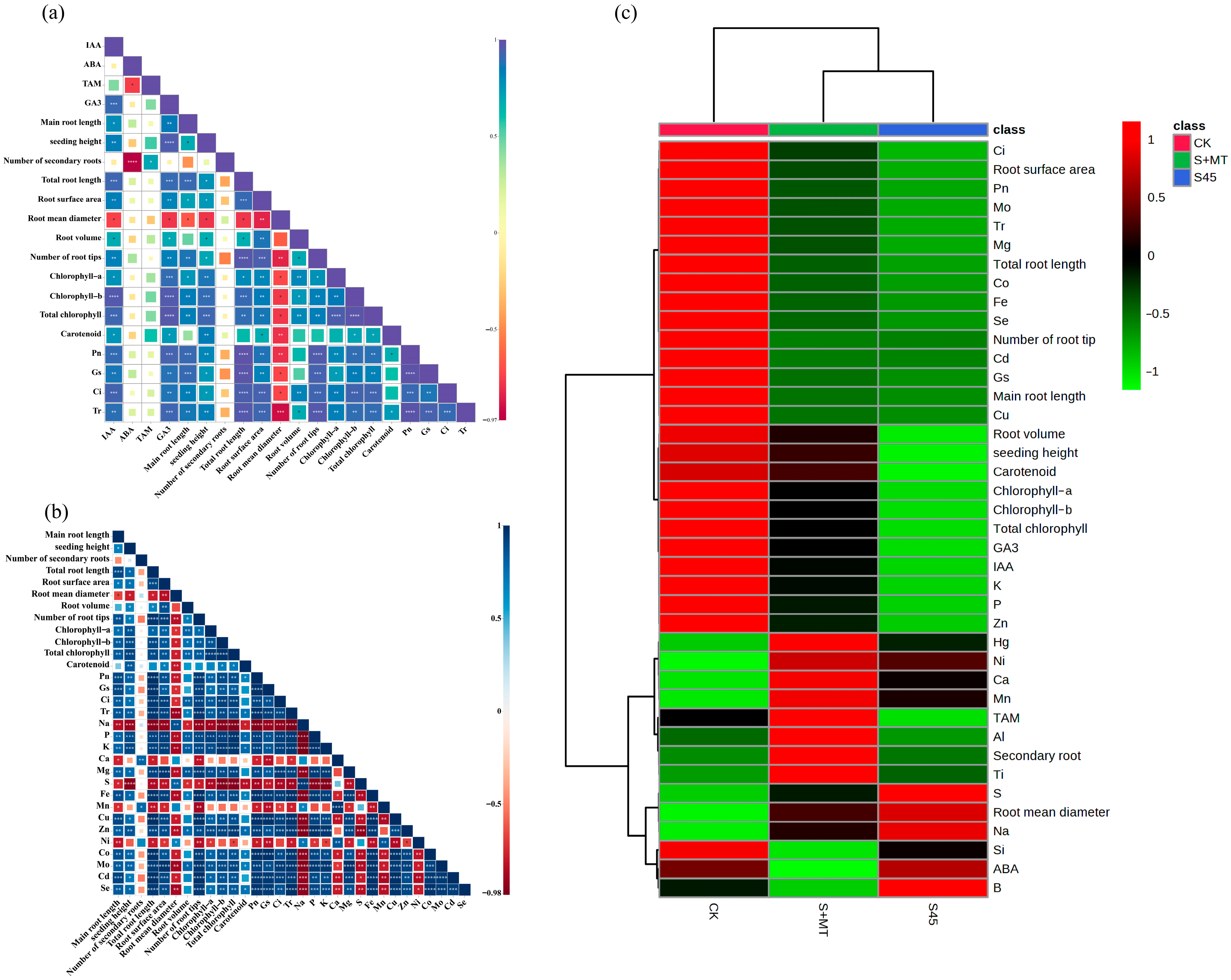
3.5. Correlation Analysis and Hierarchical Clustering Analysis of MT Treatment on Agronomic and Physiological Indicators of Oat Seedlings under Saline–Alkali Stress

Correlation analysis showed (Figure 8a) that the contents of IAA and GA3 showed a significant and positive correlation (p ≤ 0.05) with morphological indicators (excluding root mean diameter), photosynthetic pigments, and photosynthetic parameters; TAM was significantly and positively correlated (p ≤ 0.05) with the number of secondary roots, while ABA content was highly significantly negatively correlated with the number of secondary roots (p ≤ 0.01), significantly negatively correlated with TAM (p ≤ 0.05), and not correlated with other indicators (p > 0.05).
As shown in Figure 8b, there was a highly significant negative correlation (p ≤ 0.01) between Na content and morphological indicators (except for the average diameter of the root system and the number of secondary roots), photosynthetic pigments, photosynthetic parameters, and essential plant elements. Photosynthetic pigments were highly significantly and positively correlated (p ≤ 0.01) with the content of chlorophyll-synthesis-related elements such as Fe, Zn, and Mg; photosynthetic parameters were highly significantly and positively correlated (p ≤ 0.01) with P, K, Mg, Fe, Cu, Zn, Co, Mo, Cd and Se. Photosynthetic pigment content was highly significantly negatively correlated with Na and S (p ≤ 0.01); photosynthetic parameters were highly significantly negatively correlated with Na, S, Mn, and Ni (p ≤ 0.01).
Hierarchical cluster analysis showed (Figure 8c) that saline–alkali stress suppressed all morphological parameters (except root mean diameter) and decreased photosynthetic pigment content, photosynthetic parameters, endogenous hormone contents (GA3, IAA, TAM), and the elemental contents of K, P, Zn, Fe, Mg, Cu, and Se, whereas ABA content and the elemental contents of Na, Ni, Ca, and Mn were significantly increased compared to CK. Compared with saline–alkali stress, the exogenous application of MT resulted in a significant increase in morphology-related indices (especially the number of secondary roots), as well as an increase in photosynthetic pigment content, photosynthetic capacity, and endogenous hormone content (except for ABA), but a decrease in elemental content of Na and S. The exogenous application of MT resulted in a significant increase in the number of secondary roots and an increase in photosynthetic pigment content and photosynthetic capacity.

4. Discussion

4.1. MT Treatment Effect on Phenotypic Characteristics

When plants are under salinity stress, the root system first receives the stress signal and transmits this adverse effect to the stem and leaves, ultimately affecting the plant’s growth and development [30]. Numerous studies have shown that MT significantly alleviates the inhibition of plant growth due to abiotic stresses [30,31,32]. In legumes, MT could improve alfalfa morphology to enhance salt tolerance through increasing plant height, root length, fresh dry weight, and chlorophyll content [33]. This study found that saline–alkali stress markedly reduced the seedling height and main root length of oat seedlings. The root surface area, total root length, root tip number, and root volume of oats under saline–alkali stress, whether or not they were treated with MT, were lower than those of the normal culture environment; however, MT treatment alleviated this stress effect, and MT’s role revealed a gradual enhancement with the time of stress (Figure 1). The root structure of oats was significantly improved after MT treatment, which significantly increased the number of secondary roots of oats; these roots had a thicker diameter, larger surface area, and denser root hairs compared with primary roots, which further promoted the root uptake of water and other nutrients and promoted seedling growth. This is consistent with previous studies on cotton [22]. It indicates MT treatment can improve the adaptability of oats to saline–alkali environments through improving seedling root structure and aboveground morphology, which promotes the growth of oat seedlings under saline–alkali stress. This may be related to MT regulating the growth hormone response in saline–alkali stress [34].

4.2. MT Treatment Effects on Photosynthetic Physiology

Photosynthesis is an essential physiological process for synthesizing carbohydrates and maintaining normal plant growth, which is extremely sensitive to saline–alkali stress environments. Chlorophyll is the primary pigment that absorbs and converts light energy during photosynthesis in plants, and the physiological drought of cells formed by a saline–alkali stress environment will reduce chlorophyll content, affecting the absorption of light energy by leaves and reducing the photosynthetic capacity of leaves [35]. This study showed that saline–alkali stress reduced the water content of oat leaves, resulting in the yellowing of some leaves, and chlorophyll a, chlorophyll b, and total chlorophyll content was significantly decreased. Similar findings have been reported in studies on sorghum [11] and ryegrass [36]. Numerous studies have shown that MT increases photosynthetic pigment content in plant leaves under abiotic stress, delays leaf senescence, promotes light energy uptake and conversion, and ensures the accumulation of plant organic matter [37,38,39]. Our study found that exogenous MT treatment significantly increased photosynthetic pigment chlorophyll a, chlorophyll b, and total chlorophyll content in the leaves of oat seedlings under saline stress, thus ensuring the absorption of light energy by the leaves. Carotenoids act as photoprotectants, pigments, and safety valves, releasing excess energy before damaging plant cells [40]. Ahmad et al. [41] showed that abiotic stress negatively affected maize seedlings via reducing leaf chlorophyll and carotenoid contents and significantly reducing leaf photosynthetic capacity and stomatal opening, while MT treatment effectively alleviated this stress effect through increasing chlorophyll and carotenoid contents per seedling. The present study showed that MT alleviates the decreasing trend of carotenoid content in the leaves of oat seedlings under saline stress, promotes water uptake by leaves, protects chlorophyll from degradation, delays leaf senescence, improves photosynthetic efficiency, and thus improves the saline–alkali tolerance of oat seedlings.
Zhang et al. [42] found that saline–alkali stress significantly reduced wheat leaf photosynthetic parameters and chlorophyll content, inhibiting their photosynthesis and affecting plant growth. Similar findings have been reported in quinoa [43] and maize [44]. In this study, a simultaneous reduction in Gs and Ci was found. The decrease in Pn might be related to stomatal inhibition, and the lower stomatal opening further led to a decrease in Tr, thus inhibiting the photosynthetic capacity of leaves (Figure 4). Previous studies found that exogenous MT enhances the photosynthetic capacity of rice and cucumber under salt stress [26,27,38,39]. This study showed that exogenous MT treatment alleviated the prohibitive effect of saline–alkali stress on photosynthesis in oat leaves and always increased leaf Gs, Ci, and Tr after stress, which increased leaf Pn and had a more significant effect on Tr. This indicates that MT treatment ensured water uptake by seedlings, thus better regulating stomatal opening, and enhancing the adaptive capacity of oat seedlings to saline–alkali stress. We found that MT could enhance leaf photosynthetic capacity through regulating stomatal opening and playing an essential role in alleviating the inhibitory effect of salinity stress on oat photosynthesis.
In addition, correlation analysis showed (Figure 8) that Pn, Gs, Ci, Tr, and total chlorophyll, chlorophyll a, chlorophyll b, and carotenoid contents were highly significant and correlated with IAA and GA3 contents (p ≤ 0.01), so we speculate that the increase in photosynthetic pigment content and photosynthetic capacity may be related to MT enhancing the activity of IAA and GA3 in oat leaves.

4.3. Effect on Endogenous Hormones

The joint regulatory mechanisms between different endogenous hormone signaling pathways play a crucial role in improving plants’ stress tolerance when exposed to abiotic stress environments such as salt, alkali, and drought [45]. The highly osmotic cellular environment caused by saline–alkali stress has been reported to stimulate ABA biosynthesis and stomatal closure in plants, enhancing the inhibition of photosynthesis by saline–alkali stress [46]. In this study, higher ABA content was detected in oat leaves subjected to saline–alkali stress, indicating that saline–alkali stress may have promoted ABA biosynthesis in oat seedling leaves, causing the excessive accumulation of ABA and leading to stomatal closure, a significant weakening in Tr and Pn (Figure 4), and a decrease in the saline–alkali tolerance of oat seedlings. Related studies have shown that MT is directly or indirectly involved in the metabolic pathways of ABA, IAA, and GA through regulating the expression of genes related to phytohormone biosynthesis [47]. In this study, we found that the ABA content of oat leaves under saline–alkali stress decreased significantly after MT treatment, and photosynthetic parameters such as Pn and Tr were steadily improved; presumably, the reduction in endogenous ABA content improved the stomatal opening of leaves, alleviating the inhibition of leaf photosynthesis by saline–alkali stress. Furthermore, the decrease in ABA content might be related to the up-regulation of ABA catabolic genes and the downregulation of the key enzyme of ABA synthesis, 9–epoxy carotenoid dioxygenase (NCED), caused by MT treatment. This indicates that MT can improve salinity tolerance in oat seedlings through regulating the stomatal opening and decreasing the effect of saline–alkali stress on photosynthesis via regulating endogenous ABA content in leaves.
GA plays an important role in plant growth and tolerance to adversity stress, and salt stress limits plant growth through reducing GA3 bioactivity [48]. The same finding was found in saline–alkali-sensitive species where endogenous GA3 content was more inhibited by saline–alkali stress [49]. We found that saline–alkali stress significantly reduced GA3 content in oat leaves and significantly increased leaf GA3 levels after exogenous MT treatment, which could significantly alter the inhibition of GA3 biological activity due to saline-alkali stress. Correlation analysis showed (Figure 8) that GA3 content was significantly and positively correlated (p ≤ 0.01) with leaf chlorophyll and carotenoid levels, as well as photosynthetic parameters and other growth indicators. It showed that elevated endogenous GA3 content after MT treatment could alleviate the degradation of leaf chlorophyll under saline–alkali stress, increase Pn, and further average seedling growth to weaken the stress effect of saline–alkali stress on seedlings.
IAA, as a critical hormone in the hormone signaling network, is closely related to the catabolic and biosynthetic gene expression of other hormones; also, TAM is a common substrate for the biosynthetic pathway of IAA and MT in the plant body [50]. Related studies have confirmed that MT and IAA have collaborative effects on plant growth, development, and the adaptation to external stressful environments [51]. MT and IAA jointly induced the growth and development of lateral roots and adventitious under appropriate concentrations of MT treatment [7]. Our study showed that saline–alkali stress inhibited the biosynthesis of IAA in oat seedling leaves, and the content of TAM was significantly reduced; after MT treatment, it promoted the increase in TAM content and provided sufficient precursors for the synthesis of IAA, which more effectively increased the content of IAA in leaves. IAA stimulated longitudinal plant cell growth, and we found that MT treatment collaborated with developing saline–alkali-stressed oat roots and aboveground growth (correlation analysis showed (Figure 8) that IAA was closely related to seedling growth and photosynthesis). The analysis of the correlation between alterations in the hormone content in the leaves of oat seedlings under saline–alkali stress and physiological indicators such as photosynthetic pigments, photosynthesis, and root traits after MT treatment showed that MT treatment might reduce the inhibition of plant growth due to saline–alkali stress through regulating the balance of ABA, GA3 and IAA contents under saline–alkali stress, thus improving the saline–alkali tolerance of oat seedlings.

4.4. Effect on Mineral Element Content

Excessive intracellular Na+ accumulation produces ionic toxicity when plants are under saline–alkali stress. At the same time, a high-pH environment can reduce membrane stability, thus disrupting plants’ balance of mineral ion uptake. In contrast, a plant’s lack of uptake of any particular nutrient alters its metabolic capacity, leading to nutrient deficiencies and the disruption of cellular metabolism. Abiotic stress not only limits plant uptake of essential elements (K, Ca, Mg, P, S) and trace elements (Mn, Cu, Ni, Fe, Zn, B, Mo) but also nonessential elements (Na, Co, Cd, Hg, Al, Se, Ti, Si) [52,53]. Wu et al. [54] showed that salt stress significantly inhibited the uptake of important elements such as K, Ca, S, and Mg and elements such as Mn, Cu, and Zn by barley seedlings, leading to the disruption of their metabolism. Several studies have similarly shown that salt stress causes a reduction in the uptake of S, Mn, K, Ca, P, and Fe by plants [55,56,57,58]. In this study, we analyzed the changes in 20 elements in oat seedlings under 45 mmol·L−1 saline–alkali stress; the contents of Mg, Fe, Cu, Mo, Co, P, K, Zn, Cd, Al, Se, and Si in seedlings were found to decrease significantly, and the contents of S, Ca, Ni, Mn, Na, and Hg increased significantly (Supplementary Materials Figure S2). The increase in Na content was particularly significant, and the resulting Na+ toxicity may be the dominant factor affecting ionic balance.
MT promotes absorbing and utilizing mineral ions in plants under abiotic stress and reduces stress [59,60]. Severe mineral ion deficiency is closely associated with phenotypic symptoms in seedlings. Plant root development is retarded when P and Cd are deficient [61]. Our study found that MT treatment increased P and Cd uptake by oat seedlings, promoted root elongation and secondary root production, and improved root vigor, and this change in root morphology facilitated plant adaptation to stress. The same conclusion was reached in a study by Cao et al. [62] on apples. In addition, appropriate concentrations of MT can improve the salt tolerance of plants through improving the transport of Na, Ca, and Mg ions by their leaves and roots and reconfiguring the ionic balance. We found that a particular concentration of MT could enhance the uptake of Ca and Mg, protect the ionic balance of cells, and reduce the damage caused by saline–alkali stress. Fe, Mn, Cu, and Mo are involved in chlorophyll synthesis, and their specific physiological functions are closely related to the respiration and photosynthesis of plants. Reduced chlorophyll content leading to leaf yellowing is the main factor for reduced photosynthetic capacity. In this study, the application of MT promoted the uptake of Fe, Mn, Cu, and Mo in seedlings, maintained high chlorophyll levels, and prevented leaf yellowing and senescence. Studies have shown that K deficiency inhibits enzyme activity and plant stomatal opening and reduces photosynthetic rates [63]. When excessive Na+ uptake occurs, it competes for K+ sites on the inner cell and plasma membranes, inhibiting K+ uptake and disrupting cellular Na–K homeostasis. In our study, MT treatment significantly promoted K uptake by seedlings, reduced the Na/K ratio (Supplementary Materials Figure S1), and maintained the stability of membrane permeability while improving stomatal opening and enhancing leaf transpiration and the net photosynthetic rate. This is consistent with previous findings that exogenous MT application maintained higher K content and induced enhanced salt tolerance in maize [64]. This protective effect of MT on the stomatal structure and chloroplasts has been reported in previous studies [65,66]. Zn is involved in the biosynthesis of plant IAA and is closely related to plant growth and development. We found that appropriate concentrations of MT significantly promoted Zn uptake and translocation by seedlings and ensured seedling growth. We also found that MT treatment further increased the Ni content in seedlings, which may have an important role in helping seedlings resist saline–alkali stress. In contrast, variations in the content of Hg, Al, B, Ti, and Si ions are mainly caused by variations in other associated ions [67].
To assess the effect of melatonin on plant saline–alkali stress tolerance, a heatmap was combined with hierarchical clustering through correlation analysis and the hierarchical clustering analysis of multiple scores, which is a method of arranging items in a hierarchical structure based on distance or similarity between items [68]. This combined approach (Figure 8c) allowed for the precise identification of significant differences between treatments for all agronomic traits and physiological indices in this study, thus elucidating the interactions among the indices under each treatment. The correlation analysis revealed that IAA and GA3 contents were significantly and positively correlated with photosynthetic pigments and photosynthetic parameters (p ≤ 0.05). IAA is a crucial hormone regulating plant growth, and GA3 inhibits ripening and senescence. Increased contents of IAA and GA3 can promote the growth of stems and leaves and enhance the efficiency of photosynthesis in plants, which can lead to the improvement of tolerance to saline–alkali stresses in oat seedlings. While ABA content was significantly and negatively correlated with TAM content and several secondary roots, TAM was significantly and positively correlated with several secondary roots (p ≤ 0.05). As a common precursor of IAA and endogenous melatonin production, TAM was analyzed due to the potential application of exogenous MT to promote the enhancement of endogenous melatonin in the plant. Meanwhile, the plant needed to produce more IAA to cope with saline–alkali stresses, increasing TAM content. The plant differentiated more roots, allowing the oat seedlings to improve their tolerance to saline–alkali stress. There was a highly significant negative correlation (p ≤ 0.01) between Na content and morphological indicators (except for the average diameter of the root system and the number of secondary roots), photosynthetic pigments, photosynthetic parameters, and plant essential elements (Figure 8a,b). Excessive Na+ is one of the leading causes of saline–alkali stress, and excessive Na+ uptake competes for K+ sites in the inner and plasma membranes of the cell, thus inhibiting K+ uptake and breaking the cellular Na–K balance; the Na content in oat seedlings was significantly reduced after the application of MT, which greatly improved the ability of ion balance inside and outside the cell. Through correlation analysis, cluster analysis, and multiple score analysis, it can be clearly shown that the application of 100 μmol·L−1 melatonin can effectively reduce saline–alkali stress damage and facilitate the growth of seedlings through increasing the content of GA3, TAM, and IAA; promoting the absorption of mineral ions; regulating the balance of intra- and extracellular ions; and enhancing photosynthetic capacity.

5. Conclusions

The present study showed that melatonin may improve the saline–alkali tolerance of oats through (1) reducing the Na–K ratio to maintain the stability of membrane permeability, which in turn stabilizes the homeostatic balance of ions inside and outside the cell; (2) reducing the ABA content, which increases the GA3, IAA, and TAM contents; (3) increasing the content of chlorophyll a, chlorophyll b, and carotenoids, as well as enhancing the absorption of light energy in the leaves, which in turn improves the level of Pn, Gs, Ci, and Tr in the leaves; (4) increasing root surface area, root volume, and total root length, effectively improving the root structure, and increasing the root water absorption area, in terms of apparent morphology. Our study revealed the favorable effects of MT treatment on oat seedling growth from several aspects. Based on these results, we conjecture that melatonin, as a plant growth regulator, plays a role in enhancing the stress tolerance of oat seedlings.

Supplementary Materials

The following supporting information can be downloaded at: https://www.mdpi.com/article/10.3390/agronomy13092343/s1, Table S1: Nutrient composition of the test–modified Hoagland nutrient solution; Figure S1: Changes in the Na–K ratio of oats between different treatments at 7 d after MT treatment; Figure S2: Cluster analysis of ion clusters of oat seedlings under different treatments (control (CK), saline–alkali stress (S45), and saline–alkali stress + MT treatment (S + MT)) after MT treatment; color intensity in the upper right corner shows their relative values.

Author Contributions

All authors contributed to the study conception and design. Q.W. and X.L. carried out the experimental design, investigation, formal analysis, and writing. W.X. and C.Z. performed the material collection, investigation, validation, and review. C.W. carried out the investigation, verification, review, and visualization. S.Z., L.Z. and L.W. performed data validation, analysis, and visualization. C.R. contributed to the review and editing. J.W., D.X. and L.G. performed the review, supervision, editing, conceptualization, investigation, and project management, and were responsible for financial support. All authors have read and agreed to the published version of the manuscript.

Funding

This work was supported by the China Agriculture Research System (CARS-07-B-1 and CARS-07-B-2) and the National Key Research and Development Program of China (Grant No. 2022YFF1003200).

Data Availability Statement

All datasets supporting the conclusions of this article are included within the article. If not included in the manuscript, they are available from the corresponding author upon reasonable request.

Acknowledgments

We want to express our sincere gratitude to the Academy of Agricultural Sciences of Baicheng City, Jilin Province, for providing the experimental platform and financial support for this study and to Yan Wan of the School of Food and Biological Engineering, Chengdu University, for his assistance in the smooth execution of this study, and for this we salute them.

Conflicts of Interest

The authors declare no conflict of interest. All authors have read and agreed to the published version of the manuscript.

References

  1. Wang, H.; Takano, T.; Liu, S. Screening and evaluation of saline–alkaline tolerant germplasm of rice (Oryza sativa L.) in soda saline–alkali soil. Agronomy 2018, 8, 205. [Google Scholar] [CrossRef]
  2. Wang, S.; Sun, L.; Ling, N.; Zhu, C.; Chi, F.; Li, W.; Hao, X.; Zhang, W.; Bian, J.; Chen, L.; et al. Exploring Soil Factors Determining Composition and Structure of the Bacterial Communities in Saline-Alkali Soils of Songnen Plain. Front. Microbiol. 2020, 10, 2902. [Google Scholar] [CrossRef] [PubMed]
  3. Fang, S.; Hou, X.; Liang, X. Response mechanisms of plants under saline-alkali stress. Front. Plant Sci. 2021, 12, 667458. [Google Scholar] [CrossRef] [PubMed]
  4. Rao, P.S.; Mishra, B.; Gupta, S. Effects of soil salinity and alkalinity on grain quality of tolerant, semi-tolerant and sensitive rice genotypes. Rice Sci. 2013, 20, 284–291. [Google Scholar] [CrossRef]
  5. Wang, J.; Zhang, Y.; Yan, X.; Guo, J. Physiological and transcriptomic analyses of yellow horn (Xanthoceras sorbifolia) provide important insights into salt and saline-alkali stress tolerance. PLoS ONE 2020, 15, e0244365. [Google Scholar] [CrossRef]
  6. Yang, L.; Wang, Y.; Yang, K. Klebsiella variicola improves the antioxidant ability of maize seedlings under saline-alkali stress. PeerJ 2021, 9, e11963. [Google Scholar] [CrossRef]
  7. Yang, L.; You, J.; Li, J.; Wang, Y.; Chan, Z. Melatonin promotes Arabidopsis primary root growth in an IAA-dependent manner. J. Exp. Bot. 2021, 72, 5599–5611. [Google Scholar] [CrossRef]
  8. Sun, J.; He, L.; Li, T. Response of seedling growth and physiology of Sorghum bicolor (L.) Moench to saline-alkali stress. PLoS ONE 2019, 14, e0220340. [Google Scholar] [CrossRef]
  9. Wei, Y.; Liu, G.; Chang, Y.; Lin, D.; Reiter, R.J.; He, C.; Shi, H. Melatonin biosynthesis enzymes recruit WRKY transcription factors to regulate melatonin accumulation and transcriptional activity on W-box in cassava. J. Pineal. Res. 2018, 65, e12487. [Google Scholar] [CrossRef]
  10. Khan, M.S.S.; Ahmed, S.; Ikram, A.U.; Hannan, F.; Yasin, M.U.; Wang, J.; Zhao, B.; Islam, F.; Chen, J. Phytomelatonin: A key regulator of redox and phytohormones signaling against biotic/abiotic stresses. Redox Biol. 2023, 64, 102805. [Google Scholar] [CrossRef]
  11. Arnao, M.B.; Hernández-Ruiz, J. Melatonin as a regulatory hub of plant hormone levels and action in stress situations. Plant Biol. 2021, 1, 7–19. [Google Scholar] [CrossRef] [PubMed]
  12. Seybold, H.; Trempel, F.; Ranf, S.; Scheel, D.; Romeis, T.; Lee, J. Ca2+ signalling in plant immune response: From pattern recognition receptors to Ca2+ decoding mechanisms. New Phytol. 2014, 204, 782–790. [Google Scholar] [CrossRef] [PubMed]
  13. Wei, W.; Li, Q.T.; Chu, Y.N.; Reiter, R.J.; Yu, X.M.; Zhu, D.H.; Zhang, W.K.; Ma, B.; Lin, Q.; Zhang, J.S.; et al. Melatonin enhances plant growth and abiotic stress tolerance in soybean plants. J. Exp. Bot. 2015, 66, 695–707. [Google Scholar] [CrossRef]
  14. Silalert, P.; Pattanagul, W. Foliar application of melatonin alleviates the effects of drought stress in rice (Oryza sativa L.) seedlings. Not. Bot. Horti Agrobot. Cluj-Napoca 2021, 49, 12417. [Google Scholar] [CrossRef]
  15. Wang, Q.; Xu, W.; Ren, C.; Zhan, C.; Wang, C.; Li, J.; Ren, Q.; Liang, X.; Wei, L.; Xiang, D.; et al. Physiological and Biochemical Mechanisms of Exogenous Melatonin Regulation of Saline–Alkali Tolerance in Oats. Agronomy 2023, 13, 1327. [Google Scholar] [CrossRef]
  16. Qi, X.Y.; Wang, W.L.; Hu, S.Q.; Liu, M.Y.; Zheng, C.S.; Sun, X.Z. Effects of exogenous melatonin on photosynthesis and physiological characteristics of chrysanthemum seedlings under high temperature stress. Ying Yong Sheng Tai Xue Bao 2021, 32, 2496–2504. [Google Scholar] [CrossRef]
  17. Liang, C.; Zheng, G.; Li, W.; Wang, Y.; Hu, B.; Wang, H.; Wu, H.; Qian, Y.; Zhu, X.G.; Tan, D.X.; et al. Melatonin delays leaf senescence and enhances salt stress tolerance in rice. J. Pineal. Res. 2015, 59, 91–101. [Google Scholar] [CrossRef]
  18. Zhang, J.; Li, H.; Xu, B.; Li, J.; Huang, B. Exogenous melatonin suppresses dark-induced leaf senescence by activating the superoxide dismutase-catalase antioxidant pathway and down-regulating chlorophyll degradation in excised leaves of perennial ryegrass (Lolium perenne L.). Front. Plant Sci. 2016, 7, 1500. [Google Scholar] [CrossRef]
  19. Zhang, N.; Zhao, B.; Zhang, H.J.; Weeda, S.; Yang, C.; Yang, Z.C.; Ren, S.; Guo, Y.D. Melatonin promotes water-stress tolerance, lateral root formation, and seed germination in cucumber (Cucumis sativus L.). J. Pineal. Res. 2013, 54, 15–23. [Google Scholar] [CrossRef]
  20. Duan, W.; Lu, B.; Liu, L.; Meng, Y.; Ma, X.; Li, J.; Zhang, K.; Sun, H.; Zhang, Y.; Dong, H.; et al. Effects of exogenous melatonin on root physiology, transcriptome and metabolome of cotton seedlings under salt stress. Int. J. Mol. Sci. 2022, 23, 9456. [Google Scholar] [CrossRef]
  21. Li, C.; Tan, D.X.; Liang, D.; Chang, C.; Jia, D.; Ma, F. Melatonin mediates the regulation of ABA metabolism, free-radical scavenging, and stomatal behavior in two Malus species under drought stress. J. Exp. Bot. 2015, 66, 669–680. [Google Scholar] [CrossRef]
  22. Ke, Q.; Ye, J.; Wang, B.; Ren, J.; Yin, L.; Deng, X.; Wang, S. Melatonin mitigates salt stress in wheat seedlings by modulating polyamine metabolism. Front. Plant Sci. 2018, 9, 914. [Google Scholar] [CrossRef] [PubMed]
  23. Sadak, M.S.; Abdalla, A.M.; Abd Elhamid, E.M.; Ezzo, M.I. Role of melatonin in improving growth, yield quantity and quality of Moringa oleifera L. plant under drought stress. Bull. Nat. Res. Centre. 2020, 44, 18. [Google Scholar] [CrossRef]
  24. Yan, F.; Wei, H.; Ding, Y.; Li, W.; Chen, L.; Ding, C.; Tang, S.; Jiang, Y.; Liu, Z.; Li, G. Melatonin enhances Na+/K+ homeostasis in rice seedlings under salt stress through increasing the root H+-pump activity and Na+/K+ transporters sensitivity to ROS/RNS. Environ. Exp. Bot. 2021, 182, 104328. [Google Scholar] [CrossRef]
  25. Yan, F.; Zhang, J.; Li, W.; Ding, Y.; Zhong, Q.; Xu, X.; Wei, H.; Li, G. Exogenous melatonin alleviates salt stress by improving leaf photosynthesis in rice seedlings. Plant Physiol. Biochem. 2021, 163, 367–375. [Google Scholar] [CrossRef] [PubMed]
  26. Łabanowska, M.; Kurdziel, M.; Filek, M.; Wesełucha-Birczyńska, A. The impact of biochemical composition and nature of paramagnetic species in grains on stress tolerance of oat cultivars. J. Plant. Physiol. 2016, 199, 52–66. [Google Scholar] [CrossRef]
  27. Han, L.; Ma, F.; Liu, J.; Yu, S.; Liu, H.; Tan, L. Analysis of oat-straw salt ion accumulation and the potential for improving saline-alkali soils in coastal Hebei Province. Chin. J. Eco-Agric. 2012, 20, 1706–1712. [Google Scholar] [CrossRef]
  28. Sun, H.; Liu, S.; Chen, K.; Li, G. Spectrophotometric determination of chlorophylls in different solvents related to the leaf traits of the main tree species in Northeast China. IOP Conf. Ser. Earth Environ. Sci. 2021, 836, 012008. [Google Scholar] [CrossRef]
  29. Kraska, J.E.; Breitenbeck, G.A. Simple, robust method for quantifying silicon in plant tissue. Commun. Soil Sci. Plant Anal. 2010, 41, 2075–2085. [Google Scholar] [CrossRef]
  30. Lu, X.; Min, W.; Shi, Y.; Tian, L.; Li, P.; Ma, T.; Zhang, Y.; Luo, C. Exogenous melatonin alleviates alkaline stress by removing reactive oxygen species and promoting antioxidant defence in rice seedlings. Front. Plant Sci. 2022, 13, 849553. [Google Scholar] [CrossRef]
  31. Li, J.; Xie, J.; Yu, J.; Lyv, J.; Zhang, J.; Ding, D.; Li, N.; Zhang, J.; Bakpa, E.P.; Yang, Y.; et al. Melatonin enhanced low-temperature combined with low-light tolerance of pepper (Capsicum annuum L.) seedlings by regulating root growth, antioxidant defense system, and osmotic adjustment. Front. Plant Sci. 2022, 13, 998293. [Google Scholar] [CrossRef] [PubMed]
  32. Luo, C.; Min, W.; Akhtar, M.; Lu, X.; Bai, X.; Zhang, Y.; Tian, L.; Li, P. Melatonin enhances drought tolerance in rice seedlings by modulating antioxidant systems, osmoregulation, and corresponding gene expression. Int. J. Mol. Sci. 2022, 23, 12075. [Google Scholar] [CrossRef] [PubMed]
  33. Yu, R.; Zuo, T.; Diao, P.; Fu, J.; Fan, Y.; Wang, Y.; Zhao, Q.; Ma, X.; Lu, W.; Li, A.; et al. Melatonin enhances seed germination and seedling growth of Medicago sativa under salinity via a putative melatonin receptor MsPMTR1. Front. Plant Sci. 2021, 12, 702875. [Google Scholar] [CrossRef] [PubMed]
  34. Liang, C.; Li, A.; Yu, H.; Li, W.; Liang, C.; Guo, S.; Zhang, R.; Chu, C. Melatonin regulates root architecture by modulating auxin response in rice. Front. Plant Sci. 2017, 8, 134. [Google Scholar] [CrossRef] [PubMed]
  35. Zhang, P.; Liu, L.; Wang, X.; Wang, Z.; Zhang, H.; Chen, J.; Liu, X.; Wang, Y.; Li, C. Beneficial effects of exogenous melatonin on overcoming salt stress in sugar beets (Beta vulgaris L.). Plants 2021, 10, 886. [Google Scholar] [CrossRef] [PubMed]
  36. Shi, C.; Yang, F.; Liu, Z.; Li, Y.; Di, X.; Wang, J.; Lin, J. Uniform water potential induced by salt, alkali, and drought stresses has different impacts on the seedling of Hordeum jubatum: From growth, photosynthesis, and chlorophyll fluorescence. Front. Plant Sci. 2021, 12, 733236. [Google Scholar] [CrossRef]
  37. Wang, K.; He, J.; Gao, Y.; Han, K.; Liu, J.; Wang, Y. Exogenous melatonin improved the growth and development of naked oat seedlings under cadmium stress. Environ. Sci. Pollut. Res. 2022, 29, 88109–88118. [Google Scholar] [CrossRef]
  38. Wu, P.; Lyu, J.; Yu, J.H.; Liu, N.; Li, J.W.; Jin, L.; Jin, N.; Wang, S.Y. Effects of melatonin on photosynthetic properties and osmoregulatory substance contents of cucumber seedlings under salt-alkali stress. Ying Yong Sheng Tai Xue Bao 2022, 33, 1901–1910. [Google Scholar] [CrossRef]
  39. Wu, P.; Ma, Y.; Ahammed, G.J.; Hao, B.; Chen, J.; Wan, W.; Zhao, Y.; Cui, H.; Xu, W.; Cui, J.; et al. Insights into melatonin-induced photosynthetic electron transport under low-temperature stress in cucumber. Front. Plant Sci. 2022, 13, 1029854. [Google Scholar] [CrossRef]
  40. Shumskaya, M.; Wurtzel, E.T. The carotenoid biosynthetic pathway: Thinking in all dimensions. Plant Sci. 2013, 208, 58–63. [Google Scholar] [CrossRef]
  41. Ahmad, S.; Kamran, M.; Ding, R.; Meng, X.; Wang, H.; Ahmad, I.; Fahad, S.; Han, Q. Exogenous melatonin confers drought stress by promoting plant growth, photosynthetic capacity and antioxidant defense system of maize seedlings. PeerJ 2019, 7, e7793. [Google Scholar] [CrossRef] [PubMed]
  42. Zhang, K.; Tang, J.; Wang, Y.; Kang, H.; Zeng, J. The tolerance to saline–alkaline stress was dependent on the roots in wheat. Physiol. Mol. Biol. Plants 2020, 26, 947–954. [Google Scholar] [CrossRef] [PubMed]
  43. Jin, F.; Hu, Q.; Zhao, Y.; Lin, X.; Zhang, J.; Zhang, J. Enhancing quinoa growth under severe saline-alkali stress by phosphate solubilizing microorganism Penicillium funicuiosum P1. PLoS ONE 2022, 17, e0273459. [Google Scholar] [CrossRef] [PubMed]
  44. Xie, Z.; Song, R.; Shao, H.; Song, F.; Xu, H.; Lu, Y. Silicon improves maize photosynthesis in saline-alkaline soils. Sci. World J. 2015, 2015, 245072. [Google Scholar] [CrossRef] [PubMed]
  45. Ahmad, I.; Song, X.; Hussein Ibrahim, M.E.; Jamal, Y.; Younas, M.U.; Zhu, G.; Zhou, G.; Adam Ali, A.Y. The role of melatonin in plant growth and metabolism, and its interplay with nitric oxide and auxin in plants under different types of abiotic stress. Front. Plant Sci. 2023, 14, 1108507. [Google Scholar] [CrossRef]
  46. Waadt, R.; Seller, C.A.; Hsu, P.K.; Takahashi, Y.; Munemasa, S.; Schroeder, J.I. Plant hormone regulation of abiotic stress responses. Nat. Rev. Mol. Cell Biol. 2022, 23, 680–694. [Google Scholar] [CrossRef]
  47. Arnao, M.B.; Hernández-Ruiz, J. Melatonin and its relationship to plant hormones. Ann. Bot. 2018, 121, 195–207. [Google Scholar] [CrossRef]
  48. Wang, J.; Qin, H.; Zhou, S.; Wei, P.; Zhang, H.; Zhou, Y.; Miao, Y.; Huang, R. The Ubiquitin-Binding Protein OsDSK2a Mediates Seedling Growth and Salt Responses by Regulating Gibberellin Metabolism in Rice. Plant Cell 2020, 32, 414–428. [Google Scholar] [CrossRef]
  49. Kumar, M.; Kesawat, M.S.; Ali, A.; Lee, S.C.; Gill, S.S.; Kim, A.H.U. Integration of Abscisic Acid Signaling with Other Signaling Pathways in Plant Stress Responses and Development. Plants 2019, 8, 592. [Google Scholar] [CrossRef]
  50. Zhang, M.; Gao, C.; Xu, L.; Niu, H.; Liu, Q.; Huang, Y.; Lv, G.; Yang, H.; Li, M. Melatonin and indole-3-acetic acid synergistically regulate plant growth and stress resistance. Cells 2022, 11, 3250. [Google Scholar] [CrossRef]
  51. Shi, H.; Zhang, S.; Lin, D.; Wei, Y.; Yan, Y.; Liu, G.; Reiter, R.J.; Chan, Z. Zinc finger of Arabidopsis thaliana 6 is involved in melatonin-mediated auxin signaling through interacting INDETERMINATE DOMAIN15 and INDOLE-3-ACETIC ACID 17. J. Pineal Res. 2018, 65, e12494. [Google Scholar] [CrossRef]
  52. Munns, R. Approaches to identifying genes for salinity tolerance and the importance of timescale. In Plant Stress Tolerance: Methods and Protocols; Sunkar, R., Ed.; Humana Press: Totowa, NJ, USA, 2010; pp. 25–38. [Google Scholar]
  53. Tavakkoli, E.; Fatehi, F.; Coventry, S.; Rengasamy, P.; McDonald, G.K. Additive effects of Na+ and Cl ions on barley growth under salinity stress. J. Exp. Bot. 2011, 62, 2189–2203. [Google Scholar] [CrossRef]
  54. Wu, D.; Shen, Q.; Cai, S.; Chen, Z.H.; Dai, F.; Zhang, G. Ionomic responses and correlations between elements and metabolites under salt stress in wild and cultivated barley. Plant Cell Physiol. 2013, 54, 1976–1988. [Google Scholar] [CrossRef] [PubMed]
  55. Kopittke, P.M. Interactions between Ca, Mg, Na and K: Alleviation of toxicity in saline solutions. Plant Soil 2012, 352, 353–362. [Google Scholar] [CrossRef]
  56. Nazar, R.; Iqbal, N.; Masood, A.; Syeed, S.; Khan, N.A. Understanding the significance of sulfur in improving salinity tolerance in plants. Environ. Exp. Bot. 2011, 70, 80–87. [Google Scholar] [CrossRef]
  57. Pandya, D.H.; Mer, R.K.; Prajith, P.K.; Pandey, A.N. Effect of salt stress and manganese supply on growth of barley seedlings. J. Plant Nutr. 2004, 27, 1361–1379. [Google Scholar] [CrossRef]
  58. Yousfi, S.; Wissal, M.S.; Mahmoudi, H.; Abdelly, C.; Gharsalli, M. Effect of salt on physiological responses of barley to iron deficiency. Plant Physiol. Biochem. 2007, 45, 309–314. [Google Scholar] [CrossRef]
  59. Li, C.; Liang, B.; Chang, C.; Wei, Z.; Zhou, S.; Ma, F. Exogenous melatonin improved potassium content in Malus under different stress conditions. J. Pineal Res. 2016, 61, 218–229. [Google Scholar] [CrossRef]
  60. Liang, B.; Ma, C.; Zhang, Z.; Wei, Z.; Gao, T.; Zhao, Q.; Ma, F.; Li, C. Long-term exogenous application of melatonin improves nutrient uptake fluxes in apple plants under moderate drought stress. Environ. Exp. Bot. 2018, 155, 650–661. [Google Scholar] [CrossRef]
  61. Liang, B.; Li, C.; Ma, C.; Wei, Z.; Wang, Q.; Huang, D.; Chen, Q.; Li, C.; Ma, F. Dopamine alleviates nutrient deficiency-induced stress in Malus hupehensis. Plant Physiol. Biochem. 2017, 119, 346–359. [Google Scholar] [CrossRef]
  62. Cao, Y.; Du, P.; Ji, J.; He, X.; Zhang, J.; Shang, Y.; Liu, H.; Xu, J.; Liang, B. Ionomic combined with transcriptomic and metabolomic analyses to explore the mechanism underlying the effect of melatonin in relieving nutrient stress in apple. Int. J. Mol. Sci. 2022, 23, 9855. [Google Scholar] [CrossRef] [PubMed]
  63. Hafsi, C.; Debez, A.; Abdelly, C. Potassium deficiency in plants: Effects and signaling cascades. Acta Physiol. Plant. 2014, 36, 1055–1070. [Google Scholar] [CrossRef]
  64. Jiang, C.; Cui, Q.; Feng, K.; Xu, D.; Li, C.; Zheng, Q. Melatonin improves antioxidant capacity and ion homeostasis and enhances salt tolerance in maize seedlings. Acta Physiol. Plant. 2016, 38, 82. [Google Scholar] [CrossRef]
  65. Moustafa-Farag, M.; Mahmoud, A.; Arnao, M.B.; Sheteiwy, M.S.; Dafea, M.; Soltan, M.; Elkelish, A.; Hasanuzzaman, M.; Ai, S. Melatonin-induced water stress tolerance in plants: Recent advances. Antioxidants 2020, 9, 809. [Google Scholar] [CrossRef]
  66. Wei, J.; Li, D.X.; Zhang, J.R.; Shan, C.; Rengel, Z.; Song, Z.B.; Chen, Q. Phytomelatonin receptor PMTR1-mediated signaling regulates stomatal closure in Arabidopsis thaliana. J. Pineal Res. 2018, 65, e12500. [Google Scholar] [CrossRef]
  67. Guo, H.; Huang, Z.; Li, M.; Hou, Z. Growth, ionic homeostasis, and physiological responses of cotton under different salt and alkali stresses. Sci. Rep. 2020, 10, 21844. [Google Scholar] [CrossRef] [PubMed]
  68. Hossain, M.S.; Li, J.; Sikdar, A.; Hasanuzzaman, M.; Uzizerimana, F.; Muhammad, I.; Yuan, Y.; Zhang, C.; Wang, C.; Feng, B. Exogenous Melatonin Modulates the Physiological and Biochemical Mechanisms of Drought Tolerance in Tartary Buckwheat (Fagopyrum tataricum (L.) Gaertn). Molecules 2020, 25, 2828. [Google Scholar] [CrossRef]
Figure 1. Effect of MT treatment on oat phenology (A), root structure (B), seedling height (C), and main root length (D) under saline–alkali stress conditions. CK—normal culture with 50% Hoagland nutrient solution; S45—45 mmol·L−1 saline–alkali stress culture; S + MT—45 mmol·L−1 saline–alkali stress with 100 μmol·L−1 MT treatment. Based on Duncan’s multiple polar difference test, the means indicated by different lowercase letters in the graphs refer to statistically significant differences at p ≤ 0.05. Among them, *—significant (p ≤ 0.05).
Agronomy 13 02343 g001
Figure 2. Effect of MT treatment on the number of secondary roots (a), total root length (b), root surface area (c), mean diameter (d), root volume (e), and the number of root tips (f) of oats under saline–alkali stress. CK—normal culture with 50% Hoagland nutrient solution; S45—45 mmol·L−1 saline–alkali stress culture; S+MT—45 mmol·L−1 saline–alkali stress with 100 μmol·L−1 MT treatment. Among them, **, and ****—significant (p ≤ 0.01), *—significant (p ≤ 0.05); ns—not significant (p > 0.05).
Agronomy 13 02343 g002
Figure 3. Effect of MT treatment on chlorophyll a (a), chlorophyll b (b), total chlorophyll (c), and carotenoids (d) in oats under saline–alkali stress. CK—normal culture with 50% Hoagland nutrient solution; S45—45 mmol·L−1 saline–alkali stress culture; S + MT—45 mmol·L−1 saline–alkali stress with 100 μmol·L−1 MT treatment. Based on Duncan’s multiple polar difference test, the means indicated by different lowercase letters in the graphs refer to statistically significant differences at p ≤ 0.05. Among them, *—significant (p ≤ 0.05).
Agronomy 13 02343 g003
Figure 4. Effect of MT treatment on the Pn (A), Gs (B), Ci (C), and Tr (D) of oats under saline–alkali stress. CK—normal culture with 50% Hoagland nutrient solution; S45—45 mmol·L−1 saline–alkali stress culture; S + MT—45 mmol·L−1 saline–alkali stress with 100 μmol·L−1 MT treatment. Based on Duncan’s multiple polar difference test, the means indicated by different lowercase letters in the graphs refer to statistically significant differences at p ≤ 0.05. Among them, **, ***—significant (p ≤ 0.01).
Agronomy 13 02343 g004
Figure 5. Changes in the contents of oat IAA (a), TAM (b), ABA (c), GA3 (d), IAA/ABA (e), TAM/ABA (f), and GA3/ABA (g) among different treatments 14 d after MT treatment. CK—normal culture with 50% Hoagland nutrient solution; S45—45 mmol·L−1 saline–alkali stress culture; S + MT—45 mmol·L−1 saline–alkali stress with 100 μmol·L−1 MT treatment. Among them, **, ***, **** —significant (p ≤ 0.01), *—significant (p ≤ 0.05); ns—not significant (p > 0.05).
Agronomy 13 02343 g005
Figure 6. Changes in the contents of essential elements P (a), S (b), K (c), Ca (d), Mg (e), Ni (f), Fe (g), Mn (h), Cu (i), Zn (j), B (k), and Mo (l) in oats between different treatments 14 d after MT treatment. CK—normal culture with 50% Hoagland nutrient solution; S45—45 mmol·L−1 saline–alkali stress culture; S + MT—45 mmol·L−1 saline–alkali stress with 100 μmol·L−1 MT treatment. Among them, **, ***, ****—significant (p ≤ 0.01), *—significant (p ≤ 0.05); ns—not significant (p > 0.05).
Agronomy 13 02343 g006
Figure 7. Changes in the content of nonessential elements Na (a), Co (b), Cd (c), Hg (d), Al (e), Se (f), Ti (g), and Si (h) in oats between different treatments 14 d after MT treatment. CK—normal culture with 50% Hoagland nutrient solution; S45—45 mmol·L−1 saline–alkali stress culture; S + MT—45 mmol·L−1 saline–alkali stress with 100 μmol·L−1 MT treatment. Among them, **, ***, ****—significant (p ≤ 0.01), *—significant (p ≤ 0.05); ns—not significant (p > 0.05).
Agronomy 13 02343 g007
Figure 8. Pearson correlation analysis and hierarchical cluster analysis using morphological parameters, photosynthetic pigments, photosynthetic parameters, endogenous hormones, and mineral elements to evaluate the effect of exogenous MT on the growth of oat seedlings under saline–alkali stress. (a) denotes Pearson correlation analysis between endogenous hormones and morphological parameters, photosynthetic pigments, and photosynthetic parameters; (b) indicates Pearson correlation analysis between mineral elements and morphological parameters, photosynthetic pigments, and photosynthetic parameters; (c) denotes hierarchical clustering analysis among all indicators under different treatments. CK—normal culture with 50% Hoagland nutrient solution; S45—45 mmol·L−1 saline–alkali stress culture; S + MT—45 mmol·L−1 saline–alkali stress with 100 μmol·L−1 MT treatment. Pn—net photosynthetic rate; Gs—conductance to H2O; Ci—intercellular CO2 concentration; Tr—transpiration rate; IAA—growth hormone; ABA—abscisic acid; TAM—tryptamine; GA3—gibberellin. Among them, **, ***, ****—highly significant (p ≤ 0.01), *—significant (p ≤ 0.05).
Agronomy 13 02343 g008
Disclaimer/Publisher’s Note: The statements, opinions and data contained in all publications are solely those of the individual author(s) and contributor(s) and not of MDPI and/or the editor(s). MDPI and/or the editor(s) disclaim responsibility for any injury to people or property resulting from any ideas, methods, instructions or products referred to in the content.

