Article

Biological Activities of Lamiaceae Species: Bio-Guided Isolation of Active Metabolites from Salvia officinalis L.

by
Eleni Tsitsigianni
1,2,
Ekaterina-Michaela Tomou
1,
Chara Almpani
2,
Michail Ch. Rallis
2 and
Helen Skaltsa
1,*
1
Department of Pharmacognosy & Chemistry of Natural Products, School of Health Sciences, Faculty of Pharmacy, National and Kapodistrian University of Athens, Panepistimiopolis, Zografou, 15771 Athens, Greece
2
Section of Pharmaceutical Technology, School of Health Sciences, Faculty of Pharmacy, National & Kapodistrian University of Athens, Zografou, 15771 Athens, Greece
*
Author to whom correspondence should be addressed.
Agronomy 2023, 13(5), 1224; https://doi.org/10.3390/agronomy13051224
Submission received: 28 February 2023 / Revised: 14 April 2023 / Accepted: 22 April 2023 / Published: 26 April 2023
(This article belongs to the Special Issue It Runs in the Family: The Importance of the Lamiaceae Family Species)

Abstract

:
Lamiaceae family includes various medicinal and aromatic plants used in cosmetics and pharmaceutical industries. The present study aimed to investigate in vitro the cytotoxic, photoprotective and antioxidant activities of ten Lamiaceae taxa; Melissa officinalis subsp. altissima (Sm.) Arcang., Rosmarinus officinalis L., Salvia officinalis L., Sideritis cypria Post, S. euboea Heldr., S. perfoliata L. subsp. perfoliata, S. scardica Griseb., S. sipylea Boiss., Stachys iva Griseb., and Thymus vulgaris L. The aqueous extract of Salvia officinalis was bio-guided fractionated to obtain the main bioactive metabolites, which were evaluated for the aforementioned effects and their wound-healing potential. In total, five compounds were isolated and identified through NMR spectra, namely salvianic acid A, rosmarinic acid, salvianolic acid K, luteolin-3′-O-β-D-glucuronide and hispidulin-7-O-β-D-glucuronide. All the compounds were photoprotective and non cytotoxic, while no statistically significant oxidative stress reduction was obtained. Regarding the wound-healing potential, salvianolic acid K was the most promising candidate. Overall, this study suggests photoprotective natural agents from various Lamiaceae species, widely found in Greece, and provides a better insight into Salvia officinalis and its bioactive constituents.

1. Introduction

Skin is the largest organ of the human body and contributes to protecting the organism against injuries, infections, and toxic substances, as well as from ultraviolet radiation (UV) [1]. Skin aging is a multifactorial biological process affected by genetic, hormonal, metabolic, and environmental factors [2]. Extrinsic aging or photoaging could be caused by chronic exposure to solar UV radiation, resulting in gradual loss of elasticity and changes of skin connective tissue with subsequent sagging and appearance of crevices, wrinkles, dryness, and brittleness. In addition, skin aging is accompanied by impaired wound healing and skin pigmentation alterations [2,3].
Solar UV radiation is divided into three categories based on their wavelength: (i) long wave UV-A (320–380 nm), medium wave UV-B (280–320 nm), and short wave UV-C (180–280 nm) [4,5]. Of them, approximately 40% of UV-A and 10% of UV-B radiation reaches the earth’s surface and their absorptivity is not the same since the radiation’s penetration depth is relative to the wavelength. As a result, UV-A could penetrate deeper into the dermis affecting most of the skin cells and leading to skin photoaging and carcinogenic transformation, whereas UV-B could be absorbed in the epidermis by various cellular biochromes (mainly cutaneous pigments, lipids, proteins, and nucleic acids) and could reach the papillary dermis, causing erythema or sunburn [5,6]. Furthermore, UV-B could directly cause DNA damage that leads to the formation of cyclopyrimidine dimers (CPDs) and other photoproducts like pyrimidine-(6-4)-pyrimidone photoproducts (6-4PPs), responsible for skin cancer, oxidative stress, and photoaging [7,8]. However, UV radiation could also possess beneficial effects on human skin. In particular, it is mentioned that it could stimulate wound healing, exhibit antibacterial activity, and be used for the treatment of various skin conditions such as psoriasis, atopic dermatitis, vitiligo, and eczema [5,9,10].
Plant extracts and natural compounds have demonstrated important effects as photoprotective agents through different mechanisms of action such as inhibition of scavenging reactive oxygen species (ROS), stimulating the production of melanin, and absorbing UV radiation [5]. Specifically, plant polyphenols (e.g., phenolic compounds and flavonoids) exhibit photoprotective activity which could be attributed to the chromophore groups of their structures which absorb radiation, reducing its penetration to the skin and its harmful consequences [5,11,12].
Lamiaceae, being one of the largest families, includes approximately 236 genera and more than 7100 species [13]. Many plants of this widespread family have aroused significant interest due to their pharmacological properties and their use in the food industry and cosmetics [14]. Genus Salvia L. comprises over 900 species worldwide [15] and traditionally its species have been widely used to treat various diseases such as respiratory disorders and skin inflammation [16]. According to the European Medicines Agency (EMA), preparations from leaves of Salvia officinalis L., commonly known as sage, are traditional herbal medicines for four indications including the relief of minor skin inflammations [16]. Many studies have reported a broad range of pharmacological properties of S. officinalis such as antioxidant and anti-inflammatory activities [17,18,19,20]. These effects have been attributed to its rich phytochemical content, including terpenoids, flavonoids, and phenolic acids [20,21,22].
The present study aimed to investigate the biological activities of different extracts/infusions from ten Lamiaceae taxa [Melissa officinalis subsp. altissima (Sm.) Arcang., Rosmarinus officinalis L., Salvia officinalis L., Sideritis cypria Post, S. euboea Heldr., S. perfoliata L. subsp. perfoliata, S. scardica Griseb., S. sipylea Boiss., Stachys iva Griseb., and Thymus vulgaris L.]. The main objective was the bio-guided fractionation of the most potent plant with a rich phytochemical profile to explore its compounds for their cytotoxic, photoprotective, antioxidant, and wound-healing properties.

2. Materials and Methods

2.1. General Experimental Procedures

1D and 2D-NMR spectra were recorded in CD3OD on Bruker DRX 400 instrument at 295 K. Chemical shifts were given in ppm (δ) and were referenced to the solvent signals at 3.31/49.0 ppm. COSY (COrrelation SpectroscopΥ), HSQC (Heteronuclear Single Quantum Correlation), and HMBC (Heteronuclear Multiple Bond Correlation) experiments were performed using standard Bruker microprograms. Column chromatography (CC): Sephadex LH-20 (Pharmacia) and Vacuum Liquid Chromatography (VLC, Merck, Art. 9385, Darmstadt, Germany). Preparative–thin-layer chromatography (Prep-TLC) plates were pre-coated with silica gel (Merck, Art. 5721, Darmstadt, Germany). Fractionation was always monitored by TLC silica gel 60 F-254 (Merck, Art. 5554, Darmstadt, Germany) with visualization under UV (254 and 366 nm) and spraying with the vanillin-sulfuric acid reagent. All obtained extracts/infusions, fractions, and isolated compounds were evaporated to dryness in a vacuum under low temperature and then put in activated desiccators with P2O5 until their weights had stabilized.

2.2. Plant Samples

Aerial parts of 10 different taxa belonging to the Lamiaceae family were provided (Melissa officinalis subsp. altissima (Sm.) Arcang., Rosmarinus officinalis L., Salvia officinalis L., Sideritis cypria Post, Sideritis euboea Heldr., Sideritis perfoliata L. subsp. perfoliata, Sideritis scardica Griseb., Sideritis sipylea Boiss., Stachys iva Griseb., and Thymus vulgaris L.).
A detailed description of the used plant materials is shown in Table 1. All samples were authenticated and voucher specimens of wild and cultivated populations were kept at a personal herbarium of the Department of Pharmacognosy and Chemistry of Natural Products, Faculty of Pharmacy, NKUA. M. officinalis subsp. altissima; R. officinalis; Salvia officinalis; T. vulgaris, as well as Sideritis sipylea were authenticated by Assοc. Prof. Th. Constantinidis and K. Goula (voucher specimen numbers: Dimas & Skaltsa 01–04 and Lytra & Skaltsa 01, respectively). Sideritis perfoliata subsp. perfoliata were provided by the Cypriot National Centre of Aromatic Plants in trays and S. cypria from the Cypriot National Agricultural Department and seeds of the plant are kept at the Agricultural Research Institute, national gene bank (Accession number: ARI02415). S euboea; S. scardica and St. iva were provided by ELGO Dimitra and authenticated by Dr. P. Chatzopoulou (codes 19–17; GRC017 and 99/2015, respectively).

2.3. Preparation of Extracts and Infusions

Air-dried aerial parts of each plant (100 g) were cut into small pieces and then extracted at room temperature with different solvents based on their phytochemical content. Specifically, M. officinalis subsp. altissima, R. officinalis, and S. officinalis were extracted with ethyl acetate (EtOAc; 2 × 24 h) and water (2 × 24 h), successively. T. vulgaris was extracted with ethanol (EtOH; 2 × 24h) and then successively with water (2 × 24 h).
S. cypria, S. euboea, S. perfoliata subsp. perfoliata, S. scardica, S. sipylea, St. iva were extracted with methanol (MeOH; 2 × 24 h). Moreover, their infusions were independently prepared based on the monograph of the European Medicines Agency [23]. Four grams (4 g) of aerial parts of the taxa were given into 200 mL boiling distilled water for 5 min, were filtered and the solvent was removed under reduced pressure.
All obtained extracts and infusions were concentrated to dryness and were stored as solids at −20 °C until instrumental and biological analyses.

2.4. Chromatographic Separation and Isolation of the Active Metabolites from S. officinalis Aqueous Extract

Part of the aqueous extract (3.5 g) of S. officinalis was subjected to vacuum column chromatography (VLC, 10.0 cm × 3.0 cm) on silica gel, eluting with solvent mixtures of increasing polarity (EtOAc:MeOH:H2O). Ten fractions (SOW_A-SOW_J) were obtained and tested for their cytotoxicity, photoprotective activity, and their potential to reduce oxidative stress. Afterward, fractions SOW_E, SOW_F, and SOW_G were selected for further phytochemical study. Fraction SOW_E (163.5 mg; eluted with EtOAc:MeOH:H2O 75:25:2.5) was submitted in CC on Sephadex LH-20 (16.0 cm × 2.0 cm) with isocratic elution (MeOH 100%), yielding compounds rosmarinic acid (2; 11.0 mg) and luteolin-3′-O-β-D-glucuronide (4; 1.5 mg). Fraction SOW_F (93.1 mg; eluted with EtOAc:MeOH:H2O 70:30:3) was subjected to CC on Sephadex LH-20 (16.0 cm × 2.0 cm) with isocratic elution (MeOH 100%) and 27 fractions were received. The fractions SOW_FH (10.2 mg), SOW_FI (6.6 mg), and SOW_FK (4.0 mg) were separately submitted in prep-TLC over silica gel with EtOAc:MeOH:H2O 6.5:1.5:1.0 to afford compounds salvianic acid A (1; Rf: 0.36; 2.7 mg), hispidulin-7-O-β-D-glucuronide (5; Rf: 0.19; 1.0 mg), and luteolin-3′-O-β-D-glucuronide (4; Rf: 0.42; 3.0 mg), respectively. Fraction SOW_G (112.9 mg) was submitted in CC on Sephadex LH-20 (16.0 cm × 2.0 cm) with isocratic elution (MeOH 100%) and gave compounds salvianic acid A (1; 3.3 mg) and salvianolic acid K (3; 3.9 mg).

2.5. Activity of Plant Extracts/Infusions, Fractions, and Isolated Compounds in Fibroblasts

2.5.1. Materials and Equipment

A scale with precision to four decimal places from Mettler Toledo B154 College Digital Analytical Balance Scale was used. The plates were 96 and 24-well Corning 96-Well Plates—Sigma-Aldrich and the Filament flow chamber was from Abductor Telstar PV100, Spain. An Axiovert 25 ZEISS (Schwabach, Switzerland) inverted microscope and a Fluostar Galaxy BMG Microplate Photometer (Ortenberg, Germany) were used. The Laboratory Oven was a Memmert (Schwabach, Germany) and the liquid nitrogen freezing cell container was a 34 XT Taylor-Wharton (Cambridge Scientific, Merck KGaA, Darmstadt, Germany). The plate shaker was an MS1 Minishakersmall orbital shaker, (Vortex–IKA, Ludwigsburg, Germany). Also, Camera Canon PowerShot G5 Xzoom lens (7.2–28.8 mm). Haemocytometer measuring Neubauer cells and an irradiation lamp Astralux Type UVA MED, UK were used. Fluorescence images were obtained using a Canon PC1049, 16X (Tokyo, Japan) camera with Boligor (Tokyo, Japan) and Carl Zeiss 426126 and MC 80 DX-1.0X (Oberkochen, Germany) adapters for Axiovert 25 microscope, with fluorescent light MBQ 52ac LEJ (Jena, Germany) emitter.
DMEM 1X, high glucose (Dulbecco’s Modified Eagle Medium, Taufkirchen, Germany), PBS (Phosphate Buffered Saline, Cologne, Germany), Antibiotic Antimycotic solution (100×), Trypsin-EDTA 0.05% were purchased from Biosera (Cholet, France), while Sodium Lauryl Sulphate (SLS), Chlorpromazine (CPZ) were from Serva (Heidelberg, Germany). FBS (Fetal Bovine Serum) was from PanBiotech (Aidenbach, Germany) and Neutral Red Solution was from Apollo Scientific (Stockport, UK). Ethanol Absolute and Glacial Acetic Acid, as well as the water for injection, were from Millipore–Sigma (Merck KGaA, Darmstadt, Germany). 6-chloromethyl-2′,7′-dichlorodihydrofluorescein diacetate, acetyl ester (CM-H2DCFDA) was purchased from Gibco-Life Technologies (ThermoFisher Scientific, New York, NY, USA). NIH/3T3 Fibroblasts were kindly donated by Dr D. Kletsas, Laboratory of Cell Proliferation and Ageing, Demokritos, Greece.

2.5.2. Cytotoxicity Assay in NIH/3T3 Fibroblasts

Cell viability was assessed on NIH/3T3 fibroblasts. The proliferation rates of NIH/3T3 after treatment with plant extracts/infusions (Table 1) as well as fractions and isolated compounds of S. officinalis were determined by Neutral Red (NR) Uptake assay. The assay relies on the ability of living cells to incorporate and bind neutral red, a weak cationic dye, in lysosomes. Cells were maintained in the Dulbecco’s Modified Essential Medium (DMEM) high glucose, supplemented with 10% fetal bovine serum (FBS) and 1X Penicillin-Streptomycin. The cells were seeded in 96-well flat-bottomed microplates at a density of 10,000 cells/well in serum-containing medium and incubated at 37 °C, 5% carbon dioxide. After 24 h of incubation to ensure cell attachment, medium was changed to DMEM (5% FBS) containing graded concentrations of plant extracts/infusions (from 10−5 to 1.0 mg/mL), while the fractions and the isolated compounds were evaluated at concentrations ranging from 10−4 to 10−2 mg/mL and 10−7 to 10−2 mg/mL, respectively. Cultures incubated with the corresponding vehicle served as negative controls, whereas cultures incubated with serial dilutions of Sodium Lauryl Sulfate (SLS, 10−3–100 mg/mL) served as positive control. After 24 h incubation, the morphology of cells was examined under microscope. The cells were washed with DPBS. Neutral Red medium was added, followed by incubation at 37 °C in a humidified atmosphere of 5% CO2 for 3 h. After incubation, the NR medium was replaced with NR desorb solution (ETOH/acetic acid). The microtitre plate was shaken on a microtitre plate shaker for 10 min until NR was extracted from the cells and formed a homogeneous solution. The optical density was measured in a microplate reader at the wavelength of 540 nm.
Cell viability was calculated as follows:
%   cell viability = ( Absorbance sample/Absorbance control ) × 100 ,
where Absorbance control is the absorbance of cells treated DMEM (5%FBS) and Absorbance sample is the absorbance of cells treated with the test sample.
Samples with cell viability of less than 70% were considered cytotoxic.

2.5.3. Photoprotection Assay in NIH/3T3 Fibroblasts

The photoprotective potential of plant extracts/infusions (Table 1), fractions, and the isolated compounds was studied in a UVA-induced phototoxicity assay (5 J/cm2). The selected UV-A dose was slightly cytotoxic for the NIH/3T3 fibroblast cell line, in order to reveal the possible cytoprotective efficacy of the extracts/infusions. NIH/3T3 fibroblasts were seeded in 96-well clear-bottomed white microplates at a density of 10,000 cells/well in DMEM 10% FBS and were left to adhere for 24 h. Two plates per plant extract were pre-incubated for 1 h, using the concentrations described in the cytotoxicity assay (Section 2.5.2). Chlorpromazine hydrochloride was used as positive control (10−2–103 μg/mL). One of the two plates was irradiated for 50 min, whereas the other plate was kept in the dark. In both plates, the treatment buffer was replaced with fresh culture medium and cell viability was determined by NRU after an 18–24 h incubation as previously described.

2.5.4. Intracellular Reactive Oxygen Species (ROS) Assay

The NIH/3T3 fibroblasts were plated in 96-well clear-bottomed black microplates at a density of 10,000 cells/well in DMEM 10% FBS and were left to adhere for 24 h. To assess the antioxidant capacity of the plant samples, cells were incubated for 1 h in DPBS with different concentrations of plant extracts/infusions (10−5–1.0 mg/mL) or fractions (10−7–10−2 mg/mL) or isolated compounds (10−7–10−2 mg/mL). The cells were then irradiated with a low UV-A dose of 2.5 J/cm2 within 25 min, which causes non cytotoxic effects (unpublished data). H2O2, a known oxidative stress inducer, is used as positive control at concentrations of 10−3–100 μM. Oxidative stress was evaluated using CM-H2DCFDA ester (5-(and-6)-chloromethyl-2′,7′-dichlorodihydrofluorescein diacetate, acetyl ester), which reacts with free radicals (HO·, HOO·) or oxidizing molecules (H2O2 and ONOO-). The induced fluorescence is due to the existence of reactive oxygen compounds. Briefly, after the irradiation, the cells were washed with DPBS, CM-H2DCFDA ester was added in each well at a 3 μM and the cells were incubated for 45 min. The medium was replaced by DPBS, and, after 15 min, fluorescence emission was determined at 520 nm, following excitation at 485 nm in a microplate reader, and fluorescence images were obtained.

2.6. Scratch Assay

Τhe isolated compounds were tested for their healing effect by evaluating their NIH/3T3 fibroblasts migration ability. The procedure involves growing a cell monolayer to confluence in a 12-well assay plate. The ‘wound’, a cell-free zone in the monolayer, was created by a sterile micropipette yellow tip. The recolonization of the scratched region was monitored. The incision is followed by PBS wash in order to remove any dead cells and cellular debris. The isolated compounds of S. officinalis were added at two different concentrations (10−4 and 10−3 mg/mL), in DMEM (0.2% FBS) so that the cells are synchronized and cell proliferation is inhibited. DMEM (15% FBS) was used as a positive control and DMEM (0.2% FBS) as a control. Images of the incision closure were acquired at 0 and 24 h at 4× magnification. All images were analyzed using Image-J software. The reduction of the gap distance of the section was calculated by the following formula: % section closure = (A−B) * 100/A, where A = initial intersection distance in time (toh) and B = intersection distance in time (t24h).

2.7. Statistical Processing of Results

GraphPad Prism 8 software (GraphPad Software, San Diego, CA, USA) was used for statistical analysis. Experiments were performed in triplicates. Data normality was checked for all the data with the Shapiro-Wilk test. Since data followed a normal distribution, statistical significance was tested with parametric methods (One-Way Anova, Post Hoc analysis LSD, t-test). The differences were considered statistically significant at p < 0.05 which is indicated in graphs by one asterisk.

3. Results and Discussion

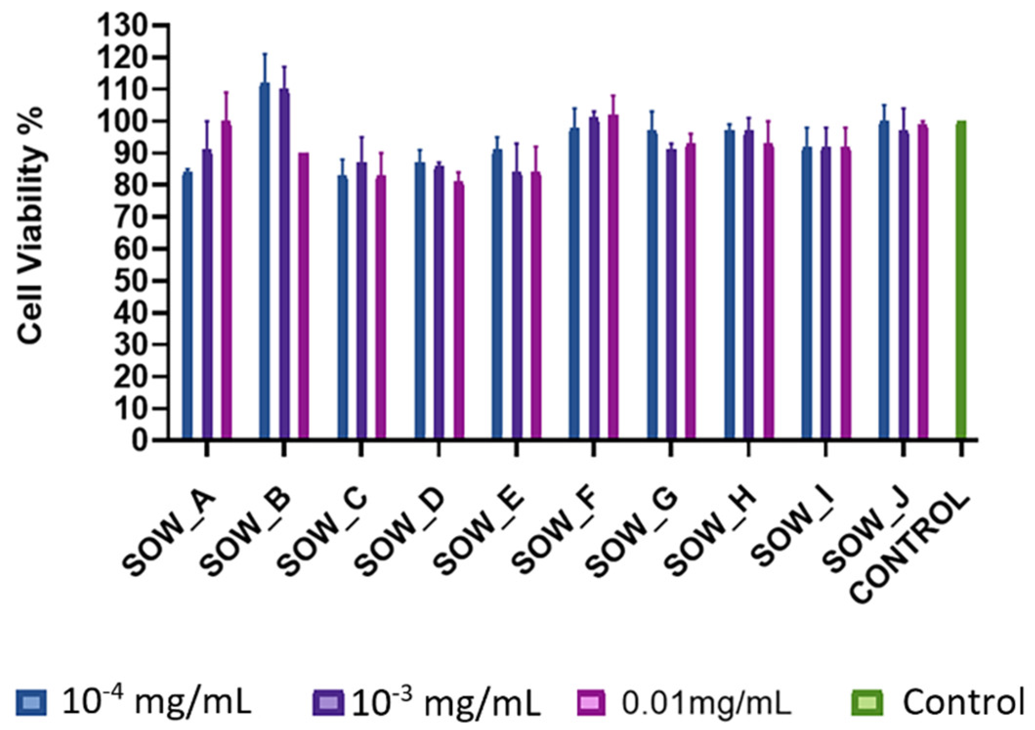
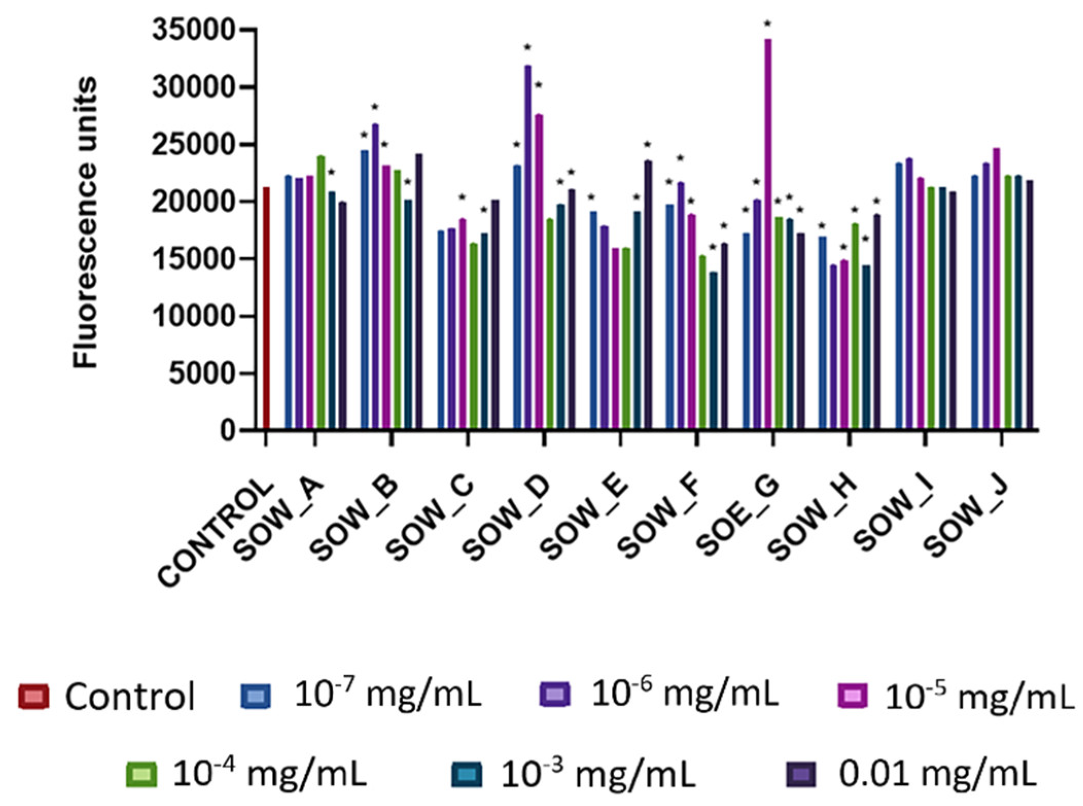
3.1. In Vitro Cytotoxicity, Photoprotection, and Antioxidant Activity of Plant Extracts/Infusions

The plant extracts and infusions did not demonstrate any cytotoxic effect at the concentrations from 10−5 to 0.1 mg/mL, apart from the ethyl acetate extract of R. officinalis (ROE), which showed a cytotoxic activity at a concentration of 0.1 mg/mL (Figures S1–S4).
The ethanol, ethyl acetate, and methanol extracts showed cytotoxicity at 1 mg/mL, the highest concentration tested, with cell viability ranging in the mean between 45–60% (Figures S1 and S2), with the exceptions of the ethyl acetate extract of M. officinalis subsp. altissima (MOE) and methanol extract of S. perfoliata subsp. perfoliata (SPM) which did not show any cytotoxicity. All aqueous extracts and infusions (Figures S3 and S4) did not exhibit any cytotoxicity with the exceptions of R. officinalis aqueous extract (ROW) and the infusions of S. sipylea (SSI) and S. euboea (SEI), which were cytotoxic at the highest tested concentration (1 mg/mL).
The photoprotective effect of the plant samples was explored at a concentration range of 10−5 to 1.0 mg/mL (Figures S5–S8). UV-A irradiation of NIH/3T3 fibroblasts with 5 J/cm2 caused an approximately 30% decrease in fibroblast viability (ΙRR control), while the non-irradiated cells (NIRR control) viability was set to 100%. The photosensitive substance, chlorpromazine (positive control), had an IC50 value of 1.0 μg/mL on cell viability after irradiation. S. officinalis ethyl acetate extract (SOE) showed important photoprotective activity at the concentration range from 10−2 to 1.0 mg/mL with cell viability ranging from 80 to 97%. In addition, the ethanol extract of T. vulgaris (TVE) demonstrated a photoprotective effect at all tested concentrations (cell viability ranges: 80–95%). Among the methanol extracts, those of S. euboea (SEM) and S. perfoliata subsp. perfoliata (SPM) had a photoprotective effect at a concentration range from 10−2 to 1.0 mg/mL and from 10−5 to 1.0 mg/mL, respectively. The aqueous extracts of S. officinalis (SOW) and T. vulgaris (TVW) unveiled the most important photoprotective effect at the concentration of 0.1 mg/mL with viability rates ranging from 85 to 94%. In addition, the aqueous extract of R. officinalis (ROW) showed moderate photoprotective activity at the lowest concentration, while greater activity was observed at higher concentrations (from 10−4 to 1.0 mg/mL). The infusions of S. euboea (SEI) and S. perfoliata subsp. perfoliata (SPI) demonstrated significant activity with cell viability ranging from 87 to 100%. It is noteworthy that both infusions showed similar viability rates to non-irradiated cells (NIRR control) at the two highest concentrations (0.1 and 1.0 mg/mL) (Figure S8).
Considering the potential of the plant extracts and infusions to reduce oxidative stress (Figures S9–S12), the aqueous extracts and infusions did not significantly reduce oxidative stress with the exception of S. sipylea infusion (SSI).
Overall, the diverse results among the tested plant samples in each biological assay could be also attributed to the different phytochemical constituents which were included in the used plants and extracts. Previous studies have reported a broad range of bioactive metabolites from these plants, belonging to triterpenoids (M. officinalis subsp. altissima, R. officinalis, Sideritis taxa, Stachys taxa, and T. vulgaris), diterpenoids (R. officinalis, S. officinalis, Sideritis taxa, Stachys taxa), iridoids (Sideritis taxa, St. iva), flavonoids (R. officinalis, S. officinalis, Sideritis taxa, St. iva, and T. vulgaris), phenylethanoid glycosides (Sideritis taxa and St. iva), and phenolic acids (M. officinalis subsp. altissima, R. officinalis, S. officinalis, Sideritis taxa, St. iva, and T. vulgaris) [21,22,24,25,26,27,28,29,30,31,32,33,34,35,36].

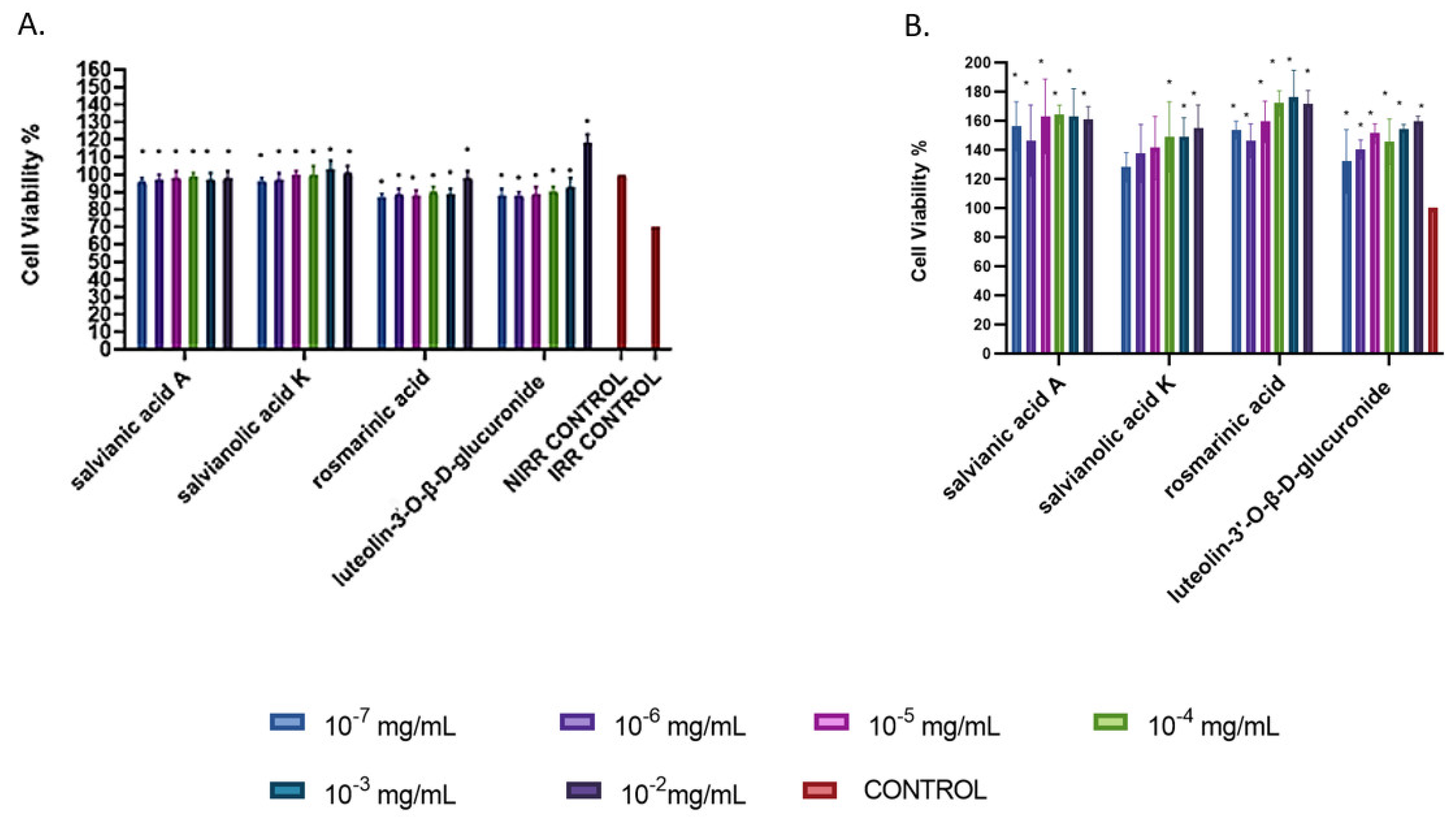
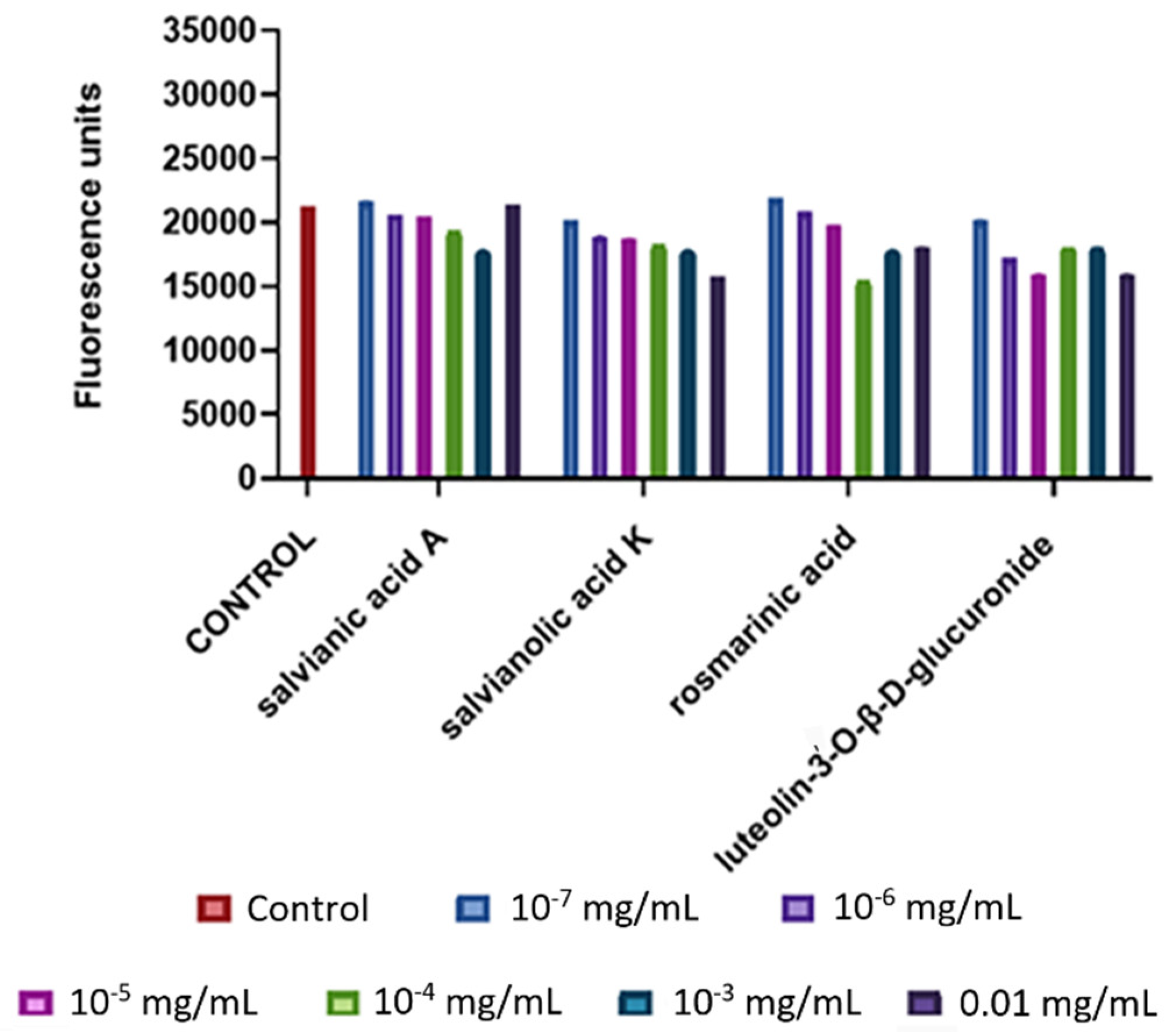
3.2. Bio-Guided Investigation of S. officinalis Aqueous Extract

Taking into consideration the overall outcomes from the plant extracts and infusions, the aqueous extract of S. officinalis (SOW) was chosen for further bio-guided isolation of the main responsible bioactive metabolites due to its rich phytochemical profile and photoprotective and non cytotoxic activity. This extract was fractionated and the obtained fractions (SOW_A-SOW_J) were evaluated for their cytotoxicity, photoprotective activity, and their potential to reduce oxidative stress. The fractions were initially tested for their in vitro cytotoxicity (Figure 1) at a concentration range from 10−4 to 0.01 mg/mL. None of the fractions were cytotoxic at all tested concentrations. All the fractions showed photoprotective activity at the tested concentrations, with the exception of SOW_H, SOW_I, and SOW_J fractions (Figure 2). It should be mentioned that a statistically significant photoprotective effect was exhibited by fractions SOW_E at the concentration of 10−2 mg/mL, SOW_F at the two highest concentrations (10−3 and 10−2 mg/mL) and SOW_G at all concentrations with the highest cell viability reaching 130 and 140% respectively, causing a 60 to 70% increase in viability compared to irradiated cells (IRR control, Figure 2). Regarding their antioxidant capacity, statistically significant effects were observed by the fractions SOW_C (at 10−5 and 10−3 mg/mL), SOW_F (at 10−3 and 0.01 mg/mL), and SOW_H (at 10−5 and 10−3 mg/mL) (Figure 3).
Based on the overall results, fractions SOW_E, SOW_F, and SOW_G were selected for further phytochemical study in order to isolate the most bioactive constituents, using different chromatographic techniques. In total, five compounds were obtained and identified through 1D- and 2D-NMR spectra, including three phenolic derivatives: salvianic acid A (1) [37], rosmarinic acid (2) [38], and salvianolic acid K (3) [38], and two flavone glucuronides namely luteolin-3′-O-β-D-glucuronide (4) [39] and hispidulin-7-O-β-D-glucuronide (5) [40]. Compounds 24 were previously found in S. officinalis [21,24,38]. To the best of our knowledge, although compounds 1 and 5 were reported in the genus Salvia [21,41,42], they were isolated for the first time from this species. In general, phenolic acids derivatives are commonly found in the genus Salvia [24]. Among them, rosmarinic acid is the most principal caffeic acid dimer, while salvianolic acids A–K are widely found in the genus Salvia. Furthermore, luteolin glycosides and their glucuronides seemed to be more common compared to those of apigenin in Salvia taxa [21].
The isolated compounds were evaluated for their cytotoxicity, photoprotective activity, and their potential to reduce oxidative stress. It should be mentioned that hispidulin-7-O-β-D-glucuronide (5) was not tested in the in vitro assays due to the fact that it was isolated in a very low amount. So, compounds 14 were tested in NIH/3T3 fibroblasts for 24 h at concentrations from 10−7 to 0.01 mg/mL. No statistically significant cytotoxic activity was observed compared to the control (Figure 4).
All isolated compounds exhibited statistically significant photoprotective activity in the tested concentrations (10−7 to 10−2 mg/mL) (Figure 5). These results are in accordance with the outcomes from the photoprotective effects of the fractions (Figure 2). It is noteworthy to mention that luteolin-3′-O-β-D-glucuronide (4) at a concentration of 0.01 mg/mL showed a cell viability value of approximately 120% (p < 0.05) compared to the control. The significant photoprotective effects of rosmarinic acid have been previously explored [43,44]. Among the different salvianolic acids, the photoprotection of salvianolic acid B has been reported [45]. Based on our knowledge this is the first report on the photoprotective activity of salvianic acid A, salvianolic acid K, and luteolin-3′-O-β-D-glucuronide.
Regardless of the potential of the isolated compounds to reduce oxidative stress, none of them demonstrated statistically significant changes in oxidative stress levels compared to the control group at the tested concentrations (Figure 6). Previous studies have mentioned that rosmarinic acid and salvianolic acids are responsible compounds for the antioxidant activity of Salvia species [21,24,46,47]. Although the initial fractions had been shown to reduce the oxidative stress in specific concentrations (Figure 3), it was not observed any effect from their isolated constituents. This might be attributed to the synergistic effects of the compounds in the fractions.

3.3. Wound-Healing Potential of the Isolated Compounds in Scratch Assay

The wound healing activity of isolated compounds (1-4) is presented in Figure 7. Mild migration was observed in the control group at 37%, while in the positive control (FBS 15%) 80% cell migration and proliferation were detected after 24 h of scratch formation. Among the isolated compounds, salvianolic acid K (3) showed the greatest, statistically significant wound healing activity at the concentration of 10−3 mg/mL, enhancing cell migration by 70%, in a similar way to positive control (80%). Previous studies have reported various pharmacological activities of this compound [46,48,49], however, this is the first time that a wound-healing activity of salvianolic acid K is reported. Among the tested compounds, rosmarinic acid (2) has already been reported as an important wound-healing agent [50].

4. Conclusions

This study explored the cytotoxic and photoprotective effects, as well as the potential to reduce oxidative stress in fibroblasts of ten Lamiaceae taxa. S. officinalis aqueous extract was bio-guided fractionated leading to the isolation and identification of the main responsible bioactive metabolites, namely salvianic acid A, rosmarinic acid, salvianolic acid K, luteolin-3′-O-β-D-glucuronide, and hispidulin-7-O-β-D-glucuronide. All the isolated compounds exhibited significant photoprotective activity in the tested concentration while none of them statistically significantly decreased oxidative stress. Salvianolic acid K wound healing effect was much enhanced, appearing as a good candidate for in vivo wound healing studies.
Overall, the present study unveiled the importance of Lamiaceae plants in skin health. Furthermore, it could provide a better insight into the traditional applications of the genus Salvia, aiming to validate ethnomedicinal use and identify the responsible metabolites.

Supplementary Materials

The following supporting information can be downloaded at: https://www.mdpi.com/article/10.3390/agronomy13051224/s1, Figure S1. Cytotoxic activity of ethanol and ethyl acetate extracts of S. officinalis (SOE), M. officinalis subsp. altissima (MOE), R. officinalis (ROE), and T. vulgaris (TVE) in mouse skin fibroblasts (NIH/3T3) after 24 h of treatment; Figure S2. Cytotoxic activity of methanol extracts of S. scardica (SSM), S. sipylea (SSMe), S. cypria (SCM), St. iva (SIM), S. euboea (SEM), and S. perfoliata subsp. perfoliata (SPM) in mouse skin fibroblasts (NIH/3T3) after 24 h of treatment; Figure S3. Cytotoxic activity of aqueous extracts of S. officinalis (SOW), T. vulgaris (TVW), M. officinalis subsp. altissima (MOW), and R. officinalis (ROW) in mouse skin fibroblasts (NIH/3T3) after 24 h of treatment; Figure S4. Cytotoxic activity of infusions of S. cypria (SCI), S. scardica (SSW), S. sipylea (SSI), S. euboea (SEI), S. perfoliata subsp. perfoliata (SPI), and St. iva (SII) in mouse skin fibroblasts (NIH/3T3) after 24 h of treatment; Figure S5. Photoprotective activity of ethanol and ethyl acetate extracts of S. officinalis (SOE), M. officinalis subsp. altissima (MOE), R. officinalis (ROE), and T. vulgaris (TVE). UV-A-induced cytotoxicity was assessed on mouse skin fibroblasts (NIH/3T3) pre-treated with the indicated extracts for 1 h (A). Extract—induced cytotoxicity after 1 h of treatment (B); Figure S6. Photoprotective activity of methanol extracts of S. scardica (SSM), S. sipylea (SSMe), S. cypria (SCM), St. iva (SIM), S. euboea (SEM), and S. perfoliata subsp. perfoliata (SPM). UV-A-induced cytotoxicity was assessed on mouse skin fibroblasts (NIH/3T3) pre-treated with the indicated extracts for 1 h (A). Extract-induced cytotoxicity after 1 h of treatment (B); Figure S7. Photoprotective activity of aqueous extracts of S. officinalis (SOW), T. vulgaris (TVW), M. officinalis subsp. altissima (MOW), and R. officinalis (ROW). UV-A-induced cytotoxicity was assessed on mouse skin fibroblasts (NIH/3T3) pre-treated with the indicated extracts for 1 h (A). Extract-induced cytotoxicity after 1 h of treatment (B); Figure S8. Photoprotective activity of infusions of S. cypria (SCI), S. scardica (SSW), S. sipylea (SSI), S. euboea (SEI), S. perfoliata subsp. perfoliata (SPI), and St. iva (SII). UV-A-induced cytotoxicity was assessed on mouse skin fibroblasts (NIH/3T3) pre-treated with the indicated extracts for 1 h (A). Extract-induced cytotoxicity after 1 h of treatment (B); Figure S9. Intracellular antioxidant activity of ethanol and ethyl acetate extracts of S. officinalis (SOE), M. officinalis subsp. altissima (MOE), R. officinalis (ROE), and T. vulgaris (TVE); Figure S10. Intracellular antioxidant activity of methanol extracts of S. scardica (SSM), S. sipylea (SSMe), S. cypria (SCM), St. iva (SIM), S. euboea (SEM), and S. perfoliata subsp. perfoliata (SPM); Figure S11. Intracellular antioxidant activity of aqueous extracts of S. officinalis (SOW), T. vulgaris (TVW), M. officinalis subsp. altissima (MOW), and R. officinalis (ROW); Figure S12. Intracellular antioxidant activity of infusions of S. cypria (SCI), S. scardica (SSW), S. sipylea (SSI), S. euboea (SEI), S. perfoliata subsp. perfoliata (SPI), and St. iva (SII).

Author Contributions

Conceptualization, M.C.R. and H.S.; methodology, M.C.R., E.-M.T. and H.S.; chemical investigation, E.T. and E.-M.T.; biological investigation: E.T.; statistical analysis, E.T.; writing—original draft preparation, E.T., E.-M.T. and C.A.; writing—review and editing, M.C.R. and H.S.; supervision, M.C.R. and H.S. All authors have read and agreed to the published version of the manuscript.

Funding

This research received no external funding.

Data Availability Statement

Not applicable.

Acknowledgments

The authors wish to thank N. Tzortzakis and A. Chrysargyris (Cyprus University of Technology) for providing the cultivated plant materials S. cypria Post and S. perfoliata subsp. perfoliata from Cyprus, as well as P. Chatzopoulou (Hellenic Agricultural Organization-ELGO Dimitra) for the cultivated plant materials of Sideritis euboea Heldr. Sideritis scardica Griseb., and Stachys iva Griseb. In addition, the authors wish to thank Th. Constantinidis and K. Goula (Section of Ecology and Systematics, Department of Biology, National and Kapodistrian University of Athens) for the identification of wild populations. E.T. thanks Maria Kostaki, for the initial training in the in vitro assays.

Conflicts of Interest

The authors declare no conflict of interest.

References

  1. Arda, O.; Göksügür, N.; Tüzün, Y. Basic histological structure and functions of facial skin. Clin. Dermatol. 2014, 32, 3–13. [Google Scholar] [CrossRef]
  2. Zouboulis, C.C.; Ganceviciene, R.; Liakou, A.I.; Theodoridis, A.; Elewa, R.; Makrantonaki, E. Aesthetic aspects of skin aging, prevention, and local treatment. Clin. Dermatol. 2019, 37, 365–372. [Google Scholar] [CrossRef]
  3. Ibrahim, N.; Wong, S.; Mohamed, I.; Mohamed, N.; Chin, K.-Y.; Ima-Nirwana, S.; Shuid, A. Wound Healing Properties of Selected Natural Products. Int. J. Environ. Res. Public. Health 2018, 15, 2360. [Google Scholar] [CrossRef]
  4. Svobodová, A.; Psotová, J.; Walterová, D. Natural phenolics in the prevention of UV-induced skin damage. A review. Biomed. Pap. Med. Fac. Univ. Palacky. Olomouc Czech Repub. 2003, 147, 137–145. [Google Scholar] [CrossRef]
  5. Solano, F. Photoprotection and Skin Pigmentation: Melanin-Related Molecules and Some Other New Agents Obtained from Natural Sources. Molecules 2020, 25, 1537. [Google Scholar] [CrossRef] [PubMed]
  6. Cavinato, M.; Jansen-Dürr, P. Molecular mechanisms of UVB-induced senescence of dermal fibroblasts and its relevance for photoaging of the human skin. Exp. Gerontol. 2017, 94, 78–82. [Google Scholar] [CrossRef] [PubMed]
  7. Cavinato, M.; Waltenberger, B.; Baraldo, G.; Grade, C.V.C.; Stuppner, H.; Jansen-Dürr, P. Plant extracts and natural compounds used against UVB-induced photoaging. Biogerontology 2017, 18, 499–516. [Google Scholar] [CrossRef] [PubMed]
  8. Hernandez, D.F.; Cervantes, E.L.; Luna-Vital, D.A.; Mojica, L. Food-derived bioactive compounds with anti-aging potential for nutricosmetic and cosmeceutical products. Crit. Rev. Food Sci. Nutr. 2021, 61, 3740–3755. [Google Scholar] [CrossRef] [PubMed]
  9. Hernández, A.R.; Vallejo, B.; Ruzgas, T.; Björklund, S. The Effect of UVB Irradiation and Oxidative Stress on the Skin Barrier-A New Method to Evaluate Sun Protection Factor Based on Electrical Impedance Spectroscopy. Sensors 2019, 19, 2376. [Google Scholar] [CrossRef]
  10. Mohania, D.; Chandel, S.; Kumar, P.; Verma, V.; Digvijay, K.; Tripathi, D.; Choudhury, K.; Mitten, S.K.; Shah, D. Ultraviolet Radiations: Skin Defense-Damage Mechanism. Adv. Exp. Med. Biol. 2017, 996, 71–87. [Google Scholar] [PubMed]
  11. Pérez-Sánchez, A.; Barrajón-Catalán, E.; Herranz-López, M.; Castillo, J.; Micol, V. Lemon balm extract (Melissa officinalis, L.) promotes melanogenesis and prevents UVB-induced oxidative stress and DNA damage in a skin cell model. J. Dermatol. Sci. 2016, 84, 169–177. [Google Scholar] [CrossRef] [PubMed]
  12. Kostyuk, V.; Potapovich, A.; Albuhaydar, A.R.; Mayer, W.; De Luca, C.; Korkina, L. Natural Substances for Prevention of Skin Photoaging: Screening Systems in the Development of Sunscreen and Rejuvenation Cosmetics. Rejuvenation Res. 2018, 21, 91–101. [Google Scholar] [CrossRef]
  13. Aghakhani, F.; Kharazian, N.; Lori Gooini, Z. Flavonoid constituents of Phlomis (Lamiaceae) species using liquid chromatography mass spectrometry. Phytochem. Anal. 2018, 29, 180–195. [Google Scholar] [CrossRef] [PubMed]
  14. Singh, P.; Pandey, A.K. Prospective of essential oils of the genus Mentha as biopesticides: A review. Front. Plant Sci. 2018, 9, 1295. [Google Scholar] [CrossRef] [PubMed]
  15. Walker, J.B.; Sytsma, K.J.; Treutlein, J.; Wink, M. Salvia (Lamiaceae) is not monophyletic: Implications for the systematics, radiation, andecological specializations of Salvia and tribe Mentheae. Am. J. Bot. 2004, 91, 1115–1125. [Google Scholar] [CrossRef] [PubMed]
  16. Committee on Herbal Medicinal Products. Assessment Report on Salvia officinalis L., Folium and Salvia officinalis L., Aetheroleum; EMA/HMPC/150801/2015; Committee on Herbal Medicinal Products: London, UK, 2016. [Google Scholar]
  17. Lamaison, J.L.; Petitjean-Freytet, C.; Carnat, A. Lamiacées médicinales à propriétés antioxydantes, sources potentielles d’acide rosmarinique [Medicinal Lamiaceae with antioxidant properties, a potential source of rosmarinic acid]. Pharm. Acta Helv. 1991, 66, 185–188. [Google Scholar]
  18. Dapkevicius, A.; Venskutonis, R.; van Beek, T.A.; Linssen, J.P.H. Antioxidant activity of extracts obtained by different isolation procedures from some aromatic herbs grown in Lithuania. J. Sci. Food Agric. 1998, 77, 140–146. [Google Scholar] [CrossRef]
  19. Hohmann, J.; Zupkó, I.; Rédei, D.; Csányi, M.; Falkay, G.; Máthé, I.; Janicsák, G. Protective effects of the aerial parts of Salvia officinalis, Melissa officinalis and Lavandula angustifolia and their constituents against enzyme-dependent and enzyme-independent lipid peroxidation. Planta Med. 1999, 65, 576–578. [Google Scholar] [CrossRef]
  20. Bonesi, M.; Loizzo, M.R.; Acquaviva, R.; Malfa, G.A.; Aiello, F.; Tundis, R. Anti-inflammatory and Antioxidant Agents from Salvia Genus (Lamiaceae): An Assessment of the Current State of Knowledge. Antiinflamm. Antiallergy Agents Med. Chem. 2017, 16, 70–86. [Google Scholar] [CrossRef]
  21. Lu, Y.; Foo, L.Y. Polyphenolics of Salvia-a review. Phytochemistry 2002, 59, 117–140. [Google Scholar] [CrossRef]
  22. Topçu, G. Bioactive triterpenoids from Salvia species. J. Nat. Prod. 2006, 69, 482–487. [Google Scholar] [CrossRef] [PubMed]
  23. Committee on Herbal Medicinal Products. European Union Herbal Monograph on Sideritis scardica Griseb.; Sideritis clandestina (Bory & Chaub.) Hayek; Sideritis raeseri Boiss. & Heldr.; Sideritis syriaca L., Herba; EMA/HMPC/39455/2015; Committee on Herbal Medicinal Products: London, UK, 2016. [Google Scholar]
  24. Lu, Y.; Foo, L.Y. Flavonoid and phenolic glycosides from Salvia officinalis. Phytochemistry 2000, 55, 263–267. [Google Scholar] [CrossRef] [PubMed]
  25. González-Burgos, E.; Carretero, M.E.; Gómez-Serranillos, M.P. Sideritis spp.: Uses, chemical composition and pharmacological activities—A review. J. Ethnopharmacol. 2011, 135, 209–225. [Google Scholar] [CrossRef] [PubMed]
  26. Fraga, B.M. Phytochemistry and chemotaxonomy of Sideritis species from the Mediterranean region. Phytochemistry 2012, 76, 7–24. [Google Scholar] [CrossRef]
  27. Tundis, R.; Peruzzi, L.; Menichini, F. Phytochemical and biological studies of Stachys species in relation to chemotaxonomy: A review. Phytochemistry 2014, 102, 7–39. [Google Scholar] [CrossRef] [PubMed]
  28. Andrade, J.M.; Faustino, C.; Garcia, C.; Ladeiras, D.; Reis, C.P.; Rijo, P. Rosmarinus officinalis L.: An update review of its phytochemistry and biological activity. Future Sci. OA 2018, 4, FSO283. [Google Scholar] [CrossRef]
  29. Mailis, T.; Skaltsa, H. Polar Constituents of Salvia willeana (Holmboe) Hedge, Growing Wild in Cyprus. Plants 2018, 7, 18. [Google Scholar] [CrossRef]
  30. Sharifi-Rad, J.; Ezzat, S.M.; El Bishbishy, M.H.; Mnayer, D.; Sharopov, F.; Kılıç, C.S.; Neagu, M.; Constantin, C.; Sharifi-Rad, M.; Atanassova, M.; et al. Rosmarinus plants: Key farm concepts towards food applications. Phytother. Res. 2020, 34, 1474–1518. [Google Scholar] [CrossRef]
  31. Żyżelewicz, D.; Kulbat-Warycha, K.; Oracz, J.; Żyżelewicz, K. Polyphenols and Other Bioactive Compounds of Sideritis Plants and Their Potential Biological Activity. Molecules 2020, 25, 3763. [Google Scholar] [CrossRef]
  32. Shimada, A.; Ueno, H.; Inagaki, M. Glutaminase inhibitory activities of pentacyclic triterpenes isolated from Thymus vulgaris L. Nat. Prod. Res. 2021, 36, 2864–2868. [Google Scholar] [CrossRef]
  33. Tomou, E.M.; Barda, C.; Skaltsa, H. Genus Stachys: A Review of Traditional Uses, Phytochemistry and Bioactivity. Medicines 2020, 7, 63. [Google Scholar] [CrossRef]
  34. Dimas, D.; Tomou, E.-M.; Karamanou, C.; Sfiniadakis, I.; Siakavella, K.I.; Liakopoulou, A.; Hatziantoniou, S.; Rallis, M.; Skaltsa, H. Melissa officinalis ssp. altissima extracts: A therapeutic approach targeting psoriasis in mice. J. Ethnopharmacol. 2019, 246, 112208. [Google Scholar]
  35. Patil, S.M.; Ramu, R.; Shirahatti, P.S.; Shivamallu, C.; Amachawadi, R.G. A systematic review on ethnopharmacology, phytochemistry and pharmacological aspects of Thymus vulgaris Linn. Heliyon 2021, 7, e07054. [Google Scholar] [CrossRef]
  36. Pritsas, A.; Tomou, E.M.; Tsitsigianni, E.; Papaemmanouil, C.D.; Diamantis, D.A.; Chatzopoulou, P.; Tzakos, A.G.; Skaltsa, H. Valorisation of stachysetin from cultivated Stachys iva Griseb. as anti-diabetic agent: A multi-spectroscopic and molecular docking approach. J. Biomol. Struct. Dyn. 2021, 39, 6452–6466. [Google Scholar] [CrossRef] [PubMed]
  37. Yahara, S.; Satoshiro, M.; Nishioka, I.; Nagasawa, T.; Oura, H. Isolation and characterization of phenolic compounds from Coptidis Rhizoma. Chem. Pharm. Bull. 1985, 33, 527–531. [Google Scholar] [CrossRef]
  38. Lu, Y.; Foo, L.Y. Rosmarinic acid derivatives from Salvia officinalis. Phytochemistry 1999, 51, 91–94. [Google Scholar] [CrossRef]
  39. Heitz, A.; Carnat, A.; Fraisse, D.; Carnat, A.-P.; Lamaison, J.-L. Luteolin 3′-glucuronide, the major flavonoid from Melissa officinalis subsp. officinalis. Fitoterapia 2000, 71, 201–202. [Google Scholar] [CrossRef]
  40. Zaabat, N.; Hay, A.E.; Michalet, S.; Skandrani, I.; Chekir-Ghedira, L.; Dijoux-Franca, M.G.; Akkal, S. Chemical Composition, Antioxidant, Genotoxique and Antigenotoxic Potentials of Phlomis bovei De Noé Aerial Parts. Iran. J. Pharm. Res. 2020, 19, 282–291. [Google Scholar]
  41. Martins, N.; Barros, L.; Santos-Buelga, C.; Henriques, M.; Silva, S.; Ferreira, I.C.F.R. Evaluation of bioactive properties and phenolic compounds in different extracts prepared from Salvia officinalis L. Food Chem. 2015, 170, 378–385. [Google Scholar] [CrossRef]
  42. Xu, J.; Wei, K.; Zhang, G.; Lei, L.; Yang, D.; Wang, W.; Han, Q.; Xia, Y.; Bi, Y.; Yang, M.; et al. Ethnopharmacology, phytochemistry, and pharmacology of Chinese Salvia species: A review. J. Ethnopharmacol. 2018, 225, 18–30. [Google Scholar] [CrossRef]
  43. Psotova, J.; Svobodova, A.; Kolarova, H.; Walterova, D. Photoprotective properties of Prunella vulgaris and rosmarinic acid on human keratinocytes. J. Photochem. Photobiol. B. 2006, 84, 167–174. [Google Scholar] [CrossRef] [PubMed]
  44. Sánchez-Campillo, M.; Gabaldon, J.A.; Castillo, J.; Benavente-García, O.; Del Baño, M.J.; Alcaraz, M.; Vicente, V.; Alvarez, N.; Lozano, J.A. Rosmarinic acid, a photo-protective agent against UV and other ionizing radiations. Food Chem. Toxicol. 2009, 47, 386–392. [Google Scholar] [CrossRef] [PubMed]
  45. Sun, Z.; Park, S.Y.; Hwang, E.; Zhang, M.; Jin, F.; Zhang, B.; Yi, T.H. Salvianolic Acid B Protects Normal Human Dermal Fibroblasts Against Ultraviolet B Irradiation-Induced Photoaging Through Mitogen-Activated Protein Kinase and Activator Protein-1 Pathways. Photochem. Photobiol. 2015, 91, 879–886. [Google Scholar] [CrossRef]
  46. Lu, Y.; Foo, Y.L. Antioxidant activities of polyphenols from sage (Salvia officinalis). Food Chem. 2001, 75, 197–202. [Google Scholar] [CrossRef]
  47. Xiao, Z.; Liu, W.; Mu, Y.P.; Zhang, H.; Wang, X.N.; Zhao, C.Q.; Chen, J.M.; Liu, P. Pharmacological Effects of Salvianolic Acid B Against Oxidative Damage. Front. Pharmacol. 2020, 11, 572373. [Google Scholar] [CrossRef] [PubMed]
  48. Kasimu, R.; Tanaka, K.; Tezuka, Y.; Gong, Z.-N.; Li, J.-X.; Basnet, P.; Namba, T.; Kadota, S. Comparative Study of Seventeen Salvia Plants: Aldose Reductase Inhibitory Activity of Water and MeOH Extracts and Liquid Chromatography-Mass Spectrometry (LC-MS) Analysis of Water Extracts. Chem. Pharm. Bull. 1998, 46, 500–504. [Google Scholar] [CrossRef]
  49. Ayoub, I.M.; George, M.Y.; Menze, E.T.; Mahmoud, M.; Botros, M.; Essam, M.; Ashmawy, I.; Shendi, P.; Hany, A.; Galal, M.; et al. Insights into the neuroprotective effects of Salvia officinalis L. and Salvia microphylla Kunth in the memory impairment rat model. Food Funct. 2022, 13, 2253–2268. [Google Scholar] [CrossRef]
  50. Küba, M.C.; Türkoğlu, A.; Oğuz, A.; Tuncer, M.C.; Kaya, Ş.; Başol, Ö.; Bilge, H.; Tatlı, F. Comparison of local rosmarinic acid and topical dexpanthenol applications on wound healing in a rat experimental wound model. Folia Morphol. 2021, 80, 618–624. [Google Scholar] [CrossRef]
Figure 1. Cytotoxic activity of S. officinalis fractions in NIH/3T3 fibroblasts after 24 h of treatment. Bars ± SD.
Agronomy 13 01224 g001
Figure 2. Photoprotective activity of S. officinalis fractions. UV-A-induced cytotoxicity was assessed on mouse skin fibroblasts (NIH/3T3) pre-treated with the indicated fractions for 1 h (A). Fraction—induced cytotoxicity after 1 h of treatment (B). PBS-treated cultures served as controls. UV-A untreated control (NIRR) set to 100%. Bars ± SD. * p < 0.05.
Agronomy 13 01224 g002
Figure 3. Intracellular antioxidant activity of S. officinalis fractions. 2′,7′-dichlorofluorescein (DCF)-fluorescence of mouse skin fibroblasts (NIH/3T3) treated with the indicated fractions for 1 h was used for the assessment of UVA-stimulated reactive oxygen species (ROS) levels. Vehicle (PBS)—treated irradiated cells were used as negative controls. Bars ± SD. * p < 0.05.
Agronomy 13 01224 g003
Figure 4. Cytotoxic activity of the isolated compounds (14) from S. officinalis in NIH/3T3 fibroblasts after 24 h of treatment. Bars ± SD.
Agronomy 13 01224 g004
Figure 5. Photoprotective activity of the isolated compounds (14) from S. officinalis. UV-A-induced cytotoxicity was assessed on mouse skin fibroblasts (NIH/3T3) pre-treated with the indicated compounds for 1 h (A). Compound—induced cytotoxicity after 1 h of treatment (B). PBS-treated cultures served as controls. UV-A untreated control (NIRR) set to 100%. Bars ± SD. * p < 0.05.
Agronomy 13 01224 g005
Figure 6. Intracellular antioxidant activity of the isolated compounds (14) from S. officinalis. 2′,7′-dichlorofluorescein (DCF)-fluorescence of mouse skin fibroblasts (NIH/3T3) treated with the indicated compounds for 1 h was used for the assessment of UVA-stimulated reactive oxygen species (ROS) levels. Vehicle (PBS)—treated irradiated cells were used as negative controls. Bars ± SD.
Agronomy 13 01224 g006
Figure 7. Wound-healing activity of the isolated compounds (14) from S. officinalis in NIH/3T3 fibroblasts. The data are expressed as mean of three experiments. Bars ± SD. * p < 0.05.
Agronomy 13 01224 g007
Table 1. Origin, extract-preparation, and abbreviation of investigated plant samples.
Plant SampleOriginExtractAbbreviation
Melissa officinalis subsp. altissima (Sm.) Arcang.wild—CreteEtOAc/AqueousMOE/MOW
Rosmarinus officinalis L.wild—Mt. PelionEtOAc/AqueousROE/ROW
Salvia officinalis L.wild—KozaniEtOAc/AqueousSOE/SOW
Sideritis cypria Postcultivated—CyprusMeOH/InfusionSCM/SCI
Sideritis euboea Heldr.cultivated—ELGO DimitraMeOH/InfusionSEM/SEI
Sideritis perfoliata L. subsp. perfoliatacultivated—CyprusMeOH/InfusionSPM/SPI
Sideritis scardica Griseb.cultivated—ELGO DimitraMeOH/InfusionSSM/SSW
Sideritis sipylea Boiss.wild—Samos IslandMeOH/InfusionSSMe/SSI
Stachys iva Griseb.cultivated—ELGO DimitraMeOH/InfusionSIM/SII
Thymus vulgaris L.wild—Mt. PelionEtOH/AqueousTVE/TVW
Disclaimer/Publisher’s Note: The statements, opinions and data contained in all publications are solely those of the individual author(s) and contributor(s) and not of MDPI and/or the editor(s). MDPI and/or the editor(s) disclaim responsibility for any injury to people or property resulting from any ideas, methods, instructions or products referred to in the content.

