Article

Genetic Diversity of Colletotrichum spp. Causing Grape Anthracnose in Zhejiang, China

1
College of Advanced Agricultural Sciences, Zhejiang A&F University, Hangzhou 311300, China
2
Research Institute for the Agriculture Science of Hangzhou, Hangzhou 310013, China
*
Authors to whom correspondence should be addressed.
Agronomy 2023, 13(4), 952; https://doi.org/10.3390/agronomy13040952
Submission received: 11 February 2023 / Revised: 15 March 2023 / Accepted: 19 March 2023 / Published: 23 March 2023
(This article belongs to the Special Issue Plant Anthracnose: Etiology and Current Management Options)

Abstract

:
Anthracnose is a fungal disease that seriously threatens grape production and quality. Multiple Colletotrichum species are detected in anthracnose grapes in vineyards. In this study, diseased grapes were collected in four counties in Zhejiang, and 43 Colletotrichum isolates were obtained. Multi-genes (ITS, TUB2, ACT, CHS-I, and GAPDH) and morphological characteristic analyses showed that C. fructicola (40 isolates, 93%), C. aenigma (two isolates, 4.7%), and C. pseudoacutatum (one isolate, 2.3%) were the cause of grape anthracnose in Zhejiang. Among the three Colletotrichum species, C. fructicola was the prevalent and dominant species in all sampled counties; C. pseudoacutatum was first identified as the pathogen responsible for grape anthracnose. There were significant differences in the sporulation among the three Colletotrichum species, as well as in the spore germination. Pathogenicity testing showed that all species can infect grapes, resulting in anthracnose. On the other hand, the virulence of species was varied and may be associated with their spore germination. This is the first study to characterize the Colletotrichum species causing grape anthracnose in Zhejiang Province and reveal that C. fructicola is the dominant species. The determination of Colletotrichum species associated with grape anthracnose may contribute to the study of epidemiology and development of an efficient strategy for controlling anthracnose in the vineyards.

1. Introduction

Grape berries are popular among customers, both as a fresh-eating food and as a material for juice and wine production. In addition, the berries are rich in various phenolic compounds, flavonoids and stilbenes, which are of benefit to human health and may help prevent several diseases and are beneficial to health [1,2]. Grape is one of the most extensively planted fruit trees worldwide, with planting ranging from temperate to tropical regions. According to the Food and Agriculture Organization (FAO), the grape-growing area is increasing by above 2% per year. China is the largest grape producer and consumer, with a total planting area of about 700,000 km2 and a production of about 1.3 million tons in 2018 [3]. Grape anthracnose or ripen rot is a serious disease; its occurrence at flowering and at harvest causes grape yield loss [4,5,6]. Furthermore, the disease also results in a bitter taste to the fruit and wine because of the increased chemical compositions (i.e., volatile acidity, residual sugar, glycerol, gluconic acid, and malic acid) in diseased fruit [7,8].
Grape anthracnose occurs in most vineyards worldwide, and the disease is caused by Colletotrichum spp. or by Elsinoe ampelina [9,10]. The symptoms induced by E. ampelina differ from those caused by Colletotrichum spp.. For instance, berries infected by E. ampelina first form dark-brown spots, then turn into sunken lesions with a grey-white center and dark-brown to purple-brown margins [10]; berries infected by Colletotrichum spp. form circular, reddish-brown spots that enlarge in concentric lesions, and acervuli with orange spore masses are observed in the spots [4,5,6,11].
Colletotrichum spp. is ranked eighth on the top 10 fungal plant pathogen list and causes anthracnose spots, wilting of plant parts, and post-harvest rot [12]. Colletotrichum spp. are important post-harvest pathogens because of latent infections: fruit are infected at the young stage, but do not show any symptoms until after the fruit have been stored or been marketed; this characteristic can cause up to 100% loss at the store stage [13]. When Colletotrichum spp. infects grapes, it causes grape anthracnose or ripen rot [9,14]. Latent infection is also observed when grapes are infected from the young green stage to the ripe stage, but do not show symptoms until ripening [4,15].
Colletotrichum spp. infects grapevine leaves, stems, and especially fruit [6,8]. It was recognized that C. gloeosporioides species complex and C. acutatum species complex are responsible for grape ripe rot [4,5,8,11,14,15,16]. Several studies have shown that C. gloeosporioides is sensitive to benomyl and that C. acutatum is generally moderately resistant to benzimidazole fungicides [4,11]. In addition, the epidemiology of C. gloeosporioides differs from C. acutatum [17,18]. Therefore, a precise determination of Colletotrichum species on the host is important for controlling Colletotrichum diseases in the fields.
In the past, morphological characterizations (i.e., colony features, size and shape of conidia and appressoria, optimal temperature, growth rate) were used for the identification of Colletotrichum species [19,20,21]. However, because of the stability of morphological traits influenced by cultivation environments and the existence of intermediate forms, differentiation of Colletotrichum species based on morphological characters is not entirely reliable enough to distinguish between Colletotrichum species. Molecular techniques, such as polymerase chain reaction (PCR), real-time PCR, restriction fragment length polymerase (RFLP), and simple sequence repeat (SSR) markers have been developed to detect pathogens [15,22]. DNA markers based on ITS regions are always used to detect fungi, and the multi-loci phylogenetic analysis method was used to distinguish Colletotrichum at the species level [22,23,24]. Combining microbiological methods with molecular phylogeny led to a better understanding and identification of Colletotrichum species, which has been used for the identification of Colletotrichum species on many hosts [25,26,27].
Zhejiang Province is one of the major grape-planting areas in southern China, and the grape industry is a characteristic agriculture for creating economic effectiveness for farmers in the region [3,28]. In Zhejiang, August and September are relatively warm summer months with the highest precipitation, which coincide with the final stages of grape ripening [28]. As we know, heat and wetness favor grape anthracnose epidemics. To provide basic knowledge of grape anthracnose and help control this fungal disease in Zhejiang, this study first investigated the genetic diversity of grape anthracnose and clarified the distribution of the Colletotrichum species in Zhejiang. Secondly, the biological characteristics and pathogenicity of each Colletotrichum species were determined.

2. Materials and Methods

2.1. Fungus Isolation

Grape fruit (Vitis vinifera ‘Kyoho’) with characteristic anthracnose symptoms were sampled from distinct vineyards in four counties in Zhejiang Province. We randomly selected six, four, five, and four vineyards in Hangzhou, Taizhou, Jinhua, and Shaoxing, respectively, for collecting samples. A total of 98 diseased fruit were collected, including 27 fruit from Hangzhou, 18 fruit from Taizhou, 27 fruit from Jinhua, and 26 fruit from Shaoxing, respectively. In the laboratory, diseased fruit were incubated in a growth chamber at 25 °C with a photoperiod of 12 h and 95% relative humidity until orange mucilage containing conidia formed on the lesions. The monospore culture method was then established as described in Arzanlou et al. [29]. In brief, conidia were picked up from diseased samples using a sterile needle, and transferred into 10 mL of sterile distilled water. Next, 100 μL of the conidia suspension was spread evenly on 2% water agar plates containing 50 mg/L streptomycin sulfate and incubated overnight at 25 °C. Then, the plates were checked under a light microscope (ZEISS Axio Scope. A1, Jena, Germany) and germinated conidia were transferred to a potato dextrose agar (PDA) plate. Ten conidia from each plate were cultured separately and the strains with different colony morphology were maintained on PDA slants at 4 °C.

2.2. Molecular Characterization

The mycelia of each field Colletotrichum isolates were harvested after 6 days of cultivation at 25 °C. Mycelia were momentarily frozen in liquid nitrogen and ground into a fine powder using a sterile mortar and pestle. The powdered mycelia were dissolved in buffer digestion (Sangon Biotech, Shanghai, China), then genomic DNA was extracted using the Rapid Fungi Genomic DNA Isolation Kit (Sangon Biotech) according to the manufacturer’s instructions. The DNA concentration was determined by spectrophotometry and diluted with distilled water for further DNA amplification reactions. Internal transcribed spacer (ITS), partial beta tubulin 2 (TUB2), acting (ACT), chitin synthase (CHS-I), and glyceraldehyde-3-phosphate dehydrogenase (GAPDH) genes were amplified using the primer pairs ITS-1F/ITS4 [30], T1/T2 [31], ACT-512F/ACT-783R [32], CHS-79F/CHS-354R [32], and GDF/GDR [33], respectively.
Each 50 μL of gene amplification reaction mixture contained 2× Taq PCR Master Mix (25 μL, Sangon Biotech), template DNA (1 μL, 20–50 ng), 10 μM forward primer and reverse primer (2 μL, each primer). PCR conditions for ITS, ACT, CHS-I and GAPDH were performed as described by Chen et al. [34]. The PCR condition for TUB2 was conducted with denaturation at 94 °C for 4 min, followed by 30 cycles of 94 °C for 30 s, 55 °C for 30 s, 72 °C for 45 s, and further extension at 72 °C for 10 min. The amplicons were separated by electrophoresis and visualized by staining with ethidium bromide (EB). The single band with expected size was cut and recovered using the SanPrep Column PCR Product Purification Kit (Sangon Biotech), then the purified products were sequenced by Sangon Biotech.

2.3. Multi-Locus Phylogenetic Analysis

Raw sequence chromatograms were manually examined, and the sequences for each fragment were assembled by primer pair of each gene using DNAStar v. 5.0 software. The assembled sequences of ITS, TUB2, ACT, CHS-I, and GAPDH were shared in the GenBank database of the National Center for Biotechnology Information (Table S1) and also were blasted against the GenBank database, respectively. Colletotrichum isolates were then first identified at the species complex level. Related gene sequences (ITS, TUB2, ACT, CHS-I, and GAPDH) of Colletotrichum species from previous publications were downloaded from GenBank [23,24]. The referenced standard strains are listed in Table S1. For each gene, sequences from the isolates belonging to the same species complex were compared using MAFFT v.7 following default parameter settings [35]. If necessary, sequences were manually adjusted in MEGA v. 6.06 [36]. The modified sequences were concatenated in Sequence Matrix 1.8. Modeltest3.7.win, Win-paup4b10-console, and Mrmodeltest2, as implemented in MrMTgui, and were used to estimate the best model of nucleotide substitution [37]. Bayesian inference (BI) was used to construct phylogenies using MrBayes v. 3.1.2 [38]. Six simultaneous Markov chains were run for 5,000,000 generations each until the standard deviation of split frequencies was below 0.01, and trees were sampled every 100th generation. The first 2000 trees, representing the burn-in phase of the analyses, were discarded, and the remaining 8000 trees were used to calculate the posterior probability (PP) in the majority rule consensus tree. Phylogenetic trees were drawn using TreeView [39]. The alignments and trees were deposited into TreeBase.

2.4. Morphological Characterization

Representative isolates of each Colletotrichum species were cultured on the PDA, malt extracts agar (MEA; Sigma Aldrich, Gillingham, UK) and oatmeal agar (OA; Sigma-Aldrich, UK), respectively. Their colonies characterization was performed following the procedures of Karimi et al. [40]. One fresh mycelium plug (5 mm in diameter) of Colletotrichum isolates was taken from the growing edge of 4-day-old cultures and was transferred onto the center of PDA, MEA, and OA plates. After cultivation for 7 days at 25 °C in darkness, the colony morphology was recorded, and the cultures on PDA plates were continued until conidiomata formed. The morphology of conidiomata were recorded by a light microscope (Nikon SMZ25, Tokyo, Japan), then conidiomata was picked up and diluted in distilled water to obtain a conidia suspension with a density of 1 × 10⁶ conidia·mL−1. Fifty spores were selected randomly to measure the length and width under a Nikon Eclipse 80i microscope (Tokyo, Japan). Conidia suspension was also used to induce appressoria as described in Wu et al. [25], and the morphology and size of 30 appressoria were recorded.

2.5. Mycelial Growth, Sporulation and Conidial Germination of Colletotrichum spp.

Isolates chosen in the previous section were also used to determine the temperature effect on colony growth. As described previously [41], for the determination of temperature effects, a mycelial plug (5 mm in diameter) from the growing edge of 4-day-old cultures was transferred onto the center of PDA plate (90 mm in diameter), and each isolate was inoculated separately at 5, 10, 15, 17, 20, 25, 27, 30, 35, and 40 °C for 5 days. Each treatment was set up for three replicates and the experiment was performed three times. The optimum temperature for the mycelial growth was estimated based on the Gaussian process (least squares fit) for nonlinear regression in Graph Prism 8.0 [42].
Additionally, sporulation and conidial germination of the selected isolates were also determined in this study. For conidia production, 10 fresh mycelial plugs were transferred to a 250-mL flask containing 100 mL potato dextrose (PD) liquid medium and were shaken at 160 rpm for 5 days at 25 °C. The conidial suspension of each isolate was filtered through three layers of muslin cloth, a 10-μL mixture was dropped on a hemocytometer and covered with a coverslip. The conidia production of each isolate was counted under a ZEISS Axio Scope A1 microscope. The mean spore numbers from ten biological replicates were used to represent sporulation capacity.
Aliquot of 6 µL suspensions with 1 × 10⁶ spores mL−1 were dropped on plastic unbreakable cover slip (size 22 × 22 mm) (Thermos Fisher Scientific, Waltham, MA, USA), and then incubated at 25 °C in darkness. After 16 h incubation, germination was quantified at five sites by counting 200 conidia in total, and the germination percentage was calculated. When a germ tube reached at least half the length of the conidium, it was scored as germinated [17]. Five biological replicates were analyzed for each isolate.

2.6. Pathogenicity Testing

Berries of ‘Kyoho’ were used for evaluating the pathogenicity of Colletotrichum spp. Healthy grapes with a uniform size and no visible surface symptoms were selected for the pathogenicity test. Inoculation was performed according to Yan et al. [9], with some modifications: grapes were surface disinfected with 2% NaOCl for 2 min followed by three rinses in sterile water, and then air-dried on sterilized filter paper. The grapes were inoculated by the wound/drop incubation method. A sterilized insect pin (0.5 mm in diameter) was used to create superficial wounds in the epidermis on the detached fruit, and an aliquot of 6 µL of conidia suspension with 1 × 10⁶ spores mL−1 was dropped on the wound. Inoculated grapes were incubated in a growth chamber at 25 °C with a photoperiod of 12 h and 95% relative humidity. Grapes treated with 6 µL of sterile water on the wound were included as a control. The experiment was performed three times, using 10 grapes per treatment per assay. The pathogens were re-isolated from the resulting lesions by tissue isolation method described above.
After 2 days, incidence in grape fruit was counted, and after 5 days, disease severity in grape fruit was assessed as the percentage of fruit area with anthracnose symptoms as previously described [43]. The rating scale for fruit was classified as follows: (0) no symptoms; (1) 0.1–5.0% of the fruit surface affected; (3) 5.1–15.0%; (5) 15.1–30.0%; (7) 30.1–50.0%; (9) 50.1–100.0% or rotten fruit. The lesion area was used to calculate the disease indicated as follows: Disease index = [(n0 × 0+ n1 × 1+ n3 × 3+ n5 × 5+ n7 × 7 + n9 × 9)/(9 × N)] × 100, where n represents the number of fruit in each class (0, 1, 3, 5, 7, 9) and N represents the total number of fruit.

2.7. Statistical Analysis

The mycelial growth, sporulation, spore germination, and virulence data were analyzed using IBM SPSS Statistics 29.0 by Fisher’s least significant difference (LSD) with a one-way ANOVA at p = 0.05.

3. Results

3.1. Colletotrichum Isolates Associated with Grape Anthracnose

Grape berries showing anthracnose symptoms were sampled in vineyards from four different geographical regions in Zhejiang Province. In total, 43 of the diseased grapes were used for monospore isolation, and finally 43 fungal isolates were recovered based on primary colony morphology, including 13 isolates obtained from Hangzhou, 14 isolates obtained from Shaoxing, 11 isolates obtained from Jinhua, and 5 isolates obtained from Taizhou.

3.2. Multi-Locus Phylogenetic Analyses

All 43 field Colletotrichum isolates were grouped into three Colletotrichum species. In the phylogenetic tree constructed for the C. gloeosporioides species complex, 42 field isolates and 26 reference isolates (Table S1), including C. boninense (CBS: 123755) as an outgroup, were subjected to multi-locus phylogenetic analyses with concatenated ITS, TUB2, ACT, CHS-I, and GAPDH sequences. The concatenated alignment of ITS, TUB2, ACT, CHS-I, and GAPDH contained 1459 characteristics (gene boundaries, ITS: 1–370, TUB2: 371–690, ACT: 691–900, CHS-I: 901–1196, GAPDH: 1197–1459). The results showed that 42 isolates clustered together with two species: 40 isolates with C. fructicola and 2 isolates with C. aenigma (Figure 1).
The remaining one isolate HZJ-6 and 31 reference isolates, including C. orchidophilum (CBS 632.80) as an outgroup, were subjected to multi-locus phylogenetic analyses with concatenated ITS, TUB2, ACT, CHS-I, and GAPDH sequences from those belonging to C. acutatum species complex. Finally, isolate HZJ-6 did not belong to the C. acutatum species complex and was clustered with C. pseudoacutatum (Figure 2).

3.3. Species Distribution

Among the three Colletotrichum species, C. fructicola was detected in each sampled county (Figure 1). In total, 40 of the 43 isolates were grouped into C. fructicola, which indicated that C. fructicola was the main pathogen associated with grape anthracnose in Zhejiang Province. C. aenigma (two isolates) and C. pseudoacutatum (one isolate) both showed a lower frequency in Zhejiang Province and were only found in Hangzhou and Shaoxing, respectively. In Jinhua and Taizhou counties, only C. fructicola isolates were isolated from grape with anthracnose symptoms (Figure 3).

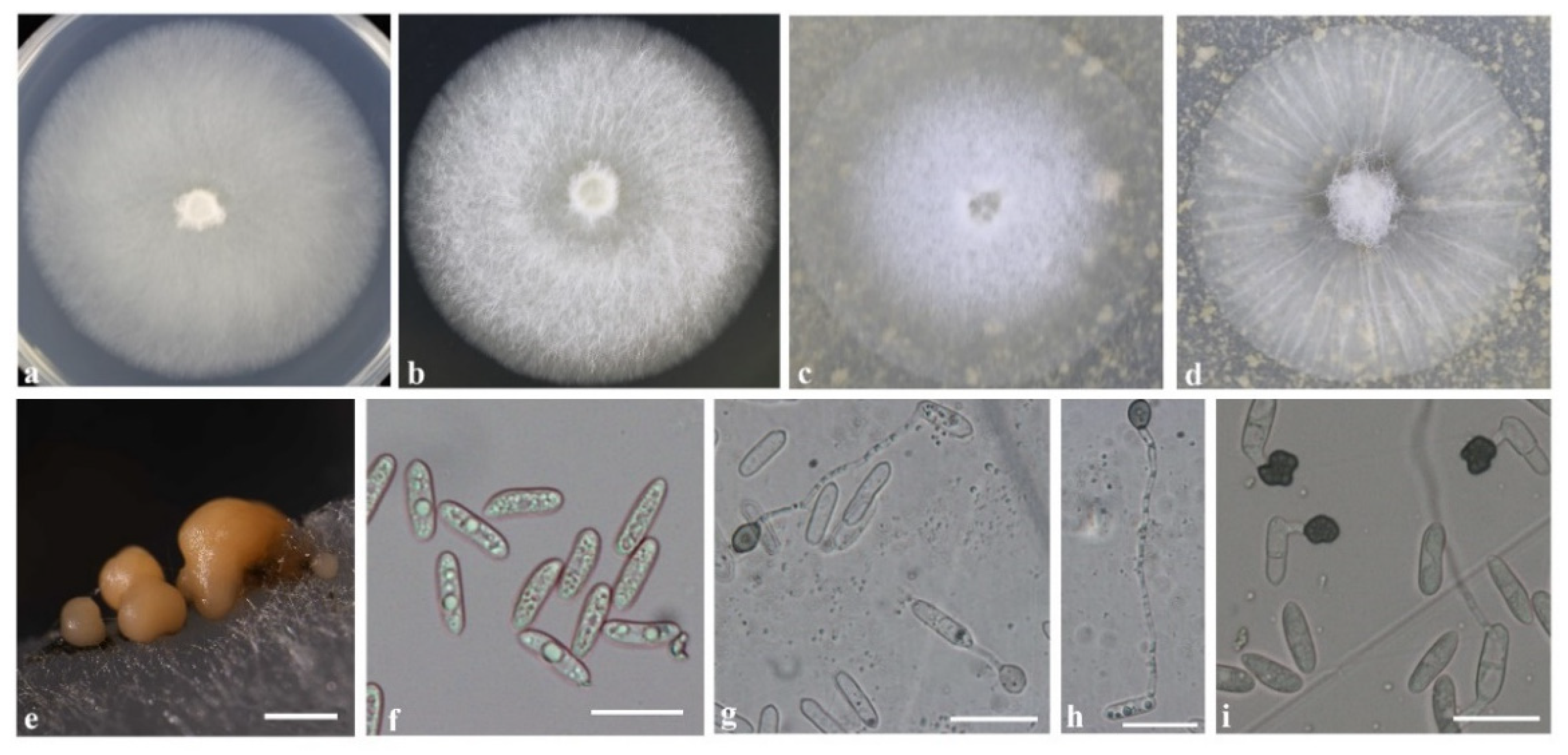
3.4. Morphology Description

Based on multi-locus sequence data, the 43 isolates were classified into three Colletotrichum species. Of these, C. pseudoacutatum was first recorded in grapes. The three Colletotrichum species were morphologically similar (Figure 4, Figure 5 and Figure 6) and their morphology characteristics are described below.
After 7 days of cultivation on a medium, colonies of C. fructicola on PDA were light gray, villous, and dense (Figure 4a); on MEA, they were white (Figure 4b), and on OA, they were white in the center with a transparent margin (Figure 4c) or grew radially with a thin, white mycelium (Figure 4d). Similar colony morphology was observed in C. aenigma (Figure 5a–c). On PDA, colonies of C. pseudoacutatum were atrovirens in the center with a gray margin; on MEA and OA, they grew with white mycelium and fluffy orange-yellow conidiomata distributed beneath the hyphae (Figure 6a–c). Conidia morphology was similar among the three species: conidia in mass orange, conidia hyaline, aseptate, smooth-walled, and cylindrical (Figure 4e,f, Figure 5d,e and Figure 6d,e). Few differences in appressoria morphology were observed among the three species. Appressoria of C. fructicola and C. pseudoacutatum were both ellipsoid or irregular (Figure 4g–i and Figure 6f,g), and appressoria of C. aenigma were subglobose (Figure 6f,g). The conidia and appressoria sizes of the tested isolates are described in detail in Table 1.

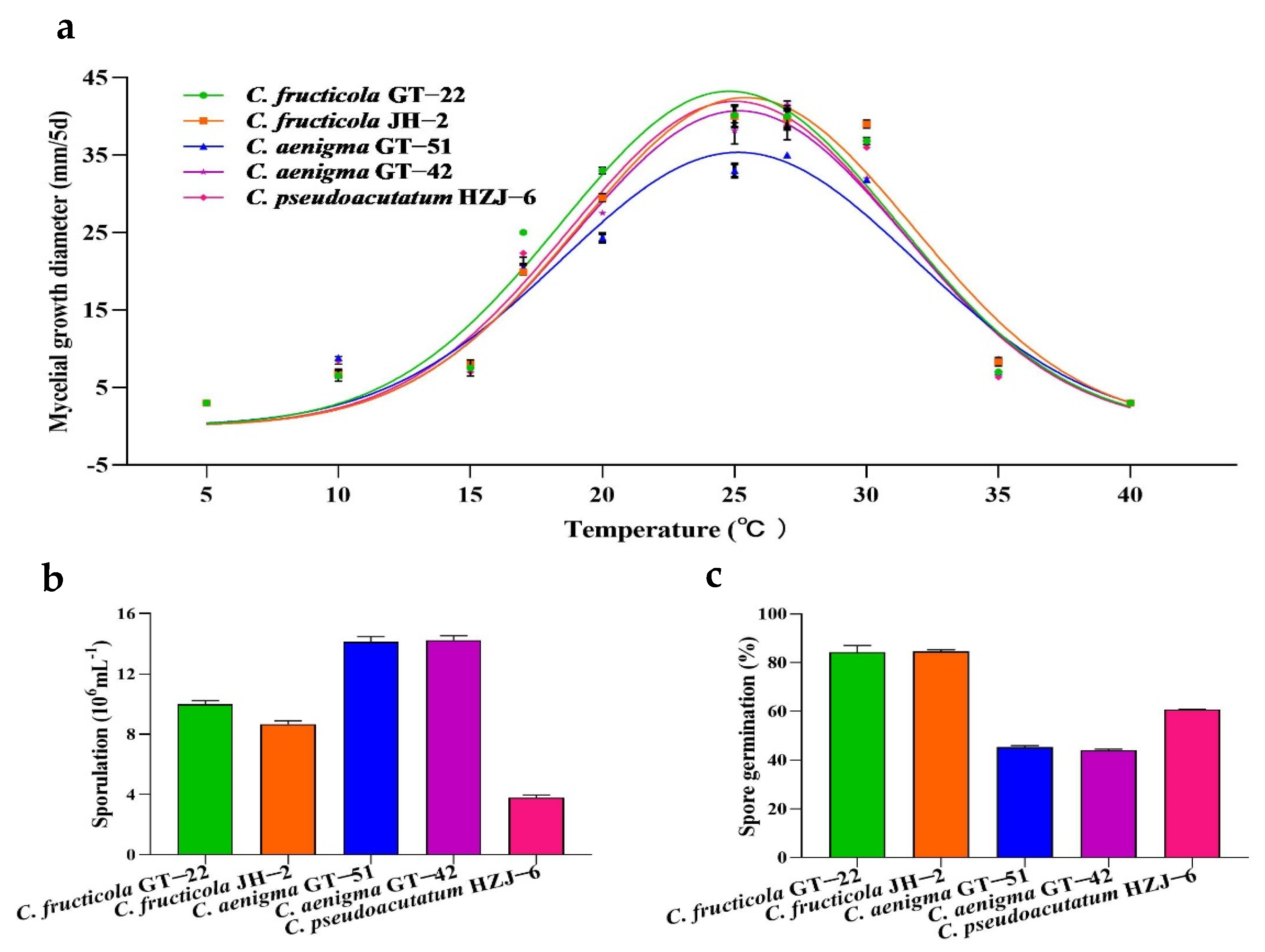
3.5. Mycelial Growth, Sporulation and Conidial Germination of Colletotrichum spp.

Five isolates selected for morphology observation were grown on PDA at 5 °C to 40 °C. The maximum colony diameter of C. fructicola GT-22, C. fructicola JH-2, C. aenigma GT-51, C. aenigma GT-42, and C. pseudoacutatum HZJ-6 after cultivation for 5 days on PDA was at 24.8 °C, 25.4 °C, 25.1 °C, 25.2 °C, and 25.0 °C, respectively (Figure 7a; Table S2). The temperature of 25 °C to 30 °C was suitable for the mycelial growth of C. fructicola and C. pseudoacutatum, and 25 °C to 27 °C was suitable for the mycelial growth of C. aenigma (Figure 7a, Tables S2 and S3).
Based on the conidia production and conidia germination results (Figure 7b,c; Table 2), the ranking of species by sporulation under the same environment was as follows: C. aenigma GT-51 and GT-42 > C. fructicola GT-22 > C. fructicola JH-2 > C. pseudoacutatum HZJ-6. The ranking of species by spore germination was as follows: C. fructicola GT-22 and JH-2 > C. aenigma GT-51 and GT-42 > C. pseudoacutatum HZJ-6.

3.6. Pathogenicity Tests

The pathogenicity test showed that the lesion area of grapes treated with sterile water on the wound was less than 0.1%, and significant differences in virulence (p < 0.05) were observed in three Colletotrichum species. Necrotic spots on grape fruit caused by C. fructicola isolates and C. pseudoacutatum isolate were visible at 2 days post inoculation (dpi). At 5 dpi, the disease index caused by C. fructicola GT-22 and JH-2 was 44.7 and 45.8, respectively, showing the greatest disease severity (Table 2). In contrast, C. aenigma species showed significantly weaker pathogenicity with a mean disease index of 14.4 (Table 2).

4. Discussion

Grape anthracnose is one of the important fungal diseases in vineyards and is epidemic worldwide. As previous research has shown [9,44,45,46], C. gloeosporioides species complex (i.e., C. fructicola, C. gloeosporioides, C. kahawae, C. viniferum, C. aenigma, and C. hebeiense), C. acutatum species complex (i.e., C. acutatum, C. limitticola, C. nymphaeae, and C. citri), and C. boninense species complex (i.e., C. karstii) are the cause of grape anthracnose. In China, C. viniferum was reported as the dominant species in Beijing, Jiangsu, Shandong, Yunnan, Guizhou, and Fujian [9,44,47,48,49]. In this study, we collected diseased fruit from vineyards in four distinct geographical regions in Zhejiang Province. Through phylogenetic analysis and inoculation tests on grapes, we revealed that three Colletotrichum species, including C. fructicola, C. aenigma, and C. pseudoacutatum, cause grape anthracnose in Zhejiang. To the best of our knowledge, it is the first description of C. pseudoacutatum causing grape anthracnose.
Colletotrichum infects many cultivated fruit crops, such as grape, pear, citrus, strawberry, cherry, and banana [9,26,27,34,50,51]. Previous studies have shown that multiple Colletotrichum species or biotypes are associated with anthracnose disease on a single host [26,43,51,52]. Variation of the morphological traits of Colletotrichum species under different cultivation environments is common [23,26,27] and the morphology is not always reliable enough to distinguish Colletotrichum at species level. In our study, multi-locus phylogenetic analyses with concatenated ITS, TUB2, ACT, CHS-I, and GAPDH sequences revealed that C. fructicola, C. aenigma, and C. pseudoacutatum were associated with grape anthracnose in Zhejiang, and the morphological characteristics of each species also were described in detail.
In the current study, C. fructicola was the dominant species (40 isolates, 93%) on grapes, followed by C. aenigma (two isolates, 4.7%), and C. pseudoacutatum (one isolate, 2.3%). Colletotrichum bunch rot disease on mature grape fruit was first documented in the USA in 1891 [53]. First C. gloeosporioides (Penz) and then C. acutatum (Simmonds) have been reported to responsible for this disease [4,8,11], but the two Colletotrichum species were not detected in our study. C. viniferum has been reported as the main cause of grape anthracnose in some grape-planting areas in China [9,47,48,49]; however this species was not found in Zhejiang Province. Here, we hypothesize that grape variety affects the pathogen diversity of anthracnose [9,11]. Although C. fructicola was detected on Colletotrichum-diseased grapes at a low frequency in some vineyards in China [47,48,49], it was a key pathogen that caused anthracnose on grapes in all sampled regions in Zhejiang. In addition, C. fructicola is prevalent on pears and strawberries [26,34]. In this study, we provide the first record of C. pseudoacutatum as a cause of grape anthracnose, which is a newly recorded species that does not belong to the C. acutatum species complex [24].
Temperature is a major factor in plant fungal disease epidemics, and rainy, humid, and warm conditions, with temperatures ranging between 20 °C and 30 °C, are beneficial for Colletotrichum-disease epidemics [54,55,56]. Temperature affects the production and maturation of conidia of Colletotrichum spp., as well as its infection rate on grapes [4,5,6]. Mycelial growth of Colletotrichum spp. at different temperatures in vitro was determined, and our results were in agreement with a previous study that revealed the majority of Colletotrichum species grew at temperatures ranging from 10 °C to 35 °C [17]. In addition, our result showed that the mycelial growth of C. fructicola, C. aenigma, and C. pseudoacutatum was inhibited at 5 °C and 40 °C, and they all had optimal growth temperatures on PDA around 25 °C, with C. aenigma having the lower growth rate.
Grape anthracnose is a polycyclic disease. Conidia are the source of infection by Colletotrichum species on grapes, and grape anthracnose epidemics are usually initiated by conidia, which settle on plant surfaces and germinate under favorable conditions and penetrate the host tissue [8,17]. Sporulation and the germination of conidia of the Colletotrichum species were determined, and the results showed that the highest sporulation was observed in two C. aenigma isolates; however, the conidial germination rate of C. fructicola was significantly higher than that of the other two species. Previous studies have shown that germination of conidia and sporulation are key aspects in the pathogen life cycle and are strongly correlated with infection of Colletotrichum species on hosts. The relative severity of disease caused by Colletotrichum species is determined by the proportion of conidia (conidia from overwintering primary sources or conidia produced on diseased tissues) that mature, disperse, and settle on plant tissue, then infect, and finally cause disease symptoms [6,17]. In our study, the disease index was used to represent the relative severity caused by Colletotrichum species on grapes. Consistent with the above studies, our results showed that the two C. aenigma isolates had poor conidial germination, resulting in lower pathogenicity.

5. Conclusions

In the present study, C. fructicola, C. aenigma, and C. pseudoacutatum were responsible for grape anthracnose in Zhejiang Province. According to our results, the C. fructicola species had the greatest spore germination rate and were the most pathogenic among the three Colletotrichum species, which may explain why it was the dominant species in vineyards in Zhejiang. C. pseudoacutatum was first recorded in grapes, and it also successfully infected strawberries and kiwifruit, indicating a lack of host specificity (data not shown). The lack of host specificity reminded us to focus on cross-infection caused by Colletotrichum species between grape and other horticultural crops. Benzimidazole resistance within the gloeosporioides complex on grapes was notable [57]. Here, we determined that Colletotrichum species caused grape anthracnose in Zhejiang vineyards, which will contribute to studying the epidemiology and fungicide sensitivity, and finally developing efficient methods for grape anthracnose management.

Supplementary Materials

The following supporting information can be downloaded at: https://www.mdpi.com/article/10.3390/agronomy13040952/s1, Table S1: List of Colletotrichum spp. strains used for multi-gene analysis in this study; Table S2: Colony diameter (mm) of Colletotrichum spp. under different temperatures (cultivation for 5 days on PDA); Table S3: Colony diameter (mm) of Colletotrichum species at 20 °C, 25 °C, 27 °C, and 30 °C (cultivation for 5 days on PDA).

Author Contributions

Conceptualization, J.W. and C.Z.; methodology, J.W., H.Y. and C.Z.; software, B.Y. and J.Z.; validation, H.Y. and C.Z.; formal analysis, J.W., J.Z. and X.C.; investigation, W.X. and X.C.; writing—original draft preparation, J.W. and B.Y.; writing—review and editing, H.Y. and C.Z.; visualization, J.W. and B.Y.; supervision, H.Y. and C.Z. All authors have read and agreed to the published version of the manuscript.

Funding

This research was funded by the National Innovation and Entrepreneurship Training Project for College Student (202110341052), sponsor: B.Y.

Institutional Review Board Statement

Not applicable.

Informed Consent Statement

Not applicable.

Data Availability Statement

Not applicable.

Conflicts of Interest

The authors declare no conflict of interest.

References

  1. Nassiri-Asl, M.; Hosseinzadeh, H. Review of the Pharmacological Effects of Vitis vinifera (Grape) and its Bioactive Constituents: An Update. Phytother. Res. 2016, 30, 1392–1403. [Google Scholar] [CrossRef] [PubMed]
  2. Xia, E.-Q.; Deng, G.-F.; Guo, Y.-J.; Li, H.-B. Biological Activities of Polyphenols from Grapes. Int. J. Mol. Sci. 2010, 11, 622–646. [Google Scholar] [CrossRef] [PubMed]
  3. Li, X.H.; Li, Y.J.; Ma, X.Q.; Guo, J.; Liu, H.J.; Zheng, G.Q.; Tao, J.M. Development Situation and Prospects of Grape Industry in China. South China Fruits 2021, 50, 161–166. [Google Scholar]
  4. Whitelaw-Weckert, M.A.; Curtin, S.J.; Huang, R.; Steel, C.; Blanchard, C.; Roffey, P.E. Phylogenetic relationships and pathogenicity of Colletotrichum acutatum isolates from grape in subtropical Australia. Plant Pathol. 2007, 56, 448–463. [Google Scholar] [CrossRef]
  5. Shiraishi, M.; Koide, M.; Itamura, H.; Yamada, M.; Mitani, N.; Ueno, T.; Nakaune, R.; Nakano, M. Screening for Resistance to Ripe Rot Caused by Colletotrichum acutatum in Grape Germplasm. Vitis 2007, 46, 196–200. [Google Scholar]
  6. Ji, T.; Salotti, I.; Dong, C.; Li, M.; Rossi, V. Modeling the Effects of the Environment and the Host Plant on the Ripe Rot of Grapes, Caused by the Colletotrichum Species. Plants 2021, 10, 2288. [Google Scholar] [CrossRef]
  7. Meunier, M.; Steel, C.C. Effect of Colletotrichum acutatum Ripe Rot on the Composition and Sensory Attributes of Cabernet Sauvignon Grapes and Wine. Aus. J. Grape Wine Res. 2010, 15, 223–227. [Google Scholar] [CrossRef]
  8. Hong, S.-K.; Kim, W.-G.; Yun, H.-K.; Choi, K.-J. Morphological Variations, Genetic Diversity and Pathogenicity of Colletotrichum species Causing Grape Ripe Rot in Korea. Plant Pathol. J. 2008, 24, 269–278. [Google Scholar] [CrossRef] [Green Version]
  9. Yan, J.-Y.; Jayawardena, R.; Goonasekara, I.D.; Wang, Y.; Zhang, W.; Liu, M.; Huang, J.-B.; Wang, Z.-Y.; Shang, J.-J.; Peng, Y.-L.; et al. Diverse species of Colletotrichum associated with grapevine anthracnose in China. Fungal Divers. 2014, 71, 233–246. [Google Scholar] [CrossRef]
  10. Li, Z.; Santos, R.F.; Gao, L.; Chang, P.; Wang, X. Current status and future prospects of grapevine anthracnose caused by Elsinoe ampelina: An important disease in humid grape-growing regions. Mol. Plant Pathol. 2021, 22, 899–910. [Google Scholar] [CrossRef]
  11. Greer, L.; Harper, J.; Savocchia, S.; Samuelian, S.; Steel, C. Ripe rot of south-eastern Australian wine grapes is caused by two species of Colletotrichum: C. acutatum and C. gloeosporioides with differences in infection and fungicide sensitivity. Aust. J. Grape Wine Res. 2011, 17, 123–128. [Google Scholar] [CrossRef]
  12. Dean, R.; Van Kan, J.A.; Pretorius, Z.A.; Hammond-Kosack, K.E.; Di Pietro, A.; Spanu, P.D.; Rudd, J.J.; Dickman, M.; Kahmann, R.; Ellis, J.; et al. The Top 10 Fungal Pathogens in Molecular Plant Pathology. Mol. Plant Pathol. 2012, 13, 414–430. [Google Scholar] [CrossRef] [PubMed] [Green Version]
  13. Prusky, D. Pathogen quiescence in postharvest diseases. Annu. Rev. Phytopathol. 1996, 34, 413–434. [Google Scholar] [CrossRef] [Green Version]
  14. Oo, M.M.; Oh, S.-K. Identification and Characterization of New Record of Grape Ripe Rot Disease Caused by Colletotrichum viniferum in Korea. Mycobiology 2017, 45, 421–425. [Google Scholar] [CrossRef] [Green Version]
  15. Freeman, S.; Katan, T.; Shabi, E. Characterization of Colletotrichum Species Responsible for Anthracnose Diseases of Various Fruits. Plant Dis. 1998, 82, 596–605. [Google Scholar] [CrossRef] [PubMed] [Green Version]
  16. Daykin, M.; Milholland, R.D.J.P. Ripe Rot of Muscadine Grape Caused by Colletotrichum gloeosporioides and Its Control. Phytopathology 1984, 74, 710–714. [Google Scholar]
  17. Salotti, I.; Ji, T.; Rossi, V. Temperature requirements of Colletotrichum spp. belonging to different clades. Front. Plant Sci. 2022, 13. [Google Scholar] [CrossRef]
  18. Zulfiqar, M.; Brlansky, R.H.; Timmer, L.W. Infection of Flower and Vegetative Tissues of Citrus by Colletotrichum acutatum and C. gloeosporioides. Mycologia 1996, 88, 121. [Google Scholar] [CrossRef]
  19. Gunnell, P.S.; Gubler, W.D. Taxonomy and Morphology of Colletotrichum Species Pathogenic to Strawberry. Mycologia 1992, 84, 157–165. [Google Scholar] [CrossRef]
  20. Smith, B.J.; Black, L.L. Morphological, Cultural, and Pathogenic Variation among Colletotrichum Species Isolated from Strawberry. Plant Dis. 1990, 74, 69–76. [Google Scholar] [CrossRef]
  21. Dickman, M.B. Colletotrichum. In Fungal Pathology; Kronstad, J.W., Ed.; Springer: Dordrecht, The Netherlands, 2000; pp. 127–147. [Google Scholar]
  22. Chavhan, R.; Sable, S.; Narwade, A.; Hinge, V.; Kalbande, B.; Mukherjee, A.; Chakrabarty, P.; Kadam, U.S. Multiplex molecular marker-assisted analysis of significant pathogens of cotton (Gossypium sp.). Biocatal. Agric. Biotechnol. 2023, 47. [Google Scholar] [CrossRef]
  23. Weir, B.S.; Johnston, P.R.; Damm, U. The Colletotrichum gloeosporioides species complex. Stud. Mycol. 2012, 73, 115–180. [Google Scholar] [CrossRef] [PubMed] [Green Version]
  24. Damm, U.; Cannon, P.F.; Woudenberg, J.H.C.; Crous, P.W. The Colletotrichum acutatum species complex. Stud. Mycol. 2012, 73, 37–113. [Google Scholar] [CrossRef] [PubMed] [Green Version]
  25. Wu, J.; Hu, S.; Ye, B.; Hu, X.; Xiao, W.; Yu, H.; Zhang, C. Diversity and Resistance to Thiophanate-Methyl of Colletotrichum spp. in Strawberry Nursery and the Development of Rapid Detection Using LAMP Method. Agronomy 2022, 12, 2815. [Google Scholar] [CrossRef]
  26. Fu, M.; Crous, P.W.; Bai, Q.; Zhang, P.F.; Xiang, J.; Guo, Y.S.; Zhao, F.F.; Yang, M.M.; Hong, N.; Xu, W.X.; et al. Colletotrichum Species Associated with Anthracnose of Pyrus spp. in China. Persoonia 2019, 42, 1–35. [Google Scholar] [CrossRef] [Green Version]
  27. Huang, F.; Chen, G.Q.; Hou, X.; Fu, Y.S.; Cai, L.; Hyde, K.D.; Li, H.Y. Colletotrichum species associated with cultivated citrus in China. Fungal Divers. 2013, 61, 61–74. [Google Scholar] [CrossRef]
  28. Chen, H.C.; Shen, B.W.; Wei, L.Z.; Fang, D.Y.; Wang, J.; Wu, J. Development Status and Countermeasures of Grape Industry in Pujiang. Sino-Overseas Grapevine Wine 2020, 4, 57–61. [Google Scholar]
  29. Arzanlou, M.; Bakhshi, M.; Karimi, K.; Torbati, M. Multigene phylogeny reveals three new records of Colletotrichum spp. and several new host records for the mycobiota of Iran. J. Plant Prot. Res. 2015, 55, 198–211. [Google Scholar] [CrossRef]
  30. White, T.; Bruns, T.; Lee, S.; Taylor, J.; Innis, M.; Gelfand, D.; Sninsky, J. Amplification and Direct Sequencing of Fungal Ri-bosomal RNA Genes for Phylogenetics. In PCR Protocols: A Guide to Methods and Applications; Elsevier: Amsterdam, The Netherlands, 1990; Volume 31, pp. 315–322. [Google Scholar]
  31. O’Donnell, K.; Cigelnik, E. Two Divergent Intragenomic rDNA ITS2 Types within a Monophyletic Lineage of the Fun-gusFusariumAre Nonorthologous. Mol. Phylogenet. Evol. 1997, 7, 103–116. [Google Scholar] [CrossRef]
  32. Carbone, I.; Kohn, L.M. A method for designing primer sets for speciation studies in filamentous ascomycetes. Mycologia 1999, 91, 553–556. [Google Scholar] [CrossRef]
  33. Templeton, M.D.; Rikkerink, E.H.; Solon, S.L.; Crowhurst, R.N. Cloning and Molecular Characterization of the glyceralde-hyde-3-phosphate dehydrogenase-encoding gene and cDNA from the Plant Pathogenic Fungus Glomerella cingulata. Gene 1992, 122, 225–230. [Google Scholar] [CrossRef]
  34. Chen, X.Y.; Dai, D.J.; Zhao, S.F.; Shen, Y.; Wang, H.D.; Zhang, C.Q. Genetic Diversity of Colletotrichum spp. Causing Strawberry Anthracnose in Zhejiang, China. Plant Dis. 2020, 104, 1351–1357. [Google Scholar] [CrossRef] [PubMed]
  35. Katoh, K.; Standley, D.M. MAFFT Multiple Sequence Alignment Software Version 7: Improvements in Performance and Usability. Mol. Biol. Evol. 2013, 30, 772–780. [Google Scholar] [CrossRef] [Green Version]
  36. Tamura, K.; Peterson, D.; Peterson, N.; Stecher, G.; Nei, M.; Kumar, S. MEGA5: Molecular Evolutionary Genetics Analysis Using Maximum Likelihood, Evolutionary Distance, and Maximum Parsimony Methods. Mol. Biol. Evol. 2011, 28, 2731–2739. [Google Scholar] [CrossRef] [PubMed] [Green Version]
  37. Vaidya, G.; Lohman, D.J.; Meier, R. SequenceMatrix: Concatenation software for the fast assembly of multi-gene datasets with character set and codon information. Cladistics 2011, 27, 171–180. [Google Scholar] [CrossRef]
  38. Ronquist, F.; Huelsenbeck, J.P. MrBayes 3: Bayesian phylogenetic inference under mixed models. Bioinformatics 2003, 19, 1572–1574. [Google Scholar] [CrossRef] [PubMed] [Green Version]
  39. Page, R.D. TreeView: An Application to Display Phylogenetic Trees on Personal Computers. Comput. Appl. Biosci. 1996, 12, 357–358. [Google Scholar]
  40. Karimi, K.; Arzanlou, M.; Pertot, I. Weeds as Potential Inoculum reservoir for Colletotrichum nymphaeae Causing Strawberry Anthracnose in Iran and Rep-PCR Fingerprinting as Useful Marker to Differentiate C. acutatum Complex on Strawberry. Front. Microbiol. 2019, 10, 129. [Google Scholar] [CrossRef] [Green Version]
  41. Dillard, H.R. Influence of Temperature, pH, Osmotic Potential, and Fungicide Sensitivity on Germination of Conidia and Growth from Sclerotia of Colletotrichum coccodes in vitro. Phytopathology 1988, 78, 1357–1361. [Google Scholar] [CrossRef]
  42. Chung, P.C.; Wu, H.Y.; Wang, Y.W.; Ariyawansa, H.A.; Hu, H.P.; Hung, T.H.; Tzean, S.S.; Chung, C.L. Diversity and Patho-genicity of Colletotrichum Species Causing Strawberry Anthracnose in Taiwan and Description of a New Species, Colletotrichum miaoliense sp. nov. Sci. Rep. 2020, 10, 14664. [Google Scholar] [CrossRef] [PubMed]
  43. Zhang, L.; Song, L.; Xu, X.; Zou, X.; Duan, K.; Gao, Q.H. Characterization and Fungicide Sensitivity of Colletotrichum Species Causing Strawberry Anthracnose in Eastern China. Plant Dis. 2020, 104, 1960–1968. [Google Scholar] [CrossRef] [PubMed]
  44. Lei, Y.; Tang, X.B.; Jayawardena, R.S.; Yan, J.Y.; Chen, Q.X. Identification and characterization of Colletotrichum species causing grape ripe rot in southern China. Mycosphere 2016, 7, 1177–1191. [Google Scholar] [CrossRef]
  45. Echeverrigaray, S.; Scariot, F.J.; Fontanella, G.; Favaron, F.; Sella, L.; Santos, M.C.; Schwambach, J.; Pedrotti, C.; Delamare, A.P.L. Colletotrichum species causing grape ripe rot disease in Vitis labrusca and V. vinifera varieties in the highlands of southern Brazil. Plant Pathol. 2020, 69, 1504–1512. [Google Scholar] [CrossRef]
  46. Kim, J.S.; Hassan, O.; Chang, T. First Report of Colletotrichum aenigma Causing Anthracnose of Grape in Korea. Plant Dis. 2021, 105, 2729. [Google Scholar] [CrossRef] [PubMed]
  47. Liu, M.; Jayawardena, R.S.; Liu, Y.; Xing, Q.K.; Yan, J.Y.; Li, X.H. Molecular Identification of the Pathogen Causing Grape Anthracnose in Beijing. J. Plant Prot. 2018, 45, 393–394. [Google Scholar]
  48. Xu, Y.; Xiao, T.; Chu, S.P.; Liu, J.X.; Rui, D.M.; Yao, K.B.; Yang, J.H. The Diversity and Resistance to Benzimidazole Fungicides of the Colletotrichum spp. Casuing Grape Anthracnose in Jurong City of Jiangsu Province. J. Nanjing Agric. Univ. 2022, 45, 78–85. [Google Scholar]
  49. Peng, L.-J.; Sun, T.; Yang, Y.-L.; Cai, L.; Hyde, K.D.; Bahkali, A.; Liu, Z.-Y. Colletotrichum species on grape in Guizhou and Yunnan provinces, China. Mycoscience 2013, 54, 29–41. [Google Scholar] [CrossRef]
  50. Tang, Z.Y.; Lou, J.; He, L.Q.; Wang, Q.D.; Chen, L.H.; Zhong, X.T.; Wu, C.F.; Zhang, L.Q.; Wang, Z.Q. First Report of Colletotrichum fructicola Causing Anthracnose on Cherry (Prunus avium) in China. Plant Dis. 2022, 106, 317. [Google Scholar] [CrossRef]
  51. Zakaria, L.; Sahak, S.; Zakaria, M.; Salleh, B. Characterisation of Colletotrichum Species Associated with Anthracnose of Banana. Trop. Life Sci. Res. 2009, 20, 119–125. [Google Scholar]
  52. Moral, J.; Agustí-Brisach, C.; Raya, M.C.; Jurado-Bello, J.; López-Moral, A.; Roca, L.F.; Chattaoui, M.; Rhouma, A.; Nigro, F.; Sergeeva, V.; et al. Diversity of Colletotrichum Species Associated with Olive Anthracnose Worldwide. J. Fungi 2021, 7, 741. [Google Scholar] [CrossRef] [PubMed]
  53. Southworth, E.A. Ripe Rot of Grapes and Apples. J. Mycol. 1891, 6, 164. [Google Scholar] [CrossRef]
  54. López-Moral, A.; Agustí-Brisach, C.; Lovera, M.; Arquero, O.; Trapero, A. Almond Anthracnose: Current Knowledge and Future Perspectives. Plants 2020, 9, 945. [Google Scholar] [CrossRef] [PubMed]
  55. Sharma, M.; Kulshrestha, S. Colletotrichum gloeosporioides: An Anthracnose Causing Pathogen of Fruits and Vegetables. Biosci. Biotechnol. Res. Asia 2015, 12, 1233–1246. [Google Scholar] [CrossRef]
  56. Ngugi, H. Epidemiology of sorghum anthracnose (Colletotrichum sublineolum) and leaf blight (Exserohilum turcicum) in Kenya. Plant Pathol. 2000, 49, 129–140. [Google Scholar] [CrossRef] [Green Version]
  57. Lin, T.; Xu, X.F.; Dai, D.J.; Shi, H.J.; Wang, H.D.; Zhang, C.Q. Differentiation in development of benzimidazole resistance in Colletotrichum gloeosporioides complex populations from strawberry and grape hosts. Australas. Plant Pathol. 2016, 45, 241–249. [Google Scholar] [CrossRef]
Figure 1. A Bayesian inference phylogenetic tree of the Colletotrichum gloeosporioides species complex. The tree was constructed with a Bayesian analysis of concatenated internal transcribed spacer (ITS), partial beta tubulin 2 (TUB2), acting (ACT), chitin synthase (CHS-I), and glyceraldehyde-3-phosphate dehydrogenase (GAPDH). The Markov chain was run for 5,000,000 generations, each locus having a separate model of DNA evaluation. C. boninense (CBS: 123755) was used as an outgroup. * The reference isolates.
Agronomy 13 00952 g001
Figure 2. A Bayesian inference phylogenetic tree of the C. acutatum species complex. The tree was constructed with a Bayesian analysis of concatenated internal transcribed spacer (ITS), partial beta tubulin 2 (TUB2), acting (ACT), chitin synthase (CHS-I), and glyceraldehyde-3-phosphate dehydrogenase (GAPDH). The Markov chain was run for 5,000,000 generations, each locus having a separate model of DNA evaluation. C. orchidophilum (CBS 632.80) was used as an outgroup. The scale bar shows 0.05 expected changes per site. * The reference isolates.
Agronomy 13 00952 g002
Figure 3. Distribution of Colletotrichum species in four grape-planting regions in Zhejiang, China. n = number of Colletotrichum isolates analyzed. 13 Colletotrichum isolates from Hangzhou contained C. fructicola (11 isolates, 85%) and C. aenigma (two isolates, 15%); 14 isolates from Shaoxing contained C. fructicola (13 isolates, 93%) and C. pseudoacutatum (one isolate, 7%); a further 11 isolates from Jinhua and 5 isolates from Taizhou all were C. fructicola.
Agronomy 13 00952 g003
Figure 4. Culture characteristics and microscopic features of C. fructicola. (a,b) Seven-day-old of colonies on PDA and MEA medium, respectively. (c,d) Seven-day-old of colonies on OA medium. (e) Conidiomata on PDA medium. (f) Conidia. (fi) Appressoria. Scale bars: (e) = 500 μm; (fi) = 20 μm. (a–c,eh): isolate GT-22; (d,i): isolate JH-2.
Agronomy 13 00952 g004
Figure 5. Culture characteristics and microscopic features of C. aenigma (isolate GT-42). (ac) Seven-day-old of colonies on PDA, MEA, and OA medium, respectively. (d) Conidiomata. (e) Conidia. (f,g) Appressoria. Scale bars: (b) = 500 μm; (eg) = 20 μm.
Agronomy 13 00952 g005
Figure 6. Culture characteristics and microscopic features of C. pseudoacutatum HZJ-6. (ac) Seven-day-old of colonies on PDA, MEA, and OA medium, respectively. (d) Conidiomata. (e) Conidia. (f,g) Appressoria. Scale bars: (d) = 500 μm; (e,f) = 20 μm.
Agronomy 13 00952 g006
Figure 7. Biological characteristics of Colletotrichum species. (a) Colony diameter of Colletotrichum species at different temperatures. The colony diameter of each Colletotrichum isolate is the mean ± standard error of three independent experiments. Different symbols represent the mean colony diameter of different species at the tested temperatures. Gaussian process regression was used to estimate the optimum temperature for mycelial growth. Comparison of sporulation (b) and spore germination (c) of Colletotrichum species.
Agronomy 13 00952 g007
Table 1. Colony morphology and morphometry of Colletotrichum species recovered form grape.
SpeciesIsolatesColony Morphology 1ConidiaAppressorium
PDA 2MEA 3OA 4Size 5 (μm)Size 5 (μm)
C. fructicolaGT-22R, C, GR, C, WR, C, W(c) to T(m)13.7–18.2 × 4.3–5.9
(16.9 ± 0.5) × (4.7 ± 0.5)
6.4–8.5 × 4.8–7.3
(7.5 ± 0.7) × (5.7 ± 0.8)
JH-2R, C, GR, C, WR, Ra, W13.1–18.6 × 4.2–5.9
(16.8 ± 0.5) × (4.9 ± 0.3)
7.2–9.9 × 6.1–8.8
(8.0 ± 0.4) × (6.9 ± 0.5)
C. aenigmaGT-51R, C, GGR, C, WR, C, W(c) to T(m)13.2–17.1 × 4.1–5.6
(14.1 ± 0.4) × (5.1 ± 0.3)
6.2–9.6 × 5.2–7.6
(6.9 ± 0.9) × (5.9 ± 0.6)
GT-42R, C, GGR, C, WR, C, W(c) to T(m)13.1–17.8 × 3.8–5.7
(13.9 ± 0.5) × (4.5 ± 0.5)
6.0–8.3 × 4.9–6.2
(6.8 ± 0.6) × (5.7 ± 0.5)
C. pseudoacutatumHZJ-6R, C, A(c) to G(m)R, C, O(c) to W(m)R, C, O(c) to W(m)14.2–18.9 × 4.9–7.2
(16.6 ± 0.5) × (5.6 ± 0.4)
7.5–10.1 × 5.2–7.2
(8.7 ± 0.9) × (5.4 ± 0.8)
1 Surface of colony: R, regular; C, circular; G, gray; GG, gray-green; A, atrovirens; W, white; O, orange; T, transparent; Ra, radiation; c, center; m, margin. 2 Potato dextrose agar. 3 Malt extracts agar. 4 Oatmeal agar. 5 Length × Width (data are mean ± standard deviation).
Table 2. Characterization of Colletotrichum species recovered from grapes.
SpeciesIsolatesSporulation 1
(×106 mL−1)
Spore Germination 1 (%)Disease Index 1
C. fructicolaGT-2210.0 ± 0.2 b 84.2 ± 2.8 a44.8 ± 5.7 a
JH-28.7 ± 0.2 c84.5 ± 0.8 a45.8 ± 5.9 a
C. aenigmaGT-5114.1 ± 0.4 a45.3 ±0.6 c14.7 ± 2.1 b
GT-4214.2 ± 0.3 a44.0 ± 0.5 c14.0 ± 3.9 b
C. pseudoacutatumHZJ-63.8 ± 0.2 d60.7 ± 0.3 b28.0 ± 1.7 ab
1 Values shown are the means ± standard error with different lowercase letters indicating significant differences (p < 0.05) according to Fisher’s least significant difference test.
Disclaimer/Publisher’s Note: The statements, opinions and data contained in all publications are solely those of the individual author(s) and contributor(s) and not of MDPI and/or the editor(s). MDPI and/or the editor(s) disclaim responsibility for any injury to people or property resulting from any ideas, methods, instructions or products referred to in the content.

