Article

Infection Biology of Bipolaris oryzae in Rice and Defence Responses in Compatible and Less Compatible Interactions

by
Tran Thi Thu Thuy
1,2,
Mette Lübeck
1,3,
Viggo Smedegaard-Petersen
1,
Eigil de Neergaard
1 and
Hans J. L. Jørgensen
1,*
1
Department of Plant and Environmental Sciences and Copenhagen Plant Science Centre, Faculty of Science, University of Copenhagen, Thorvaldsensvej 40, DK-1871 Frederiksberg C, Denmark
2
Department of Plant Protection, College of Agriculture, Can Tho University, Can Tho City 90000, Vietnam
3
Department of Chemistry and Bioscience, Aalborg University, Fredrik Bajers Vej 7H, DK-9220 Aalborg, Denmark
*
Author to whom correspondence should be addressed.
Agronomy 2023, 13(1), 231; https://doi.org/10.3390/agronomy13010231
Submission received: 4 December 2022 / Revised: 6 January 2023 / Accepted: 9 January 2023 / Published: 12 January 2023
(This article belongs to the Section Pest and Disease Management)

Abstract

:
The infection biology of Bipolaris oryzae and the defence responses of its host rice were studied using the rice cv. MTL 189 inoculated with two isolates of B5 (highly compatible with MTL 189) and K2 (less compatible). In the former interaction, the hyphal progress was accompanied by tissue degradation and extensive sporulation after 8 days, whereas in the latter interaction, only very limited tissue degradation and sporulation was observed. Quantitative microscopy of the infection showed that the percentages of conidia and appressoria causing penetration and fluorescent epidermal cells (FEC) were lower for isolate K2 than for isolate B5 at 12 and 24 hours after inoculation (hai). Fluorescent papillae (FP) were only seen in the less compatible interaction and the percentage of conidia causing single FEC was highest in the less compatible interaction at 12 hai, but not at 24 hai. Qualitative examination of other defence responses showed that H2O2 started to accumulate at 4 hai in the less compatible interaction, whereas it appeared in the compatible interaction only at 12 hai. The level of H2O2 was generally higher in the less compatible than in the compatible interaction. Cross sections of leaves showed that H2O2 accumulated in the outer walls of epidermal cells. Likewise, accumulation of callose and polyphenolic substances was most pronounced in the less compatible interaction and occurred at the same places as H2O2. To our knowledge, this is the first report implicating H2O2 as an early defence response against the hemibiotrophic pathogen B. oryzae during early infection stages in rice. Understanding defence reactions may aid in resistance breeding.

1. Introduction

Brown spot caused by Bipolaris oryzae (syn. Cochliobolus miyabeanus, Helminthosporium oryzae, Drechslera oryzae) is a common and devastating disease of rice in many rice-growing regions of the world [1,2,3]. The most prominent example of damage caused by brown spot is the outbreak in the then Bengal province (currently parts of the state of West Bengal in India, as well as Bangladesh) in 1943 [4]. The disease was a contributing factor to the Bengal famine where more than two million people died, among others because of rice yield reductions of 40–90% [4]. In Vietnam, the disease often causes problems and has been severe in several provinces of the Mekong Delta, e.g., in the provinces Tien Giang [5], Long An and Tien Giang [6]. Recently, brown spot has caused severe damage in the spring crop in the Ha Tinh Province in the middle of the country. Crop losses were estimated to be up to 90% [7].
Resistance is one of the best and most environmentally safe ways of controlling disease. However, even though cultivars with varying levels of resistance have been found, no genes for complete resistance have yet been identified [2,8]. Resistance relies mainly on quantitative trait loci (QTLs) giving varying levels of protection, which might vary under changing environmental conditions [1,2,9].
In order to utilise resistance in the best possible way, it is important to understand how the pathogen infects and how the plant defends itself against infection. However, considering the importance of the disease and the potential damage it may inflict, remarkably few studies have investigated the infection biology of the pathogen. The initial infection processes of B. oryzae in rice were studied previously [10,11,12]. On the other hand, cellular defence responses against B. oryzae infection have generally not been studied in much detail. Polyphenolic compounds [13,14,15] and callose [16] are among the substances implicated in defence. Biochemical defences have also been studied, e.g., defence-related enzymes [17] and phytoalexins [18]. Hydrogen peroxide accumulation has also been reported in relation to infection by B. oryzae [15,19,20,21], but the role appears to be unclear. Recently, Marwein et al. [22] performed a transcriptome study of resistance and susceptible rice cultivars, but they did not perform any functional characterisation of their findings.
The present work aimed at quantifying the initial infection processes of B. oryzae in a compatible and a less compatible interaction with rice and furthermore to characterise the cellular and some biochemical defence responses against infection by microscopy. Quantitative microscopy of infection stages can help by determining directly where and often how pathogen growth is arrested [23] without relying on indirect and often expensive methods. The results obtained can form the necessary basis for more detailed future studies of plant defence and thus rice resistance breeding. In the current investigation, compatibility and incompatibility was obtained by inoculating two fungal isolates on one rice cultivar. This made it difficult to make a link between resistance source and defence responses, and how defence responses may be utilised in future breeding. However, our prime goal was to characterise how rice may defend itself against the pathogen, and the hope is that the information presented here can be utilised in future resistance breeding by looking to the responses highlighted.

2. Materials and Methods

2.1. Plants

Rice cultivar MTL189 was grown in the greenhouse of University of Copenhagen, Faculty of Science (Denmark) under natural daylight conditions with supplementary light (Philips Power tone SON-T Plus, 400 w) at 25–28 °C. Plants were grown in 10 cm round pots, 15 plants per pot, containing the soil mix “Weibulls Enhetsjord”. Manganese (0.15 mL/pot) and iron (0.08 g/pot) were supplied right after sowing. Nitrogen was applied 12 days after sowing at a rate equivalent to 67 kg N/ha (0.25 g/pot of ammonium sulphate containing 21% ammonia N and 24% water-soluble S, Kemira Danmark A/S, Vedbæk). When the plants were 25–30 days old, they were transferred to a growth chamber with alternating cycles of 12 h light (OSRAM L36w/11, Tageslicht Lumilux Daylight) at 24 °C and 12 h darkness at 20 °C. Here, the fully expanded fifth leaves were fixed in a horizontal position on bent plastic plates, abaxial side upwards, using unbleached cotton strings [24].

2.2. Inoculum Production and Inoculation

Two isolates of B. oryzae from Vietnam were used, i.e., B5 (highly pathogenic on cv. MTL189) and K2 (very low pathogenicity on cv. MTL189). The isolation of B5 and K2 was described previously [25]. The inoculum was produced on diluted potato dextrose agar (19.5 g potato dextrose agar (Difco) and 11.0 g plain agar (Difco) per L distilled water). The cultures were incubated for 10–14 days under alternating cycles of 16 h near-UV light (Philips TLD 36W/08) at 20 °C and 8 h darkness at 15 °C. Conidia were harvested in glass-distilled water and the inoculum concentration adjusted to 20,000 conidia/mL.
Inoculation took place at 24 h after the plants were transferred to the growth chamber. The inoculum was applied onto the fixed leaves with a glass hand sprayer until run-off and immediately, the plants were sealed in plastic bags. Plants were kept in darkness 12 h before and until the plastic bags were removed at 24 h after inoculation. For the different investigations described below, samples were harvested at different time points as indicated.

2.3. Quantitative Study of the Initial Stages of Infection

Leaf segments (4–5 cm long) were cut 12 and 24 h after inoculation (hai) with B. oryzae, cleared and studied by light and fluorescence microscopy, as previously described [26]. At each sampling time and for each of the two isolates, eight leaves were harvested (four leaves from each of two pots at each sampling time). On each leaf, the stage of fungal development was recorded for 25 arbitrarily chosen conidia of B. oryzae (a total of 200 conidia per isolate and time point). Only conidia not in direct contact with others were examined. For each conidium, it was recorded whether it was germinated or not. For those germinated, the following parameters were recorded: the number and length of germ tubes, whether the germ tubes branched, whether appressoria were produced, the number and size of appressoria, whether the appressoria caused penetration (penetration was defined as when conidia-caused fluorescent papillae (FP), fluorescent epidermal cells (FEC), fluorescent cell walls (FCW) or further hyphal growth), whether FP formed as a response to penetration attempts, whether FEC, either single or multiple, occurred where B. oryzae actually did penetrate and finally, whether FCW formed. FP, FEC and FCW are all considered putative defence reactions.

2.4. Qualitative Examination of Other Host Responses and Later Development of B. oryzae

Hyphal growth within leaf tissues was studied at 12, 24, 48, 72, 96, 120, 144 and 192 hai. Leaf pieces (4 by 5 mm) were cut with a razor blade from the middle of the leaves at each sampling time. The leaf material was fixed in 2.5% glutaraldehyde and 24 h later dehydrated in a graded series of ethanol. Some of the leaf pieces were transferred to isopropanol (IPA) and then embedded in paraffin for sectioning. Sections 7-µm thick were cut on a rotary microtome. Before sectioning, the tissue in the blocks was softened for 24 h in a solution of IPA, propylene glycol and distilled water (9:2:9 v/v). The sections were stained with 0.05% toluidine blue O, pH 4.4 at 50 °C for 20 min in a water bath, following the procedure of Graham and Joshi [27]. Subsequently, the paraffin was removed by UltraClear (Mallinckrodt Baker B. V. Deventer, Holland) and the sections transferred to 100% IPA. Finally, the slides were air-dried and mounted with Permount (Fisher Chemical, Fair Lawn, NJ, USA).
H2O2 accumulation and localization was demonstrated with 0.05% DAB (3,3′-diaminobenzidine, D-8001, Sigma) as described by Shetty et al. [26]. Reddish-brown staining indicated accumulation of H2O2. Sampling took place at 0, 4, 8, 12, 16, 20 and 24 hai. The leaves were cut into 3–4 cm pieces and cleared as previously described. In order to observe the distribution of H2O2 inside the tissue, stained regions were cut, dehydrated, embedded in paraffin, sectioned and stained with toluidine blue O as described above.
Callose accumulation were studied in leaf samples collected at 12, 24, 48, 72, 96, 120 and 144 hai. Leaves were cleared as before and stained with aniline blue for 1 h to localize callose, as described by Shetty et al. [28]. Specimens were observed using fluorescence microscopy with an excitation filter of 330–385 nm, a dicroitic mirror DM 400 and barrier filter > 420. Regions with callose deposition emitted a greenish yellow fluorescence.
Accumulation of polyphenolic substances was studied in leaves sampled at 12, 24, 48, 72, 96, 120 and 144 hai. Leaves were cleared as described above and stained with toluidine blue O for 1 h, followed by light microscopy observations [24]. Green to turquoise staining indicated the accumulation of polyphenolics.

2.5. Statistical Analyses

Data on the average length of germ tubes and the average number and diameter of appressoria (Table 1) represent continuous variables. These parameters were analysed by analysis of variance, assuming a normal distribution. Variances were stabilized by appropriate transformation of data if necessary and means separated by LSD values. The rest of the parameters in Table 1 represent discrete variables, as it was recorded whether or not a certain event had taken place (e.g., whether or not a conidium germinated). These results were therefore analysed by logistic regression assuming a binomial distribution (corrected for over-dispersion when present) [29] or by Fisher’s exact test [30]. For comparison of discrete variables (percentages), odds ratios were calculated using isolate B5-treated plants as reference (odds ratio = 1.00). For example, the odds ratio for percentage of appressoria causing penetration at 24 hai is 10.05 (Table 1). This means that the odds (that is, P/[1 − P], where P is the probability of an appressorium causing penetration) in K2-treated leaves are approximately ten times higher than the odds for B5-treated leaves. For both continuous and discrete variables, hypotheses were rejected at p ≤ 0.05. All data were analysed by PC-SAS (release 9.4, SAS-Institute, Cary, NC, USA).

3. Results

3.1. Quantitative Studies of the Initial Stages of Infection

Bipolaris oryzae conidia generally germinated from the basal or terminal cells, rarely from the middle cells. The germ tubes often branched, and appressoria generally formed terminally on branches. Some germ tubes became very long before forming appressoria and attempting to penetrate, whereas others quickly formed appressoria. In rare cases, several appressoria formed along the germ tube. The appressoria varied in shape from hemispherical to elliptical, clavate or irregular.
In Table 1, the penetration processes of isolate B5 (compatible interaction) and isolate K2 (less compatible interaction) in the rice cultivar MTL189 are compared at 12 and 24 hai. The percentage of conidia germinating was not significantly different between the isolates at either time point (Table 1). On the other hand, germ tubes were shorter and fewer germ tubes branched in the less compatible than in the compatible interaction at both time points (Table 1). The percentage of conidia forming appressoria was higher, and the average diameter of appressoria was higher in the less compatible than in the compatible interaction at both time points (Table 1). The average number of appressoria per conidium was significantly higher for the less compatible interaction at 24 hai, but not at 12 hai (Table 1). The percentage of conidia and appressoria causing penetration was significantly lower in the less compatible than in the compatible interaction at both 12 and 24 hai (Table 1). After penetration in the compatible interaction, hyphae became thicker and often grew into the cells around the initially penetrated cell, whereas in the less compatible interaction, the hyphae remained thin and were usually restricted to the invaded cell.
Among host reactions to penetration by the pathogen, fluorescent papillae (FP) (Figure 1A) appeared as brightly autofluorescent circular structures in the outer epidermal cell wall just beneath the appressoria where penetration was attempted. FP were seen in the less compatible interaction at both 12 and 24 hai, but only for 1.5–2% of the conidia (Table 1). No FP were observed in the compatible interaction at all. However, there were no significant differences between the isolates at either time point (Table 1). Fluorescent epidermal cells (FEC) were seen as brightly autofluorescent entire cells (Figure 1B). The percentage of conidia causing single FEC was significantly higher in the less compatible than in the compatible interaction at 12 hai, whereas there was no significant difference at 24 hai (Table 1). On the other hand, the percentage of conidia causing multiple FEC was higher in the less compatible than in the compatible interaction at both 12 and 24 hai (Table 1). Likewise, fluorescent cell walls (FCW) were seen as autofluorescing parts of cell walls. The percentage of conidia causing FCW was higher in the less compatible than in the compatible interaction at both 12 and 24 hai (Table 1).
Table 1. Incidence of the various developmental steps in the infection process of compatible and less compatible interactions between Bipolaris oryzae and rice a.
Time after Inoculation of MTL189 with Two Isolates of B. oryzae
Infection Process12 h24 h
ProcessIsolate K2
Less Compatible
Isolate B5
Compatible
Odds
Ratio b
Isolate K2
Less Compatible
Isolate B5
Compatible
Odds
Ratio b
Conidia germinated (%)95.797.61.81 NS93.996.61.86 NS
Conidia with branched germ tubes (%)29.569.05.58 ***33.069.54.64 ***
Conidia forming appressoria (%)82.515.50.04 ***76.045.50.24 ***
Conidia causing penetration (%)1.55.53.83 ***5.518.53.91 ***
Appressoria causing penetration (%)0.63.118.46 ***9.723.010.05 ***
Conidia causing FP c (%)2.00.0NS d1.50.0NS d
Conidia causing single FEC (%)8.00.00.00 ***1.00.50.49 NS
Conidia causing multiple FEC e (%)25.50.00.00 ***65.022.00.15 ***
Conidia causing FCW f (%)33.01.50.03 ***29.511.50.30 **
Mean length of germ tubes per conidium (μm)211.9398.8- g181.6588.7- g
Mean diameter of appressoria (μm)12.17.5- h12.78.9- h
Mean number of appressoria per conidium1.71.4- i1.61.3- i
a. Values given are percentages (discrete data) and averages (continuous data). b. Odds ratios for comparison of isolates K2 and B5 (isolate B5 used as a reference, odds ratio = 1.00). The number of asterisks indicates the degree of significance. NS = non-significant difference, *** = significant at p ≤ 0.001, ** = significant at p ≤ 0.01. c. FP: fluorescent papillae. d. Analysed by Fisher’s exact test. e. FEC: fluorescent epidermal cells. f. FCW: fluorescent cell walls. g. Continuous variable, analysed by analysis of variance. There was a significant difference between the average length of germ tubes, p ≤ 0.001, LSD = 59.5 and LSD = 98.6 at 12 h and 24 h, respectively. h. Continuous variable, analysed by analysis of variance. There was a significant difference between average diameter of appressoria, p ≤ 0.001, LSD = 1.4 and p ≤ 0.01, LSD = 2.1 at 12 h and 24 h, respectively. i. Continuous variable, analysed by analysis of variance. There was no significant difference between average numbers of appressoria at 12 h. At 24 h, there was a significant difference, p ≤ 0.01, LSD = 0.1.

3.2. Qualitative Examination of the Later Development of Bipolaris oryzae and Host Responses

The following descriptions constitute a general overview of observations made from the examination of cleared leaves as well as sectioned, paraffin-embedded tissues at different time points. Only consistent observations are presented, meaning that they were seen regularly in the observed material.

3.2.1. Hyphal Growth

After the initial intracellular penetration of the epidermal cells in the compatible interaction, the hyphae grew into the intercellular spaces between the mesophyll cells and proliferated here. At later time points (144–192 hai), hyphae started to invade the mesophyll cells. Hyphal growth was abundant, with many thick hyphae spreading in the tissue; this was accompanied by tissue degradation. Thus, the cells were gradually degraded, making it impossible to distinguish their shapes, and at 192 hai, many lesions with abundant conidiophores and conidia were produced. In the less compatible interaction, hyphal growth was very limited, and the hyphae appeared slightly thinner than in the compatible interaction. Very limited or no tissue degradation was seen, and hyphae colonised few cells; only a few small, pinhead spots were observed as the macroscopic signs of infection. Sporulation was very restricted, i.e., only very few conidiophores and conidia were seen.

3.2.2. Localisation of H2O2

Accumulation of H2O2 was seen as a reddish-brown staining with DAB. H2O2 accumulated earlier and to a higher extent in the less compatible than in the compatible interaction. The reddish brown staining was generally darker and covered larger areas in the less compatible than in the compatible interaction.
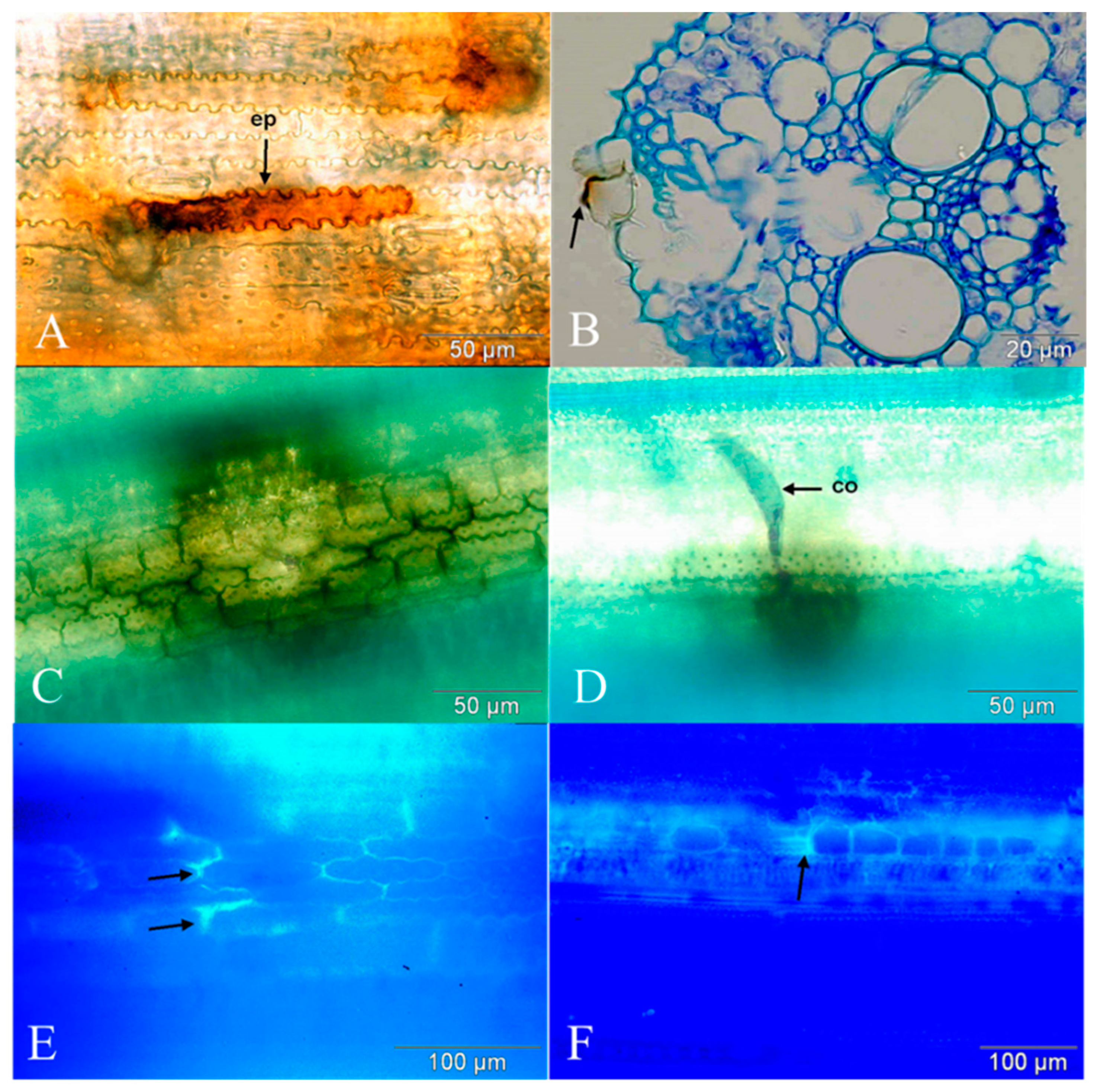
In the less compatible interaction, the accumulation of H2O2 was first seen in the epidermal cell as pale, reddish brown circles around the appressoria at 4 hai. The diameter of the brown regions increased at 8 hai and almost the entirety of the cells being penetrated were stained by 12 hai (Figure 1C). At 16 hai, H2O2 accumulation was seen in many cells to a varying degree, i.e., from faint to heavy accumulation (Figure 1D). By 20 hai, the H2O2 accumulation was seen as irregular reddish-brown staining in the epidermis and mesophyll (Figure 1E), and at 24 hai, the brown staining had further intensified both in extent and colour (Figure 1F). In the compatible interaction, H2O2 accumulation was not seen until 12 hai. H2O2 accumulation was seen in the epidermal cells beneath the appressoria (Figure 2A). At 16 hai, whole-cell staining was observed (Figure 2B). At 20 and 24 hai, there was still an increasing H2O2 accumulation in several cells, but often with a reduced intensity (Figure 2C,D). In the case of very strong accumulation of H2O2, hyphal growth in the cell was not observed. The whole-cell DAB staining in the epidermis occurred in the same cells in which autofluorescence was seen (FEC). Single-cell H2O2 accumulation was seen in the outer walls of epidermal cells in both cleared leaves and in cross sections of DAB-stained regions from the less compatible interaction (Figure 3A,B).

3.2.3. Polyphenol Accumulation

Accumulation of polyphenolics was observed by light microscopy as green to turquoise staining with toluidine blue O. Polyphenols started to accumulate in the cells and epidermal cell walls at 24 hai in both interactions, but to higher levels in the less compatible than in the compatible interaction (Figure 3C,D).

3.2.4. Callose Accumulation

Callose accumulated in the walls of epidermal cells near the appressoria of the fungus. A higher level of accumulation took place in the less compatible than in the compatible interaction. In the less compatible interaction, callose was initially seen 24 hai as bright parts of the cell walls. Generally, accumulation increased and by 144 hai, the intensity was strong, with very thick and brightly fluorescing deposits in the cell walls in the less compatible interaction (Figure 3E). In the compatible interaction, callose accumulation also started at 24 hai and slowly increased until 144 hai (Figure 3F), but the extent and amount of the callose accumulation was considerably lower and occurred slower than in the less compatible interaction.

4. Discussion

The early infection stages of B. oryzae in rice have previously been studied to some extent [10,11,12], whereas the later stages of pathogen growth apparently have not been studied in much detail. To our knowledge, the present study is one of the first reports to give a quantitative study of the early infection processes of a compatible and a less compatible interaction between rice and B. oryzae, as well as a demonstration of host responses to pathogen attack, including fluorescent papillae, fluorescent epidermal cells and accumulation of callose and H2O2. H2O2 has, however, been reported in the interaction between rice and B. oryzae before [15,19,20,21], but not as a defence response.
Most previous studies have included one fungal isolate inoculated on two or more cultivars of rice differing in resistance [11,12,22]. There are few reports that compare two isolates inoculated on one cultivar as a means to obtain compatible and less compatible interactions as in the present study. One example is Vidhyasekaran et al. [13], who investigated the role of toxins in the penetration processes of B. oryzae. The present study used an approach with one cultivar and two isolates. This was based on pathogenicity testing by Thuy [25], where 75 Vietnamese isolates of the pathogen were inoculated on eight differential rice cultivars. All isolates reacted similarly on the cultivars, i.e., a similar kind of reaction was caused in all the differentials, with no differential interactions. Either an isolate caused high levels or low levels of disease in the cultivars. There were no cases where an isolate caused high levels of disease in one cultivar and low levels in another. This made it impossible to find one isolate attacking two cultivars in a differential way. Isolate K2 had a very low compatibility to all eight cultivars tested [25]. Not even the cultivar Tetep showed a response different from other cultivars, even though this cultivar is known to possess a certain level of resistance [31]. Therefore, a highly pathogenic isolate (B5, denoted as giving a compatible interaction with MTL189) and an isolate showing a very low degree of pathogenicity (K2, denoted as giving a less compatible interaction with MTL189) were selected for the studies.
Germination of conidia was not different between the two isolates, whereas germ tubes were longer in the compatible than in the less compatible interaction. Horino and Akai [11] also found that the length of germ tubes of an isolate of B. oryzae was shorter on coleoptiles of a resistant than on a susceptible cultivar, whereas Hau and Rush [12] observed that germ tubes were longer on two resistant cultivars than on a susceptible cultivar. We also found a significantly higher percentage of conidia of K2 producing appressoria than for B5, and the average number of appressoria per conidium was higher for K2 than for B5. This is in contrast to results by Hau and Rush [12], who observed that early in the penetration process, the number of appressoria per conidium was higher on a susceptible than on a resistant cultivar. However, Hau and Rush [12] studied two cultivars inoculated with one isolate, so the results are not strictly comparable. The differences in pre-penetration growth between the two interactions, we observed, likely reflect differences between the two isolates studied. Pre-penetration growth is usually not expected to be influenced by resistance of the host, even though this has been reported from other host-pathogen systems [32,33]. There are reports on morphological variation between isolates of B. oryzae [10,34,35], and therefore it is not surprising to find significant differences in morphology and variation in the behaviour of the two isolates used in the current study. Such variation among isolates should be taken into consideration when comparing the present results with previous investigations in which two cultivars of rice and one isolate of the pathogen have been used.
The percentage of conidia causing penetration was significantly lower for isolate K2 than for isolate B5 (Table 1). This can at least partly be explained by the observation that the ability of appressoria to cause penetration was significantly lower for K2 than for B5. Again, this difference may partly be attributed to differences in the pre-penetration growth of the isolates, but the ability of the host to inhibit penetration of the two isolates is also important. Hyphal progress was accompanied by tissue degradation for B5, with many large lesions and abundant sporulation occurring at 192 hai. On the other hand, for K2, very limited or no tissue degradation was seen, lesions were very small and sporulation was highly restricted. This shows that the host succeeded in stopping pathogen development in the less compatible interaction and thus prevented tissue degradation.
Several host responses to infection were differentially expressed to infection attempts and infections by the two isolates. Formation of fluorescent papillae (FP) was seen only for K2. FP is a well-known primary resistance mechanism against penetration attempts by pathogens in species in Poaceae [36,37], but has apparently not been reported as a host response against B. oryzae before. The percentage of conidia causing FP was very low (1.5–2%), indicating that other mechanisms of resistance are involved. Likewise, the percentage of penetration attempts causing papilla formation in rice infected by Pyricularia oryzae has been found to be very low [38,39]. In the barley-B. sorokiniana interaction, Kumar et al. [40] suggested that the toxin helminthosporol produced by the fungus is able to suppress formation of papillae in the host. B. oryzae is also known to produce several toxic compounds, the ophiobolins [13,41,42,43], and therefore a similar situation could be envisaged in the rice-B. oryzae system, explaining the low occurrence of FP. In accordance with this, Vidhyasekaran et al. [13] found that a non-pathogenic isolate of B. oryzae was inhibited just after penetration. They suggested that one of the primary roles of the toxin produced by the pathogen is to suppress the synthesis of phenolic compounds, which have been suggested to act as an important defence response in rice against B. oryzae [13,14,15].
Callose deposition was also observed as a response to infection (Figure 3E,F), occurring to a higher degree in the less compatible than in the compatible interaction between rice and B. oryzae, thus potentially indicating a role in defence. On the other hand, De Vleesschauwer et al. [16] did not find any clear evidence for the role of callose in abscisic acid-induced resistance in rice against B. oryzae, based on inhibitor studies. Callose deposition constitutes a structural barrier in plants against attempted penetration and the development and spread of pathogens in the host tissue [28,44,45]. Furthermore, callose deposition in cells around the progressing hyphae could act by sealing off the flow of nutrients or water, as well as inhibiting the action of fungal toxins and/or cell wall-degrading enzymes. These functions would cause depletion of the nutrient base of B. oryzae and thus inhibit its growth.
DAB-staining showed that H2O2 accumulated in the epidermal cell walls beneath appressoria and other parts of the epidermal cells. Penetration of B. oryzae was strongly inhibited in the less compatible interaction. This inhibition cannot be explained by the formation of FP alone, but accumulation of H2O2 could be a contributing factor. Accumulation of H2O2 in the early stages of infection of biotrophic pathogens like powdery mildews is known to inhibit fungal penetration [45,46] and some hemibiotrophic pathogens such as Zymoseptoria tritici [47]. For Bipolaris species, Kumar et al. [40] observed that inhibition of B. sorokiniana penetration was spatially linked to early H2O2 accumulation, and Poudel et al. [48] also found that resistance in wheat against B. sorokiniana correlated with H2O2 accumulation and concluded that it was important in resistance. B. oryzae has often been considered a purely necrotrophic pathogen, whereas the closely related species B. sorokiniana is most often considered a hemibiotrophic pathogen [e.g., 40,48]. We consider it very likely that B. oryzae has a similar lifestyle to B. sorokiniana, and De Vleesschauwer et al. [16] also concluded that B. oryzae may have a short biotrophic phase during the initial stages of infection, during which it is sensitive to H2O2-dependent defence. This could suggest why H2O2 could be a defence response that helps in stopping infection, at least during the early stages of infection. Clearly, the specific role of H2O2 in stopping the growth of hemibiotrophic pathogens such as B. oryzae needs to be further studied and may depend on the pathogen genotype, as reviewed by Shetty et al. [49].
The percentages of conidia causing autofluorescent epidermal cells (FEC) and fluorescent cell walls (FCW) were higher for isolate K2 than for B5 at both time points (Table 1), suggesting a positive correlation between these responses and the inhibition of B. oryzae. Fluorescence in FEC and FCW occurred in the same cells, which showed whole-cell DAB staining, thus suggesting that polyphenolic substances were accumulating, as also observed by Kumar et al. [40] for barley infected by B. sorokiniana. Hyphal growth was not seen in cells showing whole-cell DAB staining and autofluorescence. The number of conidia causing single FEC was higher for isolate K2 than for B5 at 12 hai, but there was no significant difference at 24 hai due to an increasing number of conidia causing multiple FEC at that time (Table 1). Collectively, these observations could indicate that accumulation of H2O2 and polyphenolic substances have an inhibitory effect on B. oryzae during the early stages of infection, especially in the less compatible interaction. In the defence against powdery mildews, FEC constitute a well-known phenomenon, which has been taken as a manifestation of a hypersensitive reaction (HR) [45,46]. FEC effectively restrict the growth of biotrophic and often hemibiotrophic pathogens [50] such as Pyricularia oryzae in rice [31,39]. FCW are likewise known to restrict the growth of Pyricularia oryzae in rice [39]. In some cases, hemibiotrophic pathogens are not inhibited by HR. Thus, in the interaction between B. sorokiniana and barley [40], and between barley and Pyrenophora teres [24], no correlation between single FEC and inhibition of pathogen growth was found. Similarly, Ahn et al. [31] concluded that HR was not effective in stopping the growth of B. oryzae in rice. Kumar et al. [40] suggested that HR (FEC) might either occur too late to stop the fungus completely, or alternatively that the fungus might not be affected by cell death at all. On the other hand, Jørgensen et al. [24] observed that multiple FEC could actually inhibit the growth of Pyrenophora teres in induced resistant barley. Thus, the response was not effective in stopping the pathogen in control plants, but given an enhancement by an inducer treatment, it became quite effective. In the interaction between B. oryzae and rice, multiple FEC could also be involved in inhibiting the growth of the pathogen, since this response was significantly more frequent in the less compatible than in the compatible interaction. This correlated with accumulation of H2O2 and polyphenolic substances as well as the inhibition of hyphal growth, i.e., hyphae in the less compatible interaction were thin and initially inhibited. Furthermore, hyphal proliferation was severely restricted, resulting in only limited lesion formation and sporulation here.
Collectively, our data show that several potential defence reactions are activated in interactions between rice and B. oryzae, varying in compatibility. Some of these responses have not been reported before, but we were able to show correlation with incompatibility. More advanced and comprehensive studies are required to elucidate the responses’ precise role in defence and how the different responses are interconnected. Nevertheless, a first identification of potentially pathogen-restricting responses can pave the way for a better understanding of plant defence, and may ultimately benefit breeding for resistance.

Author Contributions

Conceptualization, T.T.T.T., M.L., V.S.-P., E.d.N. and H.J.L.J.; methodology, T.T.T.T., M.L., V.S.-P., E.d.N. and H.J.L.J.; formal analysis, T.T.T.T., E.d.N. and H.J.L.J.; investigation, T.T.T.T., M.L., V.S.-P., E.d.N. and H.J.L.J.; data curation, T.T.T.T.; writing—original draft preparation, T.T.T.T., M.L., V.S.-P., E.d.N. and H.J.L.J.; writing—review and editing, T.T.T.T., M.L., V.S.-P., E.d.N. and H.J.L.J.; visualization, T.T.T.T., M.L., E.d.N. and H.J.L.J.; supervision, M.L., V.S.-P., E.d.N.and H.J.L.J.; project administration, V.S-P.; funding acquisition, V.S.-P. All authors have read and agreed to the published version of the manuscript.

Funding

This research was funded by the Royal Danish Ministry of Foreign Affairs through the DANIDA-ENRECA-project “Systemic Acquired Resistance—an Ecofriendly Strategy for Managing Diseases in Rice and Pearl Millet”.

Data Availability Statement

The data presented in this study are available in Table 1 and Figure 1, Figure 2 and Figure 3.

Conflicts of Interest

The authors declare no conflict of interest.

References

  1. Barnwal, M.K.; Kotasthane, A.; Magculia, N.; Mukherjee, P.K.; Savary, S.; Sharma, A.K.; Singh, H.B.; Singh, U.S.; Sparks, A.H.; Variar, M.; et al. A review on crop losses, epidemiology and disease management of rice brown spot to identify research priorities and knowledge gaps. Eur. J. Plant Pathol. 2013, 136, 443–457. [Google Scholar] [CrossRef] [Green Version]
  2. Mizobuchi, R.; Fukuoka, S.; Tsushima, S.; Yano, M.; Sato, H. QTLs for resistance to major rice diseases exacerbated by global warming: Brown spot, bacterial seedling rot, and bacterial grain rot. Rice 2016, 9, 23. [Google Scholar] [CrossRef] [PubMed] [Green Version]
  3. Savary, S.; Willocquet, L.; Pethybridge, S.J.; Esker, P.; McRoberts, N.; Nelson, A. The global burden of pathogens and pests on major food crops. Nat. Ecol. Evol. 2019, 3, 430–439. [Google Scholar] [CrossRef]
  4. Padmanabhan, S.Y. The great Bengal famine. Annu. Rev. Phytopathol. 1973, 11, 11–26. [Google Scholar] [CrossRef]
  5. Hoang, V.T. Determination of Causal Agents of Rice Diseases in the Mekong Delta. Bachelor’s Thesis, Department of Plant Protection, Can Tho University, Can Tho, Vietnam, 1979. (In Vietnamese). [Google Scholar]
  6. Thuy, T.T.T. Determination of Causal Agents of Rice, Sugarcane, Tobacco and Banana Diseases in the Mekong Delta. Bachelor’s Thesis, Department of Plant Protection, Can Tho University, Can Tho, Vietnam, 1980. (In Vietnamese). [Google Scholar]
  7. Thanh, N. Brown Spot Disease Infected More than 2,000 Hectares of Spring Rice in Ha Tinh. 2018. Available online: https://nongnghiep.vn/benh-dom-nau-tiem-lua-an-hon-2000-ha-lua-xuan-o-ha-tinh-d216544.html (accessed on 26 November 2022). (In Vietnamese).
  8. Moriwaki, A.; Kubo, E.; Arase, S.; Kihara, J. Disruption of SRM1, a mitogen-activated protein kinase gene, affects sensitivity to osmotic and ultraviolet stressors in the phytopathogenic fungus Bipolaris oryzae. FEMS Microbiol. Lett. 2006, 257, 253–261. [Google Scholar] [CrossRef] [Green Version]
  9. Dariush, S.; Darvishnia, M.; Ebadi, A.-A.; Padasht-Dehkaei, F.; Bazgir, E. Population structure, genetic diversity, and trait association analysis in rice (Oryza sativa L.) genotypes for brown spot disease resistance. Trop. Plant Pathol. 2021, 46, 265–281. [Google Scholar] [CrossRef]
  10. Sato, H.; Ando, I.; Hirabayashi, H.; Takeuchi, Y.; Arase, S.; Kihara, J.; Kato, H.; Imbe, T.; Nemoto, H. QTL analysis of brown spot resistance in rice (Oryza sativa L.). Breed. Sci. 2008, 58, 93–96. [Google Scholar] [CrossRef] [Green Version]
  11. Nisikado, Y.; Miyake, C. Studies on the Helminthosporiose of the rice-plant. Ber. Ōhara Inst. Landwirtsch. Forsch. 1922, 2, 133–195+plate III–plate IX. [Google Scholar]
  12. Horino, O.; Akai, S. Studies in the pathological anatomy of rice plants infected by Helminthosporium oryzae I. Behavior of the causal fungus on the coleoptile of rice seedlings and its ultrafine structure. Ann. Phytopathol. Soc. Jpn. 1968, 34, 51–55. [Google Scholar] [CrossRef] [Green Version]
  13. Hau, F.C.; Rush, M.C. Preinfectional interaction between Helminthosporium oryzae and resistant and susceptible rice plants. Phytopathology 1982, 72, 285–292. [Google Scholar] [CrossRef]
  14. Vidhyasekaran, P.; Borromeo, E.S.; Mew, T.W. Helminthosporium oryzae toxin suppresses phenol metabolism in rice plants and aids pathogen colonization. Physiol. Mol. Plant Pathol. 1992, 41, 307–315. [Google Scholar] [CrossRef]
  15. Van Bockhaven, J.; Spíchal, L.; Novák, O.; Strnad, M.; Asano, T.; Kikuchi, S.; Höfte, M.; De Vleesschauwer, D. Silicon induces resistance to the brown spot fungus Cochliobolus miyabeanus by preventing the pathogen from hijacking the rice ethylene pathway. New Phytol. 2015, 206, 761–773. [Google Scholar] [CrossRef] [PubMed]
  16. Ma, Z.; Ongena, M.; Höfte, M. The cyclic lipopeptide orfamide induces systemic resistance in rice to Cochliobolus miyabeanus but not to Magnaporthe oryzae. Plant Cell Rep. 2017, 36, 1731–1746. [Google Scholar] [CrossRef]
  17. De Vleesschauwer, D.; Yang, Y.; Cruz, C.V.; Höfte, M. Abscisic acid-induced resistance against the brown spot pathogen Cochliobolus miyabeanus in rice involves MAP kinase-mediated repression of ethylene signaling. Plant Physiol. 2010, 152, 2036–2052. [Google Scholar] [CrossRef] [PubMed] [Green Version]
  18. Dorneles, K.R.; Refatti, J.P.; Pazdiora, P.C.; de Avila, L.A.; Deuner, S.; Dallagnol, L.J. Biochemical defenses of rice against Bipolaris oryzae increase with high atmospheric concentration of CO2. Physiol. Mol. Plant Pathol. 2020, 110, 101484. [Google Scholar] [CrossRef]
  19. Kariya, K.; Murata, K.; Kokubo, Y.; Ube, N.; Ueno, K.; Yabuta, Y.; Teraishi, M.; Okumoto, Y.; Mori, N.; Ishihara, A. Variation of diterpenoid phytoalexin oryzalexin A production in cultivated and wild rice. Phytochemistry 2019, 166, 112057. [Google Scholar] [CrossRef]
  20. Ahn, I.-P. Glufosinate ammonium-induced pathogen inhibition and defense responses culminate in disease protection in bar-transgenic rice. Plant Physiol. 2008, 146, 213–227. [Google Scholar] [CrossRef] [Green Version]
  21. Debona, D.; Rodrigues, F.A. A strobilurin fungicide relieves Bipolaris oryzae-induced oxidative stress in rice. J. Phytopathol. 2016, 164, 571–581. [Google Scholar] [CrossRef]
  22. Marwein, R.; Singh, S.; Maharana, J.; Kumar, S.; Arunkumar, K.P.; Velmurugan, N.; Chikkaputtaiah, C. Transcriptome-wide analysis of North-East Indian rice cultivars in response to Bipolaris oryzae infection revealed the importance of early response to the pathogen in suppressing the disease progression. Gene 2022, 809, 146049. [Google Scholar] [CrossRef]
  23. Jensen, B.; Lübeck, P.S.; Jørgensen, H.J.L. Clonostachys rosea reduces spot blotch in barley by inhibiting prepenetration growth and sporulation of Bipolaris sorokiniana without inducing resistance. Pest Manag. Sci. 2016, 72, 2231–2239. [Google Scholar] [CrossRef]
  24. Jørgensen, H.J.L.; Lübeck, P.S.; Thordal-Christensen, H.; de Neergaard, E.; Smedegaard-Petersen, V. Mechanisms of induced resistance in barley against Drechslera teres. Phytopathology 1998, 88, 698–707. [Google Scholar] [CrossRef] [PubMed]
  25. Thuy, T.T.T. Infection Biology of Bipolaris oryzae in Rice and Its Pathogenic Variation in the Mekong Delta, Vietnam. Ph.D. Thesis, Plant Pathology Section, Department of Plant Biology, The Royal Veterinary and Agricultural University, Copenhagen, Denmark, 2002. [Google Scholar]
  26. Shetty, N.P.; Kristensen, B.K.; Newman, M.-A.; Møller, K.; Gregersen, P.L.; Jørgensen, H.J.L. Association of hydrogen peroxide with restriction of Septoria tritici in resistant wheat. Physiol. Mol. Plant Pathol. 2003, 62, 333–346. [Google Scholar] [CrossRef]
  27. Graham, E.T.; Joshi, P.A. Novel fixation of plant tissue, staining through paraffin with Alcian blue and Hematoxylin, and improved slide preparation. Biotech. Histochem. 1995, 70, 263–266. [Google Scholar] [CrossRef] [PubMed]
  28. Shetty, N.P.; Jensen, J.D.; Knudsen, A.; Finnie, C.; Geshi, N.; Blennow, A.; Collinge, D.B.; Jørgensen, H.J.L. Effects of β-1,3-glucan from Septoria tritici on structural defence responses in wheat. J. Exp. Bot. 2009, 60, 4287–4300. [Google Scholar] [CrossRef] [PubMed] [Green Version]
  29. Collett, D. Modelling Binary Data; Chapman & Hall: London, UK, 2002. [Google Scholar]
  30. Cox, D.R.; Snell, E.J. Analysis of Binary Data. In Monographs on Statistics and Applied Probability, 2nd ed.; Chapman & Hall: London, UK, 1989; Volume 32. [Google Scholar]
  31. Ahn, I.-P.; Kim, S.; Kang, S.; Suh, S.-C.; Lee, Y.-H. Rice defense mechanisms against Cochliobolus miyabeanus and Magnaporthe grisea are distinct. Phytopathology 2005, 95, 1248–1255. [Google Scholar] [CrossRef] [Green Version]
  32. Bird, P.M.; Ride, J.P. The resistance of wheat to Septoria nodorum: Fungal development in relation to host lignification. Physiol. Plant Pathol. 1981, 19, 289–299. [Google Scholar] [CrossRef]
  33. Mumford, D.L. Factors associated with resistance in barley to spot blotch. Phytopathology 1966, 56, 79–82. [Google Scholar]
  34. Misra, A.P.; Chatterjee, A.K. Comparative study of two isolates of Helminthosporium oryzae Breda de Hann. Indian Phytopathol. 1963, 16, 275–281. [Google Scholar]
  35. Cholil, A.; de Hoog, G.S. Variability in Drechslera oryzae. Trans. Br. Mycol. Soc. 1982, 79, 491–496. [Google Scholar] [CrossRef]
  36. Vance, C.P.; Sherwood, R.T. Lignified papilla formation as a mechanism for protection in reed canarygrass. Physiol. Plant. Pathol. 1977, 10, 247–256. [Google Scholar] [CrossRef]
  37. Sherwood, R.T.; Vance, C.P. Resistance to fungal penetration in Gramineae. Phytopathology 1980, 70, 723–729. [Google Scholar] [CrossRef]
  38. Peng, Y.L.; Shishiyama, J. Timing of a cellular reaction in rice cultivars associated with different degrees of resistance to Pyricularia oryzae. Can. J. Bot. 1989, 67, 2704–2710. [Google Scholar] [CrossRef]
  39. Heath, M.C.; Valent, B.; Howard, R.J.; Chumley, F.G. Interactions of two strains of Magnaporthe grisea with rice goosegrass, and weeping lovegrass. Can. J. Bot. 1990, 68, 1627–1637. [Google Scholar] [CrossRef]
  40. Kumar, J.; Hückelhoven, R.; Beckhove, U.; Nagarajan, S.; Kogel, K.-H. A compromised Mlo pathway affects the response of barley to the necrotrophic fungus Bipolaris sorokiniana (teleomorph: Cochliobolus sativus) and its toxins. Phytopathology 2001, 91, 127–133. [Google Scholar] [CrossRef] [Green Version]
  41. Lindberg, G.D. Disease-induced toxin production in Helminthosporium oryzae. Phytopathology 1971, 61, 420–424. [Google Scholar] [CrossRef]
  42. Chattopadhyay, A.K.; Samaddar, K.R. Effects of Helminthosporium oryzae infection and ophiobolin on the cell membranes of host tissues. Physiol. Plant Pathol. 1976, 8, 131–139. [Google Scholar] [CrossRef]
  43. Xiao, J.Z.; Tsuda, M.; Doke, N.; Nishimura, S. Phytotoxins produced by germinating conidia of Bipolaris oryzae. Phytopathology 1991, 81, 58–64. [Google Scholar] [CrossRef]
  44. Voigt, C.A. Callose-mediated resistance to pathogenic intruders in plant defense-related papillae. Front. Plant Sci. 2014, 5, 168. [Google Scholar] [CrossRef]
  45. Shetty, R.; Jensen, B.; Shelton, D.; Jørgensen, K.; Pedas, P.; Jørgensen, H.J.L. Site-specific, silicon-induced structural and molecular defence responses against powdery mildew infection in roses. Pest Manag. Sci. 2021, 77, 4545–4554. [Google Scholar] [CrossRef]
  46. Thordal-Christensen, H.; Zhang, Z.; Wei, Y.; Collinge, D.B. Subcellular localization of H2O2 in plants: H2O2 accumulation in papillae and hypersensitive response during the barley-powdery mildew interaction. Plant J. 1997, 11, 1187–1194. [Google Scholar] [CrossRef]
  47. Shetty, N.P.; Mehrabi, R.; Lütken, H.; Haldrup, A.; Kema, G.H.J.; Collinge, D.B.; Jørgensen, H.J.L. Role of hydrogen peroxide during the interaction between the hemibiotrophic fungal pathogen Septoria tritici and wheat. New Phytol. 2007, 174, 637–647. [Google Scholar] [CrossRef] [PubMed]
  48. Poudel, A.; Navathe, S.; Chand, R.; Mishra, V.K.; Singh, P.K.; Joshi, A.K. Hydrogen peroxide prompted lignification affects pathogenicity of hemi-bio-trophic pathogen Bipolaris sorokiniana to wheat. Plant Pathol. J. 2019, 35, 287–300. [Google Scholar] [CrossRef]
  49. Shetty, N.P.; Jørgensen, H.J.L.; Jensen, J.D.; Collinge, D.B.; Shetty, H.S. Roles of reactive oxygen species in interactions between plants and pathogens. Eur. J. Plant Pathol. 2008, 121, 267–280. [Google Scholar] [CrossRef]
  50. Huysmans, M.; Lema, A.S.; Coll, N.S.; Nowack, M.K. Dying two deaths—Programmed cell death regulation in development and disease. Curr. Opin. Plant Biol. 2017, 35, 37–44. [Google Scholar] [CrossRef] [PubMed]
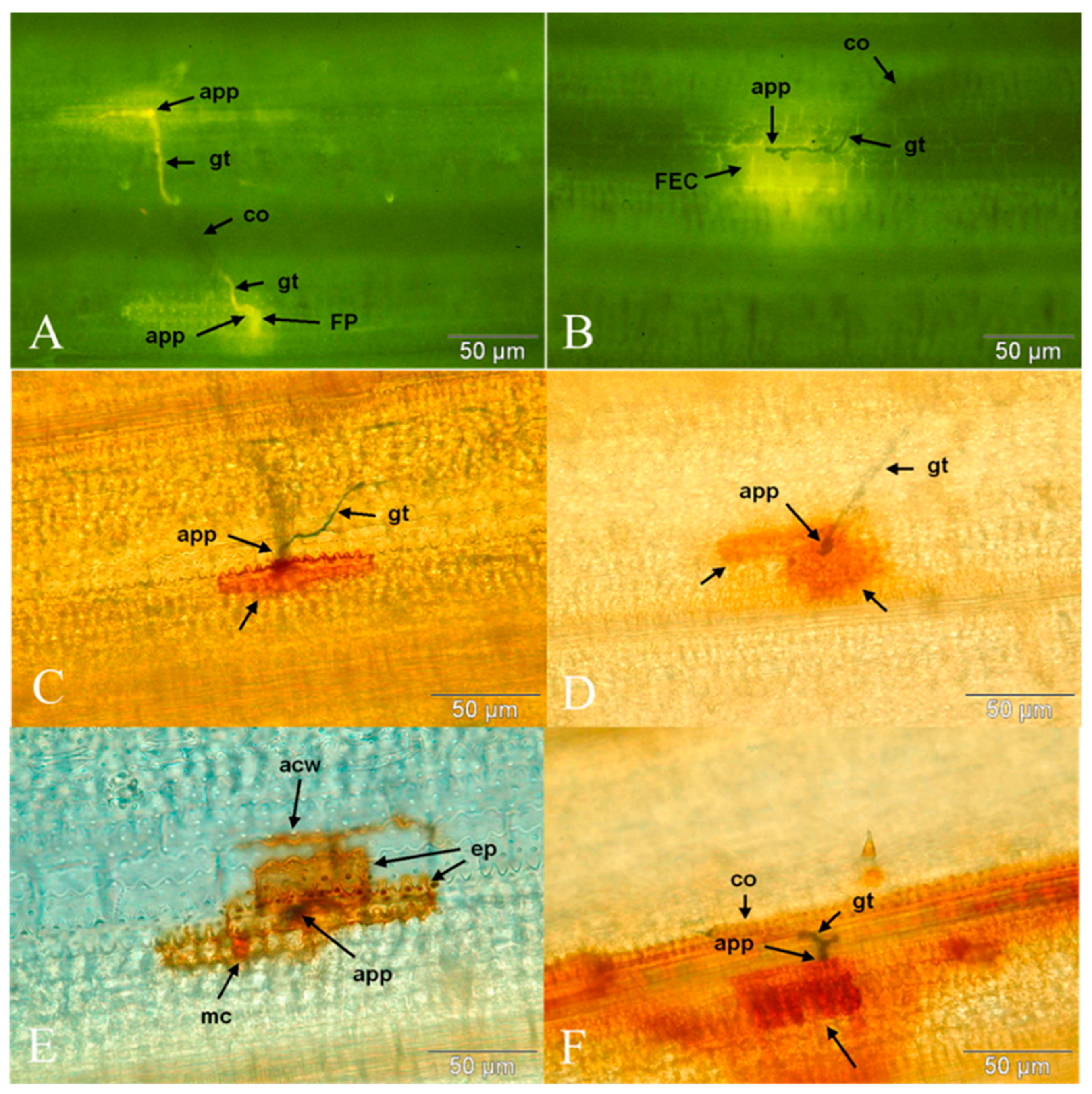
Figure 1. Details of infection biology and defence responses of rice cv. MTL189 infected by Bipolaris oryzae isolate K2 (less compatible interaction). (A) Autofluorescent papillae (FP) at penetration attempts of B. oryzae at 12 hai. (B) Multiple fluorescent epidermal cells (FEC) at 12 hai. (CF) DAB staining showing hydrogen peroxide accumulation in relation to B. oryzae. (C) FEC at 12 hai. (D) FEC 16 hai. (E) Multiple FEC at 20 hai. (F) Multiple FEC and spreading staining reaction at 24 hai. acw: anticlinal epidermal cell wall; app: appressorium; co: conidium, ep: epidermal cell; FEC: fluorescent epidermal cell; FP: fluorescent papilla; gt: germ tube; mc: mesophyll cell. Arrows indicate DAB-stained regions.
Agronomy 13 00231 g001
Figure 2. Details of infection biology and defence responses of rice cv. MTL189 infected by Bipolaris oryzae isolate B5 (compatible interaction). DAB staining showing H2O2 accumulation in relation to B. oryzae. (A) Penetration attempt resulting in hydrogen peroxide accumulation under papilla (arrow) at 12 hai. (B) Penetration resulting in hydrogen peroxide accumulation under papilla at 16 hai. Arrow shows FEC. (C) Penetration resulting in hydrogen peroxide accumulation in FEC at 20 hai. Arrow shows FEC. (D) Penetration resulting in hydrogen peroxide accumulation in FEC at 24 hai. Arrow shows FEC. Abbreviations as in Figure 1.
Agronomy 13 00231 g002
Figure 3. Details of infection biology and defence responses of rice cv. MTL189 infected by Bipolaris oryzae K2 (less compatible interaction) and isolate B5 (compatible interaction). (A) DAB staining showing hydrogen peroxide accumulation in FEC at 12 hai (isolate K2). (B) Cross section of similarly DAB-stained area (isolate K2) at 12 hai, arrow showing staining in the outer epidermal cell wall. (C) Accumulation of polyphenolic substances, seen as greenish-turquoise staining, as a response to infection attempt by isolate K2 at 72 hai. (D) Polyphenolic accumulation as a response to infection attempt by isolate B5 at 72 hai. (E) Callose indicated by fluorescence (arrows) as a response to infection attempt by isolate K2 at 144 hai. (F) Callose indicated by fluorescence (arrow) as a response to infection attempt by isolate B5 at 144 hai. Abbreviations as in Figure 1.
Agronomy 13 00231 g003
Disclaimer/Publisher’s Note: The statements, opinions and data contained in all publications are solely those of the individual author(s) and contributor(s) and not of MDPI and/or the editor(s). MDPI and/or the editor(s) disclaim responsibility for any injury to people or property resulting from any ideas, methods, instructions or products referred to in the content.