Article

The Physiological and Biochemical Response of Field Bean (Vicia faba L. (partim)) to Electromagnetic Field Exposure Is Influenced by Seed Age, Light Conditions, and Growth Media

by
Agnieszka Pawełek
1,*,
Joanna Wyszkowska
2,
Daniele Cecchetti
1,
Mergi Daba Dinka
3,
Krzysztof Przybylski
1 and
Adriana Szmidt-Jaworska
1
1
Department of Plant Physiology and Biotechnology, Faculty of Biological and Veterinary Sciences, Nicolaus Copernicus University in Toruń, Lwowska 1, 87-100 Toruń, Poland
2
Department of Animal Physiology and Neurobiology, Faculty of Biological and Veterinary Sciences, Nicolaus Copernicus University in Toruń, Lwowska 1, 87-100 Toruń, Poland
3
Department of Ecology and Biogeography, Faculty of Biological and Veterinary Sciences, Nicolaus Copernicus University in Toruń, Lwowska 1, 87-100 Toruń, Poland
*
Author to whom correspondence should be addressed.
Agronomy 2022, 12(9), 2161; https://doi.org/10.3390/agronomy12092161
Submission received: 7 August 2022 / Revised: 6 September 2022 / Accepted: 7 September 2022 / Published: 11 September 2022
(This article belongs to the Special Issue Advances in Plant Physiology of Abiotic Stresses)

Abstract

:
Research interest into the exposure of plants to magnetic fields (MF), including electromagnetic fields (EMF), has increased recently but results often vary depending on factors such as plant species and treatment dose. In this study, we exposed young (one year) and old (four years) field bean (Vicia faba L. (partim)) seeds to EMF (50 Hz, 7 mT) and observed seed germination and seedling growth under different conditions (growth media and light). The results indicated a stimulation by EMF of germination and early root growth of Petri dish-sown old seeds in continuous darkness and inhibition of germination of the pot-sown young seeds under long-day conditions. Root growth of two-week-old seedlings from pot-sown young seeds was stimulated by EMF treatment while their stem growth was inhibited. Some selected biochemical traits were examined, showing specific changes in membrane integrity, amylase activity, H2O2 levels, photosynthetic pigments, and content of the main groups of phytohormones, depending on seed age. The results indicate that priming of field bean seeds with EMF (50 Hz, 7 mT) could be a eustress factor that influences germination, early growth, and cellular activities and could positively influence the ability of field bean plants to grow and develop in more stressful conditions at later stages.

1. Introduction

The quality of seeds and other planting materials is an essential component of successful crop production. The ability of seeds to develop into healthy seedlings and subsequently produce high yields influences decisions on crop production methods. In the face of adverse environmental conditions, farmers increase the use of inputs such as fertilizers and water to ensure substantial crop yield [1,2]. The quality of seeds and crop yield is expected to be significantly reduced by abiotic stress factors, such as drought and high temperatures resulting from climate change [3]. Seed aging is an important issue in crop production, which is associated with different internal (morphological, physiological, and genetic) and external (storage temperature and humidity) factors. Old seeds that are more likely to have low germinability or vigor are often discarded. These practices tend to increase both the economic and environmental costs to crop production [4].
Legumes (Fabaceae family) serve as a fundamental, worldwide source of high-quality food and feed, as well as help to sustain soil health during intensified crop production [5]. The limited spread of the cultivation of legumes is attributed to the reduction and instability in yield and its vulnerability to biotic and abiotic stress factors [6]. Therefore, it is essential to combine both genetic and agronomic techniques to produce high yields of legumes, especially under changing environmental conditions [7]. Field bean (Vicia faba L. var. minor; Vicia faba L. partim) is one of the major leguminous crops cultivated for animal feed, green forage, hay, silage, or green manure and also serves as an alternative to transgenic soybean. Due to its ability to fix nitrogen, field bean is increasingly becoming relevant in organic production systems to limit the use of mineral fertilizer inputs [8,9].
Seed priming aims to stimulate the physiological, biochemical, and metabolic activity of seeds in order to increase germinability, improve crop yield under unfavorable conditions, and reduce the adverse environmental footprint of crop production. Conventional priming techniques include hydropriming, osmopriming, biopriming, and chemopriming [10]. In modern seed improvement techniques, utilizing physical agents to improve crop production provides some advantages over conventional methods, particularly, the use of chemical substances [11]. Among the physical stimulation methods, the use of magnetic fields (MF), which may be in the form of static magnetic fields (SMF) or electromagnetic fields (EMF), is considered an eco-friendly, relatively cheap, and non-invasive technique with proven beneficial effects in plant production [12,13]. Seed priming with different MFs (SMF and EMFs) is assumed to enhance plant vigor by influencing a plethora of biochemical and molecular processes, such as changes in reactive oxygen species (ROS) production [14] and modulation of phytohormone balance [15,16].
EMF presence in the environment, particularly from man-made sources, has increased tremendously [17] and is regarded as a silent stressor with a great impact on developmental patterns in living systems, including plants [18]. A stressor may be regarded as either a eustress, a type of physiological priming with positive effects on the performance of a living organism, or a distress, which is a continuing factor whose high dose causes negative effects [19]. The treatment of plant materials, including seeds, with different doses of MFs, has shown positive results in a number of crop plants [12]. Pea (Pisum sativum) seeds treated with MF (30 mT, 85 mT) expressed faster water uptake and enhanced germination [20], as well as faster early growth [16]. In the case of broad beans (Vicia faba), the same MF application (30 mT, 85 mT) also had a positive effect on seed emergence and crop yield during field cultivation but the efficiency of this treatment was dependent on weather conditions [21]. Soybean (Glycine max) seeds exposed to MF (10 Hz, 1.5 µT) produce plants with a greater number of leaves, pods, and seeds and also improved the length of the pods and weight of seeds [22]. Beneficial effects of the pre-sowing treatment of red clover (Trifolium pratense) seeds on their agronomic performance have been reported where seed treatment with EMF (5.28 MHz, 0.74 mT) significantly increased the number of root nodules [23] and changed the amount of flavonoids in the root exudates important for communication with nitrogen-fixing rhizobacteria [24]. Treatment of seeds with different doses of MF has also been shown to alleviate the harmful effects of various abiotic stresses. The enhancement of germination rate and seedling growth under different salinity levels was reported for magneto-primed seeds of chickpea (Cicer arietinum) (with SMF of 100 mT) [25], as well as maize (Zea mays) and soybean (with SMF of 200 mT) [26].
On the other hand, not all studies resulted in positive outcomes of agronomic importance. Aguilar et al. [27] reported that the growth response of maize seeds to EMF (60 Hz, 20–100 mT) treatment was strongly cultivar-dependent, showing positive, neutral, or even negative effects compared to the controls. The pre-sowing exposure of pea seeds to EMF (60–180 mT, 50 Hz) did not affect the chlorophyll content of one-month-old plants despite the stimulation of their stems and roots [28]. The treatment of spring wheat (Triticum aestivum) seeds with MF (30 mT) also did not produce any effect on yield [29]. Cakmak et al. [30] reported that the exposure of seeds of bean (Phaseolus vulgaris) and wheat to SMF (7 mT) significantly stimulated dry biomass accumulation of wheat but not of beans. The existing reports suggest that the positive response to MF treatment may only occur under specific conditions dictated by plant species and growth environment [13]. Moreover, a wide range of MFs are used in seed priming and variations in their duration and intensity could change a positive effect on the treated plant to either a negative or no effect [31]. These inconsistencies in reported studies serve as a drawback to the application of MFs as means of priming seeds. Therefore, further investigation is needed to determine the optimum conditions under which exposure of seeds to MFs could be used as sustainable means of enhancing crop production.
This study aims to determine how priming the seeds of field bean (Vicia faba L. (partim)) of different ages (1-year-olds vs. 4-year-olds), with EMF (50 Hz, 7 mT), affects their germination and growth in different media and under two distinct light conditions. Additionally, the mechanism of EMF action in field bean tissues is investigated by analyzing some biochemical traits, including membrane integrity, α-amylase activity, H2O2 levels, photosynthetic pigment content, and changes in phytohormones controlling growth (indole-3-acetic acid, IAA; abscisic acid, ABA; gibberellins, GAs) and stress responses (jasmonic acid, JA; salicylic acid, SA) in plants. The outcome of these studies will, thus, contribute to understanding better the effect of MF exposure on plants under different conditions.

2. Materials and Methods

2.1. Plant Material and Cultivation Conditions

Two groups of seeds of field bean, Vicia faba L. (partim) of Polish variety Fernando, were used in this study: (1) young seeds of one year old, and (2) old seeds of four years old. Both groups of seeds were harvested in the Kuyavian-Pomeranian region in Poland and stored in similar controlled conditions of temperature (approx. 15 °C), humidity (approx. 35%), and light.

2.2. Exposure to Electromagnetic Field

Before sowing in Petri dishes and in pots, dry seeds were exposed for 24 h to a sinusoidal electromagnetic field (50 Hz, 7 mT), generated in a coil of 0.1 m in radius (Elektronika i Elektromedycyna Sp. J., Otwock, Poland). A detailed description of the exposure system (Figure 1) is presented in previously published papers [32,33]. The control groups (without EMF exposure) were put in a sham exposure setup and were affected by only the local geomagnetic field. The EMF was measured before each experiment with a Gauss meter (Model GM2, AlphaLab, Inc., UT, USA). The non-homogeneity of EMF within the area where the seeds in falcon tubes were kept was less than 4%. The temperature during all experiments was set to 24 ± 1 °C and monitored using thermocouples.

2.3. Cultivation Conditions

Immediately after exposure to EMF, the dry seeds were either germinated in Petri dishes for six days or in plastic pots (11 × 11 × 21.5 cm) filled with a universal substrate (Substral, Warszawa, Poland) for two weeks. For the tests in Petri dishes, seeds were first surface-sterilized for five minutes using a mixture of 30% hydrogen peroxide and 96% ethanol (1/1 (v/v)), then washed 10 times for 30 s in sterile distilled water. Afterwards, the seeds were sown on the 9 cm Petri dishes lined with sterile filter paper moistened with 8 mL of sterile distilled water. For the Petri dish tests, seed germination and seedling growth were carried out in cultivation rooms at 21 ± 0.5 °C under two conditions: (1) long-day constituting 15 h of light and 9 h of darkness and (2) continuous darkness. The study involving seed germination and plant growth in substrate-filled pots was only carried out under long-day conditions. The light source produced a photosynthetic photon flux of 30 μmol × m−2 × s, and the humidity was 60%. In the Petri dish tests, three replicates of 50 seeds each were used with each Petri dish holding 10 seeds, while in the pot tests, five replicates of 10 seeds each were used.

2.4. Seed Germination Analysis

The germination counts of seeds sown in Petri dishes and in the pots were performed daily. Germination kinetics of young and old field bean seeds was presented on graphs and expressed as the total proportion (%) of germinated seeds on a particular day from sowing. Additionally, five different germination parameters were evaluated using the following formulas reported by Ranal and Santana [34] (parameters 1, 3, 4), Kader [35] (parameter 2), and Coolbear et al. [36] (parameter 5):
Germinability, G = 100 (N/S)
where: N is the number of total germinated seeds at end of the counts; S is the number of initial seeds used;
Germination index, GI = (6 × N1) + (5 × N2) + … + (1 × N6)
where: N1, N2N6 are the number of germinated seeds on the first, second, and subsequent days, respectively; 6, 5, …, 1 are the weights given to the days of germination;
Mean germination time, MGT = (N1T1 + N2T2 +…+ NnTn)/(N1 + N2 +…+ Nn)
where: N is the number of seeds germinated on each day; T is the time point in days;
Coefficient of the velocity of germination,
CVG = 100 [(N1 + N2 +…+ Nn)/(N1T1 + N2T2 +…+ NnTn)]
where: N is the number of seeds germinated each day; T is the number of days from sowing corresponding to N;
Median response (time to reach 50% of final germination),
t50 = Ti + [(((N + 1)/2) − Ni) (TjTi)]/(NjNi)
where N is the final number of seeds germinated and Ni and Nj are the total number of germinated seeds in adjacent counts at time Ti and Tj, respectively, when Ni < (N + 1)/2 < Nj.

2.5. Measurement of Seedling Morphological Parameters

The physiological traits of the seedlings (root and epicotyl/stem length; fresh and dry mass) grown in Petri dishes and in the pots were determined with a millimetric ruler on the 6th and 14th day after sowing, respectively. Additionally, the seedlings growing in Petri dishes, under long-day conditions and in continuous darkness, were analyzed separately for seedlings with fully emerged roots and epicotyls, and those with only protruded roots. The seedlings were oven-dried for 48 h at 70 °C to prepare for dry mass estimation.

2.6. Plant Material Collections

The plant materials collected were germinating seeds from the Petri dish tests and plant roots and leaves from the pot tests. They were frozen in liquid nitrogen and stored at −80 °C until further use. In the Petri dish tests, the germinating seeds were extracted 24 h after sowing, and this was repeated every 24 h up to the sixth day and used in the analyses described in Section 2.8 and Section 2.9. On the other hand, the plants growing in the pots were carefully removed from the pots 14 days after sowing. Their roots and leaves were separated, cleaned, and used for the analyses described in Section 2.10 and Section 2.11.

2.7. Examination of Seeds Water Uptake and Membrane Integrity

Water uptake (endpoint analysis) and membrane integrity of seeds (control and EMF-treated) were determined in four replicates with each replicate having 25 seeds. The dry seeds were weighed and soaked in 25 mL of deionized water at 23 °C for 24 h in darkness. They were then removed from the water, blotted dry, and weighed again. The change in weight due to imbibition was expressed as the amount of water absorbed per seed dry weight which was calculated by the following formula:
Water uptake [%] = ((fresh weight of seed − dry weight of seed) × 100)/(dry weight of seed)
The membrane integrity of seeds was measured based on their ion leakage. After soaking in deionized water for 24 h, the seed leachate was decanted off in a clean beaker and the electrical conductance of the leachate (mS/cm) was measured at room temperature using a digital conductivity meter (Elmetron CX-105, Zabrze, Poland).

2.8. α-Amylase Assay

The activity of α-amylase was quantified using a slightly modified 3,5-dinitrosalicylic acid (DNS) method [37]. Young and old field bean seeds germinating in Petri dishes under long-day conditions were used for this analysis and were collected every 24 h for 6 days beginning from the first 24 h after sowing. They were then homogenized in liquid nitrogen and 100 mg of each sample tissue was extracted with 2 mL of chilled distilled water, followed by centrifugation at 4500× g for 15 min. Next, 1 mL of crude enzyme extract was mixed with 0.5 mL of phosphate buffer (0.5 M; pH 6.9) and the reaction was initiated by adding 1 mL of the 1% starch solution as a substrate, incubated for 5 min at 37 °C, and terminated by adding 1% DNS (0.5 mL). Afterwards, the samples were incubated at 100 °C for 5 min. After cooling the samples on ice, 2.5 mL of distilled water was added and the amount of reducing sugar released was measured using a spectrophotometer (UV-160 1PC, Shimadzu, Kyoto, Japan) at 540 nm with maltose as the reducing sugar standard. The activity of α-amylase was calculated from a standard curve and expressed as mg of maltose per g of fresh weight (FW).

2.9. Hydrogen Peroxide Measurement

Hydrogen peroxide (H2O2) content was assayed in young and old field bean seeds germinated in Petri dishes under long-day conditions. The seeds were collected every 24 h for 6 days (0–6th day) and ground in liquid nitrogen. The 100 mg samples were homogenized with 0.5 mL of 0.1% (w/v) trichloroacetic acid in an ice bath with shaking, followed by centrifugation at 16,000× g for 10 min at 4 °C. Afterwards, 0.3 mL of supernatant was mixed with 0.3 mL of 0.1 M sodium phosphate buffer (pH 7.6) and 0.6 mL of 1 M KI. The samples were incubated in darkness for 1 h at room temperature. Next, the absorbance of the solution was measured at 390 nm in a spectrophotometer (UV-160 1PC, Shimadzu, Kyoto, Japan) and the content of H2O2 was determined using a standard curve of 0–20 µM H2O2.

2.10. Determination of Photosynthetic Pigments

Chlorophylls (a, b, and total) and total carotenoids (xanthophylls + b-carotene) concentrations were determined from leaf materials (100 mg fresh weight) of two-week-old plants growing in pots, then ground in a pre-chilled mortar and extracted in 1 mL of 80% acetone overnight in the dark at 4 °C. Afterwards, the samples were centrifuged at 12,000× g for 10 min at 4 °C and the supernatant was collected and diluted in cold 80% acetone. The absorbance of the extract was measured at 664, 647, and 470 nm using a spectrophotometer (UV-160 1PC, Shimadzu, Kyoto, Japan) and the pigment concentrations were calculated according to Lichtenthaler [38]. Three biological repetitions were performed and the data are presented as mean ± standard error (SE).

2.11. Quantification of Phytohormones by Liquid Chromatography–Tandem Mass Spectrometry (LC-MS/MS)

Mass spectrometry combined with liquid chromatography (LC-MS/MS) and the QuEChERS-based extraction method [39] were used to determine the concentrations of endogenous indole-3-acetic acid (IAA), abscisic acid (ABA), gibberellins (GA1, GA3), salicylic acid (SA), and jasmonic acid (JA). For this analysis, the roots and leaves of two-week-old pot-grown plants were ground in liquid nitrogen, and each sample (100 mg) was extracted overnight at 8 °C with shaking in a buffer containing 80% acetonitrile, 5% formic acid (FA), 15% water, 1mM butylhydroxytoluene (BHT), and stable isotope-labeled internal standards (5 ng/mL d2IAA; 5 ng/mL d6ABA; 10 ng/mL d2GA1; 15 ng/mL d2GA3; 10 ng/mL d4SA; 10 ng/mL d5JA). Afterwards, a salt mixture (magnesium sulfate heptahydrate and sodium chloride, 1/3 [m/m]) was added to the samples, vigorously mixed for 3 min, and centrifuged at 10,000× g for 10 min. The obtained supernatant was purified by adding sodium sulfate anhydrous and mixed vigorously for 1 min, followed by centrifugation (10,000× g for 10 min). Collected supernatants were dried with nitrogen gas, dissolved in 1 M FA (1 mL), and subjected to solid phase extraction (SPE) using polymer-based columns (Discovery® DSC-18 SPE Tube, Supelco, Darmstadt, Germany). The DSC-18 columns were activated and conditioned by using 100% methanol and 1M FA, respectively. The column-loaded samples were washed with 1 M FA, 1 M FA with 20% (v/v) methanol, and eluted with 80% methanol (v/v). Next, samples were lyophilized in CentriVap Centrifugal Concentrator (Labconco Corporation, Kansas City, MO, USA), resuspended in 100 µL of 35% methanol (v/v), and collected in glass vials. The concentrations of phytohormones were determined using LC-MS/MS Nexera UHPLC and LCMS-8045 integrated system (Shimadzu Corporation, Kyoto, Japan). Chromatographic separation of samples was performed on a reversed-phase C18 column (150 × 2.1 mm, 2.6 µm, Kinetex®, Phenomenex Inc., Torrance, CA, USA). Water with 0.1% FA (v/v) (A) and methanol with 0.1% FA (v/v) (B) were used as the mobile phase. The separation was carried out on a linear gradient of 40–90% (v/v) methanol for 7 min at 30 °C. The flow rate and injection volume were 0.4 mL/min and 10 µL, respectively. In mass spectrometry, the samples were subjected to negative and positive electrospray ionization (ESI) (4 kV voltage) and ions were fragmented by collision-induced dissociation (CID). Analysis of individual phytohormones was based on multiple reactions monitoring (MRM) with the LabSolutions workstation for LCMS-8045 (Shimadzu Corporation, Kyoto, Japan). Three biological repetitions were performed and the data are presented as mean ± standard error (SE).

2.12. Statistical Analysis

All data were tested for normality and homogeneity, and the level of significance was set at p < 0.05. The results were given as mean ± standard error of the mean (SE). The data obtained from the germination kinetics, other germination parameters (G, GI, MGT, CVG, t50), and the morphometric traits of field bean seedlings growing in Petri dishes and in pots, were analyzed with a one-way ANOVA test using the R package version 4.0.4 (R Core Team, Vienna, Austria) [40].
The Mann-Whitney test was applied for the analysis of water uptake, electrolyte leakage, and photosynthetic pigments. An unpaired t-test with Welch’s correction was used to determine whether exposure to EMF had an effect on the activity of α-amylases and the amount of hydrogen peroxide. Similarly, the phytohormones were analyzed by applying the Students’ unpaired t-test. These data were analyzed with the SPSS 25.0 package (IBM Inc., Armonk, NY, USA).

3. Results

3.1. Effect of Seed Age and EMF Exposure on Germination Kinetics of Petri Dish-Sown and Pot-Sown Field Bean Seeds

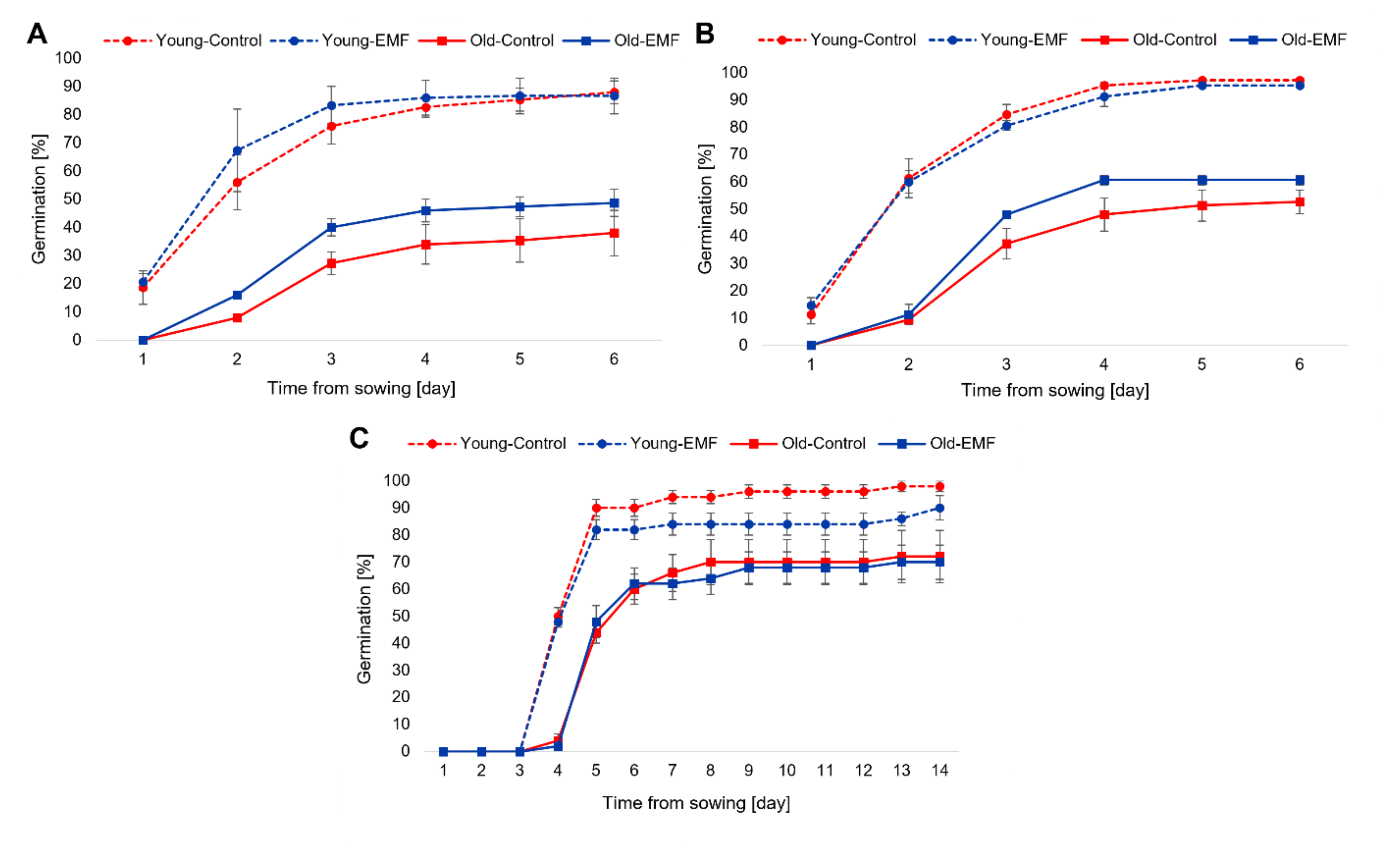
The germination kinetics of the control groups was first examined in order to observe the effects of field bean seed age on the germination process. In the Petri dish studies, young seeds germinated for 6 days at a significantly higher rate than old seeds under both the long-day conditions (p < 0.01) and in continuous darkness (p < 0.05) (Figure 2A,B). However, the germination kinetics of the control group of pot-sown seeds (young and old) under long-day conditions observed for 14 days, was not influenced by seed age (Figure 2C).
When analyzing the results for the effect of EMF treatment in all the variants of the study, the germination kinetics in continuous darkness of Petri dish-sown old seeds exposed to EMF significantly improved (p < 0.05) compared to the untreated control (Figure 2B). However, in the other experimental variants (Petri dish-sown young and old seeds under long-day conditions; Petri dish-sown young seeds in continuous darkness; and pot-sown young and old seeds under long-day conditions), EMF exposure did not affect the germination kinetics (Figure 2A–C).
Among the EMF-treated groups, Petri dish-sown young seeds under long-day conditions germinated at a significantly higher rate (p < 0.05) compared to old seeds in the same conditions (Figure 2A), thus indicating again the moderating effect of seed age on germination kinetics under certain conditions. On the other hand, seed age did not affect the germination kinetics of the EMF-treated groups of Petri dish-sown seeds in continuous darkness and pot-sown seeds under long-day conditions (Figure 2B,C).

3.2. Analysis of Germination Parameters (G, GI, MGT, CVG, t50)

In addition to the analysis of germination kinetics (Figure 2A–C), other parameters (G, GI, MGT, CVG, and t50) were evaluated to determine the germination changes in both the EMF-treated and control groups of young and old seeds.
Within the control groups, seed age was found to significantly affect these germination parameters in all of the studied media and light conditions. The germinability (G) of old seeds was significantly lower than that of young seeds by the following margins within the control groups: (a) 57% less in Petri dishes under long-day conditions; (b) 46% less in Petri dishes in continuous darkness; and (c) 27% less in pots under long-day conditions (Table 1 and Table 2). Further comparison of the control groups of young and old seeds germinated in Petri dishes under long-day conditions showed that the realized values of GI and CVG of the old seeds were significantly lower by 65% and 25%, respectively, while their MGT and t50 values were significantly higher than young seeds by 33% and 41%, respectively (Table 1). Similarly, old seeds germinated in Petri dishes in continuous darkness obtained GI and CVG values significantly lower by 56% and 26%, respectively and MGT and t50 values significantly higher by 35% and 48%, respectively when compared to young seeds in the control groups (Table 1). When comparing the germination parameters of the control groups of pot-sown young and old seeds, the GI and CVG parameters of the old seeds were significantly lower by 33% and 15%, respectively, while their t50 value was significantly higher by 16% (Table 2). These marked differences between the germination parameters of young and old seeds in the control groups clearly indicate that old seeds without any exposure to EMF germinate at a much lower rate than young seeds in all the analyzed growth media and light conditions.
Young seeds exposed to EMF and germinated in pots obtained a GI value, which is 11% lower than that of the untreated control (Table 2). However, the analysis of germination parameters in the remaining experimental variants (Petri dish-sown young and old seeds under long-day conditions and in continuous darkness, and pot-sown old seeds) showed that pre-sowing exposure of seeds to EMF did not affect their germination parameters (Table 1 and Table 2).
The EMF-treated groups of young and old seeds were analyzed to determine the influence of seed age similar to the analysis of the control groups. The comparison of the germination parameters within these groups follows a similar trend to the analysis within the control groups (apart from differences between CVG and t50 parameters for pot-sown seeds) (Table 1 and Table 2), which confirms that the germination rate reduces with increasing seed age.
The above analysis of the various germination parameters indicates that the germination process of field beans is negatively affected by seed age irrespective of the light conditions and growth media. On the contrary, pre-sowing exposure of field bean seed to EMF is found to affect germination differently and depends on seed age, light conditions, and growth media.

3.3. Morphometric Analysis of Seedling’s Growth in Control Groups

The growth parameters were measured in 6-day-old and 14-day-old seedlings growing in Petri dishes and in substrate-filled pots, respectively. Different changes in the morphometric parameters were observed depending on seed age, light conditions, and growth media.
To assess seed age’s influence on early seedling growth, the control groups in different growth media were analyzed. The control group of old seeds germinated in Petri dishes under long-day conditions did not develop seedlings with fully emerged roots and epicotyls, in contrast to the young seeds (Table 3). Analysis within the control group for differences in delayed growth (seedlings with only roots protruded) showed that old seeds germinated in Petri dishes under long-day conditions had shorter root lengths (–22%) than roots from young seeds.
Further assessment of the control groups showed that seedlings from old seeds growing in Petri dishes in continuous darkness had reduced growth parameters compared to young seeds (Table 4) at the following rates: 27% shorter root length of fully emerged seedlings, 14% less seedling fresh weight, 29% shorter root length of seedlings with only roots protruded, and 25% less emerged embryo fresh weight.
These results show that seedling growth in Petri dishes under long-day conditions and in continuous darkness was slower in seedlings from old seeds, and may be related to the similar trend of reduced rate of germination of old seeds compared to young seeds. On the contrary, analysis of the control groups of seedlings growing in substrate-filled pots shows that there are no differences in growth parameters between seedlings from young and old seeds (Table 5).

3.4. EMF Treatment Effect on Growth Parameters

Pre-sowing EMF treatment of both young and old seeds germinated in Petri dishes under long-day conditions had no statistically significant effect on the analyzed growth parameters of their respective seedlings compared to the untreated control group (Table 3). However, it can be hypothesized that EMF treatment of old seeds could be responsible for the enhanced growth of seedlings in Petri dishes under long-day conditions to the point of the appearance of fully emerged roots and epicotyls, a phenomenon which is entirely absent in seedlings from untreated samples of old seeds, although the statistical significance of this effect could not be determined (Table 3).
Contrary to the results of seedling growth in Petri dishes under long-day conditions, some selected parameters of seedling growth in Petri dishes in continuous darkness were significantly stimulated by the pre-sowing treatment of both young and old seeds. EMF treatment of young seeds enhanced the epicotyl growth of their seedlings by 14%. Compared to their untreated controls, the growth of seedlings from EMF-treated old seeds was enhanced as follows: 22% longer roots in seedlings with only protruded roots (delayed growth) and 22% more fresh weight of emerged embryos. This enhancement of seedling growth from old seeds by EMF exposure (Table 4) is associated with the observed improvement of germination kinetics in EMF-treated old seeds germinating in Petri dishes in continuous darkness (Figure 2B). This shows that the early stages of plant growth may be more prone to stimulation by seed exposure to EMF, especially if the growth process is delayed, as was observed for the old seeds.
Furthermore, the analysis of the growth parameters of 14-day-old field bean plants germinated in substrate-filled pots under long-day conditions from young seeds indicated that pre-sowing EMF treatment of the seeds significantly stimulated the root length by 13% but inhibited stem length by 10% compared to the untreated control (Table 5). Other morphometric traits of field bean plants growing in the pots were not affected by seed age or exposure to EMF.
The comparison of growth parameters within EMF-treated groups follows a similar trend to the analysis within control groups (Table 3 and Table 4), confirming that the seedling growth rate decreases with seed aging.

3.5. Assessment of Water Uptake and Membrane Integrity of Field Bean Seeds

To check whether the observed effects of seed age and EMF exposure on germination and seedling growth are related to changes in membrane permeability of young and old seeds of field beans, parameters of water uptake and membrane integrity were examined.
Among the control groups, there were no differences in water uptake for young and old seeds (Figure 3A). EMF treatment also did not affect the water uptake of young and old seeds, compared to the untreated controls. However, among the EMF-treated groups, young seeds absorbed significantly more water (+5%) than old seeds (Figure 3A).
When membrane integrity was analyzed with respect to seed age, it was observed that in the control groups, the electrolyte leakage in old seeds was significantly higher (+172%) than in young seeds (Figure 3B). Moreover, EMF treatment significantly increased the electrolyte leakage by 13% in old seeds compared to their untreated controls. In the case of young seeds, EMF exposure did not affect their electrolyte leakage compared to their untreated controls. Similarly, old seeds in the EMF-treated groups had significantly higher (+204%) values of electrolyte leakage compared to treated young seeds (Figure 3B). These results show that the integrity of cellular membranes of seed tissues was negatively affected by seed age and EMF exposure.

3.6. Amylolytic Activity and H2O2 Content in Seeds of Field Bean

For the analyses of α-amylases activity and H2O2 levels, the Petri dish-sown seeds germinating under long-day conditions were selected due to the fact that EMF-treated old seeds were able to germinate in Petri dishes under long-day conditions, to the point of the appearance of seedlings with fully emerged roots and epicotyls, which are completely absent in the untreated old seeds (Table 3). Sarraf et al. [12] have reported that amylases and reactive oxygen species (ROS) are known factors affected by seed exposure to different doses of MF. Thus, our objective was to determine whether the observed changes in seedling growth from old seeds after EMF exposure, which could not be statistically determined, would be reflected in stimulations at the cellular level, specifically, in the enzymatic activity of amylases, which hydrolyze starch reserves in seeds, and in the level of H2O2, which belongs to ROS molecules.
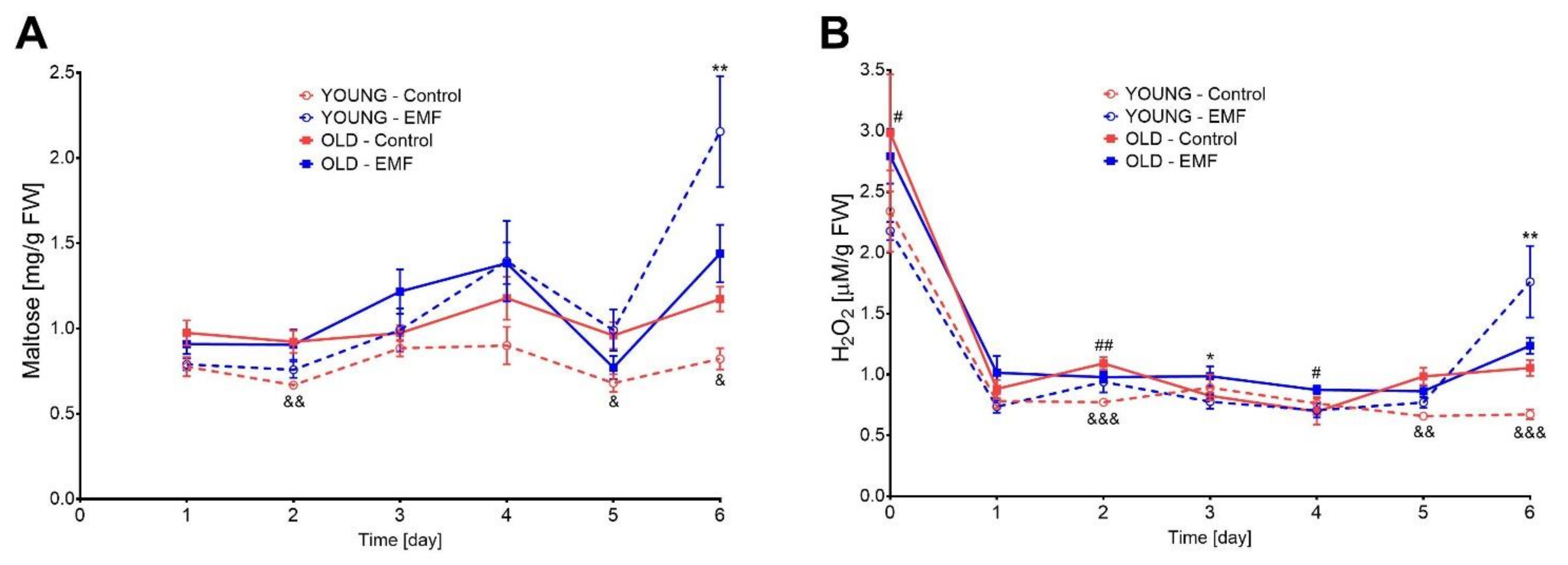
In the germinating young and old seeds of field beans, the α-amylase activity showed specific patterns of changes in the control groups and EMF-treated groups (Figure 4A). In the control groups, α-amylase activity in old seeds on the 2nd, 5th, and 6th days of germination was significantly higher (+38%, +41%, +43%), compared to young seeds.
EMF treatment significantly stimulated, by 162%, the activity of α-amylases in young seeds on the last (6th) day of germination, compared to their untreated controls. In old seeds, the activity of α-amylases was not affected by EMF exposure. This suggests that the observed appearance of seedlings with fully emerged roots and epicotyls under long-day conditions after exposure of old seeds to EMF, was not directly related to the stimulation of α-amylase activity by the EMF factor.
In contrast to the control groups, where α-amylase activity was observed to be significantly higher in old seeds than in young seeds, analysis of the EMF-treated groups showed that there were no differences in the α-amylase activity in young and old seeds. This revealed a strong stimulatory effect of EMF exposure in young seeds in the latter days of germination.
To reveal the influence of seed age on H2O2 content in field bean seeds, the control groups were analyzed and showed that on the 2nd, 5th, and 6th days of germination, the amount of H2O2 in old seeds was significantly higher (by +41%, +49%, and +57%), compared to young seeds (Figure 4B). This elevated H2O2 level in the old seeds may be responsible for the observed higher amylase activity in old seeds compared to young seeds in control groups.
The H2O2 levels were 162% higher in EMF-treated young seeds than in their controls on the 6th day of germination, while treated old seeds had 19% higher H2O2 levels than their untreated controls on the 3rd day of germination (Figure 4B). The strong stimulation of H2O2 levels in young seeds by EMF exposure recorded on the 6th day of germination is positively associated with the observed strong enhancement of α-amylase activity by EMF treatment of young seeds, also on the 6th day of germination. This again shows that during the germination process of field beans, α-amylase activity may be positively regulated by higher levels of H2O2.
Among the EMF-treated groups, the H2O2 content of old dry seeds (before sowing) and germinating old seeds on the 2nd and 4th days were found to be 28%, 4%, and 24% more than in the young seeds, respectively (Figure 4B). This increase in the H2O2 level, also revealed in the control groups, can be a consequence of metabolic changes in cells of old seeds during the senescence process. It is also worth pointing out that in both the control and EMF-treated groups, dry seeds (young and old) before sowing had about three times higher the amount of H2O2 in the imbibed seeds during the next five days of germination, showing a strong dependence of H2O2 level on the stage of the germination process.

3.7. Effect of Seed Age and EMF Exposure on Photosynthetic Pigments Content

Plant productivity depends on photosynthesis, and the level of photosynthetic pigments can be an indication of changes in plant physiology, sometimes due to the actions of different stress factors [41]. In this study, the contents of chlorophylls (a, b, and total) and total carotenoids (xanthophylls + b-carotene) were examined in two-week-old field bean plants growing in substrate-filled pots. The analysis was performed in the control groups, as well as in the EMF-treated groups, to assess the dependence of seed age and the potential physical eustress factor due to EMF on photosynthetic pigments content.
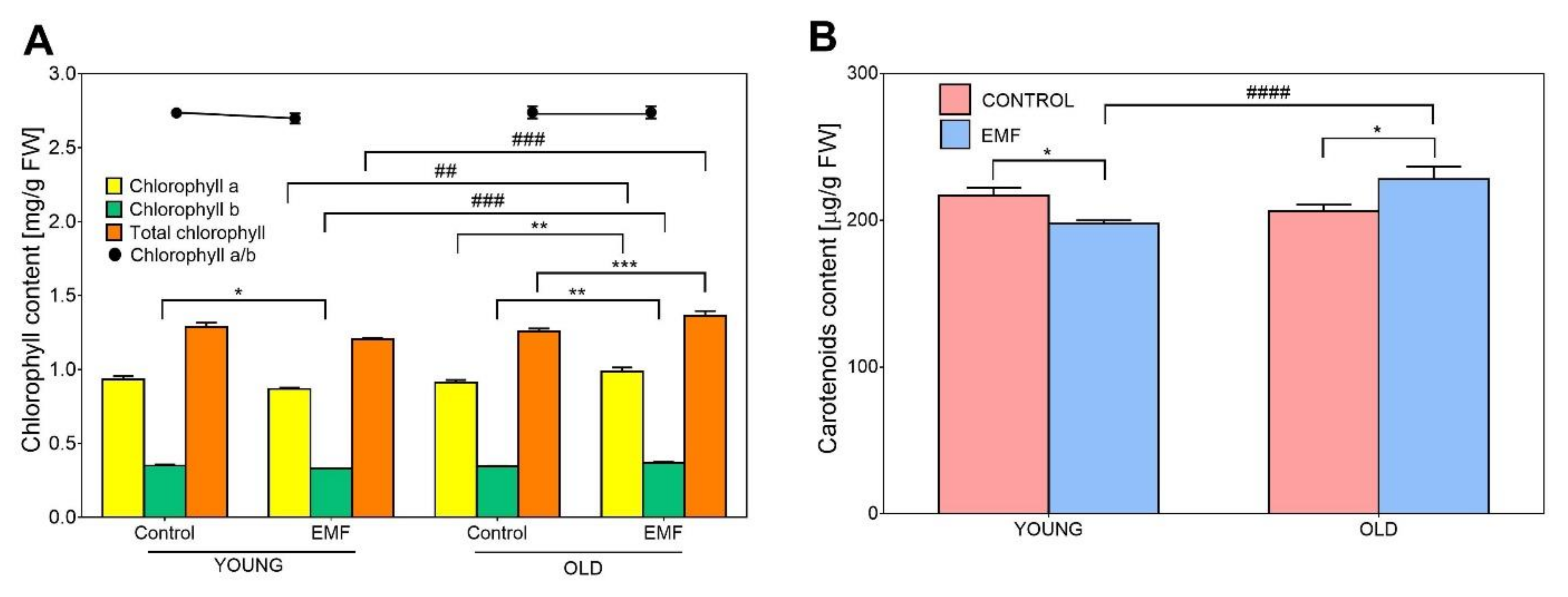
Among the control groups, there were no differences in the contents of the chlorophylls and total carotenoids in the leaves of plants growing from young and old seeds (Figure 5A,B), indicating that seed age does not influence the content of photosynthetic pigments in field bean plants.
Leaves of plants grown from EMF-treated young seeds had significantly lower levels of chlorophyll b (−5%) and carotenoids (−9%) than in their untreated controls (Figure 5A,B). This reduction in the pigment content in leaves of plants from EMF-treated young seeds can be related to the observed inhibition of growth of above-ground organs (stems) from the EMF-treated young seeds (Table 5). In contrast, leaves of plants growing from EMF-treated old seeds had a significant increase in the content of all photosynthetic pigments studied compared to their untreated controls: chlorophyll a (+8%), chlorophyll b (+6%), total chlorophyll (+8%), and total carotenoids (+11%) (Figure 5A,B).
Among the EMF-treated groups, changes in pigment content specific to seed age were observed. In these groups, leaves of plants growing from EMF-treated old seeds express significantly higher levels of chlorophyll a (+14%), chlorophyll b (+11%), total chlorophyll (+13%), and carotenoids (+15%), compared to EMF-treated young seeds (Figure 5A,B), highlighting the strong stimulatory effect of EMF exposure on photosynthetic pigments in plants growing from old seeds.
Moreover, in all the experimental variants, the chlorophyll a/b ratio did not change and therefore was not affected by seed aging or EMF exposure (Figure 5A).

3.8. Influence of Seed Age and EMF Exposure on Phytohormone Levels in Seedlings Growing in Substrate-Filled Pots

Phytohormones are essential signaling elements that control many aspects of plant growth and development, as well as their response to environmental stress [42]. This association of phytohormones to nearly all fundamental biological processes makes them a good candidate for consideration during testing and engineering stress tolerance in agronomically important crops [43].
To check if seed age and pre-sowing exposure of seeds to EMF can influence longer-term biochemical traits in field bean plants, the levels of six selected phytohormones (IAA, ABA, GA1, GA3, SA, JA) were analyzed quantitatively in the roots and leaves of two-week-old plants growing from EMF-treated young and old seeds as well as their untreated controls. Additionally, we sought to determine whether the changes observed in the growth parameters (Table 5) and the content of photosynthetic pigments (Figure 5) attributed to pre-sowing exposure of seeds to EMF will be related to changes in the levels of the main phytohormones controlling growth (IAA, ABA, GAs) and stress response (SA, JA).
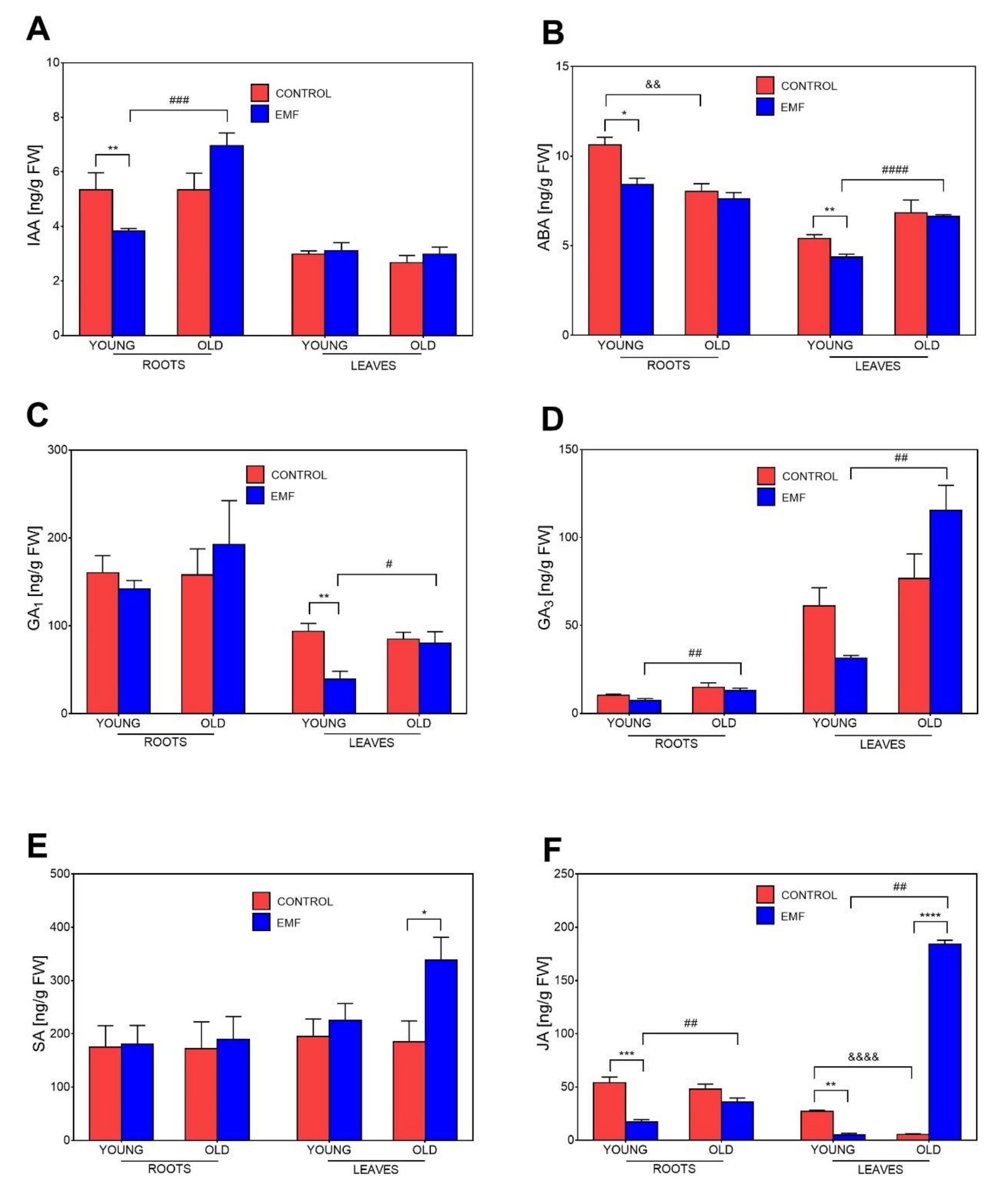
In the case of IAA, there were no differences in the auxin levels in the roots and leaves of plants growing from young and old seeds in the control groups. This indicates that seed age did not affect the IAA amount (Figure 6A). Concerning the influence of EMF exposure, the changes in IAA levels in plants from treated seeds were observed solely in underground organs (Figure 6A). The IAA amount in the roots of plants growing from EMF-treated young seeds was significantly lower (−28%) than in the untreated control. This reduction in IAA amount in the roots of treated plants is negatively associated with the observed stimulation of root growth in plants growing from EMF-treated young seeds (Table 5). Contrary to the changes in the roots of plants growing from EMF-treated young seeds, the roots of plants from EMF-treated old seeds did not express changes in IAA levels, compared to their untreated controls (Figure 6A). However, among the EMF-treated groups, changes related to seed age were detected. In these treatment groups, roots of plants growing from old seeds expressed significantly higher levels (+82%) of IAA compared to roots growing from young seeds (Figure 6A).
The ABA level changed significantly among the control groups, revealing that this hormone content was affected by seed age. In these groups, the ABA amount in the roots of plants growing from young seeds was higher compared to the roots of plants growing from old seeds (Figure 6B).
EMF-treated young seeds produced plants with significantly reduced ABA levels in their roots (−21%) and leaves (−20%) compared to their untreated controls (Figure 6B). Similar to the results from the IAA analysis, the observed reduction in ABA level in roots is negatively associated with the enhanced root growth of plants growing from EMF-treated young seeds (Table 5). Concerning the effects of EMF exposure on photosynthetic pigments, the observed reduction in ABA amount in leaves corresponds with the decrease in chlorophyll b and total carotenoids in leaves of plants growing from EMF-treated young seeds (Figure 5). The roots and leaves of plants growing from EMF-treated old seeds had no significant changes in ABA levels compared to their untreated controls (Figure 6B). Moreover, among the EMF-treated groups, the ABA level in leaves of plants growing from treated young seeds was lower (−52%) than in plants from treated old seeds, showing that in these groups the ABA level was also affected by seed age.
The analysis of the level of bioactive gibberellins showed that among the control groups, there was no difference in the GA1 levels in the roots and leaves of plants growing from young and old seeds (Figure 6C). However, the level of GA1 in leaves of plants growing from EMF-treated young seeds was significantly reduced by 58%, compared with their untreated control (Figure 6C). This reduction in GA1 level in leaves was associated with the decrease in stem length and content of particular photosynthetic pigments in leaves due to pre-sowing exposure of seeds to EMF (Table 5, Figure 5). No other significant effects of EMF exposure on the levels of GA1 in the organs of plants growing from treated young and old seeds compared to their untreated controls were detected. Concerning the effect of seed age among the EMF-treated groups, the leaves of plants growing from treated young seeds contained less GA1 (−51%) than the leaves of plants growing from treated old seeds (Figure 6C).
Among the control groups, the level of GA3 in the roots and leaves was not affected by seed age (Figure 6D). Furthermore, the pre-sowing exposure of young and old seeds to EMF did not lead to any changes in the GA3 levels in the roots and leaves of plants compared to their untreated controls. However, when the EMF-treated groups were analyzed, the age of the treated seeds had an effect on their GA3 levels. The GA3 levels in the roots and leaves of plants growing from treated old seeds were respectively, 78% and 266% higher than those of plants growing from treated young seeds (Figure 6D).
The SA level in roots and leaves in the control groups was not affected by seed age. Pre-sowing exposure of seeds to EMF led to a significant increase (+83%) in SA levels in the leaves of plants growing from old seeds (Figure 6E), which is associated with the higher content of the photosynthetic pigments in leaves of plants growing from the EMF-treated old seeds (Figure 5). No other specific changes in SA levels attributable to the pre-sowing exposure of seeds to EMF or seed age were detected (Figure 6E).
Significant changes in the amount of JA were found among the control groups of young and old seeds, indicating the influence of seed age on the JA level. In the control groups, leaves of plants growing from old seeds had an 80% lower JA level compared to leaves growing from young seeds (Figure 6F).
The JA levels in the roots and leaves of plants growing from EMF-treated young seeds were, respectively, 68% and 81% lower than in their untreated controls (Figure 6F). These results indicate a possible difference in the role of JA in the growth regulation of roots and above-ground organs since the observed reduction in JA levels attributed to EMF corresponds with root growth stimulation and the inhibition of stem growth and photosynthetic pigment content in plants growing from young treated seeds (Table 5, Figure 5). In contrast, the level of JA in the leaves of plants growing from old seeds treated with EMF was strongly stimulated (34-fold increase) compared to their untreated control. This particular change in JA level was associated with the increase in photosynthetic pigment content in leaves growing from EMF-treated old seeds (Figure 5). Among the EMF-treated groups, particular seed age-specific changes in JA levels were also detected. In these groups, the roots and leaves of plants growing from treated old seeds had significantly higher levels of JA (two-fold and 36-fold, respectively) than the organs of plants growing from treated young seeds.

4. Discussion

Plant response to MFs is regarded as a non-linear phenomenon since all living organisms, including plants, can be considered as non-linear systems which pass through different stages of developmental processes [44]. Studies on the effect of MF treatments on plant growth and development have produced varying results of stimulation, inhibition, and sometimes no effect, which indicates a complex mechanism of MF action in plant cells and the likely occurrence of different interactions between MF response and endogenous rhythms in plant cells [31].
Our current study revealed different effects of seed exposure to EMF on germination and early growth of field beans, which were dependent on seed age, light conditions, and growth media. In control groups, the germination kinetics and germination parameters of old seeds were significantly reduced compared to young seeds in all the experimental variants. On the contrary, EMF exposure significantly improved germination kinetics, but only for old seeds germinating in Petri dishes in continuous darkness (Figure 2B). However, the germination rate of EMF-treated young seeds in the substrate-filled pots was significantly reduced. In all the experimental variants, the only instance where EMF treatment led to an improvement in germination kinetics, as well as subsequent seedling growth, was during the germination and growth in Petri dishes of treated old seeds in continuous darkness (Table 4). Although the pre-sowing exposure to EMF inhibited the germination rate of young seeds in substrate-filled pots, the root length of two-week-old seedlings growing from EMF-treated young seeds was stimulated. However, the growth of stems derived from these EMF-treated young seeds in the substrate-filled pots was inhibited (Table 5). These results strongly point to the dependence of EMF effects on the physiological and developmental stage of the plant material, as well as the growth (environmental) conditions. Similar results were obtained by Ivankov et al. [23] where treatment of red clover seeds with EMF (5.28 MHz, 0.74 mT) stimulated five-week-old seedlings but only in the morphometric parameters of the roots and not in the above-ground parts. In the case of sunflower (Helianthus annus) seeds, the same dose of EMF exposure (5.28 MHz, 0.74 mT) resulted in a higher germination rate and an increase in leaf weight of two-week-old seedlings sown in a substrate, while other growth parameters remained unaffected by the EMF treatment [45]. Furthermore, the treatment of tomato (Solanum lycopersicum) seeds with EMF (3 Hz, 12.5 mT) led to the inhibition of stem length in field conditions, despite the stimulation of other growth parameters such as the number of leaves and flowers [46]. There are several other reports showing inconsistencies in the influence of MF exposure on particular physiological traits of plants, which suggests the possibility of improving plant growth without altering the germination rate of seeds. The EMF (50 Hz, 15 mT) treatment of durum wheat (Triticum durum) seeds caused no influence on the dormant seed germination process but positively affected the fresh weight of seedlings [47]. A study involving the application of EMF (5.28 MHz, 0.74 mT) on seeds of two perennial woody plants (Rhododendron smirnowii, Morus nigra) showed that the most adverse effect of EMF on the germination of seeds could be followed by an increase in the growth of leaves during the later developmental stages of the plants [48].
An earlier study showed that roots seem to be more sensitive to MF exposure than shoots and that the stimulation or inhibition of root growth by MF depends on plant species and their physiological state [49]. In addition, the roots of Arabidopsis seedlings were reported to be the most sensitive organs to MF exposure, with the observed stimulation in root growth attributed to an increase in cell number [50]. In our study, the observed stimulation of root length of field bean plants growing in the substrate-filled pots from EMF-treated young seeds can be of particular importance for crop production in field conditions. Similar results of significant improvement in the main root characteristics (length, surface area, and volume) due to MF exposure have been found in cotton (Gossypium hirsutum) (3 Hz, 12.5 mT) [51], chickpea (100 mT) [25], and maize (50–250 mT) [52]. This improvement in root growth highlights the potential of MF treatment as a useful priming method to improve crop resistance to drought stress.
Under certain stress conditions, roots are found to accelerate growth to absorb more water and nutrients for survival, which in turn increases the root-to-shoot ratio and this is attributed to the relatively greater negative consequence of the stress factor on the above-ground organs [53]. These observations support the hypothesis that particular doses of EMF exposure on some plants could be a eustress factor that affects plant growth and development. Mildaziene et al. [45] have reported that proteome analysis of two-week-old sunflower seedlings after exposure to EMF (5.28 MHz, 0.74 mT) shows a eustress-like response localized mainly in their chloroplasts.
In our Petri dish experiments, EMF effects on germination and growth were also dependent on light conditions. This is similar to the results obtained by Novitskii et al. [54], where the examination of the composition and content of lipids in five-day-old radish (Raphanus sativus) seedlings growing from seeds exposed to MF (50 Hz, 500 µT) showed existing differences in the mechanisms of MF action in light and darkness. In addition, EMF (50 Hz, 7 mT) treatment of winter wheat seeds stimulated, to a higher degree, germination and early growth in continuous darkness, compared to continuous light conditions [15]. Currently, the mechanism of this light-dependent response to MF is not fully understood, but existing reports indicate the involvement of cryptochromes in this response [55].
In old seeds, many cellular processes could change as a consequence of the aging process, including membrane damage, loss of enzymatic activity, and many oxidative damages to lipids, proteins, and DNA [4]. This, in turn, can influence specific signaling pathways in cells of seeds of different ages and cause distinct, sometimes stronger, seed responses to MF exposure [48]. In our study, the germination kinetics of only old seeds was stimulated by exposure to EMF (50 Hz, 7 mT), which later led to accelerated early root growth (Figure 2B, Table 4). Bilalis et al. [51] found that in the case of cotton seeds, EMF (3 Hz, 12.5 mT) exposure was an effective priming technique for plants growing in pots under field conditions, especially for old seeds with reduced germinability. When six-year-old pea seeds were exposed to MF (100 mT), seed vigor was enhanced, and this improvement in the quality of the aged seed was mediated by changes in free radicals by the antioxidant defense system and protein oxidation [56]. Additionally, the treatment of aged broccoli (Brassica oleracea) seeds with MF (60 Hz, 3.6 mT) to improve their germination led to different outcomes (positive, negative, and no effects) depending on the length of their aging process [57].
Many reports have indicated an increase in water uptake and improvement in seed coat membrane integrity—expressed in lower electrolyte leakage—after seed exposure to different doses of MFs, which in turn is often positively associated with an increase in germination speed [16,52,58]. In our study, EMF (50 Hz, 7 mT) exposure did not change the water uptake of field bean seeds, but reduced the already impaired membrane integrity of old seeds, and had no effect on the membrane integrity of the young seeds (Figure 3). In other studies where wheat seeds were magneto-primed, the water uptake of seeds was reduced after treatment with SMF (30 mT) but did not change after EMF (10 Hz) exposure [59]. Moreover, the treatment of two genotypes of chickpea seeds with SMF (100 mT) caused an increase in water uptake in only one genotype, while in the second (native) genotype, MF priming did not cause a significant change in seed water uptake, suggesting that the rate of water uptake by seeds may depend mainly on the internal water potential of the seeds [25]. Thus, the influence of MF on membrane permeability can depend on seed type (structure of seed coat and type of storage material) and MF characteristics.
In our study, biochemical analysis of the field bean seeds showed that in control groups, α-amylase activity in old seeds was higher than in young seeds, and was also associated with a higher level of H2O2 in old seeds. Moreover, EMF exposure (50 Hz, 7 mT) caused a significant increase in α-amylase activity on the last day of germination of young seeds, which again was associated with a large rise in H2O2 level in those seeds at the same point. Similar positive associations of H2O2 concentration with amylase activity were reported in barley (Hordeum vulgare) grains for β-amylase [60] and α-amylase [61]. Furthermore, the increased amylase activity at the later stages of germination due to MF exposure was also found in these seeds: chickpea treated with SMF of 100 mT [25]; millet (Setaria italica) treated with EMF of 10 Hz, 0.03 mT [62]; and faba bean (Vicia faba) treated with MFs of 35 mT and 80 mT seeds [63]. In the case of chickpea, the amylase activity after MF exposure was found to vary depending on plant genotype [25]. Additionally, the higher content of H2O2 after exposure of seeds to MF was associated with the reported improvement in the germination of MF-treated tomato (100 mT) [14] and cucumber (Cucumis sativus) (100–250 mT) [58]. Apart from the beneficial role of H2O2 in MF signaling, this molecule can also have a detrimental effect during seed aging, leading to a lowering of the germination rate caused by factors including membrane integrity loss [64]. In our study, the concentration of H2O2 was highest in dry seeds (young and old) compared to imbibed seeds. Moreover, within the control groups, old seeds of field beans contained more H2O2 compared to young seeds and this was also associated with higher electrolyte leakage and a reduced germination rate in the old seeds. These results are supported by existing reports showing that H2O2 accumulates in dry seeds during the after-ripening process and with seed aging during prolonged storage [64].
The content of photosynthetic pigments is a good marker of plant health and productivity, and a higher composition of chlorophylls and carotenoids can enhance photosynthesis and plant growth [65]. Existing evidence show that the effects of MF exposure on photosynthetic pigments depend on plant species and MF parameters. In this study, EMF treatment of seeds led to the photosynthetic pigment content increasing in the leaves of seedlings grown from old seeds while decreasing in seedlings from young seeds (Figure 5). Vashisth and Joshi [52] have reported a higher chlorophyll content in plants growing from MF-treated seeds of maize (50–250 mT). However, in other studies, MF exposure produced inhibitory effects or no effects on photosynthetic pigment content. EMF (50 Hz, 65 µT) decreased the content of chlorophylls a, b, and carotenoids in barley seedlings [66], while in pea seedlings, the chlorophyll content was not significantly affected after treatment of seeds with EMF (50 Hz, 60–180 mT) [28]. Similar to our results, it was reported that the exposure of durum wheat seeds to EMF (50 Hz, 15 mT) did not affect the chlorophyll a and b ratios [47].
Currently, only a few reports exist presenting hormonal changes after MF exposure and they often refer to hormonal analysis in seeds [14,23,45]. The existing knowledge base of the influence of EMF exposure on longer-term traits like phytohormone balance in growing plants is, thus, negligible. Our results show that in the case of the main growth hormones (IAA, ABA, and GAs), more changes due to exposure to EMF were observed in the roots and leaves of plants growing from young seeds. Plant hormones typical for stress response (SA and JA) were affected by EMF exposure the most in the leaves of plants growing from old seeds (Figure 6E,F). Differences in hormonal changes in roots and leaves due to seed age were detected mostly by comparing within the EMF-treated groups. In most of these cases, the levels of analyzed hormones were higher in organs of plants growing from old seeds than in organs of plants growing from young seeds (Figure 6). Khan et al. [67] have reported that the levels of IAA, ABA, SA, and JA increase in certain senescing plant organs. Some age-dependent functions of gibberellins have also been reported, indicating the role of GAs in the delay of nodule senescence in peas [68].
Regarding changes in IAA levels, our results showed that the reduction in the IAA amount in roots of plants growing from EMF-treated young seeds was associated with the stimulation of root growth from those seeds. Auxins are reported to inhibit primary root growth [69] and this supports our results. The stimulation of root growth associated with a reduction in IAA level was also reported in wheat seedlings growing from seeds exposed to EMF (50 Hz, 7 mT) [15]. However, another study indicated that exposing pea seeds to MF (30 mT and 85 mT) instead caused a significant increase in IAA levels in 6-day-old stems and roots [16]. Similarly, the treatment of 3-day-old Arabidopsis seedlings with SMF (600 mT) increased the level of auxin in the MF-treated root tip via enhanced expression of auxin influx transporter, AUX1, and decreased the expression of auxin efflux transporter, PIN3 [50]. Thus, the effect of MF treatment on IAA level in growing plants seems to be strongly dependent on MF parameters.
ABA is considered a general inhibitor of plant growth [70]. Lowering the ABA level in seeds after MF exposure is regarded as a priming mechanism to stimulate germination [15,45]. In this study, we observed a reduction in ABA level in the roots of plants growing from EMF-treated young seeds which was also associated with the stimulation of root growth from those seeds. Moreover, changes in ABA and IAA levels observed in roots of plants growing from EMF-treated young seeds were similar, which indicates possible interactions between ABA and IAA signaling pathways during root growth of field beans. Such interactions are known to occur during Arabidopsis root growth under different abiotic stress conditions [42].
Gibberellins (GAs) can enhance organ growth by stimulating cell elongation and division [43]. GA1, GA3, GA4, and GA7 are the most common biologically active forms of gibberellins found in higher plants [71]. In our study, the GA1 level was reduced in leaves of plants growing from EMF-treated young seeds. Achard et al. [72] have shown that under salt stress, the levels of bioactive gibberellins reduce, possibly through the signaling of ABA and that the reduction in the growth of Arabidopsis due to the gibberellin pathways is beneficial and enhances the survival of plants. Furthermore, results in our study indicate that leaves of plants growing from EMF-treated old seeds contained more GA1, chlorophylls, and carotenoids than leaves of plants growing from EMF-treated young seeds (Figure 6 C). This indicates the possible involvement of GA1 in the regulation of photosynthetic processes. Iftikhar et al. [73] have shown that exogenous application of GA positively affects chlorophyll content in wheat. It has also been revealed that the IAA/GA1 ratio reflects changes in growth parameters of soybean plants under specific photosynthetic conditions [74].
In our experiments, the GA3 levels were not affected by the pre-sowing exposure of seeds to EMF. However, Anand et al. [14] found that the GA3 level was higher in MF-treated (100 mT) seeds of tomato. Similarly, Podleśny et al. [16] reported that the GA3 levels in seeds of pea, along with roots and stems of their 6-day-old seedlings, increased after pre-sowing exposure of the seeds to MF (30 mT and 85 mT).
SA and JA are known to participate in defense reactions to many biotic and abiotic stressors [43]. SA content was previously investigated in dry seeds of red clover and sunflower, where exposure to EMF (5.28 MHz, 0.74 mT) resulted in SA content reduction in the red clover [23,24] but an elevation in the sunflower [45]. However, as far as we know, the effect of EMF exposure on JA content in plants has not yet been analyzed. In our study, JA level was inhibited in roots of plants growing from EMF-treated young seeds, while at the same time, the growth of those roots was stimulated. These results are supported by the fact that JA is known to inhibit root growth [75] and thus, lowering JA levels can have a stimulatory effect on root development.
The exposure of old seeds to EMF in our study led to a significant increase in SA and JA levels in the leaves of plants growing from such seeds, which resembles stressor-specific changes. SA is essential in regulating defense processes, such as hypersensitive response and systemic acquired resistance, while JA regulates biotic and abiotic stress responses [67]. Thus, the increase in SA and JA levels suggests that EMF may boost plant defense systems, preparing field bean plants for future stress conditions. One consequence of such a boost in the plant defense system could be the increase in photosynthetic pigment content, which was observed in this study for leaves of plants growing from EMF-treated old seeds. Kaya and Doganlar [76] found that tobacco (Nicotiana tabacum) plants treated with endogenous JA produce more chlorophyll and carotenoids, which helps plants to alleviate the negative effects of herbicide stressors. Brassica oleracea plants grown from seeds treated with various concentrations of JA, have also been shown to enhance photosynthetic efficiency and chlorophyll fluorescence [77].
Despite a significant increase in the level of specific stress hormones (SA and JA), the complex hormonal changes observed in the roots and leaves of plants growing from EMF-treated seeds suggests that EMF treatment of 50 Hz, 7 mT does not cause the typical stress response in field bean organs. However, the modifications in hormonal balance detected in our studies suggest a response of the plants to low-intensity stress, i.e., eustress, especially in the case of plants growing from old seeds. In recent years, it has become evident that plant hormones do not act only in a linear pathway, but also produce numerous and often complicated interactions [43,78]. Therefore, more detailed studies are necessary to elucidate the role of phytohormones in the mechanism of MF action in plant tissues and to lead to the development of more resilient crops.

5. Conclusions

We analyzed the effectiveness of the pre-sowing exposure of field bean seeds of different ages to EMF (50 Hz, 7 mT) as an alternative physical seed treatment to improve germination and growth. In control groups, old seeds expressed a reduced germination rate and slower growth compared to young seeds. After EMF treatment, different effects on seed germination, growth parameters, and biochemical traits, depending on seed age, light condition, and growth media were obtained. EMF treatment stimulated only the germination of old seeds in Petri dishes in continuous darkness and this was followed by enhanced early growth of their roots. In the studies using a universal substrate in pots, EMF exposure led to a lower rate of germination of young seeds but had no significant effect on the germination rate of old seeds. Our studies showed that membrane integrity was only affected (negatively) by EMF treatment in old seeds. Moreover, increased α-amylase activity was associated with higher H2O2 levels in the control group of old seeds and group of EMF-treated young seeds. Furthermore, the response to EMF exposure was assessed by measuring the morphological and biochemical parameters of two-week-old plants, which can affect field bean competition under field conditions. The improved root growth of plants growing from EMF-treated young seeds, corresponding to reduced IAA, ABA, and JA levels, suggests that magnetically treated field bean seeds may grow into plants with a better uptake of water under rainfed (un-irrigated) or even drought conditions. Additionally, the stimulation of photosynthetic pigment content in leaves growing from EMF-treated old seeds was associated with increased levels of SA and JA in those organs and this could help field bean plants to produce bigger biomass during field cultivation.
We conclude that the observed germination and growth effects, as well as the hormonal changes due to the pre-sowing exposure of seeds to EMF (50 Hz, 7 mT), point to a field bean response to low-intensity stress (i.e., eustress). Although pre-sowing EMF treatment did not lead to direct stimulation of germination of young and old seeds in the substrate medium, this priming method produced some positive long-term effects by improving root growth and chlorophyll content. Thus, the results of our studies indicate that pre-sowing exposure of seeds of field beans to EMF (50 Hz, 7 mT) has the potential to be an alternative method of improving their production. The effectiveness of this seed priming method to enhance long-term growth and development of field beans could also be further explored in field experiments.

Author Contributions

Conceptualization, A.P.; methodology, A.P. and J.W.; investigation, A.P., D.C., J.W., M.D.D. and K.P.; writing—original draft preparation, A.P.; writing—review and editing, A.P., J.W., D.C., M.D.D., K.P. and A.S.-J.; visualization, A.P. and J.W.; supervision, A.P. and A.S.-J. All authors have read and agreed to the published version of the manuscript.

Funding

This research was funded by 1. The EU Program Knowledge, Education, Development and the Polish National Center for Research and Development (“Universitas Copernicana Thoruniensis in Futuro”, project no. POWER.03.05.00.00-Z302/17-00); 2. INCOOP competition “Excellence Initiative—Research University” at the Nicolaus Copernicus University in Toruń.

Institutional Review Board Statement

Not applicable.

Informed Consent Statement

Not applicable.

Data Availability Statement

Relevant data are contained within the article.

Acknowledgments

We express our gratitude to Grażyna Czeszewska-Rosiak for her technical support during the laboratory work.

Conflicts of Interest

The authors declare no conflict of interest.

References

  1. Robert, M.; Thomas, A.; Bergez, J.-E. Processes of Adaptation in Farm Decision-Making Models. A Review. Agron. Sustain. Dev. 2016, 36, 64. [Google Scholar] [CrossRef]
  2. Laik, R.; Kumara, B.H.; Pramanick, B.; Singh, S.K.; Nidhi; Alhomrani, M.; Gaber, A.; Hossain, A. Labile Soil Organic Matter Pools Are Influenced by 45 Years of Applied Farmyard Manure and Mineral Nitrogen in the Wheat—Pearl Millet Cropping System in the Sub-Tropical Condition. Agronomy 2021, 11, 2190. [Google Scholar] [CrossRef]
  3. Hampton, J.G.; Conner, A.J.; Boelt, B.; Chastain, T.G.; Rolston, P. Climate Change: Seed Production and Options for Adaptation. Agriculture 2016, 6, 33. [Google Scholar] [CrossRef]
  4. Zhang, K.; Zhang, Y.; Sun, J.; Meng, J.; Tao, J. Deterioration of Orthodox Seeds during Ageing: Influencing Factors, Physiological Alterations and the Role of Reactive Oxygen Species. Plant Physiol. Biochem. 2021, 158, 475–485. [Google Scholar] [CrossRef]
  5. Kumar, M.; Mitra, S.; Mazumdar, S.P.; Majumdar, B.; Saha, A.R.; Singh, S.R.; Pramanick, B.; Gaber, A.; Alsanie, W.F.; Hossain, A. Improvement of Soil Health and System Productivity through Crop Diversification and Residue Incorporation under Jute-Based Different Cropping Systems. Agronomy 2021, 11, 1622. [Google Scholar] [CrossRef]
  6. Stagnari, F.; Maggio, A.; Galieni, A.; Pisante, M. Multiple Benefits of Legumes for Agriculture Sustainability: An Overview. Chem. Biol. Technol. Agric. 2017, 4, 2. [Google Scholar] [CrossRef]
  7. Dutta, A.; Trivedi, A.; Nath, C.P.; Gupta, D.S.; Hazra, K.K. A Comprehensive Review on Grain Legumes as Climate-smart Crops: Challenges and Prospects. Environ. Chall. 2022, 7, 100479. [Google Scholar] [CrossRef]
  8. Mariotti, M.; Andreuccetti, V.; Arduini, I.; Minieri, S.; Pampana, S. Field Bean for Forage and Grain in Short-Season Rainfed Mediterranean Conditions. Ital. J. Agron. 2018, 13, 208–215. [Google Scholar] [CrossRef]
  9. Sulas, L.; Roggero, P.P.; Canu, S.; Seddaiu, G. Potential Nitrogen Source from Field Bean for Rainfed Mediterranean Cropping Systems. Agron. J. 2013, 105, 1735–1742. [Google Scholar] [CrossRef]
  10. Devika, O.S.; Singh, S.; Sarkar, D.; Barnwal, P.; Suman, J.; Rakshit, A. Seed Priming: A Potential Supplement in Integrated Resource Management Under Fragile Intensive Ecosystems. Front. Sustain. Food Syst. 2021, 5, 654001. [Google Scholar] [CrossRef]
  11. Araújo, S.S.; Paparella, S.; Dondi, D.; Bentivoglio, A.; Carbonera, D.; Balestrazzi, A. Physical Methods for Seed Invigoration: Advantages and Challenges in Seed Technology. Front. Plant Sci. 2016, 7, 646. [Google Scholar] [CrossRef]
  12. Sarraf, M.; Kataria, S.; Taimourya, H.; Santos, L.O.; Menegatti, R.D.; Jain, M.; Ihtisham, M.; Liu, S. Magnetic Field (MF) Applications in Plants: An Overview. Plants 2020, 9, 1139. [Google Scholar] [CrossRef]
  13. Pietruszewski, S.; Muszyński, S.; Dziwulska, A. Electromagnetic Fields and Electromagnetic Radiation as Non-Invasive External Stimulants for Seeds (Selected Methods and Responses). Int. Agrophys. 2007, 21, 95–100. [Google Scholar]
  14. Anand, A.; Kumari, A.; Thakur, M.; Koul, A. Hydrogen Peroxide Signaling Integrates with Phytohormones during the Germination of Magnetoprimed Tomato Seeds. Sci. Rep. 2019, 9, 8814. [Google Scholar] [CrossRef]
  15. Cecchetti, D.; Pawełek, A.; Wyszkowska, J.; Antoszewski, M.; Szmidt-Jaworska, A. Treatment of Winter Wheat (Triticum aestivum L.) Seeds with Electromagnetic Field Influences Germination and Phytohormone Balance Depending on Seed Size. Agronomy 2022, 12, 1423. [Google Scholar] [CrossRef]
  16. Podleśny, J.; Podleśna, A.; Gładyszewska, B.; Bojarszczuk, J. Effect of Pre-Sowing Magnetic Field Treatment on Enzymes and Phytohormones in Pea (Pisum sativum L.) Seeds and Seedlings. Agronomy 2021, 11, 494. [Google Scholar] [CrossRef]
  17. Vian, A.; Davies, E.; Gendraud, M.; Bonnet, P. Plant Responses to High Frequency Electromagnetic Fields. BioMed Res. Int. 2016, 2016, 1830262. [Google Scholar] [CrossRef]
  18. Ribeiro-Oliveira, J.P. Electromagnetism and Plant Development: A New Unknown in a Known World. Theor. Exp. Plant Physiol. 2019, 31, 423–427. [Google Scholar] [CrossRef]
  19. Vázquez-Hernández, M.C.; Parola-Contreras, I.; Montoya-Gómez, L.M.; Torres-Pacheco, I.; Schwarz, D.; Guevara-González, R.G. Eustressors: Chemical and Physical Stress Factors Used to Enhance Vegetables Production. Sci. Hortic. 2019, 250, 223–229. [Google Scholar] [CrossRef]
  20. Podleśny, J.; Misiak, L.E.; Podleśna, A.; Pietruszewski, S. Concentration of Free Radicals in Pea Seeds after Pre-Sowing Treatment with Magnetic Field. Int. Agrophys. 2005, 19, 243–249. [Google Scholar]
  21. Podleśny, J.; Pietruszewski, S.; Podleśna, A. Efficiency of the Magnetic Treatment of Broad Bean Seeds Cultivated under Experimental Plot Conditions. Int. Agrophys. 2004, 18, 65–71. [Google Scholar]
  22. Radhakrishnan, R.; Ranjitha Kumari, B.D. Pulsed Magnetic Field: A Contemporary Approach Offers to Enhance Plant Growth and Yield of Soybean. Plant Physiol. Biochem. 2012, 51, 139–144. [Google Scholar] [CrossRef]
  23. Ivankov, A.; Zukiene, R.; Nauciene, Z.; Degutyte-Fomins, L.; Filatova, I.; Lyushkevich, V.; Mildaziene, V. The Effects of Red Clover Seed Treatment with Cold Plasma and Electromagnetic Field on Germination and Seedling Growth Are Dependent on Seed Color. Appl. Sci. 2021, 11, 4676. [Google Scholar] [CrossRef]
  24. Mildaziene, V.; Ivankov, A.; Pauzaite, G.; Naucienė, Z.; Zukiene, R.; Degutyte-Fomins, L.; Pukalskas, A.; Venskutonis, P.R.; Filatova, I.; Lyushkevich, V. Seed Treatment with Cold Plasma and Electromagnetic Field Induces Changes in Red Clover Root Growth Dynamics, Flavonoid Exudation, and Activates Nodulation. Plasma Processes Polym. 2021, 18, 2000160. [Google Scholar] [CrossRef]
  25. Thomas, S.; Anand, A.; Chinnusamy, V.; Dahuja, A.; Basu, S. Magnetopriming Circumvents the Effect of Salinity Stress on Germination in Chickpea Seeds. Acta Physiol. Plant. 2013, 35, 3401–3411. [Google Scholar] [CrossRef]
  26. Kataria, S.; Baghel, L.; Guruprasad, K.N. Pre-Treatment of Seeds with Static Magnetic Field Improves Germination and Early Growth Characteristics under Salt Stress in Maize and Soybean. Biocatal. Agric. Biotechnol. 2017, 10, 83–90. [Google Scholar] [CrossRef]
  27. Aguilar, C.H.; Dominguez-Pacheco, A.; Carballo, A.; Cruz-Orea, A.; Ivanov, R.; Luis, J.; Pastor, J. Alternating Magnetic Field Irradiation Effects on Three Genotype Maize Seed Field Performance. Acta Agroph. 2009, 14, 7–17. [Google Scholar]
  28. Iqbal, M.; Haq, Z.U.; Jamil, Y.; Ahmad, M.R. Effect of Presowing Magnetic Treatment on Properties of Pea. Int. Agrophys. 2012, 26, 25–31. [Google Scholar] [CrossRef]
  29. Bujak, K.; Frant, M. Influence of pre-sowing seed stimulation with magnetic field on spring wheat yielding. Acta Agroph. 2009, 14, 19–29. [Google Scholar]
  30. Cakmak, T.; Dumlupinar, R.; Erdal, S. Acceleration of Germination and Early Growth of Wheat and Bean Seedlings Grown under Various Magnetic Field and Osmotic Conditions. Bioelectromagnetics 2010, 31, 120–129. [Google Scholar] [CrossRef]
  31. Teixeira da Silva, J.A.; Dobránszki, J. Magnetic Fields: How Is Plant Growth and Development Impacted? Protoplasma 2016, 253, 231–248. [Google Scholar] [CrossRef] [PubMed]
  32. Trawiński, T.; Szczygieł, M.; Wyszkowska, J.; Kluszczyński, K. Analysis of Magnetic Field Distribution and Mechanical Vibration of Magnetic Field Exciter under Different Voltage Supply. In Information Technologies in Biomedicine; Piȩtka, E., Kawa, J., Eds.; Springer: Berlin/Heidelberg, Germany, 2010; pp. 613–622. [Google Scholar]
  33. Bieńkowski, P.; Wyszkowska, J. Technical aspects of exposure to magnetic fields of extremely low frequencies (ELF) in biomedical research. Med. Pr. 2015, 66, 185–197. [Google Scholar] [CrossRef] [PubMed]
  34. Ranal, M.A.; de Santana, D.G. How and Why to Measure the Germination Process? Braz. J. Bot. 2006, 29, 1–11. [Google Scholar] [CrossRef]
  35. Kader, M. A Comparison of Seed Germination Calculation Formulae and the Associated Interpretation of Resulting Data. J. Proc. R. Soc. N. S. W. 2005, 138, 65–75. [Google Scholar]
  36. Coolbear, P.; Francis, A.; Grierson, D. The Effect of Low Temperature Pre-Sowing Treatment on the Germination Performance and Membrane Integrity of Artificially Aged Tomato Seeds. J. Exp. Bot. 1984, 35, 1609–1617. [Google Scholar] [CrossRef]
  37. Miller, G.L. Use of Dinitrosalicylic Acid Reagent for Determination of Reducing Sugar. Anal. Chem. 1959, 31, 426–428. [Google Scholar] [CrossRef]
  38. Lichtenthaler, H.K. Chlorophylls and Carotenoids: Pigments of Photosynthetic Biomembranes. In Methods in Enzymology; Plant Cell Membranes; Academic Press: San Diego, CA, USA, 1987; Volume 148, pp. 350–382. [Google Scholar]
  39. Pu, C.-H.; Lin, S.-K.; Chuang, W.-C.; Shyu, T.-H. Modified QuEChERS Method for 24 Plant Growth Regulators in Grapes Using LC-MS/MS. J. Food Drug Anal. 2018, 26, 637–648. [Google Scholar] [CrossRef]
  40. R Core Team. R: A Language and Environment for Statistical Computing; R Foundation for Statistical Computing: Vienna, Austria, 2020. [Google Scholar]
  41. Sherin, G.; Aswathi, K.P.R.; Puthur, J.T. Photosynthetic Functions in Plants Subjected to Stresses Are Positively Influenced by Priming. Plant Stress 2022, 4, 100079. [Google Scholar] [CrossRef]
  42. Waadt, R.; Seller, C.A.; Hsu, P.-K.; Takahashi, Y.; Munemasa, S.; Schroeder, J.I. Plant Hormone Regulation of Abiotic Stress Responses. Nat. Rev. Mol. Cell Biol. 2022, 23, 1–15. [Google Scholar] [CrossRef]
  43. Salvi, P.; Manna, M.; Kaur, H.; Thakur, T.; Gandass, N.; Bhatt, D.; Muthamilarasan, M. Phytohormone Signaling and Crosstalk in Regulating Drought Stress Response in Plants. Plant Cell Rep. 2021, 40, 1305–1329. [Google Scholar] [CrossRef]
  44. Ruzic, R.; Jerman, I.; Gogala, N. Effects of Weak Low-Frequency Magnetic Fields on Spruce Seed Germination under Acid Conditions. Can. J. For. Res. 1998, 28, 609–616. [Google Scholar] [CrossRef]
  45. Mildažienė, V.; Aleknavičiūtė, V.; Žūkienė, R.; Paužaitė, G.; Naučienė, Z.; Filatova, I.; Lyushkevich, V.; Haimi, P.; Tamošiūnė, I.; Baniulis, D. Treatment of Common Sunflower (Helianthus annus L.) Seeds with Radio-Frequency Electromagnetic Field and Cold Plasma Induces Changes in Seed Phytohormone Balance, Seedling Development and Leaf Protein Expression. Sci. Rep. 2019, 9, 6437. [Google Scholar] [CrossRef]
  46. Efthimiadou, A.; Katsenios, N.; Karkanis, A.; Papastylianou, P.; Triantafyllidis, V.; Travlos, I.; Bilalis, D.J. Effects of Presowing Pulsed Electromagnetic Treatment of Tomato Seed on Growth, Yield, and Lycopene Content. Sci. World J. 2014, 2014, 369745. [Google Scholar] [CrossRef]
  47. Muszyński, S.; Gagoś, M.; Pietruszewski, S. Short-Term Pre-Germination Exposure to ELF Magnetic Field Does Not Influence Seedling Growth in Durum Wheat (Triticum Durum). Pol. J. Environ. Stud. 2009, 18, 1065–1072. [Google Scholar]
  48. Mildaziene, V.; Pauzaite, G.; Malakauskiene, A.; Zukiene, R.; Nauciene, Z.; Filatova, I.; Azharonok, V.; Lyushkevich, V. Response of Perennial Woody Plants to Seed Treatment by Electromagnetic Field and Low-Temperature Plasma. Bioelectromagnetics 2016, 37, 536–548. [Google Scholar] [CrossRef]
  49. Peñuelas, J.; Llusià, J.; Martínez, B.; Fontcuberta, J. Diamagnetic Susceptibility and Root Growth Responses to Magnetic Fields in Lens Culinaris, Glycine Soja, and Triticum Aestivum. Electromagn. Biol. Med. 2004, 23, 97–112. [Google Scholar] [CrossRef]
  50. Jin, Y.; Guo, W.; Hu, X.; Liu, M.; Xu, X.; Hu, F.; Lan, Y.; Lv, C.; Fang, Y.; Liu, M.; et al. Static Magnetic Field Regulates Arabidopsis Root Growth via Auxin Signaling. Sci. Rep. 2019, 9, 14384. [Google Scholar] [CrossRef] [Green Version]
  51. Bilalis, D.J.; Katsenios, N.; Efthimiadou, A.; Karkanis, A.; Efthimiadis, P. Investigation of Pulsed Electromagnetic Field as a Novel Organic Pre-Sowing Method on Germination and Initial Growth Stages of Cotton. Electromagn. Biol. Med. 2012, 31, 143–150. [Google Scholar] [CrossRef]
  52. Vashisth, A.; Joshi, D.K. Growth Characteristics of Maize Seeds Exposed to Magnetic Field. Bioelectromagnetics 2017, 38, 151–157. [Google Scholar] [CrossRef]
  53. Xu, W.; Cui, K.; Xu, A.; Nie, L.; Huang, J.; Peng, S. Drought Stress Condition Increases Root to Shoot Ratio via Alteration of Carbohydrate Partitioning and Enzymatic Activity in Rice Seedlings. Acta Physiol. Plant. 2015, 37, 9. [Google Scholar] [CrossRef]
  54. Novitskii, Y.I.; Novitskaya, G.V.; Serdyukov, Y.A. Lipid Utilization in Radish Seedlings as Affected by Weak Horizontal Extremely Low Frequency Magnetic Field. Bioelectromagnetics 2014, 35, 91–99. [Google Scholar] [CrossRef]
  55. Vanderstraeten, J.; Gailly, P.; Malkemper, E.P. Low-Light Dependence of the Magnetic Field Effect on Cryptochromes: Possible Relevance to Plant Ecology. Front. Plant Sci. 2018, 9, 121. [Google Scholar] [CrossRef]
  56. Bhardwaj, J.; Anand, A.; Pandita, V.K.; Nagarajan, S. Pulsed Magnetic Field Improves Seed Quality of Aged Green Pea Seeds by Homeostasis of Free Radical Content. J. Food Sci. Technol. 2016, 53, 3969–3977. [Google Scholar] [CrossRef]
  57. Martínez, F.R.; Pacheco, A.D.; Aguilar, C.H.; Pardo, G.P.; Ortiz, E.M. Effects of Magnetic Field Irradiation on Broccoli Seed with Accelerated Aging. Acta Agroph. 2014, 21, 63–73. [Google Scholar]
  58. Bhardwaj, J.; Anand, A.; Nagarajan, S. Biochemical and Biophysical Changes Associated with Magnetopriming in Germinating Cucumber Seeds. Plant Physiol. Biochem. 2012, 57, 67–73. [Google Scholar] [CrossRef]
  59. Payez, A.; Ghanati, F.; Behmanesh, M.; Abdolmaleki, P.; Hajnorouzi, A.; Rajabbeigi, E. Increase of Seed Germination, Growth and Membrane Integrity of Wheat Seedlings by Exposure to Static and a 10-KHz Electromagnetic Field. Electromagn. Biol. Med. 2013, 32, 417–429. [Google Scholar] [CrossRef]
  60. Wei, K.; Jin, X.; Chen, X.; Wu, F.; Zhou, W.; Qiu, B.; Qiu, L.; Wang, X.; Li, C.; Zhang, G. The Effect of H2O2 and Abscisic Acid (ABA) Interaction on β-Amylase Activity under Osmotic Stress during Grain Development in Barley. Plant Physiol. Biochem. 2009, 47, 778–784. [Google Scholar] [CrossRef]
  61. Ishibashi, Y.; Tawaratsumida, T.; Kondo, K.; Kasa, S.; Sakamoto, M.; Aoki, N.; Zheng, S.-H.; Yuasa, T.; Iwaya-Inoue, M. Reactive Oxygen Species Are Involved in Gibberellin/Abscisic Acid Signaling in Barley Aleurone Cells. Plant Physiol. 2012, 158, 1705–1714. [Google Scholar] [CrossRef] [Green Version]
  62. Ramesh, B.; Kavitha, G.; Gokiladevi, S.; Balachandar, R.K.; Kavitha, K.; Gengadharan, A.C.; Puvanakrishnan, R. Effect of Extremely Low Power Time-Varying Electromagnetic Field on Germination and Other Characteristics in Foxtail Millet (Setaria Italica) Seeds. Bioelectromagnetics 2020, 41, 526–539. [Google Scholar] [CrossRef]
  63. Podleśna, A.; Bojarszczuk, J.; Podleśny, J. Effect of Pre-Sowing Magnetic Field Treatment on Some Biochemical and Physiological Processes in Faba Bean (Vicia faba L. Spp. Minor). J. Plant Growth Regul. 2019, 38, 1153–1160. [Google Scholar] [CrossRef]
  64. Wojtyla, Ł.; Lechowska, K.; Kubala, S.; Garnczarska, M. Different Modes of Hydrogen Peroxide Action During Seed Germination. Front. Plant Sci. 2016, 7, 66. [Google Scholar] [CrossRef] [PubMed]
  65. Rochalska, M. Influence of Frequent Magnetic Field on Chlorophyll Content in Leaves of Sugar Beet Plants. Nukleonika 2005, 50, S25–S28. [Google Scholar]
  66. Pazur, A.; Rassadina, V.; Dandler, J.; Zoller, J. Growth of Etiolated Barley Plants in Weak Static and 50 Hz Electromagnetic Fields Tuned to Calcium Ion Cyclotron Resonance. Biomagn. Res. Technol. 2006, 4, 1. [Google Scholar] [CrossRef] [PubMed]
  67. Khan, M.; Rozhon, W.; Poppenberger, B. The Role of Hormones in the Aging of Plants–A Mini-Review. Gerontology 2014, 60, 49–55. [Google Scholar] [CrossRef] [PubMed]
  68. Serova, T.A.; Tsyganova, A.V.; Tikhonovich, I.A.; Tsyganov, V.E. Gibberellins Inhibit Nodule Senescence and Stimulate Nodule Meristem Bifurcation in Pea (Pisum sativum L.). Front. Plant Sci. 2019, 10, 285. [Google Scholar] [CrossRef] [PubMed]
  69. Qin, H.; Huang, R. Auxin Controlled by Ethylene Steers Root Development. Int. J. Mol. Sci. 2018, 19, 3656. [Google Scholar] [CrossRef] [PubMed]
  70. Humplík, J.F.; Bergougnoux, V.; Van Volkenburgh, E. To Stimulate or Inhibit? That Is the Question for the Function of Abscisic Acid. Trends Plant Sci. 2017, 22, 830–841. [Google Scholar] [CrossRef]
  71. Binenbaum, J.; Weinstain, R.; Shani, E. Gibberellin Localization and Transport in Plants. Trends Plant Sci. 2018, 23, 410–421. [Google Scholar] [CrossRef]
  72. Achard, P.; Cheng, H.; De Grauwe, L.; Decat, J.; Schoutteten, H.; Moritz, T.; Van Der Straeten, D.; Peng, J.; Harberd, N.P. Integration of Plant Responses to Environmentally Activated Phytohormonal Signals. Science 2006, 311, 91–94. [Google Scholar] [CrossRef]
  73. Iftikhar, A.; Ali, S.; Yasmeen, T.; Arif, M.S.; Zubair, M.; Rizwan, M.; Alhaithloul, H.A.S.; Alayafi, A.A.M.; Soliman, M.H. Effect of Gibberellic Acid on Growth, Photosynthesis and Antioxidant Defense System of Wheat under Zinc Oxide Nanoparticle Stress. Environ. Pollut. 2019, 254, 113109. [Google Scholar] [CrossRef]
  74. Yang, F.; Fan, Y.; Wu, X.; Cheng, Y.; Liu, Q.; Feng, L.; Chen, J.; Wang, Z.; Wang, X.; Yong, T.; et al. Auxin-to-Gibberellin Ratio as a Signal for Light Intensity and Quality in Regulating Soybean Growth and Matter Partitioning. Front. Plant Sci. 2018, 9, 56. [Google Scholar] [CrossRef]
  75. Adams, E.; Turner, J. COI1, a Jasmonate Receptor, Is Involved in Ethylene-Induced Inhibition of Arabidopsis Root Growth in the Light. J. Exp. Bot. 2010, 61, 4373–4386. [Google Scholar] [CrossRef]
  76. Kaya, A.; Doganlar, Z.B. Exogenous Jasmonic Acid Induces Stress Tolerance in Tobacco (Nicotiana tabacum) Exposed to Imazapic. Ecotoxicol. Environ. Saf. 2016, 124, 470–479. [Google Scholar] [CrossRef]
  77. Sirhindi, G.; Mushtaq, R.; Gill, S.S.; Sharma, P.; Abd_Allah, E.F.; Ahmad, P. Jasmonic Acid and Methyl Jasmonate Modulate Growth, Photosynthetic Activity and Expression of Photosystem II Subunit Genes in Brassica oleracea L. Sci. Rep. 2020, 10, 9322. [Google Scholar] [CrossRef]
  78. Vanstraelen, M.; Benková, E. Hormonal Interactions in the Regulation of Plant Development. Annu. Rev. Cell Dev. Biol. 2012, 28, 463–487. [Google Scholar] [CrossRef]
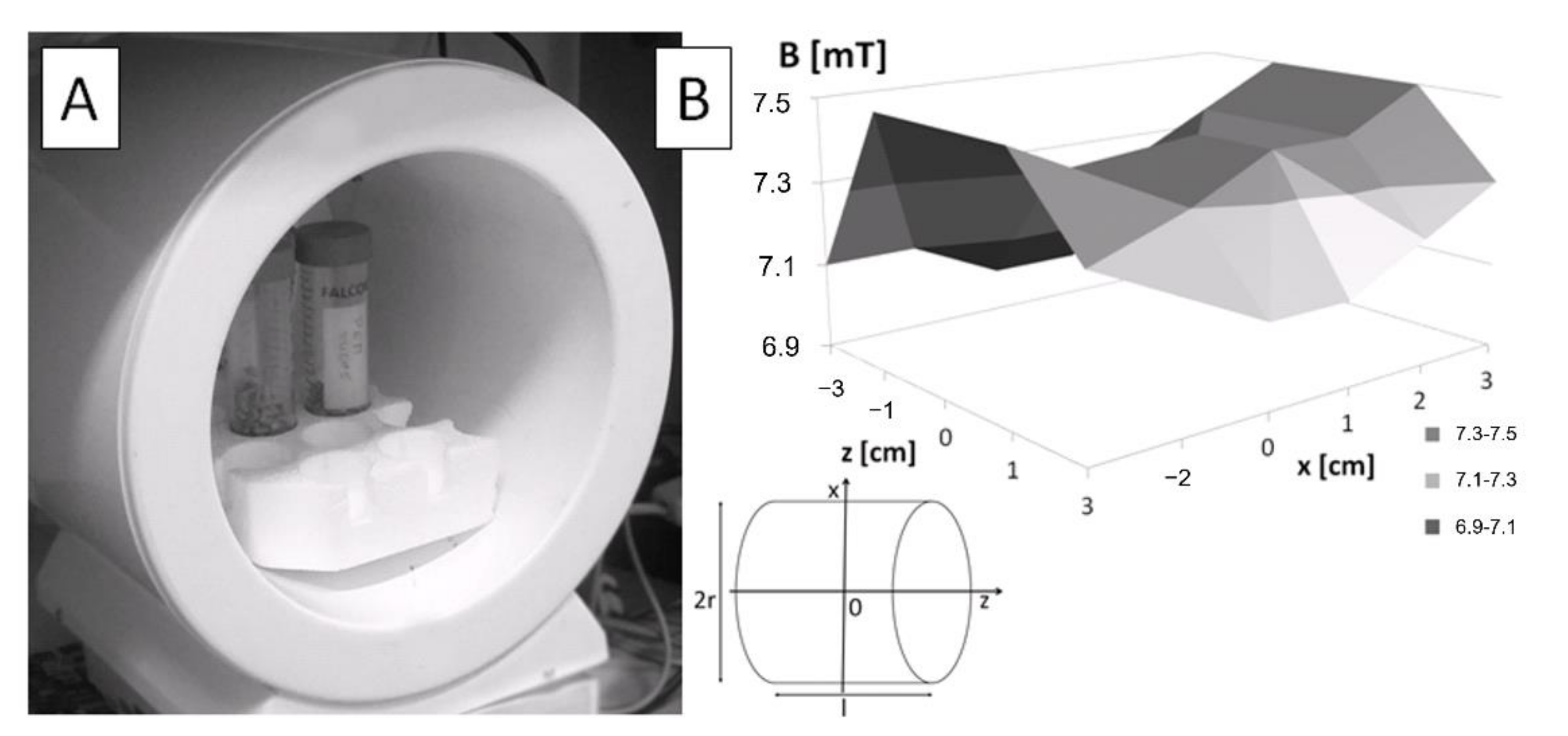
Figure 1. Exposure system. (A) Plant material in the coil. (B) The average magnetic flux density distribution inside the solenoid along the Z and X axes. Inset shows the coordinate system. 2r—diameter of the solenoid, B—magnetic flux density vector, l –coil length.
Agronomy 12 02161 g001
Figure 2. Germination kinetics of young and old field bean seeds sown in Petri dishes under long-day conditions (A), in Petri dishes in continuous darkness (B), and in pots under long-day conditions (C). The points represent mean values ± standard error (SE). Petri dish and pot tests were replicated three times (n = 50 per replicate) and five times (n = 10 per replicate), respectively. The levels of significant differences (p < 0.05; p < 0.01) between particular experimental groups are indicated in the text in the Results section.
Agronomy 12 02161 g002
Figure 3. Changes in water uptake (A) and electrolyte leakage (B) in EMF-treated and untreated young and old seeds of field bean. Data are the means ± SE (n = 4). The symbol (*) indicates significant differences between EMF-treated and control groups (* p < 0.05); “&” indicates significant differences between control groups (&&&& p < 0.0001); “#” indicates significant differences between EMF-treated groups (### p < 0.001, #### p < 0.0001).
Agronomy 12 02161 g003
Figure 4. The activity of α-amylases (A) and amount of hydrogen peroxide (H2O2) molecules (B) in young and old field bean seeds treated with EMF and their controls, germinating in Petri dishes for 6 days under long-day conditions. Data are the means ± SE (n = 3). The symbol (*) indicates significant differences between EMF-treated and control groups (* p < 0.05, ** p < 0.01); “&” indicates significant differences between control groups (& p < 0.05, && p < 0.01, &&& p < 0.001); “#” indicates significant differences between EMF-treated group (# p < 0.05, ## p < 0.01).
Agronomy 12 02161 g004
Figure 5. Photosynthetic pigments content of chlorophylls (A) and carotenoids (B) in leaves of two-week-old plants of field beans growing in pots from young and old seeds treated with EMF, and their controls. Data are the means ± SE (n = 3). The symbol (*) indicates significant differences between EMF-treated and control groups (* p < 0.05, ** p < 0.01 and *** p < 0.001); “#” indicates significant differences between EMF-treated groups (## p < 0.01, ### p < 0.001, #### p < 0.0001).
Agronomy 12 02161 g005
Figure 6. The amount of IAA (A), ABA (B), GA1 (C), GA3 (D), SA (E) and JA (F) phytohormones in roots and leaves of two-week-old plants of field bean growing in pots from young and old seeds treated with EMF, and their controls. Data are the means ± SE (n = 3). The symbol (*) indicates significant differences between EMF-treated and control groups (* p < 0.05, ** p < 0.01, *** p < 0.001, **** p < 0.0001); “&” indicates significant differences between control groups (&& p < 0.01, &&&& p < 0.0001); “#” indicates significant differences between EMF-treated groups (# p < 0.05, ## p < 0.01, ### p < 0.001, #### p < 0.0001).
Agronomy 12 02161 g006
Table 1. The effects of EMF on germination parameters of field bean seeds cultivated in Petri dishes for 6 days under long-day conditions and in continuous darkness.
Long-Day ConditionsContinuous Darkness
Young—
Control
Young—
EMF
Old—
Control
Old—EMFYoung—
Control
Young—EMFOld—
Control
Old—EMF
G88 ± 486.66 ± 6.3638 ± 8.08 &48.66 ± 4.81 ##97.33 ± 0.6695.33 ± 0.6652.66 ± 4.37 &60.66 ± 1.76 ####
GI203.33 ± 0.82215.33 ± 1.2771.33 ± 1.51 &99 ± 0.81 ##223.67 ± 0.37218.67 ± 0.2799.33 ± 1.12 &120.67 ± 0.17 ####
MGT2.38 ± 0.12.04 ± 0.053.17 ± 0.1 &2.92 ± 0.05 ##2.41 ± 0.062.41 ± 0.073.25 ± 0.08 &3.02 ± 0.04 ##
CVG42.41 ± 0.4349.12 ± 0.2631.77 ± 0.34 &34.33 ± 1.68 ##41.68 ± 0.2341.63 ± 0.2830.9 ± 0.23 &33.17 ± 0.13 #
t501.8 ± 0.15 1.64 ± 0.182.53 ± 0.16 &2.39 ± 0.03 #1.8 ± 0.121.77 ± 0.072.67 ± 0.09 &2.53 ± 0.05 ###
Note. The presented values are the average of three independent experiments ± SE. “&” indicates significant differences between control groups (& p < 0.05); “#” indicates significant differences between EMF-treated groups (# p < 0.05, ## p < 0.01, ### p < 0.001, #### p < 0.0001).
Table 2. The effects of EMF on germination parameters of field bean seeds cultivated for 14 days in pots under long-day conditions.
Young—ControlYoung—EMFOld—ControlOld—EMF
G98 ± 2 90 ± 4.47 72 ± 9.69 &70 ± 6.32 #
GI 99.8 ± 2.4 89.2 ± 3.84 *66.8 ± 7.78 &65 ± 5.67 ##
MGT 4.81 ± 0.145.06 ± 0.275.74 ± 0.435.69 ± 0.19
CVG 20.83 ± 0.58 19.98 ± 1.1117.76 ± 1.11 &17.65 ± 0.6
t504.07 ± 0.08 4.03 ± 0.094.73 ± 0.2 &4.51 ± 0.24
Note. The presented values are the average of five independent repetitions ± SE. The symbol (*) indicates significant differences between EMF-treated and control groups (* p < 0.05); “&” indicates significant differences between control groups (& p < 0.05); “#” indicates significant differences between EMF-treated groups (# p < 0.05, ## p < 0.01).
Table 3. The effects of EMF on growth parameters of field bean seedlings cultivated in Petri dishes for 6 days under long-day conditions.
Long-Day Conditions
Seedling Growth ParametersYoung—ControlYoung—EMFOld—
Control
Old—
EMF
Fully emerged roots and epicotylsEpicotyl length (mm)7.48 ± 0.378.27 ± 0.66-6.5 ± 0.5
Root length (mm)17.45 ± 1.2214.85 ± 1.05-11.5 ± 0.5
Seedling fresh weight (mg)157.70 ± 8.15142.77 ± 7.97-145.5 ± 7.5
Seedling dry weight (mg)30.33 ± 4.8147 ± 22.55-26 ± 0.1
Only roots protruded Root length (mm)8.38 ± 0.538.87 ± 0.586.56 ± 0.38 &7.13 ± 0.3 #
Emerged embryo fresh weight (mg)76.42 ± 3.483.04 ± 3.1867.21 ± 4.1475.97± 3.55
Note. The presented values are the average of three independent repetitions ± SE. “&” indicates significant differences between control groups (& p < 0.05); “#” indicates significant differences between EMF-treated groups (# p < 0.05).
Table 4. The effects of EMF on growth parameters of field bean seedlings cultivated in Petri dishes for 6 days in continuous darkness.
Continuous Darkness
Seedling Growth ParametersYoung—ControlYoung—EMFOld—
Control
Old—
EMF
Fully emerged roots and epicotylsEpicotyl length (mm)9.61 ± 0.4211 ± 0.54 *9.22 ± 0.7410 ± 0.62
Root length (mm)16.83 ± 1.0615.81 ± 0.97 12.22 ± 0.85 &11.35 ± 0.79 ##
Seedling fresh weight (mg)172.55 ± 5.25179.35 ± 7.53 147.83 ± 7.31 &142.96 ± 6.88 ##
Seedling dry weight (mg)32.66 ± 6.1229.33 ± 7.1317.33 ± 2.8520.33 ± 0.88
Only roots protruded Root length (mm)9.11 ± 0.469.72 ± 0.46 6.44 ± 0.31 &&&&7.84 ± 0.25 ***,##
Emerged embryo fresh weight (mg)104.6 ± 3.47108.62 ± 3.22 78.67 ± 3.98 &&&&95.72 ± 3.25 **,##
Note. The presented values are the average of three independent repetitions ± SE. The symbol (*) indicates significant differences between EMF-treated and control groups (* p < 0.05, ** p < 0.01, *** p < 0.001); “&” indicates significant differences between control groups (& p < 0.05, &&&& p < 0.0001); “#” indicates significant differences between EMF-treated groups (## p < 0.01).
Table 5. The effects of EMF on growth parameters of field bean seedlings cultivated for 14 days in pots under long-day conditions.
Seedling Growth ParametersYoung—ControlYoung—
EMF
Old—
Control
Old—
EMF
Stem length (cm)31.02 ± 0.8327.95 ± 1.23 *29.24 ± 1.0626.79 ± 1.39
Root length (cm)20.69 ± 0.5423.43 ± 0.93 **22.28 ± 0.6221.5 ± 1.08
Stem fresh weight (g)3.68 ± 0.143.33 ± 0.173.6 ± 0.253.05 ± 0.20
Root fresh weight (g)1.87 ± 0.091.66 ± 0.091.61 ± 0.101.61 ± 0.13
Stem dry weight (mg)232.90 ± 9.19205.19 ± 14.53216.06 ± 60.79190.78 ± 16.46
Root dry weight (mg)115.41 ± 8.85102.64 ± 5.96112.70 ± 27.0499.88 ± 7.44
Note. The presented values are the average of five independent repetitions ± SE. The symbol (*) indicates significant differences between EMF-treated and control groups (* p < 0.05, ** p < 0.01).
Publisher’s Note: MDPI stays neutral with regard to jurisdictional claims in published maps and institutional affiliations.