Article

Bioprospecting the Antibacterial Activity of Endophytic Fungi from Noni (Morinda citrifolia) against Bacterial Spot of the Passion Fruit Tree

by
Luana Cardoso de Oliveira
1,
Williams Carlos Leal da Costa
2,
Viviane Garcia Vinagre
2,
José Edson de Sousa Siqueira
1,
Sebastião da Cruz Silva
2,
Simone Yasue Simote Silva
2,
Anderson N. do Rosario Marinho
3,
Daniela Cristiane da C. Rocha
3,
Patrícia Santana Barbosa Marinho
1,2,
Alessandra Keiko Nakasone
4 and
Andrey M. do Rosario Marinho
1,2,*
1
Programa de Pós-graduação em Química, Universidade Federal do Pará, Belém 66075-110, Brazil
2
Programa de Pós-graduação em Química, Universidade Federal do Sul e Sudeste do Pará, Marabá 68507-590, Brazil
3
Instituto Evandro Chagas, Belém 67030-000, Brazil
4
Embrapa Amazônia Oriental, Belém 66095-780, Brazil
*
Author to whom correspondence should be addressed.
Agronomy 2022, 12(7), 1690; https://doi.org/10.3390/agronomy12071690
Submission received: 21 June 2022 / Revised: 11 July 2022 / Accepted: 13 July 2022 / Published: 16 July 2022

Abstract

:
Bacterial spot, which is the main disease occurring in passion fruit trees, is caused by the bacterium Xanthomonas axonopodis pv. passiflorae, leading to large annual losses in passion fruit crops. This study aims to find extracts and/or bioactive compounds of endophytic fungi of noni (Morinda citrifolia) to treat bacterial spot in passion fruit trees. Nine fungi isolated from a specimen of M. citrifolia from the Brazilian Amazon are studied. The fungus Guignardia mangiferae NF17 shows the best inhibition results and is selected for the isolation of its secondary metabolites by chromatography techniques. The isolated compounds Sydowinol (S1) and Sydowinin A (S2) are identified by nuclear magnetic resonance (NMR). Compounds S1 and S2, as well as the acetonitrile extract from the biomass of G. mangiferae NF17, are tested against four strains of X. axonopodis pv. passiflorae obtained from plants infected by bacterial spot, and which inhibited bacterial growth up to the lowest concentration tested (3.125 µg/mL). This study reports, for the first time, the antibacterial activity against X. axonopodis pv. passiflorae by the compounds Sydowinol and Sydowinin A. Compounds S1 and S2 are reported for the first time for the genus Gignardia.

Graphical Abstract

1. Introduction

Brazil is the world’s largest producer and consumer of yellow passion fruit (Passiflora edulis f. flavicarpa), with a production of 690,364 t in 2020 [1]. However, several diseases and pests threaten the expansion and yield of passion fruit crops in Brazil. Among these diseases, bacterial spot stands out [2].
The bacterial spot that occurs in passion fruit trees is caused by Xanthomonas axonopodis pv. passiflorae. There are reports that this disease was first discovered in passion fruit crops in Brazil around 1968. The disease is quite distinctive and can be visually recognized by its typical symptoms, such as small wet lesions on leaves, which soon rot and present a brownish color. The leaves becomes dry and fall as the disease develops, considerably reducing yield [3]. Currently, the use of pesticides has been the main strategy to combat bacterial spot.
Pesticides have helped to increase yield in areas where different crops are planted, which has helped to meet the world’s food demand. However, the uncontrolled use of pesticides is associated with the increase in certain types of diseases, such as cancer, Alzheimer’s disease, Parkinson’s disease, amyotrophic lateral sclerosis, asthma, bronchitis, and infertility, among others, due to their high toxicity [4]. The use of natural compounds can be a good alternative to the use of pesticides.
Morinda citrifolia is a plant originating in Southeast Asia; consumed for over 2000 years, it is commonly known as noni. Due to its wide variety of uses in folk medicine, noni has attracted the attention of researchers around the world. Chemical analyses performed on M. citrifolia reveal the existence of more than 200 phytochemical substances with bioactive properties [5]. Several benefits, such as antimicrobial, antiseptic, antioxidant, anti-inflammatory, anticancer, antidiabetic, analgesic, antiviral, antiparasitic, and antituberculosis activities, can be attributed to noni [6]. Studies have demonstrated the antibacterial effect of noni extracts against the bacterium X. axonopodis pv. passiflorae [7].
Endophytic fungi colonize the internal tissues of plants and have the potential to act as biological control agents, or elicitors, in the resistance process and in the attenuation of abiotic stresses [8]. Endophytic microorganisms represent a source of natural products with great importance for use in the pharmaceutical and agricultural industries [9,10]. The natural compounds produced by these microorganisms can also act as inhibitors of the growth of plant pathogens [11,12].
It is believed that bioactive secondary metabolites produced by endophytic microorganisms may be directly associated with the host plant through genetic recombination between species during the evolutionary phase. Some literature data have shown the ability of endophytic fungi to produce secondary metabolites identical to the compounds of their host plant [13].
To minimize phytosanitary problems, it is necessary to search for alternatives to the use of pesticides. In this context, the study of secondary metabolites of endophytic fungi represents a potential area in the development of natural pesticides. Thus, this study aims to search for extracts and/or bioactive compounds of endophytic fungi from M. citrifolia that can be used as alternative controls for bacterial spot in passion fruit.

2. Materials and Methods

2.1. Microorganisms

For this study, nine fungi isolated from a specimen of M. citrifolia belonging to a collection of the Laboratory of Phytopathology of Embrapa, Eastern Amazon, were analyzed. The strains of X. axonopodis pv. passiflorae used in the tests were obtained from passion fruit plants infected by bacterial spot in crops from four different cities in Pará state, Brazil.

2.2. Isolation of Endophytic Fungi

For this study, leaves, fruits, stems, and roots of M. citrifolia (IAN 188703) were collected in the city of Belém, Pará, Brazil, at the geographic coordinates 01°26′14.2″ S and 48°26′42.5″ W. After collection, M. citrifolia healthy tissues were washed with water and their surface was sterilized by immersion in 70% aqueous ethanol (1 min), followed by 5% aqueous sodium hypochlorite (4 min), and finally 70% aqueous ethanol (30 s). Afterwards, tissues were rinsed with sterilized water. The latter water was incubated in Petri dishes in order to guarantee the elimination of all epiphytic microorganisms. Small tissues pieces were excised and placed in Petri dishes containing potato dextrose agar (PDA) medium at 30 °C. Individual hyphal tips of the emerging fungi were removed and replaced on PDA until the isolation of endophytic fungi was obtained.

2.3. Molecular Identification of Selected Endophytic Fungi

The 9 fungi strain cultures, after 21 days of growth in PDA medium, were scraped from the Petri dish with the aid of a spatula, transferred to a chilled mortar, and macerated in the presence of liquid nitrogen. The fungal DNA extractions were performed according to the protocol of Gibbs and Mackenzie [14], with adjustments. The fungi were identified by the amplification of the ITS region of the ribosomal DNA using the primers ITS4 (5′-TCCTCCGCTTATTGATATGC-3′) and ITS5 (5′-GGAAGTAAAAGTCGTAACAAGG-3′). Samples were sequenced using the Sanger method by Helixxa, Paulínia, São Paulo, Brazil [14]. The resulting sequencing data were analyzed by the BioEdit v7.2.5 software (1997–2001, Tom Hall) for the evaluation of mutations. After the analysis of the polymorphic points, the sequences comprised a dataset with samples previously described in the GenBank (https://www.ncbi.nlm.nih.gov/genbank/, accessed on 17 January 2018) for the construction of a tree of phylogenetic relations by Neighbor Joining [15] with a bootstrap of 2000 pseudoreplications using the software MEGA 6.0 [16].

2.4. Small-Scale Extracts of Fungi

Three colonies of the studied fungi, with 25 days of growth in PDA medium, were extracted according to the following metabolite extraction method: from each colony, 25 fragments of 8 mm in diameter were removed and transferred to 50 mL Erlenmeyer flasks, and 25 mL of acetonitrile extraction solvent was added. The samples were subjected to extraction by maceration for three days, after which the solutions were filtered and the crude extracts were obtained after the evaporation of the solvent in a rotatory evaporator (Table 1). Extractions were performed in three repetitions for each fungus.

2.5. In Vitro Antibacterial Assay of Small-Scale Fungal Extracts

Antibacterial activity was determined by incorporating the extracts at a concentration of 1000 µg/mL into the culture medium 523 [17], reaching a concentration of 10 µg/mL. After the solidification of the culture medium containing the extracts, 100 µL of the bacterial suspension of X. axonopodis pv. passiflorae, at a concentration of 1.0 × 10⁸ colony-forming units (CFU)/mL with serial dilution in saline solution (0.85% NaCl) up to 10−6, was dispersed with a Drigalski loop. Only culture medium was used as a control. The plates were incubated for 48 h at 28 °C. The experimental design was completely randomized with 12 treatments and 5 replications. After the incubation period, the direct effects of the extracts on the bacteria were evaluated by counting the CFU on the plaques. An analysis of variance was performed, and means were compared by the Scott–Knott test [18] at 5% probability.

2.6. Cultivation of the Guignardia Mangiferae NF17 in Rice

The isolate fungus G. mangiferae NF17 was cultivated in rice to obtain a greater amount of extract. First, the fungus was inoculated for seven days of growth in a Petri dish containing PDA medium. Then, 1 kg of cereal was equally distributed in ten 500 mL Erlenmeyer flasks, with 100 g of cereal per flask, and 50 mL of distilled water was subsequently added. All Erlenmeyer flasks were autoclaved at 121 °C for 45 min. After reaching room temperature, three small discs of mycelium approximately 2 mm in diameter with G. mangiferae were added to eight flasks, and two flasks (of only rice) were used as a control. The fungus was inoculated and left for 25 days for the growth and production of secondary metabolites.

2.7. Obtaining Extracts and the Isolation of the Compounds from Guignardia Mangiferae NF17

After 25 days of growth in rice, 200 mL of acetonitrile (ACN) was added to each Erlenmeyer flask and left for extraction for 36 h, after which the material was filtered to obtain the ACN solution. This procedure was performed in three repetitions. The ACN solutions obtained were pooled and concentrated on a rotary evaporator, and then the ACN extract (25 g) was obtained. Part of the ACN extract (5 g) was fractionated on a silica gel column chromatography (CC) with an increasing polarity gradient using the solvents hexane (Hex), ethyl acetate (AcOEt), and methanol (MeOH) for the mobile phase. Thus, the fractions Hex 100% (FACN1), Hex/AcOEt 30% (FACN2), Hex/AcOEt 50% (FACN3), Hex/AcOEt 75% (FACN4), AcOEt 100% (FACN5), and MeOH 100% (FACN6) were obtained. The FACN4 and FACN5 fractions were re-fractionated on silica gel CC using the mixtures of hexane, ethyl acetate, and methanol in increasing polarity gradients for the mobile phase, obtaining compounds S1 (25 mg) and S2 (15 mg).

2.8. NMR Analysis

The 1D and 2D NMR spectra were obtained with an Ascend 400 spectrometer (Bruker) operating at 400 MHz for 1H NMR and at 100 MHz for 13C NMR. The samples were solubilized in a suitable deuterated solvent (CDCl3 and MeOD) to obtain the spectra. The solvent signal was used to calibrate the spectrum and the coupling constants were given in Hetz (Hz).

2.9. Antimicrobial Assays of Fungal Extracts and Isolated Compounds in 96-Well Plates

The acetonitrile extract and isolated compounds S1 and S2 were tested at concentrations from 100 to 3.125 µg/mL. In 96-well plates, 100 µL of the liquid medium 523 was added to each well. Then, each sample was added to the first well of each column, obtaining a concentration of 100 µg/mL, and the solution was homogenized. After that, successive dilutions were performed by removing 100 µL from the first well and transferring this volume to the next well, homogenizing the solution. This procedure was repeated up to the antepenultimate well of the plate, obtaining a final concentration of 3.125 µg/mL. The penultimate well was used as a control, in which 100 µL of sterile distilled water was added and homogenized, after which 100 µL was removed and discarded. Then, 5 µL of the bacterial suspension was added to each well and the plates were incubated for 24 h at 28 °C. A randomized experimental design was used with three replicates. The activity was evaluated by adding 10 µL of 2% TTC (2,3,5-triphenyltetrazolium chloride) to each well of the plate. The wells that did not show a red color were considered active. TTC is a redox indicator used to differentiate metabolically active tissues from non-active ones [19]. Then, to determine the type of activity, the sample was re-inoculated in Petri dishes containing solid medium 523 and incubated for 48 h at 28 °C. When bacterial growth was observed, it was indicated that the extract and/or compound presented bacteriostatic effects at that concentration. When there was no bacterial growth, the extract and/or compound was indicated as presenting a bactericidal effect.

3. Results

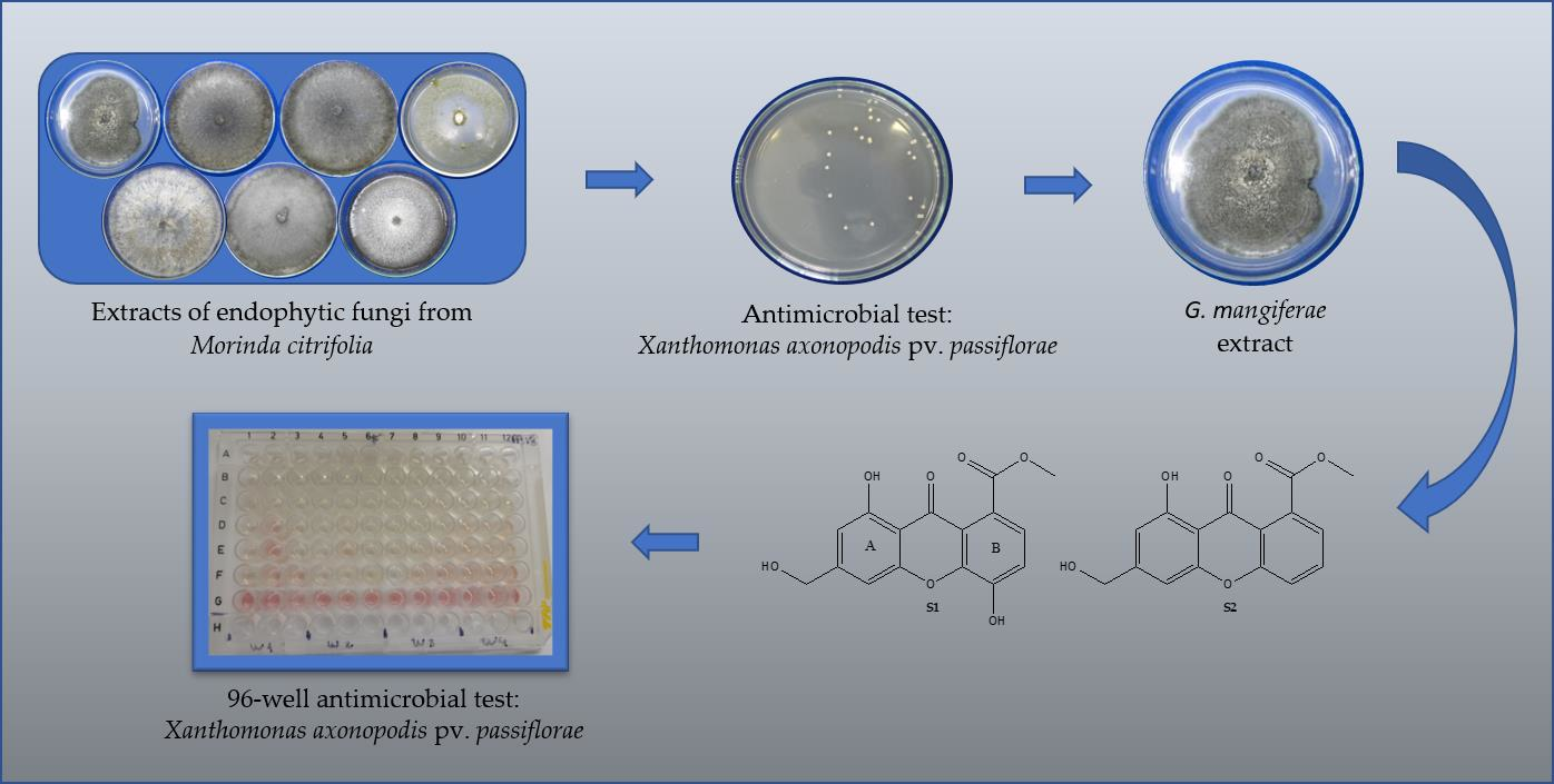
3.1. Identification of Fungi and Antimicrobial Assays with Small-Scale Extracts

The fungi studied (Figure 1) were identified by the sequencing of the ITS region with the primers ITS4 and ITS5. Then, the tree of the phylogenetic relationships was generated using Oomycetes sp. as an outgroup (Supplementary Information Figure S8), allowing the identification of the analyzed specimens (Table 2). After identification, small-scale extracts were obtained from the biomass and antibacterial assays were performed. The NFrS2 and NR7 fungi were not identified.
Only the NF17 and NFrCs16 extracts inhibited the growth of X. axonopodis pv. passiflorae and differed statistically from the control. The best result was presented by the NF17 extract, with 70.89% inhibition (Table 1, Figure 2). As the small-scale extract of the fungus G. mangiferae NF17 showed the best activity among the nine fungal strains studied, the fungus was cultivated in rice to obtain a greater amount of extract for the isolation of bioactive compounds.

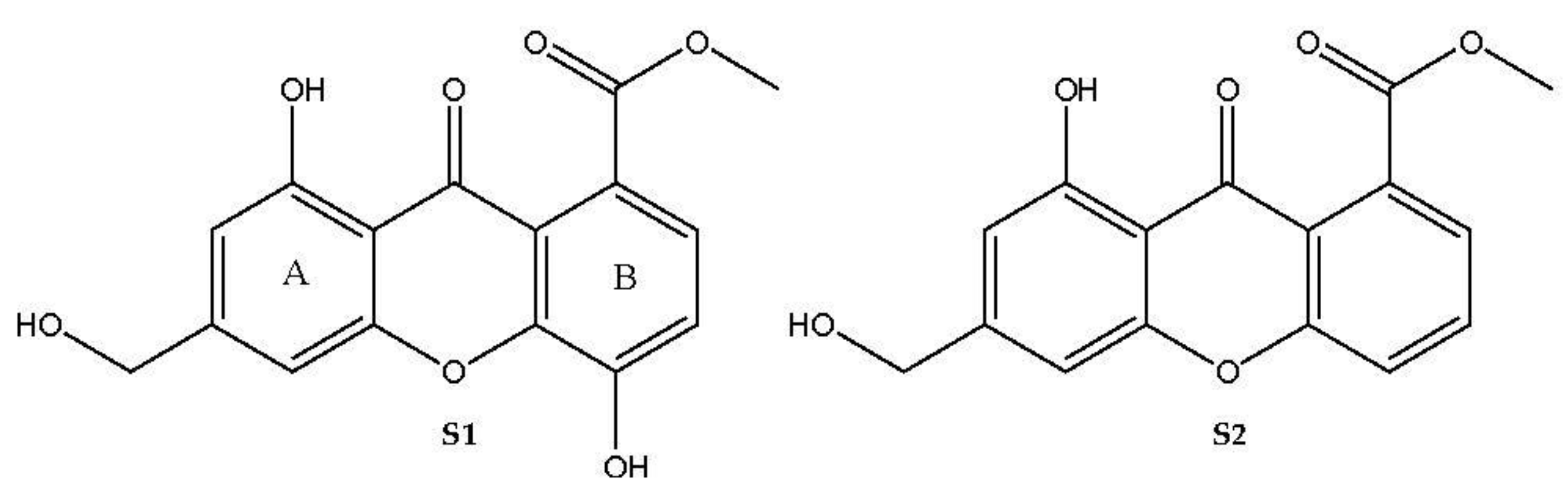
3.2. Identification of Isolated Compounds

Compounds S1 and S2 (Figure 3) were isolated using chromatography techniques from the fractions FACN5 and FACN4 of the acetonitrile extract from the biomass of the fungus G. mangiferae, respectively. The compounds were identified through 1H and 13C NMR, HMBC, HSQC, and COSY (Supplementary Information Figure S1 to S7) and compared with data described in the literature.
The 1H NMR spectrum of compound S1 (Figure 4) showed hydrogen signals that revealed the presence of two aromatic rings (ring A and B) with signals at δH 7.38 (d, 1H; 9.3 Hz) and 7.52 (d, 1H; 9.3 Hz); the constant coupling indicated that there were ortho-related aromatic hydrogens in ring B. The signals at δH 6.98 (d, 1H; 0.8 Hz) and δH 6.74 (d, 1H; 0.8 Hz) were attributed to hydrogen H-5 and H-7, which were present in the aromatic ring A of the compound S1. The 13C NMR spectra of S1 showed signals of 16 carbons, with the signal at δC 181.1 referring to a carbon belonging to the carbonyl group, which together with the 1H NMR data suggest that the compound S1 belongs to the class of xanthones [20]. As there was an HMBC correlation between H-2 (d, 7.38, 9.2 Hz) and carbon C-4 (150.1), the hydroxyl group OH-4 is positioned at C-4. The signal at δH 4.67 (s), indicating two hydrogens is typical of a hydroxymethyl group, and through the correlations observed in HBMC between H-11 with C-5 and C-7, it was possible to position the hydroxymethyl group at C-6. The signal at δH 3.95 (s), which is attributed to the presence of a methoxyl group, correlates in HMBC with the signal at δC 169.2, evidencing the presence of an acetate group linked to C-1. The NMR data of S1 were compared with the literature and this confirmed that the compound is the xanthone called Sydowinol (S1) [21].
The 1H NMR (Figure 4) and 13C NMR data of S2 are similar to that of S1, in which the characteristic signals of xanthone are observed, such as the signal for the xanthone carbonyl group at δC 180.7 (C-10), as well as the signals at δH 4.77 (H-11) and δC 64.5 (C-10), which are typical of a hydroxymethyl group -CH2OH attached to the aromatic ring. The signals at δH 6.98 (s, 1H) and δH 6.75 (s, 1H) are attributed to hydrogen H-4 and H-2, and correlate in HMBC with the hydroxymethyl group. The signals for the hydrogen of the OH group chelated with the xanthone carbonyl at δH 12.19 (s, OH) and the signals for methoxyl at δH 4.04 (s) and carbonyl at δC 170.1 were attributed to the acetate group in S2. As for the aromatic ring B, a pattern of a trisubstituted-1,2,3 aromatic ring with 1H NMR signals at δH 7.33 (dd, 7.5 and 1.2 Hz), 7.56 (dd, 7.7 and 1.2 Hz), and 7.77 (dd, 7.5 and 7.7 Hz) was observed which, together with the absence of the signal for the hydroxylated aromatic carbon in the 13C NMR spectrum, as in S1, suggests the loss of hydroxyl OH-4 at S2. These data were similar to those described in the literature by Goddard et al. (2014) for the compound Sydowinin A (S2) [21].

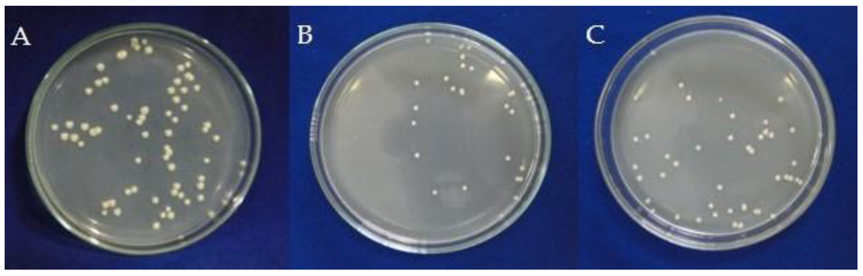
3.3. Antimicrobial Assays for the ACN Extract and Compounds S1 and S2

Assays against four different strains of X. axonopodis pv. passiflorae showed good results of bacterial growth inhibition for both the ACN extract and the compounds Sydowinol (S1) and Sydowinin A (S2), with emphasis on the bactericidal activity of S2 against the strain PA5.2 up to the lowest tested concentration: 3.125 µg/mL. The data are shown in Table 3.

4. Discussion

The bacteria of the genus Xanthomonas cause approximately 350 types of plant diseases that have been affecting the yield of agricultural crops around the world. The passion fruit crop is one of the most affected by this bacterium [22]. Due to its rapid propagation, management difficulties, problems with chemical control, and the severity of crop losses, the control of Xanthomonas is a difficult obstacle to be surpassed for agriculture worldwide [22].
Endophytic fungi are a rich source of secondary metabolites that act as biologically active agents [11,12,13,14,15,16,17,18,19,20,21,22,23] and can be applied directly or indirectly in several biotechnological areas, such as medicine, pharmacy, bioremediation, and agriculture [24].
Our studies with the extracts of the endophytic fungi from Morinda citrifolia showed that the extract of the fungus G. mangiferae NF17 inhibited the growth of the bacterium X. axonopodis pv. passiflorae at a concentration of 10 µg/mL. The search for bioactive extracts has been observed as a viable alternative to combating bacterial diseases in crops. It allows the production of antimicrobials agents without the need for elaborate purification steps. Thus, our results are corroborated by previously published studies, for example, the filamentous fungi isolated from marine sediments of the Antarctic ecosystem were tested against X. euvesicatoria and X. axonopodis pv. Passiflorae, and their extracts of the fungi inhibited the growth of X. euvesicatoria and of X. axonopodis pv. passiflorae [25]. In addition, the aqueous extracts from fruiting bodies of different isolates of Lentinula edodes showed antimicrobial activity against Xanthomonas axonopodis pv. Passiflorae [26].
Despite the antibacterial effect observed for bacteria commonly pathogenic to animals, as presented by Mai et al. [27], who evaluated the effect of hexane, dichloromethane, ethyl acetate, and methanolic extracts of two endophytic fungi isolated from M. citrifolia, and observed activity against the bacteria Escherichia coli, Bacillus subtilis, and Francisella novicida, there are not many reports of the effect of extracts of endophytic fungi of M. citrifolia on the control of X. axonopodis pv. Passiflorae, or on the control of other phytopathogenic bacteria. In a previous study, we reported antibacterial activity for the compound austdiol isolated from an endophytic fungus from Morinda citrifolia [3]. This study has now led us to study other endophytic fungi from Morinda citrifolia in search of extracts and/or bioactive compounds that act against bacterial spot. Our results show activities for the organic extracts of the endophytic fungi of M. citrifolia, which demonstrates the importance of research with this objective.
The result observed for the small-scale extract of the G. mangiferae NF17 against the bacteria X. axonopodis pv. passiflorae suggests that the extract must contain secondary metabolites that can be responsible for the observed antimicrobial activity. Some studies have shown that compounds isolated from endophytic fungi have antimicrobial activity against the phytopathogenic bacteria of the Xanthomonas genus [28,29]. Thus, the ACN extract was fractionated in CC, giving fractions FACN1 to FACN6; these fractions were analyzed by 1H NMR, and fractions FACN4 and FACN5 showed spectrums with patterns of xanthones, which is a class of compound that has reported diverse biological activities [30,31,32]. Isolation by CC procedure led to the compounds S1 and S2.
The compounds Sydowinol and Sydowinin A, as well as the ACN extract, were tested against four strains of X. axonopodis pv. passiflorae obtained from infected plants from different crops in Pará state, Brazil. The ACN extract and Sydowinol showed a bacteriostatic effect, and the substance Sydowinin A was bactericidal. The compounds Sydowinol and Sydowinin A belong to the class of xanthones.
Some xanthones have shown activity against phytopathogenic microorganisms. The xanthone γ-mangostin showed significant activity against the phytopathogenic bacterium R. solanacearum, reducing the symptoms of bacterial wilt caused by R. solanacearum in tomato and tobacco [33]. However, there are few reports of activities of xanthones against phytopathogenic bacteria, especially against the bacteria of the genus Xanthomonas.
The results obtained for both the ACN extract and the compounds Sydowinol and Sydowinin A isolated from the endophytic fungus G. mangiferae are promising and demonstrate the importance of studies aimed at finding bioactive compounds from endophytic fungi that can be used as prototypes for the development of natural pesticides.
The current work presents the in vitro results of ACN extract and the compounds Sydowinol (S1) and Sydowinin A (S2) against strains of X. axonopodis pv. Passiflorae. Despite the good in vitro results observed, some limitations of this study should be considered. The study presented here fulfills the purpose of searching for a treatment that can be effective for the control of bacterial spot in passion fruit, as an alternative to the use of synthetic pesticides. However, in vivo and field studies are still needed to complement the study, but this may take several years as they need to be completed in several locations to define appropriate dosages, determine the best application procedure, and thus confirm the real applicability of this treatment. There are also no reports of the phytotoxicity effects of xanthones against passion fruit, which still needs to be analyzed in passion fruit plants.

5. Conclusions

The ACN extract of the fungus G. mangiferae showed a good percentage of inhibition of X. axonopodis pv. passiflorae and, after isolation, compounds Sydowinol (S1) and Sydowinin A (S2) were tested against strains of X. axonopodis pv. passiflorae and showed activity up to the lowest concentration tested. This is the first report of the isolation of the compounds S1 and S2 from the genus Guignardia, and of their activities against the bacterium X. axonopodis pv. passiflorae, which causes bacterial spot. Although field studies are still needed to confirm the applicability and safety of this treatment, the in vitro results reported here suggest that the isolated compounds are promising for the control of bacterial spot in passion fruit. The search for natural compounds with antifungal activity can be a good strategy to combat bacterial spot, replacing the use of synthetic pesticides that are aggressive to the environment and public health.

Supplementary Materials

The following supporting information can be downloaded at: https://www.mdpi.com/article/10.3390/agronomy12071690/s1, Figures S1–S8: NMR spectra of the compounds are available online; Figure S1: NMR 1H spectrum to Sydowinol (S1) (400 MHz, MeOD), Figure S2: HMBC spectrum to Sydowinol (S1), Figure S3: HSQC spectrum to Sydowinol (S1), Figure S4: NMR 1H spectrum to Sydowinin A (S2) (400 MHz, CDCl3), Figure S5: NMR 13C spectrum to Sydowinin A (S2) (100 MHz, CDCl3), Figure S6: HMBC spectrum to Sydowinin A (S2), Figure S7: HSQC spectrum to Sydowinin A (S2), Figure S8: Phylogenetic analysis of the ITS region.

Author Contributions

L.C.d.O., A.M.d.R.M., A.K.N., P.S.B.M. designed the study, wrote the paper, and participated in the research; W.C.L.d.C., V.G.V., S.d.C.S., S.Y.S.S., J.E.d.S.S. participated in the phytochemical characterization of the compounds; L.C.d.O., A.K.N., A.N.d.R.M., D.C.d.C.R. performed the in vitro assays and DNA studies; A.M.d.R.M., L.C.d.O., A.K.N., P.S.B.M. contributed to the analysis of the data. All authors discussed the data obtained and collaborated in drafting the manuscript. All authors have read and agreed to the published version of the manuscript.

Funding

This research received no external funding. This study was partly funded by Federal University of Pará and Embrapa Amazônia Oriental.

Institutional Review Board Statement

Not applicable.

Informed Consent Statement

Not applicable.

Data Availability Statement

Not applicable.

Acknowledgments

The authors thank all the collaborators of the CNPq, FAPESPA, CAPES, UFPA, PROPESP/UFPA, EMBRAPA AMAZÔNIA ORIENTAL, IEC-PA for support.

Conflicts of Interest

The authors declare no conflict of interest.

References

  1. IBGE. Instituto Brasileiro de Geografia e Estatística. Produção Agrícola Municipal Culturas Temporárias e Permanentes. 2020. Available online: https://www.ibge.gov.br/estatisticas/economicas/agricultura-e-pecuaria/9117-producao-agricola-municipal-culturas-temporarias-e-permanentes.html?=&t=resultados (accessed on 28 April 2022).
  2. Nogueira, I.; Costa, A.P.; Peixoto, J.R.; Vilela, M.S. Reaction of Sour Passion Fruit Lineages and Hybrids to Bacterial Spot Caused by Xanthomonas axononopodis pv. passiflorae Under Protected Cultivation and Field Conditions. J. Agric. Sci. 2021, 13, 112–121. [Google Scholar]
  3. De Oliveira, L.C.; Ishida, A.K.N.; Da Silva, C.T.B.; Carvalho, J.M.; Feitosa, A.O.; Marinho, P.S.B.; Marinho, A.M.R. Antibacterial activity of austdiol isolated from Mycoleptodiscus indicus against Xanthomonas axonopodis pv. passiflorae. Rev. Virtual Quim. 2019, 11, 596–604. [Google Scholar] [CrossRef]
  4. Mostafalou, S.; Abdollahi, M. Pesticides: An update of human exposure and toxicity. Arch. Toxicol. 2017, 91, 549–599. [Google Scholar] [CrossRef]
  5. Almeida, E.S.; Oliveira, D.; Hotza, D. Properties and Applications of Morinda citrifolia (Noni): A Review. Compr. Rev. Food Sci. Food Saf. 2019, 18, 883–909. [Google Scholar] [CrossRef] [Green Version]
  6. Assi, R.A.; Darwis, Y.; Abdulbaqi, I.M.; Khan, A.A.; Vuanghao, L.; Laghari, M.H. Morinda citrifolia (Noni): A comprehensive review on its industrial uses, pharmacological activities, and clinical trials. Arab. J. Chem. 2017, 10, 691–707. [Google Scholar] [CrossRef]
  7. Oliveira, L.C.; Ishida, A.K.N.; Silva, C.T.B.; Marinho, P.S.B.; Marinho, A.M.R. Atividade antibacteriana de extratos de Morinda citrifolia L. (noni) contra Xanthomonas axonopodis pv. passiflorae. Rev. Cuba. Plantas Med. 2018, 23, 1–13. [Google Scholar]
  8. Fontana, D.C.; Paula, S.; Torres, A.G.; Souza, V.H.M.; Pascholati, S.F.; Schmidt, D.; Dourado Neto, D. Endophytic fungi: Biological control and induced resistance to phytopathogens and abiotic stresses. Pathogens 2021, 10, 570. [Google Scholar] [CrossRef]
  9. Newman, D.J.; Cragg, G.M. Natural products as sources of new drugs over the nearly four decades from 01/1981 to 09/2019. J. Nat. Prod. 2020, 83, 770–803. [Google Scholar] [CrossRef]
  10. Newman, D.J.; Cragg, G.M. Plant endophytes and epiphytes: Burgeoning sources of known and “Unknown” cytotoxic and antibiotic agents? Planta Med. 2020, 86, 891–905. [Google Scholar] [CrossRef] [Green Version]
  11. Sudha, V.; Govindaraj, R.; Baskar, K.; Al-Dhabi, N.A.; Duraipandiyan, V. Biological properties of endophytic fungi. Braz. Arch. Biol. Technol. 2016, 59, e16150436. [Google Scholar] [CrossRef] [Green Version]
  12. El-Baky, N.A.; Amara, A.A.A.F. Recent approaches towards control of fungal diseases in plants: An updated review. J. Fungi 2021, 7, 900. [Google Scholar] [CrossRef]
  13. Wu, L.S.; Dong, W.G.; Si, J.P.; Liu, J.J.; Zhu, Y.Q. Endophytic fungi, host genotype, and their interaction influence the growth and production of key chemical components of Dendrobium catenatum. Fungal Biol. 2020, 124, 864–876. [Google Scholar] [CrossRef] [PubMed]
  14. Rodrigues, G.N.; Alvarenga, N.; Vacondio, B.; Vasconcellos, S.P.; Passarini, M.R.Z.; Seleghim, M.H.R.; Porto, A.L.M. Biotransformation of methyl parathion by marine-derived fungi isolated from ascidian Didemnum ligulum. Biocatal. Agric. Biotechnol. 2016, 7, 24–30. [Google Scholar] [CrossRef]
  15. Saitou, N.; Nei, M. The neighbor-joining method: A new method for reconstructing phylogenetic trees. Mol. Biol. Evol. 1987, 4, 406–425. [Google Scholar] [CrossRef] [PubMed]
  16. Tamura, K.; Stecher, G.; Peterson, D.; Filipski, A.; Kumar, S. MEGA6: Molecular evolutionary genetics analysis version 6.0. Mol. Biol. Evol. 2013, 30, 2725–2729. [Google Scholar] [CrossRef] [PubMed] [Green Version]
  17. Kado, C.I.; Heskett, M.G. Selective media for isolation of Agrobacterium, Corynebacterium, Erwinia, Pseudomonas and Xanthomonas. Phytopathology 1970, 60, 969–976. [Google Scholar] [CrossRef]
  18. Scott, A.; Knott, M. Acluster-analysis method for grouping means in the analysis of variance. Biometrics 1974, 30, 507–512. [Google Scholar] [CrossRef] [Green Version]
  19. Gabrielson, J.; Hart, M.; Jarelöv, A.; Kuhn, I.; Mckenzie, D.; Möllby, R. Evaluation of redox indicators and the use of digital scanners and spectrophotometer for quantification of microbial growth in microplates. J. Microbiol. Methods. 2002, 50, 63–73. [Google Scholar] [CrossRef]
  20. Sun, R.; Miao, F.; Zhang, J.; Wang, G.; Yin, X.; Ji, N. Three new xanthone derivatives from an algicolous isolate of Aspergillus wentii. Magn. Reson. Chem. 2013, 51, 65–68. [Google Scholar] [CrossRef]
  21. Goddard, M.; Mottier, N.; Jeanneret-Gris, J.; Christen, D.; Tabacchi, R.; Abou-Mansour, E. Differential production of phytotoxins from Phomopsis sp. from grapevine plants showing esca symptoms. J. Agric. Food Chem. 2014, 62, 8602–8607. [Google Scholar] [CrossRef]
  22. Marin, V.R.; Ferrarezi, J.H.; Vieira, G.; Sass, D.C. Recent advances in the biocontrol of Xanthomonas spp. World J. Microbiol. Biotechnol. 2019, 35, 72. [Google Scholar] [CrossRef] [PubMed]
  23. Deshmukh, S.K.; Dufossé, L.; Chhipa, H.; Saxena, S.; Mahajan, G.B.; Gupta, M.K. Fungal endophytes: A potential source of antibacterial compounds. J. Fungi 2022, 8, 164. [Google Scholar] [CrossRef] [PubMed]
  24. Slama, H.B.; Bouket, A.C.; Alenezi, F.N.; Pourhassan, Z.; Golińska, P.; Tomasz Oszako, T.; Belbahri, L. Potentials of endophytic fungi in the biosynthesis of versatile secondary metabolites and enzymes. Forests 2021, 12, 1784. [Google Scholar] [CrossRef]
  25. Purić, J.; Vieira, G.; Cavalca, L.B.; Sette, L.D.; Ferreira, H.; Vieira, M.L.C.; Sass, D.C. Activity of Antarctic fungi extracts against phytopathogenic bacteria. Lett. Appl. Microbiol. 2018, 66, 530–536. [Google Scholar] [CrossRef]
  26. Tonucci-Zanardo, N.M.; Pascholati, S.F.; Di Piero, R.M. In vitro antimicrobial activity of aqueous extracts from Lentinula edodes isolates against Colletotrichum sublineolum and Xanthomonas axonopodis pv. passiflorae. Summa Phytopathol. 2015, 41, 13–20. [Google Scholar] [CrossRef] [Green Version]
  27. Mai, N.; Matainaho, T.; Rai, P.P.; Barrows, L.R. Antimicrobial activity of endophytes in six medicinal plants collected in the Central Province, Papua New Guinea. Pac. J. Med. Sci. 2013, 11, 57–69. [Google Scholar]
  28. Ataides, D.; Pamphile, J.A.; Garcia, A.; Ribeiro, M.A.S.; Polonio, J.C.; Sarragiotto, M.H.; Clemente, E. Curvularin produced by endophytic Cochliobolus sp. G2-20 isolated from Sapindus saponaria L. and evaluation of biological activity. J. Appl. Pharm. Sci. 2018, 8, 032–037. [Google Scholar] [CrossRef] [Green Version]
  29. Flores, A.C.; Pamphile, J.A.; Sarragiotto, M.H.; Clemente, E. Production of 3-nitropropionic acid by endophytic fungus Phomopsis longicolla isolated from Trichilia elegans A. JUSS ssp. elegans and evaluation of biological activity. World J. Microbiol. Biotechnol. 2013, 29, 923–932. [Google Scholar] [CrossRef]
  30. Pina, J.R.S.; Silva-Silva, J.V.; Carvalho, J.M.; Bitencourt, H.R.; Watanabe, L.A.; Fernandes, J.M.P.; Souza, G.E.; Aguiar, A.C.C.; Guido, R.V.C.; Almeida-Souza, F.; et al. Antiprotozoal and antibacterial activity of ravenelin, a xanthone isolated from the endophytic fungus Exserohilum rostratum. Molecules 2021, 26, 3339. [Google Scholar] [CrossRef]
  31. Kang, H.H.; Zhang, H.B.; Zhong, M.J.; Ma, L.Y.; Liu, D.S.; Liu, W.Z.; Ren, H. Potential antiviral xanthones from a coastal saline soil fungus Aspergillus iizukae. Mar. Drugs 2018, 16, 449. [Google Scholar] [CrossRef] [Green Version]
  32. Zhao, Y.; Liu, J.P.; Lu, D.; Li, P.Y.; Zhang, L.X. A new antioxidant xanthone from the pericarp of Garcinia mangostana Linn. Nat. Prod. Res. 2010, 24, 1664–1670. [Google Scholar] [CrossRef] [PubMed]
  33. Li, P.; Yang, Z.; Tang, B.; Zhang, Q.; Chen, Z.; Zhang, J.; Wei, J.; Sun, L.; Yan, J. Identification of xanthones from the mangosteen pericarp that inhibit the growth of Ralstonia solanacearum. ACS Omega 2020, 5, 334–343. [Google Scholar] [CrossRef] [PubMed] [Green Version]
Figure 1. Endophytic fungi from Morinda citrifolia. Colonies of the fungi studied with seven days of growth in PDA medium. (A): NF17; (B): NC4; (C): NC5; (D): NC10; (E): NFrCs4; (F): NFrCs8; (G): NFrCs16.
Agronomy 12 01690 g001
Figure 2. Effect of small-scale extracts of NF17 and NFrCs16 fungi from Morinda citrifolia on the growth of Xanthomonas axonopodis pv. passiflorae. (A): Control; (B): NF17 extract; (C): NFrCs16 extract.
Agronomy 12 01690 g002
Figure 3. Compounds isolated from Guignardia mangiferae Sydowinol (S1) and Sydowinin A (S2).
Agronomy 12 01690 g003
Figure 4. NMR 1H spectrum of (A) Sydowinol (S1) (400 MHz, MeOD) and (B) Sydowinin A (S2) (400 MHz, CDCl3).
Agronomy 12 01690 g004
Table 1. Effect of small-scale extracts of endophytic fungi from Morinda citrifolia on the growth of Xanthomonas axonopodis pv. passiflorae.
ExtractMass of Extract Obtained1 CFU/mL % Inhibition Compared to Control
NF177 mg24.80e 270.89
NFrCs163.7 mg55.40d34.98
NFrCs43 mg71.20c16.43
NFrCs85.4 mg74.40c12.68
NC43 mg75.00c11.97
NC108 mg80.00c6.10
NFrS27 mg82.40c3.29
Control10 mg85.20c-
NC58 mg89.80b-
NR724 mg102.80a-
1 CFU—colony-forming units. 2 Means followed by the same letter did not differ from each other with the Scott–Knott test at 5% probability level. CV: 13.45%.
Table 2. Identification of the endophytic fungi of Morinda citrifolia.
IsolateSpecies
NF17Guignardia mangiferae
NC4Macrophoma theicola
NC5Macrophoma theicola
NC10Trichoderma longibrachiatum
NFrCs4Diaporthe phaseolorum
NFrCs8Macrophoma theicola
NFrCs16Fusarium proliferatum
Table 3. Result of antibacterial assays for the ACN extract and compounds Sydowinol (S1) and Sydowinin A (S2) of Guignardia mangiferae against strains of Xanthomonas axonopodis pv. passiflorae (µg/mL).
Strains Sample
ACN extS1S2
PA4.350.0 (-)50.0 (-)12.5 (-)
PA5.23.125 (-)25 (=); 3.125 (-)3.125 (=)
PA1812.5 (=)12.5 (=)25.0 (=)
PA203.125 (-)50.0 (=); 12.5 (-)100.0 (=), 25.0 (-)
The symbol (-) indicates that the sample had a bacteriostatic effect up to the concentration shown; the symbol (=) indicates that the observed effect was bactericidal up to the concentration shown.
Publisher’s Note: MDPI stays neutral with regard to jurisdictional claims in published maps and institutional affiliations.

