Article

Molecular Mechanisms Regulating the Oil Biosynthesis in Olive (Olea europaea L.) Fruits Revealed by Transcriptomic Analysis

1
College of Life Sciences, Sichuan Agricultural University, Yaan 625014, China
2
Panxi Crops Research and Utilization Key Laboratory of Sichuan Province, Xichang University, Xichang 615013, China
3
College of Life Science, China West Normal University, Nanchong 637009, China
4
College of Environmental Science and Engineering, China West Normal University, Nanchong 637009, China
*
Authors to whom correspondence should be addressed.
These authors contributed equally to this work.
Agronomy 2022, 12(11), 2718; https://doi.org/10.3390/agronomy12112718
Submission received: 7 July 2022 / Revised: 10 October 2022 / Accepted: 28 October 2022 / Published: 2 November 2022

Abstract

:
As one of the most important crops for oil, olive (Olea europaea L.) is well-known worldwide for its commercial product “virgin olive oil” containing high-content fatty acids and many secondary metabolites. The molecular mechanisms underlying the enhanced oil content in olive remain unclear. To further investigate the molecular mechanisms of olive oil biosynthesis, we selected two olive cultivars, i.e., Kalinjot (JZ) and Coratina (KLD), at three maturity stages (MI-1, MI-3, and MI-6) for transcriptomic analysis based on Nanopore sequencing. Significant differences were observed in oil content between JZ and KLD during three maturity stages. Enrichment analysis revealed significant enrichment of differentially expressed genes (DEGs) in metabolic pathways of photosynthesis, amino acid biosynthesis, response to stress, and energy metabolism, in particular, fatty acid metabolism. A total of 170 (31.54% of 539 genes involved in oil synthesis) DEGs were further investigated based on expression analysis to identify their molecular functions in oil biosynthesis in olive. A co-expression network based on 714 transcription factors and their targeted genes in oil biosynthesis was constructed. Our study provided novel experimental evidence to investigate the molecular mechanisms of olive oil biosynthesis and to improve the breeding of olive varieties with enhanced oil contents.

1. Introduction

As one of the most important crop plants for oil production worldwide, olive (Olea europaea L.) is distributed mainly from the Mediterranean region to many other areas where wild olive trees generally cannot thrive [1,2]. As one of the main products derived from olive fruits, olive oil is generally considered as one of the main oils for public consumption worldwide, containing high-content fatty acids and many other types of biologically important secondary metabolites [3]. Studies have shown that with the highest abundance in one of the commercial products of olive fruits, i.e., virgin olive oil, oleic acid accounts for 55–80% of the total lipids, followed by palmitic and linoleic acids of 10–20% and 3–20%, respectively, while α–linolenic acid is less than 1% of the total fatty acids [4,5]. Due to its nutritional and therapeutic properties, olive oil consumption and demand are currently rapidly increasing worldwide [4].
To date, significant efforts and investigations have been devoted to increasing olive oil production, including improvement in oil extraction technology, breeding and identifying novel cultivars with increased oil content, and the development of efficient cultivation techniques [6,7]. These technical innovations have shown significant beneficial effects on increasing olive oil production. For example, the genes responsible for the oil biosynthesis, yield, and quality in olive have been identified [8]. However, the genetic and molecular mechanisms regulating the enhanced olive oil content remain unclear. In particular, a comprehensive investigation of the fatty acid synthesis and related metabolic pathways involved in the olive oil biosynthesis could significantly facilitate the enhancement of olive oil production. Furthermore, studies of the young seed of olive showed that the FAD2-1 gene played an important role in the desaturation of reserve lipids, whereas FAD2-2 was involved in the desaturation of storage lipids in both the mature seeds and the mesocarp of olive fruits [9]. Moreover, a previous study revealed the high oleic/linoleic acid ratio in both olive cultivars of Mari and Koroneiki due to the increased expression of stearoyl-ACP desaturase (SAD) and decreased expression of OeFAD2-2 [8], whereas FAD3A contributed to linolenic acid production in the olive seeds, and both FAD7-1 and FAD7-2 were involved in the linolenic acid production in the olive mesocarp [10]. In 2017, Unver et al. comprehensively characterized the fatty acid biosynthesis pathway in wild oleaster, showing that the complex fatty acid biosynthesis pathway contained the elongation, degradation, and biosynthesis of unsaturated fatty acids, starting from the carbon source in the plastids [11]. Specifically, the acetyl-CoA is catalyzed by the acetyl-CoA carboxylase (ACC) to generate malonyl-CoA. The ACC is composed of three separate proteins, including biotin carboxylase (BC), carboxyl transferase (CT), and biotin carboxyl carrier protein (BCCP) [12]. Then, the malonyl-CoA is catalyzed by S-malonyl transferase (SMT) to generate the malonyl-acyl carrier protein (ACP) [11]. Subsequently, the malonyl-ACP is converted into palmitoyl-ACP in six reaction cycles [11,12]. In the first step of each cycle, the malonyl-ACP reacts with 3-keto acyl-ACP to produce acetoacetyl-ACP catalyzed by β-ketoacyl-ACP synthase (KAS) III [12]. Then, the β-hydroxyacyl-ACP is generated with the catalysis of β-ketoacyl-ACP reductase and dehydrated to form enoyl-ACP catalyzed by β-hydroxyacyl-ACP dehydrase [12]. Next, the four-carbon derivative acyl-ACP is reduced by enoyl-ACP reductase [12]. The final step of each cycle is to generate the 3-keto acyl-ACP catalyzed by KAS I. After these cycles, the final product palmitoyl-ACP is generated [11,12], which is then elongated to form stearoyl-ACP catalyzed by KAS II, while the oleyl-ACP is synthesized based on stearoyl-ACP catalyzed by SAD. In particular, with the highest abundance in olive oil, the oleic acid is generated by the catalysis of oleyl-ACP thioesterase and then used to generate both linoleic acid and α–linolenic acid catalyzed by fatty-acid desaturase (FAD2) and omega-3 FAD, respectively. Studies have reported that both biotic and abiotic stresses affect the biosynthesis of fatty acids, leading to variations in the yield and quality of oil in olive fruits. For example, the expression of FAD2 gene was increased with the synthesis of linoleic and palmitolinoleic acids when the olive fruit mesocarp was wounded [13], while the olive SAD genes were involved in the generation of unsaturated fatty acids with the change in temperature, light, and wounding [5]. Furthermore, a total of four transcription factors (TFs) were revealed to regulate the olive oil biosynthesis pathway responding to heat stress [12]. Although the fatty acid synthesis pathways have been well characterized, the molecular mechanisms regulating the production of oil content in different olive cultivars remain unclear.
Nanopore sequencing is a novel next-generation sequencing technology based on a single-molecule real-time electrical signal to produce long reads in order to eliminate the restrictions associated with other sequencing methods [14,15,16,17]. Nanopore sequencing could provide comprehensive insights into the full expression profile, including both coding and non-coding RNAs, widely applied in most crop species, such as wheat, rice, grape, and Medicago falcata [18,19,20,21]. Moreover, there were several transcriptomic studies of olive fruit, mainly focused on the multiple stages of a special cultivar or a special stage of two cultivars with several known limitations [22,23,24].
In our study, Nanopore sequencing was applied to investigate the molecular mechanisms underlying the oil biosynthesis in two fruit cultivars of olive, i.e., Kalinjot (JZ) and Coratina (KLD), in three maturity stages, i.e., maturity index 1 (MI-1), MI-3, and MI-6. Our results revealed significant differences in oil contents between JZ and KLD during the three maturity stages. Our results of differential expression analysis revealed the significant enrichment of the differentially expressed genes (DEGs) in the metabolic pathways of photosynthesis, amino acid biosynthesis, response to stress, energy metabolism, and, in particular, fatty acid metabolism. Furthermore, a total of 951 up-regulated and 915 down-regulated DEGs involved in metabolism were conserved in the three pairwise comparisons between JZ and KLD at the same maturity stage. Moreover, a total of 539 genes were identified to be involved in oil biosynthesis in olive, with a total of 170 (31.54%) genes differentially expressed further investigated to identify their expression patterns in olive fruits. Lastly, a total of 714 TFs differentially expressed were identified in the pairwise comparisons between JZ and KLD, with the co-expression network constructed based on these TFs and their binding sites and the correlation between the expression of TFs and their target genes involved in olive oil biosynthesis, containing a total of 383 edges and 237 nodes. These results provided novel experimental evidence to further explore the molecular mechanisms of oil biosynthesis in olive fruits and to improve the molecular breeding of olive varieties with enhanced oil contents.

2. Materials and Methods

2.1. Plant Materials and Growth Conditions

Two olive (Olea europaea L.) cultivars with varied oil contents in fruits, Kalinjot (JZ) of low oil content and Coratina (KLD) of high oil content, were cultivated in the field of the experimental station at the Liangshan Zhongze New Technology Development Co., Ltd. (Xichang, China). For the collection of olive fruits, we marked the newborn fruits every 20 days and recorded the days of growth. According to the maturity indices of fruits described previously [25], olive fruits at three maturity stages, i.e., maturity indices 1, 3, and 6 (MI-1, MI-3, and MI-6) stages, were simultaneously collected in October 2020. Specifically, the fruit skin color of MI-1 and MI-3 was yellow/yellowish-green and reddish/light violet, respectively, while the fruit skin at MI-6 was black with violet flesh [25]. Each fruit sample of JZ and KLD at three different maturity stages contained three biological replicates for a total of 18 fruit samples with more than 50 olive fruits for each cultivar collected at each maturity stage. For each stage, four fruits of each group were firstly frozen in liquid nitrogen (−196 °C) with three biological repeats and then used to for subsequent transcriptomics sequencing and quantitative real-time PCR (qRT-PCR) validation. The rest of the fruits collected were used to measure the oil content based on three biological repeats.

2.2. Oil Content Analysis of Olive Fruits

Each fresh fruit sample of JZ and KLD was crushed three times (each of 20 s) in a grinder (FW-200, Zhongxingweiye, Beijing, China). Then, the oil content of the crushed samples was determined by a near-infrared analyzer (DA6200, Perten Instruments AB, Hägersten, Sweden) by following the manufacturer’s instructions. Each measurement was repeated thrice. The average oil contents of JZ and KLD fruits at three different maturity stages were calculated.

2.3. RNA Extraction and Nanopore Sequencing

The total RNA of a total of 18 fruit samples was extracted using RNeasy Plant Mini Kit (Qiagen, Hilden, Germany) by following the manufacturer’s instructions. DNA was removed by RNase-free DNase (Qiagen). A total of 1 μg total RNA sample was used to construct the cDNA libraries using the cDNA-PCR Sequencing Kit (SQK-PCMI109) with the experimental procedures provided by the Oxford Nanopore Technologies Ltd., Oxford, UK. The cDNA libraries were amplified for a total of 14 cycles of PCR with LongAmp Tag (New England Biolabs, New England, USA). The PCR amplification products were subjected to ONT adaptor ligation using the T4 DNA ligase (NEB). The DNA was purified and concentrated on Agencourt XP beads by following the protocols provided by ONT. The final cDNA libraries were loaded to FLO-MIN109 flowcells and sequenced using the PromethION platform (Oxford Nanopore Technologies Ltd., Oxford, UK) at the Biomarker Technology Company (Beijing, China). The transcriptomics data were deposited to the Sequence Read Archive (SRA) database of the National Center for Biotechnology Information (https://www.ncbi.nlm.nih.gov/sra/; accessed on 21 March 2022) with the accession PRJNA816306.

2.4. Transcriptomics Raw Data Processing and Analysis

Raw reads derived from the transcriptomics sequencing were processed using the in-house perl script based on the minimum average read quality score of 7 and minimum read length of 500 bps. The rRNA sequences were removed after mapping based on the rRNA database [26]. Subsequently, the full-length non-chimeric (FLNC) transcripts were identified by searching for primer sequences at both ends of the reads. Clusters of FLNC transcripts were obtained after mapping onto the olive reference genome [27] using mimimap2 (V2.16) [28]. The consensus isoforms were obtained by polishing within each cluster using pinfish V0.1 (https://github.com/nanoporetech/pinfish accessed on 21 March 2022). Redundant full-length sequences were removed from the mapped reads using the cDNA_Cupcake package (https://github.com/Magdoll/cDNA_Cupcake accessed on 21 March 2022) with minimum coverage of 85% and minimum identity of 90%. The non-redundant transcript sequences were obtained by integrating the redundant sequences of each sample.

2.5. Gene Expression Quantification and Differential Expression Analysis

The full-length transcripts were aligned based on the available olive reference transcriptome sequences [27]. Reads with “match quality > 5” were further quantified. The expression level of each gene was normalized with the counts per million (CPM) reads mapped method, with the CPM values defined as the number of reads mapped to transcripts divided by the total number of reads in a sample and then multiplied by 1,000,000.
The differentially expressed genes (DEGs) between two samples were identified by the DESeq2 R package [29] based on |log2 (fold change)| ≥ 1 and false discovery rate (FDR) < 0.01. DEGs were identified under two conditions, including maturity stages and olive cultivars. For the DEGs of different maturity stages, DEGs were identified in a total of six pairwise comparisons between three different maturity stages in the same cultivar, i.e., MI-1 vs. MI-3, MI-1 vs. MI-6, and MI-3 vs. MI-6 in both JZ and KLD, respectively. For the DEGs of two cultivars, DEGs were identified between the two cultivars in the same maturity stage, including JZ_MI-1 vs. KLD_MI-1, JZ_MI-3 vs. KLD_MI-3, and JZ_MI-6 vs. KLD_MI-6. The R package “pheatmap” was used to plot the heatmap of the gene expression levels.

2.6. Functional Annotation and Enrichment Analysis of Differentially Expressed Genes

The DEGs were annotated based on the Gene Ontology (GO) database [30]. The enrichment analysis of the DEGs was conducted based on the Kyoto Encyclopedia of Genes and Genomes (KEGG) database [31]. Both the GO annotation and KEGG enrichment analysis were performed by the R package “ClusterProfilerv4.0” based on a hypergeometric distribution [32] with the p-value adjusted using the Benjamini–Hochberg (BH) method [33]. A cutoff “p.adjust < 0.05” was used to reserve the final results of the enrichment analysis.

2.7. Prediction of Transcription Factors and Construction of Co-Expression Network

The TFs in the olive genome were identified based on the PlantTFDB [34]. To predict the potential transcription factor binding sites (TFBSs), the 2000 bp on the upstream of each of the 170 DEGs (out of a total of 539 genes identified as involved in oil biosynthesis) were extracted and used as a query based on PlantRegMap with a p-value < 1 × 10⁻⁶ [35]. The co-expression network was constructed by the correlation among different gene expression profiles using the Spearman method [36], with the interactions retained based on |R| > 0.5 and p < 0.05.

2.8. Verification of the Gene Expression Patterns Based on Nanopore Sequencing by Quantitative Real-Time PCR

Total RNA of JZ and KLD fruits at MI-1, MI-3, and MI-6 stages was extracted using the RNeasy Plant Mini Kit (Qiagen, Hilden, Germany). The concentration, quality, and purity of total RNA were assessed using the Nanodrop 2000c spectrophotometer (Thermo Fisher Scientific, Waltham, MA, USA) based on the absorptions at 230, 260, and 280 nm and the relative ratios A260/A280 and A260/A230 for protein and salt contaminations, respectively. The integrity of RNA was evaluated on the 1% agarose gel by controlling the intensity of the double bands and removing the smearing materials below the double bands. DNA contamination was eliminated by treating each sample with DNase I (Ambion). The single-strand cDNA was synthesized based on a total of 500 ng total RNA using PrimeScript™ RT reagent Kit with gDNA Eraser (Perfect Real Time, TaKaRa, Dalian, CHN) and kept at −20 °C for further eperiments.
The expression of a total of 25 genes detected in olive fruits was analyzed by qRT-PCR with the house keeping gene OeUBQ2 (GenBank accession AF429430) used as the internal reference [37]. The primers used for qRT-PCR of analysis were designed by the Primer Express 3.0 software (Thermo Fisher Scientific, Waltham, MA, USA) (Table S1). The qRT-PCR analyses were performed using the TB Green™ Premix Ex Taq™ II (TliRNaseH Plus) (TaKaRa) on the BIO-RAD CFX-96 real-time PCR platform (Bio-Rad). Each experiment was performed with three biological and two technical replicates. The gene expression level was determined using the 2−ΔΔCt method [38].

3. Results

3.1. Variations in Phenotype and Oil Content of Olive Cultivars Kalinjot (JZ) and Coratina (KLD)

Two olive cultivars, i.e., JZ and KLD, with varied phenotypes and oil contents at three different maturity stages (i.e., MI-1, MI-3, and MI-6), were selected to investigate the metabolic pathways involved in oil biosynthesis in olive fruits. Specifically, at the MI-1 stage, the fruits of both JZ and KLD were yellowish-green with the fruits of KLD generally larger than those of JZ (Figure 1A). At the MI-3 stage, the fruits of KLD turned to light violet with still obvious yellowish-green skin, while the fruits of JZ were largely red, and fruits of both KLD and JZ were black with violet fruit flesh at the MI-6 stage. Significant variations were revealed in the oil content of fruits in both KLD and JZ during the three different maturity stages (Figure 1B). Specifically, the results showed that the oil contents of KLD fruits on a dry basis at the MI-1, MI-3, and MI-6 stages was 27.99%, 31.27%, and 31.07%, respectively, while the oil contents in JZ fruits at the MI-1, MI-3, and MI-6 stages were 13.74%, 14.52%, and 20.09%, respectively. These results evidently indicated that the oil contents in KLD fruits at these three maturity stages were significantly higher than those in JZ fruits, while the MI-3 stage was the critical period for oil accumulation in olive fruits.

3.2. Transcriptomics Analysis of Olive Cultivars Kalinjot (JZ) and Coratina (KLD)

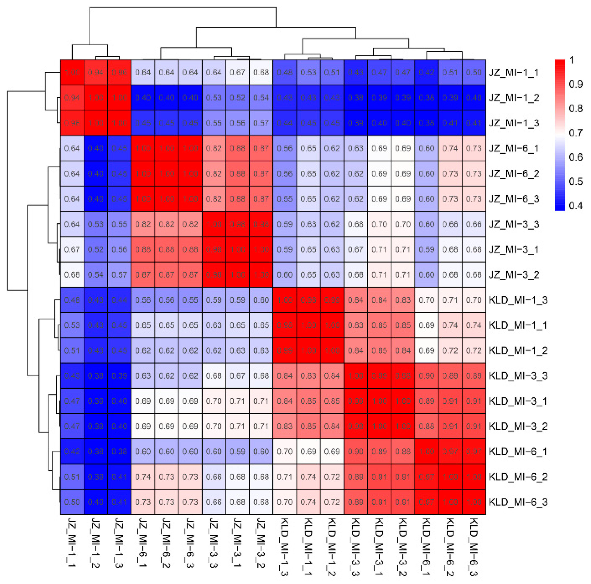
Due to the significant difference in the oil content between the fruits of KLD and JZ, we further evaluated the expression levels of oil biosynthesis genes in fruits of KLD and JZ based on full-length transcriptomics analysis of a total of 18 fruit samples of these two cultivars at three maturity stages (i.e., MI-1, MI-3, and MI-6) using Nanopore sequencing. A total of 82,065,919 raw reads were filtered to obtain a total of 71,192,844 clean reads with an average of 3,955,158 clean reads per sample (Table S2). The longest and the shortest reads were 368,449 bp and 904 bp in length, respectively. The maximum value of N50 was 1296 bp with an average value of 1173 bp in the 18 samples, strongly suggesting the continuity of the transcriptomics data. The proportions of full-length reads in the 18 samples ranged from 87.62% to 90.32%. The clean reads were mapped onto the olive reference genome with the mapping rates ranging from 97.17% to 98.32%, with an average of 97.97%. The results of principal component analysis (PCA) showed that these 18 samples were revealed in two groups, corresponding explicitly to cultivars JZ and KLD, respectively (Figure 2). A high correlation was revealed within each of the two cultivars (Figure 3). These results indicated that the transcriptomics data were of high quality and could be used for subsequent analyses.

3.3. Identification of Differentially Expressed Eenes in Olive Cultivars Kalinjot (JZ) and Coratina (KLD)

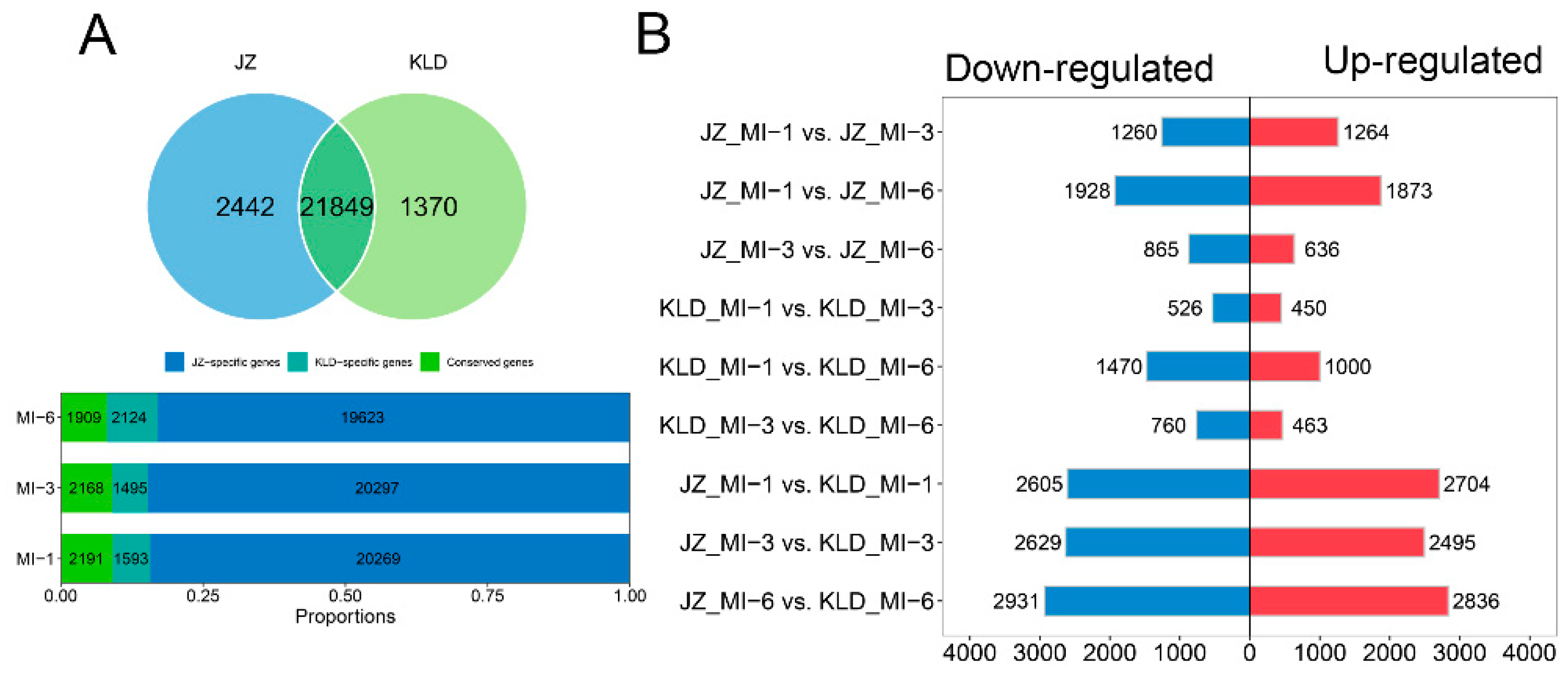
Based on the average CPM < 1, a total of 21,849 genes were identified in both JZ and KLD, with 2442 and 1370 genes exclusively expressed in JZ and KLD, respectively (Figure 4A). Generally, more genes were expressed in both JZ and KLD at the three maturity stages than those exclusively expressed in each of these two cultivars. For example, a total of 20,269 (37.87%) genes were expressed in both JZ and KLD at the MI-1 stage, whereas only a total of 2191 (4.09%) and 1593 (2.98%) genes were expressed uniquely in JZ and KLD, respectively. The same expression patterns were also observed in both MI-3 and MI-6 stages.
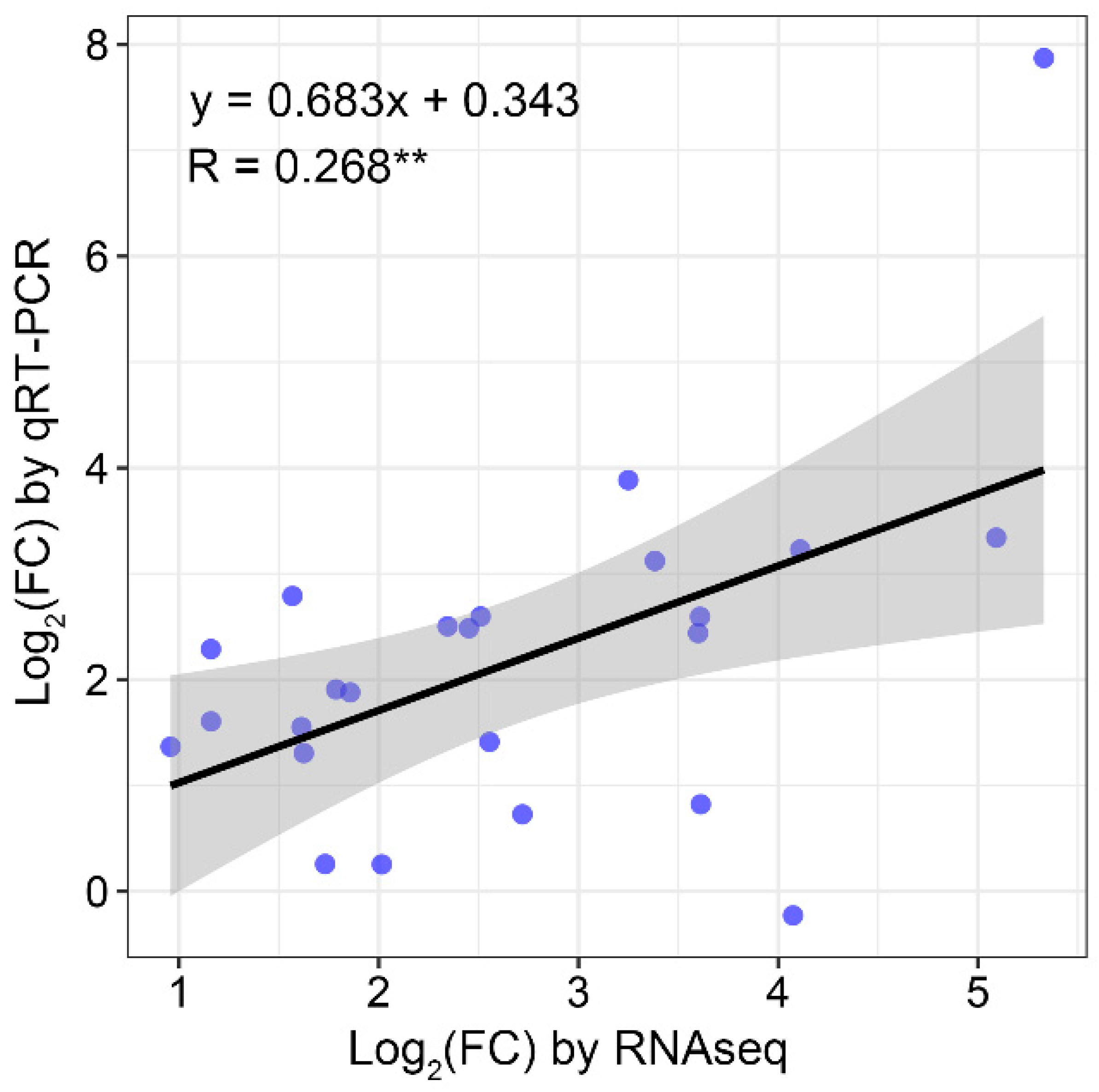
To explore the molecular variations between KLD and JZ at the transcriptional level, the DEGs in both cultivars were identified based on |log2 (fold change)| > 1 and FDR < 0.05 (Figure 4B; Table S3). The DEGs were first detected between different stages of the same cultivar. In the cultivar JZ, the highest number of DEGs (3801) was detected between MI-1 and MI-6 stages (1873 up-regulated and 1928 down-regulated), nearly two times more than the number of DEGs between MI-3 and MI-6 (636 up-regulated and 865 down-regulated). A total of 2524 DEGs were detected between MI-1 and MI-3 stages (1264 up-regulated and 1260 down-regulated). Similarly, in the cultivar KLD, the highest number of DEGs (1000 up-regulated and 1470 down-regulated) were observed between MI-1 and MI-6 stages, while similar numbers of DEGs were identified between MI-1 and MI-3 stages (450 up-regulated and 526 down-regulated) and between MI-3 and MI-6 stages (463 up-regulated and 760 down-regulated). The DEGs were also identified between these two cultivars at the same maturity stage. At the MI-1 stage, a total of 5309 DEGs (2704 up-regulated and 2605 down-regulated) were identified, while the highest number of DEGs (5767) were detected at the MI-6 stage. These results indicated that the variations between cultivars were greater than those among the developmental stages in the same cultivar. To assess the reliability and validity of the RNA-Seq data, a total of 25 DEGs were randomly selected for performing the qRT-PCR analysis. The results revealed a strong linearity between the expression patterns of these 25 genes detected by both RNA-Seq and qRT-PCR analyses (R = 0.268, p < 0.05; Figure 5), confirming the reliability of the RNA-Seq data and consistency between the RNA-Seq data and qRT-PCR analysis.

3.4. Functional Annotation of DEGs Based on Gene Ontology Database

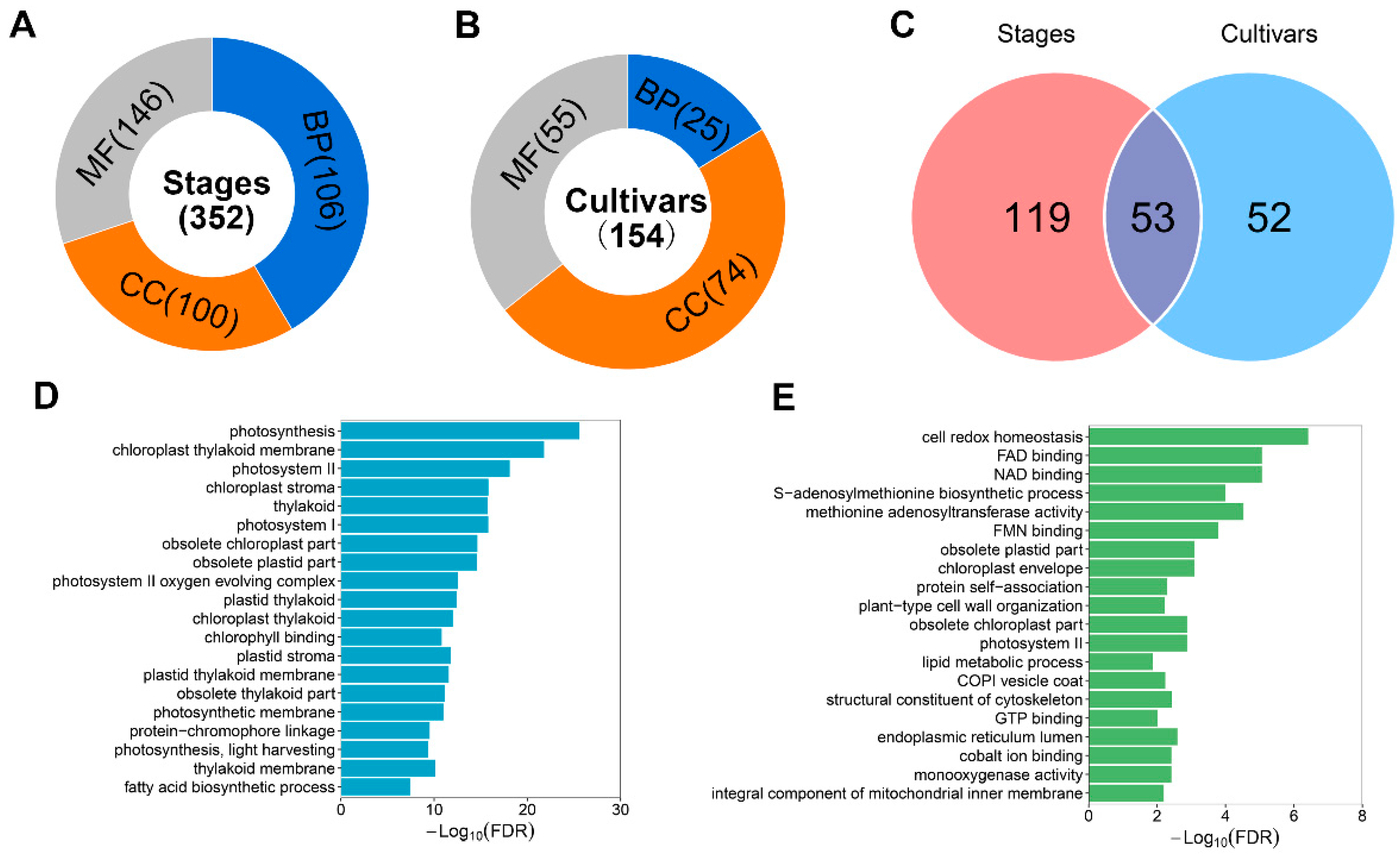
To identify the metabolic pathways involved in the oil biosynthesis in olive fruits, we further performed the GO annotation analysis based on the DEGs identified in both cultivars JZ and KLD. The results showed that based on the FDR < 0.05, a total of 352 GO terms were enriched in six pairwise comparisons among the three maturity stages, including 106, 100, and 146 in the three categories of GO terms, i.e., biological process (BP), cellular component (CC), and molecular function (MF), respectively (Figure 6A; Table S4). A total of 154 significantly enriched GO terms were identified between the two cultivars, including 25, 74, and 55 in the three categories of GO terms BP, CC, and MF, respectively (Figure 6B; Table S5). A total of 53 GO terms were shared between the maturity stages and the cultivars (Figure 6C). Among these shared GO terms, a total of 13 GO terms were found in the BP category, mainly including GO terms in energy metabolism, e.g., glycolytic process, carbon fixation, and terpenoid catabolic process, while in the CC category, the significantly enriched GO terms included photosystem I, photosystem II, chloroplast thylakoid, and chloroplast envelope, and in the MF category, it contained the GO terms of oxidoreductase activity, NAD binding, and FAD binding (Table S6).
The top 20 GO terms in the pairwise comparisons of maturity stages included photosynthesis, such as chloroplast thylakoid membrane, photosystem II, and photosynthetic membrane (Figure 6D), suggesting the importance of photosynthesis during the maturity stages of olive fruits. It was also observed that the fatty acid biosynthetic process involved in oil biosynthetic metabolism was significantly enriched. These results were consistent with the increased oil contents in both MI-3 and MI-6 stages (Figure 1B). Notably, the significant enrichment in photosynthesis was also revealed in the pairwise comparisons of olive cultivars (Figure 6E). Furthermore, the significant enrichment of lipid metabolic process evidently suggested that these DEGs were in volved in oil biosynthetic metabolism in olive fruits.

3.5. Functional Annotation and Enrichment Analysis of Conserved DEGs in Olive Cultivars Kalinjot (JZ) and Coratina (KLD)

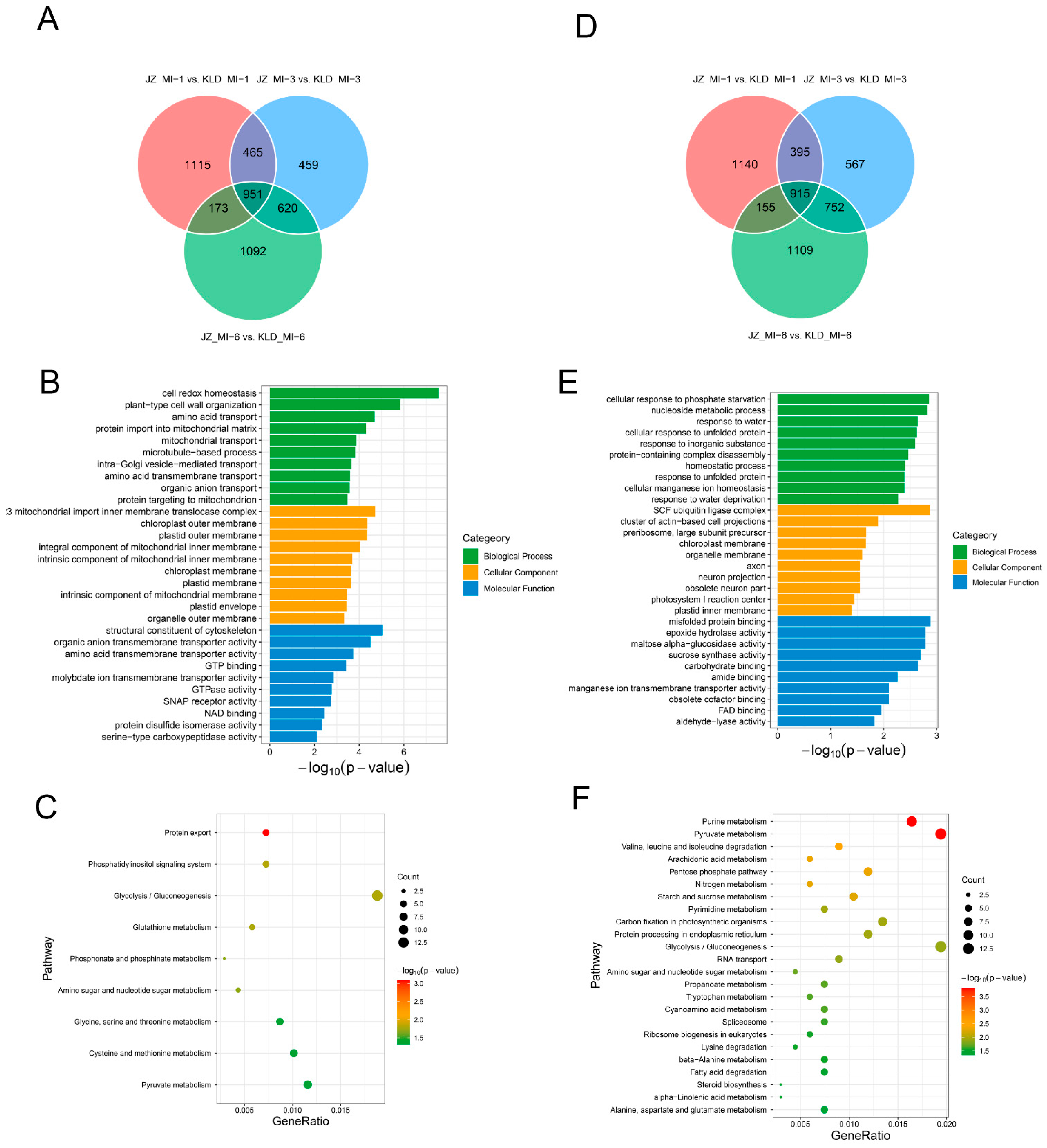
Due to the significant difference in the oil contents between the cultivars JZ and KLD, the DEGs were identified in the pairwise comparisons between the two cultivars at the same maturity stages. A total of 951 up-regulated DEGs were shared among the three pairwise comparisons (Figure 7A). The results of the GO annotation analysis showed that these DEGs were mainly enriched in cell redox homeostasis, plant-type cell wall, organization, amino acid transport, and protein import into mitochondrial matrix in the category BP of GO terms (Figure 7B). The results of the KEGG enrichment analysis showed that these conserved DEGs were mainly enriched in protein export and metabolism pathways, including glutathione metabolism, phosphonate and phosphinate metabolism, amino sugar and nucleotide sugar metabolism, and cysteine and methionine metabolism (Figure 7C). Furthermore, a total of 915 down-regulated DEGs conserved in the three pairwise comparisons between the two cultivars at the same maturity stages (Figure 7D) were significantly enriched in the process of response, such as response to water, response to inorganic substance, and response to unfolded protein based on the GO annotation analysis (Figure 7E), and were significantly enriched in the metabolic pathways of purine metabolism, pyruvate metabolism, nitrogen metabolism, and starch and sucrose metabolism based on the KEGG enrichment analysis (Figure 7F).

3.6. DEGs Involved in Oil Biosynthesis Pathway of Olive Cultivars Kalinjot (JZ) and Coratina (KLD) at Three Maturity Stages

A total of 520 DEGs were enriched in various oil biosynthetic pathways and metabolisms based on GO annotation, including fatty acid biosynthetic process (GO: 0006633), fatty acid metabolic process (GO: 0006631), and lipid biosynthetic process (GO: 0008610), and based on KEGG enrichment analysis, including fatty acid degradation (ko00071) and linoleic acid metabolism (ko00591). Previous studies have reported that a total of 87 genes were involved in fatty acid biosynthesis [27], with 68 shared with the 520 DEGs identified in our study. Therefore, a total of 539 genes with 170 (31.54%) differentially expressed in all pairwise comparisons during the three maturity stages of cultivars JZ and KLD were further investigated to reveal their molecular functions in oil biosynthesis in olive (Table S7). Overall, these results showed that these 170 DEGs played essential roles in the oil biosynthesis of olive fruits during the three maturity stages.
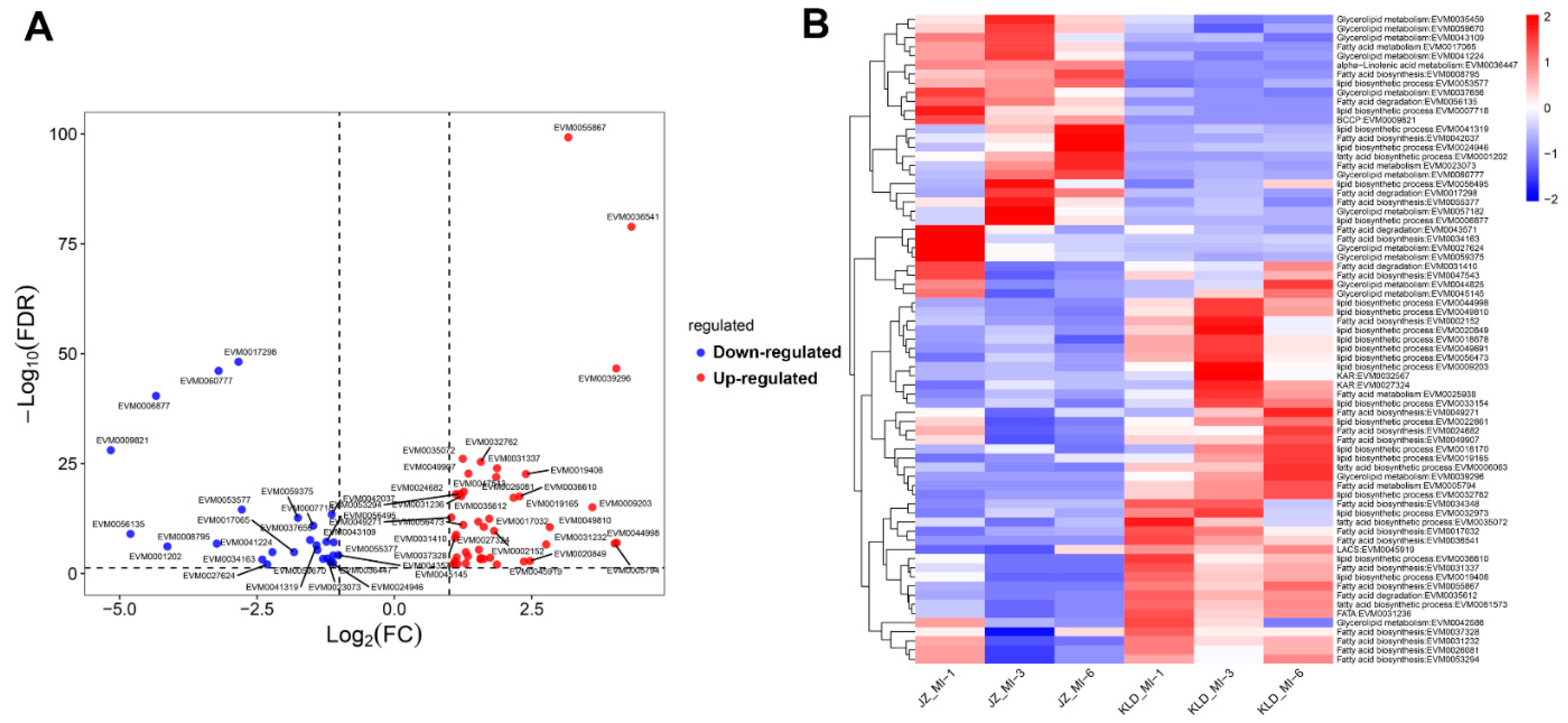
At the MI-1 stage, a total of 68 DEGs involved in oil biosynthesis were identified between the cultivars JZ and KLD with 35 up-regulated and 23 down-regulated (Figure 8A). A total of 17 down-regulated genes showed higher expression levels in JZ_MI-1than those in other samples (Figure 8B) with 4 genes (EVM0058966, EVM0027624, EVM0012991, and EVM59375) involved in glycerolipid metabolism, 2 genes (EVM0007718 and EVM0017592) in the lipid biosynthetic process, 2 genes (EVM0048444 and EVM0050078) in linoleic acid metabolism, 6 genes (EVM0003783, EVM0012415, EVM0035843, EVM0028970, EVM0034163, and EVM0061058) in fatty acid biosynthesis, 1 gene (EVM0018253) in fatty acid metabolism, and 2 genes (EVM0060777 and EVM002048) in fatty acid degradation. In particular, the fatty acid biosynthesis gene EVM0034163 was highly expressed with an average CPM value of 150.03, which was much higher than those of other samples (i.e., less than 7), while the 15 down-regulated genes were expressed at high levels in JZ fruits at three maturity stages, e.g., EVM0001202 involved in fatty acid biosynthetic process, EVM000219 in glycerolipid metabolism, and EVM0017065 in fatty acid metabolism, with most up-regulated genes showing high expression levels in KLD fruits without any maturity stage-specific expression pattern. It was noted that a total of 6 up-regulated genes in KLD_MI-1, including EVM0019165, EVM0039296, EVM0005794, EVM0032762, EVM0050882, and EVM0058699, reached their highest levels of expression at the MI-6 stage. For example, the expression of EVM0019165 involved in the lipid biosynthetic process (CPM = 23.35) was eight times higher than that in JZ_MI-1 (CPM = 3.67), while the CPM value in KLD_MI-6 was much higher (i.e., 54.09).
At the MI-3 stage, a total of 44 up-regulated and 27 down-regulated genes were identified between the JZ_MI-3 and KLD_MI-3 (Figure 9A) with 7 genes showing decreased expression levels by more than nine times, including EVM0009821 involved in biotin carboxyl carrier protein (BCCP), EVM0056135 in fatty acid degradation, EVM00687 in lipid biosynthetic process, EVM0001202 in fatty acid biosynthetic process, EVM0057182 in glycerolipid metabolism, EVM0008795 in fatty acid biosynthesis, and EVM0060777 in glycerolipid metabolism, and 7 genes showing up-regulated expression by more than nine times, including 2 fatty acid biosynthetic genes (EVM0055867 and EVM0036541), 2 lipid biosynthetic genes (EVM0009203 and EVM0044998), 1 ketoacyl-ACP reductase (KAR) gene (EVM0032567), 1 glycerolipid metabolism gene (EVM0039296), and 1 fatty acid metabolism gene (EVM0005794). The results showed that most up-regulated genes were expressed at high levels in KLD fruits compared to those in JZ fruits, indicating that these genes played important roles in the oil biosynthesis (Figure 9B). For example, the gene EVM0017032 involved in fatty acid biosynthesis was highly expressed in KLD with the CPM values of 44.20, 38.67, and 25.08 at the MI-1, MI-3, and MI-6 stages, respectively, whereas its expressions in JZ were significantly decreased with the CPM values of 6.21, 12.39, and 13.92 at the MI-1, MI-3, and MI-6 stages, respectively. Moreover, a total of 11 down-regulated genes were highly expressed at the MI-3 and MI-6 stages, suggesting their important roles in the late stages of oil biosynthesis. For example, the gene EVM0060777 involved in the glycerolipid metabolism was highly expressed with a CPM value of 81.42 and 94.41 at MI-3 and MI-6 stages in JZ, respectively, which were more than ten times higher than those of the other samples. Additionally, there were a total of 12 genes highly expressed in all three maturity stages in JZ, e.g., EVM0001202 involved in the fatty acid biosynthetic process, EVM0023073 in fatty acid metabolism, and EVM0060777 in glycerolipid metabolism.
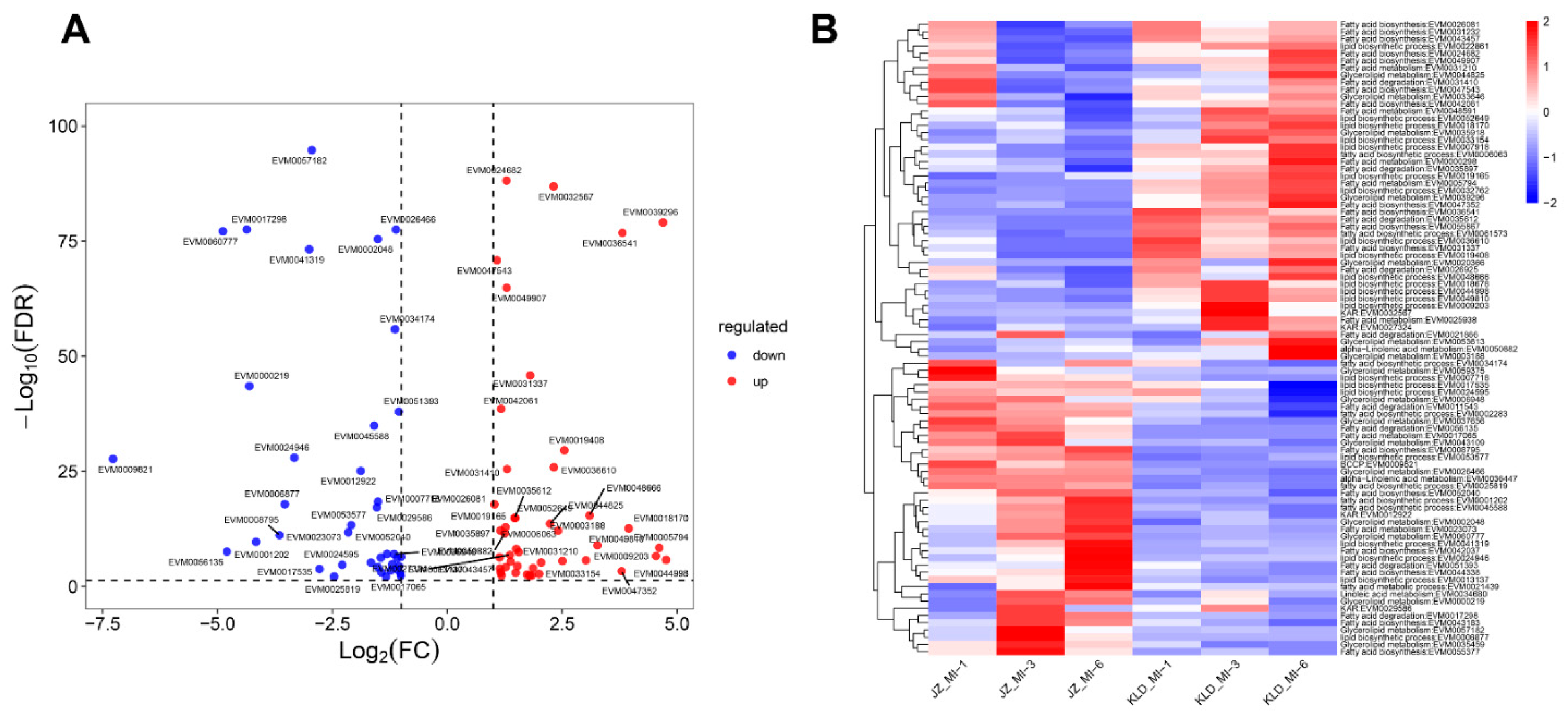
At the MI-6 stage, a total of 88 DEGs were identified between JZ_MI-6 and KLD_MI-6, with 47 up-regulated and 41 down-regulated (Figure 10A). A total of 11 genes were up-regulated more than three times, including 6 lipid biosynthetic genes (EVM0033154, EVM0048666, EVM0049810, EVM0018170, EVM0009203, and EVM0044998), 3 fatty acid biosynthetic genes (EVM005867, EVM0047352, and EVM0036541), 1 fatty acid metabolism gene (EVM005794), and 1 glycerolipid metabolism gene (EVM0039296), while a total of 10 genes were down-regulated more than nine times, i.e., 3 lipid biosynthetic genes (EVM0006877, EVM0024946, and EVM0041319), 2 fatty acid biosynthetic genes (EVM0001202 and EVM0008795), 2 fatty acid degradation genes (EVM0056135 and EVM0017298), and 2 glycerolipid metabolism genes (EVM0060777 and EVM0000219). Furthermore, results showed that most up-regulated genes were expressed with high levels in KLD at all three maturity stages (Figure 10B). For example, the gene EVM0032762 involved in the lipid biosynthetic process was highly expressed with an average CPM value of 1217.65 in all three stages in KLD fruits, which was three times higher than that (396.40) in JZ fruits. Similarly, the lipid biosynthetic gene EVM0009203 was highly expressed in all three stages in KLD but showing nearly no expression in JZ (CPM < 1). Moreover, a total of 32 (84.21%) out of 38 down-regulated genes showed a JZ-specific expression pattern, while the other 6 genes were highly expressed in KLD at both MI-1 and MI-3 stages. For example, the gene EVM0006498 was expressed with the CPM values of 45.54, 46.91, and 32.16 in JZ at the MI-1, MI-3 and MI-6 stages, respectively, while similar expression levels were revealed in KLD at MI-1 and MI-3 with the CPM values of 36.74 and 28.94, respectively, indicating the potential role of EVM0006498 in the late stages of oil biosynthesis.

3.7. Transcription Factors in Olive Fruits

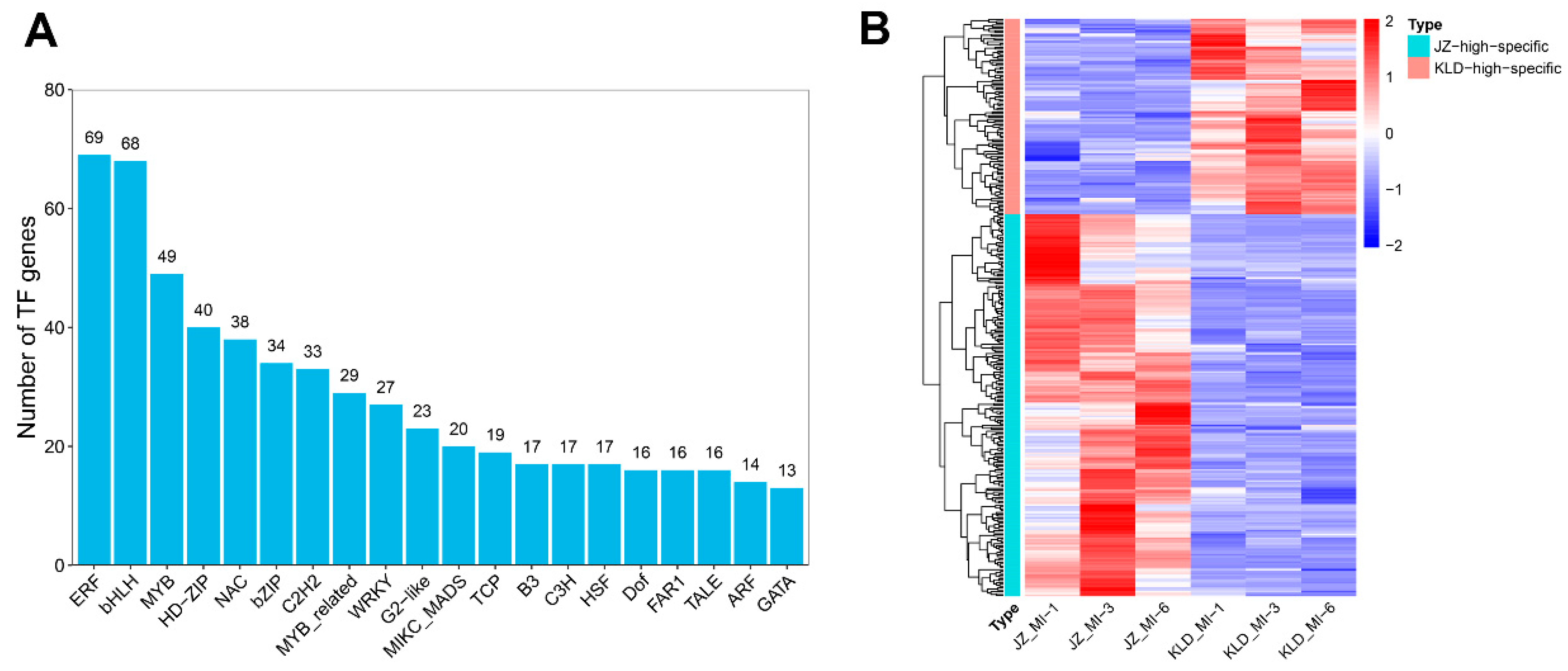
To investigate the molecular importance of TFs in oil biosynthesis, we identified the TF genes in olive cultivars JZ and KLD using the plantTFDB tool. The results showed that a total of 3587 TF genes were detected in both olive cultivars JZ and KLD, including 320 bHLH genes, 296 ERF genes, and 290 MYB genes, with a total of 714 DEGs identified in all pairwise comparisons (Table S8). The ERF and bHLH families were the most abundant, with 69 and 68 members, respectively, followed by MYB and HD-ZIP families with 49 and 40 members, respectively (Figure 11A). Furthermore, a total of 313 TFs showed cultivar-specific dominance in the three maturity stages, with 207 and 106 TFs in JZ and KLD, respectively (Figure 11B; Table S9). In JZ, the ERF family was the most abundant with 20 members, e.g., EVM0011341, EVM0022669, EVM0029736, EVM0057108, and EVM0058916, while the bHLH family contained the most member of 9 genes in KLD. It was noted that a few TF genes were expressed at extremely low levels. For example, the gene EVM0038491 of the B3 family was highly expressed with the CPM values of 4.33, 4.81, and 5.22 in KLD but was not expressed in JZ. Additionally, the TCP gene EVM0010783 was expressed specifically in JZ at three maturity stages, suggesting its important role in the oil biosynthesis in JZ. Overall, these varied expression patterns of TF genes suggested their involvement in the molecular regulations of the oil biosynthesis in the two olive cultivars with different mechanisms.

3.8. Co-Expression Network of Oil Biosynthesis Based on Transcription Factors and Their Binding Sites

It was well known that the co-expression network analysis is one of most popular methods to detect the co-expressed genes, which may share similar functions [39,40].To further investigate the molecular functions of TFs in the oil biosynthesis of olive, we further identified the transcription factor binding sites (TFBSs) related to oil biosynthesis. The results showed that a total of 461 TF genes were identified as the regulators of the oil biosynthesis in olive, including 80 ERF genes, 59 WRKY genes, 56 MYB genes, and 28 Dof TF genes (Table S10). For example, the Dof TF gene EVM0022747 targeted a total of 66 oil biosynthesis genes, such as EVM0024682, EVM0024682, and EVM0031210. A co-expression network of olive oil biosynthesis was constructed based on the TFs and their binding sites, consisting of a total of 383 edges (interactions) and 237 nodes (TFs and their target genes) (Figure 12). The core genes of the co-expression network contained three Dof genes, i.e., EVM0002986, EVM0049533, and EVM0007904. As a homologous gene of CDF5 (AT1G69570) in Arabidopsis thaliana, the gene EVM0002986 contained a total of 24 edges (10 positive and 14 negative interactions), while EVM0049533 and EVM0007904 contained 23 and 17 edges, respectively. Several oil biosynthesis genes were identified as the targets by multiple TFs. For example, EVM0056135 was identified as the target of a total of 15 TF genes, including 6 Dof genes, 8 WRKY genes, and 1 BBR-BPC gene, while EVM0002048 was targeted by a total of 11 TF genes, including 5 ERF genes, 2 MYB genes, and 4 TCP genes. Notably, several genes were independent from other genes. For instance, EVM0030047 was independently and positively associated with the HD-ZIP gene EVM0043314, and vice versa, while a total of 6 TF genes (4 bZIP genes and 2 bHLH genes) were independently positively interacted with EVM0019462. Overall, the co-expression network provided a comprehensive and enhanced understanding of the molecular mechanisms regulating the oil biosynthesis in olive based on TFs and their targets.

4. Discussion

Currently, the olive tree is an economically important oil crop with rapidly increasing consumption in America, Asia (mainly China and India), and Australia [11]. The oil accumulation in the olive fruit mesocarp is determined by many factors, such as cultivar types, climactic conditions, and biotic stresses [12,41]. However, the molecular mechanisms regulating the oil biosynthesis with varied oil contents in different olive cultivars are still unclear. Therefore, it is essential to identify the genes involved in oil biosynthesis and accumulation in different cultivars of olive. In this study, we performed a transcriptomics analysis with the fruits of two olive cultivars JZ and KLD in three maturity stages using Nanopore sequencing to identify the DEGs involved in the olive oil biosynthesis and accumulation. The molecular functions of these DEGs were further investigated using GO annotation and KEGG enrichment analysis. A co-expression network of olive oil biosynthesis was constructed based on the TFs and their target genes to explore the molecular mechanisms regulating the oil biosynthesis in olive.

4.1. Full-Length Nanopore Sequencing of Olive Cultivars Kalinjot (JZ) and Coratina (KLD)

It is well known that the full-length transcriptomics analysis based on Nanopore sequencing could facilitate the findings of novel genes and their expression profiles [42,43,44]. For example, the full-length transcriptomics analysis of Brassica napus investigated the gene expression and alternative splicing between the natural cultivar and resynthesized cultivar, demonstrating the evolution of transcription in allopolyploid B. napus [45]. Furthermore, the novel alternative polyadenylation (APA) events in transcripts were identified in rice based on the full-length transcriptomics analysis [46]. Our results of the transcriptomics analysis based on Nanopore sequencing revealed high reproducibility of multiple biological replicates in both olive cultivars JZ and KLD of three maturity stages, based on the full-length transcripts percentage and mapped rates of the clean reads (Table S2). A total of 21,849 genes were conservatively expressed among the two cultivars, with a relatively small number of genes showing cultivar-specific expressions (Figure 4A). In order to validate the accuracy and reliability of Nanopore sequencing data, a total of 25 DEGs were randomly selected for the qRT-PCR verification. The results revealed consistent gene expression patterns identified by both RNA-Seq and qRT-PCR analyses (Figure 5). This validation was also supported by the high mapping rates of the clean reads derived from the Nanopore sequencing onto the olive reference genome, indicating that the transcriptomics data were suitable for the further analysis of oil biosynthesis and accumulation in olive.

4.2. Identification of Genes Involved in Oil Biosynthesis of Olive

Oil biosynthesis is a generally complex metabolic network containing multiple biological processes in plants [47]. Among the three maturity stages of the olive fruits investigated in our study, the MI-1 stage generally indicates the beginning of harvest time based on the yellowish-green fruits of JZ and KLD. Studies have shown that during this stage, the energy and materials of olive fruits are involved in active physiological metabolism [48], while the green fruits could still photosynthesize to accumulate carbohydrates for further oil synthesis [49,50]. Furthermore, studies have shown that although the concentrations of sugars are decreased, the oil synthesis is still detected in olive fruits at the MI-1 stage [48]. Our results showed that the oil content of KLD was increased rapidly on a dry basis, indicating the importance of the MI-1 stage for oil synthesis in KLD (Figure 1B). However, the oil content of JZ fruits in the MI-1 stage was increased slightly (Figure 1B). Our results showed that a total of 68 DEGs involved in olive oil biosynthesis and metabolism were revealed to show a significant difference in their expression levels between JZ and KLD at the MI-1 stage. Among these 68 DEGs, the most up-regulated genes showed high expression levels in KLD at the three maturity stages, suggesting that these genes were responsible for the high oil content in KLD (Figure 8). Furthermore, a total of 6 reported KAR genes were significantly up-regulated, including EVM0029586, EVM0036349, EVM0017814, EVM0049914, EVM0027324, and EVM0032567. Moreover, the CPM values of a total of 14 genes, e.g., EVM0034348, EVM0040228, and EVM0035072, in KLD fruits were much higher than those of JZ, suggesting that these DEGs were responsible for the variations in the oil contents between KLD and JZ at the MI-1 stage.
The MI-3 is generally the peak stage of oil biosynthesis and accumulation in olive fruits. Studies reported that developmental stages around MI-3 appeared to be the most optimal harvest time to obtain high quality extra-virgin olive oil [51,52]. Our results showed that at the MI-3 stage, the oil accumulation in KLD reached the highest level, with the oil biosynthesis and oil content in JZ rapidly synthesized and accumulated (Figure 1B). These results indicated that the MI-3 stage was the most important stage for oil biosynthesis and metabolism in JZ, with a total of 7 and 7 genes up-regulated and down-regulated more than nine times, respectively, suggesting that these genes played important roles in oil biosynthesis in olive (Figure 9A). Furthermore, our results showed that most up-regulated genes expressed at high levels in KLD at the MI-3 stage were also detected at the MI-1 stage (Figure 8B and Figure 9B), suggesting that these genes were involved in the variations in oil content between KLD and JZ.
The MI-6 is the late stage of fruit ripening in olive. The quality of olive oil would be decreased due to the decreased contents of phenolic compounds, which were closely associated with the nutritional and sensory qualities of food [52]. The MI-6 stage is generally not considered as an appropriate time for harvesting due to the increased damage to the fruits through the handling process [25]. Our results showed that the oil content of KLD was slightly decreased in the MI-6 stage in comparison with that of the MI-3 stage (Figure 1B). These results were consistent with those reported previously [52]. Furthermore, the oil content of JZ reached its peak at the MI-6 stage (Figure 1B). During this stage, a total of 11 genes in KLD involved in oil biosynthesis and metabolism were identified with increased expressions by at least nine times more than those of JZ, including 6 lipid biosynthetic genes (EVM0033154, EVM0048666, EVM0049810, EVM0018170, EVM0009203, and EVM0044998), 3 fatty acid biosynthetic genes (EVM005867, EVM0047352, and EVM0036541), 1 fatty acid metabolism gene (EVM005794), and 1 glycerolipid metabolism gene (EVM0039296), suggesting that these 11 genes played important roles in the variations in oil content of JZ and KLD at the MI-6 stage. In particular, the CPM values of 10 genes in JZ were at least nine times higher than those of KLD, including 3 lipid biosynthetic genes (EVM0006877, EVM0024946, and EVM0041319), 2 fatty acid biosynthetic genes (EVM0001202 and EVM0008795), 2 fatty acid degradation genes (EVM0056135 and EVM0017298), and 2 glycerolipid metabolism genes (EVM0060777 and EVM0000219) (Figure 10A). Notably, the CPM value of gene EVM0039296 involved in fatty acid degradation in KLD fruits at the MI-6 stage was 28 times higher than that of JZ (Table S7). Moreover, based on the comparisons of CPM values at the MI-1 and MI-3 stages, the expression levels of these fatty acid degradation genes were greatly increased (Table S6), suggesting that the fruits of JZ or KLD should be harvested prior to the MI-6 stage in order to obtain the high level of oil contents (i.e., maintaining high quality of the oil product) without causing severe damage to the fruits due to the handling process. In addition, the CPM values of EVM0058699 gene in JZ at the MI-3 and MI-6 stages were 546 and 1729 in JZ, respectively, while the CPM values of this gene at the MI-3 and MI-6 stages were 909 and 2129 in KLD, respectively (Table S6). These results suggested that the EVM0058699 gene played an important role in the degradation of fatty acids in both JZ and KLD fruits.

4.3. Co-Expression Network of Oil Biosynthesis Based on Transcription Factors and Their Target Genes in Olive

Studies showed that the transcriptomics analysis of multiple samples at different developmental stages was an efficient strategy to construct the co-expression network [53,54]. The co-expression network analysis has been widely applied in various biological processes, e.g., plant development, stress response, and metabolism [55,56,57,58]. In our study, the co-expression network was constructed based on TFs and their target genes to investigate the molecular mechanisms regulating oil biosynthesis in olive fruits (R > 0.5; p < 0.05), containing a total of 383 edges (interactions) and 237 nodes (TFs and their target genes) as well as three Dof TF genes, i.e., EVM0002986, EVM0049533, and EVM0007904, identified as the core genes with the highest abundance of target genes. Studies have shown that a total of 91 AP2-EREBP TFs involved in α-linolenic acid synthesis are expressed in seeds of Perilla frutescens, suggesting the crucial role of the AP2-ERF family in the seed oil regulation [59]. Our study identified a total of 2 TFs in the AP2-ERF family in the co-expression network (Table S10), i.e., EVM0007215 and EVM0013234. In Arabidopsis, AtMYB96 was identified to promote seed oil biosynthesis by directly enhancing the accumulation of fatty acids, while the mutant atmyb96 accelerated oil mobilization in seeds [60,61]. In our study, a total of 8 homologous genes of MYB96 were identified in olive, including EVM0044338, EVM0048444, EVM0053577, EVM0044825, EVM0002048, EVM0039115, EVM0026081, and EVM0018170 (Table S10). Further studies are needed to explicitly investigate the functions of these TFs and their target genes in the molecular mechanisms regulating oil biosynthesis in olive fruits.

5. Conclusions

In this study, we performed the transcriptomic analysis of two cultivars of olive, i.e., JZ and KLD, with different oil contents at three maturity stages based on Nanopore sequencing to investigate the genetic and molecular mechanisms of olive oil biosynthesis. Significantly enriched metabolic pathways were identified based on both GO and KEGG databases, including photosynthesis, amino acid biosynthesis, response to stress, and energy metabolism, and in particular, fatty acid metabolism, involved in the oil biosynthesis of olive. A co-expression network based on the significantly expressed TFs and their targeted genes in oil biosynthesis was constructed to explore the molecular mechanism of oil biosynthesis in olive. Our study provided novel experimental evidence to support the further investigation of the molecular mechanisms of oil biosynthesis in olive fruits and to improve the molecular breeding of olive cultivars with enhanced oil contents.

Supplementary Materials

The following supporting information can be downloaded at: https://www.mdpi.com/article/10.3390/agronomy12112718/s1, Table S1: Primers and their sequences used for the quantitative real-time PCR (qRT-PCR) analysis. Table S2: Characteristics of transcriptomics data in olive cultivars Kalinjot (JZ) and Coratina (KLD) by Nanopore sequencing. Table S3: Differentially expressed genes (DEGs) identified among the nine pairwise comparisons of the olive cultivars Kalinjot (JZ) and Coratina (KLD) at three maturity stages (i.e., MI-1, MI-3, and MI-6). Table S4: Gene Ontology (GO) annotation based on differentially expressed genes (DEGs) derived from the pairwise comparisons between three maturity stages (i.e., MI-1, MI-3, and MI-6) in the same olive cultivars of Kalinjot (JZ) and Coratina (KLD). Table S5: Gene Ontology (GO) annotation based on differentially expressed genes (DEGs) derived from the pairwise comparisons between olive cultivars of Kalinjot (JZ) and Coratina (KLD) at three maturity stages (i.e., MI-1, MI-3, and MI-6). Table S6: Conserved Gene Ontology (GO) terms between the maturity stages (i.e., MI-1, MI-3, and MI-6) and olive cultivars Kalinjot (JZ) and Coratina (KLD). Table S7: Overview of the 539 predicted genes involved in the oil biosynthesis pathway in olive cultivars Kalinjot (JZ) and Coratina (KLD). Table S8: Overview of the 3587 transcription factors (TFs) involved in the oil biosynthesis in olive cultivars Kalinjot (JZ) and Coratina (KLD). Table S9: The 313 transcription factor (TF) genes detected in olive cultivars Kalinjot (JZ) and Coratina (KLD). Table S10: Transcription factor binding sites (TFBS) identified in olive cultivars Kalinjot (JZ) and Coratina (KLD) using PlantRegMap tool.

Author Contributions

Conceptualization, J.Q., Z.P. and C.D.; methodology, J.Q., Z.C., B.W.,S.F. and L.Z.; software, Z.T. and T.C.; validation, B.W., Z.T., T.C. and L.Z.; formal analysis, J.Q. and Z.C.; investigation, S.F. and L.Z.; resources, Z.P.; data curation, J.Q., Z.C. and B.W.; writing—original draft preparation, J.Q., Z.C., B.W. and Z.T.; writing—review and editing, C.D.; visualization, Z.C. and T.C.; supervision, Z.P. and C.D.; project administration, S.F.; funding acquisition, J.Q., Z.T., Z.P. and C.D. All authors have read and agreed to the published version of the manuscript.

Funding

This research was funded by the Sichuan Science and Technology Program, Sichuan Province, China (Grant numbers 2020YFH0207, 2022NSFSC0146, and 2020YFH0211) and the National Natural Science Foundation of China (Grant number 32060456). The APC was funded by J.Q.

Data Availability Statement

The transcriptomics data were deposited to the Sequence Read Archive (SRA) database of the National Center for Biotechnology Information (https://www.ncbi.nlm.nih.gov/sra/; accessed on 21 March 2022) with the accession PRJNA816306.

Acknowledgments

We are grateful to Liangshan Zhongze New Technology Development Co., Ltd. for providing fruit materials.

Conflicts of Interest

The authors declare no conflict of interest. The funders had no role in the design of the study; in the collection, analyses, or interpretation of data; in the writing of the manuscript; or in the decision to publish the results.

References

  1. Bervillé, A.; Breton, C. Genetic and environmental features for oil composition in olive varieties. OCL 2014, 21, D504. [Google Scholar] [CrossRef]
  2. Diez, C.M.; Trujillo, I.; Martinez-Urdiroz, N.; Barranco, D.; Rallo, L.; Marfil, P.; Gaut, B.S. Olive domestication and diversification in the Mediterranean Basin. New Phytol. 2015, 206, 436–447. [Google Scholar] [CrossRef] [PubMed]
  3. Servili, M.; Sordini, B.; Esposto, S.; Taticchi, A.; Urbani, S.; Sebastiani, L. Metabolomics of Olive Fruit: A Focus on the Secondary Metabolites. In The Olive Tree Genome; Rugini, E., Baldoni, L., Muleo, R., Sebastiani, L., Eds.; Springer International Publishing: Cham, Switzerland, 2016; pp. 123–139. [Google Scholar]
  4. Contreras, C.; Mariotti, R.; Mousavi, S.; Baldoni, L.; Guerrero, C.; Roka, L.; Cultrera, N.; Pierantozzi, P.; Maestri, D.; Gentili, L.; et al. Characterization and validation of olive FAD and SAD gene families: Expression analysis in different tissues and during fruit development. Mol. Biol. Rep. 2020, 47, 4345–4355. [Google Scholar] [CrossRef] [PubMed]
  5. Hernández, M.L.; Sicardo, M.D.; Alfonso, M.; Martínez-Rivas, J.M. Transcriptional Regulation of Stearoyl-Acyl Carrier Protein Desaturase Genes in Response to Abiotic Stresses Leads to Changes in the Unsaturated Fatty Acids Composition of Olive Mesocarp. Front. Plant Sci. 2019, 10, 251. [Google Scholar] [CrossRef] [Green Version]
  6. Servili, M.; Esposto, S.; Veneziani, G.; Urbani, S.; Taticchi, A.; Di Maio, I.; Selvaggini, R.; Sordini, B.; Montedoro, G. Improvement of bioactive phenol content in virgin olive oil with an olive-vegetation water concentrate produced by membrane treatment. Food Chem. 2011, 124, 1308–1315. [Google Scholar] [CrossRef]
  7. Inarejos-García, A.M.; Gómez-Rico, A.; Salvador, M.D.; Fregapane, G. Influence of malaxation conditions on virgin olive oil yield, overall quality and composition. Eur. Food Res. Technol. 2009, 228, 671–677. [Google Scholar] [CrossRef]
  8. Parvini, F.; Zeinanloo, A.A.; Ebrahimie, E.; Tahmasebi-Enferadi, S.; Hosseini-Mazinani, M. Differential expression of fatty acid desaturases in Mari and Shengeh olive cultivars during fruit development and ripening. Eur. J. Lipid Sci. Technol. 2015, 117, 523–531. [Google Scholar] [CrossRef]
  9. Hernández, M.L.; Mancha, M.; Martínez-Rivas, J.M. Molecular cloning and characterization of genes encoding two microsomal oleate desaturases (FAD2) from olive. Phytochemistry 2005, 66, 1417–1426. [Google Scholar] [CrossRef]
  10. Hernández, M.L.; Sicardo, M.D.; Martínez-Rivas, J.M. Differential Contribution of Endoplasmic Reticulum and Chloroplast ω-3 Fatty Acid Desaturase Genes to the Linolenic Acid Content of Olive (Olea europaea) Fruit. Plant Cell Physiol. 2016, 57, 138–151. [Google Scholar] [CrossRef] [Green Version]
  11. Unver, T.; Wu, Z.; Sterck, L.; Turktas, M.; Lohaus, R.; Li, Z.; Yang, M.; He, L.; Deng, T.; Escalante, F.J.; et al. Genome of wild olive and the evolution of oil biosynthesis. Proc. Natl. Acad. Sci. USA 2017, 114, E9413–E9422. [Google Scholar] [CrossRef]
  12. Nissim, Y.; Shlosberg, M.; Biton, I.; Many, Y.; Doron-Faigenboim, A.; Hovav, R.; Kerem, Z.; Avidan, B.; Ben-Ari, G. A High Temperature Environment Regulates the Olive Oil Biosynthesis Network. Plants 2020, 9, 1135. [Google Scholar] [CrossRef] [PubMed]
  13. Hernández, M.L.; Padilla, M.N.; Sicardo, M.D.; Mancha, M.; Martínez-Rivas, J.M. Effect of different environmental stresses on the expression of oleate desaturase genes and fatty acid composition in olive fruit. Phytochemistry 2011, 72, 178–187. [Google Scholar] [CrossRef] [PubMed]
  14. Deamer, D.; Akeson, M.; Branton, D. Three decades of nanopore sequencing. Nat. Biotechnol. 2016, 34, 518–524. [Google Scholar] [CrossRef]
  15. VanBuren, R.; Bryant, D.; Edger, P.P.; Tang, H.; Burgess, D.; Challabathula, D.; Spittle, K.; Hall, R.; Gu, J.; Lyons, E.; et al. Single-molecule sequencing of the desiccation-tolerant grass Oropetium thomaeum. Nature 2015, 527, 508–511. [Google Scholar] [CrossRef] [PubMed] [Green Version]
  16. Lan, T.; Renner, T.; Ibarra-Laclette, E.; Farr, K.M.; Chang, T.H.; Cervantes-Pérez, S.A.; Zheng, C.; Sankoff, D.; Tang, H.; Purbojati, R.W.; et al. Long-read sequencing uncovers the adaptive topography of a carnivorous plant genome. Proc. Natl. Acad. Sci. USA 2017, 114, E4435–E4441. [Google Scholar] [CrossRef] [Green Version]
  17. Chen, X.; Bracht, J.R.; Goldman, A.D.; Dolzhenko, E.; Clay, D.M.; Swart, E.C.; Perlman, D.H.; Doak, T.G.; Stuart, A.; Amemiya, C.T.; et al. The architecture of a scrambled genome reveals massive levels of genomic rearrangement during development. Cell 2014, 158, 1187–1198. [Google Scholar] [CrossRef] [PubMed] [Green Version]
  18. Karki, A.; Horvath, D.P.; Sutton, F. Induction of DREB2A pathway with repression of E2F, jasmonic acid biosynthetic and photosynthesis pathways in cold acclimation-specific freeze-resistant wheat crown. Funct. Integr. Genom. 2013, 13, 57–65. [Google Scholar] [CrossRef]
  19. Zhang, T.; Zhao, X.; Wang, W.; Pan, Y.; Huang, L.; Liu, X.; Zong, Y.; Zhu, L.; Yang, D.; Fu, B. Comparative transcriptome profiling of chilling stress responsiveness in two contrasting rice genotypes. PLoS ONE 2012, 7, e43274. [Google Scholar] [CrossRef] [Green Version]
  20. Xu, Z.; Peters, R.J.; Weirather, J.; Luo, H.; Liao, B.; Zhang, X.; Zhu, Y.; Ji, A.; Zhang, B.; Hu, S.; et al. Full-length transcriptome sequences and splice variants obtained by a combination of sequencing platforms applied to different root tissues of Salvia miltiorrhiza and tanshinone biosynthesis. Plant J. 2015, 82, 951–961. [Google Scholar] [CrossRef] [PubMed]
  21. Cui, G.; Chai, H.; Yin, H.; Yang, M.; Hu, G.; Guo, M.; Yi, R.; Zhang, P. Full-length transcriptome sequencing reveals the low-temperature-tolerance mechanism of Medicago falcata roots. BMC Plant Biol. 2019, 19, 575. [Google Scholar] [CrossRef] [PubMed]
  22. Briegas, B.; Corbacho, J.; Parra-Lobato, M.C.; Paredes, M.A.; Labrador, J.; Gallardo, M.; Gomez-Jimenez, M.C. Transcriptome and Hormone Analyses Revealed Insights into Hormonal and Vesicle Trafficking Regulation among Olea europaea Fruit Tissues in Late Development. Int. J. Mol. Sci. 2020, 21, 4819. [Google Scholar] [CrossRef] [PubMed]
  23. Rao, G.; Zhang, J.; Liu, X.; Li, X.; Wang, C. Combined Metabolome and Transcriptome Profiling Reveal Optimal Harvest Strategy Model Based on Different Production Purposes in Olive. Foods 2021, 10, 360. [Google Scholar] [CrossRef] [PubMed]
  24. Liu, X.; Guo, L.; Zhang, J.; Xue, L.; Luo, Y.; Rao, G. Integrated Analysis of Fatty Acid Metabolism and Transcriptome Involved in Olive Fruit Development to Improve Oil Composition. Forests 2021, 12, 1773. [Google Scholar] [CrossRef]
  25. Hassan, H.; El-Rahman, A.; Attia, M. Color Properties of Olive Fruits During Its Maturity Stages Using Image Analysis. AIP Conf. Proc. 2011, 1380, 101–106. [Google Scholar] [CrossRef]
  26. Quast, C.; Pruesse, E.; Yilmaz, P.; Gerken, J.; Schweer, T.; Yarza, P.; Peplies, J.; Glöckner, F.O. The SILVA ribosomal RNA gene database project: Improved data processing and web-based tools. Nucleic Acids Res. 2013, 41, D590–D596. [Google Scholar] [CrossRef]
  27. Rao, G.; Zhang, J.; Liu, X.; Lin, C.; Xin, H.; Xue, L.; Wang, C. De novo assembly of a new Olea europaea genome accession using nanopore sequencing. Hortic. Res. 2021, 8, 64. [Google Scholar] [CrossRef]
  28. Li, H. Minimap2: Pairwise alignment for nucleotide sequences. Bioinformatics 2018, 34, 3094–3100. [Google Scholar] [CrossRef] [Green Version]
  29. Love, M.I.; Huber, W.; Anders, S. Moderated estimation of fold change and dispersion for RNA-seq data with DESeq2. Genome Biol. 2014, 15, 550. [Google Scholar] [CrossRef] [Green Version]
  30. Ashburner, M.; Ball, C.A.; Blake, J.A.; Botstein, D.; Butler, H.; Cherry, J.M.; Davis, A.P.; Dolinski, K.; Dwight, S.S.; Eppig, J.T.; et al. Gene ontology: Tool for the unification of biology. The Gene Ontology Consortium. Nat. Genet. 2000, 25, 25–29. [Google Scholar] [CrossRef] [Green Version]
  31. Kanehisa, M.; Goto, S. KEGG: Kyoto encyclopedia of genes and genomes. Nucleic Acids Res. 2000, 28, 27–30. [Google Scholar] [CrossRef]
  32. Wu, T.; Hu, E.; Xu, S.; Chen, M.; Guo, P.; Dai, Z.; Feng, T.; Zhou, L.; Tang, W.; Zhan, L.; et al. clusterProfiler 4.0: A universal enrichment tool for interpreting omics data. Innovation 2021, 2, 100141. [Google Scholar] [CrossRef] [PubMed]
  33. Haynes, W. Benjamini–Hochberg Method. In Encyclopedia of Systems Biology; Dubitzky, W., Wolkenhauer, O., Cho, K.-H., Yokota, H., Eds.; Springer: New York, NY, USA, 2013; p. 78. [Google Scholar]
  34. Jin, J.; Tian, F.; Yang, D.C.; Meng, Y.Q.; Kong, L.; Luo, J.; Gao, G. PlantTFDB 4.0: Toward a central hub for transcription factors and regulatory interactions in plants. Nucleic Acids Res. 2017, 45, D1040–D1045. [Google Scholar] [CrossRef] [PubMed] [Green Version]
  35. Tian, F.; Yang, D.C.; Meng, Y.Q.; Jin, J.; Gao, G. PlantRegMap: Charting functional regulatory maps in plants. Nucleic Acids Res. 2020, 48, D1104–D1113. [Google Scholar] [CrossRef] [PubMed]
  36. Lyerly, S.B. The average spearman rank correlation coefficient. Psychometrika 1952, 17, 421–428. [Google Scholar] [CrossRef]
  37. Hernández, M.L.; Padilla, M.N.; Mancha, M.; Martínez-Rivas, J.M. Expression analysis identifies FAD2-2 as the olive oleate desaturase gene mainly responsible for the linoleic acid content in virgin olive oil. J. Agric. Food Chem. 2009, 57, 6199–6206. [Google Scholar] [CrossRef] [PubMed]
  38. Livak, K.J.; Schmittgen, T.D. Analysis of relative gene expression data using real-time quantitative PCR and the 2(-Delta Delta C(T)) Method. Methods 2001, 25, 402–408. [Google Scholar] [CrossRef]
  39. Rao, X.; Dixon, R.A. Co-expression networks for plant biology: Why and how. Acta Biochim. Biophys. Sin. 2019, 51, 981–988. [Google Scholar] [CrossRef]
  40. Zainal-Abidin, R.A.; Harun, S.; Vengatharajuloo, V.; Tamizi, A.A.; Samsulrizal, N.H. Gene Co-Expression Network Tools and Databases for Crop Improvement. Plants 2022, 11, 1625. [Google Scholar] [CrossRef]
  41. Rugini, E.; Cristofori, V.; Silvestri, C. Genetic improvement of olive (Olea europaea L.) by conventional and in vitro biotechnology methods. Biotechnol. Adv. 2016, 34, 687–696. [Google Scholar] [CrossRef]
  42. Zhang, C.; Ren, H.; Yao, X.; Wang, K.; Chang, J. Full-length transcriptome analysis of pecan (Carya illinoinensis) kernels. G3 2021, 11, jkab182. [Google Scholar] [CrossRef]
  43. Yan, H.; Zhou, H.; Luo, H.; Fan, Y.; Zhou, Z.; Chen, R.; Luo, T.; Li, X.; Liu, X.; Li, Y.; et al. Characterization of full-length transcriptome in Saccharum officinarum and molecular insights into tiller development. BMC Plant Biol. 2021, 21, 228. [Google Scholar] [CrossRef] [PubMed]
  44. Gao, X.; Guo, F.; Chen, Y.; Bai, G.; Liu, Y.; Jin, J.; Wang, Q. Full-length transcriptome analysis provides new insights into the early bolting occurrence in medicinal Angelica sinensis. Sci. Rep. 2021, 11, 13000. [Google Scholar] [CrossRef] [PubMed]
  45. Li, M.; Hu, M.; Xiao, Y.; Wu, X.; Wang, J. The activation of gene expression and alternative splicing in the formation and evolution of allopolyploid Brassica napus. Hortic. Res. 2022, 9, uhab075. [Google Scholar] [CrossRef] [PubMed]
  46. Zhang, G.; Sun, M.; Wang, J.; Lei, M.; Li, C.; Zhao, D.; Huang, J.; Li, W.; Li, S.; Li, J.; et al. PacBio full-length cDNA sequencing integrated with RNA-seq reads drastically improves the discovery of splicing transcripts in rice. Plant J. 2019, 97, 296–305. [Google Scholar] [CrossRef] [PubMed] [Green Version]
  47. Bates, P.D. Understanding the control of acyl flux through the lipid metabolic network of plant oil biosynthesis. Biochim. Biophys. Acta (BBA) Mol. Cell Biol. Lipids 2016, 1861, 1214–1225. [Google Scholar] [CrossRef] [PubMed]
  48. Kafkaletou, M.; Tsantili, E. Oil content and composition in relation to leaf photosynthesis, leaf sugars and fruit sugars in maturing Koroneiki olives—The mannitol effect on oil. J. Appl. Bot. Food Qual. 2016, 89, 1–10. [Google Scholar] [CrossRef]
  49. Proietti, P. Phtosynthesis and respiration in olive fruit. Acta Hortic. 1990, 286, 211–214. [Google Scholar] [CrossRef]
  50. Sánchez, J. Olive Oil Biogenesis. Contribution of Fruit photosynthesis. In Plant Lipid Metabolism; Kader, J.-C., Mazliak, P., Eds.; Springer: Dordrecht, The Netherlands, 1995; pp. 564–566. [Google Scholar]
  51. Bengana, M.; Bakhouche, A.; Lozano-Sánchez, J.; Youcef, A.; Youyou, A.; Segura Carretero, A.; Fernández-Gutiérrez, A. Influence of olive ripeness on chemical properties and phenolic composition of Chemlal extra-virgin olive oil. Food Res. Int. 2013, 54, 1868–1875. [Google Scholar] [CrossRef]
  52. Rotondi, A.; Bendini, A.; Cerretani, L.; Mari, M.; Lercker, G.; Toschi, T.G. Effect of olive ripening degree on the oxidative stability and organoleptic properties of cv. Nostrana di Brisighella extra virgin olive oil. J. Agric. Food Chem. 2004, 52, 3649–3654. [Google Scholar] [CrossRef]
  53. Hecker, M.; Lambeck, S.; Toepfer, S.; van Someren, E.; Guthke, R. Gene regulatory network inference: Data integration in dynamic models—A review. Biosystems 2009, 96, 86–103. [Google Scholar] [CrossRef]
  54. Haque, S.; Ahmad, J.S.; Clark, N.M.; Williams, C.M.; Sozzani, R. Computational prediction of gene regulatory networks in plant growth and development. Curr. Opin. Plant Biol. 2019, 47, 96–105. [Google Scholar] [CrossRef] [PubMed]
  55. García-Gómez, M.L.; Castillo-Jiménez, A.; Martínez-García, J.C.; Álvarez-Buylla, E.R. Multi-level gene regulatory network models to understand complex mechanisms underlying plant development. Curr. Opin. Plant Biol. 2020, 57, 171–179. [Google Scholar] [CrossRef] [PubMed]
  56. Huang, A.C.; Jiang, T.; Liu, Y.X.; Bai, Y.C.; Reed, J.; Qu, B.; Goossens, A.; Nützmann, H.W.; Bai, Y.; Osbourn, A. A specialized metabolic network selectively modulates Arabidopsis root microbiota. Science 2019, 364, eaau6389. [Google Scholar] [CrossRef] [PubMed]
  57. Wu, T.Y.; Goh, H.; Azodi, C.B.; Krishnamoorthi, S.; Liu, M.J.; Urano, D. Evolutionarily conserved hierarchical gene regulatory networks for plant salt stress response. Nat. Plants 2021, 7, 787–799. [Google Scholar] [CrossRef]
  58. Ueda, Y.; Ohtsuki, N.; Kadota, K.; Tezuka, A.; Nagano, A.J.; Kadowaki, T.; Kim, Y.; Miyao, M.; Yanagisawa, S. Gene regulatory network and its constituent transcription factors that control nitrogen-deficiency responses in rice. New Phytol. 2020, 227, 1434–1452. [Google Scholar] [CrossRef] [PubMed]
  59. Zhang, T.; Song, C.; Song, L.; Shang, Z.; Yang, S.; Zhang, D.; Sun, W.; Shen, Q.; Zhao, D. RNA Sequencing and Coexpression Analysis Reveal Key Genes Involved in α-Linolenic Acid Biosynthesis in Perilla frutescens Seed. Int. J. Mol. Sci. 2017, 18, 2433. [Google Scholar] [CrossRef] [Green Version]
  60. Lee, H.G.; Park, B.-Y.; Kim, H.U.; Seo, P.J. MYB96 stimulates C18 fatty acid elongation in Arabidopsis seeds. Plant Biotechnol. Rep. 2015, 9, 161–166. [Google Scholar] [CrossRef]
  61. Lee, K.; Lee, H.G.; Yoon, S.; Kim, H.U.; Seo, P.J. The Arabidopsis MYB96 Transcription Factor Is a Positive Regulator of ABSCISIC ACID-INSENSITIVE4 in the Control of Seed Germination. Plant Physiol. 2015, 168, 677–689. [Google Scholar] [CrossRef]
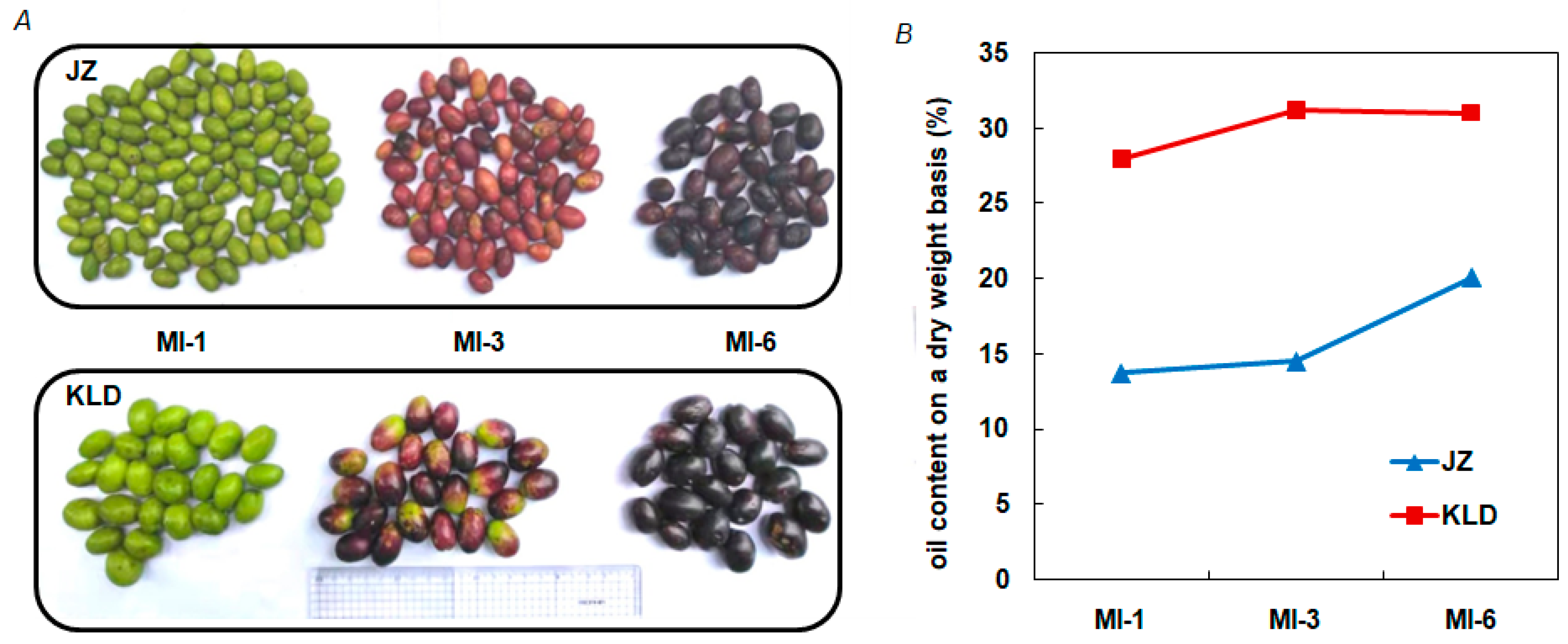
Figure 1. The phenotypes (A) and oil contents (B) of olive cultivars Kalinjot (JZ) and Coratina (KLD) in three maturity stages (i.e., MI-1, MI-3, and MI-6).
Agronomy 12 02718 g001
Figure 2. Plot of the principal component analysis (PCA) based on PC1 vs PC2 of 18 samples of olive cultivars Kalinjot (JZ) and Coratina (KLD) at three maturity stages (i.e., MI-1, MI-3, and MI-6) used in the transcriptomics analysis based on Nanopore sequencing.
Agronomy 12 02718 g002
Figure 3. The heatmap of correlation coefficients of 18 samples of olive cultivars Kalinjot (JZ) and Coratina (KLD) at three maturity stages (i.e., MI-1, MI-3, and MI-6) used in the transcriptomics analysis based on Nanopore sequencing.
Agronomy 12 02718 g003
Figure 4. The overview of the full-length transcriptomics analysis identifying the genes (A) and differentially expressed genes (DEGs) (B) in olive cultivars Kalinjot (JZ) and Coratina (KLD) at three maturity stages (i.e., MI-1, MI-3, and MI-6).
Agronomy 12 02718 g004
Figure 5. The correlation between the expression levels of 25 genes (represented by blue dots) based on RNA-Seq and qRT-PCR analyses. The expression levels of these genes based on both RNA-Seq and qRT-PCR analyses are fitted with the linear regression with the significance level set to p < 0.01 (**).
Agronomy 12 02718 g005
Figure 6. Functional annotations of the differentially expressed genes (DEGs) identified in the pairwise comparisons of maturity stages and cultivars Kalinjot (JZ) and Coratina (KLD)based on Gene Ontology (GO) database. (A) The number of GO terms enriched in the three categories of GO terms, i.e., biological process (BP), cellular components (CC), and molecular functions (MF), identified in the pairwise comparisons of maturity stages, including MI-1 vs. MI-3, MI-1 vs. MI-6, and MI-3 vs. MI-6 in JZ and KLD, respectively. (B) The number of GO terms enriched in the three categories of GO terms identified in the pairwise comparisons of cultivars JZ and KLD, including JZ_MI-1 vs. KLD_MI-1, JZ_MI-3 vs. KLD_MI-3, and JZ_MI-6 vs. KLD_MI-6, respectively. (C) The GO terms shared between the maturity stages and olive cultivars. (D) Top 20 GO terms based on the maturity stages of the two cultivars JZ and KLD. (E) Top 20 GO terms based on the olive cultivars JZ and KLD.
Agronomy 12 02718 g006
Figure 7. Functional annotation and enrichment analysis of differentially expressed genes (DEGs) between cultivars Kalinjot (JZ) and Coratina (KLD) at three maturity stages. (A,D) DEGs identified in the three pairwise comparisons between cultivars JZ and KLD at three maturity stages, respectively. (B,E) Top 30 GO terms enriched in three categories (i.e., biological process, cellular component, and molecular function) of the GO database by conserved DEGs identified in the three pairwise comparisons between cultivars JZ and KLD at three maturity stages, respectively. (C,F) KEGG metabolic pathways enriched by the conserved DEGs identified in the three pairwise comparisons between cultivars JZ and KLD at three maturity stages, respectively. The size of dots indicates proportionally the number of genes enriched.
Agronomy 12 02718 g007
Figure 8. Differentially expressed genes (DEGs) involved in oil biosynthesis of olive cultivars Kalinjot (JZ) and Coratina (KLD) at the MI−1 stage shown in the volcano plot (A) and the heatmap (B). The red and blue dots represent up-regulated and down-regulated genes, respectively. The DEGs are identified based on threshold |log2(fold change)| > 1 and FDR < 0.05, delimiting the dotted lines.
Agronomy 12 02718 g008
Figure 9. Differentially expressed genes (DEGs) involved in oil biosynthesis of olive cultivars Kalinjot (JZ) and Coratina (KLD) at the MI−3 stage presented in the volcano plot (A) and the heatmap (B). The red and blue dots represent up-regulated and down-regulated genes, respectively. The DEGs are identified based on threshold |log2(fold change)| > 1 and FDR < 0.05, delimiting the dotted lines.
Agronomy 12 02718 g009
Figure 10. Differentially expressed genes (DEGs) involved in oil biosynthesis of olive cultivars Kalinjot (JZ) and Coratina (KLD) at the MI−6 stage presented in the volcano plot (A) and the heatmap (B). The red and blue dots represent up-regulated and down-regulated genes, respectively. The DEGs are identified based on threshold |log2(fold change)| > 1 and FDR < 0.05, delimiting the dotted lines.
Agronomy 12 02718 g010
Figure 11. Transcription factors (TFs) and their expressions in JZ and KLD. (A) TF families with the number of predicted TFs genes. (B) The heatmap of differentially expressed TFs in JZ and KLD at different maturity stages.
Agronomy 12 02718 g011
Figure 12. The co-expression network of olive oil biosynthesis constructed based on transcription factors (TFs) and their target genes involved inolive oil biosynthesis. The width of each line represents proportionally the correlation coefficient. The size of each dot indicates proportionally the number of interactions.
Agronomy 12 02718 g012
Publisher’s Note: MDPI stays neutral with regard to jurisdictional claims in published maps and institutional affiliations.