Article

Optimising Sample Preparation and Calibrations in EDXRF for Quantitative Soil Analysis

1
Institute for Global Food Security, School of Biological Sciences, Queen’s University Belfast, Belfast BT9 5DL, UK
2
Environment, Soils and Land Use Department, Teagasc Johnstown Castle Research Centre, Y35 TC97 Wexford, Ireland
3
International Network for Environment and Health, School of Geography and Archaeology, NUI Galway, H91 TK33 Galway, Ireland
*
Author to whom correspondence should be addressed.
Agronomy 2020, 10(9), 1309; https://doi.org/10.3390/agronomy10091309
Submission received: 17 July 2020 / Revised: 28 August 2020 / Accepted: 29 August 2020 / Published: 2 September 2020
(This article belongs to the Special Issue Remediation of Contaminated Soil for Food Security)

Abstract

:
Energy-dispersive X-ray fluorescence spectrometry (EDXRF) is a rapid and inexpensive method for soil analysis; however, analytical results are influenced by particle size effects and spectral interferences. The objective of this study was to optimise sample preparation and calibrations to improve the accuracy of EDXRF for soil tests. Methods of sample preparation were compared by calculating the recoveries of 13 elements in four International Soil-Analytical Exchange (ISE) standards prepared as loose powder (LP), pressed pellet (PP), and pressed pellet with wax binder (PPB). A matching library (ML) was created and evaluated against the fundamental parameters (FP) calibration using 20 ISE standards. Additionally, EDXRF results of 41 tillage soils were compared with Inductively coupled plasma optical emission spectrometry (ICP-OES) results. The PPB had most recoveries within the acceptable range of 80–120%; conversely, PP yielded the poorest element recoveries. For the calibration, the ML provided better recoveries of Ni, Pb, Cu, Mg, S, P, and Cr; however, for Zn, and Mn, it had the opposite effect. Furthermore, EDXRF results compared with ICP-OES separated by soil texture class for Al, K, Mn, and Fe. In conclusion, the EDXRF is suitable for quantifying both trace elements and macronutrients in contaminated soils and has the potential to provide screening or prediction of soil texture in agriculture.

1. Introduction

Traditional wet chemistry methods for quantifying trace elements and macronutrients in soil [1,2,3] are expensive and labour-/time-intensive [4,5]. Reliant on two principal digestion schemes, both approaches have their flaws. Firstly, there is hydrofluoric (HF) acid digestion, which completely liberates silica bound elements in soil to obtain the total concentration, known as ‘real totals’ [6]. However, due to the toxicity of HF, an alternative method, aqua regia digestion [7] is used more commonly for routine analysis [6,8,9]. Aqua regia, though, provides only partial digestion and is thus referred to as a ‘pseudo’ total [5,10,11].
Alternatively, Energy-dispersive X-ray fluorescence (EDXRF) spectrometry provides total element concentrations and is effective for rapid and non-destructive multi-element analysis of numerous environmental samples, including soils [12,13,14,15]. Moreover, this technique requires minimal sample preparation and is available in portable forms, making it suitable for on-site analysis [14,15,16,17,18]. EDXRF instruments themselves have become increasingly more sensitive, stable, and affordable. However, errors in the measurement can still arise due to problems during sample preparation, namely surface irregularity, moisture, and sample inhomogeneity [19,20,21]. This can lead to a reduction in performance and that is why for many applications EDXRF is used principally as a screening/qualitative tool [22].
A further confounding issue is matrix effects, which can result in spectral interferences. These problems arise because the easiest and most widely used mode of analysis for EDXRF is a standardless, iterative concentration calibration called Fundamental Parameters (FP). These measurement errors, though, can be corrected by the development of an empirical/matrix-matched calibration [23]. However, empirical standards must be site-specific, certified using wet chemistry methods, and require a minimum of 10 soil standards for the calibration to be accurate [13,22,24]. For specialist applications, the time and costs associated with these external calibrations are warranted, but for more general agronomic soil testing the set-up is not practical. However, a compromise can be achieved by using an alternative calibration, termed a matching library (ML), that requires fewer standards [25].
An ML-like empirical calibration assumes that a linear relationship exists between the net intensity of peaks for an analyte and the concentration within a sample, i.e., holistically integrating any matrix effects into the measurement [26]. Furthermore, if commercial certified reference materials (CRMs) are used as ML standards, it is relatively easy for the optimised protocols to be replicated by other users/laboratories. Therefore, the objective of this study was to optimise sample preparation and to develop a practical/rapid/reproducible soil calibration for EDXRF as a quantitative soil analysis tool, evaluating this across a range of representative soils varying in element composition and texture class. Enhancing the analytical performance of EDXRF has extensive benefits, such as improving the speed and ease of validating soil screening methods [22], and its potential use in precision farming management.

2. Materials and Methods

2.1. Sample Archives

This study used three sets of soil archives consisting of 24 international soil-analytical exchange (ISE) samples, 4 CRMs, and 41 tillage soils from the study by Metzger et al. [27]. The ISE samples were from the Wageningen Evaluating Programs for Analytical Laboratories (WEPAL) to test laboratory proficiency across the world. The program sends samples to participating labs to compare results across different labs in different countries, and Teagasc takes part in this program and receives samples each year. The WEPAL soil samples in this study were not artificially/technically generated. The ISE samples were received as air-dried, ball-milled, homogenous soils with different concentrations of elements ranging from low to high. A summary of the distribution of each archive is shown in Figure 1.
The four CRMS were obtained from the National Research Centre for Certified Reference Materials, Beijing, China (n = 2), WEPAL, Netherlands (n = 1), and the Federal Institute for Materials Research and Testing, Germany (n = 1). The 41 tillage soils were obtained from tillage farms across the South and East of the Republic of Ireland.

2.1.1. ISE Samples for the Comparison of XRF Sample Preparation Methods and Calibrations

A total of 24 ISE used for this study were divided into two sets; four samples were kept to examine the effect of different sample preparations, and the remaining 20 samples were used to compare our XRF-determined values with those reported by WEPAL. The ISE selected were described by soil type as: clay (n = 8), sandy soil (n = 7), sandy clay (n = 1) calcareous soil (n = 2), Löss (n = 1), riverclay (n = 1), alluvium (n = 1), marshland (n = 1), Braunerde-pseudoclay (n = 1) and calcareous brown soil (n = 1), a summary of the concentration range of elements is shown in Table 1. For this study, only ISE samples with ‘real totals’ were used. The final results of the ‘real totals’ for each element reported by the participating labs were calculated and represented as a normal distribution approximation (NDA) mean by WEPAL.
The NDA mean and standard deviation indicated by WEPAL for the ‘real totals’ was considered the true value of the ISE samples for comparison and calculating the recoveries of the elements in this study. ‘Real totals’ as reported by WEPAL are determined by a range of procedures, namely: total acid digestion with HF and final medium such as HCl, H2SO4, HNO3 and HCIO4; total analysis with mixtures of acids (HNO3+conc HCl or conc.H2SO4+conc. HNO3), followed by ICP-AES, ICP-MS; real total neutron activation analysis followed by neutron activation analysis; and samples prepared as glass beads or pressed pellet followed by XRF determination. In this study, EDXRF was used to determine the element concentration of 20 ISE samples, and values were calculated as % recoveries relative to those reported by WEPAL for the same samples. The % recovery was calculated using the formula (EDXRF results/ISE NDA mean) * 100).

2.1.2. Samples for Matching Library Creation

Four CRMs were acquired to use as a matching library. These samples were used to correct or adjust XRF values determined on the ISE samples (n = 20) derived from the standardless fundamental parameters method (FP). The CRMs used in this study were BAM-U110, ISE 921, GBW07403, and GBW07405, a summary of the elemental concentration of the CRMs is shown in Appendix A, Table A1. The BAM-U110 is a contaminated soil, and thus was selected for the high concentrations of trace elements, while the GBW07403 was selected to represent low concentrations. A total of 13 elements made up the ML calibration, and Si was added as a balance compound for non-measuring components to enable the algorithms on the EDXRF to make correct calculations of element concentrations. For uniformity in the data, the element concentrations listed in the Certificate of Analysis for elements such as Si, Fe, Al, K, Mg, Mn, and P were used instead of the oxide concentration.

2.1.3. Assessing Impact of Soil Texture—Irish Tillage Soils Archive

A set of 41 tillage soil samples consisting of four texture classes—sandy loam (n = 16), clay loam (n = 6), loam (n = 18), sandy clay loam (n = 1)—was sampled from farms across the South and East of the Republic of Ireland as described and reported by Metzger et al. [27]. The samples were taken at a depth of 5–10 cm, after which they were dried at 40 °C and sieved to 2 mm. The particle size distribution was determined using the sieve-pipette method [28].

2.2. Sample Preparation for EDXRF Analysis

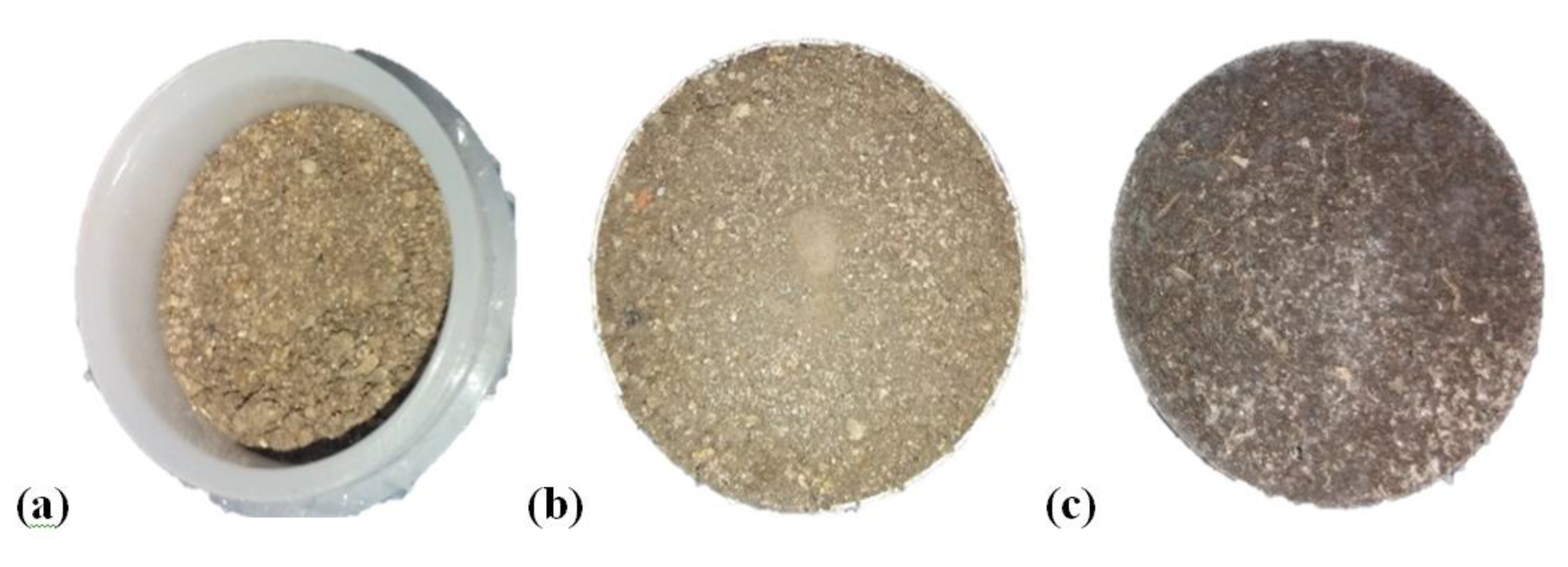
Of the four ISE samples, two were clay soil samples (ISE 952 and 961), one was sandy clay (ISE 992), and one was described as sand (ISE 995). The sample preparations for EDXRF analysis in this study included loose powder (LP), pressed powder pellet (PP), and pressed powder pellet with binder (PPB). Each sample preparation had four replicates. Preparing samples as LP involved 8 g of soil, weighed out into an XRF cup (32 mm double open-ended plastic rings with 4 µm thick propylene X-ray film at the base) [29,30]. A low pressure of 200 inch lbs was placed on the sample for 30 s, after which the height was measured and recorded. For PP, 8 g of soil in an XRF aluminium cup was placed in a die set, after which 15 tons of pressure was placed on the die set for 30 s using a Specac manual hydraulic press [31]. Samples prepared as PPB were prepared by weighing out 8 g of soil into a Fluxana mixing container with a polyamide ball, after which a Fluxana cereox binder (wax) was added at a ratio of 0.25 to the soil. The sample was mixed at high speed for 60 s with a Fluxana MU-K Mixer, and 20 tons of pressure was added to create the pellet. A visual display of the different sample preparation is shown in Appendix A, Figure A1.

2.3. EDXRF Analysis

The elemental composition of all samples was analysed with a Rigaku NEX CG benchtop EDXRF. The instrument has a nine-place sample changer with a spin function and is fitted with a Pd X-ray tube for the production of X-rays and a silicon drift detector. Additionally, the instrument uses secondary targets in Cartesian Geometry for indirect excitation to improve the sensitivity of trace element analysis by reducing the background intensity. The secondary targets used for this study were Mo, Cu, Rx9, and Si. All samples for the sample preparation study were analysed with the FP [24,32,33]. Operationally the Rigaku EDXRF system allows the creation of ‘alpha corrections’ or matching libraries to correct FP-derived analytical results. In addition, all PP and PPB samples were run under vacuum, and the LP under helium to improve light element sensitivity [34,35]. A summary of the instrument settings for the analysis of samples prepared as PP and PPB are shown in Appendix A, Table A2. To ascertain the reliability of the quantitative results from the EDXRF, a library calibration using pure elements is run weekly to check for instrumental drift. The drift monitor samples used for this study were Cu, Sn, SiO2, and a Multi-channel analyser (MCA), all supplied by the manufacturer. Additionally, the MCA was run daily before the start of any analysis. In addition, CRMs are added to every set of soil samples analysed on the EDXRF to check the accuracy of the analytical results. Furthermore, the EDXRF limit of detection (LOD) was calculated as described by Kadachi and Al-Eshaikh [36] and Scott Rees (Rigaku Tech support) [37] using the formula below:
L O D = 3 s I b T
where s = sensitivity in counts per ppm; Ib = background intensity; and T = counting time in seconds
A summary of the average LODs for the different elements is shown in Table 1.

2.4. ICP-OES Analysis

A reverse aqua regia digestion was used for the extraction of elements for the tillage soils. This involved adding 2.5 mL hydrochloric acid, 7.5 mL nitric acid and 2 mL distilled water to 0.5 g of soil followed by microwave digestion [7,38]. Digested samples were filtered with a Whatman 42 filter paper, after which they were analysed with an Agilent 5100 inductively coupled plasma optical emission spectrometer (ICP-OES).

2.5. Data Analysis

All statistics were analysed with SigmaPlot v14. A one-way ANOVA was performed to determine the significance of the difference between the EDXRF sample preparation methods for element recoveries. Prior to the analyses, tests for normality and equal variance were performed on the element recoveries using the Shapiro–Wilk and Brown–Forsythe tests, respectively. For the data that failed the equal variance test, a Kruskal–Wallis analysis was made on ranks. All pairwise multiple comparisons were made with the Holm–Sidak method. A summary of the statistical analysis is shown in Supplementary Table S1. Agreement between the ICP-OES and the EDXRF data was compared using the Bland–Altman [39,40,41] function in SigmaPlot v14. Additionally, the concordance correlation coefficient which measures agreement between methods [42] was computed using SAS 9.4. Summary data are available in Supplementary Table S2.

3. Results and Discussion

3.1. Effects of Sample Preparation on EDXRF Analytical Results

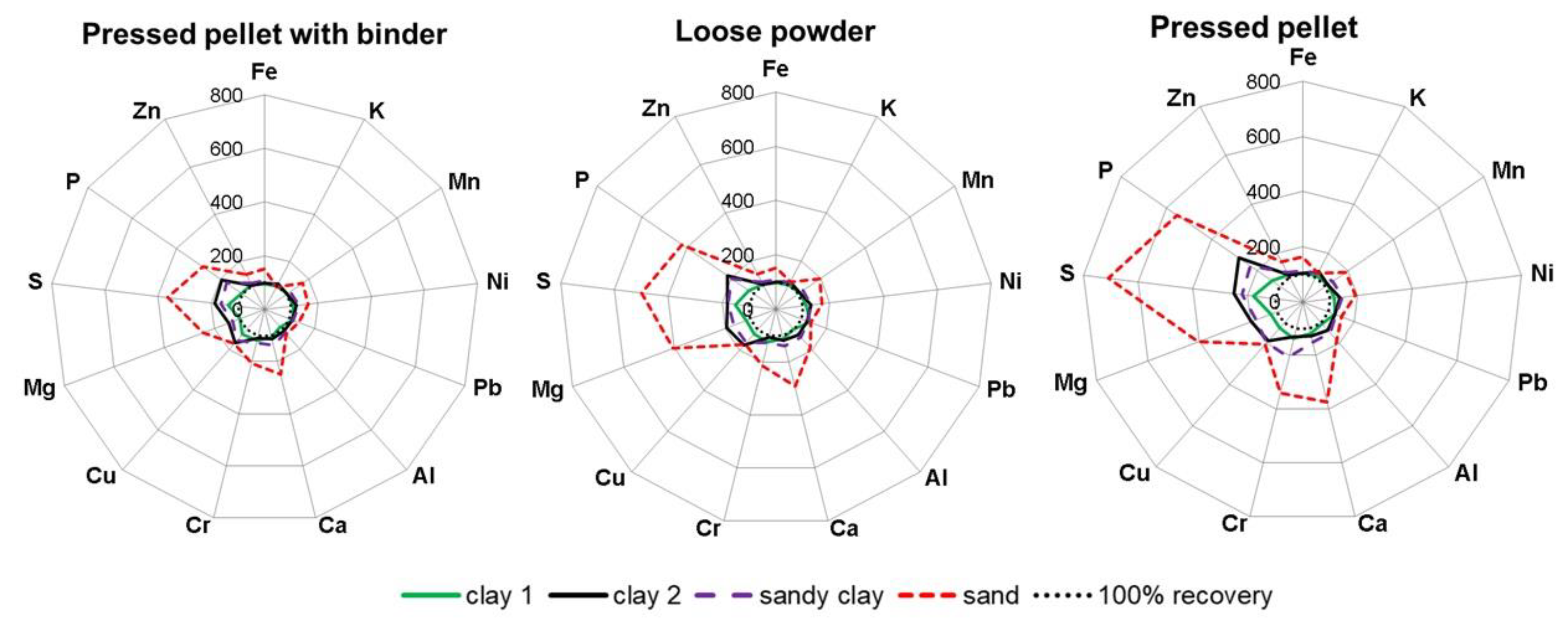
The effect of sample preparation on EDXRF results was expressed using % recovery of element concentration as a measure of accuracy and the results for four ISE samples with different sample preparation methods are presented in Figure 2. Recovery of 100% was used as the benchmark indicating the accuracy of the method; however, a deviation of ± 20% was deemed acceptable [17,43]. The recoveries of the elements for the sample preparation methods varied between ISE samples and ranged from 87–708%. The lowest recovery was reported for Al in clay 1 (ISE 952) with PPB and the highest recovery was recorded for S in the sand (ISE 995) using the PP in Figure 2.
The recoveries of K, Fe, and Zn for LP, PP, and PPB were within ± 20% for all ISE samples except for Fe and Zn in the sand sample, irrespective of the sample preparation method used. Additionally, Al, Pb, Mn, Ca, and Ni recoveries were within 100% ± 20% for all the sample preparation methods except sand (ISE 995), as shown in Figure 2. Thus, it can be inferred that when EDXRF is used for analysing soils with low silica, such as clay and sandy clay soils, high-pressure methods such as the PP and PPB may not be necessary, as the low-pressure method—the LP—is as suitable as the former. However, a one-way ANOVA showed that there were significant differences (p < 0.05) between the LP and PP, LP, and PPB methods for the recoveries of Al, Fe, and K in clay and sandy clay soils. In addition, for Mn, Ni, Zn, Pb, and Ca, the significant difference in recoveries between the methods varied for each soil (Supplementary Table S1).
Furthermore, Cu, Mg, P, and S recoveries were >120% (acceptable limit) for both LP and PP in all soil samples except clay 1 (ISE 952). PPB reduced Mg, P, and S recoveries >120% to approximately less than half of the other methods (Figure 2). Additionally, from Figure 2, the sandy soil (ISE 995) had recoveries >120% for Cu, Ca, Cr, Mg, S, and P, irrespective of the method; this is the only sample with Ca recovery >120% for all the methods. This may be due to particle size effects, especially for high silica samples; the presence of larger particles reduces the intensity reaching the XRF detector, thereby limiting the instrument’s ability to accurately quantify these elements [35]. According to Markowicz [19] and Parsons et al. [44], particle size effects are not removed even after samples have been pelletized as also observed in the current study. Also, Cu concentrations in all the soils were overestimated, irrespective of the sample preparation used. This may have resulted from the X-ray detector’s inability to properly separate different energies of two peaks whose spectra are smaller than the detector’s resolution, or elements with similar peaks overlapping with each other, such as Tantalum (Ta) Lα and Hafnium (Hf) Lα [21,45].
Another factor that may have affected the recoveries of the light elements could have been the critical analytical depth. This is the depth at which X-ray photons are absorbed or escape from a sample [46]. The critical analytical depth increases as X-ray energy and atomic number increases, thus lighter elements have a shallow critical analytical depth [46,47]. According to Ichikawa and Nakamura [46], shallow analytical depth has a negative influence on the accuracy and reproducibility of XRF analytical results. The critical analytical depth for light elements such as Mg, S, and P can sometimes be smaller than the larger particle size in soil [48]. Furthermore, for soils such as sand, pulverisation may not produce uniform particles, and if the critical analytical depth is less than the largest particle size, the EDXRF will measure a high proportion of elements in the finer fractions [22]. This may account for the high recoveries of some light elements using the LP and PP sample preparation method as shown in Figure 2. However, the addition of the binder to the PPB fills the spaces between the coarse grains, which may have reduced the particle size effects.
The effect of three different sample preparation methods on recoveries of elements in different soil analyses has been highlighted. The PP was less suitable for the sand sample due to the surface irregularity and inhomogeneity of samples, which affected the accuracy of P, S, Mg, Cr, and Ca quantification by the EDXRF. Additionally, the LP had the same problem with the quantification of P, S, Mg, Cr, and Ca elements in the sand; however, unlike the PP, the poor recoveries (>120%) were not more than 5 times above the benchmark (Figure 2). It may be that the LP is more heterogeneous than the PP. From the data shown in Figure 2, we conclude that the PPB is more suitable for multi-elemental analysis of different soils, with the advantage of reducing interference arising from particle size effects. Although this method requires an extra step for the addition and mixing of the wax binder, we have shown that this stage improves the results of EDXRF measurement.

3.2. Influence of Matching Library on EDXRF Results Using ISE Samples

The sample preparation study indicated that Cr, Cu, S, P, and Mg values would need corrections to improve results; thus, ML made up of four CRMs was created to adjust values derived from FP calibration. Additionally, the elements Al, K, Ca, Mn, Zn, Fe, Pb, and Ni were included in the ML calibration to account for spectral interference corrections [21]. Twenty ISE samples with ‘real totals’ values (NDA mean in Table 1) were compared against EDXRF values determined with ML and without a matching library (FP); all ISE samples were prepared as pressed pellet with wax binder and measured 11 times to determine the relative standard deviation (RSD), which is a measure of precision.
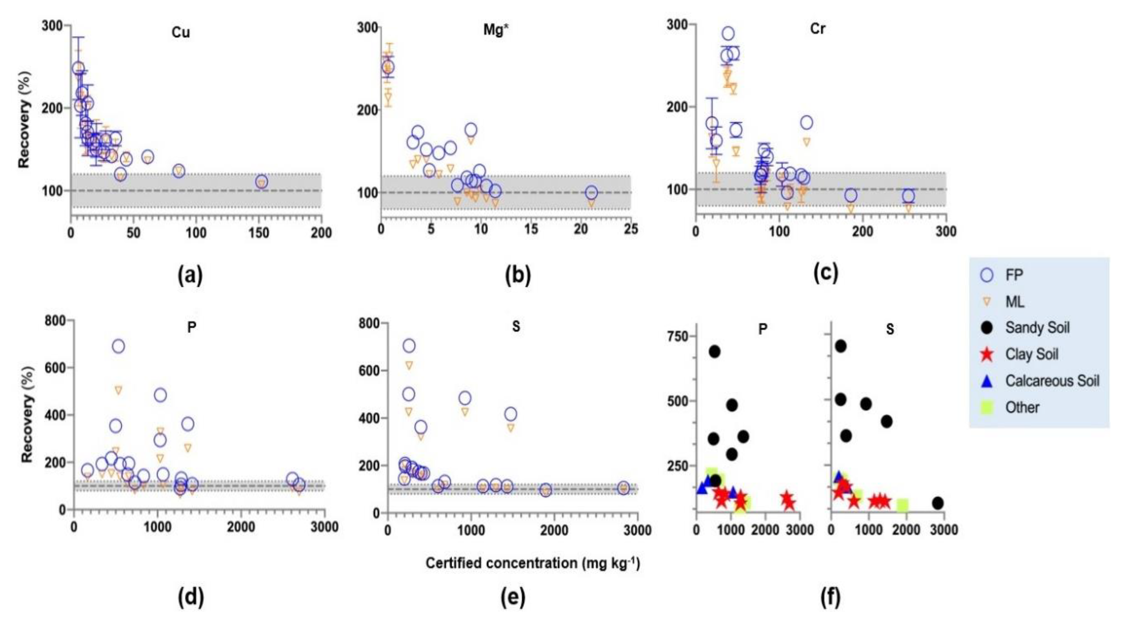
Figure 3 represents elements for which the EDXRF had a quantification problem earlier in this study, such as P, S, Cr, Cu, and Mg, while Figure 4 represents elements that did not need any corrections, namely, Pb, Ni, Zn, and Mn. At low concentrations in soils in Figure 3, element recoveries were above the maximum limit of 120% with the FP method. Rousseau [49] indicated that decreasing concentration decreases the intensity of an element line and is later overshadowed by background intensity, especially for ‘blanks’ or soils with a negligible concentration of elements. Thus, recoveries >120% observed at low concentrations in Figure 3 likely resulted from interferences from the background intensity influencing the sensitivity of the instrument [26,49]. A preliminary examination of spectra was carried out to investigate this issue, and three CRMs of varying concentrations of the elements were analysed with the FP method. After this, a visual evaluation of the spectrum was made on the Mo, Cu, Rx9, and Si secondary targets on the EDXRF. Peak interferences were not observed on any of the secondary targets for the element lines. However, on the Cu target, a lower peak than normal was observed for the element line Cr-Kα for GBW07403 (Appendix A, Table A1) at a low concentration of 32 mg kg−1. Additionally, the background intensity was 61.66 cps and higher than the intensity of 17.25 cps for Cr-Kα. The spectrum of the Cu target is shown in Appendix A, Figure A2. Furthermore, from Appendix A, Figure A2, the background intensity of 41.04 cps for Pb-Lβ1 in GBW07403 at a low concentration of 26 mg kg−1 was higher than the intensity of 25.36 cps.
However, the ML brought the recoveries closer to the target value within the 20% boundary, especially for P, Cr, and S, as illustrated in Figure 3c–e. It can be inferred that the ML reduces the effect of background intensity on the signal reaching the detector. In addition, the recoveries of P and S >150% were in the sandy soils (Figure 3f). This was also observed for Cu, Cr, and Mg (Figure 3); however, one sample with high S concentration recorded recovery within the limit of 80–120%. Sandy soils or high quartz soils analysed with EDXRF may be strongly influenced by particle size and matrix effects compared to other soil types, as observed in Figure 3f. Saini et al. [50] indicated that elements with low atomic numbers, such as P and Mg, suffer from errors arising from the complex scattering of low-energy X-rays because of inhomogeneous distribution of mineral grains and different particle size. The authors suggest that the addition of a binder in the preparation of pressed pellets reduces these effects provided the particle size is <75 µm. Although a wax binder was added to the sandy soils during sample preparation for this current study, particle size effects were not completely removed.
In addition, high-quartz soils are difficult to pelletize; thus, to reduce the effect of particle size and matrix effects, it may be necessary that samples be prepared as fused beads [51,52]. Additionally, Li et al. [53] suggests that soils with high quartz could be prepared as pressed pellet, covered with 3.6 µm polyester film and high pressure of 2000 kN added without the need for a binder. However, these additional steps may be time-consuming and reduce the advantage of the EDXRF as a rapid and inexpensive method; we recommend that for the analysis of sandy soils, an ML made up exclusively of sandy soils be used to improve analytical results.
For soils in Figure 3, it was also observed that Cu concentration <80 mg kg−1 produced recoveries >120%. The overestimation of Cu in these soils indicates that the EDXRF is not a sensitive analytical method for soils with low concentration. According to Sacristan et al. [43], the EDXRF is suitable for Cu analysis in soils, because they observed a high correlation between portable EDXRF peak height and Cu concentration in soil. However, all the soil samples used for the previous study were dosed with concentrations >500 mg kg−1. Rouillon and Taylor [17] suggest that a high concentration of Cu influences correlation, thus causing low concentration to cluster together to create a high correlation (R2 > 0.90).
In Figure 4 it was observed that at low concentrations close to the LOD (Table 1), the element recoveries were >120%, with FP similar to Figure 3. Furthermore, from Figure 3 and Figure 4, it can be observed that the EDXRF is concentration-dependent, and at certain concentration thresholds, the recoveries improve (Appendix A, Table A3). In addition, at the concentration threshold for most of the elements, the ML produced average recoveries within the range of 100% ± 20, except for Fe, K, Mn, and Zn (Appendix A, Table A3). For these elements, FP was the most suitable method of analysis compared to the ML. Additionally, when the sand samples were removed from the data, the average recoveries for P and S using the ML dropped from 130% ± 87.04 and 164% ± 130 to 85% ± 14.17 and 100% ± 10, respectively. Moreover, the average recoveries for Cu dropped from 125 ± 14 to 122 ± 14.5 with the removal of the sand samples.
It is well known that Zn, Cu, Mn, and Ni are essential for plant growth at low concentrations and toxic at high concentrations, while Pb and Cr have no known importance to plant health and are detrimental to human health [54,55]. Thus, the European Union (EU) has set a regulation for environmental monitoring of trace elements contamination in agricultural soils amended with sewage sludge. The directive 86/278/EEC [56] indicates the limit values of trace elements allowable in amended soils, which are Cu 50–140 mg kg−1, Ni 30–75 mg kg−1, Pb 50–300 mg kg−1, Zn 150–300 mg kg−1. In addition, Tóth et al. [57] indicated that lower guideline concentrations of 200 mg kg−1, 150 mg kg−1, 200 mg kg−1, 100 mg kg−1, 250 mg kg−1 for Cr, Cu, Pb, Ni, Zn, respectively, in soil, may pose ecological risks. The annual cost of management for contaminated areas in Europe alone is approximately €6 billion, and agricultural production is one of the main drivers of soil contamination [58]. Identifying contaminated soils quickly is the key to the rapid implementation of remediation measures. Thus, cost-effective and rapid analysis of contaminated soils USEPA Method 6200 [22] indicates the portable EDXRF as a screening method. However, the method states that for confirmatory results, the more expensive, laborious, and time-consuming techniques such as wet chemistry methods should be used. In this study, the use of the benchtop EDXRF is proposed as a confirmatory technique for contaminated sites identified with the portable EDXRF. The data above demonstrates that the EDXRF can accurately quantify the limit values of trace elements listed in 86/278/EEC [56] and the lower guideline limits proposed by Tóth et al. [57]. Although the FP method on the EDXRF is accurate for measuring contaminated soils, from this study it has been shown that the addition of an ML improves analytical results for trace elements and macronutrients. Furthermore, the instrument is concentration-dependent; thus, for soils deficient in trace elements and macronutrients, it has some limitations. In conclusion, the EDXRF is most suitable for rapid analysis of contaminated soils and can be used as a confirmatory technique for ‘hot spot’ identification.

3.3. Particle Size Effects on Fe, K, Mn, and Al Determined Using EDXRF Analysis in Tillage Soils

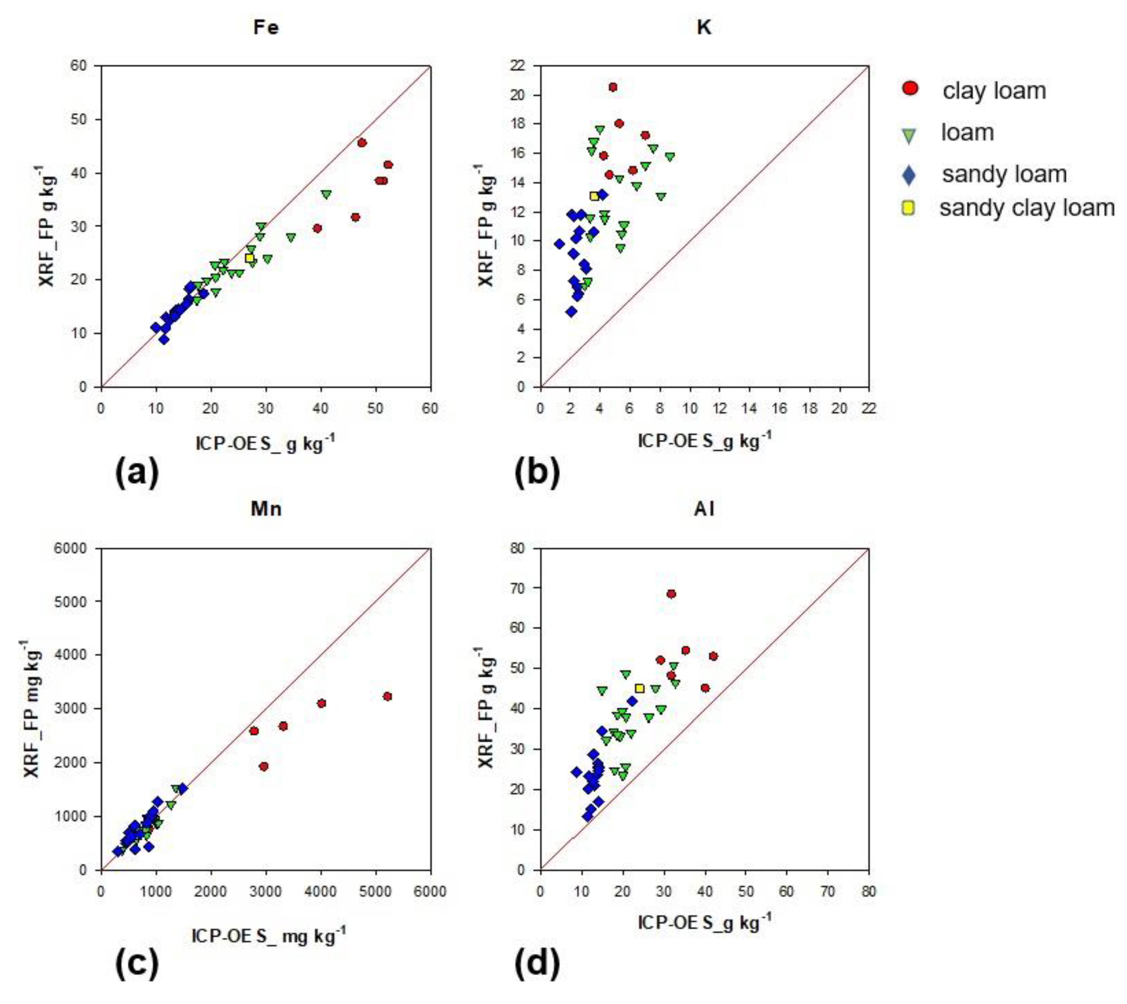
It was observed that soil texture had a strong effect on some elements, specifically Fe, K, Mn, and Al. Figure 5 highlights the influence of four soil texture classes on Fe, K, Mn, and Al values when EDXRF values are plotted against values derived from aqua regia digestion followed by ICP-OES. The concordance correlation coefficient (CCC) was computed to determine the level of agreement between the ICP-OES to the EDXRF. CCC is a product of accuracy and precision coefficients, whereby values range from −1 to 1. A value of 1 represents perfect agreement between two methods, while −1 represents disagreement, and 0 represents no agreement between the methods [42].
From Figure 5, it was observed that soils classified as clay loam drifted from the line of agreement at high concentrations of Fe (Figure 5a) and Mn (Figure 5c) compared to loam and sandy loam soils. Clay loam samples separated from the other texture classes due to the high concentration of Fe and Mn (Figure 5); however, the divergence from the line of agreement suggests EDXRF underestimated the concentrations. The sample preparation results presented earlier indicated that samples prepared as PPB underestimated Fe at high concentrations. Thus, the EDXRF underestimated Fe (Figure 5a) concentration in the clay loam samples. Agreement statistics computed showed a good agreement between the two methods with CCC = 0.898, R2 = 0.93 and CCC = 0.887, R2 = 0.92 for Fe and Mn, respectively.
For Al and K (Figure 5), a small number of the sandy loams were close to the line of agreement; the opposite is true for both clay loam and loam. This suggests that aqua regia underestimated Al and K in all the soils; thus, there was poor agreement between the EDXRF and ICP-OES for Al CCC = 0.40, R2 = 0.67, and K CCC = 0.102, R2 = 0.36. This observation is in agreement with Chen et al. [11], who found poor recoveries for Al and K in soils using aqua regia without the addition of HF. Additionally, Rao et al. [59], concluded that aqua regia was not suitable for K extraction in soils. Finally, comparative studies of the ICP methods against XRF [12,13,14,17,49,60] do not usually take into consideration the effect of soil texture on agreement between these methods. Although initially this may seem like an unwanted analytical challenge, it also offers an opportunity to potentially use EDXRF for rapid screening or prediction of soil texture.

4. Conclusions

Sample preparation methods had varying effects on multi-element EDXRF soil analysis, and the PPB produced most element recoveries within the benchmark of 80–120% for good accuracy. Thus, this method was used in the preparation of samples to examine the effect of ML on FP calibrations in EDXRF. The ML improved recoveries for Ni, Pb, Mg, S, P, Cr close to the maximum limit of 120%. However, for Zn, and Mn, the FP had recoveries closer to the 120% limit. The ML can simultaneously improve and be detrimental to soil measurements depending on the elements’ targets. An option is to have a hybrid of FP and ML for full element analysis. Additionally, the ML developed here is relatively easy to replicate, and analysts can source the same CRMs as used in this study. Additionally, it was observed that sandy soil had >120% recovery for all the elements, which may have resulted from greater homogeneity and rougher surface of samples. Furthermore, EDXRF results of Mn, Fe, K, and Al were compared to aqua regia-digested samples followed by ICP-OES in tillage soils and values separated by texture class.
This study shows that the EDXRF can be optimised for soil analysis by selecting the appropriate sample preparation and ML, with prior knowledge of texture class being a distinct advantage. However, a wide range of different textures was not included, and it will be necessary for future works to consider this. In addition, the benchtop EDXRF could be a possible confirmatory technique for verifying ‘hot spot’ contamination identified by the screening field portable XRF systems, especially for trace elements, as opposed to wet chemistry methods. This would be an advantage especially for tillage soils, whereby the reuse of sewage sludge and other organic waste is encouraged in agriculture across Europe. However, for sandy soils and soils deficient in trace elements the instrument has some limitations.

Supplementary Materials

The following are available online at https://www.mdpi.com/2073-4395/10/9/1309/s1. Table S1: One-way ANOVA results showing significant differences for recoveries for trace elements and macronutrients element for the three sample preparation methods. Table S2: Concordance correlation coefficient (CCC) and Bland-Altman results for elements concentration in tillage soils (n = 41) using fundamental parameters (FP) and matching library (ML) for EDXRF spectrometry in comparison to ICP-OES analysis. The datasets generated for this study are available at doi.org/10.17034/feca94bd-e9c7-4716-9103-33cafa09a3be.

Author Contributions

Writing—original draft preparation, investigation, methodology, formal analysis, visualization, visualization, M.E.T.C. Conceptualization, funding acquisition, supervision, project administration writing—review and editing, visualization, P.N.W. and K.D. Funding acquisition, writing—review and editing, O.F. Resources, methodology and validation, A.F. Data curation for the tillage samples K.M. All authors have read and agreed to the published version of the manuscript.

Funding

This research was funded by the Teagasc Walsh Fellowship Programme (grant number RMIS 6502) and the Institute for Global Food Security, Queen’s University Belfast.

Acknowledgments

The authors would like to acknowledge Conor Nolan, Owen Frahill, Scott Rees and Linda Moloney-Finn for technical support.

Conflicts of Interest

The authors declare no conflict of interest.

Appendix A

Table A1. Summary of certified values used in the matching library for XRF analysis. All units in mg kg−1.
Certified Reference Materials
ElementGBW07403GBW07405ISE 921BAM-U110
Al64,800114,24756,800 ± 1440* 50,382 ± 1081
Ca907167943,000 ± 1180* 40,638 ± 2105
Cu11.414493.8 ± 5.84263 ± 12
Cr32118130 ± 10.6230 ± 13
Fe13,98988,26931,900 ± 1130* 28,229 ± 663
K25,22612,44719,100 ± 640* 20,381 ± 4560
Mg3498367911,100 ± 370* 8380 ± 735
Mn30413601190 ± 43621 ± 20
Ni124042.4 ± 3.21101 ± 5
P3203901400 ± 69* 3648 ± 49
Pb26552167 ± 7.5197 ± 14
S120410702 ± 79.8* 10,593
Si349,290245,746271,000 ± 4600* 258,125
Zn31494522 ± 19.91000 ± 50
* reference value only not certified.
Table A2. Measurement conditions for analysis of the elemental composition of soil samples prepared as pressed pellet with binder using benchtop energy dispersive X-ray fluorescence spectrometer. All samples were run under vacuum atmosphere.
ElementVoltage (kv)Secondary TargetPeak Detected (KeV)Measurement
Time (s)
Cu50MoKα, 8.047100
Cr50CuKα, 5.414100
Mn50MoKα, 5.898100
Ni50MoKα, 7.477100
Zn50MoKα, 8.638100
Pb50MoLβ1, 12.611100
Al25Rx9Kα, 1.487100
S25Rx9Kα, 2.308100
P25Rx9Kα, 2.015100
Mg25SiKα,1.254300
Ca50CuKα, 3.691100
K50CuKα, 3.313100
Fe50MoKα, 6.403100
Table A3. Average recovery of elements based on the concentration threshold of the ISE samples analysed with the fundamental parameters (FP) and matching library (ML) methods on the EDXRF.
ElementsConcentration Threshold
(mg kg−1)
Average Recovery
Including Sand (%)
Average Recovery (%) Minus SandCalibration
Method
Al40760-106 ± 10.2 (n = 12)ML
Ca3750120 ± 29
(n = 17)
106 ± 12
(n = 14)
ML
Cr77.3101 ± 21
(n = 14)
97 ± 14.9
(n = 12)
ML
Cu39.66125 ± 14
(n = 5)
122 ± 14.5
(n = 4)
ML
Fe6040106 ± 10.4
(n = 17)
102 ± 4.5
(n = 14)
FP
K5988108 ± 10.1
(n = 20)
-FP
Mg4844107 ± 22.8
(n = 12)
102 ± 15.3
(n = 11)
ML
Mn238114 ± 18.3
(n = 15)
107 ± 4.9
(n = 13)
FP
Ni21.27117 ± 11.9
(n = 14)
113 ± 5.8
(n = 12)
ML
P727130 ± 87.04
(n = 12)
85 ± 14.17
(n = 9)
ML
Pb19.16113 ± 6.2
(n = 16)
-ML
S603.5164 ± 130
(n = 9)
100 ± 10
(n = 7)
ML
Zn56.45109 ± 7.3
(n = 13)
107 ± 3.6
(n = 12)
FP
Figure A1. Common methods for soil sample preparation using low and high pressure for analysis on X-ray fluorescence spectroscopy (a) loose powder (LP)-200 inch lbs pressure added for 30 s, (b) pressed powder pellet (PP)-15 tons pressure added for 30 s, (c) pressed powder pellet with wax binder (PPB)-20 tons pressure added for 30 s.
Agronomy 10 01309 g0a1
Figure A2. Spectrum of Cu target on EDXRF showing the elements on the target using three CRMs with different concentrations, whereby the red line (GBW07403) signifies elements at low concentrations, the black line (ISE 921) signifying elements at medium concentrations and the green line (BAM-U110) indicating a contaminated soil.
Agronomy 10 01309 g0a2

References

  1. Pyle, S.M.; Nocerino, J.M.; Deming, S.N.; Palasota, J.A.; Palasota, J.M.; Miller, E.L.; Hillman, D.C.; Kuharic, C.A.; Cole, W.H.; Fitzpatrick, P.M.; et al. Comparison of AAS, ICP-AES, PSA, and XRF in determining lead and cadmium in soil. Environ. Sci. Technol. 1996, 30, 204–213. [Google Scholar] [CrossRef]
  2. Gäbler, H.-E. Mobility of heavy metals as a function of pH of samples from an overbank sediment profile contaminated by mining activities. J. Geochem. Explor. 1997, 58, 185–194. [Google Scholar] [CrossRef]
  3. Correia, A.G.; Da Silva, R.J.B.; Pedra, F.; Nunes, J. Assessment of the determination of heavy metals in organic soil improvers by ICP-OES. Accredit. Qual. Assur. 2014, 19, 87–97. [Google Scholar] [CrossRef]
  4. Kilbride, C.; Poole, J.; Hutchings, T. A comparison of Cu, Pb, As, Cd, Zn, Fe, Ni and Mn determined by acid extraction/ICP-OES and ex situ field portable X-ray fluorescence analyses. Environ. Pollut. 2006, 143, 16–23. [Google Scholar] [CrossRef]
  5. Shuttleworth, E.; Evans, M.; Hutchinson, S.M.; Rothwell, J.J. Assessment of lead contamination in peatlands using field portable XRF. Water Air Soil Pollut. 2014, 225, 1844. [Google Scholar] [CrossRef]
  6. USEPA. Method 3052 Microwave Assisted Acid Digestion of Sileceous and Organically Based Matrices; United States Environmental Protection Agency: Washington, DC, USA, 1996.
  7. USEPA. Method 3050b Acid Digestion of Soils, Sediments and Sludges, Revision 2 ed; United States Environmental Protection Agency: Washington, DC, USA, 1996.
  8. Sastre, J.; Sahuquillo, Á.; Vidal, M.; Rauret, G. Determination of Cd, Cu, Pb and Zn in environmental samples: Microwave-assisted total digestion versus aqua regia and nitric acid extraction. Anal. Chim. Acta 2002, 462, 59–72. [Google Scholar] [CrossRef]
  9. Hassan, N.M.; Rasmussen, P.E.; Dabek-Zlotorzynska, E.; Celo, V.; Chen, H. Analysis of environmental samples using microwave-assisted acid digestion and inductively coupled plasma mass spectrometry: Maximizing total element recoveries. Water Air Soil Pollut. 2007, 178, 323–334. [Google Scholar] [CrossRef]
  10. Quevauviller, P.; Van Der Sloot, H.; Ure, A.; Muntau, H.; Gómez, A.; Rauret, G. Conclusions of the workshop: Harmonization of leaching/extraction tests for environmental risk assessment. Sci. Total Environ. 1996, 178, 133–139. [Google Scholar] [CrossRef]
  11. Chen, M.; Ma, L.Q. Comparison of three aqua regia digestion methods for twenty florida soils. Soil Sci. Soc. Am. J. 2001, 65, 491–499. [Google Scholar] [CrossRef] [Green Version]
  12. Anderson, P.; Davidson, C.M.; Littlejohn, D.; Ure, A.M.; Garden, L.M.; Marshall, J. Comparison of techniques for the analysis of industrial soils by atomic spectrometry. Int. J. Environ. Anal. Chem. 1998, 71, 19–40. [Google Scholar] [CrossRef]
  13. Kalnicky, D.J.; Singhvi, R. Field portable XRF analysis of environmental samples. J. Hazard. Mater. 2001, 83, 93–122. [Google Scholar] [CrossRef] [Green Version]
  14. Wu, C.-M.; Tsai, H.-T.; Yang, K.-H.; Wen, J.-C. How reliable is X-Ray fluorescence (XRF) measurement for different metals in soil contamination? Environ. Forensics 2012, 13, 110–121. [Google Scholar] [CrossRef]
  15. Thomas, C.L.; Acquah, G.E.; Whitmore, A.P.; McGrath, S.P.; Haefele, S.M. The effect of different organic fertilizers on yield and soil and crop nutrient concentrations. Agronomy 2019, 9, 776. [Google Scholar] [CrossRef] [Green Version]
  16. Melquiades, F.L.; Appoloni, C.R. Application of XRF and field portable XRF for environmental analysis. J. Radioanal. Nucl. Chem. 2004, 262, 533–541. [Google Scholar] [CrossRef]
  17. Rouillon, M.; Taylor, M.P. Can field portable X-ray fluorescence (pXRF) produce high quality data for application in environmental contamination research? Environ. Pollut. 2016, 214, 255–264. [Google Scholar] [CrossRef]
  18. Tavares, T.R.; Mouazen, A.M.; Alves, E.E.N.; Dos Santos, F.R.; Melquiades, F.L.; De Carvalho, H.W.P.; Molin, J.P. Assessing soil key fertility attributes using a portable X-ray fluorescence: A simple method to overcome matrix effect. Agronomy 2020, 10, 787. [Google Scholar] [CrossRef]
  19. Markowicz, A. An overview of quantification methods in energy-dispersive X-ray fluorescence analysis. Pramana 2011, 76, 321–329. [Google Scholar] [CrossRef]
  20. Towett, E.K.; Shepherd, K.D.; Cadisch, G. Quantification of total element concentrations in soils using total X-ray fluorescence spectroscopy (TXRF). Sci. Total Environ. 2013, 463, 374–388. [Google Scholar] [CrossRef] [Green Version]
  21. Willis, J.P.; Feather, C.E.; Turner, K. Guidelines for XRF Analysis: Setting up Programmes for WDXRF and EDXRF, 1st ed.; James Willis Consultants cc: Cape Town, South Africa, 2014; Chapter 2–16. [Google Scholar]
  22. USEPA. Method 6200 Field Portable X-ray Fluorescence Spectrometry for Determination of Elemental Concentrations in Soil and Sediment; United States Environmental Protection Agency: Washington, DC, USA, 2007.
  23. Mejia-Piña, K.; Huerta-Diaz, M.A.; González-Yajimovich, O. Calibration of handheld X-ray fluorescence (XRF) equipment for optimum determination of elemental concentrations in sediment samples. Talanta 2016, 161, 359–367. [Google Scholar] [CrossRef]
  24. Bernick, M.; Kalnicky, D.; Prince, G.; Singhvi, R. Results of field-portable X-ray fluorescence analysis of metal contaminants in soil and sediment. J. Hazard. Mater. 1995, 43, 101–110. [Google Scholar] [CrossRef]
  25. Daly, K.; Fenelon, A. A rapid and multi-element method for the analysis of major nutrients in grass (Lolium perenne) using energy-dispersive X-ray fluorescence spectroscopy. Ir. J. Agric. Food Res. 2017, 56, 1–11. [Google Scholar] [CrossRef] [Green Version]
  26. Rousseau, R.M.; Willis, J.P.; Duncan, A.R. Pratical XRF calibration procedures for major and trace elements. X-ray Spectrom. 1996, 25, 179–189. [Google Scholar] [CrossRef]
  27. Metzger, K.; Zhang, C.; Ward, M.; Daly, K. Mid-infrared spectroscopy as an alternative to laboratory extraction for the determination of lime requirement in tillage soils. Geoderma 2020, 364, 114171. [Google Scholar] [CrossRef]
  28. Gee, G.W.; Bauder, J.W. Particle-size analysis in. In Methods of Soil Analysis. Part 1. Physical and Mineralogical Methods; Part 2. Chemical and Microbiological Properties; Klute, A., Ed.; American Society of Agronomy, Inc.: Madison, WI, USA, 1986. [Google Scholar]
  29. Shibata, Y.; Suyama, J.; Kitano, M.; Nakamura, T. X-ray fluorescence analysis of Cr, As, Se, Cd, Hg, and Pb in soil using pressed powder pellet and loose powder methods. X-ray Spectrom. 2009, 38, 410–416. [Google Scholar] [CrossRef]
  30. Takahashi, G. Sample preparation for X-ray fluorescence analysis III. Pressed and loose powder methods. Rigaku J. 2015, 31, 25–30. [Google Scholar]
  31. Willis, J.P.; Turner, K.; Pritchard, G. XRF in the Workplace: A Guide to Pratical XRF Spectrometry; PANalytical Australia: Chipping Norton, Australia, 2011. [Google Scholar]
  32. Omote, J.; Kohno, H.; Toda, K. X-Ray fluorescence analysis utilizing the fundamental parameter method for the determination of the elemental composition in plant samples. Anal. Chim. Acta 1995, 307, 117–126. [Google Scholar] [CrossRef]
  33. Kataoka, Y.; Kawahara, N.; Hara, S.; Yamada, Y.; Matsuo, T.; Mantler, M. Fundamental Parameter Method Using Scattering X-rays in X-ray Fluorescence Analysis; CPDS-International Centre for Diffraction Data: Newtown Township, PA, USA; Denver, CO, USA, 2006; pp. 255–260, ISSN 1097-0002. [Google Scholar]
  34. Singh, V.; Agrawal, H. Qualitative soil mineral analysis by EDXRF, XRD and AAS probes. Radiat. Phys. Chem. 2012, 81, 1796–1803. [Google Scholar] [CrossRef]
  35. Maruyama, Y.; Ogawa, K.; Okada, T.; Kato, M. Laboratory experiments of particle size effect in X-ray fluorescence and implications to remote X-ray spectrometry of lunar regolith surface. Earth Planets Space 2008, 60, 293–297. [Google Scholar] [CrossRef] [Green Version]
  36. Kadachi, A.N.; Al-Eshaikh, M.A. Limits of detection in XRF spectroscopy. X-ray Spectrom. 2012, 41, 350–354. [Google Scholar] [CrossRef]
  37. Fees, S.; Applied Rigaku Technologies, Austin, TX, USA. Personal communication, 2019.
  38. ISO. Soil Quality, Extraction of Trace Elements Soluble in Aqua Regia; ISO: Geneva, Switzerland, 1995. [Google Scholar]
  39. Altman, D.G.; Bland, J.M. Measurement in medicine: The analysis of method comparison studies. J. R. Stat. Soc. Ser. D Stat. 1983, 32, 307–317. [Google Scholar] [CrossRef]
  40. Bland, J.M.; Altman, D. Statistical methods for assessing agreement between two methods of clinical measurement. Lancet 1986, 327, 307–310. [Google Scholar] [CrossRef]
  41. Bland, J.M.; Altman, D.G. Measuring agreement in method comparison studies. Stat. Methods Med. Res. 1999, 8, 135–160. [Google Scholar] [CrossRef] [PubMed]
  42. Lin, L.; Hedayat, A.S.; Sinha, B.; Yang, M. Statistical methods in assessing agreement. J. Am. Stat. Assoc. 2002, 97, 257–270. [Google Scholar] [CrossRef]
  43. Sacristán, D.; Viscarra-Rossel, R.; Recatalá, L. Proximal sensing of Cu in soil and lettuce using portable X-ray fluorescence spectrometry. Geoderma 2016, 265, 6–11. [Google Scholar] [CrossRef]
  44. Parsons, C.T.; Grabulosa, E.M.; Pili, E.; Floor, G.H.; Román-Ross, G.; Charlet, L. Quantification of trace arsenic in soils by field-portable X-ray fluorescence spectrometry: Considerations for sample preparation and measurement conditions. J. Hazard. Mater. 2013, 262, 1213–1222. [Google Scholar] [CrossRef]
  45. Horta, A.; Malone, B.P.; Stockmann, U.; Minasny, B.; Bishop, T.; McBratney, A.B.; Pallasser, R.; Pozza, L. Potential of integrated field spectroscopy and spatial analysis for enhanced assessment of soil contamination: A prospective review. Geoderma 2015, 241, 180–209. [Google Scholar] [CrossRef] [Green Version]
  46. Ichikawa, S.; Nakamura, T. Approaches to solid sample preparation based on analytical depth for reliable X-ray fluorescence analysis. X-ray Spectrom. 2016, 45, 302–307. [Google Scholar] [CrossRef]
  47. Potts, P.J.; Williams-Thorpe, O.; Webb, P.C. The bulk analysis of silicate rocks by portable X-ray fluorescence: Effect of sample mineralogy in relation to the size of the excited volume. Geostand. Newsl. 1997, 21, 29–41. [Google Scholar] [CrossRef]
  48. McGarry, C.; Dublin Analytical Instruments Ltd., Dublin, Ireland. Personal communication, 2020.
  49. Rousseau, R.M. Detection limit and estimate of uncertainty of analytical XRF results. Rigaku J. 2001, 18, 33–47. [Google Scholar]
  50. Saini, N.K.; Mukherjee, P.K.; Rathi, M.S.; Khanna, P.P. Evaluation of energy-dispersive x-ray fluorescence spectrometry in the rapid analysis of silicate rocks using pressed powder pellets. X-ray Spectrom. 2000, 29, 166–172. [Google Scholar] [CrossRef]
  51. Wang, X.; Li, G.; Zhang, Q.; Wang, Y. Determination of major/minor and trace elements in seamount phosphorite by XRF spectrometry. Geostand. Geoanalytical Res. 2004, 28, 81–88. [Google Scholar] [CrossRef]
  52. Matsunami, H.; Matsuda, K.; Yamasaki, S.-I.; Kimura, K.; Ogawa, Y.; Miura, Y.; Yamaji, I.; Tsuchiya, N. Rapid simultaneous multi-element determination of soils and environmental samples with polarizing energy dispersive X-ray fluorescence (EDXRF) spectrometry using pressed powder pellets. Soil Sci. Plant Nutr. 2010, 56, 530–540. [Google Scholar] [CrossRef]
  53. Li, X.; Yu, Z.; Xu, J.; Pan, Y.; Bo, W.; Liu, B.; Zhang, P.; Bai, J.; Zhang, Q. The technique of high-pressure powder pressing with polyester film covering for XRF of geochemical samples. X-ray Spectrom. 2020, 1–11. [Google Scholar] [CrossRef]
  54. Nagajyoti, P.C.; Lee, K.D.; Sreekanth, T.V.M. Heavy metals, occurrence and toxicity for plants: A review. Environ. Chem. Lett. 2010, 8, 199–216. [Google Scholar] [CrossRef]
  55. Emamverdian, A.; Ding, Y.; Mokhberdoran, F.; Xie, Y. Heavy metal stress and some mechanisms of plant defense response. Sci. World J. 2015. [Google Scholar] [CrossRef]
  56. CEC. Council directive of 12 June 1986 on the protection of the environment and in particular of the soil when sewage sludge is used in agriculture. Off. J. Eur. Communities 1986, 181, 0006–0012. [Google Scholar]
  57. Toth, G.L.; Hermann, T.; Da Silva, M.; Montanarella, L. Heavy metals in agricultural soils of the European Union with implications for food safety. Environ. Int. 2016, 88, 299–309. [Google Scholar] [CrossRef]
  58. Stolte, J.; Tesfai, M.; Øygarden, L.; Kværnø, S.; Keizer, J.; Verheijen, F.; Panagos, P.; Ballabio, C.; Hessel, R. Soil Threats in Europe; Publications Office: Luxembourg, 2015. [Google Scholar] [CrossRef]
  59. Rao, B.K.R.; Bailey, J.; Wingwafi, R.W. Comparison of three digestion methods for total soil potassium estimation in soils of papua new guinea derived from varying parent materials. Commun. Soil Sci. Plant Anal. 2011, 42, 1259–1265. [Google Scholar] [CrossRef]
  60. Vanhoof, C.; Tirez, K. Energy-dispersive X-ray fluorescence systems as analytical tool for assessment of contaminated soils. J. Environ. Monit. 2004, 6, 344–350. [Google Scholar] [CrossRef]
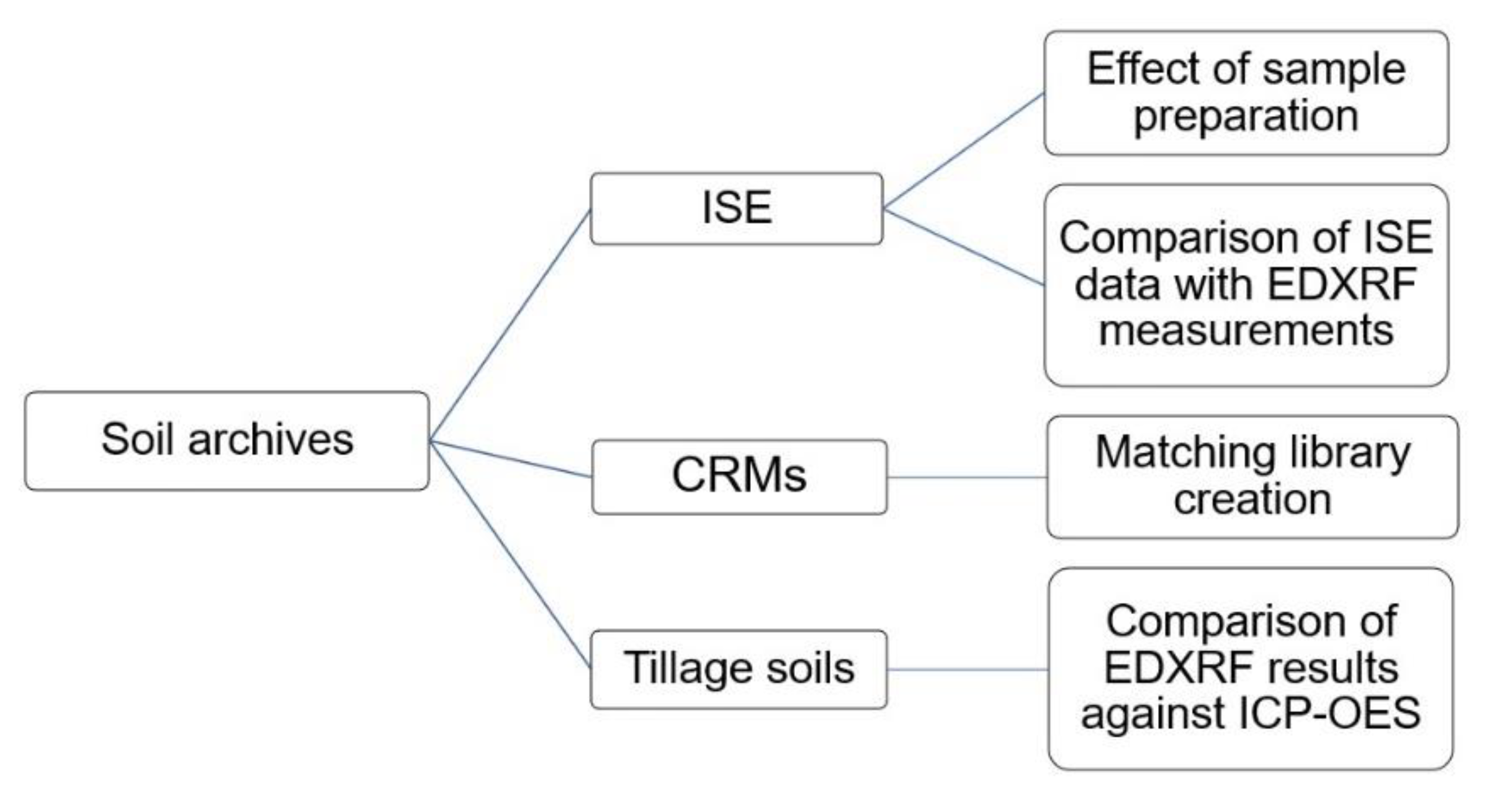
Figure 1. Distribution of sample archive showing how each objective for this study was fulfilled using the three different archives: international soil exchange (ISE), certified reference materials (CRMs), and tillage soils.
Agronomy 10 01309 g001
Figure 2. Effects of sample preparation on EDXRF analytical results using elemental recovery as a measure of accuracy with 100% recovery as the benchmark. All samples are from ISE archives (n = 4), clay 1 (ISE 952), clay 2 (ISE 961), sandy clay (ISE 992), and sand (ISE 995).
Agronomy 10 01309 g002
Figure 3. Scatterplot of (a) Cu, (b) Mg (c) Cr, (d) P, (e) S (f) P and S concentration against recovery in different soil types of ISE samples (n = 20) comparing fundamental parameters (FP)-derived results and matching library (ML)-adjusted analytical results on the energy-dispersive X-ray fluorescence spectrometer. The solid line denotes the target value of 100% recovery, and the grey shaded region denotes the acceptable limit of ± 20%. All units in mg kg−1 except Mg * which is in g kg−1.
Agronomy 10 01309 g003
Figure 4. Scatterplot of (a) Zn, (b) Pb, (c) Mn and (d) Ni concentration against recovery in different soil types of ISE samples (n = 20) comparing fundamental parameters (FP)-derived results and matching library (ML)-adjusted analytical results on the energy-dispersive X-ray fluorescence spectrometer. The solid line denotes the target value of 100% recovery and the grey shaded region denotes the acceptable limit of ± 20%. Yellow shading represents an acceptable range of element concentrations in 86/278/EEC.
Agronomy 10 01309 g004
Figure 5. Effect of soil texture on EDXRF- and ICP-OES-determined values of (a) Fe, (b) K, (c) Mn, and (d) Al concentrations separation tillage soils with the line of agreement.
Agronomy 10 01309 g005
Table 1. ISE samples reported mean total concentrations of the 13 elements selected for this study. The results below show the range of the mean results (normal distribution approximation) and the standard deviation (SD) for a minimum of 20 participating labs per analysis, which includes wet chemistry and spectrometry results. All units in mg kg−1 except Al, Ca and Fe which are in g kg−1.
ISE Normal Distribution Approximation (NDA) ResultsCalculated Limit of Detection (LOD) for the XRF Used for This Study
ElementNDA MeanNDA SDMean LOD
MaxMinMaxMinmg kg−1
Al **94.9416.185.110.5288.6
Ca **2211.18690.0856.3
Fe **60.072.623.620.165.64
K218655987.711991533.4
S2831197.152427.74
P2694162.915712.710.3
Mg21042312179463.8114.8
Mn147975.09859.0612.1
Cu152.35.067.141.051.7
Cr254.819.2923.72.285.6
Ni111.66.595.50.9981.9
Pb296.64.3221.80.9812.4
Zn103717.7432.41.2
** units in g kg−1.