
Chitosan-Based Nanoparticles of Targeted Drug Delivery System in Breast Cancer Treatment

 ,
,  , and
, and 
Abstract

1. Introduction
- Physicochemical drug characteristics such as poor solubility, leading to poor pharmacokinetic characteristics.
- Insufficient specificity for breast cancer.
- Cellular-level and tumor microenvironment drug resistance.
- Difficulties of eliminating cancer stem cells.
- Technologies to strengthen the solubility and stability of anticancer drugs [16].
- Passive and active targeting is used by nano-based modalities to selectively target malignant tissues/cells [17].
- The distribution of various drugs helps decrease drug resistance.
- The nano-based vehicles have controlled release techniques for a medication.
- Drug efflux pathways can be blocked by nano-based vehicles.
- Usage of nanoformulation dependent on stimulus-responsive.
2. Nanotechnology in Drug Targeted Delivery System in Cancer Therapy
- Protected drugs against hepatic inactivation, enzymatic degradation, and rapid clearance [26].
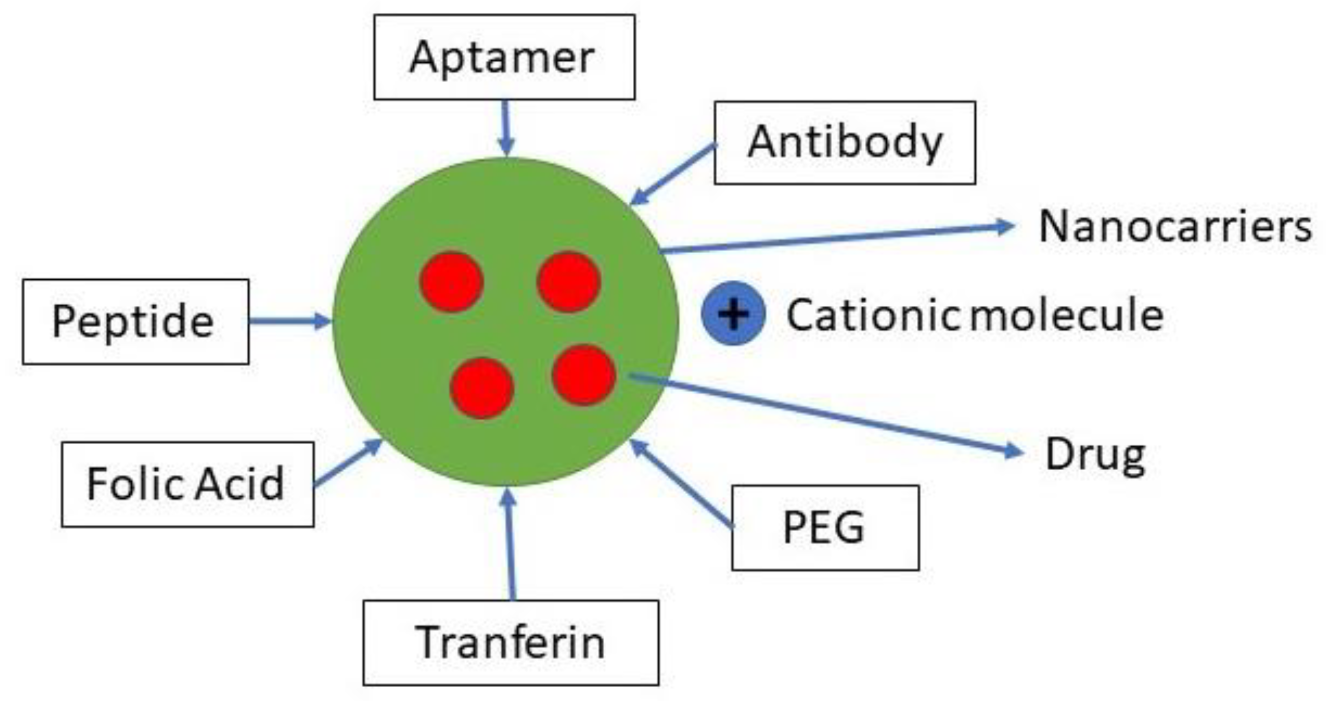
- It has enhanced cancer cell internalization by functionalizing with ligands over-expressed in tumor cells recognizable by receptors (targets) [2].
- It is influenced by cancer growth by controlling the tumor microenvironment (TME) [27]. Polymers can respond to and perceive exogenous stimulus (light, temperature, ultrasound, electrochemical triggers) or microenvironmental tumor (pH, enzyme activity, redox properties) to cause drug release to overcome these barriers [28].
- Escaped the multiple drug resistance (MDR) efflux transporters [2].
- Reduced the incidence and intensity of side effects [29].
- Carried contrast moieties contained allowing direct in vivo imaging (carrier visualization) [30].
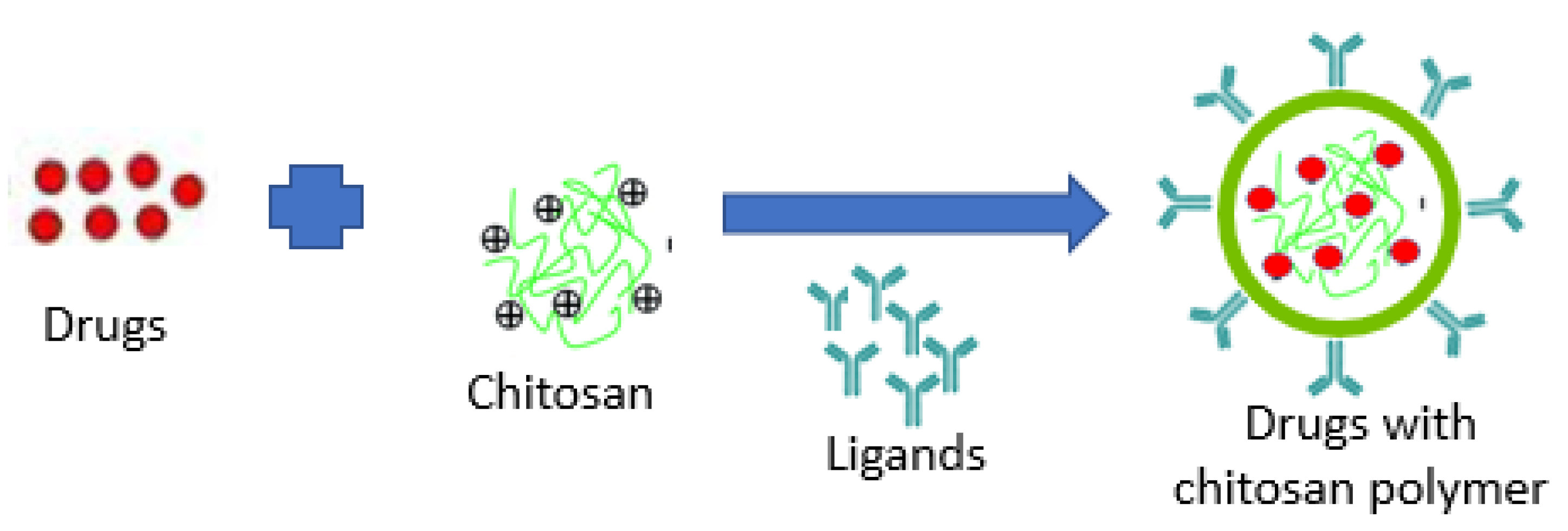
3. Biological Ligands for Nanoparticle Drug Delivery Systems
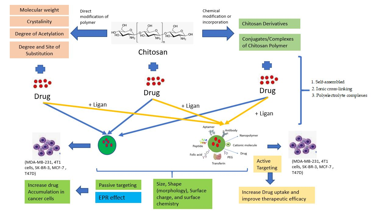
3.1. Passive Targeting
3.2. Active Targeting
- The number of target moieties is higher in tumor cells than in normal tissues [37].
- It should also be measured at locations that nanocarriers can easily reach, such as superficial receptors rather than intracellular targets.
- In order to allow competent targeting, the concentration should be high enough.
- It must ideally associate its levels with malignant activities, whether drug resistance or active targets, to target these threatening tumors on a priority basis.
- Targeting can simplify procedures that facilitate the delivery of drugs [10].
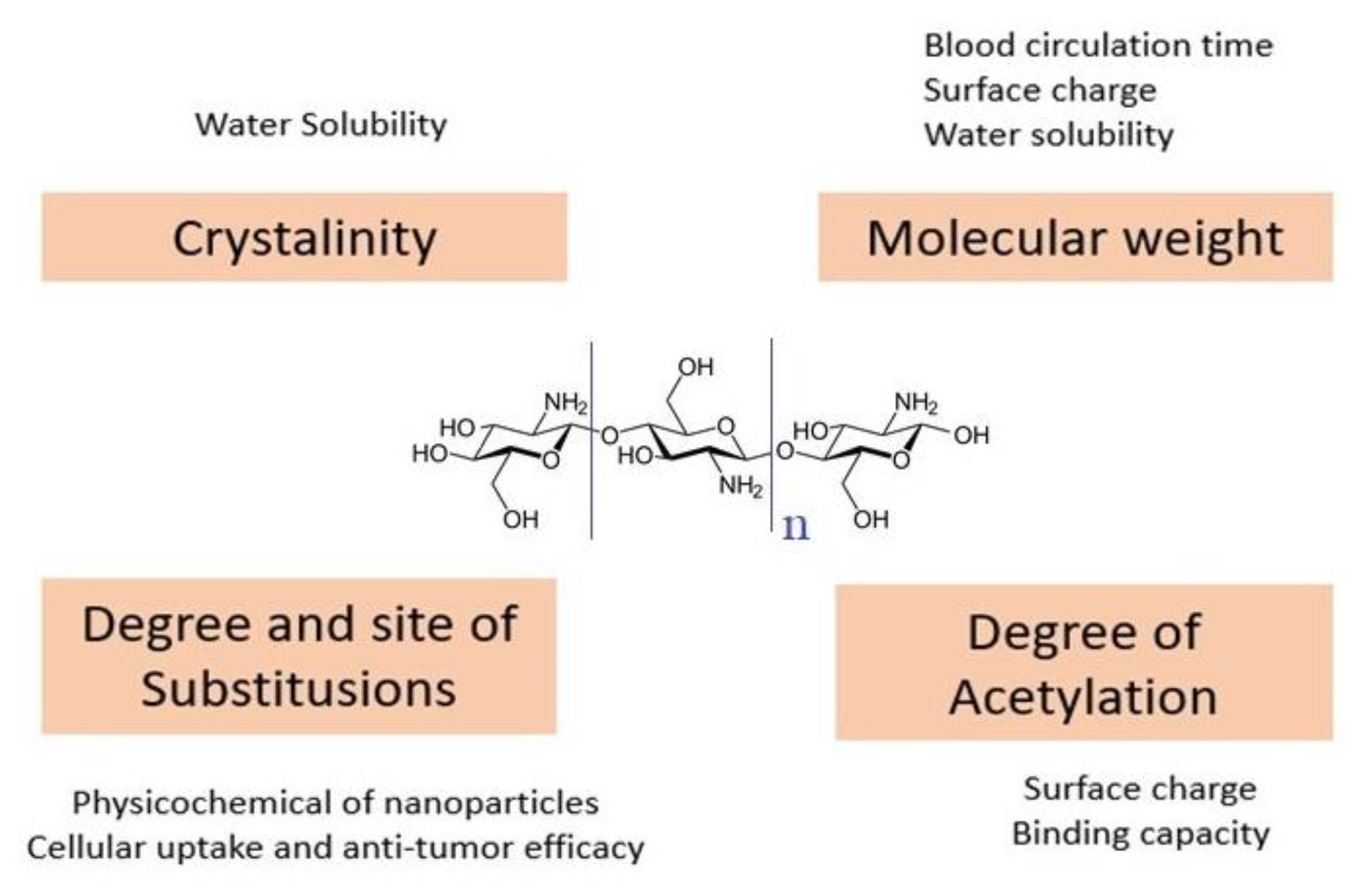
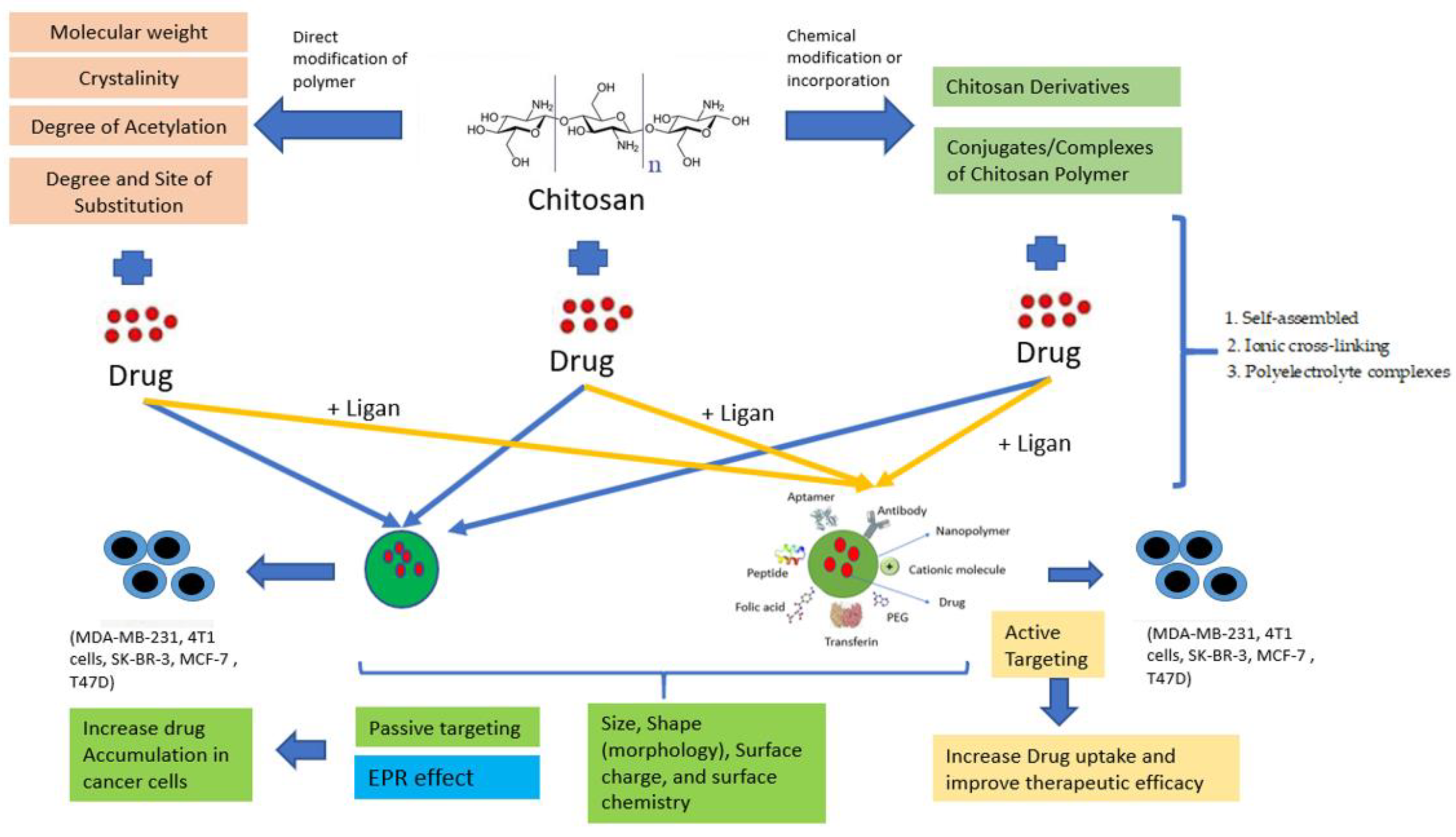
4. Chitosan-Based Nanoparticles Preparation and Modification
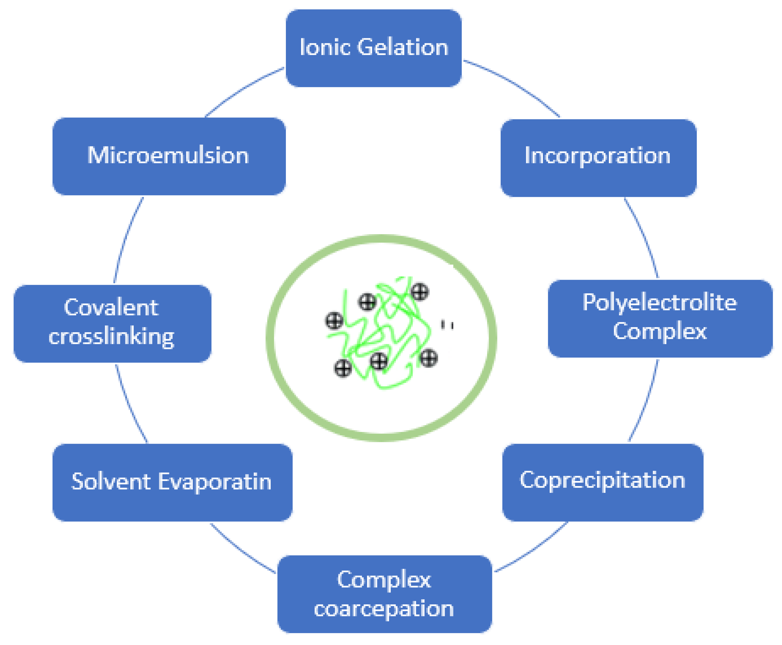
4.1. Preparing Method Chitosan-Based Nanoparticles
4.1.1. Self-Assembled
4.1.2. Ionic Cross-Linking
4.1.3. Polyelectrolyte Complexes
4.2. Chemical Modifications or Incorporation
4.2.1. Chitosan Derivatives Nanoparticles
4.2.2. Conjugates/Complexes of Chitosan-Polymer Nanoparticles
5. Chitosan-Based Systems as Drug Carriers in Targetted Drug Delivery Systems in BC Treatment
- The glucose uptake rate increases dramatically, and lactate is generated, even in the presence of oxygen and fully functioning mitochondria [53].
- Hyperpyrexia (temperatures raised to 40–42 °C due to the increasing rate of glycolysis and rapid cell proliferation) is also characterized by the tumor microenvironment [45].
- The concentration of intracellular glutathione (GSH), a tripeptide responsible for reducing disulfide bonds, is about 2–3 orders higher than the extracellular GSHH order [55].
- Positive charges on the surface of nanoparticles are also necessary for transfection into the cells because of the negative charge on membranous cells [56].
- The concept of developing targeted anticancer therapeutics has led to the development of breast cancer receptor type 2 (HER-2), estrogen receptor (ER), up-regulated with human epidermal growth factor receptor [57].
- In breast cancer cells, the folate receptors (FRs) are over-expressed [58].
6. Stimuli-Responsive Materials Based on Chitosan-Based Nanoparticles
7. Perspective
8. Conclusions
Funding
Institutional Review Board Statement
Informed Consent Statement
Data Availability Statement
Conflicts of Interest
References
- Fathi, M.; Alami-Milani, M.; Geranmayeh, M.H.; Barar, J.; Erfan-Niya, H.; Omidi, Y. Dual thermo-and pH-sensitive injectable hydrogels of chitosan/(poly(N-isopropylacrylamide-co-itaconic acid)) for doxorubicin delivery in breast cancer. Int. J. Biol. Macromol. 2019, 128, 957–964. [Google Scholar] [CrossRef]
- Mirza, Z.; Karim, S. Nanoparticles-based drug delivery and gene therapy for breast cancer: Recent advancements and future challenges. Semin. Cancer Biol. 2021, 69, 226–237. [Google Scholar] [CrossRef] [PubMed]
- Richa, M.; Muhammad, Y.; Maywan, H.; Amira, M.G.; Muchtaridi, M. α-Mangostin and its derivatives against estrogen receptor alpha. J. Biomol. Struct. Dyn. 2020, 1–14. [Google Scholar] [CrossRef]
- Mostafa, A.S.; Gomaa, R.M.; Elmorsy, M.A. Design and synthesis of 2-phenyl benzimidazole derivatives as VEGFR-2 inhibitors with anti-breast cancer activity. Chem. Biol. Drug Des. 2019, 93, 454–463. [Google Scholar] [CrossRef] [PubMed]
- Ansari, M.A.; Thiruvengadam, M.; Farooqui, Z.; Rajakumar, G.; Jamal, Q.M.S.; Alzohairy, M.A.; Almatroudi, A.; Alomary, M.N.; Chung, I.-M.; Al-Suhaimi, E.A. Nanotechnology, in silico and endocrine-based strategy for delivering paclitaxel and miRNA: Prospects for the therapeutic management of breast cancer. Semin. Cancer Biol. 2019. [Google Scholar] [CrossRef]
- Shanmuganathan, R.; Edison, T.N.J.I.; LewisOscar, F.; Kumar, P.; Shanmugam, S.; Pugazhendhi, A. Chitosan nanopolymers: An overview of drug delivery against cancer. Int. J. Biol. Macromol. 2019, 130, 727–736. [Google Scholar] [CrossRef] [PubMed]
- Elsaid Ali, A.A.; Taher, M.; Mohamed, F. Microencapsulation of alpha-mangostin into PLGA microspheres and optimization using response surface methodology intended for pulmonary delivery. J. Microencapsul. 2013, 30, 728–740. [Google Scholar] [CrossRef]
- Liyanage, P.Y.; Hettiarachchi, S.D.; Zhou, Y.; Ouhtit, A.; Seven, E.S.; Oztan, C.Y.; Celik, E.; Leblanc, R.M. Nanoparticle-mediated targeted drug delivery for breast cancer treatment. Biochim. Biophys. Acta Rev. Cancer 2019, 1871, 419–433. [Google Scholar] [CrossRef]
- Chimplee, S.; Graidist, P.; Srisawat, T.; Sukrong, S.; Bissanum, R.; Kanokwiroon, K. Anti-breast cancer potential of frullanolide from Grangea maderaspatana plant by inducing apoptosis. Oncol. Lett. 2019, 17, 5283–5291. [Google Scholar] [CrossRef] [PubMed]
- Afzal, M.; Uzzafar, A.; Alharbi, K.S.; Alruwaili, N.K.; Al-Abassi, F.A.; Al-Malki, A.A.L.; Kazmi, I.; Kumar, V.; Kamal, M.A.; Nadeem, M.S.; et al. Nanomedicine in treatment of breast cancer—A challenge to conventional therapy. Semin. Cancer Biol. 2021, 69, 279–292. [Google Scholar] [CrossRef]
- Mojeiko, G.; de Brito, M.; Salata, G.C.; Lopes, L.B. Combination of microneedles and microemulsions to increase celecoxib topical delivery for potential application in chemoprevention of breast cancer. Int. J. Pharm. 2019, 560, 365–376. [Google Scholar] [CrossRef] [PubMed]
- Din, F.; Aman, W.; Ullah, I.; Qureshi, O.S.; Shafique, S.; Zeb, A. Effective use of nanocarriers as drug delivery systems for the treatment of selected tumors. Int. J. Nanomed. 2017, 12, 7291–7309. [Google Scholar] [CrossRef] [PubMed]
- Huang, L.; Huang, J.; Huang, J.; Xue, H.; Liang, Z.; Wu, J.; Chen, C. Nanomedicine—A promising therapy for hematological malignancies. Biomater. Sci. 2020, 1–34. [Google Scholar] [CrossRef] [PubMed]
- Yetisgin, A.A.; Cetinel, S.; Zuvin, M.; Kosar, A.; Kutlu, O. Therapeutic Nanoparticles and Their Targeted Delivery Applications. Molecules 2020, 25, 2193. [Google Scholar] [CrossRef] [PubMed]
- Navya, P.N.; Kaphle, A.; Srinivas, S.P.; Bhargava, S.K.; Rotello, V.M.; Daima, H.K. Current trends and challenges in cancer management and therapy using designer nanomaterials. Nano Converg. 2019, 6. [Google Scholar] [CrossRef] [PubMed]
- Dang, Y.; Guan, J. Nanoparticle-based drug delivery systems for cancer therapy. Smart Mater. Med. 2020, 1, 10–19. [Google Scholar] [CrossRef]
- Okamatsu, A.; Motoyama, K.; Onodera, R.; Higashi, T.; Koshigoe, T.; Shimada, Y.; Hattori, K.; Takeuchi, T.; Arima, H. Folate-appended β-cyclodextrin as a promising tumor targeting carrier for antitumor drugs in vitro and in vivo. Bioconjug. Chem. 2013, 24, 724–733. [Google Scholar] [CrossRef] [PubMed]
- Qandil, A.M.; Marji, T.; Health, P.; Corporation, C.; Altaani, B.M. Depolymerization of HMW into a predicted LMW chitosan and determination of the degree of deacetylation to guarantee its quality for research use. J. Excip. Food Chem. 2018, 9, 1–13. [Google Scholar]
- Wathoni, N.; Rusdin, A.; Febriani, E.; Purnama, D.; Daulay, W.; Azhary, S.Y.; Panatarani, C.; Joni, I.M.; Lesmana, R.; Motoyama, K.; et al. Formulation and Characterization of α -Mangostin in Chitosan Nanoparticles Coated by Sodium Alginate, Sodium Silicate, and Polyethylene Glycol. J. Pharm. Bioallied Sci. 2019, 11, 1–9. [Google Scholar] [CrossRef]
- Phunpee, S.; Suktham, K.; Surassmo, S.; Jarussophon, S.; Rungnim, C.; Soottitantawat, A.; Puttipipatkhachorn, S.; Ruktanonchai, U.R. Controllable encapsulation of α-mangostin with quaternized β-cyclodextrin grafted chitosan using high shear mixing. Int. J. Pharm. 2018, 538, 21–29. Available online: https://www.sciencedirect.com/science/article/pii/S0378517317311432 (accessed on 21 January 2021). [CrossRef]
- Wathoni, N.; Rusdin, A.; Motoyama, K.; Joni, I.M.; Lesmana, R.; Muchtaridi, M. Nanoparticle drug delivery systems for α-mangostin. Nanotechnol. Sci. Appl. 2020, 13, 23–36. [Google Scholar] [CrossRef] [PubMed]
- Sun, L.; Zhou, W.; Zhang, H.; Guo, Q.; Yang, W.; Li, B.; Sun, Z.; Gao, S.; Cui, R. Modulation of Multiple Signaling Pathways of the Plant-Derived Natural Products in Cancer. Front. Oncol. 2019, 9, 1–15. Available online: https://www.frontiersin.org/articles/10.3389/fonc.2019.01153?utm_source=researcher_app&utm_medium=referral&utm_campaign=RESR_MRKT_Researcher_inbound (accessed on 21 January 2021). [CrossRef]
- Patra, J.K.; Das, G.; Fraceto, L.F.; Vangelie, E.; Campos, R.; Rodriguez, P.; Acosta, L.S.; Diaz, L.A.D.; Grillo, R.; Swamy, M.K.; et al. Nano based drug delivery systems: Recent developments and future prospects. J. Nanobiotechnol. 2018, 1–33. [Google Scholar] [CrossRef] [PubMed]
- Zubair, H.; Azim, S.; Ahmad, A.; Khan, M.A.; Patel, G.K.; Singh, S.; Singh, A.P. Cancer Chemoprevention by Phytochemicals: Nature’s Healing Touch. Molecules 2017, 22, 395. [Google Scholar] [CrossRef]
- Taghavi, S.; Ramezani, M.; Alibolandi, M.; Abnous, K.; Taghdisi, S.M. Chitosan-modified PLGA nanoparticles tagged with 5TR1 aptamer for in vivo tumor-targeted drug delivery. Cancer Lett. 2017, 400, 1–8. [Google Scholar] [CrossRef] [PubMed]
- Sun, L.; Wu, Q.; Peng, F.; Liu, L.; Gong, C. Strategies of polymeric nanoparticles for enhanced internalization in cancer therapy. Colloids Surfaces B Biointerfaces 2015. [Google Scholar] [CrossRef] [PubMed]
- Truffi, M.; Mazzucchelli, S.; Bonizzi, A.; Sorrentino, L.; Allevi, R.; Vanna, R.; Morasso, C.; Fabio Corsi, F. Nano-Strategies to Target Breast Cancer-Associated Fibroblasts: Rearranging the Tumor Microenvironment to Achieve Antitumor Efficacy. Int. J. Mol. Sci. 2019, 20, 1263. [Google Scholar] [CrossRef]
- Wang, Z.; Deng, X.; Ding, J.; Zhou, W.; Zheng, X.; Tang, G. Mechanisms of drug release in pH-sensitive micelles for tumour targeted drug delivery system: A review. Int. J. Pharm. 2018, 535, 253–260. [Google Scholar] [CrossRef]
- Allahverdiyev, A.M.; Parlar, E.; Dinparvar, S.; Bagirova, M. Current aspects in treatment of breast cancer based of nanodrug delivery systems and future prospects. Artif. Cells Nanomed. Biotechnol. 2018, 46, S755–S762. [Google Scholar] [CrossRef]
- Siddique, S.; Chow, J.C.L. Application of Nanomaterials in Biomedical Imaging and Cancer Therapy. Nanomaterials 2020, 10, 1700. [Google Scholar] [CrossRef]
- Dhanashekaran, M.; Jasmine, C.; Prabhu, V.V. Polymeric Nanoparticles- the new face in Drug Delivery and Cancer Therapy. Malaya J. Biosci. 2014, 1, 1–7. [Google Scholar]
- Silva, M.; Santini, A.; Souto, E.B. Polymeric NanopPolymeric Nanoparticles: Production, Characterization, Toxicology and Ecotoxicology. Molecules 2020, 25, 3731. [Google Scholar]
- Hoosain, F.G.; Choonara, Y.E.; Tomar, L.K.; Kumar, P.; Tyagi, C.; du Toit, L.C.; Pillay, V. Bypassing P-Glycoprotein Drug Efflux Mechanisms: Possible Applications in Pharmacoresistant Schizophrenia Therapy. Biomed. Res. Int. 2015, 2015, 1–21. [Google Scholar] [CrossRef]
- Li, F.; Chen, W.L.; You, B.G.; Liu, Y.; Yang, S.D.; Yuan, Z.Q.; Zhou, X.F.; Liu, C.; Zhang, X.N. Enhanced Cellular Internalization and On-Demand Intracellular Release of Doxorubicin by Stepwise pH-/Reduction-Responsive Nanoparticles. ACS Appl. Mater. Interfaces 2016, 8, 32146–32158. [Google Scholar] [CrossRef] [PubMed]
- Yoo, J.; Park, C.; Yi, G.; Lee, D.; Koo, H. Active Targeting Strategies Using Biological Ligands for Nanoparticle Drug Delivery Systems. Cancers 2019, 11, 640. [Google Scholar] [CrossRef] [PubMed]
- Sen, P.; Ghosh, S.S. Materials Advances Nanoparticle mediated alteration of EMT dynamics: An approach to modulate. Mater. Adv. 2020, 1, 2614–2630. [Google Scholar] [CrossRef]
- Bonafè, F.; Pazzini, C.; Marchionni, S.; Guarnieri, C.; Muscari, C. Complete Disaggregation of MCF-7-derived Breast Tumour Spheroids with Very Low Concentrations of α -Mangostin Loaded in CD44 Thioaptamer-tagged Nanoparticles. Int. J. Med. Sci. 2019, 16, 33–41. [Google Scholar] [CrossRef]
- Zhang, E.; Xing, R.; Liu, S.; Qin, Y.; Li, K.; Li, P. Advances in chitosan-based nanoparticles for oncotherapy. Carbohydr. Polym. 2019, 222, 115004. [Google Scholar] [CrossRef]
- Wathoni, N.; Yuniarsih, N.; Cahyanto, A.; Muhctaridi, M. α-Mangostin Hydrogel Film Based Chitosan—Alginate for Recurrent Aphthous Stomatitis. Appl. Sci. 2019, 9, 5235. [Google Scholar] [CrossRef]
- Hashad, R.A.; Ishak, R.A.; Geneidi, A.S.; Mansour, S. Surface functionalization of methotrexate-loaded chitosan nanoparticles with hyaluronic acid/human serum albumin: Comparative characterization and in vitro cytotoxicity. Int. J. Pharm. 2017, 522, 128–136. [Google Scholar] [CrossRef]
- Shariatinia, Z. Pharmaceutical applications of chitosan. Adv. Colloid. Interface Sci. 2019, 263, 131–194. [Google Scholar] [CrossRef] [PubMed]
- Aljebory, A.M.; Alsalman, T.M. Chitosan Nanoparticles: Review Article. Imp. J. Interdiscip Res. 2017, 3, 233–242. [Google Scholar]
- Onyebuchi, C.; Kavaz, D. Chitosan And N, N, N-Trimethyl Chitosan Nanoparticle Encapsulation of Ocimum Gratissimum Essential Oil: Optimised Synthesis, In Vitro Release and Bioactivity. Int. J. Nanomed. 2019, 14, 7707–7727. [Google Scholar] [CrossRef] [PubMed]
- Maiyo, F.; Singh, M. Folate-Targeted mRNA Delivery Using Chitosan-Functionalized Selenium Nanoparticles: Potential in Cancer Immunotherapy. Pharmaceuticals 2019, 12, 164. [Google Scholar] [CrossRef]
- Niu, S.; Williams, G.R.; Wu, J.; Wu, J.; Zhang, X.; Chen, X.; Li, S.; Jiao, J.; Zhu, L.M. A chitosan-based cascade-responsive drug delivery system for triple-negative breast cancer therapy. J. Nanobiotechnol. 2019, 17, 95. [Google Scholar] [CrossRef] [PubMed]
- Ahmed, T.A.; Aljaeid, B.M. Preparation, characterization, and potential application of chitosan, chitosan derivatives, and chitosan metal nanoparticles in pharmaceutical drug delivery. Drug Des. Devel. Ther. 2016, 10, 483–507. [Google Scholar] [CrossRef] [PubMed]
- Quiñones, J.P.; Peniche, H.; Peniche, C. Chitosan Based Self-Assembled Nanoparticles in Drug Delivery. Polymers 2018, 10, 235. [Google Scholar] [CrossRef] [PubMed]
- Palmeira, M.; Souza CDe Sábio, R.M.; Ribeiro, T.D.C.; Martins, A.; Meneguin, A.B.; Chorilli, M. Highlighting the impact of chitosan on the development of gastroretentive drug delivery systems. Int. J. Biol. Macromol. 2020, 159, 804–822. [Google Scholar]
- Ahmed, S. Chitosan & its derivatives: A review in recent innovations. IJPSR 2015, 6, 14–30. [Google Scholar]
- Bharathiraja, S.; Bui, N.Q.; Manivasagan, P.; Moorthy, M.S.; Mondal, S.; Seo, H.; Phuoc, N.T.; Vy Phan, T.T.; Kim, H.; Lee, K.D.; et al. Multimodal tumor-homing chitosan oligosaccharide-coated biocompatible palladium nanoparticles for photo-based imaging and therapy. Sci. Rep. 2018, 8, 500. [Google Scholar] [CrossRef]
- Yin, J.J.; Sharma, S.; Shumyak, S.P.; Wang, Z.X.; Zhou, Z.W.; Zhang, Y.; Guo, P.; Li, C.; Yang, T.; Kanwar, G.R.; et al. Synthesis and Biological Evaluation of Novel Folic Acid Receptor-Targeted, β-Cyclodextrin-Based Drug Complexes for Cancer Treatment. PLoS ONE 2013, 8, e62289. [Google Scholar] [CrossRef]
- Wang, W.; Meng, Q.; Li, Q.; Liu, J.; Zhou, M.; Jin, Z.; Zhao, K. Chitosan Derivatives and Their Application in Biomedicine. Int. J. Mol. Sci. 2020, 21, 487. [Google Scholar] [CrossRef] [PubMed]
- Liberti, M.V.; Locasale, J.W.; Biology, C.; Biology, C. The Warburg Effect: How Does it Benefit Cancer Cells? Trends Biochem. Sci. 2017, 41, 211–218. [Google Scholar] [CrossRef] [PubMed]
- Lokerse, W.J.M.; Bolkestein, M.; Dalm, S.U.; Eggermont, A.M.M.; de Jong, M.; Grüll, H.; Koning, G.A. Comparing the therapeutic potential of thermosensitive liposomes and hyperthermia in two distinct subtypes of breast cancer. J. Control. Release 2017, 258, 34–42. [Google Scholar] [CrossRef]
- Jordan, V.C. The new biology of estrogen-induced apoptosis applied to treat and prevent breast cancer. Endocr. Relat. Cancer 2015, 22, R1–R31. [Google Scholar] [CrossRef]
- Luesakul, U.; Komenek, S.; Puthong, S.; Muangsin, N. Shape-controlled synthesis of cubic-like selenium nanoparticles via the self-assembly method. Carbohydr. Polym. 2016, 153, 435–444. [Google Scholar] [CrossRef]
- Kumar Mehata, A.; Bharti, S.; Singh, P.; Viswanadh, M.K.; Kumari, L.; Agrawal, P.; Singh, S.; Koch, B.; Muthu, M.S. Trastuzumab decorated TPGS-g-chitosan nanoparticles for targeted breast cancer therapy. Colloids Surf B Biointerfaces 2019, 173, 366–377. [Google Scholar] [CrossRef] [PubMed]
- Carvalho, V.F.M.; Salata, G.C.; de Matos, J.K.R.; Costa-Fernandez, S.; Chorilli, M.; Steiner, A.A.; de Araujo, G.L.B.; Silveira, E.R.; Costa-Lotufo, L.V.; Lopes, L.B. Optimization of composition and obtainment parameters of biocompatible nanoemulsions intended for intraductal administration of piplartine (piperlongumine) and mammary tissue targeting. Int. J. Pharm. 2019, 567, 118460. [Google Scholar] [CrossRef]
- Hassani, S.; Laouini, A.; Fessi, H.; Charcosset, C. Preparation of chitosan-TPP nanoparticles using microengineered membranes—Effect of parameters and encapsulation of tacrine. Colloids Surf. A Physicochem. Eng. Asp. 2015, 482, 34–43. [Google Scholar] [CrossRef]
- Jonassen, H.; Kjøniksen, A.L.; Hiorth, M. Effects of ionic strength on the size and compactness of chitosan nanoparticles. Colloid. Polym. Sci. 2012, 290, 919–929. [Google Scholar] [CrossRef]
- Rampino, A.; Borgogna, M.; Blasi, P.; Bellich, B.; Cesàro, A. Chitosan nanoparticles: Preparation, size evolution and stability. Int. J. Pharm. 2013, 455, 219–228. [Google Scholar] [CrossRef]
- Aaldering, L.J.; Ritzefeld, M.; Pereira, S.; Sewald, N. Physicochemical and biological characterization of chitosan- microRNA nanocomplexes for gene delivery to MCF-7 breast cancer cells. Nat. Publ. Gr. 2015, 5, 1–15. [Google Scholar]
- Bilensoy, E. Cationic nanoparticles for cancer therapy. Expert Opin. Drug Deliv. 2014. [Google Scholar] [CrossRef] [PubMed]
- Gholami, N.; Cohan, R.A.; Razavi, A.; Bigdeli, R.; Dashbolaghi, A.; Asgary, V. Cytotoxic and apoptotic properties of a novel nano-toxin formulation based on biologically synthesized silver nanoparticle loaded with recombinant truncated pseudomonas exotoxin A. J. Cell Physiol. 2019. [Google Scholar] [CrossRef] [PubMed]
- Mahidhara, G.; Kanwar, R.K.; Roy, K.; Kanwar, J.R. Oral administration of iron-saturated bovine lactoferrin-loaded ceramic nanocapsules for breast cancer therapy and influence on iron and calcium metabolism. Int. J. Nanomed. 2015, 10, 4081–4098. [Google Scholar]
- Alinejad, V.; Hossein, M.; Baradaran, B. Direct Co-delivery of IL17RB siRNA and doxorubicin by chitosan-based nanoparticles for enhanced anticancer ef fi cacy in breast cancer cells. Science 2016, 83, 229–240. [Google Scholar]
- Sun, I.C.; Ahn, C.H.; Kim, K.; Emelianov, S. Photoacoustic imaging of cancefr cells with glycol-chitosan-coated gold nanoparticles as contrast agents. J. Biomed. Opt. 2019, 24, 1–5. [Google Scholar]
- Wang, Y.; Yang, M.; Qian, J.; Xu, W.; Wang, J.; Hou, G.; Ji, L.; Suo, A. Sequentially self-assembled polysaccharide-based nanocomplexes for combined chemotherapy and photodynamic therapy of breast cancer. Carbohydr. Polym. 2019, 203, 203–213. [Google Scholar] [CrossRef] [PubMed]
- Wang, Y.; Qian, J.; Yang, M.; Xu, W.; Wang, J.; Hou, G.; Ji, L.; Suo, A. Doxorubicin/cisplatin co-loaded hyaluronic acid/chitosan-based nanoparticles for in vitro synergistic combination chemotherapy of breast cancer. Carbohydr. Polym. 2019, 225, 115206. [Google Scholar] [CrossRef]
- Naruphontjirakul, P.; Viravaidya-Pasuwat, K. Development of anti-HER2-targeted doxorubicin-core-shell chitosan nanoparticles for the treatment of human breast cancer. Int. J. Nanomed. 2019, 14, 4105–4121. [Google Scholar] [CrossRef]
- Pertiwi, D.; Martien, R.; Ismail, H. Formulation of nanoparticles ribosome inactivating proteins from Mirabilis jalapa L. (RIP MJ) conjugated AntiEpCAM antibody using low chain chitosan-pectin and cytotoxic activity against breast cancer cell line. Pak. J. Pharm. Sci. 2018, 31, 379–384. [Google Scholar]
- Wicaksono, P.A.; Name, S.; Martien, R.; Ismail, H. Formulation and Cytotoxicity of Ribosome-Inactivating Protein Mirabilis Jalapa, L. Nanoparticles Using Alginate-Low Viscosity Chitosan Conjugated with Anti-Epcam Antibodies in the T47D Breast Cancer Cell Line. Asian Pac. J. Cancer Prev. 2016, 17, 2277–2284. [Google Scholar] [CrossRef] [PubMed][Green Version]
- Kievit, F.M.; Stephen, Z.R.; Veiseh, O.; Arami, H.; Wang, T.; Lai, V.P.; Park, J.O.; Ellenbogen, R.G.; Disis, M.L.; Zhang, M. Targeting of primary breast cancers and metastases in a transgenic mouse model using rationally designed multifunctional SPIONs. ACS Nano 2012, 6, 2591–2601. [Google Scholar] [CrossRef] [PubMed]
- Ben Djemaa, S.; David, S.; Herve-Aubert, K.; Falanga, A.; Galdiero, S.; Allard-Vannier, E.; Chourpa, I.; Munnier, E. Formulation and in vitro evaluation of a siRNA delivery nanosystem decorated with gH625 peptide for triple negative breast cancer theranosis. Eur. J. Pharm. Biopharm. 2018, 131, 99–108. [Google Scholar] [CrossRef] [PubMed]
- Hou, G.; Qian, J.; Xu, W.; Sun, T.; Wang, Y.; Wang, J.; Ji, L.; Suo, A. A novel pH-sensitive targeting polysaccharide-gold nanorod conjugate for combined photothermal-chemotherapy of breast cancer. Carbohydr. Polym. 2019, 212, 334–344. [Google Scholar] [CrossRef] [PubMed]
- Danaei, M.; Dehghankhold, M.; Ataei, S.; Hasanzadeh Davarani, F.; Javanmard, R.; Dokhani, A.; Khorasani, S.; Mozafari, M.R. Impact of particle size and polydispersity index on the clinical applications of lipidic nanocarrier systems. Pharmaceutics 2018, 10, 57. [Google Scholar] [CrossRef] [PubMed]
- Wu, M.; Guo, H.; Liu, L.; Liu, Y.; Xie, L. Size-dependent cellular uptake and localization pro fi les of silver nanoparticles. Int. J. Nanomed. 2019, 14, 4247. [Google Scholar] [CrossRef] [PubMed]
- Shafawati, N.; Rahman, A.A.; Aziz, A.A.; Shamsuddin, S.; Ibrahim, A.R. The Effect of Gold Nanoparticle Size in the Cellular Uptake. Solid State Phenom. 2019, 290, 75–80. [Google Scholar]
- Agrawal, S.; Shukla, M. PEGylated chitosan nanoparticles potentiate repurposing of ormeloxifene in breast cancer therapy. Nanomedicine 2016, 11, 2147–2169. [Google Scholar] [CrossRef] [PubMed]
- Chen, Y.; Wu, D.; Zhong, W.; Kuang, S.; Luo, Q.; Song, L.; He, L.; Feng, X.; Tao, X. Evaluation of the PEG Density in the PEGylated Chitosan Nanoparticles as a Drug Carrier for Curcumin and Mitoxantrone. Nanomaterials 2018, 8, 486. [Google Scholar] [CrossRef]
- Hu, X.; Wu, T.; Bao, Y.; Zhang, Z. Nanotechnology based therapeutic modality to boost anti-tumor immunity and collapse tumor defense. J. Control. Release 2017, 256, 26–45. [Google Scholar] [CrossRef] [PubMed]
- Dai, X.; Cheng, H.; Bai, Z.; Li, J. Breast Cancer Cell Line Classification and Its Relevance with Breast Tumor Subtyping. J. Cancer 2017, 8, 3131–3141. [Google Scholar] [CrossRef] [PubMed]
- Comşa, Ş.; Cîmpean, A.M.; Raica, M. The Story of MCF-7 Breast Cancer Cell Line: 40 years of Experience in Research. Anticancer Res. 2015, 3154, 3147–3154. [Google Scholar]
- Huang, Z.; Yu, P.; Tang, J. Characterization of Triple-Negative Breast Cancer MDA-MB-231 Cell Spheroid Model. Onco Targets Ther. 2020, 13, 5395–5405. [Google Scholar] [CrossRef] [PubMed]
- Schrörs, B.; Boegel, S.; Albrecht, C.; Bukur, T.; Bukur, V.; Holtsträter, C.; Ritzel, C.; Manninen, K.; Tadmor, A.D.; Vormehr, M.; et al. Multi-Omics Characterization of the 4T1 Murine Mammary Gland Tumor Model. Front. Oncol. 2020, 10, 1–14. [Google Scholar] [CrossRef] [PubMed]
- Chen, W.; Wei, F.; Xu, J.; Wang, Y.; Chen, L.; Wang, J.; Guan, X. Trastuzumab enhances the anti-tumor effects of the histone deacetylase inhibitor sodium butyrate on a HER2-overexpressing breast cancer cell line. Int. J. Mol. Med. 2011, 28, 985–991. [Google Scholar]
- Sutradhar, K.B.; Amin, L. Nanotechnology in Cancer Drug Delivery and Selective Targeting. ISRN Nanotechnol. 2014, 2014, 1195. [Google Scholar] [CrossRef]
- Bajwa, K.; Qorri, B.; Decarlo, A.; Szewczuk, M.R. Recent advances in “smart” delivery systems for extended drug release in cancer therapy. Int. J. Nanomed. 2018, 13, 4727–4745. [Google Scholar]
- Karimi, M.; Ghasemi, A.; Zangabad, P.S.; Rahighi, R.; Basri, S.M.M.; Mirshekari, H.; Amiri, M.; Pishabad, Z.S.; Aslani, A. Smart micro/nanoparticles in stimulus-responsive drug/gene delivery systems. Chem. Soc. Rev. 2016. [Google Scholar] [CrossRef]
- Amoozgar, Z.; Park, J.; Lin, Q.; Yeo, Y. Low Molecular-Weight Chitosan as a pH-Sensitive Stealth Coating for Tumor-Specific Drug Delivery. Mol. Pharm. 2012, 9, 1262–1270. [Google Scholar] [CrossRef]
- Madhusudhan, A.; Reddy, G.B.; Venkatesham, M. Efficient pH Dependent Drug Delivery to Target Cancer Cells by Gold Nanoparticles Capped with Carboxymethyl Chitosan. Int. J. Mol. Sci. 2014, 15, 8216–8234. [Google Scholar] [CrossRef]
- Guo, Y.; Chu, M.; Tan, S.; Zhao, S.; Liu, H.; Otieno, B.O.; Yang, X.; Xu, C.; Zhang, Z. Chitosan- g- TPGS Nanoparticles for Anticancer Drug Delivery and Overcoming Multidrug Resistance. Mol. Pharm. 2014, 11, 59–70. [Google Scholar] [CrossRef] [PubMed]
- Feng, C.; Wang, Z.; Jiang, C.; Kong, M.; Zhou, X.; Li, Y.; Cheng, X.; Chen, X. Chitosan/o-carboxymethyl chitosan nanoparticles for efficient and safe oral anticancer drug delivery: In vitro and in vivo evaluation. Int. J. Pharm. 2013, 1–10. [Google Scholar] [CrossRef] [PubMed]
- Hu, Y.; Du, Y.; Liu, N.; Liu, X.; Meng, T.; Cheng, B.; He, J.; You, J.; Yuan, H.; Hu, F. Selective redox-responsive drug release in tumor cells mediated by chitosan based glycolipid-like nanocarrier. J. Control. Release 2015, 206, 91–100. [Google Scholar] [CrossRef]
- Guerry, A.; Cottaz, S.; Fleury, E.; Bernard, J.; Halila, S. Redox-stimuli responsive micelles from DOX-encapsulating polycaprolactone- g -chitosan oligosaccharide. Carbohydr. Polym. 2014, 112, 746–752. [Google Scholar] [CrossRef] [PubMed]
- Cavalli, R.; Argenziano, M.; Vigna, E.; Giustetto, P.; Torres, E.; Aime, S.; Terreno, E. Colloids and Surfaces B: Biointerfaces Preparation and in vitro characterization of chitosan nanobubbles as theranostic agents. Colloids Surf. B Biointerfaces 2015, 129, 39–46. [Google Scholar] [CrossRef] [PubMed]
- Srinivasan, S.; Manchanda, R.; Fernandez-fernandez, A.; Lei, T.; Mcgoron, A.J. Near-infrared fluorescing IR820-chitosan conjugate for multifunctional cancer theranostic applications. J. Photochem. Photobiol. B Biol. 2013, 119, 52–59. [Google Scholar] [CrossRef] [PubMed]
- Zhong, S.; Zhang, H.; Liu, Y.; Wang, G.; Shi, C.; Li, Z.; Feng, Y.; Cui, X. Folic Acid Functionalized Reduction-Responsive Magnetic Chitosan Nanocapsules for Targeted Delivery and Triggered Release of Drugs. Carbohydr. Polym. 2017. [Google Scholar] [CrossRef] [PubMed]
- Meng, L.; Huang, W.; Wang, D.; Huang, X.; Zhu, X.; Yan, D. Chitosan-Based Nanocarriers with pH and Light Dual Response for Anticancer Drug Delivery. Biomacromolecules 2013, 4, 2601–2610. [Google Scholar] [CrossRef] [PubMed]
- Lungu, I.I.; Grumezescu, A.M.; Andronescu, E.; Volceanov, A. Nanobiomaterials Used in Cancer Therapy: An up-to-date overview. Molecules 2019, 24, 3547. [Google Scholar] [CrossRef]
- Bazak, R.; Houri, M.; Achy SEl Kamel, S. Cancer active targeting by nanoparticles: A comprehensive review of literature Cancer active targeting by nanoparticles: A comprehensive review of literature. J. Cancer Res. Clin. Oncol. 2014. [Google Scholar] [CrossRef]
- Zhang, X.; Niu, S.; Williams, G.R.; Wu, J.; Chen, X.; Zheng, H.; Zhu, L.M. Dual-responsive nanoparticles based on chitosan for enhanced breast cancer therapy. Carbohydr. Polym. 2019, 221, 84–93. [Google Scholar] [CrossRef]
- Salatin, S.; Khosroushahi, A.Y. Overviews on the cellular uptake mechanism of polysaccharide colloidal nanoparticles Mechanisms of nanoparticle endocytosis. J. Cell Mol. Med. 2017, 21, 1668–1686. [Google Scholar] [CrossRef] [PubMed]
- Moraru, C.; Mincea, M.; Menghiu, G.; Ostafe, V. Understanding the Factors Influencing Chitosan-Based Nanoparticles-Protein Corona Interaction and Drug Delivery Applications. Molecules 2020, 25, 4758. [Google Scholar] [CrossRef] [PubMed]
- Jedrzejczak-silicka, M.; Mijowska, E. General Cytotoxicity and Its Application in Nanomaterial Analysis. In Cytotoxicity; IntechOpen: London, UK, 2018; pp. 177–205. [Google Scholar]
- Sahu, D.; Kannan, G.M.; Tailang, M.; Vijayaraghavan, R. In Vitro Cytotoxicity of Nanoparticles: A Comparison between Particle Size and Cell Type. J. Nanosci. 2016, 2016, 1–9. [Google Scholar] [CrossRef]
- Grijalva, M.; Vallejo-lópez, M.J.; Salazar, L.; Camacho, J.; Kumar, B. Cytotoxic and Antiproliferative Effects of Nanomaterials on Cancer Cell Lines: A Review. In Unraveling the Safety Profile of Nanoscale Particles and Materials—From Biomedical to Environmental Applications; IntechOpen: London, UK, 2018; pp. 63–85. [Google Scholar]
- Ding, L.; Stilwell, J.; Zhang, T.; Elboudwarej, O.; Selegue, J.P.; Cooke, P.A.; Gray, J.W.; Frank, F. Molecular Characterization of the Cytotoxic Mechanism of Multiwall Carbon Nanotubes and Nano-Onions on Human Skin Fibroblast. Nano Lett. 2005, 5, 2448–2464. [Google Scholar] [CrossRef] [PubMed]
- Chivere, V.T.; Kondiah, P.P.D.; Choonara, Y.E. Nanotechnology-Based Biopolymeric Oral Delivery Platforms for Advanced Cancer Treatment. Cancers 2020, 12, 522. [Google Scholar] [CrossRef] [PubMed]
- Borkowska, M.; Siek, M.; Kolygina, D.V.; Sobolev, Y.I.; Lach, S.; Kumar, S.; Cho, Y.; Kandere-grzybowska, K.; Grzybowski, B.A. Targeted crystallization of mixed-charge nanoparticles in lysosomes induces selective death of cancer cells. Nat. Nanotechnol. 2020. [Google Scholar] [CrossRef]
- Jacob, S.; Nair, A.B.; Shah, J. Emerging role of nanosuspensions in drug delivery systems. Biomater. Res. 2020, 24, 1–16. [Google Scholar] [CrossRef]
- Yadollahi, R.; Vasilev, K.; Simovic, S. Nanosuspension Technologies for Delivery of Poorly Soluble Drugs. J. Nanomater. 2015, 2015, 1–15. [Google Scholar] [CrossRef]
- Wu, L.; Zhang, J.; Watanabe, W. Physical and chemical stability of drug nanoparticles ☆. Adv. Drug Deliv. Rev. 2011, 63, 456–469. [Google Scholar] [CrossRef] [PubMed]
- Patel, V.R.; Agrawal, Y.K. Nanosuspension: An approach to enhance solubility of drugs. J. Adv. Pharm. Technol. Res. 2011, 2, 81–88. [Google Scholar]
- Li, J.; Cai, C.; Li, J.; Li, J.; Li, J.; Sun, T.; Wang, L.; Wu, H.; Yu, G. Chitosan-Based Nanomaterials for Drug Delivery. Molecules 2018, 23, 2661. [Google Scholar] [CrossRef] [PubMed]
- Chenthamara, D.; Subramaniam, S.; Ramakrishnan, S.G.; Krishnaswamy, S.; Essa, M.M.; Lin, F.; Qoronfleh, M.W. Therapeutic efficacy of nanoparticles and routes of administration. Biomater. Res. 2019, 23, 1–29. [Google Scholar] [CrossRef] [PubMed]
- Wang, J.J.; Zeng, Z.W.; Xiao, R.Z.; Xie, T.; Zhou, G.L.; Zhan, X.R.; Wang, S.L. Recent advances of chitosan nanoparticles as drug carriers. Int. J. Nanomed. 2011, 6, 765–774. [Google Scholar]
- Garg, U.; Chauhan, S.; Nagaich, U.; Jain, N. Current Advances in Chitosan Nanoparticles Based Drug Delivery and Targeting. Adv. Pharm. Bull 2019, 7, 113–117. [Google Scholar] [CrossRef]
- Gaikwad, V.L.; Bhatia, M.S. Polymers influencing transportability profile of drug. Saudi Pharm. J. 2013, 21, 327–335. [Google Scholar] [CrossRef]
- Solnier, J.; Martin, L.; Bhakta, S. molecules Flavonoids as Novel Efflux Pump Inhibitors and Antimicrobials Against Both Environmental and Pathogenic Intracellular Mycobacterial Species. Molecules 2020, 25, 734. [Google Scholar] [CrossRef]
- Muxika, A.; Etxabide, A.; Uranga, J.; Guerrero, P.; de la Caba, K. Chitosan as a bioactive polymer: Processing, properties and applications. Int. J. Biol. Macromol. 2017, 105, 1358–1368. [Google Scholar] [CrossRef]
- Farasat, A.; Rahbarizadeh, F.; Ahmadvand, D.; Ranjbar, S.; Khoshtinat Nikkhoi, S. Effective suppression of tumour cells by oligoclonal HER2-targeted delivery of liposomal doxorubicin. J. Liposome Res. 2019, 29, 53–65. [Google Scholar] [CrossRef]
- Persi, E.; Duran-Frigola, M.; Damaghi, M.; Roush, W.R.; Aloy, P.; Cleveland, J.L.; Gillies, R.J.; Ruppin, E. Systems analysis of intracellular pH vulnerabilities for cancer therapy. Nat. Commun. 2018, 9. [Google Scholar] [CrossRef] [PubMed]
- Nate, L.; Ghandeharia, H. Polymeric conjugates for drug delivery. Chem. Mater. 2013, 24, 840–853. [Google Scholar]
- Lamberti, M.J.; Rumie Vittar, N.B.; Rivarola, V.A. Breast cancer as photodynamic therapy target: Enhanced therapeutic efficiency by overview of tumor complexity. World J. Clin Oncol. 2014, 5, 901–907. [Google Scholar] [CrossRef] [PubMed]
- Gu, M.; Wang, X.; Toh, T.B.; Chow, E.K.H. Applications of stimuli-responsive nanoscale drug delivery systems in translational research. Drug Discov. Today 2018, 23, 1043–1052. [Google Scholar] [CrossRef]
- Chen, X.; Zhang, Y.; Tang, C.; Tian, C.; Sun, Q.; Su, Z.; Xue, L.; Yin, Y.; Ju, C.; Zhang, C. Co-delivery of paclitaxel and anti-survivin siRNA via redox-sensitive oligopeptide liposomes for the synergistic treatment of breast cancer and metastasis. Int. J. Pharm. 2017, 529, 102–115. [Google Scholar] [CrossRef] [PubMed]
- Ghorbani, M.; Hamishehkar, H. Redox and pH-responsive gold nanoparticles as a new platform for simultaneous triple anti-cancer drugs targeting. Int. J. Pharm. 2017, 520, 126–138. [Google Scholar] [CrossRef]
- Zhang, S.; Guo, N.; Wan, G.; Zhang, T.; Li, C.; Wang, Y.; Wang, Y.; Liu, Y. pH and redox dual-responsive nanoparticles based on disulfide-containing poly(beta-amino ester) for combining chemotherapy and COX-2 inhibitor to overcome drug resistance in breast cancer. J. Nanobiotechnol. 2019, 17, 109. [Google Scholar] [CrossRef]


| No | Functional Groups | Chemical Reaction |
|---|---|---|
| 1 | C2–NH2 |
|
| 2 | C3–OH |
|
| 3 | C6–OH |
|
| 4 | Acetyl amino | |
| 5 | Glycosidic bond | Glycosidic bond cleavage degradation |
| Chitosan Composition | Agents of Drugs | Preparing Method | EPR Effect | Activ Targeting Receptor | BC Cell Line | Effect | Reference |
|---|---|---|---|---|---|---|---|
| Carboxymethyl dextran (CMD) Chitosan nanoparticles (ChNPs) | Co-delivery of IL17RB siRNA and DOX | The Ionic gelation Method | d = 114 nm size, PDI = 0.3 and zeta potensial = 10.1 mV | MDA-MB361 cells | A significant silencing of NF-kB and Bcl-2 relative gene expression, apoptosis induction and migration inhibition | [66] | |
| Glycol-chitosan-coated gold nanoparticles(GC-AuNPs) | AuNPs | Ethylene glycol moieties, substituting chitosan’s hydroxyl groups. | d = 94.4646.45 nm. zeta potensial 37.44.4 mV | MDA-MB-231 | Enhanced cellular uptake, tumor accumulation, improved tumor-targeting of GC-AuNPs | [67] | |
| Histidine-grafted chitosan-lipoic acid NPs (HCSL-NPs) were | doxorubicin (DOX) | Congujation | D = 106.0 nm, Z = −25.0. PDI = 0.129 | 4T1 cells | Enhanced internalization at extracellular pH, rapid release of intracellular drugs, and improved in vitro cytotoxicity against 4T1 cells were shown. | [34] | |
| D-alpha-tocopherol polyethylene glycol 1000 succinate conjugated chitosan (TPGS-g-chitosan) | Docetaxel, Trastuzumab | Combined modified solvent evaporation technique with ionic cross-linking | 186 nm 1.41 ± 0.20 mV | HER-2 receptor targetted | SK-BR-3 | Cellular uptake and cytotoxicity have been enhanced. Increase in AUC and prolonged circulation of 1.4793 and 0.2847 μg/mL and greater safety than Docel TM. | [57] |
| Multifunctional hyaluronic acid/hydroxyethyl chitosan nanocomplexes | doxorubicin and 5-aminolevulinic acid. | Self-assembly method. | 140 nm −24.6 mV, near-spherical shaped | The anti-HER2 antibody targeting moiety | MCF-7 | Enhanced the cellular uptake, displayed pH-responsive surface charge reversal, and drug release. | [68] |
| Aldehyde hyaluronic acid (AHA) and hydroxyethyl chitosan (HECS) | Doxorubicin (DOX) and cisplatin | Conjugation and Self-assembly. | ∼160 nm. −28 mV near- spherical morphology | HER2 receptor | MCF-7 | The cellular uptake of the nanoplatforms was significantly improved by HER2 receptor-mediated active targeting. Improved stability. | [69] |
| Encapsulated O-succinyl chitosan graft Pluronic® F127 (OCP) copolymer nanoparticles | Doxorubicin (DOX) with an anti-HER2 monoclonal antibody | Conjugated And Ionic cross-linking agents. | d = 34.92–48.79 nm | HER2 receptor | MCF-7 | At pH 5.0.0, the drug was quickly and fully released from the nanoparticles. It improved cytotoxicity and selectivity. High efficiency of encapsulation. | [70] |
| Chitosan and pectin | Ribosome-inactivating protein (RIP) | polyelectrolytes complex and conjugation process with antiEpCAM antibody | 376.8 nm + 36.05 mV with index polydispersity of 0.401 | epithelial cell adhesion target | T47D and Vero cell lines | Increased the cytotoxicity of RIP. Low selectivity. | [71] |
| Low viscosity Chitosan and alginate | Ribosome-inactivating protein (RIP) from M. Jalapa L. leaves (RIP) | conjugated with anti-EpCAM antibody | D = 130.7 nm, +26.33 mV polydispersity index of 0.380 | epithelial cell adhesion target | T47D Breast Cancer Cell Line | Enhance cytotoxicities, less selectivity. | [72] |
| The copolymer of chitosan and polyethylene glycol (PEG) | Superparamagnetic iron oxide nanoparticle (SPION) and fluorescent dye | A derivative of chitosan and Chitosan and polyethylene glycol (PEG) copolymer-coated conjugated SPIONs were labeled for optical detection and conjugated with a monoclonal anti-neu-receptor antibody (NP-neu). | Z = 44 and small PDI values | a monoclonal antibody at neu receptor (NP-neu). | mouse mammary carcinoma (MMC) cells | In MR images of primary breast tumors, significant contrast enhancement was provided, and high uptake was shown. | [73] |
| Folic acid-gallic acid-N, N, N-trimethyl chitosan (FA-GA-TMC) | cubic-like selenium nanoparticles (SeNPs) | Conjugation and the self-assembly method. | D = 300 nm, 30.1 mV. | Folic acid receptor | Cancer cells and normal cells (WI-38) | Improved anticancer efficacy and cellular uptake against breast cancer cells while demonstrating good selectivity. | [56] |
| Poly(N-vinylcaprolactam) (PNVCL)-chitosan (CS) nanoparticles (NPs). | doxorubicin (DOX) and cell-penetrating peptide | Conjugation and self-assembly | D = 120 ± 15 nm (n = 8). Z = −12.5 ± 2.5 mV. low polydispersity. | Cell-penetrating peptide (CPP) | MCF-7 TNB breast cancer cell line | Improved cytotoxicity, showing a selective reduction in tumor volume and prolongation of life span. | [45] |
| PEGylated chitosan and poly-L-arginine. | Superparamagnetic iron oxide nanoparticles (SPION) and siRNA | Conjugation and self-assembly | HD (nm) 213 ± 36 0.43 ± 0.05 Z = 30.7 ± 1.4 | Cell-penetrating peptide (CPP) | MDA-MB-231 triple-negative breast cancer cells. | show a high uptake, the efficacy of the siRNA retention and protection, downregulation of GFP expression. | [74] |
| Oxidized hyaluronic acid-decorated dihydroxy phenyl/hydrazide bifunctionalized hydroxyethyl chitosan (DHHC) | gold nanorod (GNR) and Doxorubicin (DOX) | Congujation, chitosan derivates. | D = 94.0 nm, Z = +25.3 mV | Hyaluronic acid | MCF-7 cells | Enhanced cellular uptake and enhance cytotoxicity | [75] |
| Chitosan and alginate nanocapsules | Iron-saturated bovine lactoferrin (Fe-bLf) | Polyelectrolyte | Spherical size and d = 322 ± 27.2, z = −1.29 mV, PDI = 0.084 | Low-density lipoprotein receptor and transferrin receptor | MDA-MB-231 | Improved antitumor activity in breast cancer by internalizing and regulating micro-RNA expression via the low-density lipoprotein receptor and transferrin receptor. | [76] |
Publisher’s Note: MDPI stays neutral with regard to jurisdictional claims in published maps and institutional affiliations. |
© 2021 by the authors. Licensee MDPI, Basel, Switzerland. This article is an open access article distributed under the terms and conditions of the Creative Commons Attribution (CC BY) license (https://creativecommons.org/licenses/by/4.0/).
Share and Cite
Herdiana, Y.; Wathoni, N.; Shamsuddin, S.; Joni, I.M.; Muchtaridi, M. Chitosan-Based Nanoparticles of Targeted Drug Delivery System in Breast Cancer Treatment. Polymers 2021, 13, 1717. https://doi.org/10.3390/polym13111717
Herdiana Y, Wathoni N, Shamsuddin S, Joni IM, Muchtaridi M. Chitosan-Based Nanoparticles of Targeted Drug Delivery System in Breast Cancer Treatment. Polymers. 2021; 13(11):1717. https://doi.org/10.3390/polym13111717
Chicago/Turabian StyleHerdiana, Yedi, Nasrul Wathoni, Shaharum Shamsuddin, I Made Joni, and Muchtaridi Muchtaridi. 2021. "Chitosan-Based Nanoparticles of Targeted Drug Delivery System in Breast Cancer Treatment" Polymers 13, no. 11: 1717. https://doi.org/10.3390/polym13111717
APA StyleHerdiana, Y., Wathoni, N., Shamsuddin, S., Joni, I. M., & Muchtaridi, M. (2021). Chitosan-Based Nanoparticles of Targeted Drug Delivery System in Breast Cancer Treatment. Polymers, 13(11), 1717. https://doi.org/10.3390/polym13111717