Article

Effect of Ag/rGO on the Optical Properties of Plasmon-Modified SnO2 Composite and Its Application in Self-Powered UV Photodetector

1
Department of Optoelectronic Information Science, School of Materials Science and Engineering, Harbin Institute of Technology, Harbin 150001, China
2
School of Applied and Industrial Science, University of Juba, P. O. Box 80 Buluk, Juba City, South Sudan
*
Authors to whom correspondence should be addressed.
Crystals 2019, 9(12), 648; https://doi.org/10.3390/cryst9120648
Submission received: 28 October 2019 / Revised: 30 November 2019 / Accepted: 30 November 2019 / Published: 6 December 2019
(This article belongs to the Special Issue Advances in Thin Film Materials and Devices)

Abstract

:
A facile hydrothermal method was employed to synthesize silver–reduced graphene oxide (Ag/rGO) plasmon-modified SnO2 composite, by incorporating Ag–reduced graphene oxide (Ag/rGO) into SnO2 nanorods as a photoanode for assembling a self-powered ultraviolet photodetector (UVPD). The as-synthesized samples were investigated in detail by X-ray diffraction (XRD), scanning electron microscopy (SEM), Raman spectroscopy, and UV visible spectrophotometer. The as-prepared Ag/rGO films show enhanced light absorption attributed to the localized surface plasmon resonance (LSPR). The optimized 1.0 wt.% Ag/rGO incorporated into SnO2-based UVPD exhibits a significant photocurrent response due to the enhanced absorption light and effective suppression of charge recombination. This UVPD demonstrates a high performance, with photocurrent density reaching 0.29 mAcm−2 compared to the SnO2-based device with 0.16 mAcm−2. This device also exhibits a high on:off ratio of 195 and fast response time, which are superior to that of the free-modified one. In addition, the UVPD based on plasmon-modified SnO2 photoanode treated with TiCl4-aqueous solution has attained a higher photocurrent with a maximum value reaching 5.4 mAcm−2, making this device favorable in ultraviolet detection.

1. Introduction

Owing to the arising demand for precise detection of ultraviolet radiation in civil and military applications, the development of ultraviolet photodetectors with energy efficiency, a sustainable optical energy detection system, and essential and cost-effective wide bandgap material, such as TiO2, SnO2, ZnO, GaN, SiC, graphene, MoS, and quantum dots, have become vital [1,2,3,4,5,6,7,8]. In addition, the development of modern ultraviolet photodetectors is necessary, because the human body is sensitive to UV radiation that may cause different diseases, including cataracts and skin cancer [9,10,11,12]. Hence, self-powered photoelectrochemical (PEC)-type UV photodetectors (UVPDs) have emerged as a promising optoelectronic device based on photovoltaic effect, that meet the demand of energyefficiency, simple fabrication process, small size, reduced weight, cost-effective, and outstanding photodetection performance for a next generation nanodevice [10,13,14,15].
Among the various nanostructured materials [16,17,18], tin oxide (SnO2) has emerged as one of the most potential wide bandgap materials owing to its outstanding properties, such as excellent optical sensitivity [19], larger bandgap (3.6–3.8 eV) [20], and higher electronic mobility (~100–200 cm2V−1s−1). A lot of progress has been made in fabricating SnO2-based photoanodes for UVPDs and dye-sensitized solar cells (DSCs) [21,22,23,24,25]. However, the SnO2-based UVPDs so far have been reported as demonstrating significantly low photocurrent and poor photoresponse, due to their ~300 mV positive shift in the conductive band edge of SnO2 and lower trapping density with numerous recombination sites, which limit their application in optoelectronics devices [26]. However, several techniques such as shape and particle size control [27,28,29,30], metal deposition [31,32,33], and hybridized carbon material [34,35] have been employed to prepare SnO2-based photoanode materials. The photonic and optoelectronic devices made up of plasmon silver and graphene are gaining considerable attention as an effective approach to boost the efficiency of these devices [36,37,38].
Recently, Ag nanoparticles and graphene hybrid composites have emerged as promising modifying plasmonic materials to improve the performance of UV photodetectors via Ag–reduced graphene oxide plasmon composite [39,40,41,42]. Graphene hybrid composites have been well-known as suitable material due to the intrinsic favorable properties of graphene and its ability to store or transport charged electrons [43,44]. Significant development has been reported regarding the synthesis and application of Ag/rGO [45]. Hence, Keshvari et al. [46] reported a triboelectric nanogenerator that works as an active self-powered UV detector with enhanced contact surface area owing to the plasmon-assisted photoresponse in reduced graphene-Ag nanoparticles heterostructure. Paria et al. [10] synthesized graphene sheets decorated by silver nanoparticles, forming plasmonic graphene–Silver hybrid composites for a sensitive photodetector, demonstrating a 3.2 AW−1 plasmon photoresponse enhancement in the wavelength range from 330 to 450 nm. Owing to the unique and excellent properties of graphene, it could be employed to stabilize silver nanoparticles as active photonic material for improving the efficiency of plasmonic UV photodetectors [47]. However, the incorporation of Ag/rGO plasmonic hybrid composites into SnO2 nanorods for self-powered UV photodetectors, to the best of our knowledge, has not much been investigated.
Herein, a silver nanoparticles and reduced graphene oxide (Ag/rGO) composite incorporated into SnO2 NRs were utilized as photoanodes for PEC-type self-powered UVPDs. The Ag/rGO plasmonic hybrid composite was synthesized by solvothermal method and simultaneously reduced without using any hazardous reducing agent. In order to gain the detailed morphological, structural, and optical information, the as-synthesized plasmon-modified SnO2 composite and SnO2 nanorods were characterized with X-ray diffraction, scanning electron microscope, Raman spectra, and UV-Visible absorption spectroscopy. The plasmonic effect of Ag/rGO hybrid composites on the performance of plasmon-modified SnO2-based PEC-type UVPD is also investigated. To evaluate the performance of PEC-type self-powered UVPDs, the on:off ratio, response time, and the photocurrent vs. light intensity were measured.

2. Experimentals

2.1. Materials

All chemicals were purchased and used without further purification, unless otherwise stated. Tin (IV) chloride pentahydrate (SnCl4.5H2O, 99%, Alddin, Shanghai, China), graphite flakes (Sigma-Aldrich, Cat # 33246, ~ 150 µm flakes, Shanghai, China), potassium permanganate (KMnO4 99%) concentrated sulfuric acid (H2SO4 95–98%), ethanol (anhydrous, 99.5%), sodium hydroxide (NaOH, 97%), and ethylene glycol were purchased from Alddin (Shanghai, China), chloroplatinic acid hexahydrate ( 37.5%) and isopropyl alcohol ( 99.5%) were purchased from Sinopharm Chemical Reagent Co., Ltd. (Shanghai, China) 1,3-dimethylimidazolium (DMII, 98%), lithium iodide (LiI, 99%), iodine (I2, 99%), tert-butylpyridine (TBP, 96%), guanidinium thiocyanate (GNCS, 99%), acetonitrile ( 99.8%), valeronitrile ( 99.5%), tert-butanol (99%), ethyl cellulose powders (Nos. 46070 and 46080), α-terpineol (96%), and chenodeoxycholic acid (98%) were purchased from Alddin (Shanghai, China).

2.2. Synthesis of Graphene Oxide

Graphene oxide was prepared from natural graphite by a modified Hummer’s method [48]. Typically, 1 g of graphite flakes, 0.5 g NaNO3, and H2SO4 were mixed, stirring in an ice bath for 1 h. Then, 3 g of KMnO4wasslowly added into the mixture till the solution turned green. The solution was transferred to water bath at 35 °C and stirred for 1 h. Hence, 40 mL of deionized water was added to the thick solution and stirred for 1 h at a temperature of 90 °C. A total of 12 ml of H2O2 was slowly added and the color changed from dark brown to yellow. The resultant solid was removed by filtration, washing with 10% HCl and deionized water several times. The graphite oxide was dried at 50 °C till dried. A total of 100 mg of the dried graphite oxide was dispersed into 100 mL deionized water under ultrasonic for 30 min. A uniform brown graphene oxide (GO) solution (1 mg/mL) was formed.

2.3. Synthesis of Ag/rGO Hybrid Composite

Briefly, the Ag–graphene hybrid composite wassynthesized as follows [49]: A uniform aqueous solution of graphene oxide (GO) was obtained when 50 mg of GO was dispersed in deionized water and sonicated for about 3 h, reaching a concentration up to 1 mg/mL. A total of 100–300 mg of silver nitrate (AgNO3) was dissolved into 10 mL of deionized water and transferred to 30 mL ethylene glycol. After stirring for 30 min, the aliquot of GO suspension was added into the silver solution and stirred. Then, 0.34 g sodium citrate was slowly added into the solution as a reducing agent, and the suspension was transferred to hydrothermal oven and heated to 110 °C for 2 h. The as-prepared product was filtered, washed with deionized water, and finally dried in vacuum oven.

2.4. Fabrication of SnO2 Nanorods and Incorporation of Ag/rGO into SnO2 Nanorods

Briefly, SnO2 NRs were synthesized by hydrothermal method [50]. Typically, 10 mL of aqueous sodium hydroxide were slowly added drop by drop to 40 ml of equal deionized water–ethanol containing 2.8 g of SnCl4.5H2O. After stirring for 30 min, the mixed solution was transferred to a Teflon-lined stainless steel autoclave and kept in hydrothermal oven for 24 h. The obtained precipitates were centrifuged, washed with deionized water and ethanol several times, and finally dried in vacuum oven at 60 °C. A known content of Ag/rGO was dissolved into 40 mL ethanol and transferred into homogenous dispersion of 1.0 g of SnO2 nanorods in 40 mL ethanol under vigorous stirring and sonication. The resultant solution was transferred to the stainless steel autoclave and treated at 120 °C for 24 h. After cooling to room temperature, the precipitate was centrifuged, washed, and dried to powder in vacuum oven at 80 °C overnight.

2.5. Device Fabrication

The SnO2 and Ag/rGO-SnO2 paste were prepared according to the standard procedures described somewhere else for DSCs [51]. Initially, the F-doped SnO2 glass (FTO Nippon sheet glass, Japan 15 Ωcm−1, 2.2 mm thick) were thoroughly washed by a detergent, deionized water, and ethanol in an ultra sonicator for 20 min each step. The SnO2 and Ag/rGO-SnO2 pastes were screen-printed on the F-doped SnO2 glass electrodes via a doctor blade technique followed by sintering at 500 °C for 30 min. The photoanodes were dipped into TiCl4 solution for 30 min at 70 °C and sintered again at 450 °C after 45 min in air, and maintained at 450 °C at a heating rate of 5 °C/min for 30 min. The PEC-type UVPDs were assembled according to the procedures similar to that reported by DSCs shown in Figure 1. Ag/rGO-SnO2 screen-printed film was employed as a photoanode with an area of ~0.28 cm2 and thickness of ~7.5 µm. The platinum (Pt) catalyst was coated onto cleaned FTO glass by a brush painting method as counter electrodes. The photoanode and counter electrode were assembled into a sealed sandwich arrangement with internal spacers separating them. The iodine-iodide (I/I3−) electrolyte was introduced into the structure by vacuum-filling method and the hole was sealed with glass cover.

2.6. Characterization

The morphology and structure of as-prepared samples were characterized by scanning electron microscopy (SEM), X-ray powder diffraction (XRD, PANalytical, X pert PRO, with Cu, Kα radiation, λ = 1.5418 Å), and Raman spectroscopy system (LabRam, HR800, Jobin Yvon, Longjumeau, France). Absorption characteristics were examined by UV-Vis spectrophotometry. The photocurrent–voltage characteristics were investigated using a Keithley 2400 SMU instrument and recorded using the custom Lab View virtual instrument program. The photoresponse switching behavior experiment was recorded using illumination of UV LED (NCSU033B, (T), Nichia) at a wavelength of 365 nm, as the source of light, with an electrochemical workstation (Autolab, PGSTAT302N).

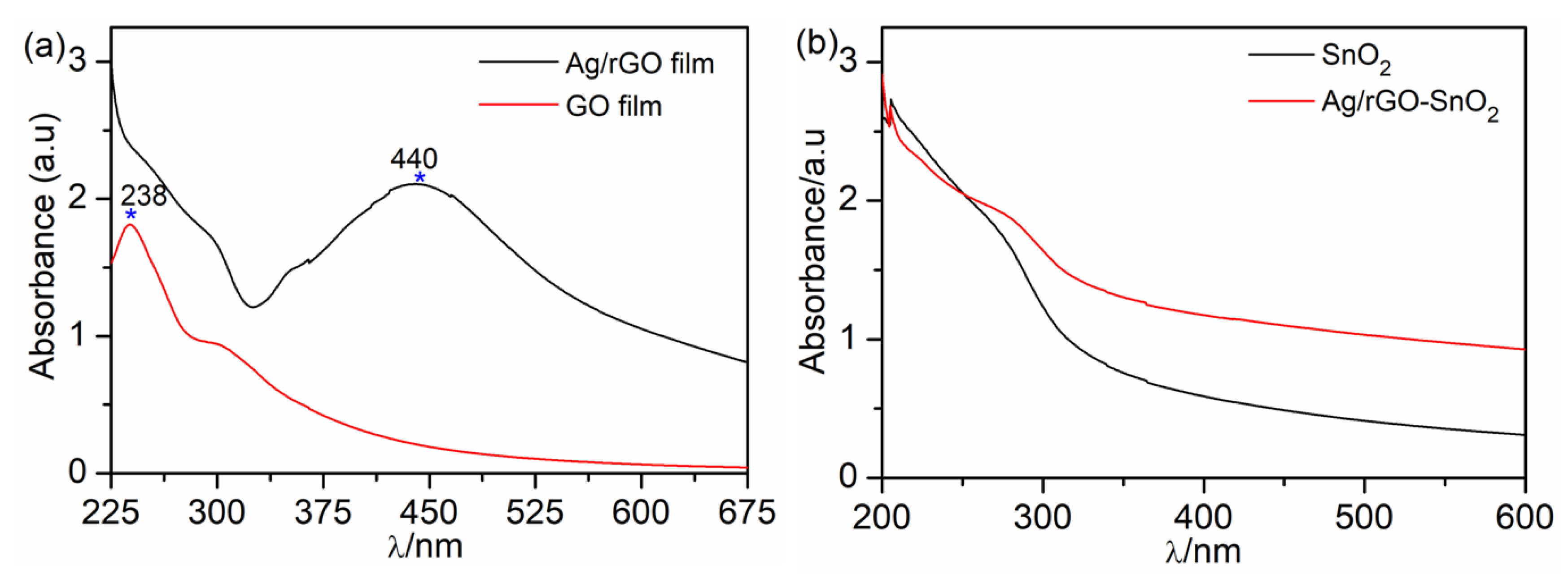
3. Results and Discussion

The synthesis mechanism for the formation of the Ag/rGO hybrid composite and plasmon-modified SnO2 composite is illustrated in Scheme 1. Firstly, ammonia solution was mixed with graphene (GO) and reacted with GO functional group to form a negative charge on the surface of GO [52]. After AgNO3 was added, the negatively-charged GO was combined with the positively-charged Ag+ ions to form a complex of Ag+-GO-NH3OH ions. Consequently, the ascorbic acid was added into the solution and heated at 110 °C for 2 h to induce reduction in both Ag+ and GO-NH3OH ions resulting into the formation of the Ag/rGO hybrid composite. Then, the Ag/rGO composite was incorporated into SnO2 nanorods.
The morphology of as-synthesized samples was examined by scanning electron microscopy (SEM). In Figure 2a, the SEM image shows that graphene (GO) sheets exhibit continuous wave-like sheets with flake-like shape and wrinkles on the surface, demonstrating their flexibility. Figure 2b shows the SEM image of Ag/reduced graphene oxide (Ag/rGO) in which Ag particles were embedded in the hybrid composite as well-dispersed minute particles, whereas graphene provides support and stability to Ag nanoparticles (NPs) from aggregation. Ag NPs act as zero-dimensional nanospacers to increase the interlayer spacing and to prevent the graphene sheets restacking. As shown in Figure 2c, the SnO2 nanorods were uniformly distributed in morphology and particle size. The SEM image of Ag/rGO incorporated into SnO2 (Ag/rGO-SnO2) in Figure 2d shows that SnO2 nanorods (NRs) were attached to the surface of Ag/rGO particles. The size of Ag NPs was reduced, which could be attributed to the incorporation of Ag/rGO into SnO2.
Structural properties of dried pristine graphite, graphene (GO), and Ag/reduced graphene oxide (Ag/rGO) composite are shown in Figure 3a. The typical X-ray diffraction (XRD) pattern of graphite displays a sharp peak located at around 26.38°, corresponding to an inter-layer spacing of 3.38 Ǻ between 002 atomic planes. The diffraction peak of GO was shifted to around 11.6°, which correspond to the inter-layer spacing of 7.64 Å for 001 atomic plane. This inter-layer spacing value was much larger than that of graphite, which confirmed the oxidation of graphite. While the diffraction peaks for the as-synthesized Ag/rGO hybrid composite revealed intense peaks at 2θ = 38.11°, 44.31°, 64.45°, and 77.41°, which were assigned to the (111), (200), (220), and (311) planes of Ag (JCPDS No. 065-8428), respectively, indicating the presence of Ag NPs in the face-centered cubic (fcc) phase.
The sharp diffraction peak for GO was absent in Ag/rGO, but a weak and broad peak appears in the range 20°–30°, attributed to a high-degree exfoliation of the graphene sheet in Ag/rGO due to the introduction of Ag NPs. The inter-layer spacing value of Ag/rGO was less than that of GO, suggesting the reduction of GO [53]. This result demonstrates that Ag NPs could be successfully incorporated with GO. Figure 3b shows the powder X-ray diffraction patterns of SnO2 nanorods and the incorporated optimized content of the Ag/rGO hybrid composite. It was observed that most diffraction peaks of SnO2 nanorods were well indexed to tetragonal rutile tin oxide (JCPDF: 41–1445). No clear diffraction peaks associated with reduced graphene oxide (rGO) were identified in composite films, which might be due to the low amount of Ag/rGO added into SnO2. The XRD analysis revealed that the lattice parameters (a, b, and c) of the tetragonal cell were achieved. The lattice parameters a and b of 1.0 wt.% Ag/rGO (a = b = 4.73 Å) became larger than that of SnO2 (a = b = 4.72 Å), while the lattice parameter of c showed negligible change (c = 7.17 Å) for both 1.0 wt.% Ag/rGO and SnO2. The enlargement of the unit cell could be due to the replacement of the smaller O2− by larger Ag+ and GO ions in the SnO2 cells. In addition, the average grain size of the 1.0 wt.% Ag/rGO crystallite estimated from the (110) Bragg reflection was ca. 106.8 nm according to Scherer’s equation, and the average grain size of the SnO2 crystallite was ca. 88.8 nm.
The Raman spectra of GO and Ag/rGO with their characteristic peaks were displayed in Figure 4a. The Raman spectra of GO exhibits the D band and G band located near 1580 and 1354 cm−1, respectively. It is noted that the intensity ratio of D and G for GO corresponds to 0.83, while the Raman spectra of the Ag/rGO hybrid composite consists of two peaks located at ~ 1354 and 1601 cm−1 corresponding to D and G bands of graphene with intensity ratio of 1.08. In contrast to GO, the response of Ag/rGO reveals sharp D and G peaks with an increase in D peak intensity relative to the G peak after reduction. The increased value of ID/IG for Ag/rGO conversely related to the broken hexagonal lattice structure and increased defects suggests a large disorder was induced when Ag NPs were assembled over graphene [54].
Hence, the main characteristic peaks of the Ag/rGO hybrid composite were significantly enhanced due to the strong plasmonic near field around silver nanoparticles. As shown in Figure 4b, the broad Raman spectrum of the Ag/rGO-SnO2 film was observed at 1353 and 1596 cm−1, corresponding to the relative intensity ratio value of 1.01. A slight red-shift was depicted at the lowest range of about 400–800 cm−1 related to SnO2 films in Ag/rGO-SnO2, which could be ascribed to the induced defects due to Ag/rGO loading [55]. Moreover, the G peak was down-shifted to 1596 cm−1on Ag/rGO-SnO2 film. This shift in G peak confirmed the occurrence of electron transfer between Ag/rGO and SnO2 in the Ag/rGO-SnO2 composite. This result suggests that electron transfer from Ag NPs to SnO2 occurs through sp2 carbon domains of graphene derivatives [56].
UV-Vis spectral analysis was conducted to examine the light absorption spectra of as-prepared samples. Figure 5a exhibits two important maxima related to GO at a peak near 238 nm, which corresponds to C–C bonds, and a shoulder at around 304 nm corresponds to transition of the C=O bond, which is attributed to the sp3 bonding in graphene [57]. The strong broad peak observed at ~440 nm is a characteristic of the Ag/rGO composite observed after Ag NPs were encapsulated with graphene nanosheets and reduced by ascorbic acid (Figure 5a) [58]. The UV-Vis absorption of the Ag/rGO red-shift to 440 nm and generate broadening as a results of strong light scattering due to the large size of nanoparticles and the formation of network structure [59,60] These experimental results suggest that absorbance of Ag/rGO could be spectrally tunable [61]. Figure 5b shows the UV-Vis spectra of SnO2 and Ag/rGO-SnO2. It could be seen that SnO2 exhibited absorption mainly in the UV region within 200 to 300 nm. The Ag/rGO-SnO2 showed the optical spectra in both the UV and the visible light region, which was significantly larger than that of SnO2. Compared to SnO2, the composite films exhibited a red-shift attributed to the optical vibration caused by surface plasmon resonance (SPR) of Ag. However, due to the low concentration of Ag and graphene, the increase of absorption of Ag/rGO-SnO2 is not prominent as it is over shadowed by more prominent SnO2 absorption [62,63,64]. Figure 6 illustrates the optical transmittance of SnO2, Ag/rGO, and Ag/rGO-SnO2. The transmittance of SnO2 and Ag/rGO film rises to 84% and 74% corresponding to wavelengths at 550 and 326 nm, respectively. The drop in transmittance of Ag/rGO is attributed to the imperfection resulting from polymeric material and graphene wrinkling [65]. It is well known that Ag atoms have high UV light absorption around the 400 nm region. However, the encapsulation of Ag by graphene as a protective layer blocks the formation of surface plasmon light and decreases the light absorption [66]. Compared to SnO2, the transmittance of Ag/rGO-SnO2 film has decreased to around 80% due to the impact of the Ag/rGO composite.
The photocurrent (J)–voltage (V) characteristics for UVPDs with optimized Ag/rGO incorporated into SnO2 as photoanodes, and compared to SnO2 photoanode under UV illumination, is shown in Figure 7a. Compared to pure SnO2 device, which shows a 0.14 mAcm−2 value, the Jsc of devices exhibited higher values when SnO2 nanorods were incorporated. A maximum value of Jsc = 0.29 mAcm−2 was obtained for 1.0 wt.% Ag/rGO hybrid composites. This increment could be ascribed to the enhancement of light absorption and the effective suppression of charge recombination. In order to examine and evaluate the photocurrent response in detail, device dynamic performances were measured under UV light irradiation of 365 nm at zero bias with a power of 40 mWcm−2.The illumination was periodically switched on:off at intervals of 10 seconds with incident light from solar simulator at zero bias. Figure 7b shows the photocurrent time response of these devices with different content of Ag/rGO incorporated into SnO2 and compared to SnO2 device. The repeatable cycles observed in photocurrent signals without decay demonstrates an outstanding reproducibility and stability for the UVPDs. The rise time and decay time of the photocurrent response was calculated from data of Figure 7b. The rise time (t r) is often defined as the time taken by the photocurrent to increase from 0% to 63% (i.e., 1–1/e) of the maximum value, and the decay time (t d) is the time taken by the photocurrent to decrease from the maximum value to 37% (i.e., 1/e) [13,67]. This device exhibited a rapid dynamic response for rise and decay time (as shown in Figure 7c). The τr and τd values for UVPDs with the 1.0 wt.% Ag/rGO photoanode were estimated to be 0.341 s and 0.068 s for the J, respectively. The τr and τd values for UVPD with the 1.5 wt.% Ag/rGO photoanode were estimated to be 0.164 s and 0.112 s, respectively, as shown in Figure S1.
The τr and τd values for UVPDs of the free-modified SnO2 photoanode were 0.13 s and 0.087 s, respectively, as shown in Figure 7d. The rapid rise time and decay time for UVPD with 1.0 wt.% Ag/rGO were shorter than that of the free-modified SnO2 photoanode device, which reveal that Ag/rGO can significantly enhance the sensitivity of UVPD. The Ag/rGO-incorporated SnO2 photoanode-based UVPD demonstrated a higher photocurrent, higher on:off ratio, and faster rise and decay time compared to the free-modified SnO2 photoanode, as summarized in Table 1. This device exhibited an on:off photocurrent contrast ratio (ratio between photocurrent and dark current) reaching the highest value of 195, compared to 66 for the free-modified SnO2-based device, suggesting that the UV photodetector would be suitable for measurement of UV light.
In addition, the TiCl4-modified SnO2 and TiCl4-treated Ag/rGO-modified SnO2 photoanodes were employed to assemble UVPDs. As shown in Figure 8a, the photocurrent–voltage characteristics of devices based on TiCl4-treated SnO2 photoanodes exhibit a significant increase. In particular, the device modified with 1.0 wt.% Ag/rGO displays a Jsc of 5.4 mAcm−2.
This enhancement is attributed to the suppressed charge recombination. Figure 8b shows the photocurrent time response of these devices with 1.0 wt.% Ag/rGO incorporated into SnO2 and compared to free-modified SnO2 device. The τr and τd values for UVPDs with 1.0 wt.% Ag/rGO photoanode were estimated to be 0.341 and 0.068 s. These are the values reported for data of Figure 8c. This device exhibits an on:off photocurrent contrast ratio reaching the highest value of 2740, compared to 544.5 for the free-modified SnO2-based device. In summary, the UVPD assembled with the Ag/rGO-incorporated SnO2 photoanode treated by TiCl4 exhibits a higher photocurrent, higher on:off ratio, and faster rise and decay times compared to that of the free-modified SnO2 photoanode, as summarized in Table 2. The increment in photocurrent is attributed to the presence of more photogenerated charge carriers produced by higher absorbed photon flux, proposing the UV photodetector be suitable for measurement of UV light. Figure 8d shows the photocurrent vs. the intensity of light for 1.0 wt.% Ag/rGO-based UVPD illuminated by UV light irradiation (λ = 365 nm) with incident light power at zero bias. The J increased linearly as the light intensity increases from 1 to 40 mWcm−2 for the UVPD-3. This increment in photocurrent varied linearly with the increase in the intensity of UV light, proposing the UV photodetector may be best suited for measurement of UV light.

4. Conclusions

In summary, we successfully prepared Ag/rGO incorporated into SnO2 nanorods by hydrothermal method. Ag/rGO-SnO2-based PEC-type self-powered UV photodetectors were fabricated incorporating Ag/rGO into SnO2 nanorods used as photoanodes. Compared to UVPDs based on pure SnO2 photoanodes, the optimized UVPD with 1.0 wt.% Ag/rGO demonstrated a higher photocurrent benefiting from plasmon-enhanced UV-absorption and the effective suppression of the charge recombination at the contact interface. Moreover, this device showed a higher on:off photocurrent ratio of 195 anda rapid response speed (rise time 0.341 s and decay time 0.068 s). The self-powered PEC-type UVPD based on the TiCl4-treated Ag/rGO-SnO2 photoanode showed a high on:off ratio of 1441, a fast rise time and decay time (τr = 0.072 s and τd = 0.05 s), together with outstanding optical signal response and good reproducibility in a wide range of switching frequencies.

Supplementary Materials

The following are available online at https://www.mdpi.com/2073-4352/9/12/648/s1, Figure S1: the enlarged rising and decaying of edges of the photocurrent response ultraviolet photodetector based on SnO2 incorporated with 1.5 wt.% Ag/rGO composite.

Author Contributions

J.T.A. investigated and wrote the original draft; J.W. supervise the work, D.W. reviewing and editing.

Funding

This research was funded by the National Natural Science Foundation of China (Nos. 51502056 and 51202046), the National Science and Technology Support Program (No. 2015BAI01B05), the Fundamental Research Funds for the Central Universities (Nos. HIT.NSRIF.2013006 and HIT.BRETIII.201403), the Natural Science Foundation of Heilongjiang Province of China (No. F2016013), and the Open Project Program of Key Laboratory for Photonic and Electric Bandgap Materials, Ministry of Education, Harbin Normal University, China.

Conflicts of Interest

The authors declare no conflicts of interest.

References

  1. Ben Elkamel, I.; Hamdaoui, N.; Mezni, A.; Ajjel, R.; Beji, L. Synthesis and characterization of Cu doped ZnO nanoparticles for stable and fast response UV photodetector at low noise current. J. Mater. Sci. Mater. Electron. 2019, 30, 9444–9454. [Google Scholar] [CrossRef]
  2. Huang, G.; Zhang, P.; Bai, Z. Self-powered UV–visible photodetectors based on ZnO/graphene/CdS/electrolyte heterojunctions. J. Alloys Compd. 2019, 776, 346–352. [Google Scholar] [CrossRef]
  3. Fang, H.; Zheng, C.; Wu, L.; Li, Y.; Cai, J.; Hu, M.; Fang, X.; Ma, R.; Wang, Q.; Wang, H. Solution-Processed Self-Powered Transparent Ultraviolet Photodetectors with Ultrafast Response Speed for High-Performance Communication System. Adv. Funct. Mater. 2019, 29, 1809013. [Google Scholar] [CrossRef]
  4. Singh, M.K.; Pandey, R.K.; Prakash, R. High-performance photo detector based on hydrothermally grown SnO2 nanowire/reduced graphene oxide (rGO) hybrid material. Org. Electron. 2017, 50, 359–366. [Google Scholar] [CrossRef]
  5. Wang, W.; Yang, Z.; Lu, Z.; Li, G. High responsivity and low dark current nonpolar GaN-based ultraviolet photo-detectors. J. Mater. Chem. C 2018, 6, 6641–6646. [Google Scholar] [CrossRef]
  6. Sarkar, K.; Hossain, M.; Devi, P.; Rao, K.D.M.; Kumar, P. Self-Powered and Broadband Photodetectors with GaN: Layered rGO Hybrid Heterojunction. Adv. Mater. Interfaces 2019, 6, 1900923. [Google Scholar] [CrossRef]
  7. Shelke, N.T.; Karche, B.R. Hydrothermal synthesis of WS2/RGO sheet and their application in UV photodetector. J. Alloys Compd. 2015, 653, 298–303. [Google Scholar] [CrossRef]
  8. Huang, Z.; Han, W.; Tang, H.; Ren, L.; Chander, D.S.; Qi, X.; Zhang, H. Photoelectrochemical-type sunlight photodetector based on MoS2/graphene heterostructure. 2D Mater. 2015, 2, 035011. [Google Scholar] [CrossRef]
  9. Choi, H.; Seo, S.; Lee, J.H.; Hong, S.H.; Song, J.; Kim, S.; Yim, S.Y.; Lee, K.; Park, S.J.; Lee, S. Solution-processed ZnO/SnO2 bilayer ultraviolet phototransistor with high responsivity and fast photoresponse. J. Mater. Chem. C 2018, 6, 6014–6022. [Google Scholar] [CrossRef]
  10. Paria, D.; Jeong, H.H.; Vadakkumbatt, V.; Deshpande, P.; Fischer, P.; Ghosh, A.; Ghosh, A. Graphene-silver hybrid devices for sensitive photodetection in the ultraviolet. Nanoscale 2018, 10, 7685–7693. [Google Scholar] [CrossRef]
  11. Deka Boruah, B. Zinc oxide ultraviolet photodetectors: Rapid progress from conventional to self-powered photodetectors. Nanoscale Adv. 2019, 1, 2059–2085. [Google Scholar] [CrossRef] [Green Version]
  12. Tian, W.; Wang, Y.; Chen, L.; Li, L. Self-Powered Nanoscale Photodetectors. Small 2017, 13. [Google Scholar] [CrossRef] [PubMed] [Green Version]
  13. Li, X.D.; Gao, C.T.; Duan, H.G.; Lu, B.G.; Pan, X.J.; Xie, E.Q. Nanocrystalline TiO2 film based photoelectrochemical cell as self-powered UV-photodetector. Nano Energy 2012, 1, 640–645. [Google Scholar] [CrossRef]
  14. Lin, C.H.; Fu, H.C.; Lien, D.H.; Hsu, C.Y.; He, J.H. Self-powered nanodevices for fast UV detection and energy harvesting using core-shell nanowire geometry. Nano Energy 2018, 51, 294–299. [Google Scholar] [CrossRef]
  15. Bai, Z.; Liu, J.; Liu, F.; Zhang, Y. Enhanced photoresponse performance of self-powered UV–visible photodetectors based on ZnO/Cu2O/electrolyte heterojunctions via graphene incorporation. J. Alloys Compd. 2017, 726, 803–809. [Google Scholar] [CrossRef]
  16. Abdalla, J.T.; Huang, Y.W.; Yu, Q.J.; Wang, J.Z.; Wang, J.N.; Yu, C.L.; Gao, S.Y.; Jiao, S.J.; Wang, D.B.; Alarabi, A.M.; et al. TiCl4 surface-treated SnO2 photoanodes for self-powered UV photodetectors and dye-sensitized solar cells. Mater. Technol. 2017, 32, 443–450. [Google Scholar] [CrossRef]
  17. Amany, A.; Wang, D.B.; Wang, J.Z.; Zeng, Z.; Jiao, S.J.; Hao, C.L.; Zhao, Y.C.; Xie, Y.; Gao, S.Y.; Ni, S.M.; et al. Enhanced the UV response of AlN coated ZnO nanorods photodetector. J. Alloys Compd. 2019, 776, 111–115. [Google Scholar] [CrossRef]
  18. Ni, S.M.; Guo, F.Y.; Wang, D.B.; Liu, G.; Xu, Z.K.; Kong, L.P.; Wang, J.Z.; Jiao, S.J.; Zhang, Y.; Yu, Q.J.; et al. Effect of MgO Surface Modification on the TiO2 Nanowires Electrode for Self-Powered UV Photodetectors. ACS Sustain. Chem. Eng. 2018, 6, 7265–7272. [Google Scholar] [CrossRef]
  19. Kar, A.; Kundu, S.; Patra, A. Surface Defect-Related Luminescence Properties of SnO2 Nanorods and Nanoparticles. J. Phys. Chem. C 2011, 115, 118–124. [Google Scholar] [CrossRef]
  20. Katoh, R.; Furube, A.; Yoshihara, T.; Hara, K.; Fujihashi, G.; Takano, S.; Murata, S.; Arakawa, H.; Tachiya, M. Efficiencies of electron injection from excited N3 dye into nanocrystalline semiconductor (ZrO2, TiO2, ZnO, Nb2O5, SnO2, In2O3) films. J. Phys. Chem. B 2004, 108, 4818–4822. [Google Scholar] [CrossRef]
  21. Gao, C.; Li, X.; Wang, Y.; Chen, L.; Pan, X.; Zhang, Z.; Xie, E. Titanium dioxide coated zinc oxide nanostrawberry aggregates for dye-sensitized solar cell and self-powered UV-photodetector. J. Power Sources 2013, 239, 458–465. [Google Scholar] [CrossRef]
  22. Gao, C.T.; Li, X.D.; Zhu, X.P.; Chen, L.L.; Wang, Y.Q.; Teng, F.; Zhang, Z.X.; Duan, H.G.; Xie, E.Q. High performance, self-powered UV-photodetector based on ultrathin, transparent, SnO2-TiO2 core-shell electrodes. J. Alloys Compd. 2014, 616, 510–515. [Google Scholar] [CrossRef]
  23. Wang, Z.R.; Ran, S.H.; Liu, B.; Chen, D.; Shen, G.Z. Multilayer TiO2 nanorod cloth/nanorod array electrode for dye-sensitized solar cells and self-powered UV detectors. Nanoscale 2012, 4, 3350–3358. [Google Scholar] [CrossRef] [PubMed]
  24. Xie, Y.; Wei, L.; Li, Q.; Chen, Y.; Yan, S.; Jiao, J.; Liu, G.; Mei, L. High-performance self-powered UV photodetectors based on TiO2 nano-branched arrays. Nanotechnology 2014, 25, 075202. [Google Scholar] [CrossRef]
  25. Xie, Y.R.; Wei, L.; Li, Q.H.; Chen, Y.X.; Liu, H.; Yan, S.S.; Jiao, J.; Liu, G.L.; Mei, L.M. A high performance quasi-solid-state self-powered UV photodetector based on TiO2 nanorod arrays. Nanoscale 2014, 6, 9116–9121. [Google Scholar] [CrossRef]
  26. Elumalai, N.K.; Jose, R.; Archana, P.S.; Chellappan, V.; Ramakrishna, S. Charge Transport through Electrospun SnO2 Nanoflowers and Nanofibers: Role of Surface Trap Density on Electron Transport Dynamics. J. Phys. Chem. C 2012, 116, 22112–22120. [Google Scholar] [CrossRef]
  27. Zamand, N.; Pour, A.N.; Housaindokht, M.R.; Izadyar, M. Size-controlled synthesis of SnO2 nanoparticles using reverse microemulsion method. Solid State Sci. 2014, 33, 6–11. [Google Scholar] [CrossRef]
  28. Vasquez, F.C.; Paraguay-Delgado, F.; Morales-Mendoza, J.E.; Antunez-Flores, W.; Lardizabal, D.; Alonso-Nunez, G.; Berhault, G. Shape and size controlled growth of SnO2 nano-particles by efficient approach. Superlattices Microstruct. 2016, 90, 274–287. [Google Scholar] [CrossRef]
  29. Sephra, P.J.; Baraneedharan, P.; Sivakumar, M.; Thangadurai, T.D.; Nehru, K. Size controlled synthesis of SnO2 and its electrostatic self-assembly over reduced graphene oxide for photocatalyst and supercapacitor application. Mater. Res. Bull. 2018, 106, 103–112. [Google Scholar] [CrossRef]
  30. Lee, J.W.; Nam, S.H.; Yu, J.N.; Kim, D.I.; Jeong, R.H.; Boo, J.H. Shape Control of Zn2SnO4/SnO2 Composites and Changes in Photocatalytic Efficiency. Sci. Adv. Mater. 2018, 10, 1045–1050. [Google Scholar] [CrossRef]
  31. Kaneti, Y.V.; Yue, J.; Moriceau, J.; Chen, C.Y.; Liu, M.S.; Yuan, Y.; Jiang, X.C.; Yu, A.B. Experimental and theoretical studies on noble metal decorated tin oxide flower-like nanorods with high ethanol sensing performance. Sens. Actuators B Chem. 2015, 219, 83–93. [Google Scholar] [CrossRef]
  32. Chew, C.K.T.; Salcianu, C.; Bishop, P.; Carmalt, C.J.; Parkin, I.P. Functional thin film coatings incorporating gold nanoparticles in a transparent conducting fluorine doped tin oxide matrix. J. Mater. Chem. C 2015, 3, 1118–1125. [Google Scholar] [CrossRef]
  33. Sevastyanov, E.Y.; Maksimova, N.K.; Novikov, V.A.; Rudov, F.V.; Sergeychenko, N.V.; Chernikov, E.V. Effect of Pt, Pd, Au additives on the surface and in the bulk of tin dioxide thin films on the electrical and gas-sensitive properties. Semiconductors 2012, 46, 801–809. [Google Scholar] [CrossRef]
  34. Hung-Low, F.; Ramirez, D.A.; Peterson, G.R.; Hikal, W.M.; Hope-Weeks, L.J. Development of a carbon-supported Sn-SnO2 photocatalyst by a new hybridized sol-gel/dextran approach. RSC Adv. 2016, 6, 21019–21025. [Google Scholar] [CrossRef]
  35. Rakibuddin, M.; Ananthakrishnan, R. Fabrication of graphene aerosol hybridized coordination polymer derived CdO/SnO2 heteronanostructure with improved visible light photocatalytic performance. Sol. Energy Mater. Sol. Cells 2017, 162, 62–71. [Google Scholar] [CrossRef]
  36. Zeng, Y.; Pan, X.; Dai, W.; Chen, Y.; Ye, Z. The enhancement of a self-powered UV photodetector based on vertically aligned Ag-modified ZnO nanowires. RSC Adv. 2015, 5, 66738–66741. [Google Scholar] [CrossRef]
  37. N’Konou, K.; Chalh, M.; Monnier, V.; Blanchard, N.P.; Chevolot, Y.; Lucas, B.; Vedraine, S.; Torchio, P. Impact of Ag@SiO2 core-shell nanoparticles on the photoelectric current of plasmonic inverted organic solar cells. Synth. Met. 2018, 239, 22–28. [Google Scholar] [CrossRef]
  38. Zhou, M.J.; Li, J.Z.; Ye, Z.F.; Ma, C.C.; Wang, H.Q.; Huo, P.W.; Shi, W.D.; Yan, Y.S. Transfer Charge and Energy of Ag@CdSe QDs-rGO Core-Shell Plasmonic Photocatalyst for Enhanced Visible Light Photocatalytic Activity. ACS Appl. Mater. Interface 2015, 7, 28231–28243. [Google Scholar] [CrossRef]
  39. An, X.; Teng, F.; Zhang, Z.; Pan, X.; Zhou, J.; Xie, E. Ultrafast UV switch based on ZnO-Ag heterostructures. Electron. Mater. Lett. 2014, 10, 95–99. [Google Scholar] [CrossRef]
  40. Dong, H.; Wu, Z.X.; Lu, F.; Gao, Y.C.; El-Shafei, A.; Jiao, B.; Ning, S.Y.; Hou, X. Optics-electrics highways: Plasmonic silver nanowires@TiO2 core-shell nanocomposites for enhanced dye-sensitized solar cells performance. Nano Energy 2014, 10, 181–191. [Google Scholar] [CrossRef]
  41. Pu, Y.C.; Wang, G.M.; Chang, K.D.; Ling, Y.C.; Lin, Y.K.; Fitzmorris, B.C.; Liu, C.M.; Lu, X.H.; Tong, Y.X.; Zhang, J.Z.; et al. Au Nanostructure-Decorated TiO2 Nanowires Exhibiting Photoactivity Across Entire UV-Visible Region for Photoelectrochemical Water Splitting. Nano Lett 2013, 13, 3817–3823. [Google Scholar] [CrossRef]
  42. Teng, F.; Zhang, G.Z.; Wang, Y.Q.; Gao, C.T.; Chen, L.L.; Zhang, P.; Zhang, Z.X.; Xie, E.Q. The role of carbon in the photocatalytic reaction of carbon/TiO2 photocatalysts. Appl. Surf. Sci. 2014, 320, 703–709. [Google Scholar] [CrossRef]
  43. Williams, G.; Kamat, P.V. Graphene-Semiconductor Nanocomposites: Excited-State Interactions between ZnO Nanoparticles and Graphene Oxide. Langmuir 2009, 25, 13869–13873. [Google Scholar] [CrossRef]
  44. Zhang, L.-S.; Jiang, L.-Y.; Yan, H.-J.; Wang, W.D.; Wang, W.; Song, W.-G.; Guo, Y.-G.; Wan, L.-J. Mono dispersed SnO2 nanoparticles on both sides of single layer graphene sheets as anode materials in Li-ion batteries. J. Mater. Chem. 2010, 20. [Google Scholar] [CrossRef]
  45. Tian, Y.; Cao, Y.-Y.; Pang, F.; Chen, G.-Q.; Zhang, X. Ag nanoparticles supported on N-doped graphene hybrids for catalytic reduction of 4-nitrophenol. RSC Adv. 2014, 4, 43204–43211. [Google Scholar] [CrossRef]
  46. Keshvari, A.; Darbari, S.; Taghavi, M. Self-Powered Plasmonic UV Detector, Based on Reduced Graphene Oxide/Ag Nanoparticles. IEEE Electron Device Lett. 2018, 39, 1433–1436. [Google Scholar] [CrossRef]
  47. Huang, Y.W.; Yu, Q.J.; Wang, J.Z.; Wang, J.A.; Yu, C.L.; Abdalla, J.T.; Zeng, Z.; Jiao, S.J.; Wang, D.B.; Gao, S.Y. Plasmon-Enhanced Self-Powered UV Photodetectors Assembled by Incorporating Ag@SiO2 Core-Shell Nanoparticles into TiO2 Nanocube Photoanodes. ACS Sustain. Chem. Eng. 2018, 6, 438–446. [Google Scholar] [CrossRef]
  48. Cote, L.J.; Kim, F.; Huang, J.X. Langmuir-Blodgett Assembly of Graphite Oxide Single Layers. J. Am. Chem. Soc. 2009, 131, 1043–1049. [Google Scholar] [CrossRef]
  49. Lim, B.C.; Singu, B.S.; Hong, S.E.; Na, Y.H.; Yoon, K.R. Synthesis and characterization nanocomposite of polyacrylamide-rGO-Ag-PEDOT/PSS hydrogels by photo polymerization method. Polym. Adv. Technol. 2016, 27, 366–373. [Google Scholar] [CrossRef]
  50. Kar, A.; Sain, S.; Kundu, S.; Bhattacharyya, A.; Pradhan, S.K.; Patra, A. Influence of Size and Shape on the Photocatalytic Properties of SnO2 Nanocrystals. Chemphyschem 2015, 16, 1017–1025. [Google Scholar] [CrossRef]
  51. Cojocaru, L.; Olivier, C.; Toupance, T.; Sellier, E.; Hirsch, L. Size and shape fine-tuning of SnO2 nanoparticles for highly efficient and stable dye-sensitized solar cells. J. Mater. Chem. A 2013, 1, 13789–13799. [Google Scholar] [CrossRef]
  52. Samantara, A.K.; Mishra, D.K.; Suryawanshi, S.R.; More, M.A.; Thapa, R.; Late, D.J.; Jena, B.K.; Rout, C.S. Facile synthesis of Ag nanowire–rGO composites and their promising field emission performance. RSC Adv. 2015, 5, 41887–41893. [Google Scholar] [CrossRef]
  53. Lee, S.C.; Some, S.; Kim, S.W.; Kim, S.J.; Seo, J.; Lee, J.; Lee, T.; Ahn, J.H.; Choi, H.J.; Jun, S.C. Efficient Direct Reduction of Graphene Oxide by Silicon Substrate. Sci. Rep. 2015, 5, 12306. [Google Scholar] [CrossRef] [Green Version]
  54. Ferrari, A.C.; Robertson, J. Interpretation of Raman spectra of disordered and amorphous carbon. Phys. Rev. B 2000, 61, 14095–14107. [Google Scholar] [CrossRef] [Green Version]
  55. Elias, D.C.; Nair, R.R.; Mohiuddin, T.M.G.; Morozov, S.V.; Blake, P.; Halsall, M.P.; Ferrari, A.C.; Boukhvalov, D.W.; Katsnelson, M.I.; Geim, A.K.; et al. Control of Graphene’s Properties by Reversible Hydrogenation: Evidence for Graphane. Science 2009, 323, 610–613. [Google Scholar] [CrossRef] [Green Version]
  56. Kim, Y.K.; Ok, G.; Choi, S.W.; Jang, H.; Min, D.H. The interfacing structural effect of Ag/graphene oxide nanohybrid films on surface enhanced Raman scattering. Nanoscale 2017, 9, 5872–5878. [Google Scholar] [CrossRef]
  57. Akbari, E.; Akbari, I.; Ebrahimi, M.R. sp2/sp3 bonding ratio dependence of the band-gap in graphene oxide. Eur. Phys. J. B 2019, 92, 71. [Google Scholar] [CrossRef]
  58. Xu, W.P.; Zhang, L.C.; Li, J.P.; Lu, Y.; Li, H.H.; Ma, Y.N.; Wang, W.D.; Yu, S.H. Facile synthesis of silver@graphene oxide nanocomposites and their enhanced antibacterial properties. J. Mater. Chem. 2011, 21, 4593–4597. [Google Scholar] [CrossRef]
  59. Hsu, K.C.; Chen, D.H. Green synthesis and synergistic catalytic effect ofAg/reduced graphene oxide nanocomposite. Nanoscale Res. Lett. 2014, 9, 484. [Google Scholar] [CrossRef] [Green Version]
  60. Beyene, H.T.; Tichelaar, F.D.; Peeters, P.; Kolev, I.; van de Sanden, M.C.M.; Creatore, M. Hybrid Sputtering-Remote PECVD Deposition of Au Nanoparticles on SiO2 Layers for Surface Plasmon Resonance-Based Colored Coatings. Plasma Process. Polym. 2010, 7, 657–664. [Google Scholar] [CrossRef]
  61. Li, J.; Liu, C.-Y. Ag/Graphene Heterostructures: Synthesis, Characterization and Optical Properties. Eur. J. Inorg. Chem. 2010, 2010, 1244–1248. [Google Scholar] [CrossRef]
  62. Khadgi, N.; Li, Y.; Upreti, A.R.; Zhang, C.; Zhang, W.; Wang, Y.; Wang, D. Enhanced Photocatalytic Degradation of 17alpha-Ethinylestradiol Exhibited by Multifunctional ZnFe2O4 -Ag/rGO Nanocomposite Under Visible Light. Photochem. Photobiol. 2016, 92, 238–246. [Google Scholar] [CrossRef]
  63. Cao, X.; Gu, L.; Lan, X.; Zhao, C.; Yao, D.; Sheng, W. Spinel ZnFe2O4 nanoplates embedded with Ag clusters: Preparation, characterization, and photocatalytic application. Mater. Chem. Phys. 2007, 106, 175–180. [Google Scholar] [CrossRef]
  64. Xie, W.; Tang, L.; Ying, M.; Liu, J.; Pan, H.; Du, M. Ag–SnO2 nano-heterojunction–reduced graphene oxide by a stepwise photocatalyzed approach and its application in ractopamine determination. RSC Adv. 2017, 7, 54506–54511. [Google Scholar] [CrossRef] [Green Version]
  65. Lee, J.; Lee, A.; Yu, H.K. Graphene protected Ag nanowires: Blocking of surface migration for thermally stable and wide-range-wavelength transparent flexible electrodes. RSC Adv. 2016, 6, 84985–84989. [Google Scholar] [CrossRef]
  66. Kim, J.M.; Seo, S.W.; Shin, D.H.; Lee, H.S.; Kim, J.H.; Jang, C.W.; Kim, S.; Choi, S.H. Ag-nanowires-doped graphene/Si Schottky-junction solar cells encapsulated with another graphene layer. Curr. Appl. Phys. 2017, 17, 1136–1141. [Google Scholar] [CrossRef]
  67. Zhou, J.Y.; Chen, L.L.; Wang, Y.Q.; He, Y.M.; Pan, X.J.; Xie, E.Q. An overview on emerging photoelectrochemical self-powered ultraviolet photodetectors. Nanoscale 2016, 8, 50–73. [Google Scholar] [CrossRef]
Figure 1. The structure of Ag/reduced graphene oxide-SnO2 based photoelectrochemical self-powered ultraviolet photodetector.
Crystals 09 00648 g001
Scheme 1. Preparation of Ag/reduced graphene oxide (Ag/rGO) and plasmon-modified SnO2 (Ag/rGO- SnO2) composites.
Crystals 09 00648 sch001
Figure 2. Morphological characterization: (ad) top surface scanning electron microscopy (SEM) images of (a) graphene (GO), (b) Ag/reduced graphene oxide (Ag/rGO), (c) SnO2 nanorods (NRs) and (d) 1.0 wt.% Ag/rGO incorporated into SnO2 nanorods (1.0 wt.% Ag/rGO).
Crystals 09 00648 g002
Figure 3. Diffraction (XRD) patterns of (a) graphite, graphene, and Ag/reduced graphene oxide (Ag/rGO) hybrid composite; (b) SnO2 nanorods and 0.5 wt.% Ag/rGO, 1.0 wt.% Ag/rGO 1.5 wt.% Ag/rGO hybrid composite, respectively, incorporated into SnO2 nanorods.
Crystals 09 00648 g003
Figure 4. Raman spectra of (a) graphene (GO) and Ag/reduced graphene oxide (Ag/rGO) films; (b) Ag/rGO, SnO2, and Ag/rGO incorporated into SnO2 (Ag/rGO-SnO2).
Crystals 09 00648 g004
Figure 5. UV-Vis absorbance of (a) graphene (GO) and Ag/reduced graphene oxide (Ag/rGO) films; (b) SnO2, Ag/rGO, and Ag/rGO incorporated into SnO2 (Ag/rGO-SnO2) composite.
Crystals 09 00648 g005
Figure 6. UV-Vis transmittance spectra for SnO2, Ag/reduced graphene oxide (Ag/rGO) hybrid composite, and Ag/rGO incorporated into SnO2 (Ag/rGO-SnO2) composite.
Crystals 09 00648 g006
Figure 7. (a) Photocurrent density (J) – voltage (V) characteristics and (b) photocurrent responses of ultraviolet photodetectors (UVPDs) based on the pure SnO2 photoanode and 0.5 wt.% Ag/rGO, 1.0 wt.% Ag/rGO, and1.5 wt.% Ag/rGO hybrid composite incorporated into SnO2photoanodesunder an irradiance of 40 mW cm−2 UV light (λ = 365 nm). (c,d) Enlarged rising and decaying edges of the photocurrent response for the Ag/rGO-SnO2 and SnO2-based UVPDs.
Crystals 09 00648 g007
Figure 8. (a) JV characteristics and (b) photocurrent responses of UVPDs based on the pure SnO2 (UVPD-1), TiCl4-treated SnO2 (UVPD-2), and 1.0 wt.% Ag/rGO (UVPD-3) under an irradiance of 40 mW cm−2 UV light (λ = 365 nm). (c) Enlarged rising and decaying edges of the photocurrent response for the 1.0 wt.% Ag/rGO. (d) J as a function of the incident UV light intensity from 1 to 40 mW cm−2.
Crystals 09 00648 g008
Table 1. Detailed data of short-circuit photocurrent density (Jsc), On:Off ratio, rise time and decay time for ultraviolet photodetector based on pure SnO2 and different contents of Ag/rGO incorporated into SnO2.
UVPDJsc (mAcm−2)On:Off RatioRise Time (s)Decay Time (s)
Pure SnO20.14660.130.087
0.5 wt.% Ag/rGO0.07410.4210.104
1.0 wt.% Ag/rGO0.291950.3410.068
1.5 wt.% Ag/rGO0.271350.1640.112
Table 2. Detailed data of short-circuit photocurrent density (Jsc), On:Off ratio, rise time and decay time for ultraviolet photodetector based on pure SnO2 (UVPD-1), TiCl4-treated SnO2 (UVPD-2) and 1.0 wt.% Ag/rGO incorporated into SnO2 (UVPD-3).
UVPDsJsc(mAcm−2)On:Off RatioRise Time (s)Decay Time (s)
UVPD-10.14660.130.087
UVPD-23.75440.140.05
UVPD-35.414410.0720.05

