Article

Hydrothermal Synthesis of (001) Facet Highly Exposed ZnO Plates: A New Insight into the Effect of Citrate

1
Department of Chemical Engineering, Tsinghua University, Beijing 100084, China
2
Key Laboratory of Synthetic and Biological Colloids, Ministry of Education, School of Chemical and Material Engineering, Jiangnan University, Wuxi 214122, China
*
Authors to whom correspondence should be addressed.
Crystals 2019, 9(11), 552; https://doi.org/10.3390/cryst9110552
Submission received: 22 September 2019 / Revised: 16 October 2019 / Accepted: 22 October 2019 / Published: 24 October 2019
(This article belongs to the Special Issue Zinc Oxide Nanomaterials and Based Devices)

Abstract

:
In this work, two synthesis routes were applied to investigate the effect of citrates on the construction of the ZnO structure. Well-dispersed ZnO plates with (001) facet highly exposed were prepared via one-step hydrothermal route, while ZnO nanoparticles were obtained via two-step route. In one-step route, citrates were added before the formation of Zn(OH)2 precursor, while citrates were added after the formation of Zn(OH)2. For the first time, the interaction between citrates and the Zn(OH)2 precursor was investigated and citrates that participated in the formation of Zn(OH)2 were the main cause for (001) facet exposed structure construction. A growth mechanism about the formation of ZnO plates in the presence of citrates was proposed. The as-prepared ZnO plates showed enhanced photocatalytic activity for the degradation of methylene blue (MB).

1. Introduction

Nanocrystals with high surface energy tend to have exceptional properties in practical applications [1]. ZnO is a promising semiconductor material which has been widely studied in many fields and it was found that ZnO with (001) facet highly exposed shows excellent performance in gas sensing [2], photocatalysis [3] and dye-sensitized solar cells [4]. However, (001) facet of ZnO tends to disappear during crystal growth due to its high surface energy, resulting in the intrinsic 1D structures such as whiskers, nanobelts and nanowires [5]. Delamination [6], catalyst-assisted CVD [7] and other methods have been developed to construct (001) facet exposed ZnO structures. Compared to the demanding condition or complex operation of other methods, capping-molecule-assisted solution synthesis provides a more practical, controllable and expandable way to get (001) facet exposed ZnO structures. For example, n-butylamine [8], sodium sulfosuccinate [9] and ethylenediamine [10] have been wildly used as capping molecule in construction of (001) facet exposed ZnO structures.
Citrates, a commonly used capping-agent and reductant in controlling metal nanocrystals structures [11,12,13], is found to be effective in controlling the growth of ZnO, especially for inhibiting the growth along [001] direction [14]. ZnO with many different morphologies such as stacked-columns [15], nanosheets [16], microsphere [17] and dumbbell [18] have been synthesized by using citrate-based additives. The directing effect is achieved by the selective combination between (001) facet and citrate [16,19,20]. David et al. [21] found that citrates can only adsorb on the (001) facet by the comparison of zeta potentials of ZnO before and after adsorption of citrates. Kathryn et al. [22] proposed that the 3-carboxylate structure has a stronger suppression effect on the axial ([001]) growth than 2-carboxylate and 1-carboxylate structure ligands. Somnath et al. [23] reported that the selective binding of citrates and (001) facet was attributed to the highest Zn2+ density of this facet. In previous reports, research on the effect of citrate mainly concentrated on the interaction between citrates and ZnO. For solution-based ZnO synthesis, the formation of ZnO undergoes the transformation from Zn(OH)2 precursor. There is no study about interaction between citrates and the precursor, which is of vital importance in the morphology evolution. Investigations on the effect of citrates on Zn(OH)2 are necessary for understanding the influence of citrate in the ZnO formation.
Here, we report a facile hydrothermal way to synthesize well-dispersed ZnO hexagonal plates with (001) facet highly exposed in the presence of sodium citrates. ZnO plates with a diameter of 1 μm and a thickness of 100 nm were obtained. Compared with ZnO nanorods, the plates showed better photocatalytic activity on the degradation of methylene blue (MB) due to their highly (001) facet exposed structure. For the first time, the influence of sodium citrates on the Zn(OH)2 precursor was investigated, and the formation mechanism of ZnO plates evolved from Zn(OH)2 in the presence of citrates was proposed.

2. Materials and Methods

2.1. Materials Synthesis

Commercial chemicals with analytical grade and deionized water with a resistivity >18 MΩ·cm−1 were used in the experiments.
Two routes were applied to synthesize ZnO samples and investigate the effect of citrates. For the synthesis of ZnO plates in the one-step (OS) route, 1.1480 g ZnSO4·7H2O (Sinopharm Chemical Reagent Co., Ltd., Beijing, China) and 0.5882 g C6H5O7Na3 (Sinopharm Chemical Reagent Co., Ltd., Beijing, China) were firstly dissolved in 40.0 mL of deionized water at room temperature. Then, the pH of the mixed solution was quickly adjusted to 9 by 4 mol·L−1 NaOH (Sinopharm Chemical Reagent Co., Ltd., Beijing, China) to form a suspension. Finally, the suspension was transferred to Teflon-lined stainless-steel autoclaves (Beijing xingde instrument equipment Co., Ltd., Beijing, China) with a volume of 80 mL. The autoclaves were put into a homogeneous reactor (Songling Chemical Equipment Co., Ltd., Yantai, China) and kept at 120 °C for 70 min. The product was filtered, washed with deionized water and ethanol (Sinopharm Chemical Reagent Co., Ltd., Beijing, China) three times and dried overnight at 60 °C.
In the two-step (TS) route, 1.1480 g of ZnSO4·7H2O were firstly dissolved in 40.0 mL of deionized water. Then, the pH of the solution was adjusted to 9 by 4 mol·L−1 NaOH to form a suspension. White precipitates were obtained via filtration of the suspension and afterwards ultrasonically dispersed in 40 mL of 0.5 mol·L−1 C6H5O7Na3 solution. After pH was adjusted to 9 by 4 mol·L−1 NaOH, the mixture was hydrothermally treated at the same condition as one-step route.
Another experiment with no C6H5O7Na3 (NC) was done to make a comparison. First, 1.1480 g of ZnSO4·7H2O were dissolved in 40.0 mL of deionized water. After pH adjustment, the suspension was directly hydrothermally treated with the same parameters as one-step route without C6H5O7Na3.

2.2. Characterization

The morphology and microstructure of ZnO plates were examined with a field emission scanning electron microscope (FESEM, JSM 7401F, JEOL, Hitachi, Japan) and a high-resolution transmission electron microscope (HRTEM, JEM-2010, JEOL, Hitachi, Japan) equipped with selected area electron diffraction (SAED), respectively. Powder X-ray diffraction (XRD) analysis was carried out for phase identification and orientation analysis using an X-ray diffractometer (D8 Advance, Bruker, Germany) with Cu Kα (λ = 0.154178 nm) radiation. The interactio53n between citrates and ZnO surface was examined using a Fourier transform infrared spectrometer (FT-IR, Nexus, Nicolet, Madison, MI, USA). The concentration of zinc ion in solution was measured by an Atomic emission spectrometer (ARCOS ICP-OED, SPECTRO, Germany) with standard Zinc solution (100 mg·L−1, HNO3) as reference. Zeta potential measurements were carried out using a Malvern Zatasizer Nano ZS instrument (ZS-90, Malvern, UK), and all samples were measured immediately after reaction without dilution.

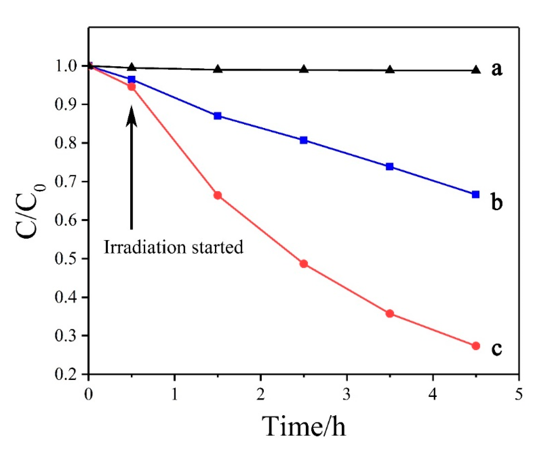
2.3. Photocatalytic Activity

The photocatalytic activity of the samples was examined for MB photodegradation in aqueous phase under UV light irradiation. The test was performed in a 100 mL beaker charged with 50 mL of 10 mg·L−1 MB solution and 30 mg of the sample. The mixture was firstly kept in a dark place for 30 min to reach adsorption–desorption equilibrium before irradiation. Then, the mixture was exposed to the radiation source. Two milliliters of the solution were taken and filtrated by a needle filter every hour to test the photodegradation performance by a UV-Vis spectrophotometer (TU-1901) (Persee Analysis General Instrument Co., Ltd., Beijing, China). The UV light irradiation source was a UV lamp (Philips, 6 W). All experiments were carried out at room temperature (25 °C) and the mixture was suspended by a magnetic stirrer.

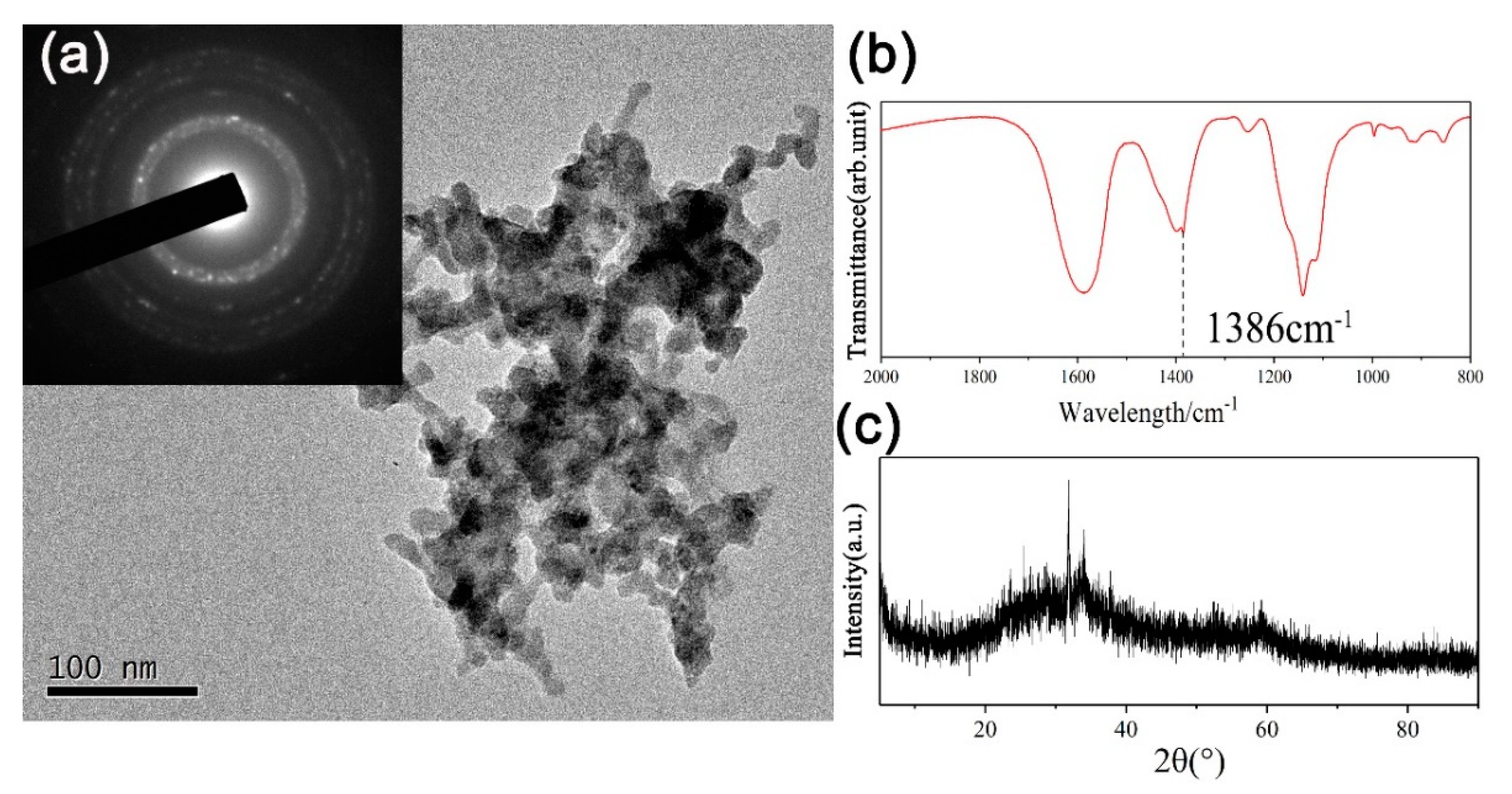
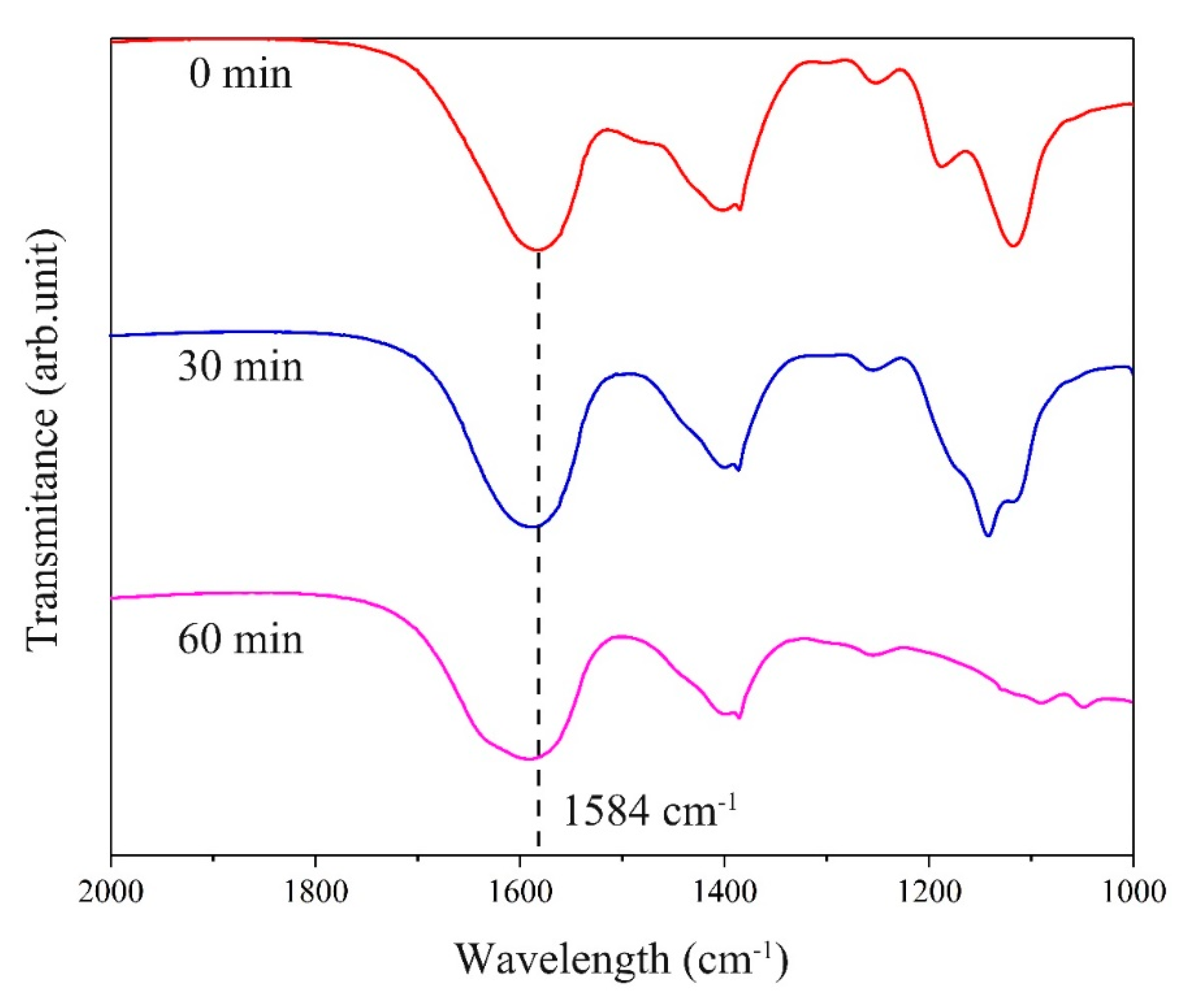
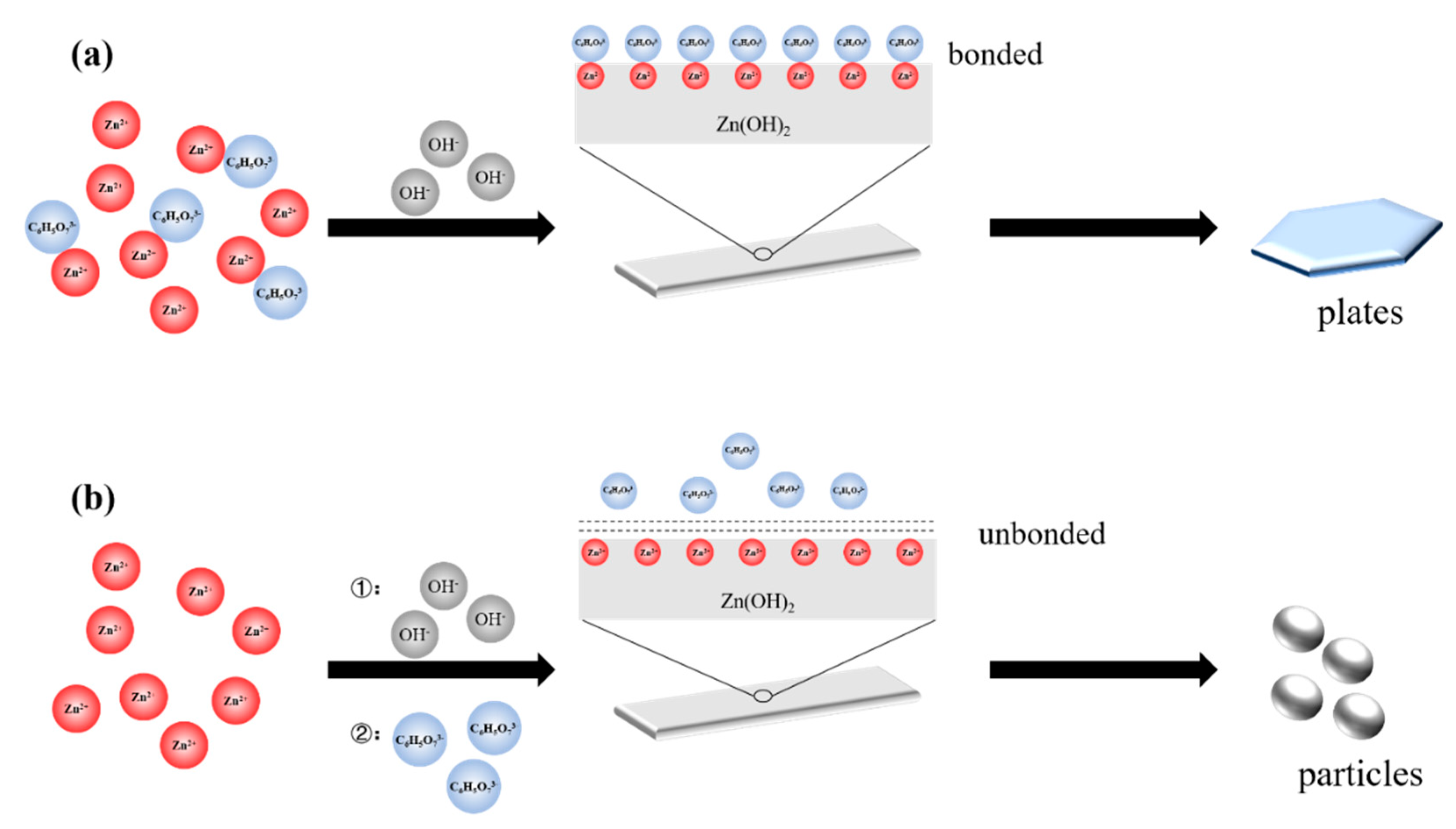
3. Results and Discussion

The phases of the three samples were firstly determined by X-ray diffraction analysis. As Figure 1 shows, the XRD peaks of all samples are well indexed to wurtzite ZnO (JCPDS 36-1451). No other peaks were observed, indicating the purity of the products. Figure 2 shows morphologies of the three products. ZnO-OS are well-dispersed hexagonal rough plates with a diameter of 1 μm and thickness of 0.1 μm. ZnO-TS are particle aggregates with a diameter of 70 nm. ZnO-NC are short rods with a diameter of 0.1 μm and length of 1 μm. The differences reveal that the existence of citrates and the way it was added had a significant impact on the structure. The rod-like ZnO-NC showed the 1D intrinsic growth, while ZnO-OS showed 2D growth. To identify the orientation of ZnO-OS, TEM observation was conducted, as shown in Figure 2d. The lattice spacing measured for the crystalline planes was 0.28 nm, corresponding to the interplanar spacing of (1010) planes. The SAED pattern of ZnO plates can be indexed to the diffraction along the [001] zone axis. The TEM and SAED results indicate the growth of ZnO-OS along [001] direction is well inhibited, and the growth along six symmetric direction perpendicular to (0110), (0110), (1010), (1010), (1100), and (1100) planes led to the hexagonal plates morphology. It can be concluded that the plates are highly (001) facet exposed, which is consistent with its highest intensity of (001) peak among three samples in Figure 1.
As reported previously [24,25,26], the selective combination of citrate with (001) facet of ZnO resulted in the final (001) exposed structures, which was supported by the complexation between carboxyl and zinc atoms. In this experiment, ZnO-NC showed rod-like shape while ZnO-OS were plate-like (Figure 2a,c), confirming that citrates are effective for ZnO shape controlling. However, ZnO-TS did not show preferential growth as ZnO-OS did in the presence of citrates. If the combination mechanism works, ZnO-TS should be plate-like as expected considering citrates also existed in hydrothermal process. Thus, the formation mechanism of ZnO plates should be reconsidered.
To investigate the effect of citrates on ZnO formation, the morphology evolution progress of ZnO-OS was observed. As Figure 3a shows, after sodium hydrate was added in pH adjustment step, particle aggregates were formed at room temperature. As the mixture underwent hydrothermal treatment, particle aggregates evolved into strip shape (Figure 3b,c). The strips gradually transformed into hexagonal ZnO plates during 30–50 min (Figure 3d,e). All strips disappeared with only hexagonal plates left at 70 min (Figure 3f). The change of soluble zinc species concentration was also measured, as displayed in Figure 4. At 0–30 min, zinc concentration decreased, along with the morphology changed from particle aggregate to strips (Figure 3a–d). The concentration reached minimum at 30 min, corresponding to the beginning of the transformation of strip-like product to hexagonal structure (Figure 3d). The concentration increased during 30–70 min, corresponding to appearance of more hexagonal structures with fewer strip structures (Figure 3e), and strip-like structures finally disappeared at 70 min (Figure 3e). The concentration variation and morphology evolution are similar to the progress in our previous study about the formation of ZnO from ε-Zn(OH)2 [27], in which the dissolution of ε-Zn(OH)2 and formation of ZnO rods happened together with soluble zinc concentration decreased. XRD test was made to identify the phases of the strip and the particle. The XRD pattern of the strip of reaction time at 20 min (Figure 3c) were well indexed to ε-Zn(OH)2 (JCPDS 38-0385) and β-Zn(OH)2 (JCPDS 20-1435), as shown in Figure 5. For particles, XRD patterns show no characteristic peaks (Figure 6c). The SAED pattern of the particle of reaction time at 0 min in OS route shows concentric rings (Figure 6a), which is the character of amorphous crystal. Figure 6b presents the Fourier transform infrared (FT-IR) spectra of the particles; the peak at 1386 cm−1 corresponds to hydroxyl bridging [28]. Based on the above results, it should be amorphous Zn(OH)2.
The formation progress of ZnO can be described as follows. In the solution containing zinc ion, amorphous Zn(OH)2 particle aggregates formed when sodium hydrate was added. Amorphous Zn(OH)2 gradually grew to ε-Zn(OH)2 and β-Zn(OH)2 strips during 0–30 min, resulting in the decrease of soluble zinc concentration. As the reaction proceeded, ε-Zn(OH)2 and β-Zn(OH)2 gradually dissolved and transformed to hexagonal ZnO plates, and soluble zinc concentration increased.
As described above, ZnO plates formed after the Zn(OH)2 precursor formed. Hydrothermal treatments in two routes were the same comparing OS route with TS route. Thus, the precursor should be the cause of the final structural difference. The condition where precursor formed differed in two routes. Citrates existed when Zn(OH)2 precipitated in OS route but did not exist in TS route. It can be inferred that citrate influenced the formation of the precursor. The soluble zinc species concentration distribution was calculated, as shown in Table 1, to analyze the solution condition before Zn(OH)2 precipitated, according to the complex constant at 25 °C (Table S1) [29] and the measured pH (5.2). The concentrations of Zn2+ and Zn2+-C6H5O73 were 4.59 × 10⁻² mol·L−1 and 4.99 × 10⁻² mol·L−1, respectively, indicating most citrates existed in the form of Zn2+-C6H5O73− due to its large complex constant (2.51 × 10¹¹). These two ions were the main species in the OS-solution, while the main species in TS-solution was Zn2+. The distribution of Zn2+-OH-H2O system was also calculated according to the complex constant in Table S1, as displayed in Figure S1 (Supplementary Materials). According to the calculation, the main species at pH = 9 was Zn(OH)2(aq). Zn(OH)2(aq) is not stable in solution (Ksp[Zn(OH)2] = 1.2 × 10⁻¹⁷). Thus, the addition of sodium hydrate caused the formation of insoluble Zn(OH)2 and the concentration of zinc species decreased to 4.42 × 10⁻² mol·L−1 (Figure 4). It can be inferred that the decrease of the concentration was caused by the precipitation of Zn2+ and the main zinc species remained in solution was Zn2+-C6H5O73−. After 30 min reaction, concentration of soluble zinc decreased to 2.25 × 10⁻² mol·L−1 from (Figure 4). It was caused by the growth of Zn(OH)2, and it can be inferred that Zn2+-C6H5O73− participated in the formation of Zn(OH)2.
To analyze the effect of citrates on the precursor, FT-IR analyses of the products at 0, 30, and 60 min were conducted. As Figure 7 shows, an absorption peak at 1589 cm−1 appeared in all three samples. This characteristic peak corresponds to the carbonyl stretching vibration when it is complexed with zinc ion. This peak indicates the complexation between citrates and zinc ions [30,31,32]. It can be inferred that citrates participated in the formation of Zn(OH)2 by complexation with zinc ions in OS route, and such complexation existed during the whole process. Zeta potential of different samples were measured, as displayed in Table 2. The adsorption of citrates can decrease the zeta potential of particles [33,34]. Compared with TS precursor dispersed in water and TS precursor dispersed in citrate solution, the OS precursor shows the most negative zeta potential (−25.8 mV), which was caused by the adsorption between citrates and the precursor. Zeta potential for the TS precursor dispersed in citrate solution (−10.3 mV) is slightly more negative than that in water (−9.5 mV), indicating that there were little citrates adsorbed on the TS precursor. Besides, zeta potential of ZnO-OS is more negative than that of ZnO-TS, which was caused by the higher exposing ratio of (001) facet in ZnO-OS, since (001) facet can adsorb more citrates than other facets. The capping effect of citrates on ZnO (001) facet is discussed in the literature [24,25,26]; the main point of them was the selective adsorption of citrates on the (001) facet inhibited the growth along [001] direction, causing (001) facet exposed structures. In our opinion, the relationship between citrates and the precursor was also the key factor for (001) capping effect. The citrates participating in the formation of precursor play the main role in capping effect. This is the reason ZnO-OS was (001) facet exposed plates while ZnO-TS was not. Citrates participated in the OS precursor formation, while no citrates existed when TS precursor formed.
Based on the above result, a possible growth mechanism is proposed, as illustrated in Figure 8. In an OS solution containing Zn2+ and C6H5O73−, the two main zinc species were Zn2+ and Zn2+-C6H5O73−. Then, Zn2+ was precipitated to form amorphous Zn(OH)2 when adding OH. Zn2+-C6H5O73− was also precipitated as amorphous Zn(OH)2 grew to ε-Zn(OH)2 and β-Zn(OH)2. The citrates that attached to the surface of the formed Zn(OH)2 played the main role in controlling ZnO growth. During the transformation from Zn(OH)2 to ZnO, the (001) hexagonal facet was mostly suppressed by those dangling citrates to form hexagonal plates structure since (001) facet has the highest zinc density. In TS route, citrates were added after the precipitation of Zn(OH)2. When TS precursor was redispersed in citrates solution, the electric double layer of Zn(OH)2 made it difficult for citrates to bond with surface Zn2+. With no citrates bonded on the surface of Zn(OH)2, there is no (001) facet inhibition effect. Thus, the growth of ZnO cannot be specifically orientated to form (001) exposed structure and ZnO particles were obtained.
The photocatalytic activity of the plate was studied by testing the degradation of MB under UV light with ZnO nanorods taken as a comparison, which were synthesized due to our previous report [35] (see Figure S2). Figure 9 shows the photocatalytic performance of the plates and nanorods, where C is the concentration of MB after irradiation for time t and C0 is the initial concentration of MB before dark treatment. The self-degradation of MB was also tested by putting MB solution under the same irradiation without any additive. The degradation ratio of plates reached 70% after 4 h of irradiation, while the nanorods showed only 30% degradation ratio. This difference is evidence of the superiority of (001) facet in photodegradation, as reported previously [36,37,38,39]. Photogenerated electrons and holes will be generated when ZnO absorb photons with energy greater than its band gap energy. Photoinduced electrons are captured by adsorbed O2 to form superoxide radicals (·O2) and holes will react with adsorbed OH to form hydroxyl radicals (·OH) [40]. These radicals are critical active species for the degradation of dyes. The positively charged Zn2+ (001) facet can adsorb more negatively charged OH in aqueous solution [41], which facilitates the formation of ·OH. Besides, the internal electric field derived by spontaneous polarization accelerates the separation of photogenerated electron–hole pairs along [001] direction [42]. Thus, as the terminated facet perpendicular to [001] direction, (001) facet shows enhanced photocatalytic activity.

4. Conclusions

This work presents the hydrothermal synthesis of well-dispersed ZnO hexagonal plates with the existence of sodium citrates, and the growth mechanism of the plates is proposed. The results show that the ZnO hexagonal plates could only be obtained when citrates participated in the formation of Zn(OH)2. Citrates that bond with the surface of precursor Zn(OH)2 played a key role in constructing (001) facet exposed structure. The suppression to (001) facet during ZnO plates growth resulted from the binding between citrates and Zn2+ on (001) facet. The growth along [001] direction was mostly inhibited due to the highest Zn2+ density on (001) facet. Compared with ZnO nanorods, the ZnO plate showed enhanced photocatalytic activity for degradation of MB due to its (001) facet exposed structure. This work gives a new insight into the effect of citrates on the ZnO structure modification by affecting the formation of the precursor.

Supplementary Materials

The following are available online at https://www.mdpi.com/2073-4352/9/11/552/s1, Figure S1: Distribution of Zn2+-OH-H2O system, Figure S2: SEM image of ZnO nanorods, Table S1: Equilibrium equation and complex constant for Zn2+-C6H5O73−-H2O system (25 °C).

Author Contributions

S.L. and R.C. conceived and designed the experiments; S.L. performed the experiments and analyzed the data; and S.L., R.C., J.W. and L.X. wrote the paper.

Funding

This research was funded by the National Science Foundation of China (Nos. 51774191, 21978153, 51802123) and the National Science Foundation of Jiangsu Province (No. BK20180630).

Acknowledgments

This work was financially supported by the National Natural Science Foundation of China (Nos. 51774191, 21978153, and 51802123).

Conflicts of Interest

The authors declare no conflict of interest.

References

  1. Zhou, Z.Y.; Tian, N.; Li, J.T.; Broadwell, I.; Sun, S.G. Nanomaterials of high surface energy with exceptional properties in catalysis and energy storage. Chem. Soc. Rev. 2011, 40, 4167–4185. [Google Scholar] [CrossRef] [Green Version]
  2. Wang, M.Z.; Chen, M.D.; Ma, Y.; Yang, J.; Yuan, Y.K.; Liu, J.F.; Liu, B.; Du, Q.; Ren, Y.; Liu, S.Z.; et al. Improving sensing performance of the ZnO foam structure with exposed {001} facets by hydrogenation and sensing mechanism at molecule level. Appl. Surf. Sci. 2019, 479, 646–654. [Google Scholar] [CrossRef]
  3. Peng, F.P.; Zhou, Q.; Lu, C.H.; Ni, Y.R.; Kou, J.H.; Xu, Z.Z. Construction of (001) facets exposed ZnO nanosheets on magnetically driven cilia film for highly active photocatalysis. Appl. Surf. Sci. 2017, 394, 115–124. [Google Scholar] [CrossRef]
  4. Wang, J.X.; Yang, Y.; Sun, X.W. ZnO disk-like structures and their application in dye sensitized solar cell. Solid State Commun. 2016, 240, 46–52. [Google Scholar] [CrossRef]
  5. Wang, Z.L. Nanostructures of zinc oxide. Mater. Today 2004, 7, 26–33. [Google Scholar] [CrossRef]
  6. Xu, C.X.; Sun, X.W.; Dong, Z.L.; Yu, M.B. Zinc oxide nanodisk. Appl. Phys. Lett. 2004, 85, 3878–3880. [Google Scholar] [CrossRef]
  7. Demel, J.; Plestil, J.; Bezdicka, P.; Janda, P.; Klementova, M.; Lang, K. Layered zinc hydroxide salts: Delamination, preferred orientation of hydroxide lamellae, and formation of ZnO nanodiscs. J. Colloid Interface Sci. 2011, 360, 532–539. [Google Scholar] [CrossRef] [PubMed]
  8. Chen, Y.; Zhao, H.; Liu, B.; Yang, H.Q. Charge separation between wurtzite ZnO polar {001} surfaces and their enhanced photocatalytic activity. Appl. Catal. B Environ. 2015, 163, 189–197. [Google Scholar] [CrossRef]
  9. Li, F.; Ding, Y.; Gao, P.X.X.; Xin, X.Q.; Wang, Z.L. Single-cystal hexagonal disks and rings of ZnO: Low-temperature, large-scale synthesis and growth mechanism. Angew. Chem. Int. Edit. 2004, 43, 5238–5242. [Google Scholar] [CrossRef]
  10. Xu, L.F.; Guo, Y.; Liao, Q.; Zhang, J.P.; Xu, D.S. Morphological control of ZnO nanostructures by electrodeposition. J. Phys. Chem. B 2005, 109, 13519–13522. [Google Scholar] [CrossRef]
  11. Zeng, J.; Zheng, Y.Q.; Rycenga, M.; Tao, J.; Li, Z.Y.; Zhang, Q.A.; Zhu, Y.M.; Xia, Y.N. Controlling the Shapes of Silver Nanocrystals with Different Capping Agents. J. Am. Chem. Soc. 2010, 132, 8552–8553. [Google Scholar] [CrossRef] [PubMed]
  12. Mallin, M.P.; Murphy, C.J. Solution-phase synthesis of sub-10 nm Au-Ag alloy nanoparticles. Nano Lett. 2002, 2, 1235–1237. [Google Scholar] [CrossRef]
  13. Hu, J.Q.; Chen, Q.; Xie, Z.X.; Han, G.B.; Wang, R.H.; Ren, B.; Zhang, Y.; Yang, Z.L.; Tian, Z.Q. A simple and effective route for the synthesis of crystalline silver nanorods and nanowires. Adv. Funct. Mater. 2004, 14, 183–189. [Google Scholar] [CrossRef]
  14. Tian, Z.R.R.; Voigt, J.A.; Liu, J.; McKenzie, B.; McDermott, M.J.; Rodriguez, M.A.; Konishi, H.; Xu, H.F. Complex and oriented ZnO nanostructures. Nat. Mater. 2003, 2, 821–826. [Google Scholar] [CrossRef] [PubMed]
  15. Tian, Z.R.R.; Voigt, J.A.; Liu, J.; Mckenzie, B.; Mcdermott, M.J. Biommetic arrays of oriented helical ZnO nanorods and columns. J. Am. Chem. Soc. 2002, 124, 12954–12955. [Google Scholar] [CrossRef]
  16. Kuo, C.L.; Kuo, T.J.; Huang, M.H. Hydrothermal synthesis of ZnO microspheres and hexagonal microrods with sheetlike and platelike nanostructures. J. Phys. Chem. B 2005, 109, 20115–20121. [Google Scholar] [CrossRef] [PubMed]
  17. Xie, Q.S.; Guo, H.Z.; Zhang, X.X.; Lu, A.L.; Zeng, D.Q.; Chen, Y.Z.; Peng, D.L. A facile approach to fabrication of well-dispersed NiO-ZnO composite hollow microspheres (vol 3, pg 24430, 2013). RSC Adv. 2017, 7, 47164. [Google Scholar] [CrossRef]
  18. Zhang, H.; Yang, D.R.; Li, D.S.; Ma, X.Y.; Li, S.Z.; Que, D.L. Controllable growth of ZnO microcrystals by a capping-molecule-assisted hydrothermal process. Cryst. Growth Des. 2005, 5, 547–550. [Google Scholar] [CrossRef]
  19. Liang, J.B.; Liu, J.W.; Xie, Q.; Bai, S.; Yu, W.C.; Qian, Y.T. Hydrothermal growth and optical properties of doughnut-shaped ZnO microparticles. J. Phys. Chem. B 2005, 109, 9463–9467. [Google Scholar] [CrossRef]
  20. Zhang, T.R.; Dong, W.J.; Keeter-Brewer, M.; Konar, S.; Njabon, R.N.; Tian, Z.R. Site-specific nucleation and growth kinetics in hierarchical nanosyntheses of branched ZnO crystallites. J. Am. Chem. Soc. 2006, 128, 10960–10968. [Google Scholar] [CrossRef]
  21. Andeen, D.; Kim, J.H.; Lange, F.F.; Goh, G.K.L.; Tripathy, S. Lateral epitaxial overgrowth of ZnO in water at 90 degrees C. Adv. Funct. Mater. 2006, 16, 799–804. [Google Scholar] [CrossRef]
  22. Meagley, K.L.; Garcia, S.P. Chemical Control of Crystal Growth with Multidentate Carboxylate Ligands: Effect of Ligand Denticity on Zinc Oxide Crystal Shape. Cryst. Growth Des. 2012, 12, 707–713. [Google Scholar] [CrossRef]
  23. Das, S.; Dutta, K.; Pramanik, A. Morphology control of ZnO with citrate: A time and concentration dependent mechanistic insight. Crystengcomm 2013, 15, 6349–6358. [Google Scholar] [CrossRef]
  24. Geng, J.; Song, G.H.; Jia, X.D.; Cheng, F.F.; Zhu, J.J. Fast One-Step Synthesis of Biocompatible ZnO/Au Nanocomposites with Hollow Doughnut-Like and Other Controlled Morphologies. J. Phys. Chem. C 2012, 116, 4517–4525. [Google Scholar] [CrossRef]
  25. Nicholas, N.J.; Franks, G.V.; Ducker, W.A. Selective Adsorption to Particular Crystal Faces of ZnO. Langmuir 2012, 28, 7189–7196. [Google Scholar] [CrossRef]
  26. Milek, T.; Zahn, D. A Surfactants Walk to Work: Modes of Action of Citrate Controlling (10-10) and (000-1) Zinc Oxide Surface Growth from Solution. Z Anorg. Allg. Chem. 2016, 642, 902–905. [Google Scholar] [CrossRef]
  27. Wang, J.; Ma, P.Y.; Xiang, L. Effects of NaOH on formation of ZnO nanorods from epsilon-Zn(OH)2. Mater. Lett. 2015, 141, 118–121. [Google Scholar] [CrossRef]
  28. Sujuan, W.Z.L. Infrared Spectra Characteristics of Zinc Hydroxide and Zinc Oxide. Chin. J. Spectrosc. Lab. 2012, 29, 2172–2175. [Google Scholar]
  29. Dean, J.A. Lange’s Handbook of Chemistry, 15th ed.; McGraw-Hill Companies: New York, NY, USA, 1998. [Google Scholar]
  30. Mandal, U.K. Ionic elastomer based on carboxylated nitrile rubber: Infrared spectral analysis. Polym. Int. 2000, 49, 1653–1657. [Google Scholar] [CrossRef]
  31. Cho, S.; Jang, J.W.; Jung, A.; Lee, S.H.; Lee, J.; Lee, J.S.; Lee, K.H. Formation of Amorphous Zinc Citrate Spheres and Their Conversion to Crystalline ZnO Nanostructures. Langmuir 2011, 27, 371–378. [Google Scholar] [CrossRef]
  32. Wang, L.; Feng, L.; Liu, J.F. Study the effect of functional molecule types on the surface properties of ZnO nanorods. RSC Adv. 2014, 4, 62505–62510. [Google Scholar] [CrossRef]
  33. Mittelman, A.M.; Fortner, J.D.; Pennell, K.D. Effects of ultraviolet light on silver nanoparticle mobility and dissolution. Environ. Sci. Nano 2015, 2, 683–691. [Google Scholar] [CrossRef]
  34. Kocsis, K.; Niedermaier, M.; Kasparek, V.; Bernardi, J.; Redhammer, G.; Bockstedte, M.; Berger, T.; Diwald, O. From Anhydrous Zinc Oxide Nanoparticle Powders to Aqueous Colloids: Impact of Water Condensation and Organic Salt Adsorption on Free Exciton Emission. Langmuir 2019, 35, 8741–8747. [Google Scholar] [CrossRef] [PubMed]
  35. Wang, J.; Liu, C.; Xiang, L. Influence of Sodium Dodecyl Sulfonate on the Formation of ZnO Nanorods from epsilon-Zn(OH)2. J. Nanomater 2013, 2013, 6. [Google Scholar]
  36. Liu, Y.J.; Huang, D.; Liu, H.X.; Li, T.D.; Wang, J.G. ZnO Tetrakaidecahedrons with Coexposed {001}, {101}, and {100} Facets: Shape-Selective Synthesis and Enhancing Photocatalytic Performance. Cryst. Growth Des. 2019, 19, 2758–2764. [Google Scholar] [CrossRef]
  37. Mclaren, A.; Valdes-Solis, T.; Li, G.Q.; Tsang, S.C. Shape and Size Effects of ZnO Nanocrystals on Photocatalytic Activity. J. Am. Chem. Soc. 2009, 131, 12540–12541. [Google Scholar] [CrossRef]
  38. Zhang, L.Y.; Yin, L.W.; Wang, C.X.; Lun, N.; Qi, Y.X. Sol-Gel Growth of Hexagonal Faceted ZnO Prism Quantum Dots with Polar Surfaces for Enhanced Photocatalytic Activity. ACS Appl. Mater. Interfaces 2010, 2, 1769–1773. [Google Scholar] [CrossRef]
  39. Wang, L.N.; Zheng, Y.Y.; Li, X.Y.; Dong, W.J.; Tang, W.H.; Chen, B.Y.; Li, C.R.; Li, X.; Zhang, T.R.; Xu, W. Nanostructured porous ZnO film with enhanced photocatalytic activity. Thin. Solid Films 2011, 519, 5673–5678. [Google Scholar] [CrossRef]
  40. Lee, K.M.; Lai, C.W.; Ngai, K.S.; Juan, J.C. Recent developments of zinc oxide based photocatalyst in water treatment technology: A review. Water Res. 2016, 88, 428–448. [Google Scholar] [CrossRef]
  41. Zeng, J.H.; Jin, B.B.; Wang, Y.F. Facet enhanced photocatalytic effect with uniform single-crystalline zinc oxide nanodisks. Chem. Phys. Lett. 2009, 472, 90–95. [Google Scholar] [CrossRef]
  42. Chen, Y.; Zhang, L.N.; Ning, L.C.; Zhang, C.J.; Zhao, H.; Liu, B.; Yang, H.Q. Superior photocatalytic activity of porous wurtzite ZnO nanosheets with exposed {001} facets and a charge separation model between polar (001) and (00(1)over-bar) surfaces. Chem. Eng. J. 2015, 264, 557–564. [Google Scholar] [CrossRef]
Figure 1. XRD patterns for all products.
Crystals 09 00552 g001
Figure 2. SEM images for all products and TEM image for ZnO-OS: (a)ZnO-OS; (b) ZnO-TS; (c) ZnO-NC; and (d)TEM and SAED images for ZnO-OS.
Crystals 09 00552 g002
Figure 3. SEM images of products obtained in OS route at different times: (a) 0 min; (b) 10 min; (c) 20 min; (d) 30 min; (e) 50 min; and (f) 70 min.
Crystals 09 00552 g003
Figure 4. Variation of soluble zinc concentration with reaction time.
Crystals 09 00552 g004
Figure 5. XRD pattern of the strip precursor taken at 20 min in OS route.
Crystals 09 00552 g005
Figure 6. Phase analysis of the particle aggregates precursor at 0 min in OS route: (a) TEM and SAED pattern; (b) FT-IR spectra; and (c) XRD pattern.
Crystals 09 00552 g006
Figure 7. FT-IR spectrum of OS products at 0, 30, and 60 min.
Crystals 09 00552 g007
Figure 8. Schematic illustration of mechanism of citrate capping effect in: (a) OS route; and (b)TS route.
Crystals 09 00552 g008
Figure 9. Photodegradation of MB solution with: (a) no samples; (b) ZnO nanorods; and (c) ZnO-OS.
Crystals 09 00552 g009
Table 1. Distribution of soluble zinc species in solution before pH adjusting.
Zinc SpeciesConcentration (OS)Ratio (OS)Concentration (TS)Ratio (TS)
Zn2+4.59 × 10−245.9%9.23 × 10−292.3%
Zn2+-C6H6O72−3.15 × 10−7000
Zn2+-C6H5O73−4.99 × 10−249.9%00
ZnSO4(aq)4.10 × 10−34.1%7.70 × 10−37.7%
Zn(OH)+1.82 × 10−603.67 × 10−60
Zn(OH)2(aq)2.30 × 10−802.92 × 10−80
Zn(OH)32.52 × 10−1402.54 × 10−140
Zn(OH)42−3.33 × 10−1901.06 × 10−190
Table 2. Zeta potential values for various samples.
SampleZeta Potential [mV]
OS precursor (0 min)−25.8
ZnO-OS (70 min)−30.2
TS precursor in water−9.5
TS precursor in citrate solution−10.3
ZnO-TS (70 min)−26.4

